antibacterial activity of the root extracts of garcinia kola
30
ANTIBACTERIAL ACTIVITY OF THE ROOT EXTRACTS OF GARCINIA KOLA AGAINST MDR STAPHYLOCOCCUS AUREUS: INVITRO AND INSILICO STUDIES Abdulbasit Haliru Yakubu 1 , Muhammed Mustapha Muhammed 2 , Anshuman Chandra 3 and Bhawna Arora 4 . 1. Department of Pharmaceutical Chemistry, Faculty of Pharmacy, PMB 1069, University of Maiduguri, Maiduguri, Borno, Nigeria. 2. Bioinformatics and Computational Biology Lab, Dept. of Microbiology, PMB 1069, University of Maiduguri, Maiduguri, Borno, Nigeria. 3. NIMR-ICMR, Dwarka, New Delhi 110077, India. 4. THSTI, NCR Biotech Science Cluster Faridabad, Haryana 121001 Corresponding author mail: [email protected] Abstract Multi Drug Resistant (MDR) Staphylococcus aureus is an important bacteria with clinical and economic implications. Plants including Garcinia kola provides bioactive principles with diverse structural and biological features. Tyrosyl-tRNA synthetase (TyrRS) is targeted in antibacterial drug discovery as its implicated in bacterial protein synthesis. The n-Butanol fraction of Garcinia kola root extract recorded the highest activity against MDR staph aureus (18.50±0.41) compared to the chloroform (10.00±2.12) and methanol (8.16±0.62) extract, with no activity recorded with the n-Hexane extract. Analysis of the n-Butanol fraction on GC-MS recorded 14 phytoconstituents with varying structural composition; containing important scaffolds & motifs of benzoquinone, imidazole[1,2-a]pyridine, chlorocarbazole and azetidine that present key pharmaceuticals as antibiotic and for drug development. Further insilico molecular docking studies of these compounds on antibacterial drug target; Tyrosyl-tRNA synthetase (PDB 1JIJ) from MDR staph aureus was documented. Nine (9) compounds had good binding scores ranging from -4.63 to -7.08 kcal/mol; with CID_590350 having the highest score. The compounds formed various bonding with the 1JIJ amino acid residues including H-bond, van der waal and π interactions. Five (5) compounds; CID_619583 (9,9-Dichloro-9-silafluorene), CID_5732 (Zolpidem), CID_616643 (Pyridazine-3,5-dicarbonitrile, 1,6-dihydro-4-amino-6-imino-1-(2- nitrophenyl)), CID_16486 ((S)-(-)-2-Azetidinecarboxlic acid) and CID_66747 (2-Hydroxyethyl
-
Upload
khangminh22 -
Category
Documents
-
view
1 -
download
0
Transcript of antibacterial activity of the root extracts of garcinia kola

ANTIBACTERIAL ACTIVITY OF THE ROOT EXTRACTS OF GARCINIA KOLA
AGAINST MDR STAPHYLOCOCCUS AUREUS: INVITRO AND INSILICO STUDIES
Abdulbasit Haliru Yakubu1, Muhammed Mustapha Muhammed2, Anshuman Chandra3
and Bhawna Arora4.
1. Department of Pharmaceutical Chemistry, Faculty of Pharmacy, PMB 1069, University of
Maiduguri, Maiduguri, Borno, Nigeria.
2. Bioinformatics and Computational Biology Lab, Dept. of Microbiology, PMB 1069,
University of Maiduguri, Maiduguri, Borno, Nigeria.
3. NIMR-ICMR, Dwarka, New Delhi 110077, India. 4. THSTI, NCR Biotech Science Cluster Faridabad, Haryana 121001
Corresponding author mail: [email protected]
Abstract
Multi Drug Resistant (MDR) Staphylococcus aureus is an important bacteria with clinical and
economic implications. Plants including Garcinia kola provides bioactive principles with diverse
structural and biological features. Tyrosyl-tRNA synthetase (TyrRS) is targeted in antibacterial
drug discovery as its implicated in bacterial protein synthesis. The n-Butanol fraction of
Garcinia kola root extract recorded the highest activity against MDR staph aureus (18.50±0.41)
compared to the chloroform (10.00±2.12) and methanol (8.16±0.62) extract, with no activity
recorded with the n-Hexane extract. Analysis of the n-Butanol fraction on GC-MS recorded 14
phytoconstituents with varying structural composition; containing important scaffolds & motifs
of benzoquinone, imidazole[1,2-a]pyridine, chlorocarbazole and azetidine that present key
pharmaceuticals as antibiotic and for drug development. Further insilico molecular docking
studies of these compounds on antibacterial drug target; Tyrosyl-tRNA synthetase (PDB 1JIJ)
from MDR staph aureus was documented. Nine (9) compounds had good binding scores ranging
from -4.63 to -7.08 kcal/mol; with CID_590350 having the highest score. The compounds
formed various bonding with the 1JIJ amino acid residues including H-bond, van der waal and π
interactions. Five (5) compounds; CID_619583 (9,9-Dichloro-9-silafluorene), CID_5732
(Zolpidem), CID_616643 (Pyridazine-3,5-dicarbonitrile, 1,6-dihydro-4-amino-6-imino-1-(2-
nitrophenyl)), CID_16486 ((S)-(-)-2-Azetidinecarboxlic acid) and CID_66747 (2-Hydroxyethyl

benzoate) showed favorable ADME properties, while their MD stimulation analysis revealed
stable binding capabilities with the drug target. CID_16486 and CID_66747 bind to the most
active binding pocket (Drug score: 0.82 and 0.72) while CID_619583 tend to bind outside the
active binding pocket. Therefore, these compounds from the root of Garcinia kola are considered
as suitable prospective bioactive compounds against MDR Staphylococcus aureus after
successful in vitro and in silico experimental validation.
Key words: Garcinia kola, Antibacterial, MDR Staphylococcus aureus, tyrosyl-tRNA synthetase
(YRS), Molecular docking, MD stimulation.
Figure legend
INTRODUCTION
In the past decades, there had been an alarming increase in the rise of antimicrobial resistance.
This translates a serious public health concern as choice of clinical drugs become limited and
ineffective. This in turn, results to negative clinical and economic consequences to patient’s
health, increase in nosocomial infection, prolonged hospital stay and mortality. The mortality
due to this infection is projected to be over a 10 million by 2050 (Ferdes, 2018). This prompt the
call for global advocacy campaign in antibiotic stewardship.

Among the common encountered bacteria of clinical importance is Staphylococcus aureus. It’s
the most common cause of nosocomial infections, bacteremia and is a bacterial pathogen with
high propensity to develop antibiotic resistance (Steinig et al., 2019 and kot et al., 2020). The
methicillin resistance Staph. aureus MRSA confers its resistance to β-lactam antibiotic by
acquiring the genetic element; staphylococcal cassette chromosome mec, which carries
the mecA gene and also that of the new variant mecC (Kot et al., 2020). This MRSA later
become resistant to other class of antibiotic including broad spectrums by acquiring new
antimicrobial resistance elements and are termed ‘Multidrug Resistant MDR’ MRSA (Kot et al.,
2020). Hiramatsu and colleagues described the genetic basis of the ability of Staph. aureus to
acquire multi-antibiotic resistance (Hiramatsu et al., 2014). MDR MRSA presents serious
challenges in the treatment and control of Staph. aureus infections; standard clinical agents used
in its treatment include vancomycin, linezolid, daptomycin, tigecycline, telavancin, and
ceftaroline (Anstead et al., 2013 and kot et al., 2020).
Aminoacyl-tRNA synthetases (AARSs) are key important enzyme that are involved in bacterial
transcription, translation and signaling pathways. They catalyse the covalent binding of relevant
amino acid with specific tRNA. Tyrosyl-tRNA synthetase (TyrRS) is a class I synthetases and
because of their role in protein synthesis, inhibitors of this enzymes are been explored for the
discovery and development of potential new antibiotic agents (Qiu et al., 2019 and Farshadfar et
al., 2020)
Plant parts had been in use since genesis for the treatment of various ailments. They are
characterized by their large structural and diverse chemical entities known as secondary
metabolites, which presents unique bioactive principles with elaborate biological activities
(Ferdes, 2018 and Othman et al., 2019). Plant phytochemicals had been explored and
documented for their potential antibacterial activity against sensitive and resistant pathogens
(Khamenah et al., 2019).
Among the Plant species explored for its antibacterial activity is Garcina kola, popularly known
as bitter kola. It’s a perennial crop that belongs to the Guttiferae family. This plant had been
utilized among locals for its medicinal importance. A review of its ethnobotanical,
phytochemistry, pharmacological & toxicological, and therapeutic properties had been
documented (Bubu et al., 2016 and Oliver et al., 2016).

A plethora of literature’s emerged on the antibacterial activity of G. kola especially the seed and
leave part. However, there’s scarce research on the antibacterial activity of the root part of
G. kola and activity of the plant against MDR staph aureus.
With the increase in antibiotic resistance and limited availability of new drug entry for possible
clinical use against MDR staph aureus, it’s imperative for the research on new bioactive
principles against this organism.
In this study, we undertook the antibacterial studies of the root extracts (n-Hexane, n-Butanol,
chloroform, and methanol) of G. kola against clinical isolates of MDR Staph. aureus, as well as
carrying out insilico molecular docking and molecular dynamic analysis of the phytoconstituents
from the active extract against Tyrosyl-tRNA synthetase.
MATERIALS AND METHOD.
Plant Collection and Identification
Fresh root parts of G.kola was collected aseptically in October, 2020 from Jos LGA, Plateau
State, Nigeria, and identified by a Medicinal Botanist (Mal Namadi Sanusi) of the Biology
department, ABU Zaria, Nigeria.
Preparation of Plant Extract and Its Fractions
The method of plant extract preparation employed in this studies was adopted from our previous
work with modification (Yakubu et al., 2020). In this study, 500mg of the pulverized root sample
material was extracted using maceration method with n-Hexane, Chloroform, n-Butanol and
Methanol and allowed for 72hrs under room condition. The mixture was filtered with a filter
paper and allowed to dry under room temperature to obtain the crude extract; stored in a
desiccator, and used for further analysis.
Biological Activity
Antimicrobial Assay
Test Organisms
Isolates of MDR staphylococcus aureus were obtained from the Microbiology department,
University of Maiduguri and were stored at 2-80C until required.
Preparation of Extract solutions and test organism for Pathogenic Assay.

In this study, we adopt the method we employed in our previous studies (Yakubu et al., 2020). A
stock solution of 1000mg/ml was prepared and a two-fold serial dilution was carried out to
obtain working solutions of varying concentrations.
Test organisms cultured for 24hrs were suspended in a sterile bottle containing pure broth.
Normal saline was added gradually and the turbidity was observed and compared to that of 0.5
Mcfarland standard; this was then diluted to produce 106cells/mL and used in the experiments.
Nutrient agar was prepared accurately to the manufacturer’s specification and sterilized at 1210C
for 15min which was then allowed to cool to 500C in a water-bath. The test organism (1mL
containing 106cells/mL) was inoculated into pre-labeled petri plates (90mm diameter), then
19mL of the molten agar was added to each petri plate, shacked and allowed to set at room
temperature on a flat surface (Yakubu et al., 2020).
Antimicrobial Susceptibility Assay (agar well diffusion method)
The antibacterial activity of the crude extracts was determined in accordance with the agar-well
diffusion method described by Indabawa and Arzai (2011) and Yakubu et al (2020). The
bacterial isolates were grown for 18 h in a nutrient broth and standardized to 0.5 McFarland
standards (10 6 cfuml-1 ). Two hundred microliter of the standardized cell suspensions were
spread on a Mueller-Hinton agar (Oxoid) and wells were bored into the agar using a sterile 6 mm
diameter cork borer. Approximately 100 µl of the crude extract at 100, 75, 50 and 25 mgml-1
were introduced into the wells, allowed to stand at room temperature for about 2 h and then
incubated at 37°C. Controls were set up in parallel using the solvents that were used to
reconstitute the extract. After 24h, the plates were observed for the zones of inhibition. The
effects were compared with those of Vancomycin at a concentration of 5 mg/ml. Antibacterial
activity was evaluated by measuring the diameters of zones of growth inhibition in triplicates and
results were presented as Mean±SEM. 00 indicates negative and no effect, diameter < 8.0mm
zone of inhibition indicates low activity, >8mm of inhibition indicates high activity (Indabawa
and Arzai, 2011).
GC-MS ANALYSIS
A GC clarus 500 Perkin Elmer system comprising a AOC-20i auto sampler and gas
chromatograph interfaced to a mass spectrometer instrument was employed for the Gas column-
Mass spectroscopy; operating with a column elite-1 fused silica capillary column (30x0.25 mm
IDx1 EM df, composed of 100% dimethyl polysiloxane), electron impact mode at 70 eV; helium

(99-999%) was used as carrier gas at a constant flow of 1 ml/min and an injection volume of 0.5
EI (Oludare et al., 2018). The oven temperature was programmed from 110⁰ C (isothermal for 2
min) with an increase of 10 C / min, to 200 ⁰C then 5 ⁰ C / min 280 ⁰ C, ending with a 9 min
isothermal at 280 ⁰ C; a scan interval of 0.5s and fragments from 40 to 550 Da.
INSILICO ANALYSIS
Preparation of protein complex
The 3D structure of the protein target staph. aureus tyrosyl-tRNA synthatase PDB 1JIJ was
retrieved from the protein data bank PDB (Pisano et al., 2019). Missing loops were checked for,
alternate conformations were removed, bond orders were determined, side chains were optimized
and fixed, improper chirality was identified, dissulfide bonds were checked, steric clashes were
identified and fixed, protonation states were determined, and the protein where optimized and
their energy calculated using a program implemented in SwissPDViewer (v4.1.0) (Isa et al.,
2018).
Molecular Docking
The most critical requirement for interaction of tyrosyl-tRNA synthatase with the ligands it’s the
proper orientation and conformation of the ligands in its active sites. Compounds from GC-MS
with desirable physicochemical properties were selected and ligands 3D structure where obtained
from PubChem. The molecular docking was analysed with AutoDock4.0. The docking analysis
was evaluated at a free binding energy of the protein-ligand complex determined with a total of
110 runs, maximum evaluation of 2500,000, maximum generation of 27,000 and with a
population size of 150. The dimension of the grid was set at 60×60×60 A with a grid spacing of
0.375 Å. A Lamarckian genetic algorithm was used to compute the free binding energy. The
protein-ligand complex was visualized using Ligplot+v.1.4.5 tool (Malik et al., 2017), Pymol
Molecular Graphics System, Version 1.8 Schrodinger, LLC (DeLano, 2002) and Bovia
Discovery Studio (Syed et al., 2021). Prediction of binding site and 2D ligand-protein complex
interaction was viewed with DoGSiteScorer and Poseview respectively from ProteinsPlus sever
(Fricker et al., 2004; Stierand et al., 2006 and Volkamer et al., 2012).
Molecular Dynamic Simulation Analysis The ligands which possessed desirable pharmacokinetic
properties were subjected to the MD simulation to determine their stability with the SK using an
AMBERTOOLS10 package (Case et al., 2010). The Antechamber and the protonate 3D used for the
addition of missing parameters and explicit hydrogen respectively. The coordinate and topology file of

the 5 complex built via Tleap. The GAFF and ff12SB force field of the protein and the ligand respectively
were assigned using the Tleap. The entire system complex neutralized with chloride ion contained in a
buffer solution of an octahedral box of TIP3P. The structural artifact of the system complex removed via
minimization. Then the system was heated with an initial and final temperature of 0 and 300k
respectively for 0.1ns via Langevin dynamics temperature regulation. Finally, the production of the
simulation was carried out at constant temperature and pressure of 300k and 1atm respectively. The
analysis of the MD simulation (root mean square deviation (RMSD), root mean square fluctuation
(RMSF), and radius of gyration) performed using the PTRAJ component of the AMBERTOOLS10 package.
Besides, the MMGBSA (Molecular Mechanics Generalized Born and Surface Area) analysis were
performed to ascertain the binding affinity of the SK-ligand complex using the MD simulation trajectory
of the last 5ns
Drug Likeness and Pharmacokinetics Analysis
All docked ligands were screened for the Lipinki’s rule of five (RO5), pharmacokinetics and
toxicity profile using the SwissADME (Daina et al., 2017). ADME/TOX program (Lipinski et al.,
2001) and DataWarrior tool (Sander et al., 2015).

RESULTS
Table 1: In vitro antimicrobial activity of G. kola extracts on MDR staph aureus.
Concentration
(mg/ml)
n-Hex
Extract
CHCl3 n-But MeOH Van(5mg/ml)
100 00.00±0.00 10.00±2.12 18.50±0.41 8.17±0.62 25.00±1.3
75 00.00±0.00 8.83±1.5 13.50±0.40 5.16±0.23 -
50 00.00±0.00 7.50±0.93 11.83±0.85 3.33±0.24 -
25 00.00±0.00 6.33±1.4 11.16±0.62 3.00±0.35 -
Key: n-Hex: n-Hexane n-But: n-Butanol Van: Vancomycin
CHCL3: Chlroform MeOH: methanol
Fig 2: In vitro antimicrobial activity of G. kola extracts on MDR staph aureus
0
5
10
15
20
25
30
100 75 50 25
zon
e o
f in
hib
itio
n (
mm
)
concentration (µg/ml)
nHex CHCl3 nBut MeOH Van
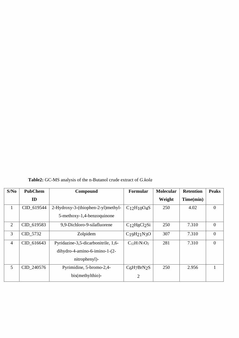
Table2: GC-MS analysis of the n-Butanol crude extract of G.kola
S/No PubChem
ID
Compound Formular Molecular
Weight
Retention
Time(min)
Peaks
1 CID_619544 2-Hydroxy-3-(thiophen-2-yl)methyl-
5-methoxy-1,4-benzoquinone
C12H10O4S
250 4.02 0
2 CID_619583 9,9-Dichloro-9-silafluorene C12H8Cl2Si 250 7.310 0
3 CID_5732 Zolpidem C19H21N3O 307 7.310 0
4 CID_616643 Pyridazine-3,5-dicarbonitrile, 1,6-
dihydro-4-amino-6-imino-1-(2-
nitrophenyl)-
C12H7N7O2 281 7.310 0
5 CID_240576 Pyrimidine, 5-bromo-2,4-
bis(methylthio)-
C6H7BrN2S
2
250 2.956 1

6 CID_622021 5H-Naphtho[1,8-bc]thiophen-5-one,
3,4-dihydro-2-phenyl-
C17H12OS 264 7.42 2
7 CID_616496 9,9'-Bis(3,6-dichlorocarbazole) C24H12Cl4
N2
468 7.42 2
8 CID_590350 3,8-Di-t-butyl-5,6-diphenyl-2,9-
dithia-1-phosphabicyclo[4.3.0]nona-
3,7-diene 1-sulfide
C26H31PS3 47/0 7.42 2
9 CID_6586 Methyl chloroformate C2H3ClO2 94 1.708 1
10 CID_249920
302
Diborane(6), μ-mercapto- B2H6S 60 1.909 2
11 CID_13770 1-Methylcyclopropanemethanol C5H10O 86 1.909 2
12 CID_16486 (S)-(-)-2-Azetidinecarboxlic acid C4H7NO2 101 2.55 1
13 CID_7983 Butanoic acid, butyl ester C8H16O2 144 3.534 2
14 CID_66747 2-Hydroxyethyl benzoate C9H10O3 166 3.534 2
Table 3: Physiochemical analysis of constituents from the n-Butanol crude extract of G.kola
S/N PubChem ID Molecular
weight
(≤500)
Number of
HBA (≤10)
Number
of
HBD (≤5)
MolLogP
(≤5)
Druglikeness
1 CID_619544 250.273 4 1 1.2614 3.7229
2 CID_619583 251.88 0 0 2.7656 -37.314
3 CID_5732 307.396 4 0 2.7975 3.3181
4 CID_616643 281.235 9 2 -0.8212 -6.832
5 CID_240576 251.172 2 0 2.1155 -3.2172
6 CID_622021 264.347 1 0 4.6609 -0.10276
7 CID_616496 470.185 2 0 7.7372 0.95217
8 CID_590350 470.704 0 0 9.3002 -9.1649
9 CID_6586 94.4968 2 0 0.8196 -12.134
10 CID_249920302 - - - - -
11 CID_13770 86.1334 1 1 0.7001 -7.3666

12 CID_16486 101.105 3 2 -2.9545 -0.55335
13 CID_7983 144.213 2 0 2.3527 -7.6022
14 CID_66747 166.175 3 1 1.0522 -1.6196
Table 4: Docking scores and residues involved in H-bond formation
S/no
.
Compound Id Docking score (kcal/mol) Residues involve in H-bonds Distance (Ǻ)
1 CID_619544 -4.63 Thr169
Thr169
2.97
3.02
2 CID_619583 -5.91 0 0
3 CID_5732 -6.30 0 0
4 CID_616643 -5.62 Thr169 2.72
5 CID_240576 -4.33 0 0
6 CID_622021 -7.01 Thr169 3.06
7 CID_616496 -7.00 0 0
8 CID_590350 -7.08 0 0
9 CID_6586 -2.95 Arg183 2.97
10 CID_249920302 - - -
11 CID_13770 -3.49 Ser202 2.74
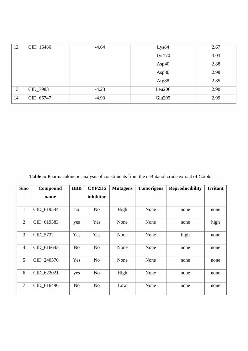
12 CID_16486 -4.64 Lys84
Tyr170
Asp40
Asp80
Arg88
2.67
3.03
2.88
2.98
2.85
13 CID_7983 -4.23 Leu206 2.90
14 CID_66747 -4.93 Glu205 2.99
Table 5: Pharmacokinetic analysis of constituents from the n-Butanol crude extract of G.kola
S/no
.
Compound
name
BBB CYP2D6
inhibitor
Mutagens Tumorigens Reproducibility Irritant
1 CID_619544 no No High None none none
2 CID_619583 yes Yes None None none high
3 CID_5732 Yes Yes None None high none
4 CID_616643 No No None None none none
5 CID_240576 Yes No None None none none
6 CID_622021 yes No High None none none
7 CID_616496 No No Low None none none

8 CID_590350 No No Low None none none
9 CID_6586 Yes No High High none low
10 CID_249920302 - - - - -
11 CID_13770 No Yes None None none none
12 CID_16486 No No None None none None
13 CID_7983 Yes No High None none None
14 CID_66747 Yes No None None high High
DISCUSSION
G. kola had been evaluated for its antibacterial properties including its activity on Staph. aureus.
Ajayi et al (2014) evaluated the antibacterial activity of the seed extracts of G. kola against
clinical dental isolates of Staph species. The ethanol extract showed an inhibitory activity of 13
and 14 at 400 mg/ml. Similar activities where reported on the seed, bark and root extract on
Staph. aureus (Akelere et al., 2008 and Indabawa and Arzai, 2011 and Ukaoma et al., 2013).
Results of this study highlighted the inhibitory antimicrobial activity of the different root extract
of G. kola on MDR Staph. aureus. Activity was recorded in the CHCl3, n-But and MeOH
extracts (highest concentration) while no activity with n-Hex fraction. The n-But extract showed
the highest activity of 18.50±0.41 at 100mg/ml, followed by CHCL3 (10.00±2.12) and MeOH
(8.166±0.62) respectively; but however, lower than that of the control vancomycin (25.00±1.3)
(Table1). This demonstrate that the active antimicrobial contents of the root of G.kola are from

the polar/non-polar intermediate solvents. This is consonant with the finding of Idu and
colleagues on the CHCl3 root extract activity of G.kola on staph aureus (Idu et al., 2014).
Subsequent analysis of the most active extract; n-But was performed with a GC-MS. 14 different
phytoconstituents with varying structural and physicochemical properties were obtained (Table
2). Their PubChem ID, molecular weight, IUPAC name, formula and retention time are also
given (Table 2). Some of the compounds exhibit benzoquinone, imidazo[1,2-a]pyridine,
Chlorocarbazole and azetidine scaffolds & motifs. Presence of this moieties in a compound
attributes to its biological activity including potent antibacterial activity. Thus, the antibacterial
activity exhibited by the n-But extracts can be linked to these compounds.
To investigate this claim, an in-silico studies was performed with these compounds and results
documented; the physiochemical analysis of drug likeness and Lipinski’s rule of five (LO5)
(Table 3) and molecular docking analysis against Staph. aureus tyrosyl-tRNA synthatase; a
Staph. aureus bacterial drug target that is implicated in protein synthesis (Table 4). The results
showed the compounds to possess good physiochemical and drug likeness score. In the
molecular docking analysis, compound CID_619544, CID_619583, CID_5732, CID_616643,
CID_622021, CID_616496, CID_590350 and CID_16486 showed good binding score of -4.63 to
-7.08 kcal/mol against the target protein and exhibit varying H-bond interaction.
DoGSiteScorer was used to predict the binding pocket of the ligand. It employs the grid-based
method that utilize the Difference of Gaussian filter to detect potential binding pockets solely
based on the 3D structure of the protein; values are between zero and one and the higher the
score the more drugable the pocket is estimated to be (Volkamer et al., 2012). Model interaction
of the Protein- ligand is also analyzed and 2D structure generated with PoseViewer.
TyrRS is characterized by a α-helical domain, linked to the catalytic domain, and a C-terminal
domain. The catalytic domain contains a six-stranded parallel β-sheet and a deep active-site cleft
(Qui et al., 2020). The authors also described the potential binding sides of inhibitors of TyrS;
tyrosine, tyrosyl-AMP, ribose and tyrosinyl-adenylate complex.
CID_619544 had a binding score of -4.63 kcal/mol. It forms a two H bonding residues with the
protein target. A terminal OH of Thr169 (2.97 Ǻ and 3.02 Ǻ) with the carbonyl and OH of the
benzoquinone ring. π interaction are seen with Phe136, Leu173 and Leu128, likewise, also Van
der waals interaction (Fig 1). The compound occupies a binding space with a Vol 242.18 A3,

Surface area 250.61 A2 and Drug score 0.45. This compound contains a 1,4-benzoquinone
moiety, which are essential structural units of quinones compounds. Many natural derivatives
had been isolated and synthesized, with potent antimicrobial activities documented (Abraham et
al., 2011).
A B
C D
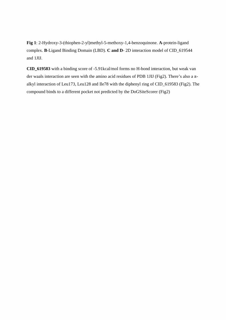
Fig 1: 2-Hydroxy-3-(thiophen-2-yl)methyl-5-methoxy-1,4-benzoquinone. A-protein-ligand
complex. B-Ligand Binding Domain (LBD). C and D- 2D interaction model of CID_619544
and 1JIJ.
CID_619583 with a binding score of -5.91kcal/mol forms no H-bond interaction, but weak van
der waals interaction are seen with the amino acid residues of PDB 1JIJ (Fig2). There’s also a π-
alkyl interaction of Leu173, Leu128 and Ile78 with the diphenyl ring of CID_619583 (Fig2). The
compound binds to a different pocket not predicted by the DoGSiteScorer (Fig2)

A B
C
Fig 2: 9,9-Dichloro-9-silafluorene A-protein-ligand complex. B-Ligand Binding Domain (LBD).
C - 2D interaction model of CID_619583 and 1JIJ.
CID_5732 with a binding score of 6.03 kcal/mol also doesn’t forms H-bond with 1JIJ, but
various π interaction including a π - π Phe136 and π -sigma Leu133 with the 4-tolyl group at
position 2. Van der Waals interaction are also seen with the imidazo[1,2-a]pyridine moeity
(Fig3). It binds and occupies in the active binding pocket with a Vol 242.18 A3, surface area
350.61 A2 and Drug score 0.45 (Fig3). The imidazo[1,2-a]pyridine presents as an important
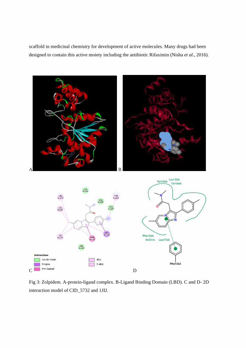
scaffold in medicinal chemistry for development of active molecules. Many drugs had been
designed to contain this active moiety including the antibiotic Rifaximin (Nisha et al., 2016).
A B
C D
Fig 3: Zolpidem. A-protein-ligand complex. B-Ligand Binding Domain (LBD). C and D- 2D
interaction model of CID_5732 and 1JIJ.

CID_616643 is a pyridazinedicarbonitrile derivative. It has a binding score of -5.62 kcal/mol and
forms two H-bond interaction with 1JIJ (Table 4). A binding of Tyr165 (3.02 Ǻ) terminal OH
with the 3,5-carbonitryl moiety at position 3 and terminal OH of Thr169 (2.72 Ǻ) with the 2-
nitrophenyl moiety. Van der Waals interaction are seen with Ile131, Ser132, Leu173 & Leu137.
Similarly, π- π interaction of Leu133 with Pyridazine-3,5-dicarbonitrile moiety and a Phen136 π
–sigma interaction with the 2-nitrophenyl ring (Fig4). The compound binds at an active binding
pocket in the target with a Vol 243.18 A3, Surface 350.61 A3 and Drug score 0.45.

A B
C D
Fig 4: Pyridazine-3,5-dicarbonitrile, 1,6-dihydro-4-amino-6-imino-1-(2-nitrophenyl)-. A-protein-
ligand complex. B-Ligand Binding Domain (LBD). C and D- 2D interaction model of
CID_616643 and 1JIJ.

CID_622021 has a binding score of -7.01kcal/mol (Table) and forms only one H-bond with
residues of 1JIJ (Fig5). A terminal carbonyl C=O of the 5H-Naphtho ring with the terminal OH
of Thr169 (3.06 Ǻ) residue. Van der Waal and π interaction are also observed (Fig5).
The compound binds in the protein target binding site with a Vol 242.18 A3, Surface area 350.61
A2 and Drug score 0.45.
A B
C D
Fig 5: 5H-Naphtho[1,8-bc]thiophen-5-one, 3,4-dihydro-2-phenyl-. A-protein-ligand complex. B-
Ligand Binding Domain (LBD). C and D- 2D interaction model of CID_622021 and 1JIJ.

CID_616496 is a dichlorocarbazole derivative. A binding score of -7.0 kcal/mol was recorded
and forms no H-bond with 1JIJ (Tab 4). A van der Waal interaction is seen with CID_616496;
Ile78, Ile172, Thr166 & Gly141. Likewise π interactions including π- π bond with Phe136 and π-
sigma bond with Leu133 residues respectively (Fig 6). The compound doesn’t occupies
completely a position predicted of an active binding pocket, however, its occupies partially in a
site in close proximity with a binding site with Vol 176.58 A3, Surface area 389.51 A2 and Drug
score of 0.34
A B C
D
Fig 6: 9,9'-Bis(3,6-dichlorocarbazole) A-protein-ligand complex. B-Ligand Binding Domain
(LBD). C and D- 2D interaction model of CID_616496 and 1JIJ.

CID_590350 occupies partially a binding pocket site with Vol 242.18 A3, Surface area 350.61
A2 and Drug score of 0.45. It recorded a binding score of -7.08 kcal/mol and also forms no H-
bond with 1JIJ residues (Tab 4). Thr169, Thr166, Ile78, Ser132 and Ile131 all form weak van der
Waal interaction with CID_590350. A π-π interact is seen with Try165 (Fig 7).
A B
C D
Fig 7: 3,8-Di-t-butyl-5,6-diphenyl-2,9-dithia-1-phosphabicyclo[4.3.0]nona-3,7-diene 1-sulfide.
A-protein-ligand complex. B-Ligand Binding Domain (LBD). C and D- 2D interaction model of
CID_590350 and 1JIJ.

CID_16486 is an aliphatic heteromonocyclic compound. It’s a natural azetidine derivative
produced by plant as a protective agent against predators like insects. In this study, the
compounds binds to the most active site of the binding pocket with a Vol 916.61A3, Surface area
894.72A2 and Drug score of 0.82. The binding score was -4.64 kcal/mol and forms five H-
bonding with residues of 1JIJ (Tab 4). Theres a H3N and HN interaction of Lys84 (2.67 Ǻ) and
Asp80 (2.98 Ǻ) respectively with the –COO- group of CID_16486. Likewise an amino H2N with
the terminal –COO- of Asp40 (2.88 Ǻ). Other H-bond interaction include that of Tyr170 (3.03 Ǻ)
and Arg88 (2.85 Ǻ), while van der Waals interaction are also seen (Fig 8). A
B
C D
Fig 8: (S)-azetidine-2-carboxylic acid. A-protein-ligand complex. B-Ligand Binding Domain
(LBD). C and D- 2D interaction model of CID_16486 and 1JIJ.

These compounds are non proteinogenic amino acid homologue of proline, making them proline
antagonist. This features make them potent antibiotics. Since then, a number of both natural
product and synthetic compounds having this amino acid moiety had been isolated and
synthesize, offering a variety of Pharmaceutical compounds including the antibiotic polyoxin(s)
(Couty and Evano, 2006).
CID_66747 is a benzoate derivatives and also binds to one of most active site of the binding
pocket with a Vol 250.3 A3, Surface area 413.0 A2 and Drug score of 0.72. It recorded a binding
score of -4.93 kcal/mol and forms two H-bond residues (Tab). The carboxylic –COO- of Glu205
(2.99 Ǻ) with the 2-OH group of CID_66747 and Leu206 with the benzoate moiety of
CID_66747. Associated van der Waals and π interactions are also witnessed (Fig 9).
A B
C D

Fig 9: 2-hydroxyethyl benzoate. A-protein-ligand complex. B-Ligand Binding Domain (LBD). C
and D- 2D interaction model of CID_66747 and 1JIJ.
CONCLUSION
The root extracts of Garcinia kola showed promising activity against MDR Staphylococcus
aureus. The n-Butanol fraction recorded the highest activity; 18.50±0.41 and its
phytoconstituents were explored using GC-MS analysis. A Total of 14 compounds were obtained
and subjected to physicochemical analysis and insilico molecular docking studies; which
determine their binding energies with Staphylococcus aureus tyrosyl-tRNA synthetase (PDB
1JIJ). The result of the docking studies showed that 9 compounds; CID_619544, CID_619583,
CID_5732, CID_616643, CID_622021, CID_ 616496, CID_590350, CID_16486 and
CID_66747 had good binding scores ranging from -4.63 to -7.08 kcal/mol; with CID_590350
having the highest score. Further analysis for Pharmacokinetic parameters was recorded. Thus,
these compounds from G.kola are proposed to be potential inhibitors of tyrosyl-tRNA synthetase
and effective against MDR Staphylococcus aureus after successful experimental validation.

REFERENCE
Abraham, Ignatious, Joshi, Rahul, Pardasani, Pushpa, & Pardasani, R.T. (2011). Recent
advances in 1,4-benzoquinone chemistry. Journal of the Brazilian Chemical Society, 22(3), 385-
421. https://dx.doi.org/10.1590/S0103-50532011000300002
Ajayi T. O , Moody J.O , Fukushi Y , Adeyemi T.A , Fakeye T.O. (2014). Antimicrobial
Activity of Garcinia kola (Heckel) Seed Extracts and Isolated Constituents Against Caries-
causing Microorganisms Afr. J. Biomed. Res. Vol.17; 165- 171 .
Anstead, G. M., Cadena, J., & Javeri, H. (2013). Treatment of Infections Due to Resistant
Staphylococcus aureus. Methicillin-Resistant Staphylococcus Aureus (MRSA) Protocols, 259–
309. doi:10.1007/978-1-62703-664-1_16
C. I. Buba, S. E. Okhale and I. Muazzam. Garcinia Kola: The Phytochemistry,
Pharmacology and Therapeutic Applications. No 2: 67-81. Doi: 10.13040/Ijpsr.0975-
8232.Ijp.3(2).67-81
Chiako Farshadfar, Adriano Mollica, Fatemeh Rafii, Akbar Noorbakhsh, Mozhgan
Nikzad, Seyed Hamid Seyedi, Fatemeh Abdi, Somayeh Abbasi Verki & Sako
Mirzaie (2020) Novel potential inhibitor discovery against tyrosyl-tRNA synthetase
from Staphylococcus aureus by virtual screening, molecular dynamics, MMPBSA and QMMM
simulations, Molecular Simulation, 46:7, 507-520, DOI: 10.1080/08927022.2020.1726911
Couty, F., & Evano, G. (2006). AZETIDINE-2-CARBOXYLIC ACID. FROM LILY OF
THE VALLEY TO KEY PHARMACEUTICALS. A JUBILEE REVIEW. Organic Preparations
and Procedures International, 38(5), 427–465. doi:10.1080/00304940609356436
Daina, A., Michielin, O. & Zoete, V. SwissADME: a free web tool to evaluate
pharmacokinetics, drug-likeness and medicinal chemistry friendliness of small molecules. Sci
Rep 7, 42717 (2017). https://doi.org/10.1038/srep42717

Devi, Nisha & Singh, Dharmender & Rawal, Ravindra & Bariwal, Jitender & Singh,
Virender. (2016). Medicinal Attributes of Imidazo[1,2-a]pyridine Derivatives: An Update.
Current topics in medicinal chemistry. 16. 10.2174/1568026616666160506145539.
Ferdes, Mariana. (2018). Antimicrobial compounds from plants. 10.5599/obp.15.15. In
book: Fighting Antimicrobial Resistance, Edited by Ana Budimir, http://iapc-
obp.com/books/release_detail/15 (pp.243-271) Chapter: Antimicrobial Compounds from plants
(pages 273-271) Publisher: IAPC-OBP, Zagreb, Croatia
Hiramatsu K, Katayama Y, Matsuo M, Sasaki T, Morimoto Y, Sekiguchi A, Baba T.
Multi-drug-resistant Staphylococcus aureus and future chemotherapy. J Infect Chemother. 2014
Oct;20(10):593-601. doi: 10.1016/j.jiac.2014.08.001. Epub 2014 Aug 27. PMID: 25172776.
Indabawa, I.I and Arzai, A.H. (2011). Antibacterial activity of Garcinia kola and Cola
nitida seeds extracts. Bayero Journal of Pure and Applied Sciences. Vol. 4 No. 1 (2011).
DOI: 10.4314/bajopas.v4i1.11
John, O. Akerele, Osahon, Obasuyi , Maureen, I. Ebomoyi Israel, E. Oboh , Osamuyi,
H. Uwumarongie (2008). Antimicrobial activity of the ethanol extract and fractions of the seeds
of Garcinia kola Heckel (Guttiferae). African Journal of Biotechnology Vol. 7 (2), pp. 169-172,
18 January, 2008.
Khameneh, B., Iranshahy, M., Soheili, V. et al. Review on plant antimicrobials: a
mechanistic viewpoint. Antimicrob Resist Infect Control 8, 118 (2019).
Kot, B., Wierzchowska, K., Piechota, M., & Grużewska, A. (2019). Antimicrobial
Resistance Patterns in Methicillin-Resistant Staphylococcus aureus from Patients Hospitalized
during 2015 - 2017 in Hospitals in Poland. Medical Principles and
Practice. doi:10.1159/000501788
MacDonald Idu, Nosa O. Obayagbona, Emmanuel O. Oshomoh , Joseph O. Erhabor
(2014) Phytochemical and Antimicrobial Properties of Chrysophyllum albidum, Dacryodes
edulis, Garcinia kola chloroform and ethanolic root extracts. Journal of Complementary
Medicine Research, 3 (1), 15-20. doi:10.5455/jice.20140109033957
Osuntokun, Oludare & Ige, Olufola & Gamberini, Maria & Idowu, Thomas. (2018). Bio-
activity and Spectral Analysis of Gas Chromatography/Mass Spectroscopy (GCMS) Profile of
Crude Spomdias mombin Extracts. Analytical Biochemistry. 2. https://doi.org/10.1186/s13756-
019-0559-6
Othman L, Sleiman A and Abdel-Massih RM (2019) Antimicrobial Activity of
Polyphenols and Alkaloids in Middle Eastern Plants. Front. Microbiol. 10:911. doi:
10.3389/fmicb.2019.00911
Pisano, M. B., Kumar, A., Medda, R., Gatto, G., Pal, R., Fais, A., Era, B., Cosentino, S.,
Uriarte, E., Santana, L., Pintus, F., & Matos, M. J. (2019). Antibacterial Activity and Molecular
Docking Studies of a Selected Series of Hydroxy-3-arylcoumarins. Molecules (Basel,
Switzerland), 24(15), 2815. https://doi.org/10.3390/molecules24152815
Qiu, X., Janson, C. A., Smith, W. W., Green, S. M., McDevitt, P., Johanson, K., Carter,
P., Hibbs, M., Lewis, C., Chalker, A., Fosberry, A., Lalonde, J., Berge, J., Brown, P., Houge-
Frydrych, C. S., & Jarvest, R. L. (2001). Crystal structure of Staphylococcus aureus tyrosyl-
tRNA synthetase in complex with a class of potent and specific inhibitors. Protein science : a
publication of the Protein Society, 10(10), 2008–2016. https://doi.org/10.1110/ps.18001
Steinig, E. J., Duchene, S., Robinson, D. A., Monecke, S., Yokoyama, M., Laabei, M., …
Tong, S. Y. C. (2019). Evolution and Global Transmission of a Multidrug-Resistant,
Community-Associated Methicillin-Resistant Staphylococcus aureus Lineage from the Indian
Subcontinent. mBio, 10(6). doi:10.1128/mbio.01105-19
Tcheghebe, Olivier & Signe, Martin & Seukep, Armel Jackson & Tatong, Francis.
(2016). Review on traditional uses, phytochemical and pharmacological profiles of Garcinia kola
Heckel. Merit Research Journal of Medicine and Medical Sciences. 4. 480-489.
Volkamer, D. Kuhn, T. Grombacher, F. Rippmann, M. Rarey. Combining global and
local measures for structure-based druggability predictions. J. Chem. Inf. Model. 2012,52,360-
372
Yakubu, Abdulbasit Haliru; Mohammed, Mohammed Mustapha; Bababe, Abdulqadir
bukar; Yesufu, Hassan (2020): Phytochemical Screening, Antioxidant and Antibacterial Activity

of the Root Extract of Cyphostemma Adenocaule (Steud. Ex A. Rich.) Wild & R.B.Drumm.
ChemRxiv. Preprint. https://doi.org/10.26434/chemrxiv.13296389.v1