Evolution of vertebrate interferon inducible transmembrane proteins
Upload
khangminh22Category
view
0download
0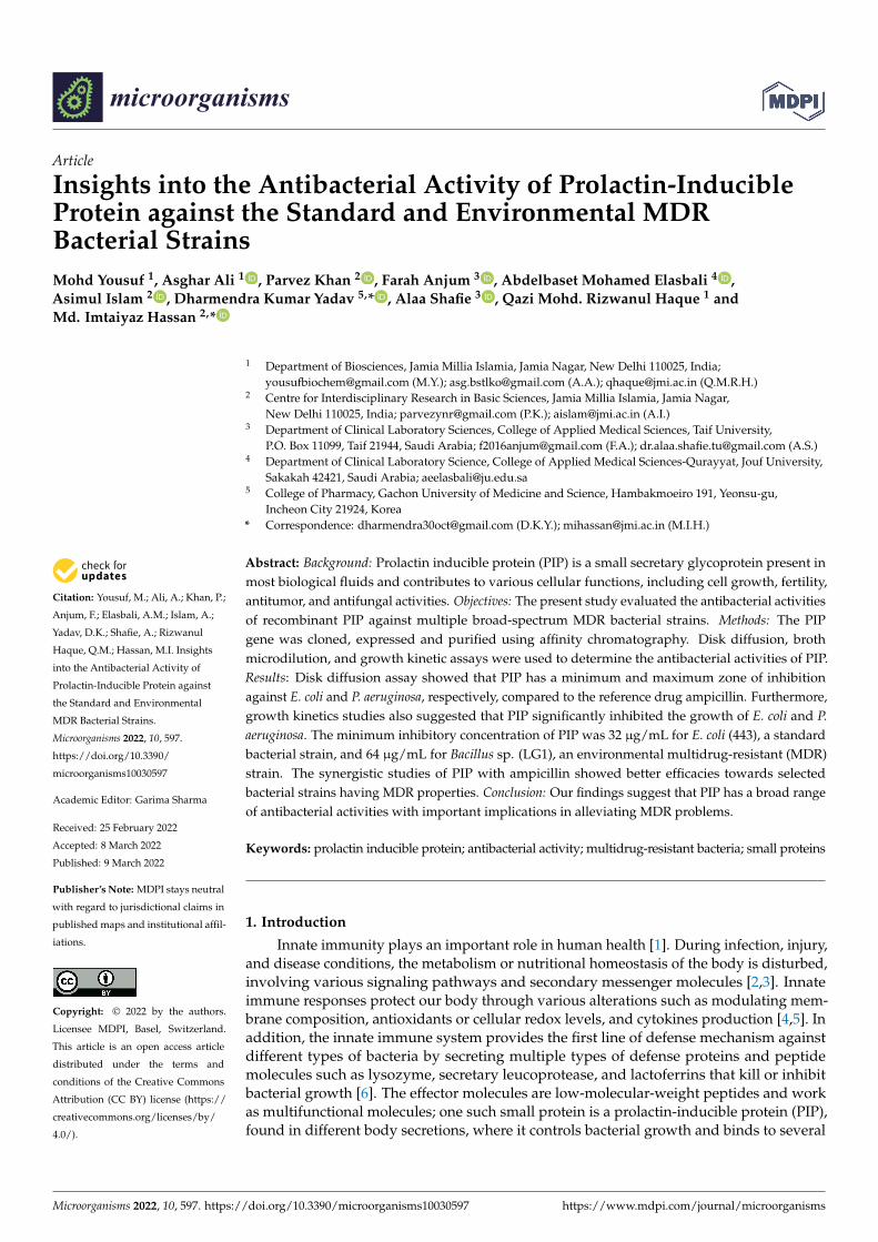
�����������������
Citation: Yousuf, M.; Ali, A.; Khan, P.;
Anjum, F.; Elasbali, A.M.; Islam, A.;
Yadav, D.K.; Shafie, A.; Rizwanul
Haque, Q.M.; Hassan, M.I. Insights
into the Antibacterial Activity of
Prolactin-Inducible Protein against
the Standard and Environmental
MDR Bacterial Strains.
Microorganisms 2022, 10, 597.
https://doi.org/10.3390/
microorganisms10030597
Academic Editor: Garima Sharma
Received: 25 February 2022
Accepted: 8 March 2022
Published: 9 March 2022
Publisher’s Note: MDPI stays neutral
with regard to jurisdictional claims in
published maps and institutional affil-
iations.
Copyright: © 2022 by the authors.
Licensee MDPI, Basel, Switzerland.
This article is an open access article
distributed under the terms and
conditions of the Creative Commons
Attribution (CC BY) license (https://
creativecommons.org/licenses/by/
4.0/).
microorganisms
Article
Insights into the Antibacterial Activity of Prolactin-InducibleProtein against the Standard and Environmental MDRBacterial StrainsMohd Yousuf 1, Asghar Ali 1 , Parvez Khan 2 , Farah Anjum 3 , Abdelbaset Mohamed Elasbali 4 ,Asimul Islam 2 , Dharmendra Kumar Yadav 5,* , Alaa Shafie 3 , Qazi Mohd. Rizwanul Haque 1 andMd. Imtaiyaz Hassan 2,*
1 Department of Biosciences, Jamia Millia Islamia, Jamia Nagar, New Delhi 110025, India;[email protected] (M.Y.); [email protected] (A.A.); [email protected] (Q.M.R.H.)
2 Centre for Interdisciplinary Research in Basic Sciences, Jamia Millia Islamia, Jamia Nagar,New Delhi 110025, India; [email protected] (P.K.); [email protected] (A.I.)
3 Department of Clinical Laboratory Sciences, College of Applied Medical Sciences, Taif University,P.O. Box 11099, Taif 21944, Saudi Arabia; [email protected] (F.A.); [email protected] (A.S.)
4 Department of Clinical Laboratory Science, College of Applied Medical Sciences-Qurayyat, Jouf University,Sakakah 42421, Saudi Arabia; [email protected]
5 College of Pharmacy, Gachon University of Medicine and Science, Hambakmoeiro 191, Yeonsu-gu,Incheon City 21924, Korea
* Correspondence: [email protected] (D.K.Y.); [email protected] (M.I.H.)
Abstract: Background: Prolactin inducible protein (PIP) is a small secretary glycoprotein present inmost biological fluids and contributes to various cellular functions, including cell growth, fertility,antitumor, and antifungal activities. Objectives: The present study evaluated the antibacterial activitiesof recombinant PIP against multiple broad-spectrum MDR bacterial strains. Methods: The PIPgene was cloned, expressed and purified using affinity chromatography. Disk diffusion, brothmicrodilution, and growth kinetic assays were used to determine the antibacterial activities of PIP.Results: Disk diffusion assay showed that PIP has a minimum and maximum zone of inhibitionagainst E. coli and P. aeruginosa, respectively, compared to the reference drug ampicillin. Furthermore,growth kinetics studies also suggested that PIP significantly inhibited the growth of E. coli and P.aeruginosa. The minimum inhibitory concentration of PIP was 32 µg/mL for E. coli (443), a standardbacterial strain, and 64 µg/mL for Bacillus sp. (LG1), an environmental multidrug-resistant (MDR)strain. The synergistic studies of PIP with ampicillin showed better efficacies towards selectedbacterial strains having MDR properties. Conclusion: Our findings suggest that PIP has a broad rangeof antibacterial activities with important implications in alleviating MDR problems.
Keywords: prolactin inducible protein; antibacterial activity; multidrug-resistant bacteria; small proteins
1. Introduction
Innate immunity plays an important role in human health [1]. During infection, injury,and disease conditions, the metabolism or nutritional homeostasis of the body is disturbed,involving various signaling pathways and secondary messenger molecules [2,3]. Innateimmune responses protect our body through various alterations such as modulating mem-brane composition, antioxidants or cellular redox levels, and cytokines production [4,5]. Inaddition, the innate immune system provides the first line of defense mechanism againstdifferent types of bacteria by secreting multiple types of defense proteins and peptidemolecules such as lysozyme, secretary leucoprotease, and lactoferrins that kill or inhibitbacterial growth [6]. The effector molecules are low-molecular-weight peptides and workas multifunctional molecules; one such small protein is a prolactin-inducible protein (PIP),found in different body secretions, where it controls bacterial growth and binds to several
Microorganisms 2022, 10, 597. https://doi.org/10.3390/microorganisms10030597 https://www.mdpi.com/journal/microorganisms

Microorganisms 2022, 10, 597 2 of 13
other partner molecules to exhibit multiple functions in cancer growth, fertility, and enamelpellicle formation [7–13].
PIP plays an important function in immune defense and the reproductive system,and its expression is upregulated by prolactin or androgens, whereas it is downregulatedby estrogens [14,15]. During pathological condition like corneal disease, keratoconous,dacryliths, and cancer, the expression of PIP get altered in several exocrine tissues ormammary glands such as sweat and salivary glands, and thus acting as a biomarkerin such diseases [16–19]. PIP is a small single polypeptide chain protein expressed invarious human body parts, including the salivary gland, lacrimal gland, trachea, prostate,muscle, mammary glands, and lungs. The highest expression of PIP was reported in thesalivary gland (~55%) [16,20]. The gene of human PIP has located on chromosome 7q32-36regions, encoding for a 146 amino-acid-residue-long polypeptide leading to the synthesisof a ~17 kDa protein [16,21,22].
PIP is a β-rich glycosylated protein. The crystal structure of PIP demonstrated that βsheets are organized around the hydrophobic amino acid residue and form a sandwich-like structure. PIP contains Asn-X-Ser/Thr motif present at position Asn77-Arg78-Thr79,representing a potential glycosylation site [21]. The structural domain of PIP representsimmunoglobulin domains [23,24]. Previous studies on the structure and function of PIPdemonstrated its isolation from different biological fluids, where it performs numerousfunctions with its interacting partner proteins [16,25–27]. The presence of immunoglobindomains suggested PIP’s immunological implications supported by multiple studies usingin vitro and in vivo (mouse models) presenting the role of PIP in innate and cell-mediatedimmunity [28,29].
The expression of PIP is used as a marker of mammary cell differentiation, and itsaltered expression is associated with breast cancer progression [29]. Recent studies sug-gested that the downregulation of PIP promotes osteogenic differentiation of periodontalstem cells [30]. Multiple reports supported the antimicrobial or anti-protozoal roles of PIP.They suggested that reduced levels of antimicrobial or immunomodulatory proteins intears lead to higher ocular infections and susceptibility to other pathogens such as Leis-mania major [31,32]. PIP acts on oral bacteria (an oral defense mechanism), promoting itsclearance from the oral cavity and modulating oral flora [33]. PIP is present in most of thebiological/physiological fluids of the human body. However, the antibacterial function ofPIP is not well-studied and little work has been conducted on evaluating its antibacterialproperties. Thus, we aimed to investigate the antibacterial activity of PIP on multiplemicrobial strains, including MDR strains.
2. Materials and Methods2.1. Bacterial Strains and Plasmids
The coding region of the PIP gene was amplified from total human cDNA throughPCR using PIP-specific primers bearing NdeI (forward primer) and XhoI (reverse primer)restriction sites. The amplified amplicon was digested with NdeI and XhoI, and subse-quently cloned into pET28a+ (Novagen, Madison, WI, USA) prokaryotic expression vector.DH5α and BL21 (DE3) strains of E. coli cells were used for PIP cloning and expression,respectively. Plasmid isolation, restriction enzyme digestion by NdeI and XhoI, and theligation experiments were performed as described previously [34]. Luria Broth (DifcoTM,Becton Dickinson, Fisher Scientific, Kansas City, KS, USA) was used for bacterial culturewith 50 µg/mL Kanamycin (Sigma, Saint Louis, MO, USA).
2.2. Expression and Purification of PIP
The pET28a+-PIP expression construct was transformed in E. coli BL21 (DE3) followingthe standard protein expression protocol as described previously [35]. The primary culturegrown overnight was used to develop secondary culture containing 50 µg/mL kanamycinand incubated at 37 ◦C with constant agitation at 180 rpm in an incubator shaker until

Microorganisms 2022, 10, 597 3 of 13
the absorbance reached 0.6 at 600 nm. The secondary culture was induced by 0.5 M IPTG(Sigma, Saint Louis, MO, USA) and incubated for an additional 4–5 h at 37 ◦C, 180 rpm.
The cell culture was centrifuged at 7000–8000× g for 10 min, and the cell pellet wasdissolved in cell lysis buffer having 50 mM Tris–HCl buffer, pH 8.0, 200 mM NaCl, 2% (v/v)glycerol, 1 mM β-mercaptoethanol, 0.1 mg/mL lysozyme, 1 mM phenyl methane sulfonylfluoride (PMSF), and 1% (v/v) triton X-100 (U. S. Biochemical Corp, Cleveland, OH, USA) andincubated for 1 h at 37 ◦C. Following the incubation, cell lysate was sonicated on ice for 15 minand centrifuged for 30 min at 13,000 rpm at 4 ◦C. The supernatant was collected, and proteinexpression was checked using SDS-PAGE. Ni-NTA affinity chromatography was used forthe purification of 6XHis-tag-PIP protein. For this, the clear supernatant was passed throughNi–NTA column pre-equilibrated with Tris buffer (50 mM Tris–HCl, pH 8.0, 200 mM NaCl,1% v/v glycerol, 20 mM imidazole). Following protein binding, the column was washed with50 mL of washing buffer (50 mM Tris–HCl, pH 8.0, 200 mM NaCl, 50 mM imidazole) at 4 ◦C.Bound protein was eluted with an imidazole gradient (100–300 mM). The eluted fractionswere run on the SDS-PAGE to check the purity of the protein. Fractions containing purifiedprotein were pooled and dialyzed in 20 mM Tris–HCl buffer, pH 8.0, 100 mM NaCl, and2% glycerol. The dialyzed protein was concentrated using Amicon Ultra 5 K device (Merck,Darmstadt, Germany). The concentrated protein sample was further loaded on Hi Trap DEAE-FF (1 mL, 7 mm × 25 mm) column (GE Healthcare, Chicago, IL, USA) pre-equilibrated with50 mM Tris–HCl buffer, pH 8.0. Bound proteins were eluted with increasing concentration ofNaCl (0–1 M) in the 50 mM Tris–HCl buffer, pH 8.0. PIP eluted at 0.50 M NaCl was pooled,concentrated, and stored for further studies. The purity of PIP was tested by SDS-PAGE andvalidated through Western blot using the luminol method [36].
2.3. Antibacterial Activity Assays
Subculturing of standard bacterial strains, including S. aureus (MTCC 902), B. sub-tilis (MTCC 736), P. aeruginosa (MTCC 2453), Escherichia coli (MTCC 443), and environ-mentally resistant (multidrug-resistant) bacterial isolates, i.e., Citrobacter werkmanii (SH52/MN267555), E. coli (SD6/MT577556), Bacillus sp. (LG1/MT576690), and Citrtobactersp. (HK 106/MT576965) was performed on nutrient agar medium through the streakingmethod [37]. Following the streaking, the plates were kept in the incubator overnight at37 ◦C, and the growth of each plate was observed on the next day.
2.4. Determination of MIC and MBC
To determine the MIC of the PIP, the microdilution method was performed accordingto the standard protocol of NCCL [38]. MIC of the PIP was determined against two Gram-positive (B. subtilis and S. aureus) and two Gram-negative (P. aeruginosa and E. coli) bacterialstrains. The MIC values were estimated and compared to ampicillin (AMP), used as acontrol antibiotic. To obtain the stock solution of 10.24 mg/mL for PIP and AMP (standardbacterial drugs), they were dissolved in Tris buffer and sterile water, respectively. The serialdilutions of broth were performed to obtain the final concentrations of 1024, 512, 256, 128,64, 32, 16, 8, 4, 2, 1, 0.5, 0.25, and 0.125 µg/mL. A control test with buffer was performedfollowing the same dilutions to check whether the buffer or solvent of PIP affected thegrowth of bacteria. The cultures were incubated at 37 ◦C for 24 h and compared with blankin terms of turbidity developed by the microbial growth. The minimum concentration ofPIP exhibiting no bacterial growth was described as MIC. For the MBC, 10 µL aliquots fromeach well that showed no growth of microorganism were plated on Mueller–Hinton Agar(MHA) and incubated at 37 ◦C for 24 h [39,40]. The MIC and MBC values were calculatedas per the previously published protocols [41] and compared with the AMP taken as areference antibiotic.
2.5. Disk Diffusion Assay
Disk diffusion assay was performed to identify the antibacterial efficacy of PIP againststandard bacterial strains by following Kirby–Bauer method [42]. Various concentrations
Microorganisms 2022, 10, 597 4 of 13
of PIP such as MIC/2, MIC and 2MIC were taken on disc for disc diffusion assay [43–46].Briefly, the bacterial cells were inoculated in a liquid broth medium and grown overnight at37 ◦C. Approximately 105 cells/mL were taken from that liquid broth medium, inoculatedinto molten nutrient agar medium, and poured into Petri plates [45,47–49]. After solidification,autoclaved Whatman papers having 4 mm diameter disks were put at suitable distances oversolid agar, and MIC/2, MIC, and 2MIC from the stock solution were put on disks. Plates wereincubated at 37 ◦C overnight, and the next day, ZOI was measured in mm.
2.6. Combination Studies of PIP against the Standard Bacterial Strains
The synergistic activity of PIP with standard drug AMP was determined using themicrodilution checkerboard method against standard bacterial strains of E. coli and P.aeruginosa as described previously [50]. AMP were serially diluted in columns from 32,16,8, 4, 2, 1, 0.5, 0.25, 0.125, and 0.0625 µg/mL, while PIP was diluted in rows from 1024,512, 256, 128, 64, 32, 16, 8, 4, 2, 1.0, and 0.5 µg/mL, respectively, in a 96-microwell plate toobtain multiple combination. The plates were inoculated with a freshly prepared cultureof bacterial isolates and incubated at 37 ◦C overnight. Following the incubation time,combinatorial MIC was determined as the concentrations at which no visible growthoccurred. Using the formula given below, the synergy of compounds in terms of FICI(fractional inhibitory concentration index) was calculated [41]:
ΣFIC = FICA + FICB = (CA/MICA) + (CB/MICB)
where MICA and MICB are the MICs of drugs A and B alone, respectively, and CA andCB are the concentrations of the drugs in combination, respectively, in all of the wellscorresponding to an MIC (isoeffective combinations).
Synergy and antagonism were defined by FICI indices ≤ 0.5 and >4, respectively, and‘indifferent’ was defined by 1 < FICI ≤4.
2.7. Growth Kinetics Assay
Based on previous experiments, we have selected E. coli and P. aeruginosa for theseexperiments. Bacterial cells were freshly revived by sub-culturing on the Luria agarplate. To obtain the fresh cultures for the experiments, an inoculum was transferredinto the Luria broth and was grown overnight at 37 ◦C. Different concentrations of PIP,equivalent to MIC/2, MIC, and 2MIC, were added separately to the respective conicalflasks containing inoculated medium and incubated at 37 ◦C. Positive control was alsotaken to observe the full growth. To observe the growth kinetics of the cultures, 1 mLaliquot of each sample was taken out from the culture flask and growth was measured at590 nm turbidometrically (optical density) using Thermo Multiskan spectrophotometerafter each one-hour interval [51]. To determine the effect of PIP on bacterial growth, agraph was plotted between OD versus time duration (hours) to obtain a growth curve andfurther analysis [52].
2.8. Effect of PIP on Environmental Resistant Bacterial Strains
Antibacterial activity of PIP was investigated against a variety of environmentalresistant strains such as HK 106 (Citrobacter sp.), LG 1 (Bacillus sp.), SD 6 (Escherichiacoli), and SH52 (Citrobacter werkmanii). All bacterial strains were isolated from differentenvironmental (Lake, Pond, effluent from slaughterhouse and wastewater treatment plant)water samples. The standard broth dilution method was used to determine the MIC ofthese isolates and compare the MIC values to the conventional antibiotics such as ampicillin(a penicillin analog). The microdilution checkerboard method has been used to examinethe synergistic activity of the PIP with the standard antibiotic AMP against Escherichia coliand Citrtobacter werkmanii. The procedure above was used for the MIC and synergisticactivity [41,53–55].

Microorganisms 2022, 10, 597 5 of 13
3. Result and Discussion3.1. Cloning, Expression, and Purification of PIP
The PIP gene (from plasmid pcDNA) was amplified using a gene-specific PCR, withNdel and XhoI sites in the forward and reverse primers, respectively. The size of theamplified product was 366-bp (Figure 1A). The amplified gene product having selectedrestriction sites was digested with NcoI and XhoI and subsequently ligated into the pET28a+backbone digested with similar restriction enzymes (Figure 1B). The ligated product wastransformed into E. coli and positive colonies were selected and confirmed using colonyPCR. The positive clones were further confirmed using restriction digestion through NcoIand XhoI endonucleases (Figure 1C). Finally, the constructed plasmid was verified by DNAsequencing (Figure S1).
Figure 1. Cloning, expression, and purification of PIP: (A) Amplification of PIP gene through PCR.Lane 1: Marker; Lane 2 and 3 are amplified products of the PIP gene. (B) Lane 1: purified pET28a+
plasmid, Lane 2: pET28a+ plasmid digested with NcoI and XhoI. (C) Restriction digestion studiesof PIP expression clone, Lane 1: marker, Lane2: digested pET28a+-PIP construct with NcoI andXhoI, Lane 3: undigested pET28a+-PIP construct. (D) Expression of recombinant PIP using E. coli asan expression host showing un-induced and IPTG-induced bacterial culture having PIP construct.(E) Elution profile of recombinant PIP protein: Lane 1: Marker, Lane 2: supernatant from wholecell lysate before binding to Ni-NTA column; Lane 3: supernatant after Ni-NTA binding; Lane 4:elution from 20 mM imidazole elution buffer; Lane 5: elution from 50 mM imidazole buffer; Lane6: elution from 200 mM imidazole buffer; Lane 7: elution from 300 mM imidazole buffer; Lane 8:elution from 500 mM imidazole buffer. (F) PIP elution profile from ion-exchange chromatography.(G) The 15% SDS-PAGE for PIP elutions; Lane 1: Marker, Lane 2: impure PIP before loading toion-exchange chromatography, Lane 3–5: showing increasing concentration gradients (5 µg, 10 µgand 15 µg, respectively) of purified PIP eluted from ion-exchange chromatography.
Microorganisms 2022, 10, 597 6 of 13
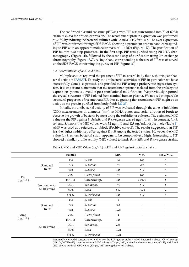
The confirmed plasmid construct pET28a+ with PIP was transformed into BL21 (DE3)strain of E. coli for protein expression. The recombinant protein expression was performedat 37 ◦C by inducing the bacterial cultures with 0.5 mM IPTG for 4–5 h. The over-expressionof PIP was confirmed through SDS-PAGE, showing a prominent protein band correspond-ing to PIP with an apparent molecular mass of ~14 kDa (Figure 1D). The purification ofPIP follows two-step processes. In the first step, PIP was purified using Ni-NTA chro-matography (Figure 1E), followed by the second step of purification using ion-exchangechromatography (Figure 1F,G). A single band corresponding to the size of PIP was observedon the SDS-PAGE, confirming the purity of PIP (Figure 1G).
3.2. Determination of MIC and MBC
Multiple studies reported the presence of PIP in several body fluids, showing antibac-terial activities [7,56,57]. To study the antibacterial activities of PIP, in particular, we havesuccessfully cloned, expressed, and purified the PIP using a prokaryotic expression sys-tem. It is important to mention that the recombinant protein isolated from the prokaryoticexpression system is devoid of post-translational modifications. We previously reportedthe crystal structure of PIP isolated from seminal human plasma. We noticed comparablestructural properties of recombinant PIP, thus suggesting that recombinant PIP might be asactive as the protein purified from body fluids [22,23].
Initially, the antibacterial activity of PIP was evaluated through the zone of inhibition(ZOI) measurements in diameter (mm) on MHA plates and serial dilution of broth toobserve the growth of bacteria by measuring the turbidity of cultures. The estimated MICvalue for the PIP against B. Subtilis and P. aeruginosa was 64 µg/mL, wh. In contrast, for E.coli and S. aureus the MIC values were 32 µg/mL and 128 µg/mL, respectively (Table 1).AMP was used as a reference antibiotic (Positive control). The results suggested that PIPhas the highest inhibitory effect against E. coli among the tested strains. However, the MICvalue for S. aureus bacterial strain appears to be comparatively high. Interestingly, PIPshowed a similar profile activity (MIC values) towards B. subtilis and P. aeruginosa strains.
Table 1. MIC and MBC Values (µg/mL) of PIP and AMP against bacterial strains.
Isolates MIC MBC MBC/MIC
PIP(µg/mL)
StandardStrains
443 E. coli 32 128 4
736 B. subtilis 64 256 4
902 S. aureus 128 512 4
2453 P. aeruginosa 64 128 2
EnvironmentalMDR strains
HK 106 Citrobacter sp. 128 >1024 8
LG 1 Bacillus sp. 64 512 8
SD 6 E.coli 512 1024 2
SH 52 B. werkmanii 128 512 4
Amp(µg/mL)
StandardStrains
443 E. coli 1 - -
736 B. subtilis 0.5 - -
902 S. aureus 0.25 - -
2453 P. aeruginosa 4 - -
MDR strains
HK 106 Citrobacter sp. 128 - -
LG 1 Bacillus sp. 256 - -
SD 6 E.coli 1024 - -
SH 52 B. werkmanii 1024 - -
Minimal bactericidal concentration values for the PIP against eight studied bacterial isolates. Citrobacter sp.(HK106/MT576965) shows maximum MBC value (>1024 µg/mL), while Pseudomonas aeruginosa (2453) and E. coli(443) shows minimal MBC value (128 µg/mL) among the tested isolates.

Microorganisms 2022, 10, 597 7 of 13
In addition, the MBC values of PIP range from 128 to 512 µg/mL (Table 1). The ratioof MBC to MIC is used to characterize the antibacterial activity of any compound. If theMBC/MIC ratio was ≤2, the compounds were termed bactericidal, and they were termedbacteriostatic if the ratio was between 2 and 16 [43,45,58–61]. The results shown in Table 1suggested that the PIP showed bactericidal and bacteriostatic effects based on the testedbacterial strains.
3.3. Disk Diffusion Assay
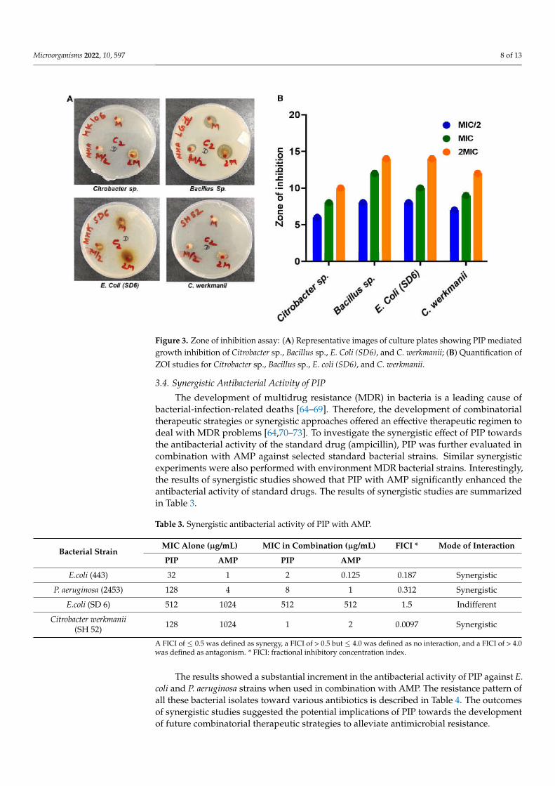
The disc diffusion test is a widely accepted and primary test to see the susceptibility ofbacteria or bacterial isolates towards antibiotics or any antibacterial molecules [46,49,62,63].To further evaluate the antibacterial activity of PIP, following the serial dilution method,the disk diffusion assay was used. The disk diffusion experiment results revealed that PIPshowed the lowest and highest ZOI against P. aeruginosa and E. coli, respectively (Figure 2).In Gram-negative E. coli culture, the treatment with PIP leads to a clear ZOI of 16, 20, and26 mm around the disc of MIC/2, MIC, and 2MIC, respectively. While in the case of P.aeruginosa (2453), 6, 8, and 13 mm clear ZOI 2343 measured around the disks of the MIC/2,MIC, and 2MIC concentrations, respectively (Figure 2, Table 2). Around the disks of MIC/2,MIC, and 2MIC, the observed ZOI was 8, 9, and 12mm with B. subtilis and 8, 13, and 17with S aureus, respectively. Antibacterial activities of PIP using agar diffusion assay arepresented in Figures 2 and 3. Diffusion assay results in terms of ZOI diameter (mm) forstandard bacterial strains are given in Table 2.
Figure 2. Zone of inhibition assay. (A) Representative images of culture plates showing PIP mediatedgrowth inhibition of Bacillus subtilis, E. coli, Staphylococcus aureus, and Pseudomonas aeruginosa. (B) Quantifica-tion of ZOI studies for Bacillus subtilis, E. coli, Staphylococcus aureus, and Pseudomonas aeruginosa.
Table 2. Zone of inhibition (in mm) as measured around the disk of various concentrations ofcompound.
Types of BacterialIsolates
Isolates Code Bacterial StrainZone of Inhibition at Different Concentrations of Test Compound
MIC/2 MIC 2MIC
StandardBacterial Strains
443 E. coli 16 20 26
736 B. subtilis 8 9 12
902 S. aureus 8 13 17
2453 P. aeruginosa 6 8 13
EnvironmentalMDR Bacterial strains
HK 106 Citrtobacter sp. 6 8 10
LG 1 Bacillus sp. 8 12 14
SD 6 E. coli 8 10 14
SH 52 C. werkmanii 7 9 12
Microorganisms 2022, 10, 597 8 of 13
Figure 3. Zone of inhibition assay: (A) Representative images of culture plates showing PIP mediatedgrowth inhibition of Citrobacter sp., Bacillus sp., E. Coli (SD6), and C. werkmanii; (B) Quantification ofZOI studies for Citrobacter sp., Bacillus sp., E. coli (SD6), and C. werkmanii.
3.4. Synergistic Antibacterial Activity of PIP
The development of multidrug resistance (MDR) in bacteria is a leading cause ofbacterial-infection-related deaths [64–69]. Therefore, the development of combinatorialtherapeutic strategies or synergistic approaches offered an effective therapeutic regimen todeal with MDR problems [64,70–73]. To investigate the synergistic effect of PIP towardsthe antibacterial activity of the standard drug (ampicillin), PIP was further evaluated incombination with AMP against selected standard bacterial strains. Similar synergisticexperiments were also performed with environment MDR bacterial strains. Interestingly,the results of synergistic studies showed that PIP with AMP significantly enhanced theantibacterial activity of standard drugs. The results of synergistic studies are summarizedin Table 3.
Table 3. Synergistic antibacterial activity of PIP with AMP.
Bacterial StrainMIC Alone (µg/mL) MIC in Combination (µg/mL) FICI * Mode of Interaction
PIP AMP PIP AMP
E.coli (443) 32 1 2 0.125 0.187 Synergistic
P. aeruginosa (2453) 128 4 8 1 0.312 Synergistic
E.coli (SD 6) 512 1024 512 512 1.5 Indifferent
Citrobacter werkmanii(SH 52) 128 1024 1 2 0.0097 Synergistic
A FICI of ≤ 0.5 was defined as synergy, a FICI of > 0.5 but ≤ 4.0 was defined as no interaction, and a FICI of > 4.0was defined as antagonism. * FICI: fractional inhibitory concentration index.
The results showed a substantial increment in the antibacterial activity of PIP against E.coli and P. aeruginosa strains when used in combination with AMP. The resistance pattern ofall these bacterial isolates toward various antibiotics is described in Table 4. The outcomesof synergistic studies suggested the potential implications of PIP towards the developmentof future combinatorial therapeutic strategies to alleviate antimicrobial resistance.

Microorganisms 2022, 10, 597 9 of 13
Table 4. Resistance pattern of environmental isolates.
Isolates Strain Accession No Resistance Pattern MIC (µg/mL) for AMP Site
HK 106 Citrtobacter sp. MT576965 AMP, ETP, IPM, CX, P/T CZ,PB, AK, CIP, CL, TR 128 Hauz Khas lake
LG 1 Bacillus sp. MT576690 AMP, CX, A/S, RIF, CZ, TR 256 Lodhi Garden Pond
SD 6 Escherichia coli MT577556 AMP, IMP, CX, RIF, CZ, PB,AK, TE, CIP, LE 1024 Shaheen Bagh Drain
SH 52 Citrtobacterwerkmanii MN267555 ETP, CX, RIF, CZ, PB, CL, TR 1024 Ghazipur Slaughterhouse
effluent
AMP: Ampicillin, A/S: Ampicillin/Sulbactam, CX: Cefoxitin, IPM: Imipenem, P/T: Piperacillin/Tazobactam, CZ:Cefazolin, ETP: Ertapenem, RIF: Rifampicin AK: Amikacin, CL: Colistin, PB: Polymyxin B, TE: Tetracycline, CIP:Ciprofloxacin, LE: Levofloxacin, TR: Trimethoprim.
3.5. Growth Kinetics
The kinetic growth studies provide an important tool to see the influence of substrateconcentration or a molecule towards the specific growth rate (versus time) and providea method to evaluate drug susceptibility to microbial growth [74,75]. Therefore, growthkinetics studies were performed to determine the effect of PIP on the growth of E. coliand P. aeruginosa. For this, we take untreated cultures as a negative control, and AMP-treated culture was taken as a positive control. The results showed that the growth curveof untreated bacterial cells (control) follows the standard growth curve with a clear lag,exponential or log, brief stationery, and decline phases of the bacterial cell. On the otherhand, at 2MIC, MIC, and MIC/2 concentration values of PIP, no growth was observed until20 h of incubation in E. coli growth experiments (Figure 4). In the case of P. aeruginosa at2MICconcentration values of PIP, no growth was observed until 24 h. At MIC concentrationvalues, a slight increase in the culture growth was observed after 20 h.
Figure 4. Growth kinetics study of (A) E. coli, (B) P. aeruginosa at different MIC concentrations of PIP.Ampicillin (AMP) was taken as a positive control.
In contrast, at the MIC/2 concentration, the lag phase was increased from 3 to 8 h,the log phase was significantly increased from 9 h to 19 h, and following the log phase, ashort stationary phase was observed from 20 h to 22 h, then a decline phase was observed.Moreover, PIP completely inhibited the growth of both bacterial strains at MIC and 2MICconcentrations. No growth was observed in AMP-treated bacterial cells for 24 h in bothselected bacterial strains.
3.6. Effect of PIP on Environmental MDR Strains
The MIC value for the PIP against environmental MDR isolates was observed from64–512 µg/mL. Minimum MIC values 64 µg/mL for LG1 64, whereas maximum MICvalues 512 µg/ml for SD6 were observed among tested MDR isolates (as shown in Table 1).Environmental MDR isolates SH52, LG1, SD6 showed a significant decrease as 4, 3, 2-fold

Microorganisms 2022, 10, 597 10 of 13
MIC values compared to AMP, respectively, while HK106 showed nearly the same MICvalue for PIP and AMP (Table 1). MBC values ranges for the resistant strain varies from> 1024 to 512 µg/mL. If the MBC/MIC ratio was 2 to 8, and the compounds were termedbactericidal and bacteriostatic. The PIP has various bactericidal and bacteriostatic effectson environment-resistant bacterial isolates. The zone of inhibition assays for PIP againstenvironmental-resistant bacterial isolate LG1 shows a maximum ZOI of 8mm at MIC/2concentration and of 12 mm at MIC concentration. Moreover, at 2MIC concentration, themaximum ZOI was 14 mm. At the same time, Citrobacter werkmanii (SH 52) shows a ZOIof 7 mm at the MIC/2 concentration, 9 mm at the MIC concentration, and 12 mm at the2MIC concentration. Furthermore, the combinational (PIP and AMP) studies against SD6and SH52 showed that the PIP has a differential mode of interaction (FICI = 1.5) with AMPagainst SD6. In contrast, it showed the synergistic mode of interaction (FICI = 0.0097)against SH52 (Table 3).
4. Conclusions
To study the antibacterial potential of PIP, we cloned, expressed, and purified PIPusing a prokaryotic expression system. The antibacterial activities of PIP were evaluatedagainst a broad spectrum of bacterial strains. The outcomes of the present study suggestthat PIP significantly inhibited a wide range of bacterial strains, including standard andenvironmental MDR strains. The enhanced antibacterial potential of standard antibioticssuch as ampicillin in synergistic studies of PIP makes it an important investigatory moleculetowards developing combinatorial antibacterial approaches to deal with the rising burdenof antimicrobial resistance. This study indicates PIP’s future investigations as a novelbioactive molecule for developing antibacterial therapies and as a preservative agent in thefood, cosmetic, and medicine industries.
Supplementary Materials: The following supporting information can be downloaded at: https://www.mdpi.com/article/10.3390/microorganisms10030597/s1. Figure S1: Sequence of PIP gene.
Author Contributions: Conceptualization, M.Y. and P.K.; methodology, M.Y.; software, A.A.; valida-tion, M.Y. and A.A.; formal analysis, F.A., A.I.; investigation, A.S.; resources, A.M.E.; data curation,Q.M.R.H.; writing—original draft preparation, A.A., M.Y.; writing—review and editing, A.I., P.K.;visualization, M.I.H.; supervision, M.I.H.; project administration, D.K.Y.; funding acquisition, M.I.H.and D.K.Y. All authors have read and agreed to the published version of the manuscript.
Funding: This work was supported by Taif University Researchers Supporting Project Number(TURSP-2020/131), Taif University, Taif, Saudi Arabia, and the Indian Council of Medical Research(Grant No. ECD/Adhoc/2/2021-22).
Institutional Review Board Statement: Not applicable.
Informed Consent Statement: Not applicable.
Data Availability Statement: Not applicable.
Acknowledgments: This work was supported by Taif University Researchers Supporting ProjectNumber (TURSP-2020/131), Taif University, Taif, Saudi Arabia. M.Y. thanks to the Indian Council ofMedical Research for the award of Senior Research Fellowship (F. No.45/54/2018-PHA/BMS/OL).M.I.H. acknowledges the Council of Scientific and Industrial Research for financial support [ProjectNo. 27(0368)/20/EMR-II]. The authors sincerely thank the Department of Science and Technology,Government of India, for the FIST support (FIST program No. SR/FST/LSII/2020/782).
Conflicts of Interest: The authors declare no conflict of interest.
References1. Boman, H. Antibacterial peptides: Basic facts and emerging concepts. J. Intern. Med. 2003, 254, 197–215. [CrossRef] [PubMed]2. Szczykutowicz, J.; Tkaczuk-Wlach, J.; Ferens-Sieczkowska, M. Glycoproteins Presenting Galactose and N-Acetylgalactosamine in
Human Seminal Plasma as Potential Players Involved in Immune Modulation in the Fertilization Process. Int. J. Mol. Sci. 2021, 22,7331. [CrossRef] [PubMed]
Microorganisms 2022, 10, 597 11 of 13
3. Roberts, L.M.; Schwarz, B.; Speranza, E.; Leighton, I.; Wehrly, T.; Best, S.; Bosio, C.M. Pulmonary infection induces persistent,pathogen-specific lipidomic changes influencing trained immunity. iScience 2021, 24, 103025. [CrossRef] [PubMed]
4. Grimble, R.F.; Grimble, G.K. Immunonutrition: Role of sulfur amino acids, related amino acids, and polyamines. Nutrition 1998,14, 605–610. [CrossRef]
5. Dumas, A.; Knaus, U.G. Raising the ‘Good’ Oxidants for Immune Protection. Front. Immunol. 2021, 12, 698042. [CrossRef]6. Rogan, M.P.; Geraghty, P.; Greene, C.M.; O’Neill, S.J.; Taggart, C.C.; McElvaney, N.G. Antimicrobial proteins and polypeptides in
pulmonary innate defence. Respir. Res. 2006, 7, 29. [CrossRef]7. Schenkels, L.C.; Veerman, E.C.; Nieuw Amerongen, A.V. EP-GP and the lipocalin VEGh, two different human salivary 20-kDa
proteins. J. Dent. Res. 1995, 74, 1543–1550. [CrossRef]8. Sivakumar, S.; Mirels, L.; Miranda, A.J. Hand, AR Secretory protein expression patterns during rat parotid gland development.
Anat. Rec. Off. Publ. Am. Assoc. Anat. 1998, 252, 485–497. [CrossRef]9. Chiu, W.W.C.; Chamley, L.W. Human seminal plasma prolactin-inducible protein is an immunoglobulin G-binding protein. J.
Reprod. Immunol. 2003, 60, 97–111. [CrossRef]10. Gaubin, M.; Autiero, M.; Basmaciogullari, S.; Métivier, D.; Misëhal, Z.; Culerrier, R.; Oudin, A.; Guardiola, J.; Piatier-Tonneau, D.
Potent inhibition of CD4/TCR-mediated T cell apoptosis by a CD4-binding glycoprotein secreted from breast tumor and seminalvesicle cells. J. Immunol. 1999, 162, 2631–2638.
11. Witkin, S.S.; Richards, J.M.; Bongiovanni, A.M.; Zelikovsky, G. An IgG-Fc binding protein in seminal fluid. Am. J. Reprod. Immunol.1983, 3, 23–27. [CrossRef] [PubMed]
12. Bronson, R.A. Antisperm antibodies: A critical evaluation and clinical guidelines. J. Reprod. Immunol. 1999, 45, 159–189. [CrossRef]13. Rathman, W.; Van, M.Z.; den Keybus Van, P.; Bank, R.; Veerman, E. Nieuw, AA Isolation and characterization of three non-
mucinous human salivary proteins with affinity for hydroxyapatite. J. Biol. Buccale 1989, 17, 199–208.14. Simard, J.; Hatton, A.C.; Labrief, C.; Dauvois, S.; Zhao, H.F.; Haagensen, D.E.; Labrie, F. Inhibitory effect of estrogens on
GCDFP-15 mRNA levels and secretion in ZR-75-1 human breast cancer cells. Mol. Endocrinol. 1989, 3, 694–702. [CrossRef]15. Blanchard, A.A.; Nistor, F.E.; Castaneda, D.; Martin, G.G.; Hicks, F.; Amara, R.P.C.; Shiu, Y.M. Generation and initial characteriza-
tion of the prolactin-inducible protein (PIP) null mouse: Accompanying global changes in gene expression in the submandibulargland. Can. J. Physiol. Pharmacol. 2009, 87, 859–872. [CrossRef] [PubMed]
16. Hassan, M.I.; Waheed, A.; Yadav, S.; Singh, T.; Ahmad, F. Prolactin inducible protein in cancer, fertility and immunoregulation:Structure, function and its clinical implications. Cell. Mol. Life Sci. 2009, 66, 447–459. [CrossRef] [PubMed]
17. Sharif, R.; Bak-Nielsen, S.; Sejersen, H.; Ding, K.; Hjortdal, J.; Karamichos, D. Prolactin-Induced Protein is a novel biomarker forKeratoconus. Exp. Eye Res. 2019, 179, 55–63. [CrossRef] [PubMed]
18. Mano, F.; Takimoto, H.; Oe, M.; Chang, K.-c.; Mano, T.; Yoshida, Y. Proteomic analysis of dacryoliths from patients with or withouttopical Rebamipide treatment. Biomed. Hub 2018, 3, 1–11. [CrossRef]
19. Urbaniak, A.; Jablonska, K.; Podhorska-Okolow, M.; Ugorski, M.; Dziegiel, P. Prolactin-induced protein (PIP)-characterizationand role in breast cancer progression. Am. J. Cancer Res. 2018, 8, 2150–2164.
20. Haagensen, D.E., Jr.; Mazoujian, G.; Dilley, W.G.; Pedersen, C.E.; Kister, S.J.; Wells Jr, S.A. Breast gross cystic disease fluid analysis.I. Isolation and radioimmunoassay for a major component protein. J. Natl. Cancer Inst. 1979, 62, 239–247.
21. Murphy, L.C.; Tsuyuki, D.; Myal, Y.; Shiu, R. Isolation and sequencing of a cDNA clone for a prolactin-inducible protein (PIP).Regulation of PIP gene expression in the human breast cancer cell line, T-47D. J. Biol. Chem. 1987, 262, 15236–15241. [CrossRef]
22. Hassan, M.I.; Kumar, V.; Singh, T.P.; Yadav, S. Purification and characterization of zinc α2-glycoprotein-Prolactin inducible proteincomplex from human seminal plasma. J. Sep. Sci. 2008, 31, 2318–2324. [CrossRef] [PubMed]
23. Hassan, M.I.; Bilgrami, S.; Kumar, V.; Singh, N.; Yadav, S.; Kaur, P.; Singh, T. Crystal structure of the novel complex formedbetween zinc α2-glycoprotein (ZAG) and prolactin-inducible protein (PIP) from human seminal plasma. J. Mol. Biol. 2008, 384,663–672. [CrossRef] [PubMed]
24. Hassan, I.; Ahmad, F. Structural diversity of class I MHC-like molecules and its implications in binding specificities. Adv. ProteinChem. Struct. Biol. 2011, 83, 223–270. [CrossRef]
25. Léon CPM, S.; Johann, S.; Els, W.-W.; Ingde, S.E.; Enno, V.; Arie, A.V. Identity of human extra parotid glycoprotein (EP-GP) withsecretory actin binding protein (SABP) and its biological properties. Biol. Chem. 1994, 375, 609–615. [CrossRef] [PubMed]
26. Schaller, J.; Akiyama, K.; Kimura, H.; Hess, D.; Affolter, M.; Rickli, E.E. Primary structure of a new actin-binding protein fromhuman seminal plasma. Eur. J. Biochem. 1991, 196, 743–750. [CrossRef] [PubMed]
27. Caputo, E.; Carratore, V.; Ciullo, M.; Tiberio, C.; Mani, J.C.; Piatier-Tonneau, D.; Guardiola, J. Biosynthesis and immunobiochemicalcharacterization of gp17/GCDFP-15: A glycoprotein from seminal vesicles and from breast tumors, in HeLa cells and in Pichiapastoris yeast. Eur. J. Biochem. 1999, 265, 664–670. [CrossRef]
28. Terceiro, L.E.L.; Blanchard, A.A.A.; Edechi, C.A.; Fresnoza, A.; Triggs-Raine, B.; Leygue, E.; Myal, Y. Generation of prolactin-inducible protein (Pip) knockout mice by CRISPR/Cas9-mediated gene engineering. Can. J. Physiol. Pharm. 2021, 100, 86–91.[CrossRef]
29. Ihedioha, O.C.; Shiu, R.P.; Uzonna, J.E.; Myal, Y. Prolactin-Inducible Protein: From Breast Cancer Biomarker to ImmuneModulator-Novel Insights from Knockout Mice. DNA Cell Biol. 2016, 35, 537–541. [CrossRef]
30. Li, X.; Zhang, Y.; Jia, L.; Xing, Y.; Zhao, B.; Sui, L.; Liu, D.; Xu, X. Downregulation of Prolactin-Induced Protein PromotesOsteogenic Differentiation of Periodontal Ligament Stem Cells. Med. Sci. Monit. 2021, 27, e930610. [CrossRef]
Microorganisms 2022, 10, 597 12 of 13
31. Kallo, G.; Varga, A.K.; Szabo, J.; Emri, M.; Tozser, J.; Csutak, A.; Csosz, E. Reduced Level of Tear Antimicrobial and Immunomod-ulatory Proteins as a Possible Reason for Higher Ocular Infections in Diabetic Patients. Pathogens 2021, 10, 883. [CrossRef][PubMed]
32. Li, J.; Liu, D.; Mou, Z.; Ihedioha, O.C.; Blanchard, A.; Jia, P.; Myal, Y.; Uzonna, J.E. Deficiency of prolactin-inducible protein leadsto impaired Th1 immune response and susceptibility to Leishmania major in mice. Eur. J. Immunol. 2015, 45, 1082–1091. [CrossRef][PubMed]
33. Nistor, A.; Bowden, G.; Blanchard, A.; Myal, Y. Influence of mouse prolactin-inducible protein in saliva on the aggregation of oralbacteria. Oral. Microbiol. Immunol. 2009, 24, 510–513. [CrossRef] [PubMed]
34. Gulzar, M.; Ali, S.; Khan, F.I.; Khan, P.; Taneja, P.; Hassan, M.I. Binding mechanism of caffeic acid and simvastatin to the integrinlinked kinase for therapeutic implications: A comparative docking and MD simulation studies. J. Biomol. Struct. Dyn. 2019, 37,4327–4337. [CrossRef] [PubMed]
35. Yousuf, M.; Shamsi, A.; Khan, P.; Shahbaaz, M.; AlAjmi, M.F.; Hussain, A.; Hassan, G.M.; Islam, A.; Rizwanul Haque, Q.M.;Hassan, M. Ellagic acid controls cell proliferation and induces apoptosis in breast cancer cells via inhibition of cyclin-dependentkinase 6. Int. J. Mol. Sci. 2020, 21, 3526. [CrossRef] [PubMed]
36. Khan, P.; Idrees, D.; Moxley, M.A.; Corbett, J.A.; Ahmad, F.; von Figura, G.; Sly, W.S.; Waheed, A.; Hassan, M.I. Luminol-basedchemiluminescent signals: Clinical and non-clinical application and future uses. Appl. Biochem. Biotechnol. 2014, 173, 333–355.[CrossRef]
37. Ali, A.; Sultan, I.; Mondal, A.H.; Siddiqui, M.T.; Gogry, F.A.; Haq, Q.M. Lentic and effluent water of Delhi-NCR: A reservoirof multidrug-resistant bacteria harbouring blaCTX-M, blaTEM and blaSHV type ESBL genes. J. Water Health 2021, 19, 592–603.[CrossRef]
38. Wayne, P. National committee for clinical laboratory standards. Perform. Stand. Antimicrob. Disc. Susceptibility Test. 2002, 12, 1–53.39. Djouossi, M.G.; Ngnokam, D.; Kuiate, J.-R.; Tapondjou, L.A.; Harakat, D.; Voutquenne-Nazabadioko, L. Antimicrobial and
antioxidant flavonoids from the leaves of Oncoba spinosa Forssk. (Salicaceae). BMC Complement. Altern. Med. 2015, 15, 134.[CrossRef]
40. Rather, L.J.; Zhou, Q.; Ali, A.; Haque, Q.M.R.; Li, Q. Valorization of agro-industrial waste from peanuts for sustainable naturaldye production: Focus on adsorption mechanisms, ultraviolet protection, and antimicrobial properties of dyed wool fabric. ACSFood Sci. Technol. 2021, 1, 427–442. [CrossRef]
41. Vaidya, M.Y.; McBain, A.J.; Butler, J.A.; Banks, C.E.; Whitehead, K.A. Antimicrobial efficacy and synergy of metal ions againstEnterococcus faecium, Klebsiella pneumoniae and Acinetobacter baumannii in planktonic and biofilm phenotypes. Sci. Rep. 2017, 7,5911. [CrossRef] [PubMed]
42. Shabbir, M.; Rather, L.J.; Azam, M.; Haque, Q.M.R.; Khan, M.A.; Mohammad, F. Antibacterial functionalization and simultaneouscoloration of wool fiber with the application of plant-based dyes. J. Nat. Fibers 2020, 17, 437–449. [CrossRef]
43. Zhu, Y.; Ramasamy, M.; Yi, D.K. Antibacterial activity of ordered gold nanorod arrays. ACS Appl. Mater. Interfaces 2014, 6,15078–15085. [CrossRef] [PubMed]
44. Thakur, D.; Ta, Q.T.H.; Noh, J.S. Photon-Induced Superior Antibacterial Activity of Palladium-Decorated, Magnetically SeparableFe3O4/Pd/mpg-C3N4 Nanocomposites. Molecules 2019, 24, 3888. [CrossRef] [PubMed]
45. Akter, S.; Huq, M.A. Biologically rapid synthesis of silver nanoparticles by Sphingobium sp. MAH-11(T) and their antibacterialactivity and mechanisms investigation against drug-resistant pathogenic microbes. Artif. Cells Nanomed. Biotechnol. 2020, 48,672–682. [CrossRef] [PubMed]
46. Tan, M.A.; Castro, S.G.; Oliva, P.M.P.; Yap, P.R.J.; Nakayama, A.; Magpantay, H.D.; Dela Cruz, T.E.E. Biodiscovery of antibacterialconstituents from the endolichenic fungi isolated from Parmotrema rampoddense. 3 Biotech 2020, 10, 212. [CrossRef] [PubMed]
47. Chandrasekaran, G.; Lee, Y.C.; Park, H.; Wu, Y.; Shin, H.J. Antibacterial and Antifungal Activities of Lectin Extracted fromFruiting Bodies of the Korean Cauliflower Medicinal Mushroom, Sparassis latifolia (Agaricomycetes). Int. J. Med. Mushrooms 2016,18, 291–299. [CrossRef]
48. Govindaraju, S.; Samal, M.; Yun, K. Superior antibacterial activity of GlcN-AuNP-GO by ultraviolet irradiation. Mater. Sci. Eng. CMater. Biol. Appl. 2016, 69, 366–372. [CrossRef]
49. Huq, M.A.; Akter, S. Biosynthesis, Characterization and Antibacterial Application of Novel Silver Nanoparticles against DrugResistant Pathogenic Klebsiella pneumoniae and Salmonella Enteritidis. Molecules 2021, 26, 5996. [CrossRef]
50. Kloezen, W.; Melchers, R.J.; Georgiou, P.-C.; Mouton, J.W.; Meletiadis, J. Activity of Cefepime in Combination with the novelß-Lactamase inhibitor taniborbactam (VNRX-5133) against ESBL-producing isolates in in vitro checkerboard assays. Antimicrob.Agents Chemother. 2021, 65, e02338-20. [CrossRef]
51. Wang, T.; Zou, C.; Wen, N.; Liu, X.; Meng, Z.; Feng, S.; Zheng, Z.; Meng, Q.; Wang, C. The effect of structural modificationof antimicrobial peptides on their antimicrobial activity, hemolytic activity, and plasma stability. J. Pept. Sci. 2021, 27, e3306.[CrossRef] [PubMed]
52. Fahimmunisha, B.A.; Ishwarya, R.; AlSalhi, M.S.; Devanesan, S.; Govindarajan, M.; Vaseeharan, B. Green fabrication, characteriza-tion and antibacterial potential of zinc oxide nanoparticles using Aloe socotrina leaf extract: A novel drug delivery approach. J.Drug Deliv. Sci. Technol. 2020, 55, 101465. [CrossRef]
53. Lee, S.Y.; So, Y.J.; Shin, M.S.; Cho, J.Y.; Lee, J. Antibacterial effects of afzelin isolated from Cornus macrophylla on Pseudomonasaeruginosa, a leading cause of illness in immunocompromised individuals. Molecules 2014, 19, 3173–3180. [CrossRef] [PubMed]

Microorganisms 2022, 10, 597 13 of 13
54. Park, S.Y.; Lee, H.U.; Lee, Y.C.; Kim, G.H.; Park, E.C.; Han, S.H.; Lee, J.G.; Choi, S.; Heo, N.S.; Kim, D.L.; et al. Wound healingpotential of antibacterial microneedles loaded with green tea extracts. Mater. Sci. Eng. C Mater. Biol. Appl. 2014, 42, 757–762.[CrossRef] [PubMed]
55. Mahajan, P.G.; Dige, N.C.; Suryawanshi, S.B.; Dalavi, D.K.; Kamble, A.A.; Bhopate, D.P.; Kadam, A.N.; Kondalkar, V.V.; Kolekar,G.B.; Patil, S.R. FRET Between Riboflavin and 9-Anthraldehyde Based Fluorescent Organic Nanoparticles Possessing AntibacterialActivity. J. Fluoresc. 2018, 28, 207–215. [CrossRef]
56. Schenkels, L.C.; Walgreen-Weterings, E.; Oomen, L.C.; Bolscher, J.G.; Veerman, E.C.; Nieuw Amerongen, A.V. In vivo bindingof the salivary glycoprotein EP-GP (identical to GCDFP-15) to oral and non-oral bacteria detection and identification of EP-GPbinding species. Biol. Chem. 1997, 378, 83–88. [CrossRef] [PubMed]
57. Priyadarsini, S.; Hjortdal, J.; Sarker-Nag, A.; Sejersen, H.; Asara, J.M.; Karamichos, D. Gross cystic disease fluid protein-15/prolactin-inducible protein as a biomarker for keratoconus disease. PLoS ONE 2014, 9, e113310. [CrossRef] [PubMed]
58. Konaté, K.; Mavoungou, J.F.; Lepengué, A.N.; Aworet-Samseny, R.R.; Hilou, A.; Souza, A.; Dicko, M.H.; M’Batchi, B. Antibacterialactivity against β-lactamase producing Methicillin and Ampicillin-resistants Staphylococcus aureus: Fractional InhibitoryConcentration Index (FICI) determination. Ann. Clin. Microbiol. Antimicrob. 2012, 11, 1–12. [CrossRef]
59. Konaté, K.; Hilou, A.; Mavoungou, J.F.; Lepengué, A.N.; Souza, A.; Barro, N.; Datté, J.Y.; M’batchi, B.; Nacoulma, O.G.Antimicrobial activity of polyphenol-rich fractions from Sida alba L. (Malvaceae) against co-trimoxazol-resistant bacteria strains.Ann. Clin. Microbiol. Antimicrob. 2012, 11, 1–6. [CrossRef]
60. Yu, J.H.; Lim, J.A.; Chang, H.J.; Park, J.H. Characteristics and Lytic Activity of Phage-Derived Peptidoglycan Hydrolase, LysSAP8,as a Potent Alternative Biocontrol Agent for Staphylococcus aureus. J. Microbiol. Biotechnol. 2019, 29, 1916–1924. [CrossRef]
61. Kang, S.M.; Jin, C.; Kim, D.H.; Park, S.J.; Han, S.W.; Lee, B.J. Structure-based design of peptides that trigger Streptococcuspneumoniae cell death. FEBS J. 2021, 288, 1546–1564. [CrossRef] [PubMed]
62. Singh, S.B.; Young, K.; Miesel, L. Screening strategies for discovery of antibacterial natural products. Expert Rev. Anti-Infect. Ther.2011, 9, 589–613. [CrossRef] [PubMed]
63. Park, J.; Lee, Y.; Hwang, Y.; Cho, S. Interdigitated and Wave-Shaped Electrode-Based Capacitance Sensor for Monitoring AntibioticEffects. Sensors 2020, 20, 5237. [CrossRef] [PubMed]
64. Harms, A.; Maisonneuve, E.; Gerdes, K. Mechanisms of bacterial persistence during stress and antibiotic exposure. Science 2016,354, aaf4268. [CrossRef]
65. Cooley, L.; Teng, J. Anaerobic resistance: Should we be worried? Curr. Opin. Infect. Dis. 2019, 32, 523–530. [CrossRef] [PubMed]66. Mun, Y.S.; Hwang, Y.J. Novel spa and Multi-Locus Sequence Types (MLST) of Staphylococcus Aureus Samples Isolated from
Clinical Specimens in Korean. Antibiotics 2019, 8, 202. [CrossRef]67. Kim, H.; Shin, J.Y.; Lee, Y.S.; Yun, S.P.; Maeng, H.J.; Lee, Y. Brain Endothelial P-Glycoprotein Level Is Reduced in Parkinson’s
Disease via a Vitamin D Receptor-Dependent Pathway. Int. J. Mol. Sci. 2020, 21, 8538. [CrossRef] [PubMed]68. Choi, Y.I.; Lee, S.M.; Chung, J.W.; Kim, K.O.; Kwon, K.A.; Kim, Y.J.; Kim, J.H.; Jeong, J.Y.; Park, D.K. Therapeutic Potential of
Sitafloxacin as a New Drug Candidate for Helicobacter Eradication in Korea: An In Vitro Culture-Based Study. Antibiotics 2021,10, 1242. [CrossRef] [PubMed]
69. Dwivedi, G.R.; Rai, R.; Pratap, R.; Singh, K.; Pati, S.; Sahu, S.N.; Kant, R.; Darokar, M.P.; Yadav, D.K. Drug resistance reversalpotential of multifunctional thieno[3,2-c]pyran via potentiation of antibiotics in MDR P. aeruginosa. Biomed. Pharm. 2021, 142,112084. [CrossRef]
70. Tung, T.T.; Tripathi, K.M.; Kim, T.; Krebsz, M.; Pasinszki, T.; Losic, D. Carbon nanomaterial sensors for cancer and diseasediagnosis. In Carbon Nanomaterials for Bioimaging, Bioanalysis, and Therapy; Wiley: Hoboken, NJ, USA, 2018; pp. 167–193.
71. Zhong, L.; Liu, H.; Samal, M.; Yun, K. Synthesis of ZnO nanoparticles-decorated spindle-shaped graphene oxide for applicationin synergistic antibacterial activity. J. Photochem. Photobiol. B 2018, 183, 293–301. [CrossRef] [PubMed]
72. Tripathi, K.M.; Ahn, H.T.; Chung, M.; Le, X.A.; Saini, D.; Bhati, A.; Sonkar, S.K.; Kim, M.I.; Kim, T. N, S, and P-Co-doped CarbonQuantum Dots: Intrinsic Peroxidase Activity in a Wide pH Range and Its Antibacterial Applications. ACS Biomater. Sci. Eng.2020, 6, 5527–5537. [CrossRef] [PubMed]
73. Singh, S.; Verma, S.; Yadav, D.K.; Kumar, A.; Tyagi, R.; Gupta, P.; Bawankule, D.U.; Darokar, M.P.; Srivastava, S.K.; Kalra, A. TheBioactive Potential of Culturable Fungal Endophytes Isolated from the Leaf of Catharanthus roseus (L.) G. Don. Curr. Top. Med.Chem. 2021, 21, 895–907. [CrossRef] [PubMed]
74. Kovarova-Kovar, K.; Egli, T. Growth kinetics of suspended microbial cells: From single-substrate-controlled growth to mixed-substrate kinetics. Microbiol. Mol. Biol. Rev. 1998, 62, 646–666. [CrossRef] [PubMed]
75. Goo, B.G.; Hwang, Y.J.; Park, J.K. Bacillus thuringiensis: A specific gamma-cyclodextrin producer strain. Carbohydr. Res. 2014, 386,12–17. [CrossRef] [PubMed]
Copyright © 2022 FDOKUMEN