Twenty-four hour rhythm of plasma prolactin in female rabbit pups
11
Animal Reproduction Science 91 (2006) 143–153 Twenty-four hour rhythm of plasma prolactin in female rabbit pups Correlation with hypothalamic and adenohypophysial dopamine, serotonin, gamma-aminobutyric acid and taurine content M.P. Alvarez a , D.P. Cardinali b , V. Jim´ enez c , M. Alvari˜ no d , A.I. Esquifino c,∗ a Departamento de Biolog´ ıa Celular, Facultad de Medicina, Universidad Complutense, 28040 Madrid, Spain b Departamento de Fisiolog´ ıa, Facultad de Medicina, Universidad de Buenos Aires, 1121 Buenos Aires, Argentina c Departamento de Bioqu´ ımica y Biolog´ ıa Molecular III, Facultad de Medicina, Universidad Complutense, 28040 Madrid, Spain d Departamento de Producci´ on Animal, E.T.S.I. Agr´ onomos, Universidad Polit´ ecnica, 28040 Madrid, Spain Received 6 September 2004; received in revised form 24 January 2005; accepted 11 March 2005 Available online 29 April 2005 Abstract Lactation in the rabbit is a nocturnal activity, extremely short and regular, that can be a strong synchronizer for the development of circadian rhythmicity in the pups. In the present study, 24-h rhythmicity of plasma prolactin and median eminence and anterior pituitary content of dopamine (DA), serotonin (5HT), gamma-aminobutyric acid (GABA) and taurine were examined in 11 days old female pups kept under 16 h light:8 h dark photoperiods (lights on at 08:00 h). Groups of six to seven female rabbit pups were killed by decapitation at six different time points throughout a 24-h cycle, starting at 09:00h. Plasma prolactin levels changed significantly throughout the day, showing two peaks, one at first half of rest span (at 13:00 h) and another one at the beginning of the scotophase (at 01:00 h), just preceding doe visit. Median eminence DA content changed in a bimodal way as a function of time of day, displaying two maxima, at the beginning of the rest span ∗ Corresponding author. Tel.: +34 913941678; fax: +34 913941691. E-mail address: [email protected] (A.I. Esquifino). 0378-4320/$ – see front matter © 2005 Elsevier B.V. All rights reserved. doi:10.1016/j.anireprosci.2005.03.007
-
Upload
independent -
Category
Documents
-
view
1 -
download
0
Transcript of Twenty-four hour rhythm of plasma prolactin in female rabbit pups

Animal Reproduction Science 91 (2006) 143–153
Twenty-four hour rhythm of plasma prolactinin female rabbit pups
Correlation with hypothalamic andadenohypophysial dopamine, serotonin,
gamma-aminobutyric acid and taurine content
M.P. Alvareza, D.P. Cardinalib, V. Jimenezc,M. Alvarinod, A.I. Esquifinoc,∗
a Departamento de Biologıa Celular, Facultad de Medicina, Universidad Complutense, 28040 Madrid, Spainb Departamento de Fisiologıa, Facultad de Medicina, Universidad de Buenos Aires,
1121 Buenos Aires, Argentinac Departamento de Bioquımica y Biologıa Molecular III, Facultad de Medicina,
Universidad Complutense, 28040 Madrid, Spaind Departamento de Produccion Animal, E.T.S.I. Agronomos, Universidad Politecnica, 28040 Madrid, Spain
Received 6 September 2004; received in revised form 24 January 2005; accepted 11 March 2005Available online 29 April 2005
Abstract
Lactation in the rabbit is a nocturnal activity, extremely short and regular, that can be a strongsynchronizer for the development of circadian rhythmicity in the pups. In the present study, 24-hrhythmicity of plasma prolactin and median eminence and anterior pituitary content of dopamine(DA), serotonin (5HT), gamma-aminobutyric acid (GABA) and taurine were examined in 11 daysold female pups kept under 16 h light:8 h dark photoperiods (lights on at 08:00 h). Groups of sixto seven female rabbit pups were killed by decapitation at six different time points throughout a24-h cycle, starting at 09:00 h. Plasma prolactin levels changed significantly throughout the day,showing two peaks, one at first half of rest span (at 13:00 h) and another one at the beginning ofthe scotophase (at 01:00 h), just preceding doe visit. Median eminence DA content changed in abimodal way as a function of time of day, displaying two maxima, at the beginning of the rest span
∗ Corresponding author. Tel.: +34 913941678; fax: +34 913941691.E-mail address: [email protected] (A.I. Esquifino).
0378-4320/$ – see front matter © 2005 Elsevier B.V. All rights reserved.doi:10.1016/j.anireprosci.2005.03.007

144 M.P. Alvarez et al. / Animal Reproduction Science 91 (2006) 143–153
and of the activity phase. Median eminence DA and plasma prolactin correlated significantly in aninverse way. Two maxima in median eminence 5HT levels were found, about 4 h in advance to theprolactin peaks. Circulating prolactin correlated inversely with median eminence 5HT content anddirectly with adenohypophysial 5HT content. Median eminence GABA content reached its maximumat the beginning of the scotophase and correlated significantly with plasma prolactin concentration.A positive correlation between plasma prolactin and adenohypophysial taurine content was observed.These results show that the circadian rhythmicity in prolactin secretory mechanisms in female rabbitpups develops during the early neonatal life.© 2005 Elsevier B.V. All rights reserved.
Keywords: Female rabbit pups; Prolactin; Dopamine; Serotonin; Gamma-aminobutyric acid; Taurine; Medianeminence; Adenohypophysis; Circadian rhythms
1. Introduction
The rabbit has been employed as a laboratory animal since long. Due to its abundance,size and importance as an agricultural pest, the rabbit is one of the best-studied laboratoryanimals in the wild. In addition, as a member of the class lagomorpha, it provides a usefulcomparative model for the studies performed on commonly used laboratory rodent species(Manning et al., 1994; Thompson and King, 1994).
The rabbit is essentially nocturnal in behavior and displays a clear daily pattern of activity(Jilge and Hudson, 2001). For example, under both laboratory and natural conditions, thedaily nursing visit of the doe is extremely short and regular, taking place typically in the dark(Broekhuizen and Mulder, 1983). The pups anticipate these visits with increased arousaland motor activity (Jilge, 1995). Despite the short duration of each nursing bout (around3 min) per day, the altricial rabbit pups (which are blind for the first 10 days of life) canlocate the mother’s nipples and suckle milk due to the perception of an olfactory signal thatis emitted by the mother’s ventrum (Distel and Hudson, 1985).
This unusual pattern of maternal care and the demands it places on the litter provide anexcellent opportunity for analyzing circadian rhythmicity during early ontogenic develop-ment (Jilge and Hudson, 2001). Indeed, many circadian rhythms in developing mammalsare entrained by the rhythmicity of the mother (Davis and Gorski, 1988; Davis and Mannion,1988). One of these rhythms is that of prolactin.
In rodents, the secretion of prolactin changes throughout the day, displaying a charac-teristic 24-h pattern with maximal values close to light–dark transition (Garcia-Bonachoet al., 2000; Castrillon et al., 2001). This pattern is not yet established in infantile ratsbefore 21 days of age (Esquifino et al., 1998). As a continuation of our previous studiesin rats, we wished to examine whether this lack of development of circadian rhythmicityin plasma prolactin was also apparent in rabbit pups. The hypothesis was that in view ofthe very restricted exposure of doe to lactation, it constitutes an effective “Zeitgeber” forplasma prolactin as it plays for many other circadian rhythms. Plasma prolactin levels anddopamine (DA), serotonin (5HT), gamma-aminobutyric acid (GABA) and taurine contentof median eminence and adenohypophysis were examined in neonatal rabbits killed on day11 of life at different time intervals during a 24-h period.

M.P. Alvarez et al. / Animal Reproduction Science 91 (2006) 143–153 145
2. Material and methods
2.1. Animals
This study was performed with 24 multiparous, lactating Californian× New ZealandWhite crossbreed female doe rabbits. Animals were housed in research facilities of theAnimal Production Department, Universidad Complutense. They were maintained undercontrolled light–dark cycles (16 h light:8 h dark; lights on at 08:00 h), housed in individ-ual metal cages, fed ad libitum using a commercial pellet diet (Lab Rabbit Chow, PurinaMills, Torrejon de Ardoz, Madrid, Spain) and had free access to tap water. On day 1 afterparturition, litter size was standardized to 8–9 by adding or removing kits to assure similarlactation conditions. Nursing visit of does under these conditions occurred during the darkphase of the photoperiod (around 02:00 a.m.) for 5 min (Hudson and Distel, 1989). Thisstudy was performed according to the CEE Council Directive (86/609, 1986) for the care ofexperimental animals. Groups of six to seven female rabbit pups were killed by decapitationon day 11 of life at six different time points every 4 h, throughout a 24-h cycle starting at09:00 h. At night intervals, animals were killed under red dim light. Number of animalsper time interval was: 09:00 h, 7; 13:00 h, 6; 17:00 h, 6; 21:00 h, 5; 01:00 h, 6; 05:00 h, 6.The brains were quickly removed, and the median eminence and the anterior pituitary weretaken out. Tissues were weighed and stored at−80◦C until assay (within 2 weeks). Thetissue was homogenized in chilled 2 M acetic acid and after centrifugation (at 15,000× gfor 30 min, at 5◦C), the samples were either analyzed for DA and 5HT or boiled for 10 minand further centrifuged at 5000× g for 20 min to measure GABA and taurine, as describedin detail elsewhere (Esquifino et al., 2000).
2.2. Prolactin assay
Plasma prolactin levels were measured by a specific radioimmunoassay method (Ubillaet al., 1992) using AFP-991086 antibody supplied by the National Institutes of Health (NIH,Bethesda, MD, USA) and Dr. A.F. Parlow (Harbour-UCLA Medical Center, CA, USA).The titer of antibody used was 1:62,500. The prolactin standard used was RbPRL-RP-1. Thehormone was labeled with125I by the chloramine-T method (Greenwood et al., 1963). Thevolume of plasma for prolactin determinations was 10�l. Staphylococcus aureus (preparedby the Department of Plant Physiology, U.A.M., Madrid, Spain) was used to precipitatethe bound fraction (Ubilla et al., 1992). All samples were measured in the same assay runto avoid inter-assay variations. The sensitivity of the assay for PRL was 0.125 ng/ml andthe intra-assay coefficient of variation was <5%. The intra-assay coefficient was calculatedusing one pool of plasma measured 10 times in the same assay.
2.3. DA and 5HT analysis
DA and 5HT were measured by high performance liquid chromatography (HPLC) us-ing electrochemical detection (Coulochem, 5100A, ESA, USA), as described elsewhere(Cano et al., 2001). A C-18 reverse phase column eluted with a mobile phase (pH 4. A0.1 M sodium acetate, 0.1 M citric acid, 0.7 mM sodium octysulphate and 0.57 mM EDTAcontaining 10% methanol, v/v), was employed. Flow rate was 1 ml/min, at a pressure
146 M.P. Alvarez et al. / Animal Reproduction Science 91 (2006) 143–153
of 2200 psi. Fixed potentials against H2/H+ reference electrode were: conditioning elec-trode:−0.4 V; preoxidation electrode: +0.10 V; working electrode: +0.35 V. Indoleamineand catecholamine concentrations were calculated from the chromatographic peak heightsby using external standards and expressed as pg/�g protein. The linearity of the detectorresponse for DA and 5HT was tested within the concentration ranges found in hypothalamicsupernatants.
2.4. Amino acid analysis
Amino acids were separated and analyzed using HPLC with fluorescence detection afterprecolumn derivatization with OPA, as described elsewhere (Esquifino et al., 2000). Analiquot of the tissue supernatant containing homoserine as internal standard was neutralizedwith NaOH (4 M), and was then incubated at room temperature with OPA reagent (4 mMOPA, 10% methanol, 2.56 mM 2-mercaptoethanol, in 1.6 M potassium borate buffer, pH 9.5)for 1 min. At the end of this period, the reaction was stopped by adding acetic acid (0.5%,v/v). Samples were loaded through a Rheodyne Model 7125) injector system with a loopsampler of 20�l to reach a C-18 reverse-phase column (4.6 mm i.d.× 150 mm, Nucleosil 5,100 A). Elution was performed by using a mobile phase consisting of 0.1 M sodium acetatebuffer (pH 6.5) containing 35% methanol, at a flow rate of 1 ml/min and at a pressureof 140 bars. The column was subsequently washed with the same buffer containing 70%methanol and was re-equilibrated with the elution buffer before re-use. The HPLC systemcomprised a solvent delivery system coupled to a filter fluorometer (excitation 340 nm,emission 455 nm). This procedure allows a clear separation and resolution of GABA andtaurine.
Median eminence amino acid content was calculated from the chromatographic peakheights by using standard curves and appropriate internal standards. The linearity of thedetector response for GABA and taurine was tested within the concentration range foundin median eminence supernatants.
2.5. Statistics
Statistical analysis of results was performed by a one-way analysis of variance (ANOVA)followed by post hoc Tukey–Kramer’s multiple comparisons tests. Curve estimation inregression analysis was made by using SPSS software, Version 10.1 (SPSS Inc., Chicago,IL, USA). p-Values lower than 0.05 were considered evidence for statistical significance.
3. Results
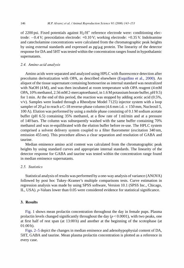
Fig. 1 shows mean prolactin concentration throughout the day in female pups. Plasmaprolactin levels changed significantly throughout the day (p < 0.0001), with two peaks, oneat first half of rest span (at 13:00 h) and another at the beginning of the scotophase (at01:00 h).
Figs. 2–5depict the changes in median eminence and adenohypophysial content of DA,5HT, GABA and taurine. Mean plasma prolactin concentration is plotted as a reference inevery case.
M.P. Alvarez et al. / Animal Reproduction Science 91 (2006) 143–153 147
Fig. 1. Twenty-four hour changes in plasma prolactin levels of 11 days old female rabbit pups. Groups of sixto seven pups were killed by decapitation at six different time intervals throughout a 24-h cycle. Bar indicatesscotophase duration. Results are the means± S.E.M. ap < 0.01 vs. 09:00, 17:00 and 21:00 h, Tukey–Kramer’smultiple comparisons test. For further statistical analysis, see text.
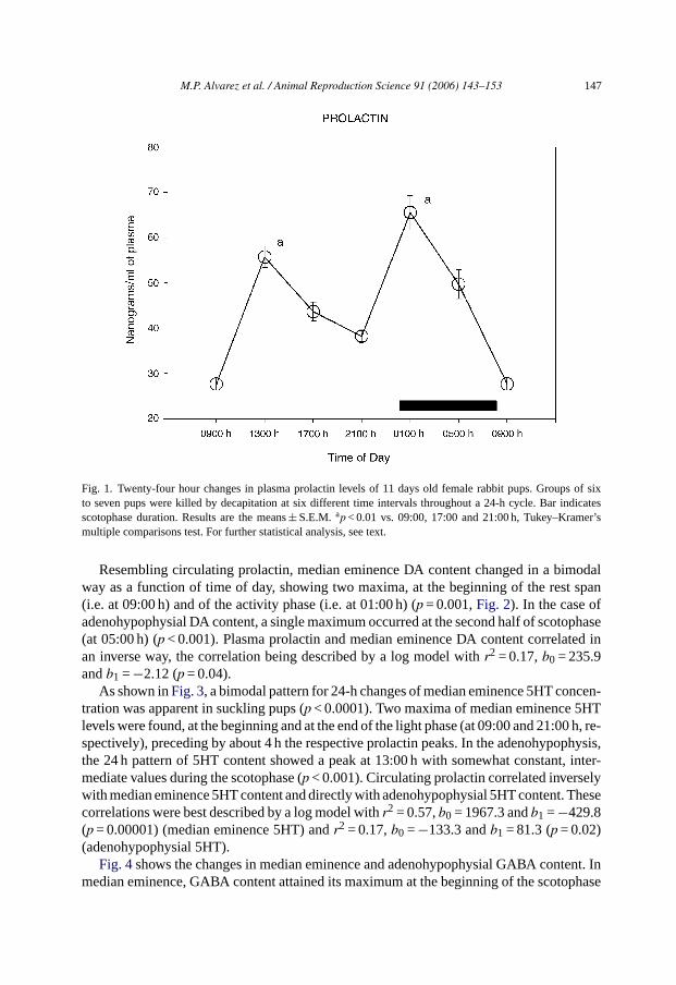
Resembling circulating prolactin, median eminence DA content changed in a bimodalway as a function of time of day, showing two maxima, at the beginning of the rest span(i.e. at 09:00 h) and of the activity phase (i.e. at 01:00 h) (p = 0.001,Fig. 2). In the case ofadenohypophysial DA content, a single maximum occurred at the second half of scotophase(at 05:00 h) (p < 0.001). Plasma prolactin and median eminence DA content correlated inan inverse way, the correlation being described by a log model withr2 = 0.17,b0 = 235.9andb1 =−2.12 (p = 0.04).
As shown inFig. 3, a bimodal pattern for 24-h changes of median eminence 5HT concen-tration was apparent in suckling pups (p < 0.0001). Two maxima of median eminence 5HTlevels were found, at the beginning and at the end of the light phase (at 09:00 and 21:00 h, re-spectively), preceding by about 4 h the respective prolactin peaks. In the adenohypophysis,the 24 h pattern of 5HT content showed a peak at 13:00 h with somewhat constant, inter-mediate values during the scotophase (p < 0.001). Circulating prolactin correlated inverselywith median eminence 5HT content and directly with adenohypophysial 5HT content. Thesecorrelations were best described by a log model withr2 = 0.57,b0 = 1967.3 andb1 =−429.8(p = 0.00001) (median eminence 5HT) andr2 = 0.17,b0 =−133.3 andb1 = 81.3 (p = 0.02)(adenohypophysial 5HT).
Fig. 4shows the changes in median eminence and adenohypophysial GABA content. Inmedian eminence, GABA content attained its maximum at the beginning of the scotophase

148 M.P. Alvarez et al. / Animal Reproduction Science 91 (2006) 143–153
Fig. 2. Twenty-four hour changes in median eminence and adenohypophysial DA content in 11 days old femalerabbit pups. Groups of six to seven pups were killed by decapitation at six different time intervals throughout a 24-hcycle. Bar indicates scotophase duration. Results are the means± S.E.M. Circulating prolactin levels are shownin shaded line. Letters indicate the existence of significant differences between time points within each tissue aftera Tukey–Kramer’s multiple comparisons test, as follows:ap < 0.01 vs. 13:00, 17:00 and 05:00 h;bp < 0.01 vs. alltime points;cp < 0.05 vs. 21:00 h. For further statistical analysis, see text.
(at 01:00 h,p < 0.001). In the anterior pituitary, GABA content showed a peak between13:00 and 17:00 h (p = 0.0008). Median eminence GABA levels correlated significantlywith plasma prolactin, this correlation being described by a linear model withr2 = 0.19,b0 = 33.7 andb1 = 0.44 (p = 0.01).
Fig. 5depicts the 24 h changes in taurine content. Median eminence taurine concentrationvaried in a bimodal way showing a peak at 13:00 h, a nadir at 17:00 h, and high levelsthroughout the end of the light period and during the scotophase (p = 0.0047). Two peakswere seen for adenohypophysial taurine content (p < 0.001), one during the light phase ofdaily photoperiod (at 13:00 h), and another during the scotophase (at 05:00 h). A directcorrelation between plasma prolactin and adenohypophysial taurine content, best describedby a log model, occurred (r2 = 0.21,b0 =−12.1 andb1 = 4.79 (p = 0.01).
4. Discussion
Foregoing results indicate that in 11 days old female rabbit pups, 24-h changes in plasmaprolactin levels are detectable. The pattern displayed two maxima, a major one at the firsthalf of the activity phase and a second one at the middle of the rest span. These findingsdiffer substantially from what was found in rat pups of a comparable age, which did not

M.P. Alvarez et al. / Animal Reproduction Science 91 (2006) 143–153 149
Fig. 3. Twenty-four hour changes in median eminence and adenohypophysial 5HT content in 11 days old femalerabbit pups. Groups of six to seven pups were killed by decapitation at six different time intervals throughouta 24-h cycle. Bar indicates scotophase duration. Results are the means± S.E.M. Circulating prolactin levels areshown in shaded line. Letters indicate the existence of significant differences between time points within eachtissue after a Tukey–Kramer’s multiple comparisons test, as follows:ap < 0.01 vs. 13:00, 17:00, 01:00 and 05:00 h;bp < 0.01 vs. 13:00, 17:00 and 01:00 h;cp < 0.01 vs. 09:00 and 17:00 h. For further statistical analysis, see text.
exhibit any prolactin circadian rhythm before weaning (Esquifino et al., 1998). Hence, afaster maturation of the circadian system seems to occur in the rabbit as compared to therat.
The nocturnal maximum in plasma prolactin values measured in 11 days old female rabbitpups was attained 1 h after switching off the light, anticipating exposure to the daily nursingvisit of the doe which is an extremely short and regular behavior (around 02:00 a.m.,Hudsonand Distel, 1989). Indeed, the anticipatory prolactin release was seen in all pups, supportingthe conclusion that this daily rhythm in maternal behavior has a very strong “Zeitgeber”influence on the newborn. In adult animals, food availability acts as a “Zeitgeber” resultingin a “food anticipatory activity” that occurs only if feeding intervals are within the circadianrange, circadian rhythms exhibiting a gradual resetting in response to mealtime shifts anda free-running during total food deprivation (Mistlberger, 1994). Since prolactin is readilyreleased following stress (Ben-Jonathan and Hnasko, 2001), it seems feasible that foodanticipatory activity acting through arousal mechanisms is responsible for the particularchanges in plasma prolactin seen in rabbit pups.
It must be noted that rabbit pups are very immature at birth and their survival is clearlydependent on the doe’s daily visit. The unusual limited maternal care pattern is under a tightcircadian control (Jilge and Hudson, 2001). At 13–18 days of age, rabbit pups have grown,improved their motor and thermoregulatory capacity and are able to leave the nest (see for

150 M.P. Alvarez et al. / Animal Reproduction Science 91 (2006) 143–153
Fig. 4. Twenty-four hour changes in median eminence and adenohypophysial GABA content in 11 days old femalerabbit pups. Groups of six to seven pups were killed by decapitation at six different time intervals throughout a24-h cycle. Bar indicates scotophase duration. Results are the means± S.E.M. Circulating prolactin levels areshown in shaded line. Letters indicate the existence of significant differences between time points within eachtissue after a Tukey–Kramer’s multiple comparisons test, as follows:ap < 0.01 vs. 09:00, 13:00, 17:00 and 05:00 h;bp < 0.05 vs. 09:00 h. For further statistical analysis, see text.
review (Hudson and Distel, 1986, 1987; Martinez-Gomez et al., 2004). Such a rapid growthis possible by a clear synchronization in behavior and physiology of the doe and the pups(Jilge and Hudson, 2001).
Prolactin is a versatile compound that has a dual function as a circulating hormone andas a cytokine. The prolactin receptor is a member of the cytokine receptor superfamily,linked to activation of signaling pathways that promote cell growth and survival. Throughthese mechanisms prolactin regulates diverse physiological functions via its effects oncellular processes such as proliferation and differentiation (Vera-Lastra et al., 2002; Yu-Lee,2002). Prolactin secretion is under inhibitory control by hypothalamic DA (Ben-Jonathanand Hnasko, 2001). DA released from tuberoinfundibular dopaminergic neurons enters thehypophysial portal blood to reach the anterior pituitary and to inhibit prolactin secretion.In turn, prolactin stimulates DA secretion from dopaminergic neurons, closing a feedbackloop. The foregoing results in 11 days old female rabbits indicate that DA content in medianeminence tended to follow a reciprocal pattern to plasma prolactin, Indeed, a negativecorrelation between plasma prolactin and DA content in median eminence was shown,suggesting that a decrease in median eminence DA content may be causally linked toprolactin release (Freeman et al., 2000).
Dopaminergic neurons in the arcuate nucleus receive a dense serotonergic innervationconsisting of a population of brainstem neurons arising mainly from the midbrain raphenuclei (Steinbusch, 1981). Fibers also originate from 5HT cell bodies located within the

M.P. Alvarez et al. / Animal Reproduction Science 91 (2006) 143–153 151
Fig. 5. Twenty-four hour changes in median eminence and adenohypophysial taurine content in 11 days old femalerabbit pups. Groups of six to seven pups were killed by decapitation at six different time intervals throughout a24-h cycle. Bar indicates scotophase duration. Results are the means± S.E.M. Circulating prolactin levels areshown in shaded line. Letters indicate the existence of significant differences between time points within eachtissue after a Tukey–Kramer’s multiple comparisons test, as follows:a p < 0.01 vs. 17:00 h;bp < 0.01 vs. 09:00,17:00, 21:00 and 01:00 h. For further statistical analysis, see text.
hypothalamus. There is a close approximation of 5HT fibers to dopaminergic cell bodiesin the arcuate nucleus (Kiss and Halasz, 1986). Therefore, the effect of 5HT on prolactinrelease has been linked to the modulation of inhibitory dopaminergic inputs to the pituitary.The foregoing results demonstrate that 5HT content in median eminence of female rabbitpups varies throughout the day inversely to the plasma prolactin, both parameters displayingmirror patterns. In fact, a significantly negative correlation between them was found. In theadenohypophysial 5HT content positively correlated with plasma prolactin, thus pointingto a possible direct effect of 5HT on the release of prolactin.
Median eminence neurons release GABA in portal blood that reaches the anterior pitu-itary (Gudelsky et al., 1983) and this release can affect prolactin secretion (Casanueva etal., 1984; Selgas et al., 1997; Duvilanski et al., 1998). The present results in female rabbitpups indicate a parallel pattern of plasma prolactin and median eminence GABA during theactivity phase. In fact, both parameters correlated positively in median eminence but not inthe adenohypophysis.
Taurine has been also implicated in prolactin secretion control (Login, 1990; Arias etal., 1998; Duvilanski et al., 1998). Female rabbit pups showed a daily pattern of taurinecontent in median eminence and in the anterior pituitary, as well as a correlation withplasma prolactin levels (in the case of adenohypophysial taurine) suggesting a direct effectof taurine on prolactin release.
152 M.P. Alvarez et al. / Animal Reproduction Science 91 (2006) 143–153
Summarizing, the present study in neonatal female rabbits demonstrates the existence of24-h variations in circulating prolactin levels and in median eminence and adenohypophysiallevels of some putative modulators of prolactin release. Plasma prolactin levels correlatedeither negatively (median eminence DA; median eminence 5HT) or positively (median em-inence GABA; adenohypophysial 5HT; adenohypophysial taurine) with putative modulatorcontent.
Acknowledgments
This work was supported by grants from DGES, PB9-0257/97, Ministerio de Educaciony Cultura, Spain.
References
Arias, P., Jarry, H., Convertini, V., Ginzburg, M., Wuttke, W., Moguilevsky, J., 1998. Changes in mediobasalhypothalamic dopamine and GABA release: a possible mechanism underlying taurine-induced prolactin se-cretion. Amino Acids 15, 5–11.
Ben-Jonathan, N., Hnasko, R., 2001. Dopamine as a prolactin (PRL) inhibitor. Endocr. Rev. 22, 724–763.Broekhuizen, S., Mulder, J., 1983. Differences and similarities in nursing behavior of hares and rabbits. Acta Zool.
Fenn. 174, 61–63.Cano, P., Cardinali, D.P., Castrillon, P., Reyes Toso, C., Esquifino, A.I., 2001. Age dependent changes in 24-h
rhythms of catecholamine concentration and turnover in hypothalamus, corpus striatum and pituitary gland ofrats injected with Freund’s adjuvant. BMC Physiol. 1, 14–25.
Casanueva, F., Apud, J.A., Masotto, C., Cocchi, D., Locatelli, V., Racagni, G., Muller, E., 1984. Daily fluctuationsin the activity of the tuberoinfundibular GABAergic system and plasma prolactin levels. Neuroendocrinology39, 367–370.
Castrillon, P.O., Cardinali, D.P., Pazo, D., Cutrera, R.A., Esquifino, A.I., 2001. Effect of superior cervical gan-glionectomy on 24-h variations in hormone secretion from the anterior hypophysis and in hypothalamicmonoamine turnover during the preclinical phase of Freund’s adjuvant arthritis in rats. J. Neuroendocrinol.13, 288–295.
Davis, F.C., Gorski, R.A., 1988. Development of hamster circadian rhythms: role of the maternal suprachiasmaticnucleus. J. Comp Physiol A 162, 601–610.
Davis, F.C., Mannion, J., 1988. Entrainment of hamster pup circadian rhythms by prenatal melatonin injectionsto the mother. Am. J. Physiol. 255, R439–R448.
Distel, H., Hudson, R., 1985. The contribution of the olfactory and tactile modalities to the nipple-search behaviourof newborn rabbits. J. Comp. Physiol. A 157, 599–605.
Duvilanski, B.H., Selgas, L., Garcia-Bonacho, M., Esquifino, A.I., 1998. Daily variations of amino acid con-centration in mediobasal hypothalamus, in rats injected with Freund’s adjuvant. Effect of cyclosporine. J.Neuroimmunol. 87, 189–196.
Esquifino, A.I., Arce, A., Villanua, M.A., Cardinali, D.P., 1998. Development of 24-hour rhythms in serum prolactinand luteinizing hormone levels in rats neonatally administered melatonin. Chronobiol. Int. 15, 21–28.
Esquifino, A.I., Garcia-Bonacho, M., Castrillon, P.O., Duvilanski, B.H., 2000. Effect of chronic hyperprolactinemiaon daily changes of glutamate and aspartate concentrations in the median eminence and different hypothalamicareas of male rats. Chronobiol. Int. 17, 631–643.
Freeman, M.E., Kanyicska, B., Lerant, A., Nagy, G., 2000. Prolactin: structure, function, and regulation of secre-tion. Physiol. Rev. 80, 1523–1631.
Garcia-Bonacho, M., Esquifino, A.I., Castrillon, P.O., Toso, C.R., Cardinali, D.P., 2000. Age-dependent effectof Freund’s adjuvant on 24-hour rhythms in plasma prolactin, growth hormone, thyrotropin, insulin, follicle-stimulating hormone, luteinizing hormone and testosterone in rats. Life Sci. 66, 1969–1977.

M.P. Alvarez et al. / Animal Reproduction Science 91 (2006) 143–153 153
Greenwood, F.C., Hunter, W.M., Glover, J.S., 1963. The preparation of 131 I-labelled human growth hormone ofhigh specific radioactivity. Biochem. J. 89, 1114–1123.
Gudelsky, G., Apud, J., Masotto, C., Locatelli, V., Cocchi, D., Racagni, G., Muller, E., 1983. Ethanolamine-O-sulfate enhances�-aminobutyric acid secretion into hypophyseal portal blood and lowers serum prolactinconcentrations. Neuroendocrinology 37, 397–399.
Hudson, R., Distel, H., 1986. Pheromonal release of suckling in rabbits does not depend on the vomeronasal organ.Physiol. Behav. 37, 123–128.
Hudson, R., Distel, H., 1987. Regional autonomy in the peripheral processing of odor signals in newborn rabbits.Brain Res. 421, 85–94.
Hudson, R., Distel, H., 1989. Temporal pattern of suckling in rabbit pups: a model of circadian synchrony be-tween mother and young. In: Reppert, S.M. (Ed.), Research in Perinatal Medicine. Development of CircadianRhythmicity and Photoperiodism in Mammals, vol. IX. Perinatology Press, Ithaca, NY, pp. 83–102.
Jilge, B., 1995. Ontogeny of the rabbit’s circadian rhythms without an external zeitgeber. Physiol. Behav. 58,131–140.
Jilge, B., Hudson, R., 2001. Diversity and development of circadian rhythms in the European rabbit. Chronobiol.Int. 18, 1–26.
Kiss, J., Halasz, B., 1986. Synaptic connections between serotonergic axon terminals and tyrosine hydroxylase-immunoreactive neurons in the arcuate nucleus of the rat hypothalamus. A combination of electron microscopicautoradiography and immunocytochemistry. Brain Res. 364, 284–294.
Login, I.S., 1990. Direct stimulation of pituitary prolactin release by glutamate. Life Sci. 47, 2269–2275.Manning, P.J., Ringler, D.H., Newcomer, C.E., 1994. The Biology of the Laboratory Rabbit. Academic Press,
New York.Martinez-Gomez, M., Juarez, M., Distel, H., Hudson, R., 2004. Overlapping litters and reproductive performance
in the domestic rabbit. Physiol. Behav. 82, 629–636.Mistlberger, R.E., 1994. Circadian food anticipatory activity: Formal models and physiological mechanisms.
Neurosci. Biobehav. Rev. 189, 171–195.Selgas, L., Arce, A., Esquifino, A.I., Cardinali, D.P., 1997. Twenty-four-hour rhythms of serum ACTH, prolactin,
growth hormone, and thyroid-stimulating hormone, and of median-eminence norepinephrine, dopamine, andserotonin, in rats injected with Freund’s adjuvant. Chronobiol. Int. 14, 253–265.
Steinbusch, H.W.M., 1981. Distribution of serotonin-immunoreactivity in the central nervous system of the rat-cellbodies and terminals. Neuroscience 6, 557–618.
Thompson, H.V., King, C.M., 1994. The European Rabbit. Oxford University Press, Oxford.Ubilla, E., Alvarino, J.M.R., Esquifino, A., Agrasal, C., 1992. Effects of induction of parturition by administration
of a prostaglandin F2 [alpha] analogue in rabbits: possible modification of prolactin. LH and FSH secretionpatterns. Anim. Reprod. Sci. 27, 13–20.
Vera-Lastra, O., Jara, L.J., Espinoza, L.R., 2002. Prolactin and autoimmunity. Autoimmun. Rev. 1, 360–364.Yu-Lee, L.Y., 2002. Prolactin modulation of immune and inflammatory responses. Recent Prog. Horm. Res. 57,
435–455.


!["A Man of the Last Hour" (1998 [1991])](https://static.fdokumen.com/doc/165x107/6332ccf74e0143040300d61f/a-man-of-the-last-hour-1998-1991.jpg)