Antibacterial Nitric Oxide-Releasing Polyester for the Coating of Blood-Contacting Artificial...
11
Antibacterial Nitric Oxide-Releasing Polyester for the Coating of Blood-Contacting Artificial Materials *Amedea B. Seabra, †Dorival Martins, *Maíra M.S.G. Simões, *Regiane da Silva, †Marcelo Brocchi, and *Marcelo G. de Oliveira *Institute of Chemistry, University of Campinas, UNICAMP; and †Department of Genetics, Evolution and Bioagents, Institute of Biology, University of Campinas, UNICAMP, Campinas SP, Brazil Abstract: The emergence of multidrug-resistant bacteria associated with blood-contacting artificial materials is a growing health problem, which demands new approaches in the field of biomaterials research. In this study, a poly(sulfhydrylated polyester) (PSPE) was synthesized by the polyesterification reaction of mercaptosuccinic acid with 3-mercapto-1,2-propanediol and blended with poly- (methyl methacrylate) (PMMA) from solution, leading to solid PSPE/PMMA films, with three different PSPE : PMMMA mass ratios. These films were subse- quently S-nitrosated through the immersion in acidified nitrite solution, yielding poly(nitrosated)polyester/PMMA (PNPE/PMMA) films. A polyurethane intravascular cath- eter coated with PNPE/PMMA was shown to release nitric oxide (NO) in phosphate buffered saline solution (pH 7.4) at 37°C at rates of 4.6 nmol/cm 2 /h in the first 6 h and 0.8 nmol/cm 2 /h in the next 12 h. When used to coat the bottom of culture plates, NO released from these films exerted a potent dose- and time-dependent antimicrobial activity against Staphylococcus aureus and a multidrug- resistant Pseudomonas aeruginosa strains. This antibacte- rial effect of PSPE/PMMA films opens a new perspective for the coating of blood-contacting artificial materials, for avoiding their colonization with highly resistant bacteria. Key Words: Antibacterial action—Biomaterials—Blood- contacting artificial materials—Catheter-related infections —Nitric oxide. The emergence of multidrug-resistant bacteria, associated with the increase in opportunistic infec- tions, is a world health problem and a major concern in hospital environments (1–3). Around the world, between 5 and 15% of the patients admitted to hospi- tals acquire one or more infections (4).The frequency of hospital-acquired infections in the USA is about 2 million cases a year, resulting in 80 000 deaths and additional costs of US$ 17–29 billion (4,5). In the UK, the number of hospital-acquired infections is about 100 000 with 5000 deaths and additional costs of £1 billion per year (6). Nosocomial pneumonia and infec- tions in burned and wounded skin are among the most common types of infections found in hospital environ- ments, frequently associated with contaminated medical devices, such as intravascular catheters. One of the strategies that have been proposed to fight infections in blood-contacting artificial materials is their coating with nitric oxide (NO)-releasing bioma- terials.This proposal is based on the key roles played by NO in host defense and its well-known antimicro- bial actions against viruses and bacteria (7), parasites (8), and fungi (9). NO was already used successfully as an antibacterial agent in several applications in vitro and in vivo. In these applications, different forms of NO have been employed such as exogenous NO gas (10), generation of NO thorough nitrite reduction by ascorbic acid (11), and NO donors such as NONOates (12). In a previous article, we have shown the dose- dependent cytotoxic effects of the NO donors S-nitroso-N-acetlycysteine and S-nitrosoglutathione (GSNO) against the intracellular pathogens Leishma- nia major and Leishmania amazonensis (13). doi:10.1111/j.1525-1594.2010.00998.x Received May 2009; revised November 2009. Address correspondence and reprint requests to Prof. Marcelo G. de Oliveira, Institute of Chemistry, University of Campinas, UNICAMP, CP 6154, CEP 13083-970 Campinas, SP, Brazil. E-mail: [email protected] First two authors share the first author designation and contrib- uted equally to this article. Artificial Organs 34(7):E204–E214, Wiley Periodicals, Inc. © 2010, Copyright the Authors Journal compilation © 2010, International Center for Artificial Organs and Transplantation and Wiley Periodicals, Inc. E204
-
Upload
independent -
Category
Documents
-
view
0 -
download
0
Transcript of Antibacterial Nitric Oxide-Releasing Polyester for the Coating of Blood-Contacting Artificial...

aor_998 204..214
Antibacterial Nitric Oxide-Releasing Polyester for theCoating of Blood-Contacting Artificial Materials
*Amedea B. Seabra, †Dorival Martins, *Maíra M.S.G. Simões, *Regiane da Silva,†Marcelo Brocchi, and *Marcelo G. de Oliveira
*Institute of Chemistry, University of Campinas, UNICAMP; and †Department of Genetics, Evolution and Bioagents,Institute of Biology, University of Campinas, UNICAMP, Campinas SP, Brazil
Abstract: The emergence of multidrug-resistant bacteriaassociated with blood-contacting artificial materials is agrowing health problem, which demands new approachesin the field of biomaterials research. In this study, apoly(sulfhydrylated polyester) (PSPE) was synthesized bythe polyesterification reaction of mercaptosuccinic acidwith 3-mercapto-1,2-propanediol and blended with poly-(methyl methacrylate) (PMMA) from solution, leadingto solid PSPE/PMMA films, with three differentPSPE : PMMMA mass ratios. These films were subse-quently S-nitrosated through the immersion in acidifiednitrite solution, yielding poly(nitrosated)polyester/PMMA(PNPE/PMMA) films. A polyurethane intravascular cath-eter coated with PNPE/PMMA was shown to release nitric
oxide (NO) in phosphate buffered saline solution (pH 7.4)at 37°C at rates of 4.6 nmol/cm2/h in the first 6 h and0.8 nmol/cm2/h in the next 12 h. When used to coat thebottom of culture plates, NO released from these filmsexerted a potent dose- and time-dependent antimicrobialactivity against Staphylococcus aureus and a multidrug-resistant Pseudomonas aeruginosa strains. This antibacte-rial effect of PSPE/PMMA films opens a new perspectivefor the coating of blood-contacting artificial materials, foravoiding their colonization with highly resistant bacteria.Key Words: Antibacterial action—Biomaterials—Blood-contacting artificial materials—Catheter-related infections—Nitric oxide.
The emergence of multidrug-resistant bacteria,associated with the increase in opportunistic infec-tions, is a world health problem and a major concernin hospital environments (1–3). Around the world,between 5 and 15% of the patients admitted to hospi-tals acquire one or more infections (4).The frequencyof hospital-acquired infections in the USA is about 2million cases a year, resulting in 80 000 deaths andadditional costs of US$ 17–29 billion (4,5). In the UK,the number of hospital-acquired infections is about100 000 with 5000 deaths and additional costs of £1billion per year (6).Nosocomial pneumonia and infec-
tions in burned and wounded skin are among the mostcommon types of infections found in hospital environ-ments, frequently associated with contaminatedmedical devices, such as intravascular catheters. Oneof the strategies that have been proposed to fightinfections in blood-contacting artificial materials istheir coating with nitric oxide (NO)-releasing bioma-terials. This proposal is based on the key roles playedby NO in host defense and its well-known antimicro-bial actions against viruses and bacteria (7), parasites(8), and fungi (9). NO was already used successfully asan antibacterial agent in several applications in vitroand in vivo. In these applications, different forms ofNO have been employed such as exogenous NO gas(10), generation of NO thorough nitrite reduction byascorbic acid (11), and NO donors such as NONOates(12). In a previous article, we have shown the dose-dependent cytotoxic effects of the NO donorsS-nitroso-N-acetlycysteine and S-nitrosoglutathione(GSNO) against the intracellular pathogens Leishma-nia major and Leishmania amazonensis (13).
doi:10.1111/j.1525-1594.2010.00998.x
Received May 2009; revised November 2009.Address correspondence and reprint requests to Prof. Marcelo
G. de Oliveira, Institute of Chemistry, University of Campinas,UNICAMP, CP 6154, CEP 13083-970 Campinas, SP, Brazil. E-mail:[email protected]
First two authors share the first author designation and contrib-uted equally to this article.
Artificial Organs34(7):E204–E214, Wiley Periodicals, Inc.© 2010, Copyright the AuthorsJournal compilation © 2010, International Center for Artificial Organs and Transplantation and Wiley Periodicals, Inc.
E204

The already reported NO-releasing biomaterialsfor antibacterial applications include polymericmaterials (14), xerogels (15), solgel films (16), andmore recently, silica nanoparticles (17). However,obtaining NO-releasing polymeric films that can beapplied as coatings on other polymeric surfaces andwhich are sufficiently stable to allow practical appli-cations is still a challenge. We have previouslydescribed the synthesis of poly(sulfhydrylated poly-esters) (PSPEs) through the polycondensation reac-tions of ethylene glycol and poly(ethylene glycol)with mercaptosuccinic acid. Nitrosation of thesePSPEs yielded NO-releasing polynitrosated polyes-ters (PNPEs), which were shown to cause localvasodilation if applied on the skin in the liquid state(18–20). More recently, we showed that liquid PNPEscan be prepared as solid films, if blended with poly-(methyl methacrylate) (PMMA). Such films can beused for the coating of other materials like metallicintracoronary stents and were shown to reduce plate-let adhesion due to NO release (21). In addition,PNPEs allow nitrosation immediately before theiruse through their immersion in acidified nitrite solu-tion, offering an option for long-term storage of thesematerials in the form of their thiolated precursors.
In this work, a new type of PNPE was synthesizedby using 3-mercapto-1,2-propanediol in place of eth-ylene glycol and poly(ethylene glycol). The productobtained in this case has a much higher surface con-centration of sulfhydry (–SH) groups and allows theloading of higher amounts of NO as S-nitrosothiol(–SNO) groups. Due to this property, new blends ofthis PNPE with PMMA were tested as antibacterialcoatings to avoid the colonization of surfaces bymultidrug-resistant gram-positive and gram-negativebacterial strains. It was shown for the first time thatthese PNPE/PMMA films applied as coatings on thebottom of culture plates have potent antibacterialactivity against two of the main agents of hospital-acquired infections, Staphylococcus aureus andPseudomonas aeruginosa strains, in a dose- and time-dependent manner.This property, associated with therelative stability of the material under storage condi-tions and resistance to sterilization by ethylene oxide(EtO), is presented in this work and makes this newNO-releasing material a promising option as an anti-bacterial coating for blood-contacting artificialmaterials.
MATERIALS AND METHODS
Materials, culture media, and bacterial strains3-Mercapto-1,2-propanediol, mercaptosuccinic
acid, sodium nitrite (NaNO2), 5′5-dithiobis(2-
nitrobenzoic acid) (DTNB), phosphate buffer saline(PBS), pH 7.4 (Sigma Aldrich Chemical Co., Inc., St.Louis, MO, USA), PMMA, average Mn 75000 (Poly-science, Niles, IL, USA), HCl (Synth, Diadema, SP,Brazil), acetonitrile (Carlo Erba, Milan, Italy), tet-rahydrofuran (THF) (high performance liquid chro-matography, Tedia Company, Fairfield, OH, USA),N-(1-naphythyl)-ethylenediamine dihydrochloride(NEED), sulfanilamide (SULF) (Merck, Darmstadt,Germany), and sodium chloride (NaCl) (J.T. Baker,Mexico City, Mexico) were used as received. Allaqueous solutions were prepared using analyticalgrade water from a Millipore Milli-Q Gradient filtra-tion system (Billerica, MA, USA). The antibacterialactivity of PSPE/PMMA and PNPE/PMMA filmswas determined in Mueller-Hinton medium (Difco,Detroit, MI, USA), as described in ref. (22). Bothsaline solution and Mueller-Hinton media were ster-ilized at 120°C for 20 min. Intravenous polyurethanecatheters Jelco Plus 18G, length 32 mm, o.d. 1.3 mm(Medex Medical Ltd., Rossendale, UK), were used asa model blood-contact artificial material.
S. aureus ATCC 25923 was used as describedin Grare et al. (23). P. aeruginosa was a capsulatedmultiresistant strain (amicacin, gentamicin, netilmy-cin, imipenem, chloramphenicol, ampicillin, ciprof-loxacin, cefotaxime) and elastase producer. Allstrains were maintained at -80°C as glycerol suspen-sions (2.5 M).
PSPE synthesisThe PSPE was synthesized following the proce-
dure described elsewhere (18). Briefly, equimolar(66 mmol) amounts of 3-mercapto-1,2-propanedioland mercaptosuccinic acid were placed in a 100-mLthree-necked, round-bottomed flask fitted with aVigreaux reflux column, under stirring and N2 flux foroxygen and water removal. Concentrated hydrochlo-ric acid (HCl 12 M, 150 mL) was added as a catalystand the reaction was carried out at 120°C in siliconeoil bath for 22 h. The PSPE, obtained as a viscousliquid, was purified by dissolution in THF followed byprecipitation in cold water for three times and freeze-dried for 24 h for the complete removal of THF.
Preparation of PSPE/PMMA solutions andsolid films
Solutions of PSPE/PMMA were prepared forobtaining solid PSPE/PMMA films with PSPE :PMMA mass ratios: 36.4:64.6, 41.7:58.3, and 50:50(wt/wt) for the measurements described below. Typi-cally, 70 mg of PMMA were dissolved in 1.0 mL ofTHF/acetonitrile (80:20 v/v). After the completePMMA dissolution, 40, 50, or 70 mg of liquid PSPE
ANTIBACTERIAL NITRIC OXIDE-RELEASING POLYESTER E205
Artif Organs, Vol. 34, No. 7, 2010

were added, and the solutions were stirred with amagnetic bar for 60 min at room temperature.
For obtaining solid PSPE/PMMA films for subse-quent S-nitrosation and spectrophotometric mea-surements, 100 mL of PSPE/PMMA (36.4:64.6 wt/wt)solution were transferred to the cavity of a quartz cellwith a detachable window (Hellma 106-QS, opticalpath 0.1 mm) and the THF/acetonitrile solvent wasallowed to evaporate over 12 h at 10°C. The film wasfurther freeze-dried for 24 h to ensure completeremoval of the solvent.
For differential scanning calorimetry (DSC) mea-surements, 1.0 mL of PMMA or 1.0 mL of PSPE/PMMA (36.4:64.6, 41.7:58.3, and 50:50 wt/wt)solution in THF/acetonitrile (80:20 v/v) was trans-ferred to the cavity of Teflon round molds (innerdiameter 2.5 cm) and the solvents were allowed toevaporate for 24 h at 10°C and further freeze-driedfor 24 h. The solid films were removed from themolds for the thermal analysis. PSPE/PMMA films(36.4:64.6 wt/wt) prepared in Teflon round molds asabove were also used for scanning electron micros-copy, stability, and sterilization experiments.
Energy dispersive X-ray fluorescence spectrometryEnergy dispersive X-ray spectrometry (EDS)
microanalysis was used to examine the compositionof bare acrylic plates and of acrylic plates coated withPSPE/PMMA (36.4:64.6 wt/wt) film. The plates werecut and mounted on aluminum stubs, sputter coatedfor 2 min in a high-vacuum gold sputter-coating unit(Balt-Tec MED 020, Balzers, Liechtenstein), andexamined using a Jeol JSM T-300 electron micro-scope (Tokyo, Japan) under 100¥ magnification, withan EDS system. The EDS technique was used to mapthe distribution of sulfur atoms on the surface of thebare and PSPE/PMMA-coated acrylic plates.
DSCThermal properties of solid PMMA and PSPE/
PMMA (36.4:64.6, 41.7:58.3, and 50:50 wt/wt) filmswere characterized by DSC, using a Q-100 calorim-eter (TA Instruments, New Castle, DE, USA). Theinstrument was calibrated with indium as a standard.Samples were cooled to -100°C and heated to 150°Cat 10°C/min and the glass-transition temperatures(Tg) were obtained from the midpoint of thetransitions. A continuous argon flow was maintainedin the sample compartment to ensure minimalsample degradation during thermal cycling.
Characterization of S-nitrosation and NO release ofPSPE/PMMS film in dry condition
The solid PSPE/PMMA (36.4:64.6 wt/wt) film pre-pared in the quartz cell (see above) was S-nitrosated
through the immersion in 50 mL of 1.0 M HCl solu-tion containing 60 mM NaNO2 at room temperaturefor 5, 10, 15, 20, 25, 30, and 35 min. Formation ofsurface S-nitrosothiol (–SNO) groups during theS-nitrosation of the film was characterized by theappearance of the characteristic absorption bandof the –SNO group with maximum at 336 nm(e = 940 mol/L/cm), using an UV-VIS spectropho-tometer (Hewlett-Packard, model 8453, Palo Alto,CA, USA) (24).
For characterizing NO released from a PNPE/PMMA film in dry condition, spectral changes asso-ciated with the bleaching of the absorption band ofthe –SNO group due to the thermal release of NOwere monitored in the range 250–450 nm for a PNPE/PMMA (36.4:64.6 wt/wt) film coated on a detachablequartz cell as described above and kept in the dark at37°C for 27 h. Spectra were acquired in time intervalsof 1 h.
NO release to PBS solution from aPNPE/PMMA-coated intravascularpolyurethane catheter
In order to evaluate the NO released from catheterscoated with PNPE/PMMA, commercial intravenouscatheters (Jelco Plus 18G) were immersed in thePSPE/PMMA solution 50:50 wt/wt in THF/acetonitrile (80:20 v/v), prepared as stated above.Thecatheters were kept in the solution for 60 s. Afterremoval from this solution, the catheters were dried ina refrigerator at 10°C for 24 h and subsequentlyfreeze-dried for 4 h. After complete drying, the cath-eters were S-nitrosated by immersion in 10 mL ofacidified sodium nitrite solution (60 mM NaNO2 inHCl 1.0 M) for 30 min followed by rinsing with ana-lytical grade water to remove nitrite/HCl and usedimmediately for measuring NO released in PBS solu-tion.This measurement was accomplished by immers-ing the catheters in a flask containing 10 mL ofdeaerated 1M PBS (pH 7.4) at 37°C and aliquots of100 mL were collected at times 0, 0.08, 2, 4, 6, 9, 16, 24,48, and 72 h and injected in 2.0 mL of Griess reactant(0.01 M sulfanilamide,0.01 M N-(1-naphtyl)ethylene-diamine dihydrochloride, 4.0M HCl) contained in aquartz cuvette. The PBS aliquots withdrawn from theflask were replaced by fresh PBS.Two hours after theinjections, the absorbance of the Griess solution wasmeasured directly in the cuvette at 540 nm. Absor-bance at this wavelength, which provides a quantita-tive measurement of NO release to the PBS solutionafter its conversion to NO2
-,was kinetically monitoredfor 72 h at 37°C, at variable time intervals. The cumu-lative amounts of NO released were calculated asmols of NO released/cm2 of coated catheter for a total
A.B. SEABRA ET AL.E206
Artif Organs, Vol. 34, No. 7, 2010
Duran
Realce
Duran
Realce

coating area of 1.31 cm2. Each point in the kineticcurves represents the average of three experiments,with the error bars expressed by their standard errorof the mean (SEM). An uncoated catheter and cath-eters coated with non-nitrosated PSPE/PMMA (50:50wt/wt) and with PNPE/PMMA (50:50 wt/wt) werephotographed in order to characterize the uniformityof the coating and the color change associated with thenitrosation reaction. A digital camera, Sony a 100DSLR-A100, fitted with a macro lens, Sony 80/100(Sony, San Diego, CA, USA), was used for recordingthe pictures.
Stability of PSPE/PMMA films under storage andafter sterilization
For evaluating the stability of the free –SH groupsin PSPE/PMMA films under sterilization conditionswith EtO, films of PSPE/PMMA (36.4:64.6, 41.7:58.3,and 50:50 wt/wt), prepared as described above, weresterilized with a gaseous mixture of EtO and CO2
(6:90, 10:90 v/v), 240 min at 45–55 � 1°C, and relativehumidity in the range 50–60%, and final vacuum of400–450 mm Hg for 30 min. Three nitrogen washeswere performed after EtO sterilization. Intra-cameraaeration was performed with five vacuum cycles fol-lowed by aeration with sterile air. Extra-camera aera-tion was performed under continuous sterile air fluxfor 48 h.
The number of –SH groups in nonsterilized andsterilized PSPE/PMMA films was measured by theDTNB reaction, based on the absorbance at 412 nm(e = 14.15 mM/cm) of the 2-nitro-5-thiobenzoateanion (TNB2-) generated in the reaction of –SHgroups with DTNB (25). Sterilized and nonsterilizedPSPE/PMMA films were immersed in 3 mL of0.01 M DTNB in PBS buffer (pH 7.4) containing1 mM of ethylenediaminetetraacetic acid.The experi-ments were performed in triplicate.
Plate sample preparation for antibacterialmeasurements
The antibacterial effect of PNPE/PMMA films wascharacterized using 24-well polystyrene pyrogen-freecell culture plates (Corning Costar, Cambridge, MA,USA).Three different groups were analyzed in quad-ruplicate: (i) growth control (uncoated wells); (ii)wells coated with PSPE/PMMA films; and (iii) wellscoated with PNPE/PMMA films. The flat bottom ofeach well (inner diameter 1.5 cm) was coated withPSPE/PMMA (36.4:64.6, 41.7:58.3, and 50:50 wt/wt)films by uniformly applying 150 mL of the corre-sponding polymeric solutions in THF/acetonitrile onthe bottom of each well. The solvent was evaporatedat 5–8°C for 1 h to avoid oxidation of the –SH groups
and further dried at 30°C for 24 h. S-nitrosation ofthe PNPE/PMMA-coated wells was obtained by theaddition of 500 mL of acidified sodium nitrite (60 mMin 1.0 M HCl) in each well. The nitrosating solutionwas maintained inside the wells for 30 min at roomtemperature. After this period, the wells were exten-sively rinsed with deionized water to remove anytrace of the nitrosating reactants.
Antibacterial activity of the NO-donorPNPE/PMMA films
The antibacterial activities of the wells coated withPMMA, PSPE/PMMA, and PNPE/PMMA films,were evaluated by adapted time–growth curves anddose–response assays, according to standard method-ologies of the Clinical and Laboratory StandardsInstitute (22). Strains were grown in Mueller-Hintonagar media (MHA) and single colonies were addedinto 1 mL of saline solution. The suspension wasadjusted to 0.5 index in McFarland scale (108 cfu.mL-1) and diluted in Mueller-Hinton broth media toreach a cell density of 105 cfu/mL. Then, 105 cfu wereadded per well into the 24-well plates (reaching afinal volume of 1 mL of bacterial suspension perwell), divided according to the groups defined above.The plates were incubated statically at 37°C and,after each treatment or exposure time, the content ofthe wells was homogenized, serially diluted in sterilesaline solution 0.85% v/v, and plated by the spreadmethod in MHA plates. Cfu counting was performedafter 0, 2, 4, 6, 9, 12, and 24 h.
For dose–response assays, the antibacterial activityof films with PNPE/PMMA ratios of 36.4:64.6,41.7:58.3, and 50:50 wt/wt was evaluated in two dif-ferent incubation times (2 and 4 h), while for time–growth assays, the PNPE/PMMA film with highestPNPE/PMMA ratio (50:50 wt/wt) was used for evalu-ating the antibacterial effect against P. aeruginosaand S. aureus over different times of exposure (0, 2, 4,6, 9, 12, and 24 h). Bacteria incubated in noncoatedwells and in PSPE/PMMA-coated wells were consid-ered 100% of cell viability. The experiments werecarried out in quadruplicate for all treatments andthe results were expressed as the logarithm of bacte-rial survival (log10[total cfu of treatments]/log10[total cfu of control]).
Statistical analysisAll biological experiments were performed at least
three times with four replicates. Data are expressedas mean � SEM. Statistical analysis was carried outby Student’s t-test and probabilities lower than 0.05(P < 0.05) were considered significant. Each point inthe kinetic curves of NO release represents the
ANTIBACTERIAL NITRIC OXIDE-RELEASING POLYESTER E207
Artif Organs, Vol. 34, No. 7, 2010
Duran
Realce

average of three experiments, with the error barsexpressed by their SEM.
RESULTS AND DISCUSSION
PNPE synthesisWe have previously described the synthesis of
PSPEs via the polycondensation of diols (ethylene-glycol and poly(ethylene glycol)) with mercaptosuc-cinic acid, followed by S-nitrosation of the –SHgroups by a gaseous NO/O2 mixture. This processyielded NO-releasing PNPEs as viscous liquids atroom temperature. These polymers were shown torelease free NO and to be able to promote localvasodilation when in contact with human skin(18,19). More recently, we have shown that liquidPNPEs can be blended with PMMA in order toobtain solid films that can be used for the coating ofmetallic surfaces, providing an active NO-releasingsurface which is able to prevent human platelet adhe-sion (20,21).
In the present work, a similar polyesterificationreaction was employed, but the mercaptosuccinicacid was reacted with a sulfhydrylated diol(3-mercapto-1,2-propanediol) instead of ethylenegly-col and poly(ethylene glycol), yielding a polyesterwith a repetition unit possessing two free thiols perunit, which were subsequently S-nitrosated (Fig. 1).This PNPE has, therefore, twice the NO loadingcapacity, relative to the former PNPEs reported,being a more appropriate candidate for applicationswhere a high local release of NO is desired. It mustbe noted that, different from the S-nitrosationwith gaseous NO/O2 employed previously, theS-nitrosation of the solid blends of this work wasobtained through the immersion of the solid blendedfilms in acidic NO2
- solution, where only the surface–SH groups of the films were S-nitrosated.
This method allowed controlling the degree ofS-nitrosation of the surface by controlling the time ofthe reaction or the PMMA : PSPE mass ratio. Inorder to avoid a fast S-NO cleavage in films with highconcentration of S-NO groups, due to the autocata-lytic effect which happens in S-nitrosothiols (24), anincubation time of 30 min was chosen for theS-nitrosation of the PSPE/PMMA films. It must benoted that full S-nitrosated films are able to releaseNO at much higher rates than those reported here(data not shown). Thus, the surface concentration ofS-NO groups can be modulated by controlling theS-nitrosation reaction, allowing the preparation ofNO-releasing films for different application such asthe inhibition of platelet adhesion, the promotion oflocal vasodilation or for cytotoxic purposes, asreported here.
In the present case, a 1.0 M HCl solution contain-ing 60 mM NaNO2 was used as S-nitrosating mediumat room temperature. The formation of surfaceS-nitrosothiol (–SNO) groups after 5, 10, 15, 20, 25,30, and 35 min of immersion of the film in the solu-tion can be observed in the spectral changes ofFig. 2A, where the appearance of the characteristicabsorption band of the S-NO group with maximum at336 nm (p → p* transition) (26) can be used tomonitor the extent of the reaction. Figure 2B showsthe spectral changes corresponding to the thermaldecomposition of PNPE in the PNPE/PMMA(36.4:64.6 wt/wt) film at 37°C with NO release to theair, in dry condition. It can be seen that the decreaseof the absorption band at 336 nm, due to the disap-pearance of the S-NO groups, is associated with anincrease in the absorption at 270 nm, due to the for-mation of sulfur (S-S) bonds, with an isosbestic pointat c. 300 nm (26). This result demonstrates that theNO release in the blended film is due to bimolecularprocesses between S-NO groups of the PNPE
PNPE
C C
H
H
C
H
SH
C
OH
OO
HO
HCl
N2
HO C C
H
H
H
C
OH
H
H
SH
mercaptosuccinic acid
3-mercapto-1,2-propanediol
HO N O
HH2O
PSPE
O C
O
C
H
H
C
H
SH
C
OH
OC C
H
CH2
HO
H
H
SH
n nO C
O
C
H
H
C
H
S
C
OH
OC C
H
CH2
HO
H
H
S NO
NO
FIG. 1. Scheme of the reactions involved in the synthesis of PSPE from the esterification reaction between 3-mercapto-1,2-propanedioland mercaptosuccinic acid, followed by the S-nitrosation of the –SH groups by nitrous acid (HONO), yielding a PNPE.
A.B. SEABRA ET AL.E208
Artif Organs, Vol. 34, No. 7, 2010
Duran
Realce
Duran
Realce

chains, similar to what is observed in the thermaldecomposition of S-nitrosothiols in both aqueoussolutions (24) or in solid polymeric films of poly(vinylalcohol)poly(vinyl pyrrolidone)-containing S-nitrosoglutathione (26,27). The primary reaction isthe homolytic cleavage of the S-N blonds accordingto:
PS-NO PS NO→ +• • (1)
where PSNO represents an –S-NO group bound tothe polyester chain, followed by the reaction betweenthe thiol radicals (PS·) formed, with a second S-NOgroup, leading to the formation of a sulfur bridge withsimultaneous release of a second NO molecule(Eq. 2)
PSNO PS PS-SP NO+ → +• • (2)
where PS-SP represents polyester chains cross-linkedby sulfur bridges. It must be considered here that theformation of both intramolecular and intermolecularsulfur bridges is conceivable in this reaction. In fact,evidence for extensive intermolecular and intramo-lecular bridges in the thermal decomposition of purePNPEs rendering these polymers insoluble hasalready been obtained (data not shown). This cross-linking reaction can also be considered interesting forstabilizing the material, leading to an increase in itsmechanical properties and reducing the possibilitiesof release of polyester chains fragments to the bio-logical milieu.
Surface composition of the solid PSPE/PMMA filmThe composition of the blended PSPE/PMMA
(36.4:64.6 wt/wt) coated on the surface of an acrylicplate film was examined by EDS. Figure 3A showsthe EDS spectra of the surface of uncoated acrylicplate which displays the presence of energy disper-sive X-ray emission peaks of carbon (C) and oxygen(O) atoms, and the absence of a peak of sulfur (S)atoms, as expected. Figure 3B shows the EDS spectra
FIG. 2. (A) Spectral changes in the UV/Vis region associatedwith the nitrosation of –SH groups obtained in the immersion of aPSPE/PMMA film (36.4:64.6 wt/wt) in aqueous solution of HCl(1.0 mol/L) containing 60 mM of NaNO2 at room temperature for5, 10, 15, 20, 25, 30, and 35 min. (B) Spectral changes associ-ated with the NO release from a PNPE/PMMA film in dry condi-tion over 27 h, showing the decrease of the absorption band dueto the S-NO group at 336 nm and the associated increasein the absorption at 270 nm, due to the formation of S-S bonds,with an isosbestic point at 300 nm. Time interval betweenspectra = 1 h.
FIG. 3. (A) Elemental analysis of a bareacrylic plate showing EDS peaks of carbonand oxygen. (B) Elemental analysis of aPSPE/PMMA-coated acrylic plate showingEDS peaks of carbon, oxygen, and sulfur.Inset: EDS dot maps of sulfur atoms on thesurfaces of (B). The peaks are labeled bytheir associated elements. The sulfur peakin (B) is due to the surface –SH groups ofthe PSPE/PMMA film.
(A)
Co
un
ts
keV
(B)
keV
Co
un
ts
ANTIBACTERIAL NITRIC OXIDE-RELEASING POLYESTER E209
Artif Organs, Vol. 34, No. 7, 2010
Duran
Realce
Duran
Realce

and EDS elemental mapping (inset) of sulfur atomsin a sampling area of 62 500 mm2. It can be seen thatthe coated plate has a clear emission peak of sulfur(in addition to the peaks of carbon and oxygen) anda homogeneous distribution of dots of islands ofsulfur atoms in the mapped area. This result confirmsthe formation of a homogeneous PSPE/PMMAcoating on the acrylic surface (used in this case as amodel surface), demonstrating that the coating ofsurfaces with PSPE/PMMA solutions followed bysolvent evaporation is a feasible process.
DSC analysisDSC analysis was used to investigate the thermal
properties of the pure synthesized polyester and of itsblend with PMMA. Figure 4 shows the thermogramsobtained for PMMA (i), PSPE/PMMA (36.4:64.6wt/wt) (ii), PSPE/PMMA (41.7:58.3 wt/wt) (iii), andPSPE/PMMA (50:50 wt/wt) (iv) films. The thermo-gram of PMMA (Fig. 4 [i]) shows a characteristic Tg
at c. +110°C, as previously reported for PMMA (28).In the presence of increasing PSPE amounts, the Tg ofpure PMMA is shifted toward decreasing Tg values:from 109 to 83°C for the 36.4:64.6 wt/wt film (Fig. 4[ii]) to 76°C for the (41.7:58.3 wt/wt) film (Fig. 4 [iii])and to 59°C for the PSPE/PMMA (50:50 wt/wt)film (Fig. 4 [iv]). This result is a strong evidence of aplasticizing effect of the PSPE on the amorphousPMMA phase, associated with an increase in its freevolume, due to the interpenetration of PSPS andPMMA chains.
NO release to PBS solution from aPNPE/PMMA-coated intravascularpolyurethane catheter
Figure 5 shows the kinetic curve of NO releasefrom a polyurethane intravascular catheter coatedwith a PNPE/PMMA film 50:50 wt/wt, immersed inPBS solution (pH 7.4) at 37°C.The NO released fromthis coated catheter to deaerated aqueous PBS solu-tion was subsequently detected and quantified by theGriess assay. It must be noted that the S-nitrosationof PSPE films was performed using a molar excess ofSH groups, relative to the molar nitrous acid(HONO) species. In addition, the films were rinsedwith water after S-nitrosation to eliminate any freeNO2
- ions. Therefore, NO measured as NO2- ions by
the Griess assay can be assigned solely to NO comingfrom the spontaneous thermal decomposition of theSNO groups of the PNPE/PMMA films. The curve inFig. 5 shows that the cumulative amount of NOreleased increases sharply in the first 6 h, and contin-ues to increase at progressively lower rates, after theinitial burst with a rate of NO release of 4.6 nmol/cm2/h. The slopes of the curve of Fig. 5 allow estimat-ing that the average rate of NO release of the PNPE/PMMA-coated catheter decreased to c. 0.8 nmol/cm2/h in the period 6–18 h and to c. 5 ¥ 10-2 nmol/cm2/h in the period 18–72 h. The NO flux obtained inthe first 6 h is on the same magnitude order of the NOflux of 3.6 nmol/cm2/h, as reported by Schoenfisch etal. (15, 16, 29) for NO-releasing materials with anti-bacterial action.
Due to its higher S-NO surface groups, the PNPE/PMMA film 50:50 wt/wt was selected for coating thebottom of the wells in the incubation experiments
FIG. 4. Thermograms obtained by DSC of (i) PMMA; (ii) PSPE;(iii) PSPE/PMMA (36.4:64.6 wt/wt); (iv) PSPE/PMMA (41.7:58.3wt/wt); and (v) PSPE/PMMA (50:50 wt/wt) film.
0 10 20 30 40 50 60 70
0
10
20
30
40
To
tal
NO
re
lea
se
d (
nm
ol
cm
-2)
Time (h)
FIG. 5. (A) Kinetic curve of the total NO released (nmol/cm2)versus time (h) for PNPE/PMMA 50:50 wt/wt-coated catheters,immersed in PBS solution over 72 h at 37°C. The amounts of NOeluted were assessed by the Griess assay.
A.B. SEABRA ET AL.E210
Artif Organs, Vol. 34, No. 7, 2010

with both S. aureus and P. aeruginosa. Based on thekinetic curve of Fig. 5, it is possible to estimate that atan initial rate of NO release of 4.6 nmol/cm2/h,26.2 nmol of NO/cm2 was released after 6 h. As thearea of the flat bottom of each well of the plate is1.8 cm2, a corresponding amount of 47.2 nmol of NOwas released at this time in a total volume of 1.0 mL,leading to a NO concentration of 47.2 mM. The per-centages of survival of both P. aeruginosa and S.aureus were reduced to virtually zero after 10 h ofexposure to this film (Fig. 8). The cumulative NOconcentration after 9 h is expected to have increasedby 8.2 mM (corresponding to the additional amountreleased in the following 3 h), resulting in a totalconcentration of c. 55.4 mM. This concentration canbe considered cytotoxic according to several reportsin the literature (13,30) explaining the antibacterialeffect obtained (see below).
It must be considered that the main possibleunwanted side effect that could be associated withthe release of NO by a coated intravascular device isa systemic vasodilation, leading to a reduction in themean arterial pressure. This effect would be impor-tant only if the amount of NO released by the deviceis enough to overcome the basal plasma NO level.It is known that NO released luminally into thebloodstream reacts readily with intraerythrocyticoxyhemoglobin-forming nitrate (NO3
-), and that aportion of the remaining NO is oxidized to nitrite(NO2
-). Plasma levels of nitrite ions have been usedas an index of nitric oxide synthase activity in vivoand are associated with the intravascular NO storage.Recently, Dejam et al. (31) found that the basal wholeblood NO2
- levels in healthy resting humans areabout 176 � 17 nM. If one considers the total amountof NO released in the standard blood volume of anadult (4.5 L) by the intravenous catheter Jelco Plus18 G coated with the PNPE/PMMA blend whichreleased the highest amount of NO after 5 min, 2, 4, 6,and 48 h, the corresponding cumulative NO concen-trations achieved in the blood would be: 0.4, 4.0, 5.2,7.6, and 12.4 nM. The maximum concentration of12.4 nM is therefore below the experimental uncer-tainty in the NO2
- levels, measured in healthy restinghumans. In addition, it must be considered that NO
released in the blood from such a catheter is notexpected to accumulate with time, as it would bedepleted due to penetration in the local endothelialcells and in erythrocytes being converted into NO2
-
and NO3-. Therefore, a NO-releasing intravascular
device similar to this catheter can be considered tohave a large margin of safety regarding unintendedsystemic vasodilation.
Polyurethane catheter coating withPNPE/PMMA films
Figure 6 shows the pictures of an uncoated cath-eter, a PSPE/PMMA coated catheter, and a PNPE/PMMA coated catheter. It can be observed that thePSPE/PMMA formed a homogeneous coating on thecatheter body, without the presence of bubbles or anyother coating defect. The coating was also observedto be strongly adhered to the polyurethane surfaceand to withstand flexion without detachment.Figure 6B,C shows that the S-nitrosation reactionchanges the coating from colorless to orange, which isthe expected color for the presence of S-NO moieties,as those groups have a visible absorption band withmaximum at 550 nm. After 72 h of NO elution, thisorange color bleaches, but a pale orange color is stillvisible confirming the NO release and showing thatthe catheter still has residual intact S-NO groups(data not shown).
Antibacterial activityFigure 7 shows the antibacterial effect of PNPE/
PMMA films (36.4:64.6, 41.7:58.3, and 50:50 wt/wt)against S. aureus ATCC 25923 and P. aeruginosastrains after 2 and 4 h of incubation. It can be seenthat in this composition range, dose- and time-dependent antibacterial actions were obtainedagainst both strains. After 2 h, the percentage of sur-vival of S. aureus was smaller than for P. aeruginosa atall compositions showing a higher susceptibility of S.aureus to the release of NO at this time. However,after 4 h, there is no significant difference betweenthe percentages of survival of S. aureus and P. aerugi-nosa in all PNPE/PMMA compositions.
Figure 8 shows the time-kill curves over 24 h ofincubation of S. aureus and P. aeruginosa strains in
FIG. 6. Pictures of polyurethane intravas-cular catheters: (A) noncoated; (B) coatedwith non-nitrosated PSPE/PMMA; (C)coated with PNPE/PMMA. The orangecolor in (C) is due to the presence of S-NOgroups, formed in the S-nitrosation of thePSPE/PMMA coating.
A B C
ANTIBACTERIAL NITRIC OXIDE-RELEASING POLYESTER E211
Artif Organs, Vol. 34, No. 7, 2010
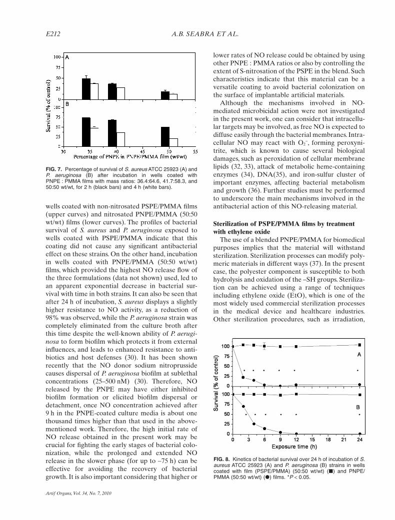
wells coated with non-nitrosated PSPE/PMMA films(upper curves) and nitrosated PNPE/PMMA (50:50wt/wt) films (lower curves). The profiles of bacterialsurvival of S. aureus and P. aeruginosa exposed towells coated with PSPE/PMMA indicate that thiscoating did not cause any significant antibacterialeffect on these strains. On the other hand, incubationin wells coated with PNPE/PMMA (50:50 wt/wt)films, which provided the highest NO release flow ofthe three formulations (data not shown) used, led toan apparent exponential decrease in bacterial sur-vival with time in both strains. It can also be seen thatafter 24 h of incubation, S. aureus displays a slightlyhigher resistance to NO activity, as a reduction of98% was observed, while the P. aeruginosa strain wascompletely eliminated from the culture broth afterthis time despite the well-known ability of P. aerugi-nosa to form biofilm which protects it from externalinfluences, and leads to enhanced resistance to anti-biotics and host defenses (30). It has been shownrecently that the NO donor sodium nitroprussidecauses dispersal of P. aeruginosa biofilm at sublethalconcentrations (25–500 nM) (30). Therefore, NOreleased by the PNPE may have either inhibitedbiofilm formation or elicited biofilm dispersal ordetachment, once NO concentration achieved after9 h in the PNPE-coated culture media is about onethousand times higher than that used in the above-mentioned work. Therefore, the high initial rate ofNO release obtained in the present work may becrucial for fighting the early stages of bacterial colo-nization, while the prolonged and extended NOrelease in the slower phase (for up to ~75 h) can beeffective for avoiding the recovery of bacterialgrowth. It is also important considering that higher or
lower rates of NO release could be obtained by usingother PNPE : PMMA ratios or also by controlling theextent of S-nitrosation of the PSPE in the blend. Suchcharacteristics indicate that this material can be aversatile coating to avoid bacterial colonization onthe surface of implantable artificial materials.
Although the mechanisms involved in NO-mediated microbicidal action were not investigatedin the present work, one can consider that intracellu-lar targets may be involved, as free NO is expected todiffuse easily through the bacterial membranes. Intra-cellular NO may react with O2
-, forming peroxyni-trite, which is known to cause several biologicaldamages, such as peroxidation of cellular membranelipids (32, 33), attack of metabolic heme-containingenzymes (34), DNA(35), and iron-sulfur cluster ofimportant enzymes, affecting bacterial metabolismand growth (36). Further studies must be performedto underscore the main mechanisms involved in theantibacterial action of this NO-releasing material.
Sterilization of PSPE/PMMA films by treatmentwith ethylene oxide
The use of a blended PNPE/PMMA for biomedicalpurposes implies that the material will withstandsterilization. Sterilization processes can modify poly-meric materials in different ways (37). In the presentcase, the polyester component is susceptible to bothhydrolysis and oxidation of the –SH groups. Steriliza-tion can be achieved using a range of techniquesincluding ethylene oxide (EtO), which is one of themost widely used commercial sterilization processesin the medical device and healthcare industries.Other sterilization procedures, such as irradiation,
FIG. 7. Percentage of survival of S. aureus ATCC 25923 (A) andP. aeruginosa (B) after incubation in wells coated withPNPE : PMMA films with mass ratios: 36.4:64.6, 41.7:58.3, and50:50 wt/wt, for 2 h (black bars) and 4 h (white bars).
FIG. 8. Kinetics of bacterial survival over 24 h of incubation of S.aureus ATCC 25923 (A) and P. aeruginosa (B) strains in wellscoated with film (PSPE/PMMA) (50:50 wt/wt) (�) and PNPE/PMMA (50:50 wt/wt) (�) films. *P < 0.05.
A.B. SEABRA ET AL.E212
Artif Organs, Vol. 34, No. 7, 2010

heat, steam, or acid, cause extensive deformation ofthe devices and accelerated polymer degradation(38). The –SH groups of the present material are thetarget sites for NO loading.Therefore, if these groupsundergo chemical changes such as oxidation to S-Sduring sterilization, the film will lose its capacity forNO loading through the S-nitrosation reaction. Eth-ylene oxide microbiologic inactivation properties aredirectly related to the addition of alkyl groups toproteins, DNA, and RNA in microorganisms bybinding to the sulfhydryl, hydroxyl, amino, and car-boxyl groups, which inactivates normal cellularmetabolism and cell division (39). Therefore, alkyla-tion is probably the main cause for a depletion of the–SH groups of PSPE in EtO sterilization.To evaluatethe extent of –SH loss in PSPE/PMMA, films steril-ized with EtO were subsequently incubated in the–SH specific reagent, DTNB, which is water solubleand reacts rapidly with free surface –SH groups atneutral pH-forming TNB2- (25). Figure 9 shows thepercentage of –SH groups per cm2 measured byDTNB titration of nonsterilized and sterilized PSPE/PMMA (36.4:64.6, 41.7:58.3, and 50:50 wt/wt) films. Itcan be seen that c. 40% of the –SH groups are stillpreserved on the surface of the PSPE/PMMA films36.4:64.6 wt/wt (corresponding to 80.5 � 4.5 nmol–SH/cm2) and 41.7:58.3 wt/wt (corresponding to108.2 � 2.3 nmol–SH/cm2),while 25% of the –SHgroups are preserved in the PSPE/PMMA 50:50wt/wt (corresponding to 112.0 � 4.0 nmol–SH/cm)after sterilization with EtO. These percentages ofremaining SH groups can be considered enough forloading these films with high NO amounts, once theextent of S-nitrosation performed in the film whichdisplayed high antibacterial activity (Figs. 7 and 8)was only partial and could be greatly increased.Therefore, this result indicates that this procedure is
viable for sterilizing such films for biomedicalapplications.
CONCLUSIONS
A PSPE, synthesized by the polyesterification reac-tion of mercaptosuccinic acid with 3-mercapto-1,2-propanediol, can be blended with PMMA leadingto solid PSPE/PMMA, which can be subsequentlyS-nitrosated in aqueous solution, yielding PNPE/PMMA films, with high NO-loading capacity. Thesefilms can be used to coat polyurethane intravascularcatheters which are able to release NO in PBS solu-tion (pH 7.4) at 37°C at rates of 4.6 nmol/cm2/h in thefirst 6 h and 0.8 nmol/cm2/h in the next 12 h. PNPE/PMMA films used to coat the bottom of wells ofculture plates exert potent dose- and time-dependentantimicrobial actions against S. aureus and P. aerugi-nosa strains and are able to reduce the viability ofthese multidrug-resistant bacteria by more than 98%after 12 h of incubation. The antibacterial action andNO-releasing capacity of PSPE/PMMA films open anew perspective for the coating of blood-contactingartificial materials, to avoid their colonization withmultidrug-resistant gram-positive and gram-negativebacterial strains.
REFERENCES
1. Lipsky BA, Weigelt JA, Gupta V, Killian A, Peng MM.Skin, soft tissue, bone, and joint infections in hospitalizedpatients: epidemiology and microbiological, clinical, andeconomic outcomes. Infect Control Hosp Epidemiol2007;28:1290–8.
2. Grundmann H, Aires-de-Sousa M, Boyce J, Tiemersma E.Emergence and resurgence of methicillin-resistant Staphylo-coccus aureus as a public-health threat. Lancet 2006;368:874–85.
3. Strateva T, Ouzounova-Raykova V, Markova B, Todorova A,Marteva-Proevska Y, Mitov I. Problematic clinical isolates ofPseudomonas aeruginosa from the university hospitals inSofia, Bulgaria: current status of antimicrobial resistance andprevailing resistance mechanisms. J Med Microbiol 2007;56:956–63.
4. Jarvis WR. The Lowbury Lecture. The United States approachto strategies in the battle against healthcare-associatedinfections, 2006: transitioning from benchmarking to zerotolerance and clinician accountability. J Hosp Infect 2007;65:3–9.
5. Wenzel R, Edmond MD. The impact of hospital acquiredblood stream infections. Emerg Infect Dis 2001;7:174–9.
6. Pittet D, Allegranzi B, Storr J, Donaldson L. “Clean Care isSafer Care”: the Global Patient Safety Challenge 2005–2006.Int J Infect Dis 2006;6:419–24.
7. Benz D, Cadet P, Mantione K, Zhu W, Stefano G. Tonal nitricoxide and health: anti-bacterial and -viral actions and implica-tions for HIV. Med Sci Monit 2002;8:RA27–31.
8. Dondji B, Bungiro RD, Harrison LM, et al. Role for nitricoxide in hookworm-associated immune suppression. InfectImmun 2008;76:2560–7.
9. Lazar EE, Wills RH, Ho BT, Harris AM, Spohr LJ. Antifungaleffect of gaseous nitric oxide on mycelium growth, sporulationand spore germination of the postharvest horticulture patho-
0
20
40
60
80
100
120
PSPE:PMMA mass ratio
50.0: 50.036.6: 63.4 41.7: 58.3
% o
f S
H g
rou
ps/c
m2
sterilized non-sterilized
FIG. 9. Percentage of remaining –SH groups per cm2 on thesurface of nonsterilized and sterilized PSPE/PMMA 36.4:64.6,41.7:58.3, and 50:50 wt/wt films.
ANTIBACTERIAL NITRIC OXIDE-RELEASING POLYESTER E213
Artif Organs, Vol. 34, No. 7, 2010
Duran
Realce
Duran
Realce

gens, Aspergillus niger, Monilinia fructicola and Penicilliumitalicum. Lett Appl Microbiol 2008;46:688–92.
10. Ghaffari A, Miller CC, McMullin B, Ghahary A. Potentialapplication of gaseous nitric oxide as topical antimicrobialagent. Nitric Oxide 2006;14:21–9.
11. Carlson S, Weitzberg E, Wiklund P, Lundberg JO. Intravesicalnitric oxide delivery for prevention of catheter-associatedurinary tract infection. Antimicrob Agents Chemother 2005;49:2352–5.
12. Chen C, Shi YQ, Song J, Qi QS, Gu L, Wang PG. Delivery ofnitric oxide released from b-galactosidase and its activityagainst Escherichia coli. Biol Pharm Bull 2006;29:1239–41.
13. de Souza GFP, Yokoyama-Yasunaka JKU, Seabra AB, MiguelDC, Uliana SRB. Leismanicidal activity of primaryS-nitrosothiols against L. major and L. amazonensis: implica-tions for the treatment of cutaneous leishmaniasis. NitricOxide 2006;15:209–16.
14. Shoenfisch MH, Rothrock AR, Shin JH, Polizzi MA, BrinkleyMF, Dobmeir KP. Poly(vinylpyrrolidone)-doped with nitricoxide-releasing xerogels as glucose biosensor membranes.Biosens Bioelectron 2006;22:306–12.
15. Hetrick EM, Schoefisch MH. Antibacterial nitric oxide-releasing xerogels: cell viability and parallel plate flow celladhesion studies. Biomaterials 2007;28:1948–56.
16. Nablo BJ, Prichard HL, Butler RD, Klitzman B, SchoenfischMH. Inhibition of implant-associated infections via nitricoxide release. Biomaterials 2005;26:6984–90.
17. Hetrick EM, Shin HH, Stasko NA, et al. Bactericidal efficacyof nitric oxide-releasing silica nanoparticles. ACS Nano2008;2:235–46.
18. Seabra AB, da Silva R, de Oliveira MG. Polynitrosated poly-esteres: preparation, characterization and potential use fornitric oxide release. Biomacromolecules 2005;6:2512–20.
19. de Oliveira MG, Seabra AB. Process for the preparation ofpolynitrosated polyesters as nitric oxide donors for biomedicalapplications. BR Patent No PI 000.784-7, 2003.
20. de Oliveira MG, Seabra AB, Laurindo FRM. Process for thecoating of implantable intracoronary stents with polymerichydrophobic nitric oxide blend. BR Patent No PI0401977-A,2004.
21. Seabra AB, da Silva R, de Souza GFP, de Oliveira MG. Nitricoxide releasing from polynitrosated polyester poly(methyl-metacrylate) blend for the coating of blood-contactingsurfaces. Artif Organs 2008;32:262–7.
22. Clinical Laboratory Standards Institute—CLSI. Methods forDilution Antimicrobial Susceptibility Tests for Bacteria ThatGrow Aerobically, 6th Edition. Wayne, PA: CLSI, 2003.
23. Grare M, Mourer M, Fontanay S, Regnouf-de-Vains JB,Finance C, Duval RE. In vitro activity of para-guanidinoethylcalix[4]arene against susceptible and antibiotic-resistant Gram-negative and Gram-positive bacteria. JAntimicrob Chemother 2007;60:575–81.
24. de Oliveira MG, Shishido SM, Seabra AB, Morgon NH.Thermal stability of primary S-nitrosothiols: roles of autocaly-sis and structural effects on the rate of nitric oxide release. JPhys Chem A 2002;106:8963–70.
25. Riddles PW, Blakeley RL, Zerner B. Reassessment of Ell-man’s reagent. Methods Enzymol 1983;91:49–60.
26. Seabra AB, da Rocha LL, Eberlin MN, de Oliveira MG. Solidfilms of blended poly(vinyl alcohol)/poly(vinyl pyrrolidone)for topical S-nitrosoglutathione and nitric oxide release. JPharm Sci 2005;95:994–1003.
27. Seabra AB, de Oliveria MG. Poly(vinyl alcohol) and poly(vi-nyl pyrrolidone) blended for local nitric oxide release. Bioma-terials 2004;25:3773–82.
28. Poomalai P, Ramaray B, Siddaramaiach BVSK. Thermal andmechanical properties of poly(methylmetacrylate) and ethyl-ene vinyl acetate copolymer blends. J Appl Poly Sci 2007;106:684–91.
29. Dobmeier KP, Schoenfisch MH. Antibacterial properties ofnitric oxide-releasing sol-gel microarrays. Biomacromolecules2004;5:2493–5.
30. Barraud N, Hassett DJ, Hwang SH, Rice SA, Kjelleberg S.Involvement of nitric oxide in biofilm dispersal of Pseudomo-nas aeruginosa. J Bacteriol 2006;188:7344–53.
31. Dejam A, Hunter CJ, Pelletier MM, et al. Erythrocytes are themajor intravascular storage sites of nitrite in human blood.Blood 2005;106:734–9.
32. Bartesaghi S, Valez V, Trujillo M, et al. Mechanistic studies ofperoxynitrite-mediated tyrosine nitration in membranes usingthe hydrophobic probe N-t-BOC-L-tyrosine tert-butyl ester.Biochemistry 2006;45:6813–25.
33. Radi R. Nitric oxide, oxidants, and protein tyrosine nitration.Proc Natl Acad Sci USA 2004;101:4003–8.
34. Alam MS, Akaike T, Okamoto S, et al. Role of nitric oxide inhost defense in murine salmonellosis as a function of its anti-bacterial and antiapoptotic activities. Infect Immun 2002;70:3130–42.
35. Delaney S, Delaney JC, Essigmann JM. Chemical-biologicalfingerprinting: probing the properties of DNA lesions formedby peroxynitrite. Chem Res Toxicol 2007;20:1718–29.
36. Djaman O, Outten FW, Imlay JA. Repair of oxidized iron-sulfur clusters in Escherichia coli. J Biol Chem 2004;279:44590–9.
37. Nair PD. Currently practiced sterilization methods—someinadvertent consequences. J Biomater Appl 1995;10:121–35.
38. Chantal EH, Cheng C, Davies JE, Shoichet MS. Optimizingthe sterilization of PLGA scaffolds for use in tissueengineering. Biomaterials 2001;22:25–31.
39. Mendes GCC, Brandão TRS, Silva CLM. Ethylene oxide ster-ilization of medical devices: a review. Am J Infect Control2007;35:574–81.
A.B. SEABRA ET AL.E214
Artif Organs, Vol. 34, No. 7, 2010