Effect of raised serum prolactin on breast development* - NCBI
13
J. Anat. (1989), 162, pp. 249-261 249 With 1O figures Printed in Great Britain Effect of raised serum prolactin on breast development* B. M. J. STRINGER$, J. ROWSONT, AND E. D. WILLIAMSt t Department of Pathology, University of Wales College of Medicine, Heath Park, Cardiff CF4 4XN, U.K. and $ Biological and Medical Research Department, King Faisal Specialist Hospital and Research Centre, P.O. Box 3354, Riyadh 11211, Kingdom of Saudi Arabia (Accepted 2 June 1988) INTRODUCTION It is well established that serum prolactin plays a major role in breast development. Factors which lead to the elevation of serum prolactin such as pregnancy (Amenomori, Chen & Meites, 1970), administration of dopamine antagonists (Sulman, 1970) and grafting of multiple pituitary isografts (Everett, 1954) also lead to breast growth. Early qualitative studies (Cole & Hopkins, 1962; Ben-David & Chrambach, 1970) suggested that growth was due to an increase in both the number and size of differentiated lobular structures in the breast, this being accompanied by increased ductular and lobular luminal volume. Previous attempts to quantify these changes in morphology, however, have suffered from a number of drawbacks. First, and without exception, only a few of the component tissues of the breast have been studied in a single investigation and a distinction between myoepithelium and epithelium has not been made on separate structures (Ben-David, Dirkstein & Sulman, 1965; Ben-David, 1968; Sulman, 1970). Secondly, in almost all cases, unreliable sampling methods have been employed without reference to stereological principles, often employing only single breast pads for study in the rat and mouse (Cowie & Fowley, 1947; Danon, Weller & Sulman, 1970; Sulman, 1970; Silver, 1983), and therefore without determination of changes in the total volumes of components. Finally, no attempt has been made to relate changes in morphology to the level of prolactin stimulus. The purpose of this study was to provide a reliable, quantitative assessment of the individual components of the breast during breast growth in the rat and to correlate these changes with the level of circulating prolactin. In view of the studies which have underlined the role of prolactin in mammary tumorigenesis (Lacassagne & Duplan, 1959; Welsch, Jenkins & Meites, 1970), it was hoped that the results obtained would provide an accurate baseline for further investigations now being undertaken to elucidate the mechanisms regulating breast growth. MATERIALS AND METHODS Female Sprague-Dawley rats aged 7 weeks and weighing 182+7 g (k ± S.E.) were obtained from A. Tuck & Son, Essex, England, and housed 5 to a cage in a thermostatically controlled room (21 + 2 °C, temperature + accepted range) with a constant 12 hour light/12 hour dark cycle (light 07.00-19.00). Animals were fed on a * Reprint requests to Dr E. D. Williams. 9 ANA 161
-
Upload
khangminh22 -
Category
Documents
-
view
1 -
download
0
Transcript of Effect of raised serum prolactin on breast development* - NCBI

J. Anat. (1989), 162, pp. 249-261 249With 1OfiguresPrinted in Great Britain
Effect of raised serum prolactin on breast development*
B. M. J. STRINGER$, J. ROWSONT, AND E. D. WILLIAMSt
t Department of Pathology, University of Wales College of Medicine, Heath Park,Cardiff CF4 4XN, U.K. and $ Biological and Medical Research Department, KingFaisal Specialist Hospital and Research Centre, P.O. Box 3354, Riyadh 11211,
Kingdom of Saudi Arabia
(Accepted 2 June 1988)
INTRODUCTION
It is well established that serum prolactin plays a major role in breast development.Factors which lead to the elevation of serum prolactin such as pregnancy (Amenomori,Chen & Meites, 1970), administration of dopamine antagonists (Sulman, 1970) andgrafting of multiple pituitary isografts (Everett, 1954) also lead to breast growth.
Early qualitative studies (Cole & Hopkins, 1962; Ben-David & Chrambach, 1970)suggested that growth was due to an increase in both the number and size ofdifferentiated lobular structures in the breast, this being accompanied by increasedductular and lobular luminal volume.
Previous attempts to quantify these changes in morphology, however, have sufferedfrom a number of drawbacks. First, and without exception, only a few of thecomponent tissues of the breast have been studied in a single investigation and adistinction between myoepithelium and epithelium has not been made on separatestructures (Ben-David, Dirkstein & Sulman, 1965; Ben-David, 1968; Sulman, 1970).Secondly, in almost all cases, unreliable sampling methods have been employedwithout reference to stereological principles, often employing only single breast padsfor study in the rat and mouse (Cowie & Fowley, 1947; Danon, Weller & Sulman,1970; Sulman, 1970; Silver, 1983), and therefore without determination of changes inthe total volumes of components. Finally, no attempt has been made to relate changesin morphology to the level of prolactin stimulus.The purpose of this study was to provide a reliable, quantitative assessment of the
individual components of the breast during breast growth in the rat and to correlatethese changes with the level of circulating prolactin.
In view of the studies which have underlined the role of prolactin in mammarytumorigenesis (Lacassagne & Duplan, 1959; Welsch, Jenkins & Meites, 1970), it washoped that the results obtained would provide an accurate baseline for furtherinvestigations now being undertaken to elucidate the mechanisms regulating breastgrowth.
MATERIALS AND METHODS
Female Sprague-Dawley rats aged 7 weeks and weighing 182+7 g (k ± S.E.) wereobtained from A. Tuck & Son, Essex, England, and housed 5 to a cage in athermostatically controlled room (21 +2°C, temperature+ accepted range) with aconstant 12 hour light/12 hour dark cycle (light 07.00-19.00). Animals were fed on a
* Reprint requests to Dr E. D. Williams.9 ANA 161
250 B. M. J. STRINGER, J. ROWSON AND E. D. WILLIAMS
standard laboratory diet (Pilbury's rat and mouse breeding diet) and given free accessto drinking water. Vaginal smears were taken daily between 08.00 and 12.00 hours andonly rats undergoing 3 or more regular 4 day oestrous cycles were chosen for thisstudy. The stage in the cycle was assessed by light microscopic examination of vaginalsmears after Papanicoulou staining. The oestrous cycle was defined as: oestrus, theday of cornified cells only; metoestrus, the first day of leukocytes; dioestrus, thesecond day of leukocytes together with mucus production; pro-oestrus, the day withnucleated cells present in the absence of leukocytes.
Fifty one rats were used for serum prolactin studies. Thirty rats were givenperphenazine orally as a 0-01 % solution, made up from Fentazin syrup (2 mgperphenazine/ml syrup, Allen & Hanbury Ltd, Greenford, England) in light-tightcontainers, the solution being renewed every two days. This was commenced 48 hoursafter a dioestrus smear. No further smears were taken before blood sampling becauseof the known effects of handling on serum prolactin levels. Twenty one animals werenot given perphenazine; sixteen were killed at the beginning of the experiment (fourin each stage of the oestrous cycle), and a further five were killed at the end of theexperiment. The perphenazine-treated animals were killed in groups of five at 2, 4, 7,14, 27 and 54 days after the start of the experiment.
All animals were killed rapidly between 13.00 and 13.10 hours by decapitation in aseparate room in order to avoid the known effects of stress on the pituitary secretionof prolactin (Krulich, Hefco, Ilner & Read, 1974). Trunk blood was collected from theneck, and serum separated for subsequent prolactin assay. Vaginal smears were alsotaken from all rats after killing and the stage of the oestrous cycle confirmed for theuntreated rats and determined for the perphenazine-treated animals.
Since decapitation can lead to breast tissue loss as well as damage, a separate groupof forty female rats was used for morphometric studies. Thirty were given perphenazine48 hours after a dioestrus smear and killed in groups of five on Days 2, 4, 7, 14, 27 and54 after the start of treatment as described for the prolactin studies. The remaining tenrats were not given perphenazine and were killed five on Day 0 and five on Day 54 ofthe experiment. Groups were killed by ether overdose and the skin and breast tissueremoved whole and pinned out flat onto cork boards. The tissue was then immersedin chilled methacarn fixative (a mixture of methanol, chloroform and acetic acid at6:3: 1) and fixed for 24 hours at 4 'C. After fixation all breast tissue including thesurrounding fat matrix was removed from the skin by blunt dissection. The totalvolume of breast tissue plus surrounding matrix of each rat was assessed by measuringthe volume of methacarn displaced in a volumetric cylinder after tissue immersion.
Before processing, tissue architecture was physically randomised as describedpreviously (Stringer, Wynford-Thomas & Williams, 1982). The tissue was diced intoapproximately 400 pieces of equal size. The diced tissue was then processed inchloroform and embedded into 5 wax blocks per rat. Each block containedapproximately 80 pieces of breast tissue in random orientation within the block. Twoserial sections of thickness 4,um were cut from each block. In one section themyoepithelium was localised by immunocytochemistry, using antimyosin antiserumand an indirect immunoperoxidase technique and counterstained with Harris'haematoxylin (Fig. 1). The remaining sections from each block were stained withHarris' haematoxylin only.

Serum prolactin and breast development
Fig. 1. 4msection through normal rat breast. The myoetheal cells_havebee
Compoet quant.9:fiedi
Theunepitohelicalelemeignts-ofothebrastipeneraten theisubcutaneousnfatrand,ainthcnqe.
rodent during pregnancy, they spread widely through the subcutis virtually joiningacross the back of the animals. Breast tissue for the purpose of this morphometricanalysis was defined as the epithelial component from nipple to alveolus together withthe myoepithelial cells, lumen and associated specialised stroma. The relative volumeof breast tissue and fat was measured at low magnification and the absolute volumethen calculated by reference to the earlier displacement measurement. Fourcomponents of breast tissue were then measured at high magnification, as follows.
(1) Lumen component: the space surrounded by either alveolar or duct epithelium.(2) Epithelial component: epithelial cells characterised by their morphological
appearance, their position in relation to the breast duct and alveolar lumen and theirstaining properties.
(3) Myoepithelial component: defined both by the relationship of the cell to theepithelium and by the immunocytochemical demonstration of intracellular myosin.
(4) Stromal component: defined as the connective tissue and blood vessels (includinglumen) of the specialised stroma surrounding the epithelium; excluding thesurrounding fat cells.
Each component was subdivided into that associated with ducts or ductules andthat associated with alveoli (including alveolar buds).
251

B. M. J. STRINGER, J. ROWSON AND E. D. WILLIAMS
4.i s a ,4
p rV AP,
4 0~~~~0JWi~~~~~~~~~~~~~~~~~~~~~94t f
75 m-m:
Fig. 2. Low power view of H and E stained 4 ,cm section through normal rat breast. The majorityof the breast pad is fat matrix penetrated by occasional breast epithelial elements. In the unstimulatedgland, no marked lumen distension is seen.
Quantitative microscopic analysisTotal breast tissue was quantified semi-automatically with the use of an IBAS II
Image Analyser. To do this, sections stained with haematoxylin were viewed at lowpower (field size of 510 x 510 ,um), the field of view being processed via a video camerainto the computer of the image analyser and displayed on the video screen. Breasttissue was discriminated from the fat matrix by the use of a programme designed todistinguish differences in 'grey-level' within the picture. Image analysis discriminationwas visually confirmed on the video screen by a colour overlay. Any discrepanciesbetween observed and expected discrimination were edited manually with a hand-heldcursor. Once editing was completed, the area of the breast tissue was automaticallycomputed as a percentage of the whole field viewed.The first whole field from the top left hand corner of each of 50 tissue piece profiles
chosen at random was quantified for each rat and the mean percentage area of breasttissue ascertained.The individual compartments of the breast were quantified manually. Sections were
viewed at high magnification ( x 860) on a projecting microscope (Reichert Visopan)with the screen overlayed with a square lattice consisting of 100 individual points. Tenfields were viewed from each of 5 sections per rat. Each field (1 16 x 116 ,um) was selectedrandomly from a separate tissue piece within the section. Each point within the latticewas scored as either epithelial, myoepithelial, luminal or stromal and was subclassifiedas either alveolar or ductular. Occasionally points were found lying on the boundariesbetween compartments. On these occasions, the component to the left and/or abovethe points was scored. The points scored totalled 5000 for each animal.
252

Serum prolactin and breast development
75gm ~ q~
Fig. 3. Low power view of H and E stained 4 ,um section through 14 day prolactin-stimulated ratbreast. A substantial increase in the breast tissue is seen with marked lumen distension and thepresence of fully differentiated lobular structures.
Assay for serum prolactinSerum samples from each rat were assayed for prolactin by radioimmunoassay
using the NIADDK kit. 200 ,ul serum samples were assayed in triplicate from each rat.Results were expressed in terms of the NIADDK-rPRL-RP-3 standard. Assayprecision and sensitivity were ascertained using the method of Ekins (1976): prolactinassay sensitivity in our hands was shown for triplicate samples to be 5 6 ng/ml whichwas less than 11 % of the overall mean serum prolactin levels present in our untreatedcontrol rats. The intra-assay coefficient of variation was found to be 8-2-8&9 % over the'normal' range and 11 9-13 4% over the range covering the perphenazine-inducedhyperprolactinaemic rat serum concentrations.
Statistical treatment of resultsResults are expressed as the mean+ standard error unless otherwise stated. Where
tests of significance were made Student's t-independent test was used.
RESULTS
The vaginal smears taken from groups of control rats on Days 1-4 at the start ofthe experiment showed the expected positions in the oestrous cycle predicted from thevaginal smears taken two days prior to killing. Four groups of four rats were in eachof the four stages of the oestrous cycle.The effects of perphenazine administration on vaginal smear patterns of rats during
the first 7 days of treatment were variable both within and between groups. Ten of thevaginal smears taken showed patterns similar to dioestrus, three showed a metoestrus
253
10 ANA 162

B. M. J. STRINGER, J. ROWSON AND E. D. WILLIAMS
300 -
250
E 200
--150
E2100
a)
C')
-1
-3-202 4 7 14 27 54Days of perphenazine administration
Fig. 4. Effect of oral administration of perphenazine on rat serum prolactin (i ± S.E.M. 4 or 5 rats/point; see text). 0-*, normal rat serum prolactin levels at 13.00 hours on day of metoestrus (Day3), dioestrus (Day 2), pro-oestrus (Day 1) and oestrus (Day 0). A-A, serum prolactin levels at 13.00hours in rats administered perphenazine orally for varying times up to 54 days. 0, normal rat(unselected for phase of oestrus) serum prolactin levels maintained as controls for the 54 dayperphenazine-treated group.
pattern and for the remaining two rats, a pattern equivalent to pro-oestrus was seen. Ingeneral, as the treatment time increased, the tendency for the vaginal smears to givea predominantly dioestrus appearance also increased. None of the treated rats showeda vaginal smear pattern equivalent to the oestrus stage at any time point.The effects of the oestrous cycle and oral administration of perphenazine on rat
serum prolactin are illustrated in Figure 4. The serum prolactin levels of untreated ratssampled at 13.00 hours on each day of the 4 day oestrous cycle were: 24-3+ 6-2,29-8+ 7-4, 108-6 + 24-4, and 56-3 + 18-4 for metoestrus, dioestrus, pro-oestrus andoestrus respectively.The pro-oestrus prolactin levels were significantly greater than those of the
metoestrus (P < 0 02) and dioestrus (P < 0 05) phases. No significant difference wasseen between prolactin levels of pro-oestrus and oestrus, nor between oestrus andmetoestrus or dioestrus.
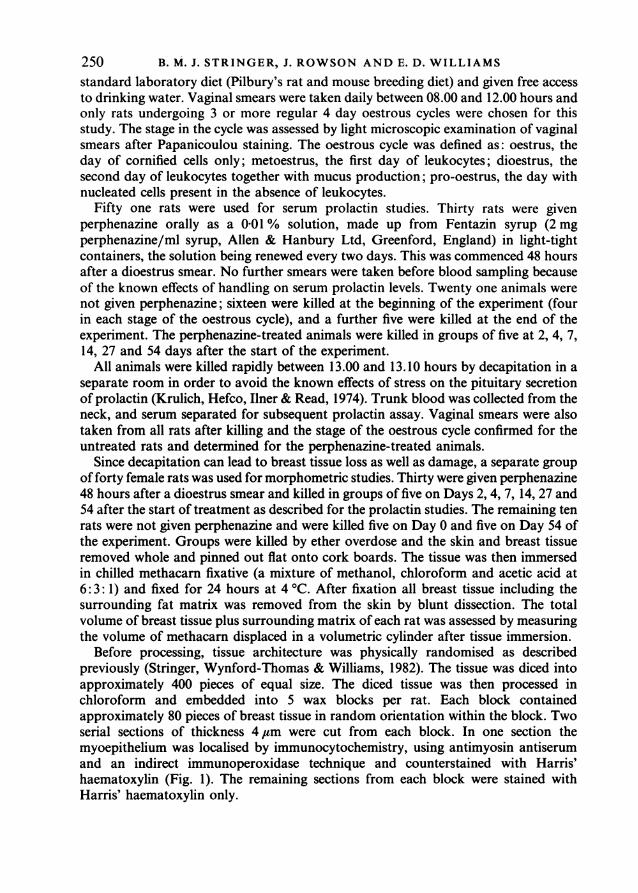
Oral administration of perphenazine (0-01 %) led to a marked rise in serumprolactin. This was seen at the first time point sampled (2 days) when it was alreadysignificantly increased when compared to the level of prolactin at any phase ofoestrous cycle (metoestrus P < 0.001, dioestrus P < 0.001, pro-oestrus P < 0 05,oestrus P < 0-0 1). Further rises occurred at 4 and at 7 days after commencingperphenazine administration, and a sustained high level was maintained until thestudy was concluded at 54 days. The prolactin levels of the control group of animalskilled at the end of the experiment were not significantly different from the levels ofthe control animals killed at the start of the experiment.The effect of orally administered perphenazine on rat body weight is shown in
254
Serum prolactin and breast development
300
250
200
40)
~3150-
0m 100
50 -
0 2 4 7 14 27 54Days of perphenazine administration
Fig. 5. Effect of oral administration of perphenazine (0-01 % in drinking water) on rat body weight(X±S.E.M. 5 rats/point). *-*, untreated control rats; A-A, rats given oral perphenazine.
Figure 5. Body weights of the treated groups were compared with age-matcheduntreated control rats for each time-point. A significant (P < 0-05) fall in the bodyweights of treated rats was seen after two days of perphenazine administration, butthis returned to basal values by Day 4 of the treatment. From Day 4 of treatment, theweight gains of both the control and treated groups were similar with no significantdifference between the groups at matched time-points up to 27 days. The mean bodyweight of the 54 days perphenazine-treated group, however, was slightly butsignificantly (P < 0-05) lower than that of the age-matched untreated controlgroup.The effect of raised serum prolactin on total rat breast volume is shown in Figure
6. A significant (P < 0-01) 3-4-fold increase in breast volume was seen at 4 days afterbeginning treatment, with a continued rise up to an 8-9-fold peak by Day 14. A slightbut significant (P < 0-05) fall in the 14 days value was seen at 27 days but no significantdifference was seen between the 14 days and 54 days levels, nor between the 27 daysand 54 days levels.Changes in the volumes of the epithelial components are shown in Figure 7. A small
but significant (P < 0-05) 2-6-fold increase in the volume of the ductular epitheliumwas found 4 days after the start of perphenazine administration. This remainedelevated for the duration of the experiment but no further significant increases wereseen. A highly significant (P < 0-001) 5-6-fold increase in the alveolar epitheliumvolume was seen by Day 4 with a continued rise to a 15-6-fold peak by Day 14. A slightbut significant (P < 0-05) fall from this peak occurred by Day 27 but as with the totalbreast volume, this was later reversed so that the 54 day measurement was notsignificantly different from either the 14 or 27 day levels.Myoepithelium component volume changes in response to serum prolactin elevation
are shown in Figure 8. A significant (P < 0-05) 2-1-fold rise in the ductularmyoepithelial component was also seen by Day 4 and it continued to rise to reach a2-9-fold (P < 0-01) peak by Day 7. The peak was not sustained, however, and the
10-2
255

B. M. J. STRINGER, J. ROWSON AND E. D. WILLIAMS
8
6
4E> 3
2
1
0 4 8 12 16 20 24 28Days of perphenazine administration
54
Fig. 6. Effect of perphenazine administration on total rat breast volume (X±S.E.M. 5 rats/point).
1 -8
1-6
1-4
1.2I-. _
-2 1.0
E= 0-8
> 06
0-4-
0-2
4 8 12 16 20 24 28Days of perphenazine administration
Fig. 7. Effect of perphenazine administration on breast alveolar (-f*) and ductular (O-O)epithelial volume (X+S.E.M. 5 rats/point).
ductular myoepithelial volume declined to just above normal levels at the 14, 27 and54 day time-points. A significant (P < 005) 3-7-fold increase in the alveolarmyoepithelial compartment volume was seen 4 days after beginning treatment. Thisrose further to a 7-2-fold (P < 0O001) peak by Day 14 and remained at this levelthroughout the rest of the treatment period.Both the ductular and alveolar lumen compartment volumes rose in response to
raised serum prolactin (Fig. 9). A marked 63-fold increase (P < 0001) in the alveolarlumen compartment volume was seen by Day 14 but this declined to a 36-fold increaseby Day 54. A significant but more gradual increase in the lumen compartment was alsoseen in the ducts leading a stable 16-fold (P < 0001) plateau by Day 14.
256

Serum prolactin and breast development
0-4
0-3
o 0-2E
01
0 4 8 12 16 20 24 28 54Days of perphenazine administration
Fig. 8. Effect of perphenazine administration on breast alveolar (U-U) and ductular ([I-[])myoepithelial volume (X±S.E.M. 5 rats/point).
4
3
E
1 X
0 4 8 12 16 20 24 28 54Days of perphenazine administration
Fig. 9. Effect of perphenazine administration on breast alveolar (0-U) and ductular (O-EC)luminal volume (X±S.E.M. 5 rats/point).
The stromal volume related to alveoli rose to a 3-7-fold (P < 0-001) plateau by Day14 of treatment (Fig. 10). A significant (P < 0 01) 2 1-fold increase in the duct stromalvolume was also seen by Day 7, with no further significant changes by Day 54.No significant changes in any breast components were seen between Day 0 and Day
54 control groups.
257

B. M. J. STRINGER, J. ROWSON AND E. D. WILLIAMS
1 *0
0-8
En 065
Eo 0*4
0-2
0 4 8 12 16 20 24 28 54Days of perphenazine administration
Fig. 10. Effect of perphenazine administration on breast alveolar (f--) and ductular (O-J)stromal volume (i ± S.E.M. 5 rats/point).
DISCUSSION
Our study showed serum prolactin levels in normal rats to vary at different times ofthe oestrous cycle. Investigations performed by other workers have conclusivelydemonstrated an oestrous rhythm of prolactin in both 4 and 5 days cycling rats (Gay,Midgley & Niswender, 1970; Midgley, Niswender, Gay & Reichert, 1971; Butcher,Collins & Fugo, 1974). Also, a circadian rhythm of prolactin has been reported whichdiffers at different times of the cycle (Butcher, Fugo & Collins, 1970). We have notinvestigated the oestrous or circadian rhythms but as in other studies, high serumprolactin levels were found during pro-oestrus, our reported levels being comparableto those of other workers.
Oral administration of perphenazine led to a dramatic increase in serum prolactin.A highly significant (P < 0-001) 8-8-fold elevation above basal values was seen at thefirst time-point sampled and was maintained for the duration of the experiment.Prolactin elevation caused a disruption of the oestrous cycle, leading to apredominantly dioestrus appearance of vaginal smear patterns after the first few days.The dioestrus pattern induced by elevated prolactin has been reported previously(Ben-David, 1968) and is most likely due to the loss of the gonadotrophic surge whichis thought to be negated by hyperprolactinaemia (Muralidhar, Maneckjee & Moudgal,1977). Figure 5 shows the effect of orally administered perphenazine on rat bodyweight. A small but significant fall was seen after 2 days and 54 days of treatment butotherwise the dopamine antagonist was well tolerated.
Hyperprolactinaemia led to considerable changes in breast morphology during thefirst few days of serum prolactin elevation. Structures common to unstimulated breasttissue, notably the terminal end buds, lateral buds and terminal ducts (Russo, Tay &Russo, 1982) virtually disappeared by Day 7, being replaced by the more differentiated
258

Serum prolactin and breast development 259alveolar buds and lobular structures. By Day 14 non-ductular breast epitheliumconsisted almost entirely of fully differentiated lobules, each containing large numbersof individual alveoli. A noticeable increase in the lumen volume of alveoli could beseen by Day 14 (Fig. 3). The qualitative microscopic changes in breast tissue inresponse to elevated serum prolactin were similar to those reported previously(Sulman, 1970).Although an increase in breast tissue volume was obvious on subjective light
microscopy, there was no significant difference in the volumes of breast tissue with itsaccompanying fat between any of the groups. Studies by Cole & Hopkins (1962) alsoshowed no significant difference in the wet weight of abdomino-inguinal breast padstaken from untreated and prolactin-treated rats, despite very significant increases inDNA content in response to daily prolactin injections. It is clear from theseinvestigations therefore that the total breast fat pad volume/weight cannot be reliedupon as an indicator of true breast tissue volume. Quantitative microscopic analysismust therefore be employed to determine changes in breast volume independent of thesurrounding fat matrix.
Unlike previous investigations concerned with changes in breast growth anddifferentiation in response to prolactin stimulation, we have randomly sampled theentire breast tissue complement included within the fat matrix. This, along with themeasurement of the tissue sample volume, has enabled us to provide reliable estimatesof changes in the total breast volume. Furthermore, we have studied the changes ineight separate components: epithelium, myoepithelium, lumen and stroma each beingsubdivided into either ductular or alveolar compartments.The results show a highly significant (P < 0-001) 9-2-fold increase in total breast
volume from 0-80 + 0-21 cm3 in the untreated group to 7 15 + 0-78 cm3 after 14 daysstimulation. Other workers have shown that short term serum prolactin elevation inthe rat leads to mammary gland differentiation (Ben-David & Chrambach, 1970;Sulman, 1970) but have not quantified breast volume changes or considered the ductsand alveoli as independent compartments.
Separation of the breast components into different compartments has clearlyidentified different patterns of responses. Of particular interest is the finding that theepithelial, myoepithelial and stromal responses of the ducts resembled each other butdiffered from the epithelial, myoepithelial and stromal responses of the alveoli. Theductal responses reached an early relatively low peak: 2*6-fold increase by 4 days forduct epithelium, 2-9-fold increase by 7 days for duct myoepithelium and 3 1-foldincrease by 7 days for duct stroma. In contrast, the alveolar responses reached a latepeak, and showed a greater increase: 15-6-fold by 14 days for alveolar epithelium, 7-2-fold increase by 14 days for alveolar myoepithelium and an 8-7-fold increase by 14days for alveolar stroma. This suggests that the separate but coordinated responsesseen for these three compartments in duct and alveolar tissue may be regulated bylocal controlling mechanisms. More detailed analysis however, including cell turnoverstudies, will be required to show whether not only the tissue volumes but also mitoticrates are coordinated.The changes in lumen volume are of less interest than are those of the cellular
compartments but the alveolar lumen showed the expected increase from a very lowto a high level. Identification of the various components proved easy, although therecognition of intracellular myosin by immunocytochemistry proved essential for thequantification of the myoepithelial compartment.Although there have been studies on the myoepithelial cell during breast
differentiation and development (Radnor, 1972 a, b) we believe this to be the first
B. M. J. STRINGER, J. ROWSON AND E. D. WILLIAMS
investigation to quantify changes in total myoepithelium volume in normal andstimulated breast.
SUMMARY
The effect of serum prolactin elevation on the growth and development of the ratbreast was investigated. Oral administration of the dopamine antagonist, perphen-azine, led to a 5-10-fold elevation of serum prolactin after two days of treatmentwhich was maintained for the 54 days of study. A significant (P < 0.01) 3-4-foldincrease in total breast volume was seen by Day 4 of serum prolactin elevation. Breastvolume continued to rise up to Day 14 reaching an 8-9-fold peak (P < 0-001) whichwas maintained for the duration of the experiment. Epithelial, myoepithelial, lumenand stromal volume changes in the ductular and alveolar compartments werequantified separately. Highly significant (P < 0-01) volume increases were seen in allcomponents within the first few days of prolactin elevation.
Similar time courses of the growth response to elevated serum prolactin were seenin the ductal tissues reaching an approximate 3-fold peak by 7 days in duct epithelium,myoepithelium and duct stroma. Time coordinated growth responses were also seenin the alveolar tissues with larger (7-15-fold) increases in alveolar epithelium, alveolarmyoepithelium and alveolar stroma, reaching a peak by 14 days.
We would like to thank the Medical Research Council for grant support. We aregrateful to Dr Parlow of the NIADDK, NIH, Bethesda, U.S.A., for the supply of thereagents used in this study.
REFERENCES
AMENOMORI, Y., CHEN, C. L. & MEITES, J. (1970). Serum prolactin in rats during different reproductivestates. Endocrinology 86, 506-510.
BEN-DAVID, M. (1968). Mechanism of induction of mammary differentiation in Sprague-Dawley female ratsby perphenazine. Endocrinology 83, 1217-1223.
BEN-DAVID, M. & CHRAMBACH, A. (1970). Monographs on Endocrinology, Vol. 3. Hypothalamic Control ofLactation, Part II. Mammotrophic effect of phenothiazine derivatives (ed. F. Gross, A. Labhart, T. Mann,L. T. Samuels & J. Zander). New York: Springer-Verlag.
BEN-DAVID, M., DIRKSTEIN, S. & SULMAN, F. G. (1965). Production oflactation by non-sedative phenothiazinederivatives. Proceedings of the Society for Experimental Biology and Medicine 118, 265-270.
BUTCHER, R. L., COLLINS, W. E. & FuGo, N. W. (1974). Plasma concentrations of LH, FSH, Prolactin,Progesterone and Estradiol-17B throughout the 4 day oestrus cycle of the rat. Endocrinology 94,1704-1708.
BUTCHER, R. L., FuGo, N. W. & COLLINS, W. E. (1970). Semicircadian rhythm in plasma levels of prolactinduring early gestation in the rat. Endocrinology 90, 1215-1217.
COLE, R. D. & HOPKINS, T. R. (1962). Maintenance of the mammary of glands in hypophysectomised-oophorectomised rats by injections of prolactin. Endocrinology 71, 395-398.
COwIE, A. T. & FowLEY, S. J. (1947). The role of the adrenal cortex in mammary development and its relationto the mammogenic action of the anterior pituitary. Endocrinology 40, 274-285.
DANON, A., WELLER, C. P. & SULMAN, F. G. (1970). Role of oestrogens and gonadotrophins in perphenazine-induced mammogenesis. Journal of Endocrinology 48, 365-37 1.
EKINS, R. P. (1976) In Hormone Assays and Their Clinical Applications, 4th ed. (ed. J. A. Loraine & E. C. Bell),pp. 1-72. Edinburgh: Churchill Livingstone.
EvERETT, J. W. (1954). Luteotrophic function of autografts of the rat hypophysis. Endocrinology 54,685-690.
GAY, V. L., MIDGLEY, A. R. & NISWENDER, G. D. (1970). Patterns of gonadotrophin secretion associated withovulation. Federation Proceedings 29, 1880-1887.
KRULICH, L., HEFco, E., ILLNER, P. & READ, C. B. (1974). The effects of acute stress on the secretion of LH,FSH, prolactin and GH in the normal male rate with comments on their statistical evaluation.Neuroendocrinology 16, 293-311.
LACASSAGNE, A. & DUPLAN, T. F. (1959). Mecanisme de la canc6risation de la mamelle chez la souris.Considere d'apres lesresultats d'experiences au moyen de lareserpine. Compte rendu hebdomadaire desseances del'Academie des sciences 249, 810-812.
260

Serum prolactin and breast development 261MIDGLEY, A. R., NISWENDER, G. D., GAY, V. L. & REICHERT, L. E. (1971). Use of antibodies for
characterisation of gonadotrophins and steroids. Recent Progress in Hormone Research 27, 235-286.MURALIDHAR, K., MANECKJEE, R. & MOUDGAL, N. R. (1977). Inhibition of in-vivo release of luteinizinghormone in lactating rats by exogenous prolactin. Endocrinology 100, 1137-1142.
RADNOR, C. J. P. (1972a). Myoepithelial cell differentiation in rat mammary glands. Journal ofAnatomy 111,381-398.
RADNOR, C. J. P. (1972b). Myoepithelium in the prelactating and lactating mammary glands of the rat. Journalof Anatomy 112, 337-353.
Russo, J., TAY, L. K. & Russo, I. H. (1982). Differentiation of the mammary gland and its susceptibility tocarcinogenesis. Breast Cancer Research and Treatment 2, 5-73.
SILVER, M. (1983). A quantitative analysis of the role of oestrogen in mammary development of the rat. Journalof Endocrinology 10, 17-34.
STRINGER, B. M. J., WYNFORD-THoMAs, D. & WILLIAMS, E. D. (1982). Physical randomization of tissuearchitectures - an alternative to systematic sampling. Journal of Microscopy 126, 179-182.
SULMAN, F. G. (1970). Monographs on Endocrinology, Vol. 3. Hypothalamic Control of Lactation, Part II.Mammotrophic effect of phenothiazine derivatives (ed. F. Gross, A. Labhart, T. Mann, L. T. Samuels &J. Zander). New York: Springer-Verlag.
WELSCH, C. W., JENKINS, T. W. & MEITES, J. (1970). Increased incidence of mammary tumours in the femalerat grafted with multiple pituitaries. Cancer Research 30, 1024-1029.