![Natural product leads for drug discovery: Isolation, synthesis and biological evaluation of 6-cyano-5-methoxyindolo[2,3-a]carbazole based ligands as antibacterial agents](https://static.fdokumen.com/doc/165x107/63372f4e605aada553005d0d/natural-product-leads-for-drug-discovery-isolation-synthesis-and-biological-evaluation.jpg)
Beehive Products as Antibacterial Agents: A Review - MDPI
25
antibiotics Review Beehive Products as Antibacterial Agents: A Review Rita Abou Nader 1,† , Rawan Mackieh 1,† , Rim Wehbe 2 , Dany El Obeid 3 , Jean Marc Sabatier 4, * and Ziad Fajloun 1,5, * Citation: Nader, R.A.; Mackieh, R.; Wehbe, R.; El Obeid, D.; Sabatier, J.M.; Fajloun, Z. Beehive Products as Antibacterial Agents: A Review. Antibiotics 2021, 10, 717. https:// doi.org/10.3390/antibiotics10060717 Academic Editor: Adelaide Almeida Received: 24 May 2021 Accepted: 12 June 2021 Published: 15 June 2021 Publisher’s Note: MDPI stays neutral with regard to jurisdictional claims in published maps and institutional affil- iations. Copyright: © 2021 by the authors. Licensee MDPI, Basel, Switzerland. This article is an open access article distributed under the terms and conditions of the Creative Commons Attribution (CC BY) license (https:// creativecommons.org/licenses/by/ 4.0/). 1 Faculty of Sciences 3, Department of Biology, Lebanese University, Campus Michel Slayman Ras Maska, Tripoli 1352, Lebanon; [email protected] (R.A.N.); [email protected] (R.M.) 2 Biology Department, Faculty of Arts and Sciences, American University of Beirut, Beirut 1107 2020, Lebanon; [email protected] 3 Faculty of Agriculture & Veterinary Sciences, Lebanese University, Dekwaneh, Beirut 2832, Lebanon; [email protected] 4 Faculté de Médecine Secteur Nord, 51, Université Aix-Marseille, Institut de Neuro-Physiopathologie, UMR 7051, Boulevard Pierre Dramard-CS80011, CEDEX 15, 13344 Marseille, France 5 Laboratory of Applied Biotechnology (LBA3B), Azm Center for Research in Biotechnology and its Applications, EDST, Lebanese University, Tripoli 1300, Lebanon * Correspondence: [email protected] (J.M.S.); [email protected] (Z.F.) † These authors contributed equally to this work. Abstract: Honeybees are one of the most marvelous and economically beneficial insects. As pollina- tors, they play a vital role in every aspect of the ecosystem. Beehive products have been used for thousands of years in many cultures for the treatment of various diseases. Their healing properties have been documented in many religious texts like the Noble Quran and the Holy Bible. Honey, bee venom, propolis, pollen and royal jelly all demonstrated a richness in their bioactive compounds which make them effective against a variety of bacterial strains. Furthermore, many studies showed that honey and bee venom work as powerful antibacterial agents against a wide range of bacteria including life-threatening bacteria. Several reports documented the biological activities of honeybee products but none of them emphasized on the antibacterial activity of all beehive products. Therefore, this review aims to highlight the antibacterial activity of honey, bee venom, propolis, pollen and royal jelly, that are produced by honeybees. Keywords: antibacterial activity; honeybee products; honey; bee venom; propolis; royal jelly; pollen 1. Introduction Honeybees, also known as the ‘’Golden insect‘’, belong to the genus Apis, which is the Latin word for ‘’bee”. These marvelous social insects live in a well-organized community and belong to the order Hymenoptera and to the family Apidae [1]. Honeybees can be found all around the world and are used for their vital role as pollinators in agriculture, but the main used species for crop pollination is Apis mellifera [2]. Since ancient times, honeybee products have been used for medicinal purposes. They have been also cited in many religious books [3]. Egyptians, Romans, Chinese and Persians have all documented for thousands of years the nutritional and medicinal values of bee products [4]. Apitherapy is a branch of unconventional medicine that relies on the usage of bee products which consist of honey, pollen, propolis, royal jelly and bee venom (BV). Besides having high nutritional importance and health benefits, honey showed antifungal, antiviral, antiseptic, anticancer, anti-diabetic, anti-inflammatory and cardio protective activities [5]. As for the BV, despite its possible adverse effects like the allergic reactions that might occur after the bee sting, one cannot disregard its various therapeutic effects. BV exerts anticancer, antiviral, antifungal and antibacterial effects, it is also used for the treatment of many neurodegenerative diseases [6–10]. Regarding propolis, it has the capacity to fight against cancer and many microorganisms [4,11,12]. Moreover, pollen possesses antioxidant, anticoagulant and anti- inflammatory properties [13–15]. Also, royal jelly exhibits several interesting biological Antibiotics 2021, 10, 717. https://doi.org/10.3390/antibiotics10060717 https://www.mdpi.com/journal/antibiotics
-
Upload
khangminh22 -
Category
Documents
-
view
4 -
download
0
Transcript of Beehive Products as Antibacterial Agents: A Review - MDPI

antibiotics
Review
Beehive Products as Antibacterial Agents: A Review
Rita Abou Nader 1,†, Rawan Mackieh 1,†, Rim Wehbe 2, Dany El Obeid 3, Jean Marc Sabatier 4,*and Ziad Fajloun 1,5,*
�����������������
Citation: Nader, R.A.; Mackieh, R.;
Wehbe, R.; El Obeid, D.; Sabatier, J.M.;
Fajloun, Z. Beehive Products as
Antibacterial Agents: A Review.
Antibiotics 2021, 10, 717. https://
doi.org/10.3390/antibiotics10060717
Academic Editor: Adelaide Almeida
Received: 24 May 2021
Accepted: 12 June 2021
Published: 15 June 2021
Publisher’s Note: MDPI stays neutral
with regard to jurisdictional claims in
published maps and institutional affil-
iations.
Copyright: © 2021 by the authors.
Licensee MDPI, Basel, Switzerland.
This article is an open access article
distributed under the terms and
conditions of the Creative Commons
Attribution (CC BY) license (https://
creativecommons.org/licenses/by/
4.0/).
1 Faculty of Sciences 3, Department of Biology, Lebanese University, Campus Michel Slayman Ras Maska,Tripoli 1352, Lebanon; [email protected] (R.A.N.); [email protected] (R.M.)
2 Biology Department, Faculty of Arts and Sciences, American University of Beirut, Beirut 1107 2020, Lebanon;[email protected]
3 Faculty of Agriculture & Veterinary Sciences, Lebanese University, Dekwaneh, Beirut 2832, Lebanon;[email protected]
4 Faculté de Médecine Secteur Nord, 51, Université Aix-Marseille, Institut de Neuro-Physiopathologie,UMR 7051, Boulevard Pierre Dramard-CS80011, CEDEX 15, 13344 Marseille, France
5 Laboratory of Applied Biotechnology (LBA3B), Azm Center for Research in Biotechnology and itsApplications, EDST, Lebanese University, Tripoli 1300, Lebanon
* Correspondence: [email protected] (J.M.S.); [email protected] (Z.F.)† These authors contributed equally to this work.
Abstract: Honeybees are one of the most marvelous and economically beneficial insects. As pollina-tors, they play a vital role in every aspect of the ecosystem. Beehive products have been used forthousands of years in many cultures for the treatment of various diseases. Their healing propertieshave been documented in many religious texts like the Noble Quran and the Holy Bible. Honey, beevenom, propolis, pollen and royal jelly all demonstrated a richness in their bioactive compoundswhich make them effective against a variety of bacterial strains. Furthermore, many studies showedthat honey and bee venom work as powerful antibacterial agents against a wide range of bacteriaincluding life-threatening bacteria. Several reports documented the biological activities of honeybeeproducts but none of them emphasized on the antibacterial activity of all beehive products. Therefore,this review aims to highlight the antibacterial activity of honey, bee venom, propolis, pollen androyal jelly, that are produced by honeybees.
Keywords: antibacterial activity; honeybee products; honey; bee venom; propolis; royal jelly; pollen
1. Introduction
Honeybees, also known as the ‘’Golden insect‘’, belong to the genus Apis, which is theLatin word for ‘’bee”. These marvelous social insects live in a well-organized communityand belong to the order Hymenoptera and to the family Apidae [1]. Honeybees can befound all around the world and are used for their vital role as pollinators in agriculture, butthe main used species for crop pollination is Apis mellifera [2]. Since ancient times, honeybeeproducts have been used for medicinal purposes. They have been also cited in manyreligious books [3]. Egyptians, Romans, Chinese and Persians have all documented forthousands of years the nutritional and medicinal values of bee products [4]. Apitherapy isa branch of unconventional medicine that relies on the usage of bee products which consistof honey, pollen, propolis, royal jelly and bee venom (BV). Besides having high nutritionalimportance and health benefits, honey showed antifungal, antiviral, antiseptic, anticancer,anti-diabetic, anti-inflammatory and cardio protective activities [5]. As for the BV, despiteits possible adverse effects like the allergic reactions that might occur after the bee sting, onecannot disregard its various therapeutic effects. BV exerts anticancer, antiviral, antifungaland antibacterial effects, it is also used for the treatment of many neurodegenerativediseases [6–10]. Regarding propolis, it has the capacity to fight against cancer and manymicroorganisms [4,11,12]. Moreover, pollen possesses antioxidant, anticoagulant and anti-inflammatory properties [13–15]. Also, royal jelly exhibits several interesting biological
Antibiotics 2021, 10, 717. https://doi.org/10.3390/antibiotics10060717 https://www.mdpi.com/journal/antibiotics
Antibiotics 2021, 10, 717 2 of 25
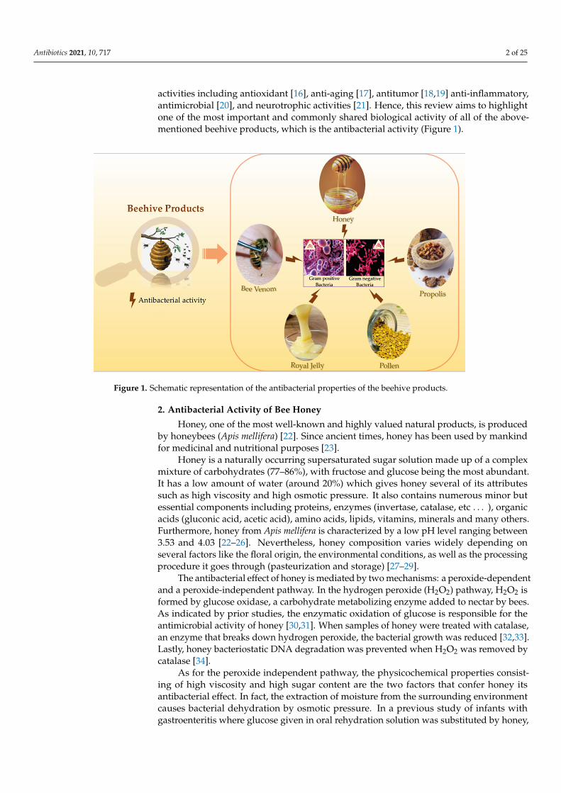
activities including antioxidant [16], anti-aging [17], antitumor [18,19] anti-inflammatory,antimicrobial [20], and neurotrophic activities [21]. Hence, this review aims to highlightone of the most important and commonly shared biological activity of all of the above-mentioned beehive products, which is the antibacterial activity (Figure 1).
Antibiotics 2021, 10, x FOR PEER REVIEW 2 of 24
Antibiotics 2021, 10, x. https://doi.org/10.3390/xxxxx www.mdpi.com/journal/antibiotics
and many microorganisms [4,11,12]. Moreover, pollen possesses antioxidant, anticoagu-lant and anti-inflammatory properties [13–15]. Also, royal jelly exhibits several interesting biological activities including antioxidant [16], anti-aging [17], antitumor [18,19] anti-in-flammatory, antimicrobial [20], and neurotrophic activities [21]. Hence, this review aims to highlight one of the most important and commonly shared biological activity of all of the above-mentioned beehive products, which is the antibacterial activity (Figure 1).
Figure 1. Schematic representation of the antibacterial properties of the beehive products.
2. Antibacterial Activity of Bee Honey Honey, one of the most well-known and highly valued natural products, is produced
by honeybees (Apis mellifera) [22]. Since ancient times, honey has been used by mankind for medicinal and nutritional purposes [23].
Honey is a naturally occurring supersaturated sugar solution made up of a complex mixture of carbohydrates (77–86%), with fructose and glucose being the most abundant. It has a low amount of water (around 20%) which gives honey several of its attributes such as high viscosity and high osmotic pressure. It also contains numerous minor but essential components including proteins, enzymes (invertase, catalase, etc…), organic acids (glu-conic acid, acetic acid), amino acids, lipids, vitamins, minerals and many others. Further-more, honey from Apis mellifera is characterized by a low pH level ranging between 3.53 and 4.03 [22–26]. Nevertheless, honey composition varies widely depending on several factors like the floral origin, the environmental conditions, as well as the processing pro-cedure it goes through (pasteurization and storage) [27–29].
The antibacterial effect of honey is mediated by two mechanisms: a peroxide-de-pendent and a peroxide-independent pathway. In the hydrogen peroxide (H2O2) pathway, H2O2 is formed by glucose oxidase, a carbohydrate metabolizing enzyme added to nectar by bees. As indicated by prior studies, the enzymatic oxidation of glucose is responsible for the antimicrobial activity of honey [30,31]. When samples of honey were treated with catalase, an enzyme that breaks down hydrogen peroxide, the bacterial growth was re-duced [32,33]. Lastly, honey bacteriostatic DNA degradation was prevented when H2O2 was removed by catalase [34].
As for the peroxide independent pathway, the physicochemical properties consisting of high viscosity and high sugar content are the two factors that confer honey its antibac-terial effect. In fact, the extraction of moisture from the surrounding environment causes
Figure 1. Schematic representation of the antibacterial properties of the beehive products.
2. Antibacterial Activity of Bee Honey
Honey, one of the most well-known and highly valued natural products, is producedby honeybees (Apis mellifera) [22]. Since ancient times, honey has been used by mankindfor medicinal and nutritional purposes [23].
Honey is a naturally occurring supersaturated sugar solution made up of a complexmixture of carbohydrates (77–86%), with fructose and glucose being the most abundant.It has a low amount of water (around 20%) which gives honey several of its attributessuch as high viscosity and high osmotic pressure. It also contains numerous minor butessential components including proteins, enzymes (invertase, catalase, etc . . . ), organicacids (gluconic acid, acetic acid), amino acids, lipids, vitamins, minerals and many others.Furthermore, honey from Apis mellifera is characterized by a low pH level ranging between3.53 and 4.03 [22–26]. Nevertheless, honey composition varies widely depending onseveral factors like the floral origin, the environmental conditions, as well as the processingprocedure it goes through (pasteurization and storage) [27–29].
The antibacterial effect of honey is mediated by two mechanisms: a peroxide-dependentand a peroxide-independent pathway. In the hydrogen peroxide (H2O2) pathway, H2O2 isformed by glucose oxidase, a carbohydrate metabolizing enzyme added to nectar by bees.As indicated by prior studies, the enzymatic oxidation of glucose is responsible for theantimicrobial activity of honey [30,31]. When samples of honey were treated with catalase,an enzyme that breaks down hydrogen peroxide, the bacterial growth was reduced [32,33].Lastly, honey bacteriostatic DNA degradation was prevented when H2O2 was removed bycatalase [34].
As for the peroxide independent pathway, the physicochemical properties consist-ing of high viscosity and high sugar content are the two factors that confer honey itsantibacterial effect. In fact, the extraction of moisture from the surrounding environmentcauses bacterial dehydration by osmotic pressure. In a previous study of infants withgastroenteritis where glucose given in oral rehydration solution was substituted by honey,

Antibiotics 2021, 10, 717 3 of 25
the recovery time of patients was significantly reduced [35]. This is likely due to the factthat the high sugar content in honey improves electrolyte and water reabsorption in theintestine. Moreover, the low pH level halts microbial growth [36,37]. Methylglyoxal (MGO)is another bioactive compound involved in non-peroxide antibacterial function. It is pro-duced by non-enzymatic conversion of dihydroxyacetone (found in high concentrationin the nectar of Leptospermum scoparium flowers) [38]. Manuka honey, a honey known topossess non-peroxide antibacterial activity due to MGO, is produced from the Myrtaceaefamily manuka tree, L. scoparium [39,40]. This honey has received a lot of attention fromscientists around the world due to the biological properties it has, notably its antioxidantand antibacterial properties. Furthermore, Manuka honey has been used in the treatmentof infections such as, surgical wounds, abscesses, traumatic wounds, burns, and ulcers ofvarious origins and has demonstrated its potency in inhibiting human pathogens and pre-vent the formation of biofilms [41–43]. In addition to the bioactive compounds responsibleof non-peroxide activity, an antimicrobial peptide present in honey called bee defensin-1acts against several Gram-positive bacteria such as Stephalococcus aureus, Bacillus subtilis,and Paenibacillus larvae, the main cause of the ravaging American bee larvae disease [37].
With the emergence and spread of resistant pathogens, the properties of honey couldbe potentially clinically relevant. Honey demonstrates a wide range of antibacterial activ-ity encompassing both Gram-negative and Gram-positive bacteria. In a previous study,Escherichia coli strains with ampicillin-resistance gene (β–lactamase) were found to besensitive to honey [44]. In addition, Mokaya et al. collected 16 different types of honeyfrom various Kenyan locations. They found that all of the 16 samples prevented E. coligrowth and had a significant amount of non-peroxide antimicrobial activity [45]. Anotherstudy examined two natural honeys against Methicillin-resistant Staphylococcus aureus(MRSA), vancomycin-sensitive enterococci (VSE), and vancomycin-resistant enterococci(VRE) and then compared their antibacterial activity to that of artificial honey. All MRSAstrains were resistant to artificial honey but sensitive to both honey types. As for VRE andVSE, both were susceptible to artificial and natural honey, with lower minimum inhibitoryconcentration (MIC) for natural honey [46]. These findings are in agreement with the studyof Katrina et al. where MRSA and VRE were sensitive to the selected honeys and showedthat the bacteriostatic effect was H2O2 dose-dependent [47].
In addition honey has been discovered to be a powerful inhibitor of Helicobacter pylori,causative agent of peptic ulcers [48].
As Manuka honey possesses an exceptional antibacterial activity, Brown et al. tried itagainst Staphylococcus pseudintermedius which cause serious infections in domesticated ani-mals and can be transmitted to their owner. They demonstrated that Manuka honey is notjust effective against novel multidrug-resistant S. pseudintermedius isolates, but additionallyworks in tandem with clinically important antibiotics and reduces its virulence [49].
A previous study has assessed the capacity of honey to prevent the adherence ofSalmonella interitidis to the intestinal epithelial cells in vitro and has demonstrated thathoney at dilutions 1:8 diminished the bacterial adherence from 25.6 ± 6.5 (control) to6.7 ± 3.3 bacteria/epithelial cell [50].
Furthermore, another study found that a 10% concentration of honey could preventoral bacteria like Streptococcus mutans from forming a biofilm, implying that honey couldhelp to minimize oral pathogens in dental plaque [51]. Honey was also effective againstbiofilms produced by methicillin-susceptible S. aureus (MSSA), MRSA, and Pseudomonasaeruginosa, with bactericidal rates of 63–82%, 73–63%, and 91–91%, respectively, whichwere higher than the effect of widely used single antibiotics [52].
In another study, honey demonstrated antibacterial activity against Burkholderia cepacia,which is responsible of pulmonary infections especially in patients with cystic fibrosis andchronic granulomatous disease. Twenty strains of B. cepacia isolated from the sputum ofpatients with cystic fibrosis were screened for their susceptibility to eight antibiotics andtwo types of honey (with and without peroxide activity). All strains were resistant to theantibiotics tested but susceptible to honey at concentrations less than 6% (v/v) [53]. In ad-

Antibiotics 2021, 10, 717 4 of 25
dition, 13 honeys samples from different plant source were tested against P. aeruginosa andagainst E. coli at different concentrations (10%, 5%, 2.5%, and 1% w/v). All honey samplesdisplayed an inhibitory effect on the growth of P. aeruginosa and E. coli for concentrationsabove 2.5%. Only four samples were still effective at a concentration of 2.5%: one againstE. coli and three against P. aeruginosa. No activity was observed at concentrations below2.5% [54].
The antimicrobial effect of honey has been tested against two Gram-positive organisms:Streptococcus pyogenes and S. aureus. Four honey samples were taken from beekeepersand tested at various concentrations (undiluted honey, 10 %, 30%, 50% and 70% w/v),then compared to standard antibiotics. Honey samples exhibited antibacterial activitywith a diameter of the zone of inhibition (ZDI) ranging between 0 and 46 mm for S.aureus and between 0 and 44 mm for S. pyogenes [55]. Other studies also indicated theantibacterial activity of honey against human pathogens and foodborne pathogens such asE.coli, Klebsiella pneumonia, Salmonella spp., etc . . . [33,56].
Honey can be utilized to treat a wide range of oral diseases, including periodontaldisease which is caused by Porphyromonas gingivalis, a Gram-negative bacterium. A previ-ous study has shown that honey helps to prevent periodontal disease by killing anaerobicbacteria [57].
It is worth mentioning that the antibacterial activity of honey differs widely dependingon the plant source [29,54]. Also, adulteration, thermal treatment, and long-term storagecan all affect honey’s antibacterial function [58].
Fuertes et al. have explored the repressive mechanism of honey on the growth of bac-teria. They found that when E. coli and S. aureus were exposed to different honey samples,physiological changes in membrane integrity and polarization occurred. Honey also causeda major metabolic disturbance in S. aureus as a primary physiological consequence [59].
Comparing the antibacterial potency of honey to that of antibiotics was also reported.A recent study compared the activity of three honeys with gentamicin against E. coli andP. aeruginosa. None diluted honey and its 1:2 to 1:6 aqueous dilutions had 100% and96.4% activity against P. aeruginosa and E. coli, respectively. Gentamicin, on the other hand,showed lower antibacterial activity when used at concentrations of 8.0 and 4.0 g/mL [60].
Combining honey with other antibacterial compounds increases their inhibitory rateagainst microorganisms. In fact, when honey was combined with the gentamicin, the killingrate increased to more than 92–93% while the killing rate was around 77% for gentamicinalone and around 45% for honey alone [61]. Another study attempted to combine honeywith ethanolic extract of cinnamon bark and found that the mixture had additive activityagainst acne-causing bacteria [62]. More studies have shown that the combination of honeywith other bioactive compounds has increased its antibacterial activity [41,63–65].
Concerning clinical trials on honey, Blaser et al. were reported a full recovery in sevenconsecutive patients with MRSA infected or colonized wounds. Alternatively, antisepticsand antibiotics had previously failed to eradicate infection-related symptoms [66]. Fur-thermore, another study has demonstrated that consuming honey at least once a weekconsiderably reduced the chance of H. pylori infection in a group of 150 dyspeptic pa-tients [67]. Also, in a clinical trial involving 90 patients with infected wounds, non- gammairradiated honey was associated with gradual decrease of bacterial load over a period of4 weeks [68].
Studies used different methods to measure the antibacterial activity of honey. Themost common methods were agar diffusion assay and serial dilution approach in microtiterplates. The first method, while being effortless and quick in performance, presents manylimitations: (1) difficulty in loading a specific volume of the product sample into the agarwells due to the high viscosity of honey, (2) diffusion issues with active components suchas defensin-1 and glucose oxidase, which have a large molecular weight across the agarmatrix, (3) low reproducibility, (4) difficulty in comparing the results with those of otherauthors, (5) inability to discriminate between bacteriostatic and bactericidal activity. Forthe second method, serial dilution assay, the bacteriostatic (MIC) and bactericidal activity

Antibiotics 2021, 10, 717 5 of 25
(MBC) of examined honey samples can be determined quantitatively using this approachin contrary to that of the first method. The only challenge that this method presents is thepreparation of honey output solution [69].
In light of these findings, honey appears to exert wide antibacterial activity againsta variety of microorganisms. It has been suggested in the literature that these propertiesmake the development of honey-resistance unlikely and extrapolate that honey may havean important clinical utility in the treatment of antibiotic-resistant bacteria [70–72]. Also,the use of honey as an antibacterial agent has no negative side effects for patients, in con-trast to antibiotics, and is cost effective [69]. Finally, honey may present some limitationswhen used as a treatment, like the presence of some toxic substances such as herbicides orpesticides in it, as well as its possible contamination by clostridium endospores. Therefore,it is crucial to follow specific criteria and standards when using honey as a therapeuticagent [23,73]. It is also mandatory to create a sterilizing procedure that is safe for honey pro-teinaceous antibacterial components (glucose oxidase, bee defensin-1). Gamma-irradiationsterilization procedure appears to be promising [74]. Also, it is important to agree onone standard method to determine the honey’s antimicrobial activity, making it easy tocompare the activity of the product evaluated in different studies.
3. Antibacterial Activity of Bee Venom
Bee venom (BV) is a widely recognized toxin secreted by female worker bee’s poisonglands as a protection mechanism [75]. Although it is toxic to predators, BV has been usedfor many medicinal purposes since Ancient Egypt (4000 BC). In bee venom therapy (BVT),the toxin is applied directly or indirectly into the body for the treatment of certain diseases,such as rheumatism arthritis [76–78]. Venoms from different animals or organisms areconsidered as promising antimicrobial agents that work against several bacteriologicalpathogenesis [79]. The antimicrobial activity and medical use of BV is due to the presenceof bioactive molecules in particular peptides which are the main components and constitute48–50% of dry BV weight [80]. The Apis mellifera venom is an odorless and transparentliquid that is made up of 88% of water and only 0.1 µg of dry venom [78]. The dry venomitself is highly rich in peptides notably melittin, apamin, adolapin, and mast cell degranu-lating (MCD) peptide, enzymes such as phospholipase A2 (PLA2) and hyaluronidas [81,82].Among these peptides, melittin represents the main biological active compound in beevenom. It represents about 50% of the dry BV [83]. Another major component is phospho-lipase A2, being the most toxic bee venom peptide [84]. These two components can actsynergically and damage the cell membrane [8]. Melittin possesses a low selectivity to thecell membrane and acts strongly on its lipids via the process of pore formation. This processcauses the release of the cell cytoplasmic contents and leads to cell lysis [85]. This modeof action is most likely to be responsible of the antibacterial properties of BV [86]. Severalstudies have demonstrated the ability of BV to kill both Gram-negative and Gram-positivebacteria [87,88]. Indeed, the antibacterial activity of BV from Apis mellifera purebred as wellas hybrid was tested against five bacterial strains. The results showed that BV displayedan antibacterial activity against all five bacterial strains. Other studies proved that BV canbe used as a complementary antimicrobial agent against pathogenic bacteria even if it iscollected by different methods (i.e., from the top of the frames or from under the frameswith distance between the bottom board and the brood chamber) [89]. Additionally, thebacterium Borrelia burgdorferi is known to be the main cause of Lyme disease. Researchersfound that both BV and melittin possess significant effects on all the morphological formsof B. burgdorferi, even on antibiotic resistant attached biofilms. However, antibiotics whenadministrated alone or combined together had limited effects [90]. Food-borne pathogenspresent a risk to develop antimicrobial resistance and generate biofilms such as Salmonella.Sixteen Salmonella strains isolated from poultry were tested against apitoxin (Bee venom).In 14 of the 16 tested strains, apitoxin reduced the biofilm formation and destroyed thepreformed biofilm by 47%. Also, apitoxin increased the mobility of the bacteria and theMIC varied between 1024–256 µg/mL [7]. Aiming to find an alternative to antibiotics in the

Antibiotics 2021, 10, 717 6 of 25
poultry industry, broiler chicks were sprayed with BV to evaluate its immunoprophylacticeffects against Salmonella gallinarum. The results showed that BV increased the antibodyproduction against formalin-killed S. gallinarum [91].
Others previous studies focused on the antibacterial effect of bee venom against ad-ditional type of bacteria such as S. aureus and E. coli. The results proved BV to be activein killing both bacterial strains. At 10 × MIC, the regrowth of E. coli was not observedat 18 h while For S. aureus at 5 × MIC [92,93]. Regarding the MIC for Gram-positivestrains, the range was from 200 µg/mL to 8 µg/mL for the most sensitive species B. subtilis.Alternatively, Gram-negative strains were found to be more resistant to BV (MIC 60 to>500 µg/mL). This can be explained by the nature of the bacterium cell membrane [87,93];the outer membrane of Gram-negative bacteria encompasses lipopolysaccharides (LPS)which obstruct the penetration of melittin presented in the BV. Thus, making melittinresponsible for the antibacterial activity. While in Gram-positive bacteria, LPS are absent,indicating that melittin can penetrate the cell membrane more easily to form pores andincreases the cell permeability through the cytoplasmic membrane [94]. PLA2, which is alsopresented in abundance in the crude BV can disrupt the cell membrane of Gram-negativebacteria indirectly by hydrolyzing phospholipids enzymatically. Other works reportedthe antibacterial activity of the crude BV and its two main biopeptides melittin and PLA2against oral pathogen responsible of tooth decay: Lactobacillus casei, Streptococcus salivar-ius, S. mutans, Staphylococcus mitis, Staphyslococcus sobrinus, Staphylococcus sanguinis andEnterococcus faecali. Melittin was twice as active as the crude BV against the tested bacteria(4 to 40 µg/mL). As for PLA2, it was only active against L. casei at >400 µg/mL [95,96].Similarly, melittin showed antimicrobial activity against Staphylococcal strains as wellas MRSA strains while PLA2 did not show any effect on the same strains [97]. Also, ithas been demonstrated that certain amino acids and their positions play a crucial rolein the antibacterial activity exerted by melittin. For example, the absence of a prolineresidue in position 14 significantly diminishes the antimicrobial activity of melittin [98].Likewise, two synthetic melittins, one asparagine-substituted melittin (Mel-N) and theother serine-substituted melittin (Mel-S) were able to penetrate the membrane of E. coli butthe latter was more efficient [99]. Intriguingly, the antibacterial mechanism of new melittinderived-peptides MDP1 (GIGAVLKVLTTGLPALIKRKRQQ) and MDP2 (GIGAVLKWLPA-LIKRKRQQ) was evaluated against multidrug resistant strains such as E. coli, P. aeruginosaand MRSA. The two derived peptides showed strong antibacterial activity against referencestrains of the three multidrug resistant strains compared to the indigenous melittin viamembrane alteration and damage [100].
On the other hand, antimicrobial treatments are becoming less efficient by the day dueto the increasing number of multi-drug resistant (MDR) bacteria as well as bacterial biofilmswhich are very hard to treat. Between 317 positive specimens for bacterial growth, 124 wereMDR isolate of Gram-negative and Gram-positive bacteria. BV demonstrated a completegrowth inhibition percentage (100%) against all tested MDR-isolates with a wide range ofMICs and MLCs concentration-ranging between 3.125–50 µg/mL and 6.25–100 µg/mL,respectively against all MDR-GNB and GPB one [101]. The antibacterial activity of BVwas tested alone or in combination with antibiotics (ampicillin, gentamicin, penicillin, orvancomycin) on the growth of MRSA strains. A partial synergetic effect was indicated bythe index of inhibitory concentration ranging from 0.631 to 1.002 for the combination ofBV with ampicillin or penicillin which made the MRSA strains more susceptible to thecombination of BV with gentamicin or vancomycin rather than the combination of BVwith ampicillin or penicillin [102]. BV was studied for its antibacterial activity against fourMDR-Acinetobacter baumannii bacterial strains. Results showed an inhibition of the bacterialgrowth of MDR-A. baumannii strains with MIC value 31.25 mg/mL [103]. Melittin combinedwith doripenem caused a significant decrease in the MIC of MDR- A. baumannii strains. Thecombination of melittin–ceftazidime and melittin–doripenem was administrated to MDR-P.aeruginosa strains. This caused a decrease in melittin dosage which led to a decrease inmelittin cytotoxicity. Therefore, the combination of melittin–doripenem (for A. baumannii)

Antibiotics 2021, 10, 717 7 of 25
and melittin–doripenem with melittin– ceftazidime (for P. aeruginosa) at their MIC dosemight serve as a promising therapeutical treatment against MDR bacteria [104].
The investigation of BV in pre-clinical and clinical application is still very slow de-spite its efficacity when combined with other drugs to treat different types of bacteria.Researchers found that BV and melittin combined with other antibiotics like vancomycin,oxacillin, and amikacin yielded fractional inhibitory concentration (FIC) indices fluctuatingbetween 0.24 and 0.5. Apitoxin and melittin tested against 51 strains of bacteria exhibitedstrong antibacterial activity against both Gram-negative bacteria (MIC values between 30and >500 µg/mL) and Gram-positive (MIC values between 10 and 100 µg/mL). Moreover,a strong anti-MRSA and anti-VRE activity was shown by BV and melittin at MIC values of6–800 µg/mL [87]. When combined with oxacillin, BV and melittin showed an antibacterialactivity against MRSA ATCC 3359 [84]. The antibacterial activity of melittin as well as itssynergistic effect with β-lactam antibiotics against A. baumannii was evaluated by meansof the broth microdilution method. The MIC value of melittin was 4 µg/mL and resultsshowed inhibition of bacterial growth. Furthermore, FIC indices for combination of melittinwith co-amoxiclav, ceftazidime and imipenem showed a synergetic effect [105].
All of the above- mentioned information confirm that BV and its two major com-pounds melittin and PLA2 exhibit a very promising antibacterial activity against severalpathogenic bacterial strains even against multidrug resistant bacteria. However, one shouldalways keep in mind that these biopeptides are venomous and might act as toxic agentsif administrated the wrong way. Melittin is the most toxic component in BV. While beingresponsible for the majority of the pain associated with bee stings, it only induces a minorallergic reaction. Contrary to PLA2 which is the most allergenic and immunogenic proteinin bee venom [106,107]. Bee envenomation can lead to many clinical manifestations suchas local inflammatory reactions, allergic reactions, anaphylactic shocks, and systemic toxicreactions [108]. The ideal treatments against the toxic effect of bee venom are corticos-teroids, antihistamine, and intramuscular adrenaline depending on the severity of the beeenvenomation [109,110].
Many clinical studies on bee venom have shown promising results. Acne, one of themost common problems among teenagers, is usually treated with antibiotics to kill thecausative bacteria but in some cases bacterial resistance can occur [111]. In a clinical studyconducted by Han et al., skincare products with or without were tested on 12 subjectssuffering from acne. The results showed a 57.7% decrease in ATP measured to evaluate thedecrease in skin microbes. Thus, cosmetics and skincare products containing BV can usedas a promising therapeutic anti-acne agent [112].
4. Antibacterial Activity of Propolis
Among the many products of honeybees, propolis is considered as one of the mostinteresting products utilized as the main defensive element and building block in the hives.Propolis is used by bees to fill the cavity in the walls of the hive, it is also used to repaircombs and strengthen its thin borders. On the other hand, propolis can mummify intrudersthat cannot be transported outside thus preventing their decay [113]. Since ancient times,the anti-putrefactive property has been used by humans, notably by Egyptians. Theorigin of propolis, also known as “bee glue”, had long been a subject of discussion, somethought it came from bees themselves while others thought it originated from plants.Nowadays, the approximate composition of propolis and the factors affecting it becameclearer [114]. Propolis is now confirmed to be a bee product made from plants. Beescollect resinous vegetable matter from different parts of the plant such as lipophilic matteron leaves and their buds, latex as well as mucilage [115]. Also, the bees are able tocut the fragments of vegetative tissues in order to extract the products necessary forthe production of propolis [116]. Furthermore, propolis is a complex mixture with avariable composition that depends on the geographical region and the plant species usedin its production. Surprisingly, having different composition does not indicate differentbiological activities [12]. Propolis is a resin that can occur in different colors and exhibits

Antibiotics 2021, 10, 717 8 of 25
a pleasant aromatic resin smell of great value. Propolis is mainly composed of resins,flavonoids, polyphenols, terpenoids, essential oils, and other organics and minerals [117].The extraction of propolis and its dissolution is needed in order to release its most activeingredients. The extraction process is also required for the later usage of propolis formedicinal purposes. It is also important to mention that solvent types used in extractioncan affect the biological activity obtained from propolis. For the antibacterial activity, theabundancy of flavonoids and phenols plays a key role in conferring this bioactivity [118].Additionally, the presence of many bioactive ingredients and in different concentrationsis crucial in preventing the occurrence of bacterial resistance [119]. As for the mechanismof action, propolis can act directly on the microorganism or indirectly by triggering theimmune system which results in the activation of the body natural defense mechanism.Studies showed that Gram-negative bacteria are more resistant to propolis than Gram-positive bacteria. This can be explained by two main factors, the first one being the specificstructure of the Gram negative bacteria membrane and the second one being the secretionof hydrolytic enzymes which destroy the active ingredients present in propolis [120].
Artepillin C (3, 5-diprenyl-p-coumaric acid) is considered as one of the most impor-tant phenolic compounds found in propolis. Propolis ethanolic extract showed higherantibacterial activity against MRSA in comparison with hexane extract due to the higherconcentration of Artepillin C in ethanolic extract [121]. The antibacterial activity of ethanolextracted propolis and its derivative compounds was investigated against P. gingivalis,a bacterium responsible for periodontal diseases. Artepillin C showed a bacteriostaticeffect with membrane blebbing [122]. Propolis contains other phenyl derivatives such as2-dimethyl-8-prenylchromene and 3-prenylcinnamic acid allyl ester. After investigatingthe chemical constituents and the antibacterial activity of the ethanolic extract of propolis,the results showed a high concentration of p-coumaric acid, artepillin-C, drupanin andkaempferide alongside an antibacterial activity against Listeria monocytogenes, Enterococcusfaecalis, S. aureus and Staphylococcus saprophyticus [123]. Also, propolis contains pinocembrinand apigenin. Chilean propolis was subjected to a study and researchers found that theantibacterial activity of pinocembrin and apigenin is higher than that of polyphenols mix-ture or even chlorhexidine against S. aureus [124]. Numerous work reported the presenceof antibacterial activity of pinocembrin against different bacterial strains such as S. mutans,S. sobrinus, S. aureus, E. faecalis, L. monocytogenes, P. aeruginosa and K. pneumoniae [125,126].
Nepalese ethanolic extract of propolis was evaluated for both its antimicrobial activityand its chemical composition. The main components were flavonoid aglycones (mainlyneoflavonoids, isoflavonoids) and pterocarpans. Nepalese propolis exhibited its highestantibacterial activity against H. pylori, S. aureus and Shigella flexneri. When combined withamikacin and tetracycline, the same propolis yielded the strongest effect against S. au-reus [126]. The ethanolic extract of polish propolis (EEPP) was examined for its antibacterialactivity against MRSA and MSSA. The average MIC was 0.54 mg/mL and EEPP waseffective against 12 S. aureus strains. Also, EEPP combined with 8 antistaphylococcal drugs,provided a stronger antibacterial effect against the tested strains than EEPP alone [127].The geographical location affects the composition of propolis as well as its antimicrobialactivity. 40 ethanol extract of propolis (EEP) collected worldwide were evaluated for theirantibacterial activity against S. aureus strains. Results showed a moderate activity forAsian and African samples with MICS ranging from 0.0156 to >0.5 mg/mL and 0.0078to >0.5 mg/mL, respectively with a similar results displayed by samples collected fromNorth and South America and Europe [128]. EEP samples from three different regions ofturkey were analyzed. MIC values varied and amounted to 0.018, 0.162, and 0.101 mg/mLand samples showed high antibacterial activity [129].
The number of studies concerning the antibacterial effect of propolis against anaer-obic bacteria remains limited. However, it has been presented that propolis is moreactive on Gram-positive anaerobic bacteria than on Gram-negative ones. Propolis showedantibacterial activity against Lactobacillus acidophilus, Prevotella oralis, P. gingivalis, Fu-sobacterium nucleatum, Peptostreptococcus anaerobius, Actinomyces naeslundii and Veillonella

Antibiotics 2021, 10, 717 9 of 25
parvula [130]. Moreover, research shows an important activity of propolis against Porphy-romona, Fusobacterium, Propionibacterium, Clostridium, Prevotella, Actinomyces and Bacteroidesspecies [131,132].
Propolis displays different antibacterial mechanisms such as inhibition of cell division,collapse of membranes and cell walls of the microbial cytoplasm, decreasing bacterialmotility, enzymatic inactivation, bacteriolysis and inhibition of protein synthesis [133]. Allthe different compounds that constitute the propolis composition are rich in polyphenolswhich work together with different microbial proteins to form both ionic and hydrogenicbonds. This contributes to the modification of the three-dimensional structure of proteinshence the alteration of their function. These effects have encouraged researchers to com-bine propolis with antibiotics in order to overcome the problem of bacterial resistanceto drugs [134]. A synergy has been observed between EEP, vancomycin and oxacillin;antibiotics that inhibit the synthesis of the bacterial cell wall, against S. pyogenes, VREATCC 51299 and MRSA NCTC 10442 [135]. Also, synergism was present between EEP anddrugs targeting microbial ribosomes (neomycin) but absent with drugs interfering in thebiosynthesis of folic acid or DNA (ciprofloxacin and norfloxacin) as well as those inhibitingmetabolic pathways (cotrimoxazole) [134]. EEP has proved to be the most effective whencombined with antibiotics that interfere with bacterial protein biosynthesis such as tetracy-cline, linezolid, chloramphenicol, gentamicin, tobramycin and netilmicin against MRSAand MSSA [127].
Propolis is widely used in medicine due to its various bioactivities. The effect oftopical propolis extract was tested on facial vulgaris acne where subjects were treatedwith a topical solution of ethanolic extract of propolis. This solution showed a significantbacteriological activity on Propionibacterium acnes and Staphylococcus epidermidis, as wellas being effective against facial acne [136]. To test the efficiency of propolis in inhibitingdental caries, propolis fluoride was applied to children’s teeth with active dentinal carriessurface. Carries were stopped in 99.80% of the cases after just one month of applicationwithout causing any black discoloration of the teeth [137]. In another study, researcherstreated patients suffering from chronic periodontitis with an irrigation of the selectedsites with a hydro alcoholic solution of propolis extract propolis. Results showed thatpropolis exhibits a considerable activity against chronic periodontitis. This treatment wasmore effective than the conventional treatment as it significantly decreased the viability ofP. gingivalis [138].
5. Antibacterial Activity of Bee Pollen
Pollen of various flowers is collected by field bees and agglutinated by bees enzymessecreted by the salivary glands and by the nectar. Then, the pollen is placed in corbiculaesituated on the tibia of the bees hind legs to form the pollen loads, commonly known asthe bee pollen, in the form of granules [139]. Pollen is the raw material used by bees toproduce bee bread, the basis hives food [140]. Pollen stored in honeycomb cells is coveredwith a thin layer of honey and wax. This results in the formation of bee bread whichundergoes anaerobic fermentation and is preserved by the lactic acid which formed [141].In comparison to honey, small amounts of bee pollen are stocked in the hives and areused during forage absence [142]. Many studies have shown that bees collect pollen fromvery few plant species. Bee pollen contains about 250 substances including amino acids,lipids, vitamins, macro- and micronutrients, flavonoid, and organic carotenoid pigmentswhich makes it an ideal food [143]. Its composition varies depending on the biogeographicorigin, ecological habitat, season, weather conditions during collection, as well as on thebee race and even beekeeping management [144]. Phenolic compounds are responsible ofthe bee pollen bioactivity [141]. For centuries, bee pollen has been used for its medicinaland nutritional properties. Likewise, many ancient philosophers highly valued pollen as apart of their healthy diet [15].
Bee pollen is often collected by using a pollen trap at the entrance or in the hives.Sometimes, it is directly collected from the bee hind legs [145]. Water, ethanol and methanol
Antibiotics 2021, 10, 717 10 of 25
are the most commonly used solvents for the extraction of bee pollen. Studies showed thatthe highest content of bioactive compounds is present in ethanol or water extracts in com-parison to natural bee pollen [146]. The antibacterial activity of bee pollen was investigatedand results showed a positive effect on both Gram-positive and Gram-negative bacteriawith a higher sensitivity in Gram positive bacteria. A study on the antibacterial activity ofMoroccan bee pollen demonstrated an inhibition effect against S. aureus, Streptococcus spp.,E. coli and P. aeruginosa with a higher sensitivity in Gram-positive bacteria [145]. Similarly,Bacillus cereus, S. aureus, S. typhi and E. coli were sensitive to Portugal bee pollen [147].Applying a concentration of 0.02% to 2.5% (vol/vol) of bee pollen extract to B. cereus, B.subtilis, E. coli, Salmonella typhimurium, S. aureus, Yersinia enterocolitica, E. faecalis and Listeriamonocytogenes did not exert any antimicrobial effect on these bacterial strains indicatingthat the activity is concentration dependent [148]. The antimicrobial activity of bee pollenwas also assessed on Clostridia class organisms, i.e., Clostridium butyricum, Clostridiumhystoliticum, Clostridium intestinale, Clostridium perfringens and Clostridium ramosum. Resultsshowed an antibacterial activity against all of the above mentioned strains of clostridiawith that against C. butyricum and C. perfringens being the most potent one [149]. Theantimicrobial activity of a Chilean bee pollen extract was investigated against humanpathogenic bacteria E. coli, P. aeruginosa, S. aureus and S. pyogenes showed a high sensitivityto the bioactivity of bee pollen in opposition to E. coli and P. aeruginosa [150].
Furthermore, ethanol and methanol extracts of Slovakian bee pollen were tested fortheir antibacterial property against L. monocytogenes, P. aeruginosa, S. aureus, S. enterica andE. coli. Results showed that S. aureus was the most sensitive bacteria to the ethanolic beepollen extract while S. enterica was the most sensitive one to the methanolic extract [139]. E.coli, S. enteritidis, S. aureus, L. monocytogenes were tested with ethanol pollen extract. It wasfound that S. aureus and L. monocytogenes growth was inhibited while this was not the casefor both E. coli and Salmonella enteritidis which both showed resistance to ethanol extracts ofpollen [151]. In some studies, the nature of the solvent used might affect the antibacterialactivity of bee pollen where the same sample can be more effective against Gram-negativebacteria than Gram-positive bacteria. Slovenian bee pollen ethanol extract showed a higherantibacterial activity against Gram-negative bacteria (E. coli and Campylobacter jejuni) thanGram-positive bacteria (L. monocytogenes) [152]. The same result was observed for honeybeepollen collected from a region of Chile who revealed antibacterial activity against S. pyogenesonly and showed no activity against E. coli, S. aureus and P. aeruginosa [14]. Moreover, E.coli was the most sensitive strain to 70% ethanol bee pollen extract but resistant to 96%ethanol extract which confirms the fact that bacterial strains are specific to the solvent andits concentration [153].
The antibacterial mechanism of bee pollen remains unclear until now. Studies suggestthat the antibacterial activity of bee pollen is associated with glucose oxidase; an enzymeproduced by honeybees and added to pollen during the process of granules formation [154].Additionally, this activity might be correlated with the total phenolic content and phenolcomposition. Phenolic acids and flavonoids present in pollen can act against bacterialcells by degrading the cytoplasmic membrane which leads to loss of potassium ions andinitiation of the cell’s autolysis [148]. Other researchers argue that the composition ofthe pollen is associated with its antibacterial activity. This is most likely due to the factthat many bee pollen extracts with the lowest total phenol concentrations were the mosteffective against microorganisms [155,156]. In addition to the presence of many bioactivecompounds such as fatty acids, EXLVs and presumably microbial metabolites known fortheir antibacterial activity in bee pollen. Surprisingly, bee pollen exerts an inhibition effectagainst many pathogenic bacterial strains but does not act against lactic acid starter cultures.Hence, bee pollen could serve as a potential candidate that is more suitable than antibioticswhich kill both pathogens and probiotics [157].

Antibiotics 2021, 10, 717 11 of 25
6. Antibacterial Activity of Royal Jelly
Royal jelly (RJ), a yellowish white, sticky acidic secretion, is produced by worker beesand secreted from hypopharyngeal and mandibular glands. Bee larvae at early stages oftheir life rely on royal jelly as a primary source of food. Only the queen bee is fed by royaljelly until it dies, that’s why royal jelly is widely recognized as a ‘’super food” [158,159].
Water is the major molecule forming RJ (50 to 60%). In addition to water, RJ containsother components such as proteins (18%), carbohydrates (15%), lipids (3 to 6%), mineralsalts (1.5%), and other minor constituents such as free amino acids, vitamins (particularlyvitamin B) and phenols such as flavonoids [160].
RJ exhibits important antibacterial activity. This antibacterial potential of RJ is dueto the existence of some main bioactive constituents. Proteins play an important rolein the antibacterial activity of RJ. In fact, proteins are the major constituents of the drymatter of RJ (50% of the dry matter of RJ) [161–164]. The MRJPs glycoproteins familyform 82–90% of the protein constitution in RJ and has nine identified members MRJP1-MRJP9 [165]. MRJP1, also known as royalactin [166] or apalbumin [167], is the firstidentified and major protein in the MRJPs family [165]. It has been discovered that MRJP1possesses an indirect antibacterial activity. Moreover, there are jelleines, short antimicrobialpeptides (eight to nine amino acids), which have four different peptide sequences: jelleine-I (PFKLSLHL-NH2), jelleine-II (TPFKLSLHL-NH2), jelleine-III (EPFKLSLHL-NH2), andjelleine-IV (TPFKLSLH-NH2). Jelleine I, II, and III are derived from MRJP1 cleavageproduct and are responsible for the antibacterial activity of MRJP1 [168,169]. A previousstudy has tested jelleines I, II and III against Gram-positive (S. aureus, S. saprophyticusand B. subtilis) and Gram-negative bacteria (E. coli, Enterobacter cloacae, K. pneumoniae andP. aeruginosa). The results revealed that jelleines-I and II had a broad-spectrum activity,while jelleine-III was less active, and jelleine-IV had no antimicrobial activity [168]. Hence,jelleines I, II and III are responsible for the antibacterial activity of MRJP1. On another hand,MRJP2 (apalbumin2) exhibited an antibacterial activity against P. larvae, B. subtilis and E.coli. This antibacterial activity of MRJP2 is attributable to its high mannose carbohydratelevels and its complex-type antennary carbohydrate structures [170]. In addition, a studyhas demonstrated that the levels of MRJP3 (apalbumin 3) increased after bacterial infection.Future work should investigate the antimicrobial activity of this protein against a varietyof pathogens [171]. It is important to mention that the dominant allergens of royal jelly areMRJ1 and MRJ2, which could present a limitation to the usage of royal jelly as a therapeuticagent [172].
Royalisin, another antibacterial protein found in RJ, prevents RJ from becoing contam-inated and infected by Gram-positive bacteria. Royalisin has been shown to have antibacte-rial activity against Gram-positive bacteria but not Gram-negative bacteria [168]. However,it has been demonstrated that royalisin plays a role in the honeybee protection systemagainst bacterial invasion especially against P. larvae, the main pathogen of Americanfoulbrood disease. Royalisin also acts on other Gram-positive bacteria like Staphylococcus,Streptococcus, B. subtilis, Sarcina lutea, Micrococcus luteus and Gram-positive rods such asCorynebacterium, Clostridium, Lactobacilus helveticus and Leuconostoc. However, no inhibitionof Gram-negative bacteria like E. coli and Serratia marcescens was detected [173–175].
A study was conducted with the aim to assess the amount of royalisin in RJ obtainedfrom honeybee colonies with different genotypes. It was found that there is an obviouslink between the amount of royalisin and the antibacterial potential of RJ. Furthermore, theamount of royalisin in RJ was discovered to differ between colonies [176].
The mechanism of action of the majority of RJ peptides remains unrecognized. For thefirst time, the analysis of RAcc-Royalisin (recombinant protein RAcc-Royalisin expressedand purified from E. coli) showed that royalisin inhibits bacterial growth by disruptingthe permeability of the cell membrane as well as reducing the hydrophobicity of bacterialcells [177].
Moreover, trans-10-hydroxy-2-decenoic acid (10-HDA) fatty acid, is the most abundantcomponent in the lipids fraction (80%) and can only be found in RJ [178]. Unlike other

Antibiotics 2021, 10, 717 12 of 25
acids, which have 14–20 carbon atoms, RJ fatty acids are shorter (8–10 carbon atoms).Furthermore, RJ fatty acids are either dicarboxylic or hydroxy acids. Other fatty acids, onthe other hand, are normally triglyceride fatty acids [179]. Thus, this particular fatty acidmay be used to distinguish RJ from other bee products and to confirm the authenticity offormer [180]. Early studies on 10-HDA antimicrobial activity found that 10-HDA has ahigh capability to inhibit the growth of both Gram-negative bacteria (E. coli) and Gram-positive (Micrococcus pyogenes, B. subtilis, S. aureus) [181]. Also, a comparative study of theantimicrobial capacity of the ether-soluble fatty acids, which primarily contain 10-HDA,and the ether-insoluble fractions, which include royalisin, was also performed. Severalpathogenic bacteria were tested to see if these fractions had antibacterial properties againststrains of Streptomyces species, S. aureus and E. coli. The results show that at concentrationsof 30 mg/mL, ether-soluble fractions of RJ strongly reduced the growth of the previouslydescribed microorganisms, while non-soluble fractions had almost no antimicrobial ac-tivity [182]. Furthermore, glucosyltransferases (gtfs) genes, especially gtfB and gtfC, areessential in S. mutans colonization and pathogenesis. Yousefi et al. discovered that 10-HDAinhibits gene expression and mRNA transcription while also effectively reducing S. mutansadhesion to the cell surface [183].
Milliou and Chinou reported other fatty acids that displayed an antibacterial activity.Oral pathogens S. mutans and Streptococcus viridans, as well as S. aureus and S. epidermidis,were efficiently inhibited by 3-hydroxydodecanedioic acid, 10-acetoxy-2-decenoic acid,and (11S)-hydroxydodecanoic acid. However, most of these fatty acids occurred to beineffective against Gram-negative bacteria. Moreover, 3-hydroxydodecanedioic acid, witha MIC of 0.47 mg/mL, appeared to have a minor effect on K. pneumonia [184].
Consequently, some of MRJPs, jelleines, royalisin and 10-HAD may act synergicallyand confer the RJ an effective and promising antibacterial activity.
Raw RJ has been tested against human pathogens in many studies. Two RJ samples,from Argentina, A and B were tested against Gram-positive and Gram-negative bacteriathat cause infections in cutaneous wounds. Both samples had the ability to inhibit Gram-positive and Gram-negative bacteria except for K. pneumoniae which was not inhibited byneither samples. Sample A also had no effect on Streptococcus uberis [185]. Differences inactivities between samples of RJ could be linked to the differences in geographical locationsand changes in components due to the genetic variations between bee colonies [185].Another study has tested RJ against Gram-positive (M. luteus, B. cereus and S. aureus) andGram-negative bacteria (S. flexneri, S. typhi, E. coli, P. vulgaris and P. aeruginosa). Resultsdemonstrated that RJ exhibited an antibacterial activity and had two distinct forms ofeffect, one being bactericidal and the other being bacteriostatic against the studied bacteria.Likewise, they investigated the impact of storage time on RJ antibacterial activity. Within24 h of being frozen, the highest antibacterial activity of RJ was observed. This activitydecreased gradually over time until it reached a steady value [186]. A recent study hascompared the activity of RJ to chlorhexidine (gold standard) against the periodontopathicbacteria (aerobic and anaerobic) in subgingival plaque. It was found that chlorhexidine hasa greater inhibitory effect than RJ. These results indicated that RJ should be administratedat high concentrations if it was to be used as an alternative to chlorhexidine [187].
The combination of RJ with other compounds was also studied in order to enhancethe effectiveness of RJ antibacterial activity. Boukraa et al. investigated the effect of mixingRJ with starch. They found that starch alone does not have an antibacterial activity. RJ MICagainst S. aureus and E. coli was 1.7% (v/v) and 2% (v/v), respectively. When mixed withstarch, the MIC decreased by 61% and 30% against S. aureus and E. coli, respectively. It wassuggested that this decrease in MIC values is due to the existence of amylases in RJ whichmight be a possible explanation for the increase in the antibacterial activity after addingstarch. This enzyme breaks down starch into dextrin and maltose, increasing RJ osmoticcapacity and, as a consequence, its antibacterial impact [188]. Surprisingly, when RJ wasmixed with honey, which is known to have a strong antibacterial activity, it was found thatMIC values decreased significantly against P. aeruginosa [189].

Antibiotics 2021, 10, 717 13 of 25
Furthermore, Romanelli et al. found that when jelleine-I was combined with temporinA or temporin B (peptides from the temporin family that are active against Gram-positivebacteria at low concentrations), the antibacterial activity of jelleine-I was increased againstS. aureus and L. monocytogenes [190].
7. Conclusions
The use of beehive products for medicinal purposes has long been exploited bymankind. Here, the antibacterial properties of honey, bee venom, propolis, pollen androyal jelly have been discussed. Each of these products is characterized by the presence ofbioactive compounds which makes the formers powerful inhibitors of pathogenic bacterialstrains. Table 1 summarizes all the bacterial strains susceptible to honeybee productswith the MIC and MBC determined. Many clinical trials are being conducted to test thesafety and effectiveness of beehive products on infected wounds as well as on variousbacterial diseases such as gastroenteritis. Studies showed that bee venom exhibits bothantibacterial and anti-inflammatory activity and can be used in acne treatment, and propoliswas demonstrated to be efficient in both skin and dental treatment. Finally, although beeproducts are considered as promising and potent antibacterial candidates, future workshould emphasize on their possible adverse effects when applied or administrated directlyto the human body.
Table 1. Summary of all the bacterial strains susceptible to each honeybee product. The minimum inhibitory effect(MIC) as well as the minimum bactericidal concentration (MBC) of each of the beehive products are also shown (ND:none determined).
Honeybee Product Bacterial Strains MIC/MBC Reference
Honey
Raw honey
Manuka honey Shigella sonnei MIC 9%(v/v)[191]
Talah honey MIC 20%(v/v)
Saudi honey S. flexneri MIC 10–20%(v/v) [192]
Canadian honey
E. coli
MIC 6.25% (w/v)MBC 6.25–12.5%(w/v) [44]
Saudi honey MIC 20–40%(v/v) [192]
Korean honey MIC 25–50%(w/v) [193]
Omani and African honey ND [194]
Ulmo honey MIC 12.5%(v/v) [195]
Manuka honey
MRSA
MIC 9.98%(v/v)[46]
Pasture honey MIC 3.07%(v/v)
Ulmo honey MIC 3.1–6.3%(v/v) [195]
Turkish honey
S. aureus
ND [196]
Saudi honey MIC 20–40%(v/v) [192]
Brazilian honey MIC 126.23–185.70 mg.mL−1 [197]
Korean honey MIC 25–50%(w/v) [193]
Omani and African honey ND [194]
Algerian honey MIC 12–95%(v/v) [55]
Saudi honey Staphylococcus epidermis MIC 20–40%(v/v) [192]
Manuka honey vancomycin-sensitiveEnterococci
MIC 4.92%(v/v)
[46]Pasture honey MIC 9.66%(v/v)
Manuka honey vancomycin-resistantEnterococci
MIC 4.61%(v/v)
Pasture honey MIC 8.25%(v/v)

Antibiotics 2021, 10, 717 14 of 25
Table 1. Cont.
Honeybee Product Bacterial Strains MIC/MBC Reference
Manuka honey Coagulase-negativestaphylococci
MIC 3.4%(v/v)[198]
Pasture honey MIC 3.6%(v/v)
Manuka honey Campylobacter spp. MIC ≈ 1% (v/v) [199]
Turkish honeyH. pylori ND
[196]
Cider honey [48]
Turkish honey B. subtilis ND [196]
Saudi honey Proteus mirabilis MIC 10–20%(v/v) [192]
Korean honey L. monocytogenes MIC 25% (w/v)[193]
Korean honey Salmonella typhymurium MIC 25–50% (w/v)
Omani honey
P. aeruginosa
ND [194]African honey
Ulmo honey MIC 12.5% (v/v) [195]
Tualang honey MIC 18.5% (w/v)MBC 25% (w/v) [200]
Manuka honey MIC 7.5%(v/v)MBC 9.71%(v/v)
[201]Pasture honey MIC 6.8%(v/v)
MBC 9%(v/v)
Tualang honey S. pyogenesMIC 13% (w/v)MBC 25% (w/v) [200]
Algrian honey 25–73%(v/v) [55]
Manuka honey S. mutans MIC 100–200 µg/mLMBC 200–500 µg/mL [51]
Manuka honey B. cepacia MIC 2.9%(v/v)[53]
Pasture honey MIC 3.6%(v/v)
P. gingivalis MIC 2–10% (w/v) [57]
Hydrogenperoxide
Melilot honey(sample 5) S. aureus
MIC 12.5% (w/v) [32]
Dutch Gold Honey ND [33]
Dutch Gold honey S. sonnei ND [33]
Melilot honey(sample 5) Salmonella spp. MIC 12.5% (w/v) [32]
Melilot honey(sample 5) E. coli MIC 12.5% (w/v) [32]
Dutch Gold honey L. monocytogenes ND [33]
Methylglyoxal Manuka honey
B. subtilis MIC 0.8 mM [202]
S. aureus MIC 1.2 mM [202]
P. aeruginosa MIC 1.0 mM [202]
E. coli MIC 1.2 mM [202]

Antibiotics 2021, 10, 717 15 of 25
Table 1. Cont.
Honeybee Product Bacterial Strains MIC/MBC Reference
Beevenom
Crude extract
MRSA CCARM 3366 MIC 0.085 µg/mLMBC 0.106 µg/ mL [102]
MRSA ATCC 33591 MIC90% 7.2 µg/mLMBC90% 28.7 µg/mL [84]
S. aureus CCARM 3708 MIC 0.11 µg/mLMBC 0.14 µg/mL [102]
S. aureus enterotoxinATCC 23235 MIC 0.7 µg/mL [84]
S. salivarius MIC 20 µg/mL
[95]
S. sanguinis MIC 30 µg/mL
S. sobrinus MIC 40 µg/mL
S. mitis MIC 40 µg/mL
S. mutans MIC 20 µg/mL
K. pneumonia MIC 30 µg/mL for 24 h [87]B. subtilis
E. faecalis MIC 20 µg/mLMIC 3.7 µg/mL [95]
L. casei MIC 20 µg/mLMIC 0.9 µg/mL
Borrelia spirochetes MIC 200 µg/mLMIC 10 µg/mL [90]
E. coli MIC 0.25 µg/mL [92]S. aureus MIC 0.06 µg/mL
S. salivarius MIC 10 µg/mL
[95]
E. FAECALIS MIC 6 µg/mL
L. casei MIC 4 µg/mL
Melittin
S. sanguinis
MIC 10 µg/mLS. sobrinus
S. mitis
S. mutans MIC 40 µg/mL
K. pneumonia MIC 8 µg/mL throughout24 h [87]
B. subtilis MIC 6 µg/mL for 24 h
L. monocytogenes F4244 MIC 0.315 µg/mLMBC 3.263 µg/mL [94]
E. coli MIC 0.125 µg/mL[92]
S. aureus MIC 0.06 µg/mL
MRSA ATCC 33591 MIC90% 6.7 µg/mLMBC90% 26 µg/mL [84]
S. aureus enterotoxinATCC 23235 MIC 3.6 µg/mL [84]
B. spirochetes MIC 200 µg/mL [90]
Mutant melittin I17KL. monocytogenes F4244
MIC 0.814 µg/mLMBC 7.412 µg/mL
[94]
Mutant melittin G1I MIC 0.494 µg/mLMBC 5.366 µg/mL
PLA2 L. casei MIC 400 µg/mL [95]

Antibiotics 2021, 10, 717 16 of 25
Table 1. Cont.
Honeybee Product Bacterial Strains MIC/MBC Reference
Propolis
S. aureus MIC90 246.3 µg/mL [121]
P. gingivalis ATCC 33277 MIC 64–128 µg/mL [122]
S. aureus ATCC 25923
MIC 6.2 mg/mL [123]L. monocytogenes
E. faecalis ATCC 19433
S. saprophyticus ATCC15305
S. aureus (ATCC 25923)
MIC 0.39 mg/mL
[125]
E. faecalis (ATCC 29212)
E. coli (ATCC 25922)
P. aeruginosa (ATCC27853)
L. monocytogenes (ATCC,19111) MIC 0.10 mg/mL
K. pneumonia (ATCC13883) MIC 0.78 mg/mL
S. aureusMIC50 0.39 mg/mLMIC90 0.78 mg/mL
MBC 0.78 to 3.13 mg/mL[127]
S. typhi MIC 9.90% v/vMIC 10.0% v/v [134]
Royal jelly
S. aureus
MIC 3.4–9 mg/mLMBC < 250 mg/mL [185]
MIC 12.5 mg/mL [186]
MIC 1.7% (v/v) [188]
MRSA MIC 8–14.5 mg/mLMBC < 250 mg/mL [185]
E. coliMIC 13.5 mg/mL [186]
MIC 2% (v/v) [188]
S. epidermis MIC 8.7–10.3 mg/mLMBC 125 mg/mL
[185]M. luteus MIC 7.5–11.8 mg/mL
MBC 125 mg/mL
E. faecalis MIC 3.7–17.7 mg/mLMBC < 250mg/mL
P. aeruginosa MIC 3.3–14.4 mg/mL
MIC 15.5 mg/mL
[186]
P. vulgaris MIC 15.5 mg/mL
S. typhi MIC 14.5 mg/mL
B. cereus MIC 12.5 mg/mL
Sarcina lutia MIC 0.30 mg/mL
S. flexneri MIC 14.5 mg/mL

Antibiotics 2021, 10, 717 17 of 25
Table 1. Cont.
Honeybee Product Bacterial Strains MIC/MBC Reference
Pollen
B. cereus MIC 0.17% (w/v)[147]
S. aureus MIC 0.21% (w/v)
E. coli MIC 82.4 mg/mL
[150]P. aeruginosa MIC 41.2 mg/mL
S. aureus pyogenes MIC 20.6 mg/mL
E. coli MIC 2.68 mg/mL
[152]C. jejuni MIC 9.93 mg/mL
L. monocytogenes MIC > 6.25 mg/m
MIC 320 µg/mL [157]E. coli
S. enteritidis
S.aureus
S. pyogenes MIC 0.78–6.25 mg/mL [14]
P. aeruginosa MIC 640 µg/mL [157]
Author Contributions: Conceptualization, Z.F., D.E.O., and J.M.S.; formal analysis, R.W. and Z.F.;writing—original draft preparation, R.A.N. and R.M.; writing—review and editing, R.W., D.E.O.;J.M.S. and Z.F.; supervision, Z.F.; project administration, Z.F.; funding acquisition, Z.F. All authorshave read and agreed to the published version of the manuscript.
Funding: This research was funded by Lebanese University.
Institutional Review Board Statement: Not applicable.
Informed Consent Statement: Not applicable.
Data Availability Statement: Not applicable.
Conflicts of Interest: The authors declare no conflict of interest.
References1. D’Apolito Pessoa, S.M.; Balestieri, F.C.; de LM Balestieri, J.B.P. Pollen harvest by Apis mellifera L. (Hymenoptera: Apidae) in the
Dourados region, Mato Grosso do Sul state (Brazil). Acta Bot. Bras. 2010, 24, 898–904. [CrossRef]2. Greenleaf, S.S.; Kremen, C. Wild bees enhance honey bees’ pollination of hybrid sunflower. Proc. Natl. Acad. Sci. USA 2006, 103,
13890–13895. [CrossRef]3. Bogdanov, S. Pollen: Nutrition, Functional Properties, Health. Bee Prod. Sci. 2016, 20, 350.4. Kuropatnicki, A.K.; Szliszka, E.; Krol, W. Historical aspects of propolis research in modern times. Evid Based Complement. Altern.
Med. 2013, 2013, 964149. [CrossRef]5. Kassim, M.; Achoui, M.; Mansor, M.; Yusoff, K.M. The inhibitory effects of Gelam honey and its extracts on nitric oxide and
prostaglandin E2 in inflammatory tissues. Fitoterapia 2010, 81, 1196–1201. [CrossRef] [PubMed]6. Khalil, W.K.B.; Assaf, N.; ElShebiney, S.A.; Salem, N.A. Neuroprotective effects of bee venom acupuncture therapy against
rotenone-induced oxidative stress and apoptosis. Neurochem. Int. 2015, 80, 79–86. [CrossRef] [PubMed]7. Arteaga, V.; Lamas, A.; Regal, P.; Vazquez, B.; Miranda, J.; Cepeda, A.; Franco, C.M. Antimicrobial activity of apitoxin from Apis
mellifera in Salmonella enterica strains isolated from poultry and its effects on motility, biofilm formation and gene expression.Microb. Pathog. 2019, 137, 103771. [CrossRef] [PubMed]
8. Abd El-Wahed, A.A.; Khalifa, S.A.M.; Sheikh, B.Y.; Farag, M.A.; Saeed, A.; Larik, F.A. Chapter 13 Bee Venom Composition:From Chemistry to Biological Activity. In Studies in Natural Products Chemistry; Rahman, A.U., Ed.; Elsevier: Amsterdam, TheNetherlands, 2019.
9. Yaacoub, C.; Rifi, M.; El-Obeid, D.; Mawlawi, H.; Sabatier, J.-M.; Coutard, B.; Fajloun, Z. The Cytotoxic Effect of Apis melliferaVenom with a Synergistic Potential of Its Two Main Components-Melittin and PLA2-On Colon Cancer HCT116 Cell Lines.Molecules 2021, 26, 2264. [CrossRef]
10. Cornara, L.; Biagi, M.; Xiao, J.; Burlando, B. Therapeutic Properties of Bioactive Compounds from Different Honeybee Products.Front Pharmacol. 2017. Available online: https://www.frontiersin.org/articles/10.3389/fphar.2017.00412/full (accessed on 2June 2021).

Antibiotics 2021, 10, 717 18 of 25
11. Paulino, N.; Abreu, S.R.L.; Uto, Y.; Koyama, D.; Nagasawa, H.; Hori, H.; Koca-Caliskan, U.; AlAjmi, M.F.; Hassan, M.; Wahabi,H.A.; et al. Anti-inflammatory effects of a bioavailable compound, Artepillin C, in Brazilian propolis. Eur. J. Pharmacol. 2008, 587,296–301. [CrossRef]
12. Schnitzler, P.; Neuner, A.; Nolkemper, S.; Zundel, C.; Nowack, H.; Sensch, K.H.; Reichling, J. Antiviral activity and mode of actionof propolis extracts and selected compounds. Phytother. Res. 2010, 1, S20–S28. [CrossRef]
13. Buchwald, R.; Breed, M.D.; Greenberg, A.R.; Otis, G. Interspecific variation in beeswax as a biological construction material. J.Exp. Biol. 2006, 209, 3984–3989. [CrossRef] [PubMed]
14. Bridi, R.; Atala, E.; Pizarro, P.N.; Montenegro, G. Honeybee Pollen Load: Phenolic Composition and Antimicrobial Activity andAntioxidant Capacity. J. Nat. Prod. 2019, 82, 559–565. [CrossRef] [PubMed]
15. Campos, M.; Frigerio, C.; Lopes, J.; Bogdanov, S. What is the future of Bee-Pollen? J. ApiProduct ApiMedical Sci. 2010, I, 131–144.[CrossRef]
16. Ahmed, R.; Tanvir, E.M.; Hossen, M.S.; Afroz, R.; Ahmmed, I.; Rumpa, N.-E.-N.; Paul, S.; Gan, S.H.; Sulaiman, S.A.; Khalil, M.I.Antioxidant Properties and Cardioprotective Mechanism of Malaysian Propolis in Rats. Evid. Based Complementary Altern. Med.2017, 2017, e5370545. [CrossRef] [PubMed]
17. Inoue, S.; Koya-Miyata, S.; Ushio, S.; Iwaki, K.; Ikeda, M.; Kurimoto, M. Royal Jelly prolongs the life span of C3H/HeJ mice:Correlation with reduced DNA damage. Exp. Gerontol. 2003, 38, 965–969. [CrossRef]
18. Simúth, J.; Bíliková, K.; Kovácová, E.; Kuzmová, Z.; Schroder, W. Immunochemical approach to detection of adulteration inhoney: Physiologically active royal jelly protein stimulating TNF-alpha release is a regular component of honey. J. Agric. FoodChem. 2004, 52, 2154–2158. [CrossRef]
19. Nakaya, M.; Onda, H.; Sasaki, K.; Yukiyoshi, A.; Tachibana, H.; Yamada, K. Effect of Royal Jelly on Bisphenol A-InducedProliferation of Human Breast Cancer Cells. Biosci. Biotechnol. Biochem. 2007, 71, 253–255. [CrossRef]
20. Kohno, K.; Okamoto, I.; Sano, O.; Arai, N.; Iwaki, K.; Ikeda, M.; Kurimoto, M. Royal jelly inhibits the production of proinflamma-tory cytokines by activated macrophages. Biosci. Biotechnol. Biochem. 2004, 68, 138–145. [CrossRef] [PubMed]
21. Hattori, N.; Ohta, S.; Sakamoto, T.; Mishima, S.; Furukawa, S. Royal jelly facilitates restoration of the cognitive ability intrimethyltin-intoxicated mice. Evid Based Complement Altern. Med. 2011, 2011, 165968. [CrossRef] [PubMed]
22. Blasa, M.; Candiracci, M.; Accorsi, A.; Piacentini, M.; Albertini, M.; Piatti, E. Raw Milefiori honey is packed full of antioxidant.Food Chem. 2006, 97, 217–222. [CrossRef]
23. Ahmed, A.K.J.; Hoekstra, M.J.; Hage, J.J.; Kariml, R.B. Honey-medicated dressing: Transformation of an ancient remedy intomodern therapy. Ann. Plast. Surg. 2003, 50, 143–147. [CrossRef] [PubMed]
24. Moniruzzaman, M.; Khalil, M.I.; Sulaiman, S.A.; Gan, S.H. Physicochemical and antioxidant properties of Malaysian honeysproduced by Apis cerana, Apis dorsata and Apis mellifera. BMC Complementary Altern. Med. 2013, 13, 43. [CrossRef] [PubMed]
25. Khalil, M.; Moniruzzaman, M.; Boukraâ, L.; Benhanifia, M.; Islam, M.; Sulaiman, S.A.; Gan, S.H. Physicochemical and AntioxidantProperties of Algerian Honey. Molecules 2012, 17, 11199–11215. [CrossRef]
26. Brown, E.; O’Brien, M.; Georges, K.; Suepaul, S. Physical Characteristics and Antimicrobial Properties of Apis Mellifera,Frieseomelitta Nigra and Melipona Favosa Bee Honeys from Apiaries in Trinidad and Tobago. BMC Complement Med. Ther.2020, 20. Available online: https://www.ncbi.nlm.nih.gov/pmc/articles/PMC7076972/ (accessed on 27 April 2021). [CrossRef][PubMed]
27. Gheldof, N.; Wang, X.-H.; Engeseth, N.J. Identification and quantification of antioxidant components of honeys from variousfloral sources. J. Agric. Food Chem. 2002, 50, 5870–5877. [CrossRef] [PubMed]
28. Da CAzeredo, L.; Azeredo, M.A.; De Souza, S.R.; Dutra, V.M. Protein contents and physicochemical properties in honey samplesof Apis mellifera of different floral origins. Food Chem. 2003, 80, 249–254. [CrossRef]
29. Kumar, P.; Sindhu, R.K.; Narayan, S.; Singh, I. Honey collected from different floras of Chandigarh Tricity: A comparative studyinvolving physicochemical parameters and biochemical activities. J. Diet Suppl. 2010, 7, 303–313. [CrossRef]
30. Kus, P.M.; Szweda, P.; Jerkovic, I.; Tuberoso, C.I.G. Activity of Polish unifloral honeys against pathogenic bacteria and itscorrelation with colour, phenolic content, antioxidant capacity and other parameters. Lett. Appl. Microbiol. 2016, 62, 269–276.[CrossRef]
31. Bang, L.M.; Buntting, C.; Molan, P. The effect of dilution on the rate of hydrogen peroxide production in honey and its implicationsfor wound healing. J. Altern. Complement Med. 2003, 9, 267–273. [CrossRef]
32. Sowa, P.; Grabek-Lejko, D.; Wesołowska, M.; Swacha, S.; Dzugan, M. Hydrogen peroxide-dependent antibacterial action ofMelilotus albus honey. Lett. Appl. Microbiol. 2017, 65, 82–89. [CrossRef]
33. Taormina, P.J.; Niemira, B.A.; Beuchat, L.R. Inhibitory activity of honey against foodborne pathogens as influenced by thepresence of hydrogen peroxide and level of antioxidant power. Int. J. Food Microbiol. 2001, 69, 217–225. [CrossRef]
34. Brudzynski, K.; Abubaker, K.; Miotto, D. Unraveling a mechanism of honey antibacterial action: Polyphenol/H2O2-inducedoxidative effect on bacterial cell growth and on DNA degradation. Food Chem. 2012, 133, 329–336. [CrossRef] [PubMed]
35. Abdulrhman, M.A.; Mekawy, M.A.; Awadalla, M.M.; Mohamed, A.H. Bee honey added to the oral rehydration solution intreatment of gastroenteritis in infants and children. J. Med. Food 2010, 13, 605–609. [CrossRef]
36. Ratiu, I.A.; Al-Suod, H.; Bukowska, M.; Ligor, M.; Buszewski, B. Correlation Study of Honey Regarding their PhysicochemicalProperties and Sugars and Cyclitols Content. Molecules 2019, 20, 34. [CrossRef]
Antibiotics 2021, 10, 717 19 of 25
37. Kwakman, P.H.S.; te Velde, A.A.; de Boer, L.; Speijer, D. Vandenbroucke-Grauls CMJE, Zaat SAJ. How honey kills bacteria. FASEBJ. 2010, 24, 2576–2582. [CrossRef] [PubMed]
38. Adams, C.J.; Manley-Harris, M.; Molan, P.C. The origin of methylglyoxal in New Zealand manuka (Leptospermum scoparium)honey. Carbohydr. Res. 2009, 344, 1050–1053. [CrossRef]
39. Mavric, E.; Wittmann, S.; Barth, G.; Henle, T. Identification and quantification of methylglyoxal as the dominant antibacterialconstituent of Manuka (Leptospermum scoparium) honeys from New Zealand. Mol. Nutr. Food Res. 2008, 52, 483–489. [CrossRef]
40. Kato, Y.; Umeda, N.; Maeda, A.; Matsumoto, D.; Kitamoto, N.; Kikuzaki, H. Identification of a Novel Glycoside, Leptosin, as aChemical Marker of Manuka Honey. J. Agric. Food Chem. 2012, 60, 3418–3423. [CrossRef]
41. Jenkins, R.; Cooper, R. Improving antibiotic activity against wound pathogens with manuka honey in vitro. PLoS ONE 2012, 7,e45600. [CrossRef] [PubMed]
42. Müller, P.; Alber, D.G.; Turnbull, L.; Schlothauer, R.C.; Carter, D.A.; Whitchurch, C.B.; Harry, E.J. Synergism between Medihoneyand Rifampicin against Methicillin-Resistant Staphylococcus aureus (MRSA). PLoS ONE 2013, 8, e57679. [CrossRef]
43. Gethin, G.; Cowman, S.; Conroy, R. The impact of Manuka honey dressings on the surface pH of chronic wounds. Int. Wound J.2008, 5, 185–194. [CrossRef]
44. Brudzynski, K.; Sjaarda, C. Antibacterial compounds of Canadian honeys target bacterial cell wall inducing phenotype changes,growth inhibition and cell lysis that resemble action of β-lactam antibiotics. PLoS ONE 2014, 9, e106967. [CrossRef]
45. Mokaya, H.O.; Bargul, J.L.; Irungu, J.W.; Lattorff, H.M.G. Bioactive constituents, in vitro radical scavenging and antibacterialactivities of selected Apis mellifera honey from Kenya. Int. J. Food Sci. Technol. 2020, 55, 1246–1254. [CrossRef]
46. Cooper, R.A.; Molan, P.C.; Harding, K.G. The sensitivity to honey of Gram-positive cocci of clinical significance isolated fromwounds. J. Appl. Microbiol. 2002, 93, 857–863. [CrossRef] [PubMed]
47. Brudzynski, K.; Lannigan, R. Mechanism of Honey Bacteriostatic Action Against MRSA and VRE Involves Hydroxyl RadicalsGenerated from Honey’s Hydrogen Peroxide. Front. Microbiol. 2012, 3, 36. [CrossRef] [PubMed]
48. Header, E.; Hashish, A.; ElSawy, N.; Alkushi, A.; El-Boshy, M.; Elsawy, N. Gastroprotective effect of dietary honey againstacetylsalicylate induced expermental ulcer in albino rat. Life Sci. J. 2016, 13, 42–47.
49. Brown, H.; Metters, G.; Hitchings, M.; Wilkinson, T.; Sousa, L.; Cooper, J.; Dance, H.; Atterbury, R.; Jenkins, R. Antibacterial andanti-virulence activity of manuka honey against genetically diverse Staphylococcus pseudintermedius. Appl. Environ. Microbiol.2020, 86, e01768-20. [CrossRef] [PubMed]
50. Alnaqdy, A.; Al-Jabri, A.; Al Mahrooqi, Z.; Nzeako, B.; Nsanze, H. Inhibition effect of honey on the adherence of Salmonella tointestinal epithelial cells in vitro. Int. J. Food Microbiol. 2005, 103, 347–351. [CrossRef]
51. Badet, C.; Quero, F. The in vitro effect of manuka honeys on growth and adherence of oral bacteria. Anaerobe 2011, 17, 19–22.[CrossRef]
52. Alandejani, T.; Marsan, J.; Ferris, W.; Slinger, R.; Chan, F. Effectiveness of honey on Staphylococcus aureus and Pseudomonasaeruginosa biofilms. Otolaryngol. Head Neck Surg. 2009, 141, 114–118. [CrossRef]
53. Cooper, R.A.; Wigley, P.; Burton, N.F. Susceptibility of multiresistant strains of Burkholderia cepacia to honey. Lett. Appl. Microbiol.2000, 31, 20–24. [CrossRef]
54. Wilkinson, J.M.; Cavanagh, H.M.A. Antibacterial activity of 13 honeys against Escherichia coli and Pseudomonas aeruginosa. J.Med. Food. 2005, 8, 100–103. [CrossRef]
55. Moussa, A.; Noureddine, D.; Mohamed, H.S.; Abdelmelek, M.; Saad, A. Antibacterial activity of various honey types of Algeriaagainst Staphylococcus aureus and Streptococcus pyogenes. Asian Pac. J. Trop. Med. 2012, 5, 773–776. [CrossRef]
56. Lusby, P.E.; Coombes, A.L.; Wilkinson, J.M. Bactericidal activity of different honeys against pathogenic bacteria. Arch Med. Res.2005, 36, 464–467. [CrossRef]
57. Eick, S.; Schäfer, G.; Kwiecinski, J.; Atrott, J.; Henle, T.; Pfister, W. Honey a potential agent against Porphyromonas gingivalis: Anin vitro study. BMC Oral Health 2014, 25, 24. [CrossRef]
58. Bucekova, M.; Bugarova, V.; Godocikova, J.; Majtan, J. Demanding New Honey Qualitative Standard Based on AntibacterialActivity. Foods 2020, 9, 1263. [CrossRef]
59. Combarros-Fuertes, P.M.; Estevinho, L.; Teixeira-Santos, R.G.; Rodrigues, A.; Pina-Vaz, C.; Fresno, J.M.; Tornadijo, M.E. Antibac-terial Action Mechanisms of Honey: Physiological Effects of Avocado, Chestnut, and Polyfloral Honey upon Staphylococcusaureus and Escherichia coli. Molecules 2020, 25, 1252. [CrossRef]
60. Adeleke, O.E.; Olaitan, J.O.; Okpekpe, E.L. Comparative Antibacterial Activity of Honey and Gentamicin Against EscherichiaColi and Pseudomonas Aeruginosa. Ann. Burn. Fire Disasters 2006, 19, 201–204.
61. Al-Jabri, A.A.; Al-Hosni, S.A.; Nzeako, B.C.; Al-Mahrooqi, Z.H.; Nsanze, H. Antibacterial activity of Omani honey alone and incombination with gentamicin. Saudi Med. J. 2005, 26, 767–771. [PubMed]
62. Julianti, E.; Rajah, K.K.; Fidrianny, I. Antibacterial Activity of Ethanolic Extract of Cinnamon Bark, Honey, and Their CombinationEffects against Acne-Causing Bacteria. Sci. Pharm. 2017, 85, 19. [CrossRef] [PubMed]
63. Campeau, M.E.M.; Patel, R. Antibiofilm Activity of Manuka Honey in Combination with Antibiotics. Int. J. Bacteriol. 2014, 2014,795281. [CrossRef]
64. Liu, M.; Lu, J.; Müller, P.; Turnbull, L.; Burke, C.M.; Schlothauer, R.C.; Carter, D.A.; Whitchurch, C.B.; Harry, E.J. Antibiotic-specificdifferences in the response of Staphylococcus aureus to treatment with antimicrobials combined with manuka honey. Front.Microbiol. 2014, 5, 779. [CrossRef] [PubMed]

Antibiotics 2021, 10, 717 20 of 25
65. Jenkins, R.; Cooper, R. Synergy between oxacillin and manuka honey Sensitizes methicillin-resistant Staphylococcus aureus tooxacillin. J. Antimicrob. Chemother. 2012, 67, 1405–1407. [CrossRef] [PubMed]
66. Blaser, G.; Santos, K.; Bode, U.; Vetter, H.; Simon, A. Effect of medical honey on wounds colonised or infected with MRSA. J.Wound Care 2007, 16, 325–328. [CrossRef] [PubMed]
67. Boyanova, L.; Ilieva, J.; Gergova, G.; Vladimirov, B.; Nikolov, R.; Mitov, I. Honey and green/black tea consumption may reducethe risk of Helicobacter pylori infection. Diagn. Microbiol. Infect. Dis. 2015, 82, 85–86. [CrossRef]
68. Yousaf, I.; Ishaq, I.; Hussain, M.B.; Inaam, S.; Saleem, S.; Qamar, M.U. Antibacterial activity of Pakistani Beri honey comparedwith silver sulfadiazine on infected wounds: A clinical trial. J. Wound Care 2019, 28, 291–296. [CrossRef]
69. Szweda, P. Antimicrobial Activity of Honey [Internet]. Honey Analysis. IntechOpen. 2017. Available online: https://www.intechopen.com/books/honey-analysis/antimicrobial-activity-of-honey (accessed on 5 June 2021).
70. Cooper, R.A.; Jenkins, L.; Henriques, A.F.M.; Duggan, R.S.; Burton, N.F. Absence of bacterial resistance to medical-grade manukahoney. Eur. J. Clin. Microbiol. Infect. Dis. 2010, 29, 1237–1241. [CrossRef]
71. Combarros-Fuertes, P.; Fresno, J.M.; Estevinho, M.M.; Sousa-Pimenta, M.; Tornadijo, M.E.; Estevinho, L.M. Honey: AnotherAlternative in the Fight against Antibiotic-Resistant Bacteria? Antibiotics 2020, 9, 774. [CrossRef] [PubMed]
72. Blair, S.E.; Cokcetin, N.N.; Harry, E.J.; Carter, D.A. The unusual antibacterial activity of medical-grade Leptospermum honey:Antibacterial spectrum, resistance and transcriptome analysis. Eur. J. Clin. Microbiol. Infect. Dis. 2009, 28, 1199–1208. [CrossRef][PubMed]
73. Hermanns, R.; Mateescu, C.; Thrasyvoulou, A.; Tananaki, C.; Wagener, F.; Cremers, N. Defining the standards for medical gradehoney. J. Apic. Res. 2019, 59, 1–11. [CrossRef]
74. Molan, P.C.; Allen, K.L. The Effect of Gamma-irradiation on the Antibacterial Activity of Honey. J. Pharm. Pharmacol. 1996, 48,1206–1209. [CrossRef] [PubMed]
75. Baracchi, D.; Francese, S.; Turillazzi, S. Beyond the antipredatory defence: Honey bee venom function as a component of socialimmunity. Toxicon 2011, 58, 550–557. [CrossRef] [PubMed]
76. Lee, J.A.; Son, M.J.; Choi, J.; Yun, K.-J.; Jun, J.H.; Lee, M.S. Bee venom acupuncture for rheumatoid arthritis: A systematic reviewprotocol. BMJ Open 2014, 4, e004602. [CrossRef] [PubMed]
77. Zhang, S.; Liu, Y.; Ye, Y.; Wang, X.-R.; Lin, L.-T.; Xiao, L.-Y.; Zhou, P.; Shi, G.X.; Liu, C.Z. Bee venom therapy: Potential mechanismsand therapeutic applications. Toxicon 2018, 148, 64–73. [CrossRef]
78. Wehbe, R.; Frangieh, J.; Rima, M.; El Obeid, D.; Sabatier, J.-M.; Fajloun, Z. Bee Venom: Overview of Main Compounds andBioactivities for Therapeutic Interests. Molecules 2019, 24, 2997. [CrossRef]
79. Wang, L.; Zhao, X.; Zhu, C.; Zhao, Y.; Liu, S.; Xia, X.; Liu, X.; Zhang, H.; Xu, Y.; Hang, B.; et al. The antimicrobial peptide MPXkills Actinobacillus pleuropneumoniae and reduces its pathogenicity in mice. Vet Microbiol. 2020, 243, 108634. [CrossRef]
80. Owen, M.D.; Pfaff, L.A. Melittin synthesis in the venom system of the honey bee (Apis mellifera L.). Toxicon 1995, 33, 1181–1188.[CrossRef]
81. Son, D.J.; Lee, J.W.; Lee, Y.H.; Song, H.S.; Lee, C.K.; Hong, J.T. Therapeutic application of anti-arthritis, pain-releasing, andanti-cancer effects of bee venom and its constituent compounds. Pharm. Ther. 2007, 115, 246–270. [CrossRef] [PubMed]
82. Frangieh, J.; Salma, Y.; Haddad, K.; Mattei, C.; Legros, C.; Fajloun, Z.; El Obeid, D. First Characterization of The Venom from Apismellifera syriaca, A Honeybee from The Middle East Region. Toxins 2019, 11, 191. [CrossRef] [PubMed]
83. Han, S.M.; Park, K.K.; Nicholls, Y.M.; Macfarlane, N.; Duncan, G. Effects of honeybee (Apis mellifera) venom on keratinocytemigration in vitro. Pharm. Mag. 2013, 9, 220–226. [CrossRef]
84. Marques Pereira, A.F.; Albano, M.; Bérgamo Alves, F.C.; Murbach Teles Andrade, B.F.; Furlanetto, A.; Mores Rall, V.L.; Delazari,D.S.L.; de Oliveira, O.R.; Fernandes, J.A. Influence of apitoxin and melittin from Apis mellifera bee on Staphylococcus aureusstrains. Microb. Pathog. 2020, 141, 104011. [CrossRef]
85. Ostroumova, O.S.; Efimova, S.S.; Malev, V.V. Chapter Six Modifiers of Membrane Dipole Potentials as Tools for InvestigatingIon Channel Formation and Functioning. In International Review of Cell and Molecular Biology; Jeon, K.W., Ed.; Academic Press:Cambridge, MA, USA, 2015; pp. 245–297.
86. Asthana, N.; Yadav, S.P.; Ghosh, J.K. Dissection of antibacterial and toxic activity of melittin: A leucine zipper motif plays acrucial role in determining its hemolytic activity but not antibacterial activity. J. Biol. Chem. 2004, 279, 55042–55050. [CrossRef]
87. AL-Ani, I.; Zimmermann, S.; Reichling, J.; Wink, M. Pharmacological synergism of bee venom and melittin with antibioticsand plant secondary metabolites against multi-drug resistant microbial pathogens. Phytomedicine 2015, 22, 245–255. [CrossRef][PubMed]
88. Yacoub, T.; Rima, M.; Karam, M.; Sabatier, J.-M.; Fajloun, Z. Antimicrobials from Venomous Animals: An Overview. Molecules2020, 25, 2402. [CrossRef] [PubMed]
89. Hegazi, A.; Abdel-Rahman, E.H.; Alfattah, A. Antibacterial Activity of Bee Venom Collected from Apis Mellifera Carniolan Pureand Hybrid Races by Two Collection Methods. Undefined. 2015. Available online: /paper/Antibacterial-Activity-of-Bee-Venom-Collected-from-Hegazi-Abdel-Rahman/92bff1e2eff3d71ca4eb0ada4876e4b68d24d3fe (accessed on 8 April 2021).
90. Socarras, K.; Theophilus, P.; Torres, J.; Gupta, K.; Sapi, E. Antimicrobial Activity of Bee Venom and Melittin against Borreliaburgdorferi. Antibiotics 2017, 6, 31. [CrossRef] [PubMed]

Antibiotics 2021, 10, 717 21 of 25
91. Jung, B.-G.; Lee, J.-A.; Park, S.-B.; Hyun, P.-M.; Park, J.-K.; Suh, G.-H.; Lee, B.G. Immunoprophylactic Effects of AdministeringHoneybee (Apis melifera) Venom Spray against Salmonella Gallinarum in Broiler Chicks. J. Vet Med. Sci. 2013, 75, 1287–1295.[CrossRef] [PubMed]
92. Han, S.; Yeo, J.; Baek, H.; Lin, S.-M.; Meyer, S.; Molan, P. Postantibiotic effect of purified melittin from honeybee (Apis mellifera)venom against Escherichia coli and Staphylococcus aureus. J. Asian Nat. Prod. Res. 2009, 11, 796–804. [CrossRef]
93. Picoli, T.; Peter, C.M.; Zani, J.L.; Waller, S.B.; Lopes, M.G.; Boesche, K.N.; Vargas, G.D.Á.; Hübner, S.O.; Fischer, G. Melittin and itspotential in the destruction and inhibition of the biofilm formation by Staphylococcus aureus, Escherichia coli and Pseudomonasaeruginosa isolated from bovine milk. Microb. Pathog. 2017, 112, 57–62. [CrossRef]
94. Wu, X.; Singh, A.K.; Wu, X.; Lyu, Y.; Bhunia, A.K.; Narsimhan, G. Characterization of antimicrobial activity against Listeria andcytotoxicity of native melittin and its mutant variants. Colloids Surf. B Biointerfaces 2016, 143, 194–205. [CrossRef] [PubMed]
95. Leandro, L.F.; Mendes, C.A.; Casemiro, L.A.; Vinholis, A.H.C.; Cunha, W.R.; de Almeida, R.; Martin, C.H. Antimicrobial activityof apitoxin, melittin and phospholipase A2 of honey bee (Apis mellifera) venom against oral pathogens. Acad. Bras. Cienc. 2015,87, 147–155. [CrossRef]
96. Bitar, L.; Jundi, D.; Rima, M.; Al Alam, J.; Sabatier, J.-M.; Fajloun, Z. Bee Venom PLA2 versus Snake Venom PLA2: Evaluation ofStructural and Functional Properties. Venoms Toxins 2019, 1, 1–12.
97. Choi, J.H.; Jang, A.Y.; Lin, S.; Lim, S.; Kim, D.; Park, K.; Han, S.M.; Yeo, J.H.; Seo, H.S. Melittin, a honeybee venom-derivedantimicrobial peptide, may target methicillin-resistant Staphylococcus aureus. Mol. Med. Rep. 2015, 12, 6483–6490. [CrossRef]
98. Jamasbi, E.; Batinovic, S.; Sharples, R.A.; Sani, M.-A.; Robins-Browne, R.M.; Wade, J.D.; Separovic, F.; Hossain, M.A. Melittinpeptides exhibit different activity on different cells and model membranes. Amino Acids 2014, 46, 2759–2766. [CrossRef]
99. Park, D.; Jung, J.W.; Lee, M.O.; Lee, S.Y.; Kim, B.; Jin, H.J.; Kim, J.; Ahn, Y.J.; Lee, K.W.; Song, Y.S.; et al. Functional characterizationof naturally occurring melittin peptide isoforms in two honey bee species, Apis mellifera and Apis cerana. Peptides 2014, 53,185–193. [CrossRef] [PubMed]
100. Akbari, R.; Hakemi vala, M.; Hashemi, A.; Aghazadeh, H.; Sabatier, J.-M.; Pooshang Bagheri, K. Action mechanism of melittin-derived antimicrobial peptides, MDP1 and MDP2, de novo designed against multidrug resistant bacteria. Amino Acids 2018, 50,1231–1243. [CrossRef] [PubMed]
101. Fadl, A. Antibacterial and antibiofilm effects of bee venom from (Apis mellifera) on multidrug-resistant bacteria (MDRB). Al-AzharJ. Pharm. Sci. 2018, 58, 60–80. [CrossRef]
102. Han, S.M.; Kim, J.M.; Hong, I.P.; Woo, S.O.; Kim, S.G.; Jang, H.R.; Pak, S.C. Antibacterial Activity and Antibiotic-EnhancingEffects of Honeybee Venom against Methicillin-Resistant Staphylococcus aureus. Molecules 2016, 21, 79. [CrossRef] [PubMed]
103. Al-Safar, M.; Salman, J. Antibacterial activity of bee venom against multidrug resistant Acinetobacter baumannii locally isolates.Int. J. Res. Pharm. Sci. 2018, 9, 1510–1514.
104. Akbari, R.; Hakemi-Vala, M.; Pashaie, F.; Bevalian, P.; Hashemi, A.; Pooshang Bagheri, K. Highly Synergistic Effects of Melittinwith Conventional Antibiotics Against Multidrug-Resistant Isolates of Acinetobacter baumannii and Pseudomonas aeruginosa.Microb. Drug Resist. 2019, 25, 193–202. [CrossRef] [PubMed]
105. Giacometti, A.; Cirioni, O.; Kamysz, W.; D’Amato, G.; Silvestri, C.; Del Prete, M.S.; Łukasiak, J.; Scalise, G. Comparative activitiesof cecropin A, melittin, and cecropin A-melittin peptide CA(1-7)M(2-9)NH2 against multidrug-resistant nosocomial isolates ofAcinetobacter baumannii. Peptides 2003, 24, 1315–1318. [CrossRef] [PubMed]
106. Pucca, M.B.; Cerni, F.A.; Oliveira, I.S.; Jenkins, T.P.; Argemí, L.; Sørensen, C.V.; Ahmadi, S.; Barbosa, J.E.; Laustsen, A.H. BeeUpdated: Current Knowledge on Bee Venom and Bee Envenoming Therapy. Front. Immunol. 2019, 10, 2090. [CrossRef]
107. Cherniack, E.P.; Govorushko, S. To bee or not to bee: The potential efficacy and safety of bee venom acupuncture in humans.Toxicon 2018, 154, 74–78. [CrossRef] [PubMed]
108. Almeida, R.A.M.; Olivo, T.E.T.; Mendes, R.P.; Barraviera, S.R.C.S.; Souza, L.; Martins, J.G.; Hashimoto, M.; Fabris, V.E.; FerreiraJunior, R.S.; Barraviera, B. Africanized honeybee stings: How to treat them. Rev. Soc. Bras. Med. Trop. 2011, 44, 755–761. [CrossRef][PubMed]
109. Kragballe, K. Topical corticosteroids: Mechanisms of action. Acta Derm. Venereol. Suppl. Stockh. 1989, 151, 7–10. [PubMed]110. Fitzgerald, K.T.; Flood, A.A. Hymenoptera stings. Clin. Tech. Small Anim. Pr. 2006, 21, 194–204. [CrossRef] [PubMed]111. Kim, H.; Park, S.-Y.; Lee, G. Potential Therapeutic Applications of Bee Venom on Skin Disease and Its Mechanisms: A Literature
Review. Toxins 2019, 11, 374. [CrossRef]112. Han, S.M.; Lee, K.G.; Pak, S.C. Effects of cosmetics containing purified honeybee (Apis mellifera L.) venom on acne vulgaris. J.
Integr. Med. 2013, 11, 320–326. [CrossRef]113. Park, J.H.; Yim, B.K.; Lee, J.-H.; Lee, S.; Kim, T.-H. Risk Associated with Bee Venom Therapy: A Systematic Review and
Meta-Analysis. PLoS ONE 2015, 10, e0126971. [CrossRef]114. Popova, M.; Silici, S.; Kaftanoglu, O.; Bankova, V. Antibacterial activity of Turkish propolis and its qualitative and quantitative
chemical composition. Phytomedicine 2005, 12, 221–228. [CrossRef]115. Crane, E. A short history of knowledge about honey bees (Apis) up to 1800. Bee World. 2015, 85, 6–11. [CrossRef]116. Galeotti, F.; Maccari, F.; Fachini, A.; Volpi, N. Chemical Composition and Antioxidant Activity of Propolis Prepared in Different
Forms and in Different Solvents Useful for Finished Products. Foods 2018, 7, 41. [CrossRef]117. Pimenta, H.C.; Violante, I.M.P.; Musis, C.R.; de Borges, Á.H.; Aranha, A.M.F. In vitro effectiveness of Brazilian brown propolis
against Enterococcus faecalis. Braz. Oral Res. 2015, 29, 1–6. [CrossRef]

Antibiotics 2021, 10, 717 22 of 25
118. Miguel, M.G.; Nunes, S.; Dandlen, S.A.; Cavaco, A.M.; Antunes, M.D. Phenols and antioxidant activity of hydro-alcoholic extractsof propolis from Algarve, South of Portugal. Food Chem. Toxicol. 2010, 48, 3418–3423. [CrossRef] [PubMed]
119. Inui, S.; Hatano, A.; Yoshino, M.; Hosoya, T.; Shimamura, Y.; Masuda, S.; Ahn, M.R.; Tazawa, S.; Araki, Y.; Kumazawa, S.Identification of the phenolic compounds contributing to antibacterial activity in ethanol extracts of Brazilian red propolis. Nat.Prod. Res. 2014, 28, 1293–1296. [CrossRef] [PubMed]
120. Bastos, E.M.A.; Simone, M.; Jorge, D.M.; Soares, A.E.E.; Spivak, M. In vitro study of the antimicrobial activity of Brazilian propolisagainst Paenibacillus larvae. J. Invertebr. Pathol. 2008, 97, 273–281. [CrossRef] [PubMed]
121. Veiga, R.S.; Mendonça, S.D.; Mendes, P.B.; Paulino, N.; Mimica, M.J.; Netto, A.A.L.; Lira, I.S.; López, B.G.; Negrão, V.; Marcucci,M.C. Artepillin C and phenolic compounds responsible for antimicrobial and antioxidant activity of green propolis and Baccharisdracunculifolia DC. J. Appl. Microbiol. 2017, 122, 911–920. [CrossRef] [PubMed]
122. Yoshimasu, Y.; Ikeda, T.; Sakai, N.; Yagi, A.; Hirayama, S.; Morinaga, Y.; Furukawa, S.; Nakao, R. Rapid Bactericidal Action ofPropolis against Porphyromonas gingivalis. J. Dent. Res. 2018, 97, 928–936. [CrossRef] [PubMed]
123. Seibert, J.B.; Bautista-Silva, J.P.; Amparo, T.R.; Petit, A.; Pervier, P.; dos Santos Almeida, J.C.; Azevedo, M.C.; Silveira, B.M.;Brandão, G.C.; de Souza, G.H.B.; et al. Development of propolis nanoemulsion with antioxidant and antimicrobial activity foruse as a potential natural preservative. Food Chem. 2019, 287, 61–67. [CrossRef] [PubMed]
124. Veloz, J.J.; Alvear, M.; Salazar, L.A. Antimicrobial and Antibiofilm Activity against Streptococcus mutans of Individual andMixtures of the Main Polyphenolic Compounds Found in Chilean Propolis. Biomed Res. Int. 2019, 2, 7602343. [CrossRef]
125. Kharsany, K.; Viljoen, A.; Leonard, C.; van Vuuren, S. The new buzz: Investigating the antimicrobial interactions betweenbioactive compounds found in South African propolis. J. Ethnopharmacol. 2019, 238, 111867. [CrossRef]
126. Okinczyc, P.; Paluch, E.; Franiczek, R.; Widelski, J.; Wojtanowski, K.K.; Mroczek, T.; Krzyzanowska, B.; Skalicka-Wozniak, K.;Sroka, Z. Antimicrobial activity of Apis mellifera L. and Trigona sp. propolis from Nepal and its phytochemical analysis. Biomed.Pharmacother. 2020, 129, 110435. [CrossRef] [PubMed]
127. Wojtyczka, R.D.; Dziedzic, A.; Idzik, D.; Kepa, M.; Kubina, R.; Kabała-Dzik, A.; Smolen-Dzirba, J.; Stojko, J.; Sajewicz, M.; Wasik,T.J. Susceptibility of Staphylococcus aureus Clinical Isolates to Propolis Extract Alone or in Combination with AntimicrobialDrugs. Molecules 2013, 18, 9623–9640. [CrossRef]
128. Seidel, V.; Peyfoon, E.; Watson, D.G.; Fearnley, J. Comparative study of the antibacterial activity of propolis from differentgeographical and climatic zones. Phytother Res. 2008, 22, 1256–1263. [CrossRef] [PubMed]
129. Kilic, A.; Baysallar, M.; Besirbellioglu, B.; Salih, B.; Sorkun, K.; Tanyuksel, M. In Vitro Antimicrobial Activity of Propolisagainst Methicillin-Resistant Staphylococcus Aureus and Vancomycin-Resistant Enterococcus Faecium. 2005. Available on-line: https://avesis.hacettepe.edu.tr/yayin/3ef4193c-cc98-4711-bfca-d5a21f1fa7ee/in-vitro-antimicrobial-activity-of-propolis-against-methicillin-resistant-staphylococcus-aureus-and-vancomycin-resistant-enterococcus-faecium (accessed on 26 April2021).
130. Koru, O.; Toksoy, F.; Acikel, C.H.; Tunca, Y.M.; Baysallar, M.; Uskudar Guclu, A.; Akca, E.; Ozkok Tuylu, A.; Sorkun, K.; Tanyuksel,M.; et al. In vitro antimicrobial activity of propolis samples from different geographical origins against certain oral pathogens.Anaerobe 2007, 13, 140–145. [CrossRef] [PubMed]
131. Shabbir, A.; Rashid, M.; Tipu, H.N. Propolis, A Hope for the Future in Treating Resistant Periodontal Pathogens. Cureus 2016, 8, 7.[CrossRef] [PubMed]
132. Boyanova, L.; Kolarov, R.; Gergova, G.; Mitov, I. In vitro activity of Bulgarian propolis against 94 clinical isolates of anaerobicbacteria. Anaerobe 2006, 12, 173–177. [CrossRef] [PubMed]
133. Fernandes Júnior, A.; Balestrin, E.C.; Betoni, J.E.C.; Orsi, R.; de Cunha, M.; Montelli, A.C. Propolis: Anti-Staphylococcus aureusactivity and synergism with antimicrobial drugs. Mem. Inst. Oswaldo Cruz. 2005, 100, 563–566. [CrossRef] [PubMed]
134. Orsi, R.O.; Fernandes, A.; Bankova, V.; Sforcin, J.M. The effects of Brazilian and Bulgarian propolis in vitro against SalmonellaTyphi and their synergism with antibiotics acting on the ribosome. Nat. Prod. Res. 2012, 26, 430–437. [CrossRef]
135. AL-Ani, I.; Zimmermann, S.; Reichling, J.; Wink, M. Antimicrobial Activities of European Propolis Collected from VariousGeographic Origins Alone and in Combination with Antibiotics. Medicines 2018, 5, 2. [CrossRef]
136. Ali, B.M.M.; Ghoname, N.F.; Hodeib, A.; Elbadawy, M. Significance of topical propolis in the treatment of facial acne vulgaris.Egypt. J. Dermatol. Venerol. 2015, 35, 29.
137. Darwita, R.R.; Finisha, A.; Nur Wahyuni, H.; Ghina, S.; Muhammad, R.; Satyanegara, A.; Setiawati, F.; Adiatman, M. Theeffectiveness of propolis fluoride application in inhibiting dental caries activity in school children age 6-9 years old. Int. J. Appl.Pharm. 2018, 9, 1. [CrossRef]
138. Gebara, E.C.E.; Pustiglioni, A.N. Propolis Extract as an Adjuvant to Periodontal Treatment. Oral Health 2003, 1, 7.139. Fatrcová-Šramková, K.; Nôžková, J.; Kacániová, M.; Máriássyová, M.; Rovná, K.; Stricík, M. Antioxidant and antimicrobial
properties of monofloral bee pollen. J. Environ. Sci. Health B 2013, 48, 133–138. [CrossRef] [PubMed]140. Bakour, M.; Fernandes, Â.; Barros, L.; Sokovic, M.; Ferreira, I.C.F.R.; Badiaa, L. Bee bread as a functional product: Chemical
composition and bioactive properties. LWT 2019, 109, 276–282. [CrossRef]141. Kaškoniene, V.; Adaškeviciute, V.; Kaškonas, P.; Mickiene, R.; Maruška, A. Antimicrobial and antioxidant activities of natural and
fermented bee pollen. Food Biosci. 2020, 34, 100532. [CrossRef]142. Schmickl, T.; Crailsheim, K. Cannibalism and early capping: Strategy of honeybee colonies in times of experimental pollen
shortages. J. Comp. Physiol. A 2001, 187, 541–547.

Antibiotics 2021, 10, 717 23 of 25
143. Taha, E.-K.A.; Al-Kahtani, S.; Taha, R. Protein content and amino acids composition of bee-pollens from major floral sources inAl-Ahsa, eastern Saudi Arabia. Saudi J. Biol. Sci. 2019, 26, 232–237. [CrossRef]
144. Silva, G.R.; Natividade, T.B.; Camara, C.A.; Silva, E.M.S.; Santos, F.; Silva, T.M.S. Identification of Sugar, Amino Acids andMinerals from the Pollen of Jandaíra Stingless Bees (Melipona subnitida). Food Nutr. Sci. 2014. [CrossRef]
145. Abouda, Z.; Zerdani, I.; Kalalou, I.; Faid, M.; Ahami, M.T. The Antibacterial Activity of Moroccan Bee Bread and Bee-Pollen(Fresh and Dried) against Pathogenic Bacteria. Res. J. Microbiol. 2011, 6, 376–384.
146. Kroyer, G.; Hegedus, N. Evaluation of bioactive properties of pollen extracts as functional dietary food supplement. Innov. FoodSci. Emerg. Technol. 2001, 2, 171–174. [CrossRef]
147. Morais, M.; Moreira, L.; Feás, X.; Estevinho, L.M. Honeybee-collected pollen from five Portuguese Natural Parks: Palynologicalorigin, phenolic content, antioxidant properties and antimicrobial activity. Food Chem. Toxicol. 2011, 49, 1096–1101. [CrossRef]
148. Erkmen, O.; Ozcan, M.M. Antimicrobial effects of Turkish propolis, pollen, and laurel on spoilage and pathogenic food-relatedmicroorganisms. J. Med. Food 2008, 11, 587–592. [CrossRef]
149. Kacaniova, M. Antimicrobial Activity of Bee Collected Pollen against Clostridia. Sci. Pap. Anim. Sci. Biotechnol. 2015, 47, 362–365.150. Cabrera, C.; Montenegro, G. Pathogen control using a natural Chilean bee pollen extract of known botanical origin. Cienc. Investig.
Agrar. 2013, 40, 223–230. [CrossRef]151. Karadal, F.; Onmaz, N.E.; Abay, S.; Yildirim, Y.; Al, S.; Tatyuz, I.; Ackay, A. A Study of Antibacterial and Antioxidant Activities of
Bee Products: Propolis, Pollen and Honey Samples. Ethiop. J. Health Dev. 2018, 32, 2.152. Šimunovic, K.; Abramovi, H.; Lilek, N.; Angelova, M.; Podržaj, L.; Smole Možina, S. Microbiological quality, antioxidantive and
antimicrobial properties of Slovenian bee pollen. AGROFOR 2019, 4, 82–92. [CrossRef]153. Kacaniova, M.; Vukovic, N.; Chlebo, R.; Hašcík, P.; Rovná, K.; Cubon, J.; Dzugan, M.; PASTERNAKIEWICZ, A. The antimicrobial
activity of honey, bee pollen loads and beeswax from Slovakia. Arch. Biol. Sci. 2012, 64, 927–934. [CrossRef]154. Campos, M.; Bogdanov, S.; Almeida-Muradian, L.; Szczesna, T.; Mancebo, Y.; Frigerio, C.; Ferreira, F. Pollen composition and
standardisation of analytical methods. J. Apic. Res. Bee World 2008, 47, 156–163.155. Freire, K.R.L.; Lins, A.C.S.; Dórea, M.C.; Santos, F.A.R.; Camara, C.A.; Silva, T.M.S. Palynological Origin, Phenolic Content, and
Antioxidant Properties of Honeybee-Collected Pollen from Bahia, Brazil. Molecules 2012, 17, 1652–1664. [CrossRef]156. Velásquez, P.; Rodríguez, K.; Retamal, M.; Giordano, A.; Valenzuela, L.; Montenegro, G. Relation between composition, antioxidant
and antibacterial activities and botanical origin of multi-floral bee pollen. J. Appl. Bot. Food Qual. 2017, 90, 306–314.157. Khider, M.; Elbanna, K.; Mahmoud, A.; Owayss, A. Egyptian Honeybee Pollen as Antimicrobial, Antioxidant Agents, and Dietary
Food Supplements. Food Sci. Biotechnol. 2013, 22, 2013. [CrossRef]158. Buttstedt, A.; Moritz, R.F.; Erler, S. More than royal food Major royal jelly protein genes in sexuals and workers of the honeybee
Apis mellifera. Front. Zool. 2013, 10, 72. [CrossRef]159. Fujita, T.; Kozuka-Hata, H.; Ao-Kondo, H.; Kunieda, T.; Oyama, M.; Kubo, T. Proteomic analysis of the royal jelly and characteri-
zation of the functions of its derivation glands in the honeybee. J. Proteome Res. 2013, 12, 404–411. [CrossRef]160. Nagai, T.; Inoue, R. Preparation and functional properties of water extract and alkaline extract of royal jelly. Food Chem. 2004, 84,
181–186. [CrossRef]161. Bíliková, K.; Hanes, J.; Nordhoff, E.; Saenger, W.; Klaudiny, J.; Simúth, J. Apisimin, a new serine-valine-rich peptide from
honeybee (Apis mellifera L.) royal jelly: Purification and molecular characterization. FEBS Lett. 2002, 528, 125–129. [CrossRef]162. Buttstedt, A.; Moritz, R.F.A.; Erler, S. Origin and function of the major royal jelly proteins of the honeybee (Apis mellifera) as
members of the yellow gene family. Biol. Rev. Camb. Philos. Soc. 2014, 89, 255–269. [CrossRef] [PubMed]163. Furusawa, T.; Rakwal, R.; Nam, H.W.; Shibato, J.; Agrawal, G.K.; Kim, Y.S.; Ogawa, Y.; Yoshida, Y.; Kouzuma, Y.; Masuo, Y.; et al.
Comprehensive royal jelly (RJ) proteomics using one- and two-dimensional proteomics platforms reveals novel RJ proteins andpotential phospho/glycoproteins. J. Proteome Res. 2008, 7, 3194–3229. [CrossRef]
164. Scarselli, R.; Donadio, E.; Giuffrida, M.G.; Fortunato, D.; Conti, A.; Balestreri, E.; Felicioli, R.; Pinzauti, M.; Sabatini, A.G.; Felicioli,A. Towards royal jelly proteome. Proteomics 2005, 5, 769–776. [CrossRef]
165. Hanes, J.; Šimuth, J. Identification and partial characterization of the major royal jelly protein of the honey bee (Apis mellifera L.).J. Apic. Res. 1992, 31, 22–26. [CrossRef]
166. Kamakura, M. Royalactin induces queen differentiation in honeybees. Nature 2011, 473, 478–483. [CrossRef]167. Šimúth, J. Some properties of the main protein of honeybee (Apis mellifera) royal jelly. Apidologie 2001, 32, 69–80. [CrossRef]168. Fontana, R.; Mendes, M.A.; de Souza, B.M.; Konno, K.; César, L.M.M.; Malaspina, O.; Palma, M.S. Jelleines: A family of
antimicrobial peptides from the Royal Jelly of honeybees (Apis mellifera). Peptides 2004, 25, 919–928. [CrossRef] [PubMed]169. Bucekova, M.; Majtan, J. The MRJP1 honey glycoprotein does not contribute to the overall antibacterial activity of natural honey.
Eur. Food Res. Technol. 2016, 242, 625–629. [CrossRef]170. Bíliková, K.; Mirgorodskaya, E.; Bukovská, G.; Gobom, J.; Lehrach, H.; Simúth, J. Towards functional proteomics of minority
component of honeybee royal jelly: The effect of post-translational modifications on the antimicrobial activity of apalbumin2.Proteomics 2009, 9, 2131–2138. [CrossRef] [PubMed]
171. Okamoto, I.; Taniguchi, Y.; Kunikata, T.; Kohno, K.; Iwaki, K.; Ikeda, M.; Kurimoto, M. Major royal jelly protein 3 modulatesimmune responses in vitro and in vivo. Life Sci. 2003, 73, 2029–2045. [CrossRef]
172. Rosmilah, M.; Shahnaz, M.; Patel, G.; Lock, J.; Rahman, D.; Masita, A.; Noormalin, A. Characterization of major allergens of royaljelly Apis mellifera. Trop Biomed. 2008, 25, 243–251. [PubMed]

Antibiotics 2021, 10, 717 24 of 25
173. Fujiwara, S.; Imai, J.; Fujiwara, M.; Yaeshima, T.; Kawashima, T.; Kobayashi, K. A potent antibacterial protein in royal jelly.Purification and determination of the primary structure of royalisin. J. Biol. Chem. 1990, 265, 11333–11337. [CrossRef]
174. Bilikova, K.; Wu, G.; Šimúth, J. Isolation of a peptide fraction from honeybee royal jelly as a potential antifoulbrood factor.Apidologie 2001, 32, 275–283. [CrossRef]
175. Bachanov, K.; Klaudiny, J.; Kopernick, J.; Šimúth, J. Identification of honeybee peptide active against Paenibacillus larvae larvaethrough bacterial growth-inhibition assay on polyacrylamide gel. Apidologie 2002, 33, 259–269. [CrossRef]
176. Klaudiny, J.; Bachanová, K.; Kohútová, L.; Dzúrová, M.; Kopernický, J.; Majtan, J. Expression of larval jelly antimicrobial peptidedefensin1 in Apis mellifera colonies. Biologia 2012, 67, 200–211. [CrossRef]
177. Shen, L.; Liu, D.; Li, M.; Jin, F.; Din, M.; Parnell, L.D.; Lai, C.Q. Mechanism of action of recombinant acc-royalisin from royal jellyof Asian honeybee against Gram-positive bacteria. PLoS ONE 2012, 7, e47194. [CrossRef]
178. Zhou, J.; Xue, X.; Li, Y.; Zhang, J.; Zhao, J. Optimized Determination Method for trans-10-Hydroxy-2-Decenoic Acid Content inRoyal Jelly by High-Performance Liquid Chromatography with an Internal Standard. J. Aoac Int. 2007, 90, 244–249. [CrossRef][PubMed]
179. Šedivá, M.; Laho, M.; Kohútová, L.; Mojžišová, A.; Majtán, J.; Klaudiny, J. 10-HDA, A Major Fatty Acid of Royal Jelly, Exhibits pHDependent Growth-Inhibitory Activity Against Different Strains of Paenibacillus larvae. Molecules 2018, 23, 3236. [CrossRef][PubMed]
180. Bc, B.; Cs, H.; Ct, H.; Yo, B. Liquid chromatographic determination of trans-10-hydroxy-2-decenoic acid content of commercialproducts containing royal jelly. J. Aoac Int. 1995, 78, 1019–1023.
181. Blum, M.S.; Novak, A.F.; Taber, S. 10-Hydroxy-delta 2-decenoic acid, an antibiotic found in royal jelly. Science 1959, 130, 452–453.[CrossRef]
182. Moselhy, W.A.; Fawzy, A.M.; Kamel, A.A. An evaluation of the potent antimicrobial effects and unsaponifiable matter analysis ofthe royal jelly. Life Sci. J. 2013, 10, 290–296.
183. Yousefi, B.; Ghaderi, S.; Rezapoor-Lactooyi, A.; Amiri, N.; Verdi, J.; Shoae-Hassani, A. Hydroxy decenoic acid down regulatesgtfB and gtfC expression and prevents Streptococcus mutans adherence to the cell surfaces. Ann. Clin. Microbiol. Antimicrob. 2012,11, 21. [CrossRef]
184. Melliou, E.; Chinou, I. Chemistry and bioactivity of royal jelly from Greece. J. Agric. Food Chem. 2005, 53, s8987–s8992. [CrossRef]185. Celeste García, M.; Silvia Finola, M.; Miguel Marioli, J. Antibacterial activity of Royal Jelly against bacteria capable of infecting
cutaneous wounds. Int. Bee Res. Assoc. 2021, 2, 93–99.186. Ratanavalachai, T.; Wongchai, V. Antibacterial Activitv of Intact Roval Jelly, Its Lipid Extract and Its Defatted Extract. Sci. Technol.
Asia 2002, 7, 5–12.187. Khosla, A.; Gupta, S.J.; Jain, A.; Shetty, D.C.; Sharma, N. Evaluation and comparison of the antimicrobial activity of royal jelly A
holistic healer against periodontopathic bacteria: An in vitro study. J. Indian Soc. Periodontol. 2020, 24, 221–226.188. Boukraâ, L.; Meslem, A.; Mokhtar, B.; Hammoudi, S. Synergistic Effect of Starch and Royal Jelly Against Staphylococcus aureus
and Escherichia coli. J. Altern. Complement. Med. 2009, 15, 755–757. [CrossRef]189. Boukraa, L. Additive activity of royal jelly and honey against Pseudomonas aeruginosa. Altern. Med. Rev. 2008, 13, 330–333.190. Romanelli, A.; Moggio, L.; Montella, R.C.; Campiglia, P.; Iannaccone, M.; Capuano, F. Peptides from Royal Jelly: Studies on the
antimicrobial activity of jelleins, jelleins analogs and synergy with temporins. J. Pept. Sci. 2011, 17, 348–352. [CrossRef] [PubMed]191. Al-Masaudi, S.B.; Hussain, M.B.; Al-Maaqar, S.M.; Al Jaouni, S.; Harakeh, S. In vitro antibacterial activity of honey against
multidrug-resistant Shigella sonnei. Complement.Ther. Clin. Pract. 2020, 41, 101257. [CrossRef] [PubMed]192. Ghramh, H.A.; Khan, K.A.; Alshehri, A.M.A. Antibacterial potential of some Saudi honeys from Asir region against selected
pathogenic bacteria. Saudi J. Biol. Sci. 2019, 26, 1278–1284. [CrossRef]193. Park, S.H.; Kim, Y.K.; Kim, M.S.; Lee, S.H. Antioxidant and Antibacterial Properties of Hovenia (Hovenia dulcis) Monofloral
Honey Produced in South Korea. Food Sci. Anim. Resour. 2020, 40, 221–230. [CrossRef]194. Al-Jabri, A.A.; Nzeako, B.; Al Mahrooqi, Z.; Al Naqdy, A.; Nsanze, H. In vitro antibacterial activity of Omani and African honey.
Br. J. Biomed. Sci. 2003, 60, 1–4. [CrossRef]195. Sherlock, O.; Dolan, A.; Athman, R.; Power, A.; Gethin, G.; Cowman, S. Comparison of the antimicrobial activity of Ulmo honey
from Chile and Manuka honey against methicillin-resistant Staphylococcus aureus, Escherichia coli and Pseudomonas aeruginosa.BMC Complement. Altern Med. 2010, 10, 47. [CrossRef]
196. Biological Activities and Chemical Composition of Three Honeys of Different Types from Anatolia ScienceDirect. Availableonline: https://www.sciencedirect.com/science/article/abs/pii/S0308814605008964 (accessed on 10 June 2021).
197. Miorin, P.L.; Junior, N.C.L.; Custodio, A.R.; Bretz, W.A.; Marcucci, M.C. Antibacterial activity of honey and propolis from Apismellifera and Tetragonisca angustula against Staphylococcus aureus. J. Appl. Microbiol. 2003, 95, 913–920. [CrossRef]
198. French, V.M.; Cooper, R.A.; Molan, P.C. The antibacterial activity of honey against coagulase-negative staphylococci. J. Antimicrob.Chemother. 2005, 56, 228–231. [CrossRef]
199. Lin, S.M.; Molan, P.C.; Cursons, R.T. The in vitro susceptibility of Campylobacter spp. to the antibacterial effect of manuka honey.Eur. J. Clin. Microbiol. Infect. Dis. 2009, 28, 339–344. [CrossRef]
200. Al-Kafaween, M.A.; Al-Jamal, H.A.N.; Hilmi, A.B.M.; Elsahoryi, N.A.; Jaffar, N.; Zahri, M.K. Antibacterial properties of selectedMalaysian Tualang honey against Pseudomonas aeruginosa and Streptococcus pyogenes. Iran J. Microbiol. 2020, 12, 565–576.

Antibiotics 2021, 10, 717 25 of 25
201. Cooper, R.A.; Halas, E.; Molan, P.C. The efficacy of honey in inhibiting strains of Pseudomonas aeruginosa from infected burns. J.Burn. Care Rehabil. 2002, 23, 366–370. [CrossRef] [PubMed]
202. Rabie, E.; Serem, J.C.; Oberholzer, H.M.; Gaspar, A.R.M.; Bester, M.J. How methylglyoxal kills bacteria: An ultrastructural study.Ultrastruct. Pathol. 2016, 40, 107–111. [CrossRef]