Protective Effect of Date Palm Extracts on Cadmium-Induced ...
10
The Egyptian Journal of Hospital Medicine (October 2017) Vol.69 (4), Page 2181-2190 2181 Received: 5 / 8 /2017 DOI : 10.12816/0041514 Accepted:15 /8 /2017 Protective Effect of Date Palm Extracts on Cadmium-Induced Infertility in Male Rats El-Sayed Mohamed El-Habibi; Magda Mahmoud El-Komy and Heba Osama Saad Physiology Division, Zoology Department, Faculty of Science, Mansoura University, Mansoura, Egypt Corresponding author: Heba Osama Saad, [email protected] ABSTRACT Background: Infertility is a problem which affects one in six couples. However, male factor considers solely responsible in about 20% of infertile couples and contributory in another 30–40%. Objective: The aim of this study was to elucidate the effect of date palm pollen (DPP) and date palm seed extract (DPS) on cadmium-induced infertility in male rats. Materials and Methods: Thirty six male albino rats were divided into six groups (n=6) and received their treatment orally for 30 days: group 1, control; group 2, (DPP): (240mg/kg) daily, group 3, (DPS): (100mg/kg) daily, group 4, (CdCl 2 ): (5mg/kg) every other day, group 5, (CdCl 2 + DPP): CdCl 2 as group 4, followed by DPP as group 2 (each dose given 2 hours after CdCl 2 ); group 6, (CdCl 2 + DPS): CdCl 2 as group 4, followed by DPS as group 3 (each dose was given 2 hours after CdCl 2 ). Results: The current data exhibited significant decrease in the sperm quality, T, E2, FSH, LH, aromatase enzyme, TAC, GSH, SOD and CAT, with marked increase in MDA& XO and severe destruction in testis histoarchitecture in CdCl 2 treated rats. However, there was a significant improvement in all these parameters with DPP& DPS administration. Conclusion: consumption of DPP or DPS might be considered as a functional treatment for retarding risks of infertility associated with cadmium exposure. Keywords: Male infertility; Cadmium; Date palm pollen; Date palm seed; Antioxidants; Testis. INTRODUCTION Infertility is commonly defined as the failure of conception after at least 12 months of unprotected intercourse 1 . It is a major health problem which affects approximately 15% of all couples 2 , however, male factor considers responsible in about 20% of infertile couples and contributory in another 30–40% 3 . Factors like diabetes, bronchiectasis, high grade fever, long term medication, urinary tract infection, sexually transmitted diseases, epididymitis, testicular injury, un-descended testis, mumps, orchitis, excessive alcohol, smoking, exposure to heat and certain chemicals affect spermatogenesis 4 . Cadmium (Cd) and other heavy metals and estrogenic-based compounds (e.g., bisphenols) may account for the recent declining fertility in men by reducing sperm count and testis function 5 . Cadmium can affect human health through several sources such as drinking water, food 6 , manufacturing of batteries and pigments that utilize Cd 7 , cigarette smoke 8 , pesticides, rubber processing 9 , electroplating, mining, alloy preparation and plastic stabilizers manufacturing 7,10 . Cadmium acts as an endocrine disruptor and oxidative stress inducer in humans and rodents 5,11 . It has long been known to damage the hepatic, respiratory and reproductive systems including the ovary and testes 12 , red blood cells 13 , the heart 14 , bone diseases 15 and the skeletal muscles of rats 16 . Renal tubular damage is probably the most common adverse effect 12,15 . Herbal medicines are gaining importance and nowadays are being studied to find the scientific basis of their therapeutic actions 17 . Date palm (Phoenix dactylifera L., Palmae) is native to the Middle East region over centuries ago 18 . In traditional medicine, a suspension of date palm pollen (DPP) is widely used as a folk remedy for curing male infertility 19,20 . DPP mainly contains cholesterol, rutin, carotenoids, and estrone which are known to exhibit gonadotrophin activity in the rat 21 . Its extracts contain oestrogenic compounds, oestrones, gonad stimulating compounds that can improve male infertility and elicit gonadotrophin activity in rat models. The antioxidative effect of DPP is mainly due to phenolic components, such as flavonoids 22,23 , phenolic acids, and phenolic diterpenes 24 , anthocyanins, procyanidins and coumaric acid 25 . Studies have been increasingly showing that date seeds possess significant nutritional value, especially in terms of their fiber and antioxidant content 26,27 . Date seed contain different chemical compounds such as saturated fatty acids as stearic and palmitic acid, unsaturated fatty acids
-
Upload
khangminh22 -
Category
Documents
-
view
3 -
download
0
Transcript of Protective Effect of Date Palm Extracts on Cadmium-Induced ...

The Egyptian Journal of Hospital Medicine (October 2017) Vol.69 (4), Page 2181-2190
2181
Received: 5 / 8 /2017 DOI : 10.12816/0041514
Accepted:15 /8 /2017
Protective Effect of Date Palm Extracts on Cadmium-Induced
Infertility in Male Rats El-Sayed Mohamed El-Habibi; Magda Mahmoud El-Komy and
Heba Osama Saad
Physiology Division, Zoology Department, Faculty of Science,
Mansoura University, Mansoura, Egypt Corresponding author: Heba Osama Saad, [email protected]
ABSTRACT Background: Infertility is a problem which affects one in six couples. However, male factor considers
solely responsible in about 20% of infertile couples and contributory in another 30–40%. Objective: The
aim of this study was to elucidate the effect of date palm pollen (DPP) and date palm seed extract (DPS) on
cadmium-induced infertility in male rats. Materials and Methods: Thirty six male albino rats were
divided into six groups (n=6) and received their treatment orally for 30 days: group 1, control; group 2,
(DPP): (240mg/kg) daily, group 3, (DPS): (100mg/kg) daily, group 4, (CdCl2): (5mg/kg) every other day,
group 5, (CdCl2 + DPP): CdCl2 as group 4, followed by DPP as group 2 (each dose given 2 hours after
CdCl2); group 6, (CdCl2 + DPS): CdCl2 as group 4, followed by DPS as group 3 (each dose was given 2
hours after CdCl2). Results: The current data exhibited significant decrease in the sperm quality, T, E2,
FSH, LH, aromatase enzyme, TAC, GSH, SOD and CAT, with marked increase in MDA& XO and severe
destruction in testis histoarchitecture in CdCl2 treated rats. However, there was a significant improvement
in all these parameters with DPP& DPS administration. Conclusion: consumption of DPP or DPS might
be considered as a functional treatment for retarding risks of infertility associated with cadmium exposure.
Keywords: Male infertility; Cadmium; Date palm pollen; Date palm seed; Antioxidants; Testis.
INTRODUCTION
Infertility is commonly defined as the
failure of conception after at least 12 months of
unprotected intercourse1. It is a major health
problem which affects approximately 15% of all
couples2, however, male factor considers
responsible in about 20% of infertile couples and
contributory in another 30–40%3.
Factors like diabetes, bronchiectasis, high
grade fever, long term medication, urinary tract
infection, sexually transmitted diseases,
epididymitis, testicular injury, un-descended testis,
mumps, orchitis, excessive alcohol, smoking,
exposure to heat and certain chemicals affect
spermatogenesis4.
Cadmium (Cd) and other heavy metals
and estrogenic-based compounds (e.g., bisphenols)
may account for the recent declining fertility in
men by reducing sperm count and testis function5.
Cadmium can affect human health through several
sources such as drinking water, food6,
manufacturing of batteries and pigments that
utilize Cd7, cigarette smoke
8, pesticides, rubber
processing9, electroplating, mining, alloy
preparation and plastic stabilizers
manufacturing7,10
.
Cadmium acts as an endocrine disruptor
and oxidative stress inducer in humans and
rodents5,11
. It has long been known to damage the
hepatic, respiratory and reproductive systems
including the ovary and testes12
, red blood cells13
,
the heart14
, bone diseases15
and the skeletal
muscles of rats16
. Renal tubular damage is
probably the most common adverse effect 12,15
.
Herbal medicines are gaining importance
and nowadays are being studied to find the
scientific basis of their therapeutic actions17
. Date
palm (Phoenix dactylifera L., Palmae) is native to
the Middle East region over centuries ago18
. In
traditional medicine, a suspension of date palm
pollen (DPP) is widely used as a folk remedy for
curing male infertility19,20
. DPP mainly contains
cholesterol, rutin, carotenoids, and estrone which
are known to exhibit gonadotrophin activity in the
rat21
. Its extracts contain oestrogenic compounds,
oestrones, gonad stimulating compounds that can
improve male infertility and elicit gonadotrophin
activity in rat models. The antioxidative effect of
DPP is mainly due to phenolic components, such
as flavonoids22,23
, phenolic acids, and phenolic
diterpenes24
, anthocyanins, procyanidins and
coumaric acid25
.
Studies have been increasingly showing
that date seeds possess significant nutritional
value, especially in terms of their fiber and
antioxidant content26,27
. Date seed contain different
chemical compounds such as saturated fatty acids
as stearic and palmitic acid, unsaturated fatty acids
Protective Effect of Date Palm Extracts…
2182
such as linoleic, oleic acids which could inhibit
the 5-α reducates enzyme, Zinc (Zn), Calcium
(Ca), potassium (K)28
, protein, fat, ash dietary
fiber. Also, seeds contain high levels of phenolics,
antioxidants26
, p-coumaric, ferulic and sinapic
acids, flavonoids and procyanidins25,29
and lipids,
which is either in wax, fat or oil form30
.
According to these findings, the present
study was undertaken to evaluate the positive
effect of date palm pollen (DPP) and date palm
seed extracts (DPS) in treatment of male infertility
induced by cadmium in rats.
MATERIALS AND METHODS
Chemicals
Cadmium Chloride (CdCl2) was purchased
from Sigma-Aldrich (Saint Louis, Missouri, USA).
Ethanol was purchased from (Al-Gomohria
Company for chemicals, Abou-Zabal, Egypt). All
other chemicals were of analytical grade.
Preparation of DPP and DPS extracts
2 plant products (DPP and DPS) were
purchased from local market in Mansoura, Egypt.
DPP suspension was freshly prepared
daily by adding 5 ml distilled water to 1.2 gram of
powdered pollen with stirring for 10 minutes till
complete dispersion22
.
The (DPS) ethanol extract was prepared
by adding 120 ml of ethanol to 63gm of DPS
powder and left for 48 hours. Then the mixture
was filtrated and the precipitate was left to dry.
The dried powder was weighted to give (2.1gm)
DPS extract which was used to prepare the
required dose by adding 2.5 gm of the extract to
50ml of water.
Experimental animals
Adult male Albino rats weighing 185±
10g obtained from the Egyptian Organization for
Biological products and vaccines, Cairo, Egypt,
were used in this study. They were allocated in
stainless steel cages in an automatically
illuminated and thermally controlled room (22-
25°C and 12 hrs light / dark cycle) at the Animal
House, Faculty of Science, Mansoura University,
Mansoura, Egypt. They were fed on standard
chewing diet according to the National Research
Council, committee on Animal Nutrition (1995),
purchased from a local market at El Mansoura
city, Egypt. Animals received human care and the
present study conformed to the instruction’s
guidelines. The local committee approved the
design of the experiments, and the protocol
complied with the guidelines of the National
Institutes of Health (NIH).
Study design
After acclimation period of one week, rats
were divided into six equal groups (6rats/ group).
Rats were fed on standard diet and received their
respective treatment orally via stomach tube for 30
days as follow: group 1; normal control (NC)
group fed on standard diet without any
supplementation; group 2; (DPP): rats received
DPP suspension at a dose of 240 mg/kg daily22
,
group 3; (DPS): rats received DPS ethanol extract
at a dose of 100 mg/kg daily31
, group 4; (CdCl2):
rats received CdCl2 solution at a dose of 5 mg/kg
every other day23
, group 5; (Cdcl2+DPP): rats
received the same dose of CdCl2 solution every
other day as group 4, followed by an identical dose
of DPP suspension daily as group 2 (each dose
given 2 hours after CdCl2) and group 6;
(CdCl2+DPS): rats received the same dose of
CdCl2 solution every other day as group 4,
followed by an identical dose of DPS extract daily
as group 2 (each dose given 2 hours after CdCl2).
Sample collection and tissue preparation
At the end of the experimental period, rats
were fasted for 12 hrs, weighed and then sacrificed
under ether anesthesia. Blood samples were
collected in clean dry non heparinized centrifuge
tubes. Sera were separated by centrifugation at 860
Xg for 20 min at 4°C, and frozen at -20°C for
future biochemical analysis.
Rats were dissected, and the two testes
and epididymis from each rat were removed.
Cauda epididymys were cut into pieces in 5 ml
normal saline solution and used immediately for
sperm analysis.
The right testis was homogenized by
tephlon homogenizer in a 10 fold volume of ice-
cold saline solution, centrifuged at 860 Xg for 20
min at 4°C and the resultant supernatants were
frozen at -20°C for biochemical analysis, while the
left testis was kept in 10% neutral formalin
fixative for histopathological examination.
METHODS
Sperm count was estimated using the
haemocytometer following the method of
Majumder and Biswas32
. Percent of sperm
abnormalities and viability were estimated using
Casa Device.

El-Sayed El-Habibi et al.
2183
Serum testosterone was estimated by using
ELISA Kit, Diagnostic Biochem Canada Inc.
according to Winters et al.33
. Serum estradiol (E2)
was estimated by Enzyme linked Flurorescent
Assay (ELFA) technique using kits of Biomerieux
according to the methods of Dupont et al.34
.
Serum FSH & LH were measured by IMMULITE
1000 analyzers using IMMULITE FSH & LH Kit
purchased from Siemens Health Care Diagnostics
Products Ltd, USA according to Babson35
.
The quantitative measurement of
aromatase activity was performed by a solid phase
enzyme-linked immunosorbent assay (ELISA)
(obtained from Beckman Coulter, Brea, Calif.,
USA), based on the sandwich principle, as
described by Roselli36
. Malondialdehyde (MDA)
(the end product of lipid peroxidation) level was
measured according to Rat Malondialdehyde,
MDA ELISA Kit, Catalog No. MBS268427.
Superoxide dismutase (SOD) was measured by
Rat Superoxide Dimutase (SOD) ELISA Kit,
Catalog No. CSB-E08555r.
Reduced glutathione (GSH) content was
determined according to Rat glutathione, GSH
ELISA Kit, Catalog No: E0294r. Catalase activity
(CAT) was determined by Rat Catalase (CAT)
ELISA Kit, Cat No. MBS2600683. The total
antioxidant capacity (TAC) was determined
according to Rat Total Antioxidant Capacity
(TAC) ELISA kit, Catalog Number:
MBS733414_48T. The Xanthine oxidase enzyme
activity was measured by Rat XDH / Xanthine
Oxidase ELISA Kit (Sandwich ELISA) Catalog
No. LS-F12985. For histopathology, the fixed
testis tissues, in neutral formalin, were dehydrated
through the ascending series of ethyl alcohol,
cleared in xylene, infiltrated and embedded in
paraffin wax. Transverse sections of testis were
cut at thickness of 5 µm and stained with Mayer's
hematoxylin and eosin stains according to
Weesner37
for further examination.
The study was approved by the Ethics
Board of Mansoura University.
Statistical analysis The results obtained were evaluated by
One Way ANOVA (analysis of variance) test and
post comparison was carried out with Duncan test.
The data were expressed as mean ± standard error
(mean±SE), where p≤0.05 is considered
statistically significant38
.
RESULTS
As shown in tables 1, 2 and 3, the
recorded data showed some significant increases
between the normal control (NC) rats group and
normal rats administrated with DPP or DPS
groups, indicating their safety and nontoxic effect.
However, obtained data recorded a significant
decrease in sperm count and viability with
significant elevation in sperm abnormalities of
CdCl2 rats group comparing to NC rats. While,
CdCl2+DPP or CdCl2+DPS groups showed a
significant amelioration in all the above mentioned
changes comparing to CdCl2 rats group (Table 1).
Additionally, a significant decrease in the level of
sex hormones (T, E2, FSH& LH) as well as serum
and testicular aromatase enzyme activity were
recorded in CdCl2 rats compared to NC group.
Meanwhile, administration of DPP or DPS after
CdCl2 succeeded to induce a significant
improvement in these biochemical parameters
(Table 2).
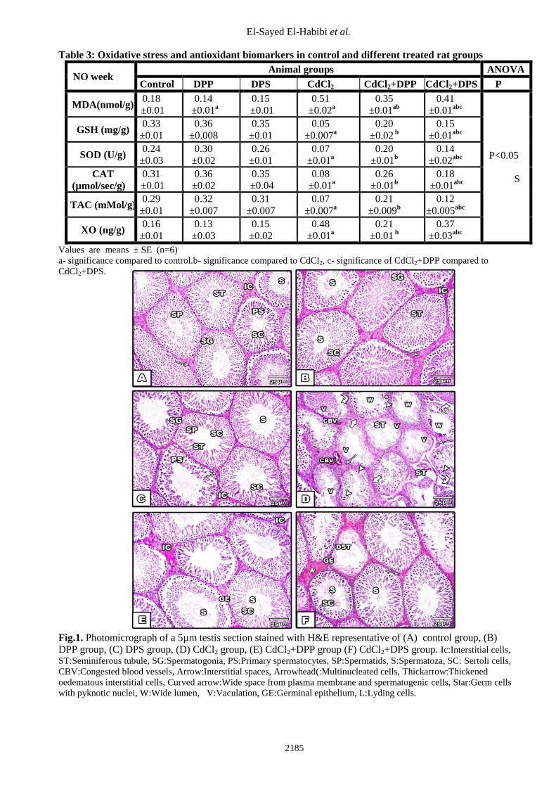
Table 3 recorded a significant increase in
testicular MDA level and XO activity associated
with significant decrease in TAC, GSH level, SOD
and CAT activities in CdCl2 rats compared to NC
rats group. While the co-administration of DPP or
DPS with CdCl2 caused a significant elevation of
these parameters.
The histopathological observation of testis
in control, DPP and DPS groups showed a normal
testis histoarchitecture. Testis sections of DPP and
DPS treated groups showed histological
appearance of seminiferous tubules (ST), with
arrangement of different stages of spermatogenic
cells, interstitial cells (Ic), sertoli cells (Sc) and
many sperms (S) were observed. However the
examination of testis sections of CdCl2 group
showed distorted and necrotic seminiferous
tubules (ST) and thickened oedematous interstitial
cells (thick arrow) with dilated and congested
blood vessels (CBV). There was also wide lumen
(W) with no sperms, multinucleated cells (arrow
head) disorganized germinal epithelium with
marked vaculation (V) and wide space from
plasma membrane and spermatogenic cells
(curved arrow). Also degenerated germ cells with
pyknotic nuclei (star) and fragmented sertoli cells
were seen. On the other hand, testis sections of
CdCl2+DPP and CdCl2+DPS treated animals
showed an improvement in the testicular histology
compared with the CdCl2 treated group, however
the co-administration of DPP showed more
improvement than DPS (Fig 1).

Protective Effect of Date Palm Extracts…
2184
Table 1: Sperm quality in control and different treated rat groups
NO week
Animal groups ANOVA
Control DPP DPS CdCl2 CdCl2+DPP CdCl2+DPS P
No of sperms
(x 104/g
epididymal
tissue)
8.31
±0.11
9.39
±0.16a
9.05
±0.13a
3.17
±0.10a
6.59
±0.09ab
5.23
±0.09abc
P<0.05
S
Sperm
Abnormalities
%
9.6
±0.58
7.4
±0.60
8.4
±0.69
52.5
±3.07a
25.8
±2.02ab
32.7
±2.00abc
sperm
viability%
73.0
±4.15
86.0
±2.08a
79.3
±2.61
25.0
±2.36a
58.2
±2.50ab
41.5
±2.92abc
Values are means ± SE (n=6)
a- significant compared to control.
b- significant compared to CdCl2.
c- significant of CdCl2+DPP compared to CdCl2+DPS.
Table 2: Biochemical parameters in control and different treated rat groups
NO week
Animal groups ANOVA
Control DPP DPS CdCl2 CdCl2+DPP CdCl2+DPS P
T (ng/dl) 752.6
±17.6
778.1
±7.48
790.3
±26.3
267.3
±27.7a
647.0
±24.3b
401.6
±28.7abc
P<0.05
S
E2 (pg/ml) 43.5
±4.66
52.6
±2.56a
46.1
±4.45
15.5
±1.49a
37.8
±1.05b
27.1
±1.81abc
FSH (mlU/mL) 0.52
±0.03
0.59
±0.04
0.53
±0.01
0.14
±0.01a
0.41
±0.01b
0.22
±0.02abc
LH (mlU/mL) 0.90
±0.10
1.50
±0.09a
1.20
±0.15
0.09
±0.01a
0.38
±0.08 ab
0.20
±0.06ab
Serum
Aromatase
(U\ml)
56.9
±5.83
60.0
±6.98
63.5
±6.37
13.4
±1.88a
44.2
±1.54b
26.9
±4.36abc
Testicular
Aromatase
(U/mg)
57.5
±8.22
63.2
±6.17
60.0
±4.92
12.5
±2.00a
39.9
±1.54b
20.9
±3.72abc
Values are means ± SE (n=6)
a- significance compared to control.
b- significance compared to CdCl2.
c- significance of CdCl2+DPP compared to CdCl2+DPS.
El-Sayed El-Habibi et al.
2185
Table 3: Oxidative stress and antioxidant biomarkers in control and different treated rat groups
NO week Animal groups ANOVA
Control DPP DPS CdCl2 CdCl2+DPP CdCl2+DPS P
MDA(nmol/g) 0.18
±0.01
0.14
±0.01a
0.15
±0.01
0.51
±0.02a
0.35
±0.01ab
0.41
±0.01abc
P<0.05
S
GSH (mg/g) 0.33
±0.01
0.36
±0.008
0.35
±0.01
0.05
±0.007a
0.20
±0.02 b
0.15
±0.01abc
SOD (U/g) 0.24
±0.03
0.30
±0.02
0.26
±0.01
0.07
±0.01a
0.20
±0.01b
0.14
±0.02abc
CAT
(µmol/sec/g)
0.31
±0.01
0.36
±0.02
0.35
±0.04
0.08
±0.01a
0.26
±0.01b
0.18
±0.01abc
TAC (mMol/g) 0.29
±0.01
0.32
±0.007
0.31
±0.007
0.07
±0.007a
0.21
±0.009b
0.12
±0.005abc
XO (ng/g) 0.16
±0.01
0.13
±0.03
0.15
±0.02
0.48
±0.01a
0.21
±0.01 b
0.37
±0.03abc
Values are means ± SE (n=6)
a- significance compared to control.b- significance compared to CdCl2, c- significance of CdCl2+DPP compared to
CdCl2+DPS.
Fig.1. Photomicrograph of a 5µm testis section stained with H&E representative of (A) control group, (B)
DPP group, (C) DPS group, (D) CdCl2 group, (E) CdCl2+DPP group (F) CdCl2+DPS group. Ic:Interstitial cells,
ST:Seminiferous tubule, SG:Spermatogonia, PS:Primary spermatocytes, SP:Spermatids, S:Spermatoza, SC: Sertoli cells,
CBV:Congested blood vessels, Arrow:Interstitial spaces, Arrowhead(:Multinucleated cells, Thickarrow:Thickened
oedematous interstitial cells, Curved arrow:Wide space from plasma membrane and spermatogenic cells, Star:Germ cells
with pyknotic nuclei, W:Wide lumen, V:Vaculation, GE:Germinal epithelium, L:Lyding cells.
Protective Effect of Date Palm Extracts…
2186
DISCUSSION
Infertility is a major health problem that
affects one in six couples39
, however, male
infertility causes was found to be about 50% of
infertile couples40
.
Exposure of the reproductive system to
heavy metals has been reported to be a major risk
factor for infertility and there has been an
increasing interest in the contribution of
occupational and environmental exposures to
toxic metals in declining sperm concentration and
human male fertility41
.
Cadmium is one of the most toxic
industrial and environmental heavy metals. It acts
as an endocrine disruptor and oxidative stress
inducer in humans and rodents5,11
.
In the present study, the obtained results
indicated that CdCl2 treated rats showed a
significant drop in sperm count with a significant
decrease in sperm viability, while the sperm
abnormalities were elevated significantly. These
results run parallel to Eleawa et al.42
who revealed
that the main cause of these results is the reduced
spermatogenesis due to increased oxidative stress
and the apoptotic mechanism observed in the
testes of the treated rats. This reduction in sperm
quality may be also due either to impairment of
the H2O2 removal system, which leads to
inhibition of steroidogenesis in the Leydig cells
due to an accumulation of H2O243
or to membrane
damage or macromolecular degradation incurred
by ROS44
.
Furthermore the administration of CdCl2
caused a significant reduction of sex hormones
(T,E2,FSH,LH), and this is in accordance with
Yang et al.45
who suggested that CdCl2 stimulated
apoptosis of the anterior pituitary. Additionally,
the loss of testosterone feedback can result in
pituitary cell hypertrophy, hyperplasia and
eventually pituitary neoplasia46
. Thus the
disruption of the testes-pituitary axis may
contribute to the causation of both testicular and
pituitary destructions. Alternatively, this
reduction could result from decreased viability of
Leydig cells as a consequence of the necrobiotic
effects of toxicants such as Cd47,48
.
Additionally, the data revealed a
significant reduction in both serum and testicular
aromatase in Cd treated rats, while the
administration of either DPP or DPS caused a
significant elevation in this parameter. The ability
of the testis to convert irreversibly androgens into
estrogens is related to the presence of a
microsomal enzymatic complex named
aromatase, which is composed of a specific
glycoprotein, the cytochrome P450 aromatase
(P450arom). In the rat testis the P450arom has
been immunolocalized not only in Leydig cells
but also in germ cells and especially in elongated
spermatids49
producing a significant amount of
the estradiol in the testes50
. It can be concluded
that a complex balance of testosterone, estradiol
and aromatase in the testes confirms a highly
regulated hormonal interaction in the male51
.
As evident from the present data there is
a significant decrease in testicular TAC and GSH
levels as well as SOD and CAT activities
accompanied with significant increase in the lipid
peroxidation product (MDA) level and XO
activity in CdCl2 treated group confirming that
Cd caused testicular oxidative stress. Ikediobi et
al.52
demonstrated that the toxic effect of CdCl2 on
the testes is known to deplete glutathione and
protein-bound sulfhydryl groups, which caused
enhanced production of reactive oxygen species
(ROS) such as superoxide ion, hydroxyl radicals
and hydrogen peroxide. El-Neweshy et
al.53
observed a significant increase in testicular
oxidative stress reflected by the significant
elevation of MDA and a significant depletion of
GSH, which caused irreversible testicular cell
damage. The decreased level of GSH in the testis,
as recorded by Imafidon et al.48
can be attributed
to its excessive use by the testicular tissue to
scavenge the free radicals that were generated
following exposure to Cd toxicity and or reduced
GSH production by the tissue; a consequence
evidently enhanced by the increased use of SOD
in the oxidative process which resulted in its
reduced testicular level.
The administration of CdCl2 also resulted
in histopathological gonadal lesions, such as
testicular damage in seminiferous tubules (STs),
Leydig cells and Sertoli cells in addition to
decreases in the spermatogonial population and
number of spermatozoa. These results run parallel
with the previous studies of El-Neweshy et
al.53
and Eleawa et al.42
who revealed that the
testicular injury was confirmed by marked
alterations in the histological structure of the Cd-
treated rats testes where it exhibited degeneration,
necrosis and atrophy of almost all of the STs with
incomplete to complete spermatogenic arrest. Cd
enters the seminiferous tubules through a breach
of the blood-testis barrier and causes focal
testicular necrosis and dystrophy with consequent
reduction in germ cell numbers, leading to
infertility54
.
El-Sayed El-Habibi et al.
2187
On the other hand, the supplementation
of DPP or DPS extracts significantly improved
the sperm quality, which may be due to DPP
estradiol and flavonoid components55
or to its
scavenging properties that is said to be the main
important effects on the sperm parameters56,57
.
Furthermore, phenol and tocopherol profiles of
DPS were found to be a rich source of natural
phenolic compounds, which was one of the main
reasons for its better oxidative stability58
.
Moreover, DPP and DPS co-
administration showed a significant increase in
sex hormones, which is in accordance with Abedi
et al.59
. This effect may be due to the presence of
flavonoids, steroids, saponins and estradiol
compounds in DPP that affect the hypothalamus
pituitary axis and have thus increased
concentrations of these hormones through raising
the level of luteinizing hormones (LH) leading to
stimulate estradiol levels and endogenous
testosterone levels57,60
.
Shagauo and Davidson61
showed that
DPP is capable of releasing LH hormone by
affecting hypothalamus axis which increases
secretion of gonadotropin releasing hormone
(GnRH). Concerning DPS extract, Ammar et
al.62
reported the presence of different sterols
such as; campasterol, stigmasterol, b-amyrin and
b-sitosterol which exhibited a remarkable
antioxidant and estrogen like activity which can
lead to increase the mentioned hormones.
Additionally, there was a significant
increase in the levels of TAC, GSH, as well as
SOD and CAT activities associated with
significant decrease in MDA and XO activities in
the testis of rats co-administrated with either DPP
or DPS. These results are in harmony with24
who
reported that the antioxidative effect of DPP is
mainly due to its phenolic components, such
anthocyanins, procyanidins and coumaric
acid25
.The antioxidant activity of phenolic
compounds is a result of their redox properties,
which can play an important role in absorbing
and neutralizing free radicals, quenching singlet
and triplet oxygen, or decomposing peroxides63
.
Additionally, the decreased MDA level
associated with elevated other antioxidants in
DPS rat group may be attributed also to the wide
range of phenolic compounds in DPS including p-
coumaric, ferulic and sinapic acids, flavonoids
and procyanidins25
.
The administration of DPP or DPS
caused significant suppress of Cd induced
histopathological damage in testicular tissues. It
is reported that free radicals induce oxidative
damage that is resulted in destructive testicular
architecture causing male infertility, so the DPP
administration showed an antioxidant effect, as
evidenced by improved GSH and restored LPO in
the Cd-treated rats' testes. Thus, DPP can
ameliorate Cd-induced oxidative stress in the
testicular tissues, as evidenced by the renewal of
spermatogenesis in the seminiferous tubules and
normalisation of the testicular
histoarchitecture53,64
. Investigations have also
reported that DPP extracts have the capacity to
improve this ultration is attributed to its content
of estrogenic materials that are considered as
gonad-stimulating compounds65
where estrogen is
involved in regulating the renewal of
spermatogonial stem cell66
. Vayalil55
and Bashir
et al.67
also attributed the positive effect of DPS
on the testicular architecture to its antioxidant
effect that was reported to possess both
androgenic and antioxidant properties.
CONCLUSION
In this study, the data showed that Cd can
induce significant spermatological damage,
oxidative stress and histopathological alterations
in the testicular tissue of male rats 30 days after
exposure, ultimately resulting in infertility.
Remarkably, administration of DPP and DPS
once daily for 30 days effectively prevented the
deleterious effects of Cd. The pro-fertility
properties of DPP and DPS are mainly achieved
through its endocrine-mediated effects, consistent
with their vital role in the antioxidant systems
that protect against Cd damage, and possibly due
to their prevention of oxidative damage to
testicular tissues. Also it can be concluded that
DPP is more effective than DPS in ameliorating
the above mentioned infertility features.
Taken together, our findings support the
hypothesis that the testis is very sensitive to Cd,
which can induce testicular damage and infertility
that can be blocked by the therapeutic
administration with DPP or DPS.
REFERENCES
1-Fallahi S, Rajaei M, Malekzadeh K and Kalantar
SM(2015): Would Phoenix Dactyflera Pollen (palm
seed) be considered as a treatment agent against
Males’ infertility? A systematic review.
Electro.Phys.,7(8):1590-1596.
Protective Effect of Date Palm Extracts…
2188
2-Sharlip ID, Jarow JP and Belker AM(2002): l.Best
practice policies for male infertility.
Fertil.Steril.,77:873-882.
3-Wang Y, Junyi F, Shaoxin H, Chen L and
Guangqin F(2013): The chronic effects of Nrf2 and
Mrp1 of the testis in the rats.
Enviro.Toxic.Pharmacol.,35:109-116.
4-Pant PR(2009): Factors affecting male infertility.
J.Inst.Med.,31(3):10-12.
5-Siu ER, Mruk DD, Porto CS and Cheng
CY(2009): Cadmium-induced testicular injury.
Toxicol.Appl.Pharmacol.,238(3):240–249.
6-Jamshidi B, Ehrampoush MH and Dehvari
M(2013): Utilization of Olive Kernel Ash in
Removal of RB19 from Synthetic Textile
Wastewater. J.Environ.Treat.Tech.,1(3):150-156.
7-Aziz R, Rafiq MT, Yang J, Liu D, Lu L, He Z,
Daud MK, Li T and Yang X(2014): Impact
Assessment of Cadmium Toxicity and Its
Bioavailability in Human Cell Lines (Caco-2 and HL-
7702). Bio.Med.Res.Int.,1-8
8-El-Dars FMSE, Bakr MHM and Gabre AM(2013): Reduction of COD in Resin Production Wastewater
Using Three Types of Activated Carbon.
J.Environ.Treat.Tech.,1(3):126-136.
9-Kidambi SS, Lee DK and Ramamoorthy A(2003): Interaction of Cd and Zn with biologically important
ligands characterized using solid-state NMR and ab
initio calculations. Inorg.Chem.,42:3142-51.
10-J¨arup L (2003): Hazards of heavy metal
contamination. British Medical Bulletin,68:167–182.
11-Cheng CY, Wong EW, Lie PP, Li MW, Su L, Siu
ER, Yan HH, Mannu J, Mathur PP, Bonanomi M,
Silvestrini B and Mruk DD(2011): Environmental
toxicants and male reproductive function.
Spermatogenesis,1(1):2–13.
12-Haidry MT and Malik A(2014): Hepatoprotective
and antioxidative effects of terminalia arjuna against
cadmium provoked toxicity in albino rats (Ratus
norvigicus).Biochem.Pharmacol.,3:130.
13-Kostic MM, Ognjanovic B, Dimitrijevic S, Zikic
RV, Stajn A and Rosic GL(1993): Cadmium –
induced changes of antioxidant and metyabolic status
in red blood cells of rat: In vivo effects.
Eur.J.Haematol.,51:86 – 92
14-Zikic RV, Stajn AS, Ognjanovic BI, Saicic ZS,
Kostic MM, Pavlovic SZ and Petrovic VM(1998): The effect of cadmium and selenium on the
antioxidant enzyme activities in rat heart.
J.Environ.Pathol.Toxicol.Oncol.,17:259 – 264.
15-Satarug S, Garrett SH, Sens MA and Sens
DA(2011): Cadmium, environmental exposure, and
health outcomes. Cien Saude Colet.,16:2587–2602.
16-Pavlovic SZ, Ognjanovic BI, Stajn AS, Zikic RV,
Saicic ZS and Petrovic VM(2001): Antioxidant
defense system in skeletal muscle of rats treated with
cadmium. A possible protective role of coenzyme
Q10. Jugoslav Med.Biochem.,20:229–235.
17-Gupta YK and Briyal S(2004): Animal models of
cerebral ischemia for evaluation of drugs.
Ind.J.Physiol.Pharmacol.,48:379–394.
18-Adaay MH and Mattar AG(2012): Effect of
Aqueous and Ethanolic Extracts of Tribulus terrestris,
Phoenix dactylifera and Nasturtium officinale
Mixture on Some Reproductive Parameters in Male
Mice. J.Baghdad Sci.,9(4):640-650.
19-Khare CP(2007): Indian Medicinal Plants: An
Illustrated Dictionary. Springer Reference, Springer,
New York.
20-El-Neweshy MS, El-Maddawy ZK and El-Sayed
YS(2013): Therapeutic effects of date palm (Phoenix
dactylifera L.) pollen extract on cadmium-induced
testicular toxicity. Andrologia, 45: 369-378.
21-Dostal LA, Faber CK and Zandee J(1996): Sperm
motion parameters in vas deferens and cauda
epididymal rat sperm. Reprod.Toxicol.,10:231-235.
22-Bahmanpour S, Talaei T, Vojdani Z,
Panjehshahin MR, Poostpasand A, Zareei S and
Ghaeminia M(2006): Effect of Phoenix Dactylifera
Pollen on Sperm Parameters and Reproductive
system of Adult Male Rats. IJMS., 31(4):208-212.
23-Hassan WA, El-kashlan AM and Ehssan
NA(2012): Egyptian Date Palm Pollen Ameliorates
Testicular Dysfunction Induced by Cadmium
Chloride in Adult Male Rats. J.Am.Sci.,8(4):659-669.
24-Bahmanpour S, Kavoosi F, Talaei T and
Panjehshahin MR(2013): Effects of Date Palm
(Phoenix Dactylifera) Gemmule Extract on
Morphometric Parameters of Reproductive Tissues,
Hormones and Sperm Quality in Rat.
Anatom.Sci,10(3):144-150.
25-Al-Farsi M, Alasalvar C, Morris A, Baron M
and Shahidi F(2005): Compositional and sensory
characteristics of three native sun dried date (Phoenix
dactylifera L.) varieties grown in Oman. J.Agric.Food
Chem.,53:7586-7591.
26-Al-Farsi M, Alasalvar C, Al-Abid M, Al-Shoaily
K, Al-Amry M and Al-Rawahy F(2007): Compositional and functional characteristics of dates,
syrups, and their by-products. Food
Chem.,104(3):943–947.
27-Habib HM and Ibrahim WH(2008): Nutritional
quality evaluation of eighteen date pit varieties.
Int.J.Food Sci.Nutr.,16:1–13.
28-Shariati M, Sharifi E and Kaveh M(2008): The
effect of phoenix dactylifera (date-palm) pit powder
on testosterone level and germ cells in adult male
rats. J. Zanjan. Uni.Med.Sci. Health Sci.,15:21-27.
29-Gu L, Kelm MA, Hammerstone JF, Beecher G,
Holden J, Haytowitz D and Prior RL(2003): Screening of foods containing proanthocyanidins and
their structural characterization using LCMS/ MS and
thiolytic degradation. J.Agric. Food
Chem.,51(25):7513-7521.
30-Abdul Afiq MJ, Abdul Rahman R, Che Man YB,
AL-Kahtani HA and Mansor TS(2013): Date seed
and date seed oil. IFRJ.,20(5):2035-2043.
El-Sayed El-Habibi et al.
2189
31-Orabi SH and Shawky SM(2014): Effect Of Date
Palm (Phoenix Dactylifera) Seeds Extracts On
Hematological, Biochemical Parameters And Some
Fertility Indices In Male Rats. International Journal
of Sciences: Basic and Applied Research
(IJSBAR),,17(1):137-147.
32-Majumder GC and Biswas R(1979): Evidence for
the occurance of ecto (adenosine triphosphatase) in
rat epididymal spermatozoa. Biochem.,183:737-743.
33-Winters SJ, Kelley DE and Goodpaster B(1998):
The analog free testosterone assay: Are the results in
men clinically useful. Clin.Chem.,44(10):2178-2182.
34-Dupont A, Dupont P, Cusan L, Tremblay M,
Rioux J, Cloutier D, Mailloux J, De Lignieres B,
Gutkowska J, Boucher H, Belanger A, Moyer DL,
Moorjani S and Labier F(1991): Comparative
endocrinological and clinical effects of percutaneous
estradiol and oral conjugated estrogens as
replacement therapy in menopausal women.
Malturitas.,13(4):297-311.
35-Babson AL(1991): The immulite automated
immunoassay system. J.Clin.Immunoass.,14:83-88.
36-Roselli CE(1998): The effect of anabolic-
androgenic steroids on aromatase activity and
androgen receptor binding in the rat preoptic area.
Brain Res.,792(2):271-276.
37-Weesner FM(1968): General Zoological
Microtechinques. Scientific Book Agency, Calcutta,
India,p.86.
38-Snedecor GW and Cochran WG(1982): Statistical
Methods 7th ed. The State University Press
Ametican, Iowa.pp593.
39-Balen AH and Rutherford AJ(2007): Management
of infertility. BMJ., 335(7620):608-611.
40-WHO(2000): Air Quality Guidelines. World Health
Organization.Regional Office for Europe;
Copenhagen, Denmark. Cadmium.
www.euro.who.int/__data/assets/pdf_file/0005/74732
/E71922.pdf
41-Akinloye O, Arowojolu AO, Shittu OB and
Anetor JI(2006): Cadmium toxicity: a possible cause
of male infertility in Nigeria. Reprod.Biol.,6:17–30.
42-Eleawa SM, Alkhateeb MA, Alhashem FH, Bin-
Jaliah I, Sakr HF, Elrefaey HM, Elkarib AO,
Alessa RM, Haidara MA, Shatoor AS and Khalil
MA(2014): Resveratrol Reverses Cadmium
Chloride-induced Testicular Damage and Subfertility
by Downregulating p53 and Bax and Upregulating
Gonadotropins and Bcl-2 gene Expression.
J.Reprod.Develop.,60(2):115-127.
43-Diemer T, Allen JA, Hales KH and Hales
DB(2003): Reactive oxygen disrupts mitochondria in
MA-10 tumor Leydig cells and inhibits steroidogenic
acute regulatory (StAR) protein and steroidogenesis.
Endocrinol.,144:2882-2891.
44-Qadori YT and Al-shaikh MN(2012): Effects of
high and low dose of cadmium chloride on male
Reproductive system in mice. J.Fac.Med.
Baghdad,54(1):110-114.
45-Yang HJ, Lee SH, Jin Y, Choi JH, Han CH and
Lee MH(2005): Genotoxicity and toxicological
effects of acrylamide on reproductive system in male
rats. J.Vet.Sci.,6:103-109.
46-Nyska A, Leininger JR, Maronpot RR, Haseman
JK and Hailey JR(1998): Effects of individual
versus group caging on the incidence of pituitary and
leydig cell tumors in F344 rats: Proposed
Mechanism. Med.Hypoth.,50:525-529.
47-Yang JM, Arnush M, Chen QY, Wu XD, Pang B
and Jiang XZ(2003): Cadmium-induced damage to
primary cultures of rat Leydig cells.
Reprod.Toxicol.,17:553–560.
48-Imafidon CE, Risikat OT, Samuel BF, Esther OO
and Aderonke AK(2016): Cadmium-induced
testicular toxicity, oxidative stress and histopathology
in Wistar rats: Sustained effects of polyphenol-rich
extract of Vernonia amygdalina (del.) leaf.
J.Interdiscipl.Histopathol.,4(3):54-62.
49-Carreau S, Lambard1 S, Delalande C, Denis-
Galeraud I, Bilinska B and Bourguiba S(2003):
Aromatase expression and role of estrogens in male
gonad: a review. Reprod.Biol.Endocrinol.,1:35.
55-Genissel C and Carreau S(2001): Regulation of
the aromatase gene expression in mature rat Leydig
cells. Mol. Cell Endocrinol.,178:141-146.
51-Schulster M, Bernie AM and Ramasamy
R(2016): The role of estradiol in male reproductive
function. Asian J.Androl.,18:1-6.
52-Ikediobi CO, Badisa VL, Ayuk-Takem LT,
Latinwo LM and West J(2004): Response of
antioxidant enzymes and redox metabolites to
cadmium-induced oxidative stress in CRL-1439
normal rat liver cells. Int.J.Mol.Med.,14:87–92.
53-El-Neweshy MS, El-Maddawy ZK and El-Sayed
YS(2012): Therapeutic effects of date palm (Phoenix
dactylifera L.) pollen extract on cadmium-induced
testicular toxicity. Androl.,1–10.
54-Monsefi M, Alaee S, Moradshahi A and Rohani
L(2010): Cadmium-induced infertility in male mice.
Environ.Toxicol.,25:94–102.
55-Vayalil PK(2002): Antioxidant and antimutagenic
properties of aqueous extract of date fruit (Phoenix
dactylifera L. Arecaceae). J.Agric. Food
Chem.,50(3):610-617.
56-Kostyuk VA, Potapovich AI, Strigunova EN and
Afanas'ev I(2004): Experimental evidence that
flavonoid metal complexes may act as mimics of
superoxide dismutase.
Arch.Biochem.Biophys.,428:204-208.
57-Kobeasya MI, El-Naggara AY and Abdallahd
AA(2015): A novel methods for protective role
against reproductive toxicity of carbofuren in male
rats using palm pollen grains and vanadyl(II)folate as
a new compound. J.Chem.Pharm.Res.,7(4):1142-
1148.
58-Fatma BA, Nozha CF, Ines D, Hamadi A, Basma
H and Leila AK(2009): Sperm quality improvement
after date seed oil in vitro supplementation in
Protective Effect of Date Palm Extracts…
2190
spontaneous and induced oxidative stress. Asian
J.Androl.,11:393–398.
59-Abedi A, Parviz M, Karimian SM and Rodsari
HR(2012): The Effect of Aqueous Extract of Phoenix
Dactylifera Pollen Grain on Sexual Behavior of Male
Rats. J.Phys.Pharm.Adv.,2(6):235-242.
60-Jahromi VH, Parivar K and Forozanfar
M(2011): The Effect of cinna-mon extracton
spermatogenesis hormonal axis of pituitary gonad in
mice. Iran.J.Appl.Anim.Sci.,1:99-103
61-Shagauo RB and Davidson AM(2006): The effect
of Cinnamom umzeylanicum on histological structure
of testis in rats. Endocrinol.,63:241-252.
62-Ammar NM, Al-Okbi SY, Mohamed DA and
Abou El-Kassem LT(2009): Antioxidant and
Estrogen Like Activity of the Seed of Phoenix
dactylifera L. Palm Growing in Egyptian Oases.
Report and Opinion,1(3):1-8.
63-Shahidi F, Janitha PK and Wanasundara
PD(1992): Phenolic antioxidants. Critical Reviews of
Food Science & Nutrition,32(1),67–103.
64- Park MS, Shin HS, Lee J, Kil GS and Choi
CY(2010): Influence of quercetin on the
physiological response to cadmium stress in olive
flounder, Paralichthys olivaceus: effects on
hematological and biochemical parameters. Mol. Cell
Toxicol.,6:151–159.
65-Zargari A(1999): Medical plants. University of
Tehran Press,3:33-40.
66-Miura T, Ohta T, Miura CI and Yamauchi
K(2003): Complementary deoxyribonucleic acid
cloning of spermatogonial stem cell renewal factor.
Endoc.,144:5504-5510.
67-Bashir A, Tahir M, Sammee W and Munir
B(2009): Effects of Tribulus Terrestris on Testicular
Development of Immature Albino Rats. Biomed.,25:
63-68.