Seaweed Extracts as Substitutes of Synthetic Hormones for ...
Upload
khangminh22Category
view
1download
0
565
Veterinarni Medicina, 62, 2017 (10): 565–578 Original Paper
doi: 10.17221/161/2016-VETMED
Efficacy of Chromolaena odorata leaf extracts for the healing of rat excision wounds
K. Vijayaraghavan1, J. Rajkumar2, M.A. Seyed3*1Agni College of Technology, Chennai, India2Rajalakshmi Engineering College, Chennai, India3University of Tabuk, Tabuk, Kingdom of Saudi Arabia*Corresponding author: [email protected]
ABSTRACT: Injury to the soft tissues is followed by wound healing, which consists of four stages: haemostasis, inflammation, proliferation and remodelling. Chromolaena odorata is a weed that is traditionally used for the treatment of various ailments in humans and animals. The present study was aimed at exploring the wound healing potential of aqueous and ethanolic extracts of C. odorata in a rat excision wound model. This investigation involved phytochemical screening and in vitro analyses of various parameters such as antioxidant activity, lipid peroxide inhibitory activity and the effects of extracts on contraction and epithelialisation of the rat excision wounds. The phytochemical screening of both ethanolic and aqueous extracts showed that they were rich in secondary metabo-lites such as alkaloids, flavonoids, tannins, saponins, terpenoids, anthraquinones, cardiac glycosides and carbo-hydrates. The aqueous extract showed high antioxidant and lipid peroxide inhibitory activity, while the ethanolic extract showed high total phenol content and hydrogen peroxide inhibitory activity at concentrations of 50, 100 and 250 μg/ml. Our results also indicate that the most effective concentration of the C. odorata extract for excision wound healing was 5.0% (w/w). C. odorata-treated groups exhibited a faster reduction in wound area compared to control and Betadine-treated groups. In addition, the topical application of C. odorata extract increased collagen synthesis and its stabilisation at the wound site, as evidenced by the increase in hydroxyproline and hexosamine levels and expression of collagen. The present investigation demonstrates that aqueous and ethanolic extracts of C. odorata of varying concentrations promote an accelerated wound healing process and might represent a novel healing agent. Our findings are of potential clinical relevance and might be highly beneficial for drug discovery and development in the area of both human and veterinary medicine.
Keywords: Betadine; hydroxyproline; hexosamine; collagen; siam weed; traditional medicine; phytochemicals; antioxidant; invasive species; ornamental plant
Wounds are physical injuries to the skin that take various forms including lacerated wounds, bruises, burns, etc. Based on the physiology of wound heal-ing and its various phases, wounds can be classified as open or closed wounds, and acute or chronic wounds (Meenakshi et al. 2006; Nagori and Solanki 2011). The proper healing of wounds is essential for restoration of disrupted anatomical integrity and altered function of the affected area (Edlich et al. 2005). Healing is a complex and difficult process ini-tiated in response to an injury that serves to restore
the function and integrity of the damaged tissues (Wietecha and DiPietro 2013). Chronic wounds, in particular, are a major concern for animals, hu-mans and clinicians, as they affect a large number of patients, leading to a significant reduction in their quality of life (Hostetler et al. 2006; Belmont et al. 2010). Wound healing is a normal biological process in the body and is achieved through four precisely and highly programmed phases, i.e., haemosta-sis, inflammation, proliferation and remodelling. Wound healing also involves a variety of processes

566
Original Paper Veterinarni Medicina, 62, 2017 (10): 565–578
doi: 10.17221/161/2016-VETMED
such as inflammation, cell proliferation and con-traction of the collagen lattice (Burford et al. 2007). However, the presence of oxygen free radicals or microbial infection hampers healing processes.
Despite remarkable development and advance-ments in the pharmaceutical industry, the search for more effective and low-cost therapeutic ap-proaches for wound healing remains a challenge for modern medicine because of the need for long-term therapy and associated side-effects (Divakar et al. 2000; Soni and Singhai 2013). Naturally de-rived organic compounds/substances that are syn-thesised by primary or secondary metabolism in living organisms have been known for years (Estork et al. 2014; Rastogi et al. 2015), and are widely used in human therapy, veterinary medicine, agriculture and other areas (Martinez and Lujan 2011; Yakubu 2012; Moosavi et al. 2015).
Plants and their metabolites are the primary sources of novel bioactive compounds and are at-tractive alternative therapeutic options to synthetic drugs. The use of medicinal plants in health care is now a common practice (Hashemi and Davoodi 2011; Borges et al. 2016). Many studies have de-scribed the wound healing activity of medicinal plants (Purna and Sabu 2006; Kumar et al. 2007; Dawn et al. 2008).
Chromolaena odorata (L.) R.M. King and H. Robinson, is an ornamental plant usually consid-ered to be one of the top 100 most invasive en-vironmental weeds of wastelands, roadsides and other exposed areas in the world (GISD 2006; Chakraborty et al. 2011). This flowering shrub is native to North and Central America, and was later introduced to parts of Asia, Africa and Australia. C. odorata is also known by various other names such as Armstrong’s weed, baby tea, bitter bush, butterfly weed, Christmas bush, devil weed, eupatorium, Jack in the bush, king weed, paraffin bush, paraffin weed, Siam weed, turpentine weed and triffid weed (Mc Fadyen 2004). It possesses insecticidal properties and is used as a green manure. It is also used for the preservation of dead bodies (Ukwueze et al. 2013).
The fresh leaves of C. odorata or the decoction has been used by practitioners of traditional medi-cine for the treatment of human burns, soft tissue wounds, ulcerated wounds, burn wounds, post-natal wounds and also for the treatment of leech bites, indigestion and skin infection (Panyaphu et al. 2011). The decoctions of the stems were report-ed to be effective against skin disease caused by
Propionibacterium acnes (Pandurangan et al. 2015). It is also used for the treatment of various ailments, such as amenorrhea, catarrh, cold-associated na-sal congestion, diabetes, diarrhoea, fever, pertussis and rheumatism, and as a vermifuge (Goodall and Erasmus 1996). Other pharmacological proper-ties of this plant include anthelminthic (Patel et al. 2010), antimalarial (Ongkana 2003), analgesic (Chakraborty et al. 2011), anti-inflammatory, anti-pyretic, antispasmodic (Oludare et al. 2000), anti-mycobacterial, insecticidal, antioxidant (Phan et al. 2001a), anti-gonorrhoeal (Caceres et al. 1995), fungicidal, diuretic (Gopinath et al. 2009), blood coagulating (Triaratana et al. 1991), and antimi-crobial effects (Borges et al. 2016). However, at present there is only limited information available pertaining to the wound healing properties of the plant (Biswal et al. 1997). Hence, this study was designed to determine the phytochemical composi-tion as well as the in vivo wound healing properties of ethanolic and aqueous extracts of C. odorata using an excision wound model.
The investigation of potent wound healing agents is one of the most promising areas in the field of bi-omedical sciences (Majumder 2012; Kameshwaran et al. 2014). Phytochemical screenings are impor-tant and are considered as the first step towards the discovery of potentially useful drugs (Sivamani et al. 2012). To understand such plants in detail and for them to assume their proper role in contribut-ing to affordable healthcare, a robust scientific as-sessment is needed. Successive solvent extraction techniques, as well as chromatographic separations and spectroscopic methods have been used to de-termine the chemical constituents of plants and to study their bioactivity. Previous results have dem-onstrated that several secondary metabolites such as phenolic acids (protocatechuic, p-hydroxyben-zoic, p-coumaric, ferulic and vanillic acids) isolated from various plants including C. odorata facilitate wound healing in various animal models (Phan et al. 2001a; Phan et al. 2001b; Paiva et al. 2002). Thus, we hypothesised that various phytochemicals pre-sent in C. odorata leaf extracts could have promis-ing synergistic wound healing properties.
MATERIAL AND METHODS
Plant material collection and extraction. The leaves of C. odorata (Figure 1) were collected from

567
Veterinarni Medicina, 62, 2017 (10): 565–578 Original Paper
doi: 10.17221/161/2016-VETMED
with ethanol in a ratio of 1 : 20. The extract was then filtered through Whatmann filter paper, and the filtrate was evaporated using a rotary flash evapo-rator to obtain a concentrated extract. The crude extract was then stored at –20 °C. Phytochemical analysis was carried out to identify the secondary metabolites present in the extracts using standard methods (Harborne 1984).
Determination of total phenol and lipid per-oxidation inhibition assay. Total phenolic content in the leaf extracts was determined using standard methods (Gulcin et al. 2003). One millilitre aliquots of extract or a standard solution of gallic acid were added to a volumetric flask containing 9 ml of wa-ter. Then, 1 ml of the Folin-Ciocalteu reagent was added to the mixture followed by vortexing. After 5 min, 10 ml of 7% sodium carbonate was added to
the hilly regions of Ootacamund (Ooty), India and authenticated by Dr. B. Saraswathi (Director and Head, Siddha Medicines Division, Government Siddha Medical College, Chennai, India with voucher number LR/0078/MP/2014). Fresh leaves were washed thoroughly with distilled water and dried in the shade in a clean environment. The dry leaves were powdered and soaked in distilled water (1 : 5 w/v) at 37 °C. After 24 h, the supernatant was removed and the residue was soaked again in fresh distilled water for another 24 h. The whole process was repeated to ensure a complete extraction. The supernatants were pooled, filtered and centrifuged at 5000 g and 4 °C for 30 min. After centrifugation, the obtained supernatants were frozen at –20 °C and then lyophilised. For the preparation of the ethanol extract, the dried plant material was mixed
SUBMITTED ON LINE © VETERINARY RESEARCH INSTITUTE, BRNO, CZECH REPUBLIC
(Hruska and Zalmanek, 2010: http://vetmed.vri.cz) 23
O
O
OO
O
OFigure 1. Image of Chromolaena odorata plant (A), its collection area in southern India (B) and the chemical structures of its major bioactive components (C) stig-masterol, (D) scutellarein tetramethyl ether
(D)
(B)
SUBMITTED ON LINE © VETERINARY RESEARCH INSTITUTE, BRNO, CZECH REPUBLIC
(Hruska and Zalmanek, 2010: http://vetmed.vri.cz) 23
H
HHH
HHHO
(C)
(A)

568
Original Paper Veterinarni Medicina, 62, 2017 (10): 565–578
doi: 10.17221/161/2016-VETMED
the mixture, which was then incubated for 90 min at 37 °C. The blank was prepared using distilled water and the absorbance against the blank was determined at 750 nm after the incubation period. The amount of phenolic content in the extract was estimated from the standard curve produced with different concentrations (10, 20, 30, 40, 50 μg/ml) of gallic acid. The total phenolic content of the plant was calculated using the following formula:
C = c x m/V
where: C = total content of phenolic compound in gallic acid equivalent/g, c = the concentration of gallic acid established from the calibration curve (μg/ml), m = weight of the crude plant extract (g), V = volume of extract (ml)
The samples were analysed in triplicate. The lipid peroxidation inhibition ability of the extracts was evaluated using a modified procedure described by Lizcano et al. (2012). The percentage inhibition was calculated as follows:
I = [(ODc – ODs)/ODc] × 100
where: I = lipid peroxidation inhibition (%), ODc = optical density control, ODs = optical density sample
The analysis was performed in triplicate.Animals. World Health Organisation (WHO
2014, http://www.who.int/mediacentre/factsheets/fs365/en/), Geneva, and the Indian National Science Academy, New Delhi, guidelines for ani-mal handling and care were followed in this study. This study was conducted based on the guidelines and approval of the Institutional Animal Ethical Committee (IAEC No. Biotech REC. 005/10). Eight-to-ten-week-old female Wistar albino rats (Rattus norvegicus) weighing 120–180 g were selected from an inbred colony and maintained under controlled conditions of temperature (23 ± 2 °C) and light (10 and 14 hours of light and dark, respectively). The animals had free access to sterile food and water. Three animals were housed in a polypropylene cage containing sterile paddy husk (procured locally) as bedding throughout the experiment. The animals were examined by a qualified veterinary surgeon and declared free of any parasites or worms be-fore the experiment began. The cages were changed every two days to maintain aseptic conditions. The food and water consumption was monitored daily
throughout the duration of the experiment. Our animal weight observation chart (data not shown) showed that there was no change in the weight of the animals in any of the groups.
Excision wounds. The animals were anaesthe-tised using diethyl ether and then excision wounds were created on the rats as described by Morton and Malone (1972). The fur on the dorsal side (below the rib cage) of each animal was removed using a sterile shaving blade and the surface was decontaminated by wiping with sterile disinfect-ant. Then, on the shaved dorsal surface, a wound was inflicted by excising the skin flap in an aseptic fashion using sterile surgical knife, scissors and forceps. Each wounded rat was housed in a sepa-rate sterile polypropylene cage. The leaf extract was applied on alternate days on the wound surface and the wound was covered using sterile gauze. The wound size was calculated and granulation tissue was removed on the appropriate termination day (days 4, 8 and 16). Simple ointment base white petroleum jelly was used as vehicle control and the same was used to impregnate leaf extracts for the test group. Ten grams of petroleum jelly were weighed into a beaker and then melted in a water bath. The required quantities of the extract were weighed, added to the molten ointment base and triturated. The rats were divided into six groups of six to eight rats each. Group I rats were treated with simple ointment base (control), group II rats treated with a reference standard Betadine oint-ment, and groups III, IV, V and VI were treated with different (2.5, 5.0, 7.5, and 10%, respectively; w/w) concentrations of the extract (ethanolic or water extract) for 20 days. The ointment base and Betadine were applied in the same quantity to serve as control and standard, respectively. The wound contraction was calculated as the percent-age reduction in wound area. On the fourth, eighth, twelfth and sixteenth days after the infliction of the initial wound, progressive change was observed in the wound areas. The wound surface areas were outlined using transparent paper and a graph sheet was used to measure the area of the wound. No animal death was observed in the control group, or in the reference standard drug or extract-treated group. The biochemical indicators hydroxypro-line, hexosamine (Futamura et al. 2013; Gangwar et al. 2015) and total protein content were esti-mated from the excised tissue (Inkinen et al. 2000; Upadhyay et al. 2009).

569
Veterinarni Medicina, 62, 2017 (10): 565–578 Original Paper
doi: 10.17221/161/2016-VETMED
Histopathological examination. The experi-ment was terminated after appropriate treatment periods, i.e., after 0, 4, 8, 12 or 16 days of extract treatment and tissue was excised from the wound for histological examination. The sections were stained with haematoxylin and eosin (H&E) stains and the tissue samples were evaluated for the re-epithelisation of tissues, the width of the granular cell layer and the extent of tissue formation.
Statistical analysis. All experiments were per-formed in triplicate (n = 3), and the results were compared using a one-way analysis of variance (ANOVA). P-values of less than 0.05 (P < 0.05) were considered to indicate statistical significance.
RESULTS
Phytochemical screening conducted on leaf ex-tracts of C. odorata revealed the presence of the following classes of primary and secondary me-
tabolites: alkaloids, flavonoids, tannins, saponins, anthraquinones, cardiac glycosides and terpenoids in both ethanolic and water extracts, but with sig-nificant differences in relative concentrations, i.e., low, average or high (Table 1). Phytochemical screening of the aqueous extracts in this study re-vealed that they were rich in saponins and alkaloids, which have numerous pharmacological effects, and have been extensively used as drugs in the medical field. The detection of high levels of alkaloids in the leaf extracts of C. odorata further supports the presence of alkaloids in this plant, whereas etha-nol extracts were rich in terpenoids and cardiac glycosides. However, both aqueous and ethanolic extracts showed high total phenol contents and also demonstrated high antioxidant and lipid peroxide inhibitory activity at 100, 200 and 300 μg/ml. The antioxidant activity for the aqueous and ethanolic extracts relative to gallic acid is shown in Table 2, the total phenol content for the aqueous and etha-nolic extracts is shown in Table 3 and lipid peroxide inhibitory activity for the aqueous and ethanolic extracts is shown in Table 4.
Our results demonstrated improved wound healing within 16 days in C. odorata-treated rats, whereas it took more than 16 days in the standard and 20 days in the control rats. Wounds treated with the C. odorata extract showed no evidence of wound haemorrhage, oedema, inflammation and exudate. In contrast, untreated wounds displayed characteristic oedema associated with typical in-flammation. In the C. odorata-treated rats, the skin surrounding the wound appeared to be regular and soft, while in the Betadine-treated group the skin
Table 1. Phytochemical analysis of aqueous and etha-nolic leaf extracts of Chromolaena odorata
Phytochemicals Ethanol AqueousTannins + +Saponins + ++Flavonoids + +Anthocyanins – –Betacyanins + –Quinones + –Glycosides + –Cardiac glycosides ++ –Terpenoids ++ –Phenols ++ ++Coumarins – +Steroids + –Alkaloids – ++
++ = abundant, + = trace, – = absent
Table 2. Antioxidant activity (%) of Chromoleana odo-rata extracts
Concentration of extracts (µg/ml)
Aqueous extract
Ethanolic extract
Gallic acid
100 75.00 ± 0.08 75.21 ± 0.01 80.06 ± 0.03200 76.32 ± 0.09 77.06 ± 0.01 81.12 ± 0.02300 77.12 ± 0.08 78.02 ± 0.02 81.72 ± 0.02
Table 4. Lipid peroxidation inhibition activity (%) of C. odorata extracts. Values are presented as the means of triplicate measurements ± SD
Concentration of extracts (µg/ml)
Aqueous extract
Ethanolic extract
100 10.48 ± 0.09 12.78 ± 0.08200 22.29 ± 1.35 28.48 ± 1.33300 41.25 ± 2.22 31.11 ± 2.10
Table 3. Total phenol content of Chromolaena odorata leaf fractions
Extract (fractions) Total phenol content equivalent to gallic acid (mg/g GAE)
Aqueous 0.22 ± 0.01Ethanol 0.85 ± 0.06

570
Original Paper Veterinarni Medicina, 62, 2017 (10): 565–578
doi: 10.17221/161/2016-VETMED
appeared totally dehydrated. Among various concen-trations, 5.0% (w/w) was the most effective concentra-tion of the C. odorata leaf extract for wound healing. The effect of topical application of different concen-trations of the C. odorata leaf extract on the time required for wound healing is shown in Table 5. The animals treated with 5.0% C. odorata extract showed a significantly faster reduction in wound area than the untreated group on day 8 (114.18 versus 140.5 mm2) and day 16 (51.35 versus 89.33 mm2). Levels of hy-droxyproline, hexosamine and total protein were also significantly increased in the rats treated with the 5.0% C. odorata extract (Table 6).
Haematoxylin and eosin (H&E) staining was per-formed to study the anatomical restoration elicited by treatment with the extract of C. odorata. Our results indicate that there was an increased forma-tion of blood vessels and an improvement in cell proliferation after treatment with C. odorata leaf extract. When treated with different concentrations of the extract, the group of animals with excision wounds (Figure 2) showed significant differences compared to the normal and standard reference control (Figure 3).
DISCUSSION
Wound management entails a hospital stay of specific duration, expensive medication and vari-
ous surgical modalities followed by a long process of rehabilitation (Thomas et al. 2009). Most topical antibacterial agents and disinfectants are effective in protecting against infections, but the occurrence of irritations in the skin and allergy due to these agents reduces the rate of skin regeneration and increases the time needed for complete recovery (Burks 1998). Moreover, most expensive tissue-en-gineered wound dressings are out of reach for the patients residing in developing and underdeveloped countries. Many medicinal plants and products de-rived from medicinal plants have been shown to possess satisfactory wound healing efficacy with no or little toxicity and are less expensive than syn-thetic drugs. Some of the medicinal plants that have been investigated for their wound healing activ-ity have already been used in traditional medicine for wound care (Phan et al. 2001b; Upadhyay et al. 2009; Hashemi and Davoodi 2011; Vaisakh and Pandey 2012).
Our phytoscreening results are in line with previ-ous studies (Ling et al. 2005; Pandith et al. 2013) that were conducted using ethanolic seed extracts of C. odorata. However, the present investigation revealed that C. odorata aqueous extracts of vary-ing concentrations can also exert significant cuta-neous wound healing activity. The effects of the extract, Betadine ointment (standard), and normal base (control) in the excision would model was evaluated by measuring the wound area and wound
Table 5. Effect of topical application of C. odorata lyophilised aqueous leaf extract on wound area contraction (mm2). Values are presented as mean ± SE; n = 6; (values do not differ significantly at P < 0.05)
Post wounding day
Excised wound control
C. odorata aqueous leaf extract concentration Betadine concentration 5.0% (w/w)2.5% (w/w) 5.0% (w/w) 7.5% (w/w) 10.0% (w/w)
Day 0 175.83 ± 0.87 177.67 ± 1.26 178.33 ± 0.99 179.17 ± 0.60 177.33 ± 0.99 176.5 ± 1.80Day 8 140.50 ± 4.60 124.50 ± 2.79 114.18 ± 3.48 121.33 ± 3.57 133.00 ± 3.17 128.83 ± 5.28Day 16 89.33 ± 2.63 71.67 ± 3.68 51.35 ± 3.8 63.33 ± 3.79 79.00 ± 3.48 72.00 ± 4.33
Wound area contraction (mm2) in response to different concentrations of C. odorata extract was calculated relative to excised wound control and standard reference control (Std-RC, Betadine) on days 0, 8 and 16
Table 6. Effect of topical application of C. odorata aqueous leaf extract for seven days on various biochemical param-eters in granulation tissue. Values are presented as mean ± SE; n = 6
Group (mg/g tissue wt)
Excised wound control
C. odorata aqueous leaf extract concentration Betadine concentration 5.0% (w/w)2.5% w/w 5.0% w/w 7.5% w/w 10.0% w/w
Hydroxyproline 21.80 ± 0.32 28.12 ± 0.70* 30.99 ± 0.82* 26.72 ± 0.73* 23.19 ± 0.80 25.73 ± 0.67*Hexosamine 0.50 ± 0.04 0.55 ± 0.03 0.69 ± 0.04* 0.61 ± 0.03* 0.60 ± 0.03 0.53 ± 0.03Hexuronic acid 88.74 ± 4.18 114.33 ± 7.40* 123.77 ± 7.77* 109.11 ± 3.17* 97.51 ± 3.61 109.78 ± 4.16*
*P < 0.05 compared with excised wound control

571
Veterinarni Medicina, 62, 2017 (10): 565–578 Original Paper
doi: 10.17221/161/2016-VETMED
contraction. The results indicate that healing is de-pendent on both the concentration as well as the nature of the different extracts (water vs ethanol). Our observations indicate that extracts with con-centrations of more than 10% w/w could be lethal for animals. The potency of the extracts was found to be inversely proportional to the time needed for the healing of the wound. The activity of C. odorata was comparable with that of the reference drug Betadine, confirming the effectiveness of this medicinal plant in wound healing.
Wound repair consists of a cascade of events that re-establish the integrity of damaged tissues. The sequence of events that repairs the damage is cat-egorised into four overlapping phases: haemostasis, inflammation, proliferation and tissue remodelling. Healing requires the collaborative efforts of numer-ous different tissues and cell lineages (Ling et al. 2005; Odunbaku et al. 2008). It involves platelet aggregation and blood clotting, fibrin formation, inflammatory responses to the injury, alteration in the inflammatory mediators, angiogenesis and re-epithelialisation. The healing process is not
complete until the disrupted surfaces are firmly closed together by collagen (Owoyele et al. 2008). The basic principle of optimal wound healing is to minimise tissue damage and to provide adequate tissue perfusion and oxygenation, proper nutrition, and a moist wound healing environment to restore the anatomical continuity and function of the af-fected region (Bamba et al. 1993).
The most established and discussed activity of C. odorata is its wound healing effect. The con-stituents of the plant’s extracts modulate one or more of the overlapping wound healing stages. Leaf extracts and other plant parts of C. odorata (Table 6) have been shown to be beneficial in the treatment of wounds and other disorders (Bani 2002; Gosain and DiPietro 2004; Raina et al. 2008; Ayyanar and Ignacimuthu 2009; Nguyen et al. 2009). Traditionally, its leaves were ground into a paste and applied topically to the affected areas (Pandith et al 2013). Both in vitro wound assay and in vivo studies have demonstrated that these extracts enhance the proliferation of fibroblasts, endothelial cells and keratinocytes; stimulate ke-
Figure 2. Various wound healing phases in negative control (NC) (A, B, C), standard reference control (Std.RC) and in response to 5% Betadine (D, E, F) and 5% C. odorata leaf extract (G, H, I)
(I) (G)
(F) (E) (D)
(C) (B) (A)
(H)
C. odorata
Std.RC
NC
Day 0 Day 8 Day 16

572
Original Paper Veterinarni Medicina, 62, 2017 (10): 565–578
doi: 10.17221/161/2016-VETMED
ratinocyte migration; upregulate the keratinocyte-induced production of extracellular matrix proteins and basement membrane components including collagen VII, anchoring fibrils and fibrillin micro-fibrils; and inhibit collagen lattice contraction by fibroblasts (Kusano et al. 2001; Subramoniam et al. 2012).
Despite the natural cascades of the healing pro-cess, the process may be delayed by an infection caused by various mechanisms, such as decreased blood supply, which can promote impaired leuko-cyte function, prolong inflammatory and debride-ment phases, and produce proteolytic enzymes. Therefore, infection is the major impediment to the wound healing process and antibacterial com-pounds play a major role in the process of heal-ing (Annan and Houghton 2008; Kahkeshani et al. 2013; Kumari et al. 2013). Medicinal plants, with their antioxidant, anti-inflammatory, and antimi-crobial activities, harbour great potential for the management and treatment of wounds (Trabucchi et al. 1986; Trombetta et al. 2005; Akinmoladun et al. 2010). In the present study, leaf extracts of C. odorata induced better wound healing than
Betadine, a finding which was confirmed by the histopathological evaluation which showed com-plete re-epithelialisation, well-formed granulation tissue and neovascularisation. Betadine cream is the most commonly used topical antimicrobial agent for the treatment of injuries and wounds. It is widely used because it rapidly kills all bacteria and fungi commonly responsible for wound and skin infections. The present study suggests that the use of C. odorata leaf extracts would be highly beneficial for the treatment of wounds.
Another major challenge of pharmacological vali-dation is that the exact mechanism of the wound healing process is often not clearly understood; hence, most research studies are restricted to screening plants to simply evaluate their wound healing effects and do not go into the mechanistic details. It is important to note that wound healing involves a number of processes, including inflam-mation, epithelisation, antioxidant defence, bio-chemical change, granulation, neovascularisation and wound contraction. The possible mechanisms by which C. odorata can influence the different wound healing phases are briefly shown in Figure 4.
Figure 3. Histopathological evaluation demonstrates the healing effects observed (during the study period; day 0, day 8 and day 12) in the negative control (NC) (A, D, G), standard reference control (Std.RC) and in response to 5% Betadine (B, E, H) and 5% C. odorata leaf extract (C, F, I). Arrows indicate the wound healing granulation process
(I) (H) (G)
(F) (E) (D)
(C) (B) (A)
NC
Std.RC
C. odorata
Day 0 Day 8 Day 16

573
Veterinarni Medicina, 62, 2017 (10): 565–578 Original Paper
doi: 10.17221/161/2016-VETMED
Figure 4. Schematic diagram showing the cellular and physiological events associated with C.odorata wound healing mechanisms and other pharmacological activities. C. odorata extract or its bioactive compounds stimulate haemostatic activity by stimulation of TXS and repression of MMP-9 expression. It also activates MEK, p38MAPK, AKT and JNK kinase pathways which initiate the expression of HO-1. The induction of HO-1 inhibits inflammation and stimulates cell proliferation, thereby enhancing the neovascularisation and cell migration that helps to accomplish wound healing. The dotted arrow represents bioactive compounds isolated from C. odorata that may be involved in the wound healing process individually or in combination, whereas the bold arrow indicates the definite involvement of Scu, Stigmasterol and C1 in the process
Chromolena odorata
alkaloids, flavonoids, tannins, saponins, anthraquinones, cardiac glycosides, terpenoids, stigmasterol, scutellarin terramethyl ether (scu),
chromomoric acid C-1
Hours
Hours
Weeks to months
TXS
MM
P9
Plat
elet
ag
greg
atio
n Pl
atel
et
JNK
A
KT
P38
MA
PK
MEK
HO
-1
gene
/pro
tein
Pr
omot
er
Hem
osta
sis
Infla
mm
atio
n Pr
olife
ratio
n Platelets
Neutrophil, monocyte macrophage and lymphocyte
Keratinocytes, fibroblasts, endothelial cells
Vascular constriction, platelet aggregation, degranulation, fibrin plug formation (thrombus), hypoxia and release of proinflammatory mediators (cytokines)
Neutrophil, monocyte infiltration and differentiation to macrophage, lymphocyte infiltration, fibrin formation (thrombus)
Re-epithelialisation, angiogenesis (VEGF, TGF-β), fibroblasia, cytokine production, collagen synthesis, ECM formation, epidermal surfacing
Rem
odel
ling
mat
urat
ion Myofibroblasts Phenotypic switch from
myofibroblasts to fibroblasts, wound contraction, mature scar formation
Time
Possible signalling
mechanisms
Phases of
wound healing
Main cell types
involved
Main events involved in wound
healing
Figure 4. Schematic diagram showing the cellular and physiological events associated with C. odorata wound healing mechanisms and other pharmacological activities. C. odorata extract or its bioactive compounds stimulate haemo-static activity by stimulation of TXS and repression of MMP-9 expression. It also activates MEK, p38MAPK, AKT and JNK kinase pathways which initiate the expression of HO-1. The induction of HO-1 inhibits inflammation and stimulates cell proliferation, thereby enhancing the neovascularisation and cell migration that helps to accomplish wound healing. The dotted arrow represents bioactive compounds isolated from C. odorata that may be involved in the wound healing process individually or in combination, whereas the bold arrow indicates the definite involvement of scutellarein tetramethyl ether, stigmasterol and C1 in the process
574
Original Paper Veterinarni Medicina, 62, 2017 (10): 565–578
doi: 10.17221/161/2016-VETMED
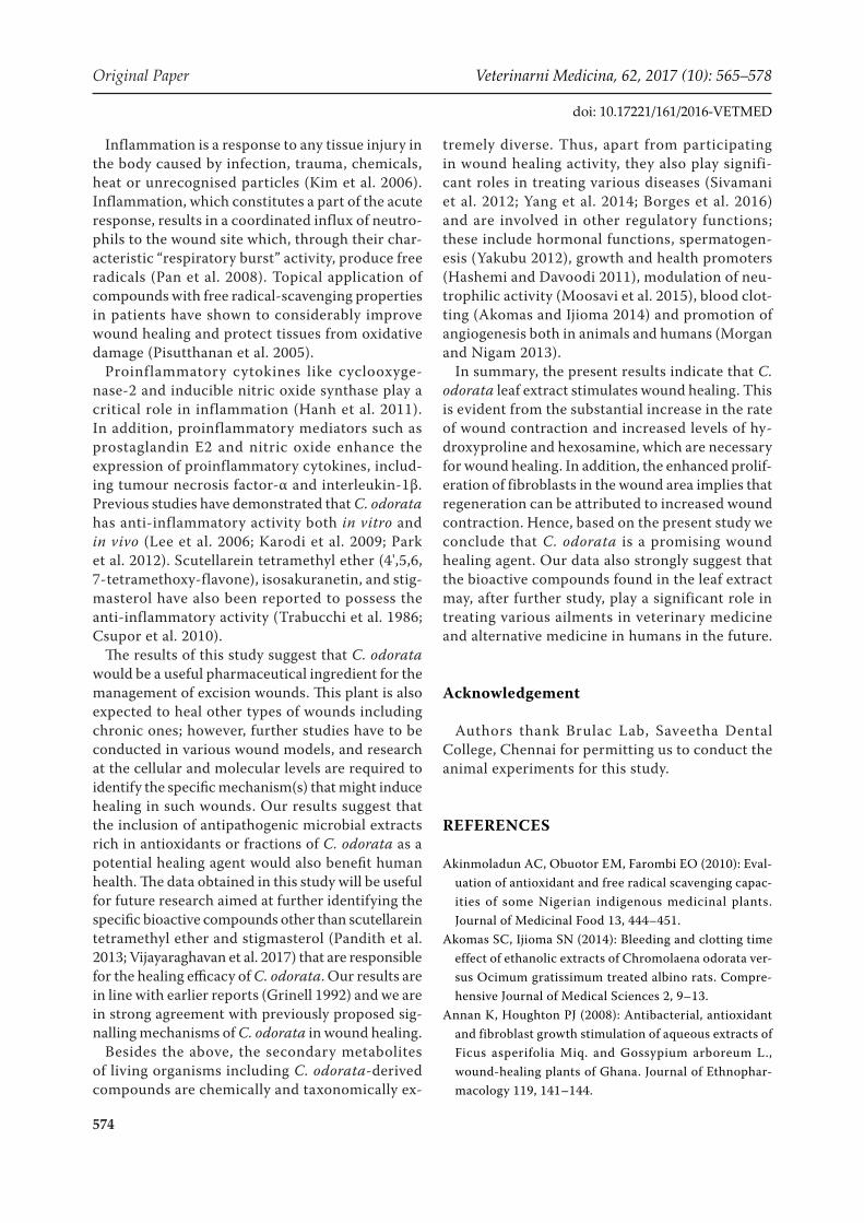
Inflammation is a response to any tissue injury in the body caused by infection, trauma, chemicals, heat or unrecognised particles (Kim et al. 2006). Inflammation, which constitutes a part of the acute response, results in a coordinated influx of neutro-phils to the wound site which, through their char-acteristic “respiratory burst” activity, produce free radicals (Pan et al. 2008). Topical application of compounds with free radical-scavenging properties in patients have shown to considerably improve wound healing and protect tissues from oxidative damage (Pisutthanan et al. 2005).
Proinflammatory cytokines like cyclooxyge-nase-2 and inducible nitric oxide synthase play a critical role in inflammation (Hanh et al. 2011). In addition, proinflammatory mediators such as prostaglandin E2 and nitric oxide enhance the expression of proinflammatory cytokines, includ-ing tumour necrosis factor-α and interleukin-1β. Previous studies have demonstrated that C. odorata has anti-inflammatory activity both in vitro and in vivo (Lee et al. 2006; Karodi et al. 2009; Park et al. 2012). Scutellarein tetramethyl ether (4',5,6, 7-tetramethoxy-flavone), isosakuranetin, and stig-masterol have also been reported to possess the anti-inflammatory activity (Trabucchi et al. 1986; Csupor et al. 2010).
The results of this study suggest that C. odorata would be a useful pharmaceutical ingredient for the management of excision wounds. This plant is also expected to heal other types of wounds including chronic ones; however, further studies have to be conducted in various wound models, and research at the cellular and molecular levels are required to identify the specific mechanism(s) that might induce healing in such wounds. Our results suggest that the inclusion of antipathogenic microbial extracts rich in antioxidants or fractions of C. odorata as a potential healing agent would also benefit human health. The data obtained in this study will be useful for future research aimed at further identifying the specific bioactive compounds other than scutellarein tetramethyl ether and stigmasterol (Pandith et al. 2013; Vijayaraghavan et al. 2017) that are responsible for the healing efficacy of C. odorata. Our results are in line with earlier reports (Grinell 1992) and we are in strong agreement with previously proposed sig-nalling mechanisms of C. odorata in wound healing.
Besides the above, the secondary metabolites of living organisms including C. odorata-derived compounds are chemically and taxonomically ex-
tremely diverse. Thus, apart from participating in wound healing activity, they also play signifi-cant roles in treating various diseases (Sivamani et al. 2012; Yang et al. 2014; Borges et al. 2016) and are involved in other regulatory functions; these include hormonal functions, spermatogen-esis (Yakubu 2012), growth and health promoters (Hashemi and Davoodi 2011), modulation of neu-trophilic activity (Moosavi et al. 2015), blood clot-ting (Akomas and Ijioma 2014) and promotion of angiogenesis both in animals and humans (Morgan and Nigam 2013).
In summary, the present results indicate that C. odorata leaf extract stimulates wound healing. This is evident from the substantial increase in the rate of wound contraction and increased levels of hy-droxyproline and hexosamine, which are necessary for wound healing. In addition, the enhanced prolif-eration of fibroblasts in the wound area implies that regeneration can be attributed to increased wound contraction. Hence, based on the present study we conclude that C. odorata is a promising wound healing agent. Our data also strongly suggest that the bioactive compounds found in the leaf extract may, after further study, play a significant role in treating various ailments in veterinary medicine and alternative medicine in humans in the future.
Acknowledgement
Authors thank Brulac Lab, Saveetha Dental College, Chennai for permitting us to conduct the animal experiments for this study.
REFERENCES
Akinmoladun AC, Obuotor EM, Farombi EO (2010): Eval-uation of antioxidant and free radical scavenging capac-ities of some Nigerian indigenous medicinal plants. Journal of Medicinal Food 13, 444–451.
Akomas SC, Ijioma SN (2014): Bleeding and clotting time effect of ethanolic extracts of Chromolaena odorata ver-sus Ocimum gratissimum treated albino rats. Compre-hensive Journal of Medical Sciences 2, 9–13.
Annan K, Houghton PJ (2008): Antibacterial, antioxidant and fibroblast growth stimulation of aqueous extracts of Ficus asperifolia Miq. and Gossypium arboreum L., wound-healing plants of Ghana. Journal of Ethnophar-macology 119, 141–144.

575
Veterinarni Medicina, 62, 2017 (10): 565–578 Original Paper
doi: 10.17221/161/2016-VETMED
Ayyanar M, Ignacimuthu S (2009): Herbal medicines for wound healing among tribal people in Southern India: Ethnobotanical and Scientific evidences. International Journal of Applied Research in Natural Products 2, 29–42.
Bamba D, Bessiere JM, Marion C, Pelissier Y, Fouraste I (1993): Essential oil of Eupatorium odoratum. Planta Medica 59, 184–185.
Bani G (2002): Status and management of Chromolaena odorata in Congo. In: Zachariades C, Muniappan R, Strathie L (eds): Proceedings of the Fifth International Workshop on Biological Control and Management of Chromolaena odorata. Durban, South Africa, October 2000. 71–73.
Belmont PJ, Schoenfeld AJ, Goodman G (2010): Epidemiol-ogy of combat wounds in operation iraqi freedom and operation enduring freedom: Orthopaedic burden of disease. Journal of Surgical Orthopedic Advances 19, 2–7.
Biswal PR, Sardar KK, Parija SC, Mishra PR, Mishra SN (1997): Wound healing effect of Eupatorium odoratum Linn, and Himax in rabbits. Indian Journal of Indigenous Medicine 19, 71–74.
Borges A, Abreu AC, Dias C, Saavedra MJ, Borges F, Simoes M (2016): New perspectives on the use of phytochemicals as an emergent strategy to control bacterial infections including biofilms. Molecules 21, doi: 10.3390/mole-cules21070877.
Burford G, Bodeker G, Ryan TJ (2007): Skin and wound care: traditional, complementary and alternative medi-cine in public health dermatology. In: Bodeker G, Burford G (eds): Traditional, Complementary and Alternative Medicine: Policy and Public Health Perspectives. 1st edn. Imperial College Press, London. 472 pp.
Burks RI (1998): Povidone-iodine solution in wound treat-ment. Physical Therapy 78, 212–218.
Caceres A, Menendez H, Mendez E, Cohobon E, Samayoa BE, Jauregui E, Peralta E, Carrillo G (1995): Antigonor-rhoeal activity of plants used in Guatemala for the treat-ment of sexually transmitted diseases. Journal of Ethnopharmacology 48, 85–88.
Chakraborty AK, Sujit R, Umesh KP (2011): Chromolaena odorata (L.): An overview. Journal of Pharmacy Research 43, 573–576.
Csupor D, Blazso G, Balogh A, Hohmann J (2010): The tra-ditional Hungarian medicinal plant Centaurea sadleriana Janka accelerates wound healing in rats. Journal of Eth-nopharmacology 127, 193–195.
Dawn TA, Jialin W, Zhihong J, Hubiao C, Guanghua L, Zhongzhen Z (2008): Ethnobotanical study of medicinal plants used by Hakka in Guangdong, China. Journal of Ethnopharmacology 117, 41–50.
Divakar MC, Devi LS, Kumar S, Rao SB (2000): Studies on wound healing property of Polyscias scutellaria leaf sap-onins. Indian Journal of Natural Products 17, 37–42.
Edlich RF, Winters KL, Britt LD, Long WB, Gubler KD, Drake DB (2005): Difficult wounds: an update. Journal of Long Term Effective Medical Implants 15, 289–302.
Estork DM, Gusmao DF, Paciencia ML, Diaz IE, Varella AD, Younes RN, Reis LF, Montero EF, Bernardi MM, Suffredini IB (2014): First chemical evaluation and toxicity of Cas-inga-cheirosa to Balb-c male mice. Molecules 19, 3973–3987.
Futamura A, Higashiguchi T, Ito A, Kodama Y, Chihara T, Kaneko T, Tomatsu A, Shimpo K (2013): Experimental research on stimulation of wound healing by n-3 fatty acids. Wounds – A Compendium of Clinical Research and Practice 25, 186–192.
Gangwar M, Gautam MK, Ghildiyal S, Nath G, Goel RK (2015): Mallotus philippinensis Muell. Arg fruit glandu-lar hairs extract promotes wound healing on different wound model in rats. BMC Complementary Alternative Medicine 15, doi: 10.1186/s12906-015-0647-y.
GISD – Global Invasive Species Database (2006): Global Invasive Species Database online data sheet. Chromo-laena odorata (herb). Available at http://www.iucngisd.org/gisd/species.php?sc=47. (Accessed November 1, 2016).
Goodall JM, Erasmus DJ (1996): Review of the status and integrated control of the invasive alien weed, Chromo-laena odorata, in South Africa. Agriculture, Ecosystems and Environment 56, 151–164.
Gopinath R, Sunilson JAJ, Radhamani S, Das A, Nilugal K (2009): Diuretic activity of Eupatorium odoratum (L). Journal of Pharmaceutical Research 2, 844–846.
Gosain A, DiPietro LA (2004): Aging and wound healing. World Journal of Surgery 28, 321–326.
Grinell F (1992): Cell adhesion. In: Cohen IK, Diegelmann RF, Lindblad WJ (eds): Wound Healing: Biochemistry of Clinical Aspects. Saunders WB, Philadelphia. 209–222.
Gulcin L, Sat GI, Beydemir S, Elmastas M, Kufrevioglu OI (2003): Comparison of antioxidant activity of clove (Eu-genia caryophylata Thumb.) buds and lavender (Lavan-dula stoechas L). Food Chemistry 87, 393–400.
Hanh TT, Hang DT, Van Minh C, Dat N (2011): Anti-in-flammatory effects of fatty acids isolated from Chromo-laena odorata. Asian Pacific Journal of Tropical Medicine 4, 760–763.
Harborne JB (1984): Phytochemical methods – a guide to modern techniques of plant analysis. 2nd edn. Springer Netherlands. 4–16.
Hashemi SR, Davoodi H (2011): Herbal plants and their derivatives as growth and health promoters in animal
576
Original Paper Veterinarni Medicina, 62, 2017 (10): 565–578
doi: 10.17221/161/2016-VETMED
nutrition. Veterinary Research Communications 35, 169–180.
Hostetler SG, Xiang H, Gupta S, Sen C, Gordillo JM (2006): Discharge patterns of injury-related hospitalizations with an acute wound in the United States. Wounds 1, 340–351.
Inkinen K, Turakainen H, Wolff H, Ravanti L, Kahari VM, Ahonen J (2000): Expression and activity of matrix met-alloproteinase-2 and-9 in 649 experimental granulation tissue. Apmis 108, 318–328.
Kahkeshani N, Farahanikia B, Mahdaviani P, Abdolghaffari A, Hassanzadeh G, Abdollahi M (2013): Antioxidant and burn healing potential of Galium odoratum extracts. Re-search in Pharmaceutical Sciences 8, 197–203.
Kameshwaran S, Senthilkumar R, Thenmozhi S, Dhanalakshmi M (2014): Wound healing potential of ethanolic extract of Tecoma stans flowers in rats. Pharmacology 5, 215–221.
Karodi R, Jadhav M, Rub R, Bafna A (2009): Evaluation of the wound healing activity of a crude extract of Rubia cordifolia L. (Indian madder) in mice. International Jour-nal of Applied Research and Natural Products 2, 12–18.
Kim JH, Kim DH, Baek SH, Lee HJ, Kim MR, Kwon HJ, Lee CH (2006): Rengyolone inhibits inducible nitric oxide synthase expression and nitric oxide production by down-regulation of NF-κB and p38 MAP kinase activity in LPS stimulated RAW 264.7 cells. Biochemical Pharmacology 71, 1198–1205.
Kumar B, Vijayakumar M, Govindarajan R, Pushpangadan P (2007): Ethnopharmacological approaches to wound healing – Exploring medicinal plants of India. Journal of Ethnopharmacology 114, 103–113.
Kumari P, Yadav P, Verma PR, Kumar S, Arya A (2013): A review on wound healing properties of Indian medicinal plants. Indian Journal of Fundamental and Applied Life Sciences 3, 220–232.
Kusano A, Seyama Y, Nagai M, Shibano M, Kusano G (2001): Effects of fukinolic acid and cimicifugic acids from Cimicifuga species on collagenolytic activity. Biological Pharmaceutical Bulletin 24, 1198–1201.
Lee IS, Jin W, Zhang X, Hung TM, Song KS, Seong YH, Bae K (2006): Cytotoxic and COX-2 inhibitory constituents from the aerial parts of Aralia cordata. Archives of Phar-macal Research 29, 548–555.
Ling SK, Mazura MP, Khoo MGH, Virmala S, Ong BK, Mastura M, Nor Azah MA, Salbiah M, Siti Asha ABL and Rasadah MA (2005): Chemical constituents and thera-peutic potential of the leaf extracts from Chromolaena odorata. In: Nik Zanariah NM, Norhara H, Nor Azman H, Chan HT (eds): Highlights of FRIM’s IRPA Projects 2005. Identifying Potential Commercial Collaborations Project Evaluation Meeting. Forest Research Institute Malaysia, Malaysia. 108–114.
Lizcano LJ, Viloria BM, Vicente R, Berrueta LA, Gallo B, Martinez-Canamero M, Ruiz-Larrea MB, Ruiz-Sanz JI (2012): Lipid oxidation inhibitory effects and phenolic composition of aqueous extracts from medicinal plants of Colombian Amazonia. International Journal of Mo-lecular Sciences 13, 5454–5467.
Majumder P (2012): Taxo-cognostic, Phyto-physicochem-ical & Biological screening of a plant: Taxo-cognostic, Phytochemical, Physicochemical parameters & Biological screening of Zyziphus oenoplia (L.) Mill plant root. LAP Lambert Academic Publishing. 72 pp.
Martinez GJ, Lujan MC (2011): Medicinal plants used for traditional veterinary in the Sierras de Cordoba (Argen-tina): An ethnobotanical comparison with human me-dicinal uses. Journal of Ethnobiology and Ethnomedicine 7, 23.
McFadyen RE (2004): Chromolaena in East Timor: history, extent and control. In: Day MD, McFadyen RE (eds): Chromolaena in the Asia-Pacific Region. Proceedings of the 6th International Workshop on Biological Control and Management of Chromolaena odorata. ACIAR Technical Reports 55, Canberra, Australia: ACIAR. 8–10.
Meenakshi S, Raghavan G, Nath V, Ajay Kumar SR, Shanta M (2006): Antimicrobial, wound healing and antioxidant activity of Plagiochasma appendiculatum Lehm. et. Lind. Journal of Ethnopharmacology 107, 67–72.
Morgan C, Nigam Y (2013): Naturally derived factors and their role in the promotion of angiogenesis for the heal-ing of chronic wounds. Angiogenesis 16, 493–502.
Morton JJ, Malone MH (1972): Evaluation of vulnerary ac-tivity by an open wound procedure in rats. Archives of International Pharmacodynamics and Therapeutics 196, 117–126.
Moosavi F, Hosseini R, Saso L, Firuzi O (2015): Modulation of neurotrophic signalling pathways by polyphenols. Drug Design Development Therapy 10, 23–42.
Nagori BP, Solanki R (2011): Role of medicinal plants in wound healing. Research Journal of Medicinal Plants 5, 392–405.
Nguyen DT, Orgrill DP, Murphy GF (2009): The patho-physiologic basis for wound healing and cutaneous re-generation. In: Orgill D, Blanco G (eds): Biomaterials for Treating Skin Loss. Woodhead Publishing. 25–57.
Odunbaku OA, Ilusanya OA, Akasoro KS (2008): Antibac-terial activity of ethanolic leaf extract of Ficus exasperata on Escherichia coli and Staphylococcus albus. Scientific Research and Essays 3, 562–564.
Oludare TB, Olumayokun OA, Olufunmilola SO, Modupe MJ (2000): Anti-inflammatory, antipyretic and antispas-modic properties of Chromolaena odorata. Pharmaceu-tical Biology 38, 367–370.
577
Veterinarni Medicina, 62, 2017 (10): 565–578 Original Paper
doi: 10.17221/161/2016-VETMED
Ongkana R (2003): Phytochemistry and antimalarial activ-ity of Eupatorium odoratum L. International Journal of Pharmaceutical Research 4, 573–576.
Owoyele BV, Oguntoye SO, Dare K, Ogunbiyi BA, Aruboula EA, Soladoye AO (2008): Analgesic, anti-inflammatory and antipyretic activities from flavonoid fractions of Chromolaena odorata. Journal of Medical Plant Research 2, 219–225.
Paiva LA, de Alencar Cunha KM, Santos FA, Gramosa NV, Silveira ER, Rao VS (2002): Investigation on the wound healing activity of oleo-resin from Copaifera langsdorffi in rats. Phytotherapy Research 16, 737–739.
Pan CH, Kim ES, Jung SH, Nho CW, Lee JK (2008): Tecto-rigenin inhibits IFN-γ/LPS-induced inflammatory re-sponses in murine macrophage RAW 264.7 cells. Archives of Pharmacal Research 31, 1447–1456.
Pandith H, Zhang X, Thongpraditchote S, Wongkrajang Y, Gritsanapan W, Baek SJ (2013): Effect of Siam weed ex-tract and its bioactive component scutellarein tetramethyl ether on anti-inflammatory activity through NF-κB path-way. Journal of Ethnopharmacology 147, 434–441.
Pandurangan A, Kavita R, Apoorva S (2015): Evaluation of antimicrobial and anthelmentic activity of leaves of Chro-molaena odorata. International Bulletin of Drug Research 5, 64–71.
Panyaphu K, On TV, Sirisa-Ard P, Srisa-Nga P, ChansaKaow S, Nathakarnkitkul S (2011): Medicinal plants of the Mien (Yao) in Northern Thailand and their potential value in the primary healthcare of postpartum women. Journal of Ethnopharmacology 135, 226–237.
Park JW, Kwon OK, Jang H, Jeong H, Oh SR, Lee HK, Han SB, Ahn KS (2012): A leaf methanolic extract of Wercklea insignis attenuates the lipopolysaccharide-induced inflam-matory response by blocking the NF-κB signaling pathway in RAW 264.7 macrophages. Inflammation 35, 321–331.
Patel J, Kumar GS, Qureshi MS, Jena PK (2010): Anthel-minthic activity of ethanolic extract of whole plant of Eupatorium odoratum. International Journal of Phy-tomedicine 2, 127–132.
Phan TT, Wang L, See P, Grayer RJ, Chan SY, Lee ST (2001a): Phenolic compounds of Chromolaena odorata protect cultured skin cells from oxidative damage: Implication for cutaneous wound healing. Biological and Pharmaceu-tical Bulletin 24, 1373–1379.
Phan TT, Hughes MA, Cherry GW (2001b): Effects of an aqueous extract from the leaves of Chromolaena odorata (Eupolin) on the proliferation of human keratinocytes and on their migration in an in vitro model of reepithe-lialization. Wound Repair Regeneration 9, 305–313.
Pisutthanan N, Liawruangrath S, Bremner J, Liawruangrath B (2005): Chemical constituents and biological activities
of Chromolaena odorata. Journal of Chiang Mai Univer-sity 32, 139–148.
Purna SK, Babu M (2006): Collagen based dressings – a review. Burns 26, 54–62.
Raina R, Parwez S, Verma PK, Pankaj NK (2008): Medicinal plants and their role in wound healing. Online Veterinary Journal 3, 21.
Rastogi S, Pandey MK, Prakash J, Sharma A, Singh GN (2015): Veterinary herbal medicines in India. Pharma-cognosy Review 9, 155–163.
Sivamani RK, Ma BR, Wehrli LN, Maverakis E (2012): Phy-tochemicals and naturally derived substances for wound healing. Advances in Wound Care (New Rochelle) 1, 213–217.
Soni H, Singhai AK (2013): A recent update of botanicals for wound healing activity. International Research Jour-nal of Pharmacy 3, 1–7.
Subramoniam A, Asha VV, Nair SA, Sasidharan SP, Suresh-kumar PK, Rajendran KN, Karunagaran D, Ramalingam K (2012): Chlorophyll revisited anti-inflammatory ac-tivities of chlorophyll a and inhibition of expression of TNF-α gene by the same. Inflammation 35, 959–966.
Thomas GW, Rael LT, Bar-Or R, Shimonkevitz R, Mains CW, Slone DS, Craun ML, Bar-Or D (2009): Mechanisms of delayed wound healing by commonly used antiseptics. Journal of Trauma 66, 82–90.
Trabucchi E, Preis-Baruffaldi F, Baratti C, Montorsi W (1986): Topical treatment of experimental skin lesions in rats: macroscopic, microscopic and scanning electron-microscopic evaluation of the healing process. Interna-tional Journal of Tissue Reaction 8, 533–544.
Triaratana T, Suwannuraks R, Naengchomnong W (1991): Ef-fects of Eupatorium odoratum on blood coagulation. Jour-nal of the Medical Association of Thailand 74, 283–287.
Trombetta D, Castelli F, Sarpietro MG, Venuti V, Cristani M, Daniele C, Saija A, Mazzanti G, Bisignano G (2005): Mechanisms of antibacterial action of three monoterpe-nes. Antimicrobial Agents and Chemotherapy 49, 2474–2478.
Ukwueze ES, Duru OM, Shorinwa OA (2013): Evaluation of the cutaneous wound healing activity of solvent frac-tions of chromolaena odorata linn. Indo American Jour-nal of Pharmaceutical Research 3, 3316–3323.
Upadhyay NK, Kumar R, Mandotra SK (2009): Safety and healing efficacy of Sea buckthorn (Hippophae rham-noides L.) seed oil on burn wounds in rats. Food and Chemical Toxicology 47, 1146–1153.
Vaisakh MN, Pandey A (2012): Assessment of flavonoid release with different permeation enhancers. Interna-tional Journal of Pharmaceutical Science and Drug Re-search 4, 154–156.
578
Original Paper Veterinarni Medicina, 62, 2017 (10): 565–578
doi: 10.17221/161/2016-VETMED
Vijayaraghavan K, Rajkumar J, Bukhari SN, Al-Sayed B, Seyed MA (2017): Chromolaena odorata: A neglected weed with a wide spectrum of pharmacological activities (Review). Molecular Medicine Reports 15, 1007–1016.
Wietecha MS, DiPietro LA (2013): Therapeutic approaches to the regulation of wound angiogenesis. Advances in Wound Care (New Rochelle) 2, 81–86.
Yakubu MT (2012): Effect of a 60-day oral gavage of a crude alkaloid extract from Chromolaena odorata leaves on
hormonal and spermatogenic indices of male rats. Jour-nal of Andrology 33, 1199–1207.
Yang X, Wen K, Tin C, Li G, Wang H, Kocher J, Pelzer K, Ryan E, Yuan L (2014): Dietary rice bran protects against rotavirus diarrhea and promotes Th1-type immune re-sponses to human rotavirus vaccine in gnotobiotic pigs. Clinical Vaccine Immunology 21, 1396–1403.
Received: November 14, 2016Accepted after corrections: September 9, 2017
Copyright © 2022 FDOKUMEN