In Vitro Antimicrobial Activity of Medicinal Plant Extracts ...
Upload
khangminh22Category
view
0download
0
Oxidative Medicine and Cellular Longevity
Biological Efficacy of Medicinal Plant Extracts in Preventing Oxidative Damage
Lead Guest Editor: Jaideep BanerjeeGuest Editors: Amitava Das, Mithun Sinha, and Sudipta Saha
Biological Efficacy of Medicinal Plant Extractsin Preventing Oxidative Damage
Oxidative Medicine and Cellular Longevity
Biological Efficacy of Medicinal Plant Extractsin Preventing Oxidative Damage
Lead Guest Editor: Jaideep BanerjeeGuest Editors: Amitava Das, Mithun Sinha, and Sudipta Saha

Copyright © 2018 Hindawi. All rights reserved.
This is a special issue published in “OxidativeMedicine and Cellular Longevity.” All articles are open access articles distributed under theCreative Commons Attribution License, which permits unrestricted use, distribution, and reproduction in any medium, provided theoriginal work is properly cited.
Editorial Board
Dario Acuña-Castroviejo, SpainFabio Altieri, ItalyFernanda Amicarelli, ItalyJosé P. Andrade, PortugalCristina Angeloni, ItalyAntonio Ayala, SpainElena Azzini, ItalyPeter Backx, CanadaDamian Bailey, UKGrzegorz Bartosz, PolandSander Bekeschus, GermanyJi C. Bihl, USAConsuelo Borrás, SpainNady Braidy, AustraliaDarrell W. Brann, USARalf Braun, GermanyLaura Bravo, SpainVittorio Calabrese, ItalyAmadou Camara, USAGianluca Carnevale, ItalyRoberto Carnevale, ItalyAngel Catalá, ArgentinaGiulio Ceolotto, ItalyShao-Yu Chen, USAFerdinando Chiaradonna, ItalyZhao Zhong Chong, USAAlin Ciobica, RomaniaAna Cipak Gasparovic, CroatiaGiuseppe Cirillo, ItalyMaria R. Ciriolo, ItalyMassimo Collino, ItalyManuela Corte-Real, PortugalMark Crabtree, UKManuela Curcio, ItalyAndreas Daiber, GermanyFelipe Dal Pizzol, BrazilFrancesca Danesi, ItalyDomenico D’Arca, ItalyClaudio De Lucia, ItalyYolanda de Pablo, SwedenSonia de Pascual-Teresa, SpainCinzia Domenicotti, ItalyJoël R. Drevet, FranceGrégory Durand, France
Javier Egea, SpainErsin Fadillioglu, TurkeyIoannis G. Fatouros, GreeceQingping Feng, CanadaGianna Ferretti, ItalyGiuseppe Filomeni, ItalySwaran J. S. Flora, IndiaTeresa I. Fortoul, MexicoJeferson L. Franco, BrazilRodrigo Franco, USAJoaquin Gadea, SpainJosé Luís García-Giménez, SpainGerardo García-Rivas, MexicoJanusz Gebicki, AustraliaAlexandros Georgakilas, GreeceHusam Ghanim, USARajeshwary Ghosh, USAEloisa Gitto, ItalyDaniela Giustarini, ItalySaeid Golbidi, CanadaAldrin V. Gomes, USATilman Grune, GermanyNicoletta Guaragnella, ItalySolomon Habtemariam, UKEva-Maria Hanschmann, GermanyTim Hofer, NorwayJohn D. Horowitz, AustraliaSilvana Hrelia, ItalyStephan Immenschuh, GermanyMaria Isaguliants, LatviaLuigi Iuliano, ItalyVladimir Jakovljevic, SerbiaMarianna Jung, USAPeeter Karihtala, FinlandEric E. Kelley, USAKum Kum Khanna, AustraliaNeelam Khaper, CanadaThomas Kietzmann, FinlandDemetrios Kouretas, GreeceAndrey V. Kozlov, AustriaJean-Claude Lavoie, CanadaSimon Lees, CanadaChristopher Horst Lillig, GermanyPaloma B. Liton, USA
Ana Lloret, SpainLorenzo Loffredo, ItalyDaniel Lopez-Malo, SpainAntonello Lorenzini, ItalyNageswara Madamanchi, USAKenneth Maiese, USAMarco Malaguti, ItalyTullia Maraldi, ItalyReiko Matsui, USAJuan C. Mayo, SpainSteven McAnulty, USAAntonio Desmond McCarthy, ArgentinaBruno Meloni, AustraliaPedro Mena, ItalyVíctor Manuel Mendoza-Núñez, MexicoMaria U Moreno, SpainTrevor A. Mori, AustraliaRyuichi Morishita, JapanFabiana Morroni, ItalyLuciana Mosca, ItalyAnge Mouithys-Mickalad, BelgiumIordanis Mourouzis, GreeceDanina Muntean, RomaniaColin Murdoch, UKPablo Muriel, MexicoRyoji Nagai, JapanDavid Nieman, USAHassan Obied, AustraliaJulio J. Ochoa, SpainPál Pacher, USAPasquale Pagliaro, ItalyValentina Pallottini, ItalyRosalba Parenti, ItalyVassilis Paschalis, GreeceDaniela Pellegrino, ItalyIlaria Peluso, ItalyClaudia Penna, ItalySerafina Perrone, ItalyTiziana Persichini, ItalyShazib Pervaiz, SingaporeVincent Pialoux, FranceAda Popolo, ItalyJosé L. Quiles, SpainWalid Rachidi, France
Zsolt Radak, HungaryNamakkal S. Rajasekaran, USAKota V. Ramana, USASid D. Ray, USAHamid Reza Rezvani, FranceAlessandra Ricelli, ItalyPaola Rizzo, ItalyFrancisco J. Romero, SpainJoan Roselló-Catafau, SpainH. P. Vasantha Rupasinghe, CanadaGabriele Saretzki, UKNadja Schroder, BrazilSebastiano Sciarretta, Italy
Honglian Shi, USACinzia Signorini, ItalyMithun Sinha, USACarla Tatone, ItalyFrank Thévenod, GermanyShaneThomas, AustraliaCarlo Tocchetti, ItalyAngela Trovato Salinaro, JamaicaPaolo Tucci, ItalyRosa Tundis, ItalyGiuseppe Valacchi, ItalyJeannette Vasquez-Vivar, USADaniele Vergara, Italy
Victor M. Victor, SpainLászló Virág, HungaryNatalie Ward, AustraliaPhilip Wenzel, GermanyAnthony R. White, AustraliaGeorg T. Wondrak, USAMichal Wozniak, PolandSho-ichi Yamagishi, JapanLiang-Jun Yan, USAGuillermo Zalba, SpainJacek Zielonka, USAMario Zoratti, Italy
Contents
Biological Efficacy of Medicinal Plant Extracts in Preventing Oxidative DamageJaideep Banerjee , Amitava Das, Mithun Sinha, and Sudipta SahaEditorial (2 pages), Article ID 7904349, Volume 2018 (2018)
Ferulated Arabinoxylans andTheir Gels: Functional Properties and Potential Application asAntioxidant and Anticancer AgentMayra Alejandra Mendez-Encinas, Elizabeth Carvajal-Millan , Agustín Rascon-Chu,Humberto Francisco Astiazaran-Garcia, and Dora Edith Valencia-RiveraReview Article (22 pages), Article ID 2314759, Volume 2018 (2018)
Structural Characterization and Repair Mechanism of Gracilaria lemaneiformis SulfatedPolysaccharides of Different Molecular Weights on Damaged Renal Epithelial CellsDa Guo, Kai Yu, Xin-Yuan Sun, and Jian-Ming OuyangResearch Article (15 pages), Article ID 7410389, Volume 2018 (2018)
Therapeutic Potential of Salviae Miltiorrhizae Radix et Rhizoma against Human Diseases Based onActivation of Nrf2-Mediated Antioxidant Defense System: Bioactive Constituents and Mechanism ofActionGuo-Hui Li, Yan-Ru Li, Ping Jiao, Yu Zhao, Hui-Xin Hu, Hong-Xiang Lou, and Tao ShenReview Article (13 pages), Article ID 7309073, Volume 2018 (2018)
Antioxidant Activity and Genotoxic Assessment of Crabwood (Andiroba, Carapa guianensis Aublet)Seed OilsCarlos F. Araujo-Lima , Andreia S. Fernandes, Erika M. Gomes, Larisse L. Oliveira, Andrea F. Macedo,Rosemar Antoniassi, Allan E. Wilhelm, Claudia A. F. Aiub , and Israel FelzenszwalbResearch Article (11 pages), Article ID 3246719, Volume 2018 (2018)
Evaluation of the Protective Effect of Olive Leaf Extract on Cisplatin-Induced Testicular Damage in RatsRafa S. Almeer and Ahmed E. Abdel MoneimResearch Article (11 pages), Article ID 8487248, Volume 2018 (2018)
Resveratrol Inhibits ROS-Promoted Activation and Glycolysis of Pancreatic Stellate Cells viaSuppression of miR-21Bin Yan , Liang Cheng , Zhengdong Jiang , Ke Chen , Cancan Zhou , Liankang Sun,Junyu Cao , Weikun Qian , Jie Li, Tao Shan , Jianjun Lei , Qingyong Ma , and Jiguang MaResearch Article (15 pages), Article ID 1346958, Volume 2018 (2018)
TheCritical Role of IL-10 in the Antineuroinflammatory and Antioxidative Effects of Rheumtanguticum on Activated MicrogliaJie Meng , Junjun Ni , ZhouWu , Muzhou Jiang, Aiqin Zhu , Hong Qing, and Hiroshi NakanishiResearch Article (12 pages), Article ID 1083596, Volume 2018 (2018)
Antifibrogenic Influence ofMentha piperita L. Essential Oil against CCl4-Induced Liver Fibrosis in Rats
Hanan A. Ogaly , Nadia A. Eltablawy, and RehamM. Abd-ElsalamResearch Article (15 pages), Article ID 4039753, Volume 2018 (2018)
Recovery of Cardiac Remodeling and Dysmetabolism by Pancreatic Islet Injury Improvement inDiabetic Rats after Yacon Leaf Extract TreatmentKlinsmann Carolo dos Santos , Sarah Santiloni Cury , Ana Paula Costa Rodrigues Ferraz,José Eduardo Corrente , Bianca Mariani Gonçalves , Luiz Henrique de Araújo Machado,Robson Francisco Carvalho, Ana Cláudia de Melo Stevanato Nakamune, Alexandre Todorovic Fabro,Paula Paccielli Freire , and Camila Renata CorrêaResearch Article (10 pages), Article ID 1821359, Volume 2018 (2018)
The Encapsulation of Lycopene in Nanoliposomes Enhances Its Protective Potential inMethotrexate-Induced Kidney Injury ModelNenad Stojiljkovic , Sonja Ilic , Vladimir Jakovljevic , Nikola Stojanovic , Slavica Stojnev ,Hristina Kocic, Marko Stojanovic , and Gordana KocicResearch Article (11 pages), Article ID 2627917, Volume 2018 (2018)
EditorialBiological Efficacy of Medicinal Plant Extracts in PreventingOxidative Damage
Jaideep Banerjee ,1 Amitava Das,2 Mithun Sinha,2 and Sudipta Saha 3
1George Washington University, Washington, USA2The Ohio State University, Columbus, USA3Babasaheb Ambedkar University, Lucknow, India
Correspondence should be addressed to Jaideep Banerjee; [email protected]
Received 12 August 2018; Accepted 13 August 2018; Published 13 September 2018
Copyright © 2018 Jaideep Banerjee et al. This is an open access article distributed under the Creative Commons AttributionLicense, which permits unrestricted use, distribution, and reproduction in any medium, provided the original work isproperly cited.
Reactive oxygen species (ROS) and reactive nitrogen species(RNS) are important signaling molecules that maintain cellu-lar homeostasis. Redox imbalance or production of excessamounts of ROS and RNS, however, is either a cause or animportant mediator in the pathogenesis and pathophysiologyofmany diseases. It results in oxidative damage to various bio-logical macromolecules including DNA, lipids, and proteins,thereby altering several signaling pathways that ultimatelypromote cellular damage and death.
Natural product-based medicines have been used in med-ical practices for centuries. Naturally derived compoundshave fewer reported side effects than allopathic medicineand may be safer to use over a longer period of time. F. Zhuet al. had reported in 2012 in Plos One that the active ingredi-ents in combinations of natural products can achieve the samelevel of potency as synthetic drugs, although they may have tobe taken in larger quantities or for a longer period. About 8%of hospital admissions in theUnited States of America are dueto adverse or side effects of synthetic drugs, and approxi-mately 100,000 people each year die due to these toxicities,as reported in J Appl Pharmaceut Sci in 2011 by G Philomena.However, toxicity of herbal medicines needs to be seen incontext, and although generally considered safe, it can stillhave side effects.
Many natural compounds and natural product mimicsare potential antioxidants that protect against oxidativedamage in chronic diseases. Understanding and validatingthe bioactivities of the natural compounds and the molecular
mechanisms are essential for a solid scientific foundation fortheir clinical use, improvement in their efficacy, and to meetthe regulatory challenges. This special issue on the “Biologi-cal Efficacy of Medicinal Plant Extracts in Preventing Oxida-tive Damage” presents a collection of original reports andreview articles on the scientific mechanism of action of somenovel as well as traditionally used medicinal extracts inpreventing oxidative damage-related diseases.
G.-H. Li et al. describe the bioactive constituents andthe mechanism of action of Salviae Miltiorrhizae Radix etRhizoma (SMRR), which is a traditional Chinese medicineand is commonly used for the therapy of cardiac cerebraldiseases. The authors discuss the effect of the SMRRextract as well as the purified constituents tanshinone I,tanshinone IIA, and salvianolic acids A and B on theNrf2 pathway and the resulting antioxidant therapeuticeffects on cardiovascular diseases, neurodegenerative dis-eases, diabetes, nephropathy, inflammation, liver diseases,and lung diseases.
K. C. dos Santos et al. evaluated the effect of the leavesof Yacon (Smallanthus sonchifolius) on dysmetabolism andcardiomyopathy in type 1 diabetic rats. Yacon is a nativeAndean plant that is rich in phenolic compounds, andthe treatment increased the activity of the antioxidantenzymes (catalase, superoxide dismutase, and glutathioneperoxidase). This was also associated with reduced glycemia,increased insulin concentration, decreased serum triacyl-glycerol and fatty acid content, and decreased fibrosis and
HindawiOxidative Medicine and Cellular LongevityVolume 2018, Article ID 7904349, 2 pageshttps://doi.org/10.1155/2018/7904349
cellular disorganization in the pancreas and cardiac tissue ofdiabetic animals.
Carapa guianensis (Aublet) is a neotropical tree found inthe north of South America, Central America, Caribbean,and Sub-Saharan Africa. The seed oil is widely used inBrazilian traditional medicine because of its multiple curativeproperties against fever and rheumatism and as an anti-inflammatory agent, antibacterial agent, and insect repellant.Authors C. F. Araujo-Lima et al. have evaluated the chemicalcomposition, free-radical scavenging activity, and mutagenicand genotoxicity properties of three C. guianensis oilsobtained by different extraction methods and have identifiedthe best procedure to extract the oil which makes it safefor use.
Authors D. Guo et al. report that natural Gracilaria lema-neiformis sulfated polysaccharide increased the cell viabilityand restored the cell morphology of human kidney proximaltubular epithelial cells (HK-2) damaged by oxalate. Adecrease in released lactate dehydrogenase and an increasein mitochondrial membrane potential were observed. Theauthors also found that the repair ability of the GLP fractionsare closely correlated with the molecular weight of the frac-tions, with GLP2 exhibiting the strongest repair effect. Theseresults can therefore provide references for inhibiting the for-mation of kidney stones and developing original antistonepolysaccharide drugs.
In the review article by M. A. Mendez-Encinas et al., theauthors describe the functional properties and potentialapplication as an antioxidant and anticancer agent of feru-lated arabinoxylans, which are polysaccharides obtainedfrom the cell walls of cereal grains. They also discuss thegel-forming characteristic of these polysaccharides, whichhas characteristics such as high water absorption capacity,stability to pH, temperature, and ionic charges, thus makingthem an excellent drug delivery system.
J. Meng et al. report a potential use of a traditionalTibetan medicine, Rheum tanguticum (Rt), for treatment inAlzheimer’s disease. Rt has anti-inflammatory and antioxida-tive properties and inhibits the expression and production ofinflammatory and oxidative molecules such as IL-1β, TNF-α,and nitric oxide by microglia. They further found that aloe-emodin and (+)-catechin are responsible for these propertiesthrough the secretion of IL-10 from microglia.
The effect of olive leaf extract (OLE) on testicular damagewas tested in rats by R. S. Almeer et al. Cisplatin is widelyused as an antineoplastic drug for treating various cancers.However, its use is mainly limited by severe toxicity tonormal tissues, especially nephrotoxicity, neurotoxicity, andtesticular damage. Cisplatin causes disorganization of germi-nal epithelium and apoptosis. And testicular weights, catalase,serum testosterone, and testicular enzymes are significantlyreduced. The authors report that OLE treatment can mark-edly attenuate both biochemical and histopathologicalchanges and is mediated, at least partly, by inducing thenuclear factor erythroid 2-related factor 2 (Nrf2)/hemeoxygenase 1 (HO-1) pathway.
H. A. Ogaly et al., in their manuscript, have investigatedthe efficacy of Mentha piperita L. essential oil (MPEO)against liver fibrosis in rats and have explored this use of
MPEO as an antifibrotic treatment for treating chronic liverdiseases. Hepatoprotective effects of MPEO were observedas documented by the reduction of liver injury markersand lipid peroxidation (LPO) with ameliorated pathologicaland fibrotic liver injuries. Furthermore, reduced expressionof desmin, α-SMA, TGF-β1, and SMAD3 proteins indi-cated that reduced hepatic stellate cell (HSC) activation.MPEO also resulted in downregulation of CCl4-stimulatedp53 expression.
Lycopene, which is a potent antioxidant carotenoid, hasbeen evaluated by N. Stojiljkovic et al. in methotrexate-induced kidney damage in rats. Lycopene was administeredin two different forms: dissolved in corn oil or encapsulatedin nanoliposomes. Application of both forms of lycopeneconcomitantly with methotrexate was found to be effectiveagainst changes in serum urea and creatinine and oxidativedamage markers and markedly reversed structural changesof kidney tissue, with the nanoliposome-encapsulated formbeing more effective for recovery.
Resveratrol (RSV), a natural polyphenol, is known for itspotent antioxidant and anticancer effects. Authors B. Yanet al. studied the effect of RSV on the biological propertiesof activated pancreatic stellate cells that initiate pancreaticfibrosis in chronic pancreatitis. The authors report thatRSV downregulates miR-21 expression and induces PTENexpression, resulting in impeded reactive oxygen speciesinduction in PSCs. Collectively, the authors conclude thatRSV inhibits invasion and migration of pancreatic cancercells through suppression of ROS/miR-21-mediated activa-tion and glycolysis in PSCs and thus may serve as a newstrategy for clinical prevention or treatment of pancreaticductal adenocarcinoma.
Taken together, the articles in this special issue contrib-uted by the experts in the fields of oxidative stress biologyhighlight the increasing importance of investigating the effectof natural products on ameliorating oxidative damage andthus identify safe therapeutic treatments for the plethora ofoxidative stress-related diseases.
Conflicts of Interest
The authors declare that there is no conflict of interestregarding the publication of this article.
Jaideep BanerjeeAmitava DasMithun SinhaSudipta Saha
2 Oxidative Medicine and Cellular Longevity

Review ArticleFerulated Arabinoxylans and Their Gels: Functional Propertiesand Potential Application as Antioxidant and Anticancer Agent
Mayra Alejandra Mendez-Encinas,1 Elizabeth Carvajal-Millan ,1 Agustín Rascon-Chu,2
Humberto Francisco Astiazaran-Garcia,3 and Dora Edith Valencia-Rivera4
1Biopolymers, Research Center for Food and Development, CIAD, A.C. Carretera a La Victoria Km. 0.6, 83304 Hermosillo,SON, Mexico2Biotechnology, Research Center for Food and Development, CIAD, A.C. Carretera a La Victoria Km. 0.6, 83304 Hermosillo,SON, Mexico3Nutrition, Research Center for Food and Development, CIAD, A.C. Carretera a La Victoria Km. 0.6, 83304 Hermosillo, SON, Mexico4Department of Chemical Biological and Agropecuary Sciences, University of Sonora, Avenida Universidad e Irigoyen,83621 Caborca, SON, Mexico
Correspondence should be addressed to Elizabeth Carvajal-Millan; [email protected]
Received 12 December 2017; Revised 19 May 2018; Accepted 2 July 2018; Published 16 August 2018
Academic Editor: Jaideep Banerjee
Copyright © 2018 Mayra Alejandra Mendez-Encinas et al. This is an open access article distributed under the Creative CommonsAttribution License, which permits unrestricted use, distribution, and reproduction in any medium, provided the original work isproperly cited.
In the last years, biomedical research has focused its efforts in the development of new oral delivery systems for the treatment ofdifferent diseases. Ferulated arabinoxylans are polysaccharides from cereals that have been gaining attention in thepharmaceutical field due to their prebiotic, antioxidant, and anticancer properties. The antioxidant and anticancer properties ofthese polysaccharides make them attractive compounds for the treatment of cancer, particularly colon cancer. In addition,ferulated arabinoxylans can form covalent gels through the cross-linking of their ferulic acids. Due to their particularcharacteristics, ferulated arabinoxylan gels represent an excellent alternative as colon-targeted drug delivery systems. The aim ofthe present work is to review the physicochemical and functional properties of ferulated arabinoxylans and their gels and topresent the future perspectives for potential application as antioxidant and anticancer agents.
1. Introduction
Consumption of whole grains is associated with the preven-tion of cardiovascular diseases, diabetes, obesity, and cancer[1]. The dietary fiber and the antioxidant compounds ofgrains play an important role providing such benefits.
An inverse relationship between the dietary fiber ofwhole grain consumption and total cancer death has beenestablished [2]. Particularly, a case study suggested thathigher intake of dietary fiber reduces the risk of incidentcolorectal adenoma and distal colon cancer [3]. In addition,dietary fiber has shown to reduce type 2 diabetes mellitus riskthrough glycemic control or decrease energy intake, reduceblood glucose excursions, and lower insulin responses [4].Moreover, insoluble antioxidants of whole grains which are
bounded to arabinoxylan side chains can be released bymicrobial enzymatic hydrolysis in the colon and adsorbed,exhibiting an antioxidant protection [1]. On the other hand,certain components of dietary fiber such as arabinoxylans(AX) have prebiotic properties. In fact, phenolic acids of AX,such as ferulic acid (FA), exert antioxidant activity [5]. In thisway, AX have gained attention in the pharmaceutical field dueto their interesting functional and biological properties.
AX are nonstarch polysaccharides in the cell wall ofcereal grains. A unique property of AX is their ability to formcovalent gels by the oxidative coupling of the FA [6]. Due totheir covalent nature, these gels have interesting characteris-tics such as high water absorption capacity and stability topH, temperature, and ionic charges [7]. In addition, AX gelsexhibit antioxidant activity [8] and can be fermented by the
HindawiOxidative Medicine and Cellular LongevityVolume 2018, Article ID 2314759, 22 pageshttps://doi.org/10.1155/2018/2314759
colonic microbiota [9–11]. Furthermore, several studies havedemonstrated the biological properties of AX, particularlytheir prebiotic, antioxidant, and more recently anticancerproperties [12–16]. The anticancer activity of AX has beenlargely related to their prebiotic and antioxidant properties[17–19]. Thus, the particular characteristics and functionalproperties of AX make them promising polysaccharides forbiopharmaceutical purposes.
The prebiotic and antioxidant properties of AX dependon its structural characteristics. It has been established thatthe presence and appearance of FA in AX impacts directlyin its antioxidant and prebiotic properties [20]. A previousstudy showed that highly feruloylated AX oligosaccharides(AXOS), hydrolytic degradation products of AX, were lessfermented than AXOS depleted in FA [20]. This appears tobe a great advantage due to the selective inhibition of thegrowth of certain nonbeneficial bacteria, but the growth ofprobiotic bacteria, such as Lactobacillus and Bifidobacterium,which are able to produce FA esterases to release FA fromAXOS and AX [21, 22]. In addition, the presence andamount of FA result to be the principal factor in providingthe antioxidant capacity to AX and AXOS as has been welldocumented previously [10, 20]. Then, it could be interest-ing to investigate how the cross-linking of AX couldimpact on the prebiotic and antioxidant properties of AXgels.
Recently, researchers have focused their attention on thedevelopment of novel bioactive materials as colon-targetedoral delivery systems for the treatment of diseases such ascolon cancer [23, 24]. AX gels with anticancer activity couldbe potential candidates for use as matrices for drug deliveryin the treatment of colon cancer. AX with a high content ofFA lead to the formation of high cross-linked density gels[25]. Since AX exhibit anticancer activity, the effect of theoxidative gelation and the cross-linking density of the gelson such property need to be investigated. In this context,the objective of the present review focuses on the functionaland biological properties of AX and their gels and theirpotential application as antioxidant and anticancer agents.
2. Arabinoxylans
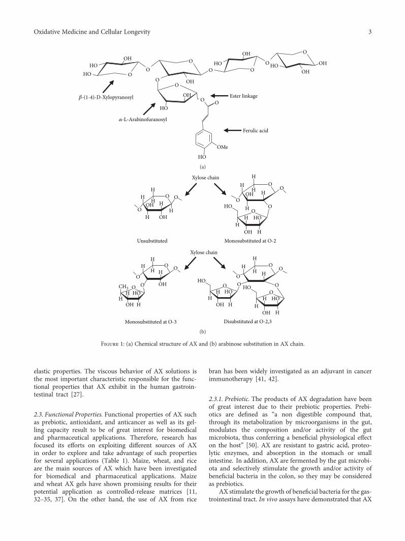
2.1. Chemical Structure. AX are polysaccharides from cerealgrains constituted by a linear β-(1-4)-xylopyranosyl chain.Some α-L-arabinofuranosyl residues are linked to the mainxylose chain at O-3 and/or O-2 positions, resulting in fourdifferent structures (monosubstituted at O-3 or O-2, disub-stituted at O-2,3, and unsubstituted) (Figure 1(b)). Theamount and distribution of these branches can vary depend-ing on the source of the polysaccharide [26]. In addition toarabinose, some galactose, xylose, and glucuronic acid resi-dues can exist as side branches in the main chain of AX [27].
A particular structural characteristic of AX is the pres-ence of phenolic acids. Some FA and cumaric acid residuescan be esterified to arabinose at the O-5 position [28](Figure 1(a)). FA is the most abundant phenolic acid in AX,and its content depends on the origin of the tissue (Table 1).AX from endosperm contains very small amounts of FA,while AX from pericarp and aleurone layer are highly
esterified to FA [20]. The FA contents in AX vary from0.001 to 7.00μg/mg AX [29, 30]. High contents of FA(6–7.00μg/mg AX) have been detected in maize bran AX[25, 30], while AX extracted from finger millet bran and ispa-ghula seed contain very low or even undetectable amounts ofFA (0.001μg/mg AX) [29, 31]. These differences could berelated to the source of the polysaccharide as well as themethod used for its extraction.
Usually, the structure of AX in different cereal tissues issimilar, although some differences in the fine structure candrastically modify its functional properties. These differencesare reflected in the degree of polymerization (DP), arabinox-ylan to xylose ratio (A/X), amount and sequence of glycosidicbonds, and the presence of other substituents [7]. AX can beclassified according to their solubility in aqueous solvents aswater-extractable (WE-AX) and water-unextractable (WU-AX) AX. In cereals, the cross-linking between AX and othercomponents from the cell wall form structures that areinsoluble in water. Alkali treatments are used to hydrolyzesuch cross-links, allowing the release of AX chains fromthe cell wall and making them soluble in the aqueous envi-ronment [27].
The substitution degree in the AX structure can be deter-mined by the A/X. The A/X may vary from 0.3 to 1.1 in AXfrom different cereals depending on the origin of the polysac-charide; AX from pericarp present higher A/X values thanthose for endosperm or aleurone layer [46, 47]. However, thisparameter does not describe detailed and exhaustive struc-tural characteristics of AX, and therefore, it cannot be usedto characterize its fine structure.
The molecular weight (Mw) of AX can vary dependingon the polysaccharide origin and the method used for itsdetermination. The average Mw estimated for AX rangesfrom 10 to 10,000 kDa [7]. The Mw and the Mw distribution(polydispersity index (PI)) of AX can be affected by theextraction conditions (time, pH, and temperature) [48].
2.2. Physicochemical Characteristics. AX show physicochem-ical characteristics, such as solubility and viscosity, whichprovide them different functional properties. Similar to otherpolysaccharides, the water solubility of AX depends on cer-tain parameters such as the chain-chain and chain-solventinteractions. In addition, some structural factors includingthe chain length and the presence and distribution of sidegroups can also modify the solubility of the polymers. Thesubstitution pattern of the polysaccharide chain is the mainparameter controlling the solubility of AX. Since the mecha-nism of aggregation in the AX is due to the intermolecularinteractions of the unsubstituted regions of the polysaccha-ride chain, the presence of arabinose residues in the xylosechain is determinant for the solubility of AX [48].
AX form very high viscous solutions in aqueous envi-ronments. The apparent viscosity of the AX solutions isconcentration- and shear rate-dependent. The viscosityvalues increase as the polymer concentration increases, anddecrease as the shear rate increases [27]. The Mw is anotherimportant factor that determines the viscosity of the AX solu-tions. Izydorczyk and Biliaderis [49] demonstrated thatwheat AX solutions with high Mw fractions showed weak
2 Oxidative Medicine and Cellular Longevity
elastic properties. The viscous behavior of AX solutions isthe most important characteristic responsible for the func-tional properties that AX exhibit in the human gastroin-testinal tract [27].
2.3. Functional Properties. Functional properties of AX suchas prebiotic, antioxidant, and anticancer as well as its gel-ling capacity result to be of great interest for biomedicaland pharmaceutical applications. Therefore, research hasfocused its efforts on exploiting different sources of AXin order to explore and take advantage of such propertiesfor several applications (Table 1). Maize, wheat, and riceare the main sources of AX which have been investigatedfor biomedical and pharmaceutical applications. Maizeand wheat AX gels have shown promising results for theirpotential application as controlled-release matrices [11,32–35, 37]. On the other hand, the use of AX from rice
bran has been widely investigated as an adjuvant in cancerimmunotherapy [41, 42].
2.3.1. Prebiotic. The products of AX degradation have beenof great interest due to their prebiotic properties. Prebi-otics are defined as “a non digestible compound that,through its metabolization by microorganisms in the gut,modulates the composition and/or activity of the gutmicrobiota, thus conferring a beneficial physiological effecton the host” [50]. AX are resistant to gastric acid, proteo-lytic enzymes, and absorption in the stomach or smallintestine. In addition, AX are fermented by the gut microbi-ota and selectively stimulate the growth and/or activity ofbeneficial bacteria in the colon, so they may be consideredas prebiotics.
AX stimulate the growth of beneficial bacteria for the gas-trointestinal tract. In vivo assays have demonstrated that AX
OO
O
OHOHHOO
O
OH
HOO
O
OHO
OO
OHHO
HO
O
HO
OH
HO
OMe
Ferulic acid
Ester linkage�훽-(1-4)-D-Xylopyranosyl
�훼-L-Arabinofuranosyl
(a)
OO
HO
H
H
OHH
HO
OHH
H
OOH
H
H
O
OO
HO
H
H
OHH
HO
OHH
H
O
H
O
OHO
H
H
OHH
HOO
O
HHH
H
OOH
H OH
OH
OH
OHH
H
OO
HO
OHO
H
H
OH
CH3
H
Unsubstituted Monosubstituted at O-2
Monosubstituted at O-3 Disubstituted at O-2,3
Xylose chain
Xylose chain
(b)
Figure 1: (a) Chemical structure of AX and (b) arabinose substitution in AX chain.
3Oxidative Medicine and Cellular Longevity
promote the growth of Bifidobacterium and Lactobacillus,both considered as beneficial species for the gut [51]. Thesemodifications in the gut microbiota are associated withhealth benefits, reduction in gastrointestinal infections, andimprovement in the mineral absorption and the suppressionof colon cancer [52, 53].
It has been established that some phenolic acids modu-late the composition of microbiota through the selective inhi-bition of some pathogenic bacteria, while the growth ofcommensal anaerobes and probiotic bacteria is less affectedor even increased [22, 54]. In this regard, the growth of pro-biotic such as Bifidobacterium and Lactobacillus during AXfermentation can be explained by the fact that these bacte-ria produce the FA esterases to release the FA residuesfrom AX [21, 55, 56].
Another benefit of AX as a prebiotic is the production ofbeneficial bacterial metabolites, such as short-chain fattyacids (SCFA). The fermentation of AX increases the produc-tion of acetic, propionic, and butyric acids. Particularly, AXare characterized by increasing the butyric acid levels, whichplay an important role in the maintenance of health and gas-trointestinal function [57]. The fermentation of AX is associ-ated with the growth of butyric acid-producing bacteria, suchas Eubacterium and Roseburia [58]. Nielsen et al. [59] evalu-ated the effect of AX-rich and high-fat diets on the produc-tion of SCFA in pigs. The supplementation of the AX-richdiet increased the SCFA levels, particularly the butyric acidlevel which was 5-fold higher compared with that obtainedfor the high-fat diet.
Butyric acid is considered an essential metabolite for thehuman colon as it is the main source of energy for its epithe-lial cells (colonocytes), contributes to the maintenance of thegut barrier functions, and has immunomodulatory and anti-
inflammatory properties [58]. In addition, the proliferationof butyrate-producing bacteria protects the colon from path-ogenic microbiota [60]. For those reasons, AX have beenconsidered as polysaccharides with excellent prebiotic prop-erties. The consumption of this polysaccharide providesmany health benefits, especially those related with the pre-vention of colon cancer.
Recently, long-chain AX (LC-AX) have demonstrated tomodulate the luminal and mucosal microbiota. Experimentsusing a dynamic in vitro model of the human digestive tract(M-SHIME) showed that supplementation of LC-AX to theproximal colon compartments of the M-SHIME increasedBifidobacterium population in both lumen and mucuscompared with the control. The levels of propionate aswell as the activity of enzymes β-xylanase, β-xylosidase, andα-arabinofuranosidase were also increased in the lumenregion.Thesefindings suggest thatLC-AXcould exert apoten-tial prebiotic effect on the host, as the mucosa-associatedmicrobiota impacts directly in health by protecting againstpathogen colonization and host immunity [61].
2.3.2. Antioxidant. The antioxidant activity of AX has beenmainly associated with their content of phenolic acids, par-ticularly FA. Phenolic acids have beneficial effects againstchronic and cardiovascular diseases, cancer, diabetes, inflam-matory diseases, and aging [62]. Phenolic acids exhibittheir antioxidant activity through diverse mechanisms suchas free radical scavenging, metal chelation, and reducingpotential, blocking the free radical chain, modulation ofenzymatic activity, and alteration of signal transductionpathways [63–65].
The main function of antioxidants is delaying or preven-tion of the oxidation produced by free radicals [66]. The free
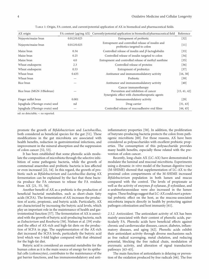
Table 1: Origin, FA content, and current/potential application of AX in biomedical and pharmaceutical fields.
AX origin FA content (μg/mg AX) Currently/potential application in biomedical/pharmaceutical field Reference
Nejayote/maize bran 0.012/0.025 Entrapment of probiotic [32]
Nejayote/maize bran 0.012/0.025Entrapment and controlled release of insulin and
probiotics targeted to colon[11]
Maize bran 0.34 Controlled release of insulin and β-lactoglobulin [33]
Maize bran 0.25 Controlled release of insulin targeted to colon [34]
Maize bran 4.0 Entrapment and controlled release of methyl xanthine [35]
Wheat endosperm 2.3 Controlled release of proteins [36]
Wheat endosperm 0.53 Entrapment of probiotics [37]
Wheat bran 0.435 Antitumor and immunomodulatory activity [16, 38]
Wheat bran — Prebiotic [39]
Rice bran — Antitumor and immunomodulatory activity [40]
Rice bran (MGN-3/Biobran) —Cancer immunotherapy
Prevention and inhibition of cancerSynergistic effect with chemotherapeutic agents
[15, 41, 42]
Finger millet bran 0.001 Immunomodulatory activity [29]
Ispaghula (Plantago ovata) seed nd Drug carrier [31, 43]
Ispaghula (Plantago ovata) seed — Controlled release of mucoadhesive oral films [44, 45]
nd: no detectable; −: no reported.
4 Oxidative Medicine and Cellular Longevity
radicals are generated by diverse factors such as normalmetabolic activity, diet, and environment. In a natural man-ner, the body uses antioxidant endogenous enzymes as adefense mechanism against the free radicals. An increasein the production of free radicals and other reactive oxygenspecies exceeding normal levels in the body results in oxida-tive stress. This imbalance causes damage to biomoleculessuch as membrane lipids, lipoproteins, and DNA, increasingthe risk of developing chronic diseases [67]. Therefore, theuse of antioxidants results to be adequate to decrease theeffects of free radicals and the risk of chronic diseases, suchas cancer.
Although the antioxidant activity of AX is associated withthe presence of phenolic acids, some studies suggest that thisactivity is mainly attributed to FA. The antioxidant activity ofFA is attributed to its structural characteristics. The presenceof electron-donating groups on its benzene ring gives it theproperty of terminating the free radical chain reactions. Inaddition, its COOH– group can bind to the lipid bilayer, pro-viding protection against the free radicals attack and the lipidperoxidation [62]. Since FA is the most abundant phenolicacid in AX, it may be the main responsible for the antioxidantactivity of the polysaccharide, as has been observed in differ-ent studies.
Recently, Kamboj and Rana [68] compared the antioxi-dant activity of maize bran gum with the antioxidant activityfrom other natural gums as xanthan and guar gums. Theyfound that the maize fiber gum exhibited a higher antioxi-dant activity compared to the other gums, regardless of themethod used for the determination. They suggested thatmaize fiber gum could be a promising excipient with antiox-idant activity in the food and pharmaceutical industry.
Feruloyl oligosaccharides (FOS), hydrolytic products ofAX, exhibit a protective effect on the cells against the damageproduced by the free radicals. Wang et al. [14] evaluated theprotective effect of FOS against the oxidative stress in ratplasma. The levels of oxidized glutathione and malondialde-hyde and the activity of antioxidant enzymes in plasma fromrats fed with the FOS diet decreased with respect to the con-trol group. In a previous study, the protective activity of FOSagainst the oxidative DNA damage in normal human lym-phocytes induced by hydrogen peroxide was investigated.The DNA damage was inhibited by FOS, observing a 91%inhibition of lymphocyte DNA damage at 500μmol/L ascompared with control [69].
The content and appearance of FA determine the antiox-idant capacity of AX. Higher contents of trimers of FA (tri-FA) in AX result in higher antioxidant activity [5]. Thisbehavior is attributed to three units of FA, which providehigher amounts of OH– groups, increasing the hydrogendonor capacity and, therefore, protecting from radical scav-enging [70]. This highlights the close relationship existingbetween the structural characteristics of the polysaccharideand its functional properties. Therefore, when discussingabout the antioxidant capacity of AX, not only the FA con-tent should be considered but also how it is found in the mol-ecule. The knowledge of such structural characteristics wouldhelp to predict the antioxidant activity of AX in order to con-sider it for specific applications.
2.3.3. Anticancer. Cancer is among the leading causes ofdeath worldwide, and the second most common in theUnited States [71]. Usually, conventional cancer treatmentsincluding surgery, chemotherapy, and radiotherapy whichfocus on eliminating cancer cells are short-term effective,but not enough for a complete eradication of all cancercells, resulting in recurrence of disease [72]. The repeatedsessions of treatments lead to the suppression of the immunesystem and promote multidrug resistance and toxicity[41, 73]. Therefore, the search for natural products withchemopreventive properties and without side effects hasbeen increasing.
AXOS exhibit protective effects against colon cancer,which have been related to their prebiotic effect. Femiaet al. [17] observed that the administration of AXOS reducedthe preneoplastic lesions in the colon of rats. The authorssuggest that AXOS exhibited a chemopreventive effect oncolon carcinogenesis due to their prebiotic activity. On theother hand, Glei et al. [74] showed that the fermentationproducts of wheat AX (SCFA) inhibited the growth of coloncancer cells (HT29) and induced the antioxidant activity ofthe endogenous enzyme glutathione transferase.
A close relationship between the proliferation of cancercells and the antioxidant systems has been suggested. Cancercells produce large amounts of hydrogen peroxide, whichmay favor mutations, damage, and invasion of other tissues.Then, cancer cell proliferation impacts directly in the antiox-idant machinery, and according to this, some anticanceragents can act as antioxidants [19]. Thus, the antitumorpotential of AX has been related to its antioxidant effect.Noaman et al. [19] supplied AX from rice to rats, which werepreviously inoculated with Erlich cancer cells. The resultsshowed the inhibition of the development and growth oftumors, as well as a decrease in lipid peroxidation andincrease in the activity of endogenous antioxidant enzymes(catalase, superoxide dismutase, and glutathione transferase).The authors suggested that AX exerted an antioxidant effectthrough its ability to increase the gene expression and activityof endogenous antioxidant enzymes in the cells and normal-ize the lipid peroxidation in blood, liver, and tumor tissue inanimals bearing tumors.
In a previous study, the antitumor activity of MGN-3/Biobran on mice bearing a solid Erlich carcinoma (SEC)tumor was attributed to mechanisms involving induction ofapoptosis and immune modulation. The administration ofMGN-3 significantly decreased tumor volume (63.27%) andtumor weight (45.2%) in comparison to the control group.Flow cytometry and histopathological analyses showed anincrease in the number of apoptotic SEC cells. In addition,an improvement of cytokine production was observed asshown by increasing levels of tumor necrosis factor-α andinterferon-γ, while the levels of immune suppressing IL-10were downregulated. In addition, the activity of natural killer(NK) cells was also increased [75].
The synergistic anticancer effect of MGN-3/Biobran withnatural anticancer agents as well as chemotherapeutic drugshas been widely explored using in vitro studies. The synergis-tic apoptotic potential of MGN-3 and curcumin on humanmultiple myeloma cell line U266 was determined. Treatment
5Oxidative Medicine and Cellular Longevity
of MGN-3 or curcumin alone showed an inhibition of cellproliferation in a dose-dependent manner. The combinationof MGN-3 and curcumin caused a synergistic effect char-acterized by a decrease in cell number and an increasein apoptotic cells. The expression of the proapoptotic pro-tein (Bax) increased, while antiapoptotic protein (Bcl-2)decreased, which favored apoptosis. These findings indicatedthat Biobran and curcumin synergize in the induction of apo-ptosis [76]. A similar behavior was observed when Biobranwas combined with paclitaxel in order to sensitize humanand murine breast cancer cells (MCF-7 and 4T1) to pacli-taxel. A synergistic effect between Biobran and paclitaxelresulted in damage of DNA, enhancement of apoptosis, andinhibition of 4T1 cell proliferation [77].
In sum, researches have demonstrated the anticancerpotential of the AX. Such property is attributed to both theantioxidant and prebiotic capacity of the polysaccharide. Inaddition, it is suggested that AX exerts its anticancer effectby a mechanism which involves its immune-modulationability. In vitro as well as in vivo studies have evidenced theanticancer effect of AX on different types of cancer. Never-theless, most studies highlight its beneficial effects in the pre-vention of colon cancer, due to the benefits of its prebioticactivity and immunostimulatory activity.
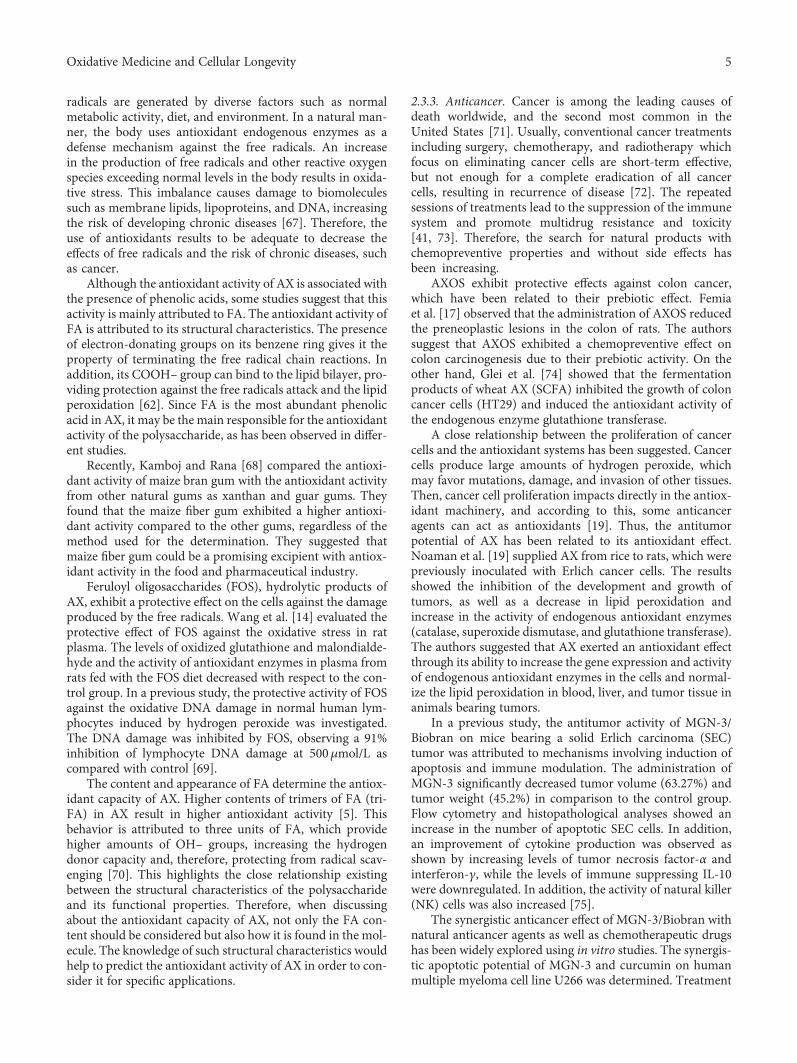
2.3.4. Gelling. A particular property of AX is their capacity toform covalent hydrogels. AX can gel through the covalentcross-linking of FA [6], under the action of different chemical(ferric chloride, ammonium persulphate) or enzymatic (lac-case/O2, peroxidase/H2O2, linoleic acid/lipoxygenase) oxi-dizing agents [78–83] (Figure 2(b)). The ability of AX to geldepends on the concentration of the polysaccharide, Mw,and particularly the FA content [27].
The oxidative gelation of AX results from the dimeriza-tion of FA residues of adjacent polysaccharide chains leadingto the formation of the three-dimensional network where theaqueous phase is retained. The FA dimerization mechanismoccurs as follows: first, an oxidizing agent attacks the H atomof the OH group at the ring position of FA resulting in a phe-noxy radical. Then, this radical is stabilized by resonance andlocated at three different positions onto the whole molecule,two on the aromatic ring (C-4 and C-5) and one at the doublebound (C-8) of its side chain. In the next step, the cross-linking between two phenoxy radicals is carried out; the cou-pling of unpaired electrons of two different radicals forms acovalent linkage which connects the two polysaccharidechains. Thus, the structure of the dimers formed during gela-tion will depend on the radical position [48] (Figure 2(a)).
During gelling, the coupling of FA results in different struc-tures. In AX gels, five di-FA, 8-5′, 8-O-4′, 5-5′, 8-5′ benzo, and8-8′, have been detected, with 8-5′ and 8-O-4′ being the mostabundant [48], and one tri-FA (4-O-8′/5′-5′) [84]. In addition,the presence of noncovalent weak interactions (hydrogenbonds) may contribute to the stability of the gel [85, 86].
During the formation of the AX gel, the FA is oxidizedand disappears as a result of the formation of cross-links(di-FA and tri-FA). Nevertheless, the concentrations ofdi-FA and tri-FA formed at the end of gelation do notcompensate for the decrease in the FA monomers. There-fore, the formation of superior FA oligomers (FA tetramers,FA pentamers) have been proposed by several authors[84, 86]. In the cell wall of cereals, five tri-FA, 5-5′/8-O-4′,8-O-4/8-O-4′, 8-8′ (cyclic)/8-O-4′, 8-O-4′/8-5′ (noncyclic), 5-5/8-O-4′(H2O), and two FA tetramers, 4-O-8′/5–5′/8-O-4′and 4-O-8′/5–5′/8–5′, have been identified and characterized
AX chain O
OH
FA
OMeO
OMeO
O AX chainMeO
O
MeOO
CO
C
AX chain
AX chain O
O
OHOMe
MeOHO
AX chain8-5′ di-FA
Phenoxy radicals
C OMeO
OMe
Laccase
AX chain AX chainO O AX chain O
C
O2
(a)
HOHO O
OO
O
O O HO
HO
8-5′ di-FAOMe
MeOOH
OH
OHOH
OHOH
OHOH
OHOH
HO
O O
OO
OO
OOO
OO
AX chain
AX chain
OO
HO
HOOH
OO
O
OHOHHO
OH
OH
OH
(b)
Figure 2: (a) Schematic representation of FA dimerization. (b) Covalent cross-linking of ferulated AX. Formation of 8-5′ di-FA is presentedas an example. AX: arabinoxylan; FA: ferulic acid; di-FA: ferulic acid dimer.
6 Oxidative Medicine and Cellular Longevity
[87]. The latest evidence suggests the possibility that themissingFA at the end of the gelling process could be related with theformation of superior structures that are not yet identified.
The AX gels present interesting features with a wide rangeof applications. These gels have neutral flavor, odor, and coloras well as high water absorption and exhibit pH, temperature,and ionic stability [7]. They usually form quickly, and they arestrong and thermostable [88]. In addition, they acquire ameso- and macroporous structure and have a dietary fibernature. Due to their interesting characteristics, AX gels couldbe good candidates for their use as controlled release matricesfor bioactive agents in the pharmaceutical, cosmetic, and foodindustries [7].
As previously mentioned, the antioxidant and prebioticproperties depend on the structure of AX. The presence ofphenolic acids in AX, particularly FA, has been related toits prebiotic as well as antioxidant capacities. In addition,the resulting products of the enzymatic hydrolysis of AXexert important prebiotic properties. Then, it can be possiblethat the cross-linking process and formation of superior
ferulate structures may contribute to some extent to the anti-oxidant and prebiotic properties of AX gels.
2.4. Preclinical Studies. Several in vivo studies have been per-formed in order to explore the potential of AX to exert itsprebiotic, antioxidant, and anticancer effects. Following, thecharacteristics and findings of some of the most recent stud-ies evaluating the effects of AX administration on animalmodels (mainly rats and mice) are presented.
2.4.1. Prebiotic Effect. The prebiotic effect of AX and theirderivatives, xylooligosaccharides (XOS) and AXOS, has beentested in vivo. Several studies using animal models (mainlyrats) demonstrate the potential prebiotic effect of AX throughdifferent observations: showing a bifidogenic effect, modula-tion of mucosa and gut microbiota, and increasing SCFAlevels (mainly propionic and butyric acid), among others.Table 2 shows the characteristics and observations of somestudies performed during the last 10 years related to the eval-uation of the prebiotic properties of AX.
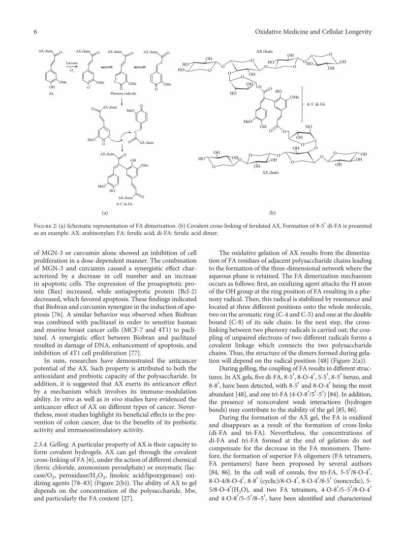
Table 2: In vivo studies on the evaluation of the prebiotic effect of AX.
Animal model Diet/experimental time Findings Reference
Male chickensControl diet (CT), diet supplemented withXOS, wheat bran-derived AXOS, wheatendosperm alkali-solubilized AX. 2 w
All treatments increased bifidobacteria.AX decreased body weight gain after 2weeks of feeding compared with CT.
[89]
Male C57bl6/J miceControl diet, high-fat (HF) diet, HFdiet supplemented with AX. 4 w
HF diet supplemented with AX restoredmicrobiota with a major effect on Roseburia
spp., Bacteroides-Prevotella spp., andbifidobacteria.
Improvement of gut barrier function,decrease in adipocyte size, fatty acid uptake,fatty acid oxidation and inflammation, anddecrease in key lipogenic enzyme activity
in the subcutaneous adipose tissue.
[39]
Male germ-free Fisher 344albino rats inoculated withhuman faecal microbiota
Control diet, diet supplemented withlong-chain AX (LC-AX) and dietsupplemented with inulin (IN). 6 w
LC-AX and IN increased SCFA levels(propionate and butyrate, resp.). Stimulation
of butyrate-producing bacteria and bifidobacteria,respectively. Reduction of mucin-degradingAkkermansia muciniphila and more mucinproduction by the host. Less weight gain.
[90]
Male Wistar ratsDiets supplemented with WU-AX,
WE-AX, and AXOS. 14 days
WU-AX supplementation increased butyrateproduction and butyrate-producing bacteria.WE-AX and/or AXOS reduced pH, suppressedrelevant markers of proteolytic breakdown,and induced selective bifidogenic response.
Combination of WU-AX, WE-AX, and AXOSshowed a synergic effect.
[91]
Male C57bl6/J miceControl diet, high-fat (HF) diet, HFdiet supplemented with AXOS. 8 w
AXOS supplementation exerted a bifidogeniceffect. Improvement of the HF-induced body
weight gain, fat mass development,hyperinsulinemia, insulin resistance,
endotoxemia, and inflammatory disorders ina model of HF diet-induced obesity.
[13]
PigsLow dietary fiber and high-fat
diet (WSD), AX-rich diet (AXD),and resistant starch diet (RS). 3 w
AXD feeding shifted the microbial compositiontowards butyrogenic species in the faeces andincreased the large-intestinal butyrate pool size.
[59]
w: week.
7Oxidative Medicine and Cellular Longevity
The supplementation of wheat AX to high-fat (HF) dietin mice led to an increase in bifidobacteria, particularly B.animalis lactis. In addition, AX modulated the microbiotaby restoring the levels of bacteria (Bacteroides-Prevotellaspp., Roseburia spp.) that were decreased with the HF. Thebifidogenic effect of AX was correlated with lower levels ofinflammatory markers in the serum which resulted in theimprovement of the gut barrier functions. The consumptionof AX also decreased body weight gain and fat mass develop-ment in the HF diet-induced obesity group. Moreover, thehypercholesteremia and the content of free cholesterol inthe liver decreased in HF diet feeding. The presence ofsmaller adipocytes in the group treated with HF diet andAX was also observed which was attributed to the ability ofAX to decrease the expression of genes involved in adipocytedifferentiation, fatty acid uptake and oxidation, lipolysis, andfatty acid synthesis. The authors proposed a positive correla-tion between the modulation of microbiota and the antiobe-sity action as well as the cholesterol-lowering effect observedin the experiment [39]. Similar effects were observed whendiet-induced obese mice were fed with a HF diet supple-mented with wheat-derived AXOS. A positive correlationbetween the increase of bifidobacteria and improvement ofmetabolic endotoxemia and inflammatory markers was alsoobserved with administration of AXOS. A higher expressionof the tight junction proteins Z01 and claudin 3 led to a betterfunction of the gut barrier. Moreover, the peptides GLP-1 andPYY, which are involved in the regulation of food intake andglucose homeostasis, respectively, increased after AXOS sup-plementation. This increase could be related with lower foodintake and the improvement of insulin sensitivity inmice [13].
The supplementation of long-chain AX (LC-AX) in ratsinoculated with human faecal microbiota has been associatedwith the production of propionic acid and stimulation of B.longum. LC-AX administration also increased the produc-tion of mucin, while it shifted mucin degradation from thecaecum to the colon. The degradation of mucin in the distalregions of colon could be beneficial as most chronic colonicdiseases, such as ulcerative colitis and colorectal cancer, orig-inate in this region [90].
2.4.2. Antioxidant Effect. The antioxidant property of AX hasbeen related with several beneficial effects. In vivo studies,using rats, show that AX exerts its antioxidant effect by mod-ulating lipid peroxidation, improving the activity of antioxi-dant enzymes, and protecting against oxidative stress.These positive effects have been related to the mechanismsof AX to exert its anticancer effect as well as improve lipidmetabolic disorder and suppress lipid peroxidation.
Recently, male Sprague-Dawley fed with a HF dietsupplemented with AX (HF-AX) showed lower triglycerideconcentration in serum in comparison with the HF dietgroup. Higher lipoprotein lipase (LPL), hepatic lipase (HL),total lipase, and acyl-CoA oxidase (ACO) activities and lowertriglyceride and cholesterol levels in the liver of the HF-AXgroup were observed. The authors suggest that intake of AXhelped to alleviate lipid metabolic disorder by reducing tri-glycerides and low-density lipoprotein in serum of rats. Theadministration of HF-AX changed the lipid metabolism by
improving the activity of fatty acid oxidation enzymes(LPL, HL, and ACO) which helped to reduce the triglyceridelevels in liver. AX could help to maintain normal fat levels byactivating lipid catabolism and oxidation rather than inhibit-ing lipid synthesis. Moreover, the activity of antioxidantenzymes glutathione peroxidase and total superoxide dis-mutase was also improved by the ingestion of AX resultingin a reduction of the oxidative stress in serum and tissues.The results also indicated that AX may alleviate the damageof hepatic morphology by regulation of liver cell apoptosis(Bax). These findings showed that supplementation of AXimproved lipid metabolic disorder and alleviated liver dam-age by activation of liver lipid catabolism and suppressionof lipid peroxidation in rats [92].
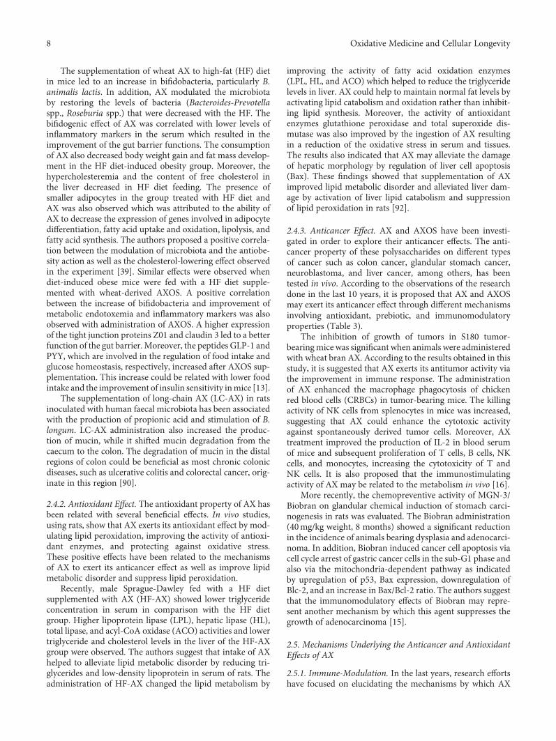
2.4.3. Anticancer Effect. AX and AXOS have been investi-gated in order to explore their anticancer effects. The anti-cancer property of these polysaccharides on different typesof cancer such as colon cancer, glandular stomach cancer,neuroblastoma, and liver cancer, among others, has beentested in vivo. According to the observations of the researchdone in the last 10 years, it is proposed that AX and AXOSmay exert its anticancer effect through different mechanismsinvolving antioxidant, prebiotic, and immunomodulatoryproperties (Table 3).
The inhibition of growth of tumors in S180 tumor-bearingmice was significant when animals were administeredwith wheat bran AX. According to the results obtained in thisstudy, it is suggested that AX exerts its antitumor activity viathe improvement in immune response. The administrationof AX enhanced the macrophage phagocytosis of chickenred blood cells (CRBCs) in tumor-bearing mice. The killingactivity of NK cells from splenocytes in mice was increased,suggesting that AX could enhance the cytotoxic activityagainst spontaneously derived tumor cells. Moreover, AXtreatment improved the production of IL-2 in blood serumof mice and subsequent proliferation of T cells, B cells, NKcells, and monocytes, increasing the cytotoxicity of T andNK cells. It is also proposed that the immunostimulatingactivity of AX may be related to the metabolism in vivo [16].
More recently, the chemopreventive activity of MGN-3/Biobran on glandular chemical induction of stomach carci-nogenesis in rats was evaluated. The Biobran administration(40mg/kg weight, 8 months) showed a significant reductionin the incidence of animals bearing dysplasia and adenocarci-noma. In addition, Biobran induced cancer cell apoptosis viacell cycle arrest of gastric cancer cells in the sub-G1 phase andalso via the mitochondria-dependent pathway as indicatedby upregulation of p53, Bax expression, downregulation ofBlc-2, and an increase in Bax/Bcl-2 ratio. The authors suggestthat the immunomodulatory effects of Biobran may repre-sent another mechanism by which this agent suppresses thegrowth of adenocarcinoma [15].
2.5. Mechanisms Underlying the Anticancer and AntioxidantEffects of AX
2.5.1. Immune-Modulation. In the last years, research effortshave focused on elucidating the mechanisms by which AX
8 Oxidative Medicine and Cellular Longevity
exerts its anticancer effects. In this regard, several studieshave demonstrated that one of such mechanisms couldinvolve the immune-modulation properties of AX.
It has been proposed that MGN-3/Biobran (AX fromrice) exhibits its anticancer effects due to its ability to act asa biological response modifier (BRM). BRM are designed toactivate the host immune response to destroy cancer cells
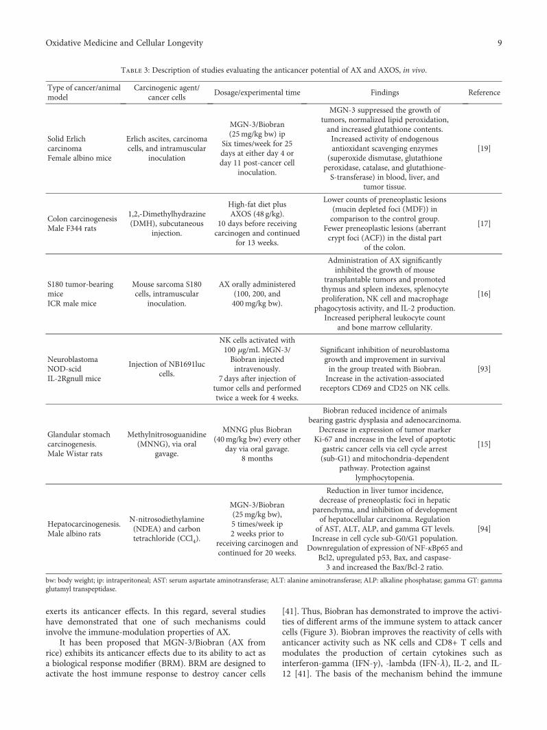
[41]. Thus, Biobran has demonstrated to improve the activi-ties of different arms of the immune system to attack cancercells (Figure 3). Biobran improves the reactivity of cells withanticancer activity such as NK cells and CD8+ T cells andmodulates the production of certain cytokines such asinterferon-gamma (IFN-γ), -lambda (IFN-λ), IL-2, and IL-12 [41]. The basis of the mechanism behind the immune
Table 3: Description of studies evaluating the anticancer potential of AX and AXOS, in vivo.
Type of cancer/animalmodel
Carcinogenic agent/cancer cells
Dosage/experimental time Findings Reference
Solid ErlichcarcinomaFemale albino mice
Erlich ascites, carcinomacells, and intramuscular
inoculation
MGN-3/Biobran(25mg/kg bw) ip
Six times/week for 25days at either day 4 orday 11 post-cancer cell
inoculation.
MGN-3 suppressed the growth oftumors, normalized lipid peroxidation,and increased glutathione contents.Increased activity of endogenousantioxidant scavenging enzymes
(superoxide dismutase, glutathioneperoxidase, catalase, and glutathione-S-transferase) in blood, liver, and
tumor tissue.
[19]
Colon carcinogenesisMale F344 rats
1,2,-Dimethylhydrazine(DMH), subcutaneous
injection.
High-fat diet plusAXOS (48 g/kg).
10 days before receivingcarcinogen and continued
for 13 weeks.
Lower counts of preneoplastic lesions(mucin depleted foci (MDF)) incomparison to the control group.
Fewer preneoplastic lesions (aberrantcrypt foci (ACF)) in the distal part
of the colon.
[17]
S180 tumor-bearingmiceICR male mice
Mouse sarcoma S180cells, intramuscular
inoculation.
AX orally administered(100, 200, and400mg/kg bw).
Administration of AX significantlyinhibited the growth of mouse
transplantable tumors and promotedthymus and spleen indexes, splenocyteproliferation, NK cell and macrophage
phagocytosis activity, and IL-2 production.Increased peripheral leukocyte count
and bone marrow cellularity.
[16]
NeuroblastomaNOD-scidIL-2Rgnull mice
Injection of NB1691luccells.
NK cells activated with100 μg/mL MGN-3/Biobran injectedintravenously.
7 days after injection oftumor cells and performedtwice a week for 4 weeks.
Significant inhibition of neuroblastomagrowth and improvement in survivalin the group treated with Biobran.Increase in the activation-associated
receptors CD69 and CD25 on NK cells.
[93]
Glandular stomachcarcinogenesis.Male Wistar rats
Methylnitrosoguanidine(MNNG), via oral
gavage.
MNNG plus Biobran(40mg/kg bw) every other
day via oral gavage.8 months
Biobran reduced incidence of animalsbearing gastric dysplasia and adenocarcinoma.
Decrease in expression of tumor markerKi-67 and increase in the level of apoptotic
gastric cancer cells via cell cycle arrest(sub-G1) and mitochondria-dependent
pathway. Protection againstlymphocytopenia.
[15]
Hepatocarcinogenesis.Male albino rats
N-nitrosodiethylamine(NDEA) and carbontetrachloride (CCl4).
MGN-3/Biobran(25mg/kg bw),5 times/week ip2 weeks prior to
receiving carcinogen andcontinued for 20 weeks.
Reduction in liver tumor incidence,decrease of preneoplastic foci in hepatic
parenchyma, and inhibition of developmentof hepatocellular carcinoma. Regulationof AST, ALT, ALP, and gamma GT levels.Increase in cell cycle sub-G0/G1 population.
Downregulation of expression of NF-κBp65 andBcl2, upregulated p53, Bax, and caspase-3 and increased the Bax/Bcl-2 ratio.
[94]
bw: body weight; ip: intraperitoneal; AST: serum aspartate aminotransferase; ALT: alanine aminotransferase; ALP: alkaline phosphatase; gamma GT: gammaglutamyl transpeptidase.
9Oxidative Medicine and Cellular Longevity
modulatory effects of Biobran is not completely elucidated.However, it is proposed that these AX are hydrolyzed inorder to reduce its MW so they can diffuse through intestinalwalls or directly into the blood stream and then be trans-ported to the lymph nodes where immune cells reside [95, 96].
NK cells play an important role in the natural defense ofthe immune system against cancer and viral infections [93].These cells work by attaching to cancer cells and releasingtheir granules which form holes causing cell death [41].AX has the ability to increase the cytotoxicity activity ofNK cells as confirmed by several in vitro and in vivo studies[16, 93]. Dendritic cells (DCs) are important antigen-presenting cells (APCs) involved in generating antitumorimmune response. Biobran upregulates CD80 and CD86which are molecules expressed on mature DCs. This stimu-lation promotes the production of proinflammatory andimmune-regulatory cytokines. It is proposed that Biobrancould bind to the cell surface receptors (TLRs and/or C typelectins) or to intracellular receptors (NLRP3 inflammasome)and trigger signaling pathways involved in cell activationand cytokine production [41].
A recent study evaluated the potential capacity of Biobranto activate and improve the cytotoxicity of NK cell activityagainst neuroblastoma in vitro, using several pediatric celllines (acute leukaemia, neuroblastoma, Ewig sarcoma, embry-onic rhabdomyosarcoma, and alveolar rhabdomyosarcoma)and in vivo (NOD/scid/IL-2Rγnull neuroblastoma model)[93]. The stimulation of NK cells with Biobran resulted in ahigher expression of the activation-associated receptorsCD25 and CD69 than in unstimulated cells. In addition, thestimulation increased NK cell cytotoxicity against cancer celllines and reduced the neuroblastoma growth in vivo. Severalmechanisms are proposed in order to explain how Biobrancould stimulate NK cell activity. One of those theories estab-lishes anapoptotic effectmediated by the activationofNKcellsreleasing TNF-α and IFN-γ [75]. Other mechanisms could berelated with the increasing of activating receptors on Biobran-stimulated NK cells. The authors observed an increase in theactivation-associated receptor CD69 and CD25 on stimulatedNK cells. The increase of CD69 is associated with a higher
cytotoxicity of NK cells, while CD25 expression on NK cellsis indicative of proliferation potential [97, 98]. NK cells attackcancer and viral cells through the release of their granules thatcausecell death. In this regard,Biobran treatment increases thegranular content (perforin and granzyme) ofNKcells favoringits activity against malignant cells [99]. In addition, the treat-ment with Biobran helps NK cells to attach cancer cells [41].
AX can stimulate the production of interleukins such ascytokines IL-2 and IL-12 which are the main anticancer cyto-kines in humans [41]. In S180 tumor-bearing mice, the treat-ment with AX led to an increase in the secretion of IL-2 in theblood of mice. It is postulated that increase of IL-2 may be amechanism for AX to exert antitumor effects, as IL-2 canimprove the proliferation of T cells, B cells, NK cells, andmonocytes and increase the cytotoxicity of T cells and NKcells [16, 100]. In addition, the ingestion of Biobran increasedthe production of IL-12 in multiple myeloma patients at oneand two months post-ingestion [101].
AX can induce the production of TNFs (IFN-λ andIFN-γ) which have been found to exhibit antitumor activity[75, 101, 102]. In a previous study, the oral administrationof partially hydrolyzed AX from corn husk to mice increasedthe production of IL-2 and IFN-γ and slightly increased IL-4in mitogen-induced proliferation spleen cells. In addition, anincrease in the activity of NK cells in spleen cells fromtransplanted-tumor mice was observed [102]. T helper 1(Th1) promotes antitumor immunity through the produc-tion of IL-2 and IFN-γ, which activates NK cells to attackcancer and virus-infected cells [103]. In this study, theadministration of AX also decreased ear inflammation of amodel mouse of atopic dermatitis. According to the resultsobtained, it is suggested that the anti-inflammatory effect ofAX is attributed to the activation of an IFN-γ-dependentTh1-like immune response in mice. In another study, multi-ple myeloma patients presented an increase of IFN-γ at twomonths post-treatment with MGN-3 [101].
T regulatory lymphocytes (T reg) or CD4+CD25+ lym-phocytes act by suppressing the antitumor cytotoxic immuneresponse [104]. In this regard, it is proposed that the counter-acting of T reg cell activity could interfere in a positive way
NK cells
CD4+T cells
NK cells
Cancer cell
CD8+T cellDCs
A
IFN-�훾
IL-2 ,IL-12IFN-�휆
Figure 3: MGN-3/Biobran enhances the cytotoxicity reactivity of immune cells with anticancer effect and the production of certain cytokines(adapted from [41]).
10 Oxidative Medicine and Cellular Longevity
with the progression of neoplastic diseases by improving theefficacy of the anticancer immune response [41]. AX fromrice was given orally to 22 patients with solid tumor for twomonths. The results showed an increase in TH cells, whileT reg cells decreased, but the differences were not statisticallysignificant. On the contrary, the TH/T reg ratio significantlyenhanced after AX therapy [40].
2.5.2. Induction of Apoptosis. Induction of apoptosis appearsto be another mechanism by which MGN-3/Biobran mayexert its anticancer effects. In vivo studies evaluated the effectof Biobran on mice bearing a SEC tumor as well as in chemi-cally induced glandular stomach adenocarcinoma rats. Accord-ing to the results observed in those experiments, the anticanceractivity exhibited by Biobran was explained through a mecha-nism via induction of apoptosis.
The administration of Biobran in mice bearing a SECtumor caused a significant delay in the volume and weightof the tumor in comparison to the control. The authors pro-posed that the antitumor activity of Biobran was related to itsability to induce apoptosis and immune modulation. Theintraperitoneal treatment of Biobran on mice increased thenumber of apoptotic SEC cells. Moreover, cytokine produc-tion was influenced by increasing the levels of tumor necrosisfactor-α and IFN-γ, while a downregulation of the immunesuppressing cytokine IL-10 was observed. In addition, a con-siderable increase in NK cell activity was observed in micetreated with Biobran [75]. Biobran can cause tumor regres-sion by the induction of cancer cell apoptosis via its immunu-modulatory effects on NK cells and cytokine production. It isknown that NK cells kill cancer cells by different pathways,and one of those involves the ligation of FasL to its Fas recep-tor to induce apoptosis [105]. It is also possible that Biobrancould exert its apoptotic effect via the increase of TNF-α andIFN-γ. In this regard, both TNF-α and IFN-γ have beenshown to act synergistically to induce cancer cell deaththrough apoptotic and necrotic effects [106, 107].
Recently, the chemopreventive activity of Biobran againstchemical induction of glandular stomach carcinogenesis inrats was associated with the ability of Biobran to induce apo-ptosis via the mitochondrial-dependent pathway in gastriccancer cells [15]. Biobran treatment caused a significantreduction in the incidence of animals bearing gastric dyspla-sia and adenocarcinoma in comparison to the untreatedgroup. The upregulation in p53 expression, Bax expression,downregulation in Bcl-2 expression, the increase in Bax/Blc-2 ratio, and the activation of caspase-3 as well as theinduction of cell-cycle arrest in the sub-G1 phase mayexplain the mitochondria-dependent pathway as the mecha-nism involved in the anticancer effect observed in the presentstudy. Changes in p53, Bax, and Bcl-2 can alter the outermitochondrial membrane and subsequent release of cyto-chrome C, which finally activates caspase-3. In this regard,Biobran has shown to induce apoptosis via activation of cas-pase-8, -9, and -3 [108]. Although the mechanism by whichBiobran exerts its apoptotic effect is not completely eluci-dated, it could be related to the capacity of Biobran to sensi-tize the surface CD95 receptor that is involved in thetriggering of apoptosis [109]. On the other hand, another
possible mechanism by which Biobran suppresses the growthof tumor could be related to its immunomodulatory proper-ties. It was observed that treatment with Biobran protectedagainst chemical-induced lymphocytopenia in rats. Lympho-cytes are white blood cells which are part of the immunesystem [15].
2.5.3. Antioxidant. AX has been found to exert antioxidanteffects through the modulation of lipid peroxidation, pro-moting the antioxidant defense system and protectingagainst oxidative stress [19]. Although the mechanismsbehind the antioxidant property of AX are not fully under-stood, some studies suggest that its capacity to increase theactivity of endogenous antioxidant enzymes, suppress lipidperoxidation, and induce apoptosis could be involved in suchmechanisms [19, 92]. The antioxidant activity of AX has beenrelated to its capacity to exert anticancer effect, improve lipidmetabolic disorder, and alleviate liver damage in rats.
Noaman et al. [19] evaluated the antioxidant activity as apossible mechanism of Biobran to exert its antitumor poten-tial on mice inoculated with Erlich ascites carcinoma (EAC)cells. Biobran administration suppressed tumor growth bynormalizing the lipid peroxidation level and augmentationof glutathione (GSH) contents. In addition, the expressionand activity of endogenous antioxidant scavenging enzymes(superoxide dismutase, glutathione peroxidase, catalase, andglutathione-S-transferase) in the cells of normal and tumor-bearing animals were increased in blood, liver, and tumortissue. The ability of Biobran to induce apoptosis was pro-posed as the mechanism by which it could exert those antiox-idant effects. Reactive oxygen species (ROS) act as signalingmolecules for the initiation and execution of apoptosis.GSH and thioredoxin not only regulate ROS levels but couldact as reversible redox modifiers of enzyme function [110]. Inthis regard, higher levels of GSH content in tissues of micetreated with Biobran were observed in comparison to theuntreated mice. Among other functions, glutathione-S-transferase enzymes detoxify carcinogens [111]. This alsocould be a possible mechanism for Biobran to prevent canceras elevated levels of such enzymes were observed in micetreated with Biobran.
The antioxidant capacity of AX has been also related tothe improvement of metabolic disorder and alleviate liverdamage in rats induced by high-fat diet [92]. Lipid peroxida-tion is one of the most common free radical chain reactionsthat causes oxidative damage [112]. In this study, mice fedwith a high-fat diet presented an increase in serum and tissueoxidative stress and subsequent reduction of the antioxidantenzymes glutathione peroxidase and total superoxide dis-mutase as well as an increase in malondialdehyde level. Onthe contrary, supplementation of AX in high-fat diet cata-lyzed the dismutation of the superoxide (O−2) radical intoeither molecular oxygen (O2) or hydrogen peroxide (H2O2)by the promotion of total superoxide dismutase. In addition,the increase in glutathione peroxidase activity reduced lipidhydroperoxides [92]. On the other hand, AX supplementa-tion could alleviate damage liver morphology by regulatingthe liver cell apoptosis through the modulation of the expres-sion of proapoptotic and antiapoptotic proteins, Bax and Bcl-
11Oxidative Medicine and Cellular Longevity
2. Moreover, Biobran has the ability to change the lipidmetabolism by regulating the expression of UCP2, a mito-chondrial membrane protein that works by accelerating thefatty acid β-oxidation and minimizing the production ofreactive oxygen species [113]. These results may suggest thatBiobran improved lipid catabolism and protected liver dam-age via an antioxidant mechanism.
3. Arabinoxylan Gels
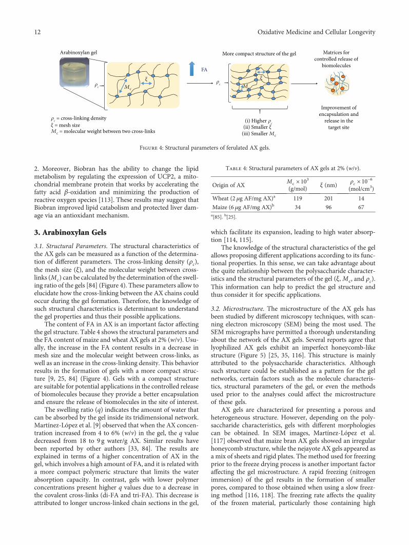
3.1. Structural Parameters. The structural characteristics ofthe AX gels can be measured as a function of the determina-tion of different parameters. The cross-linking density (ρc),the mesh size (ξ), and the molecular weight between cross-links (Mc) can be calculated by the determination of the swell-ing ratio of the gels [84] (Figure 4). These parameters allow toelucidate how the cross-linking between the AX chains couldoccur during the gel formation. Therefore, the knowledge ofsuch structural characteristics is determinant to understandthe gel properties and thus their possible applications.
The content of FA in AX is an important factor affectingthe gel structure. Table 4 shows the structural parameters andthe FA content of maize and wheat AX gels at 2% (w/v). Usu-ally, the increase in the FA content results in a decrease inmesh size and the molecular weight between cross-links, aswell as an increase in the cross-linking density. This behaviorresults in the formation of gels with a more compact struc-ture [9, 25, 84] (Figure 4). Gels with a compact structureare suitable for potential applications in the controlled releaseof biomolecules because they provide a better encapsulationand ensure the release of biomolecules in the site of interest.
The swelling ratio (q) indicates the amount of water thatcan be absorbed by the gel inside its tridimensional network.Martínez-López et al. [9] observed that when the AX concen-tration increased from 4 to 6% (w/v) in the gel, the q valuedecreased from 18 to 9 g water/g AX. Similar results havebeen reported by other authors [33, 84]. The results areexplained in terms of a higher concentration of AX in thegel, which involves a high amount of FA, and it is related witha more compact polymeric structure that limits the waterabsorption capacity. In contrast, gels with lower polymerconcentrations present higher q values due to a decrease inthe covalent cross-links (di-FA and tri-FA). This decrease isattributed to longer uncross-linked chain sections in the gel,
which facilitate its expansion, leading to high water absorp-tion [114, 115].
The knowledge of the structural characteristics of the gelallows proposing different applications according to its func-tional properties. In this sense, we can take advantage aboutthe quite relationship between the polysaccharide character-istics and the structural parameters of the gel (ξ,Mc, and ρc).This information can help to predict the gel structure andthus consider it for specific applications.
3.2. Microstructure. The microstructure of the AX gels hasbeen studied by different microscopy techniques, with scan-ning electron microscopy (SEM) being the most used. TheSEM micrographs have permitted a thorough understandingabout the network of the AX gels. Several reports agree thatlyophilized AX gels exhibit an imperfect honeycomb-likestructure (Figure 5) [25, 35, 116]. This structure is mainlyattributed to the polysaccharide characteristics. Althoughsuch structure could be established as a pattern for the gelnetworks, certain factors such as the molecule characteris-tics, structural parameters of the gel, or even the methodsused prior to the analyses could affect the microstructureof these gels.
AX gels are characterized for presenting a porous andheterogeneous structure. However, depending on the poly-saccharide characteristics, gels with different morphologiescan be obtained. In SEM images, Martínez-López et al.[117] observed that maize bran AX gels showed an irregularhoneycomb structure, while the nejayote AX gels appeared asa mix of sheets and rigid plates. The method used for freezingprior to the freeze drying process is another important factoraffecting the gel microstructure. A rapid freezing (nitrogenimmersion) of the gel results in the formation of smallerpores, compared to those obtained when using a slow freez-ing method [116, 118]. The freezing rate affects the qualityof the frozen material, particularly those containing high
Arabinoxylan gel More compact structure of the gel Matrices forcontrolled release of
biomolecules
Improvement ofencapsulation and
release in thetarget site
�휌c = cross-linking density
�휉 = mesh sizeMc = molecular weight between two cross-links
�휌c
�휌c
FA
(i) Higher �휌c
(ii) Smaller �휉(iii) Smaller Mc
Mc
�휉Mc
�휉
Figure 4: Structural parameters of ferulated AX gels.
Table 4: Structural parameters of AX gels at 2% (w/v).
Origin of AXMc × 103(g/mol)
ξ (nm)ρc × 10−6(mol/cm3)
Wheat (2 μg AF/mg AX)a 119 201 14
Maize (6 μg AF/mg AX)b 34 96 67a[85]. b[25].
12 Oxidative Medicine and Cellular Longevity
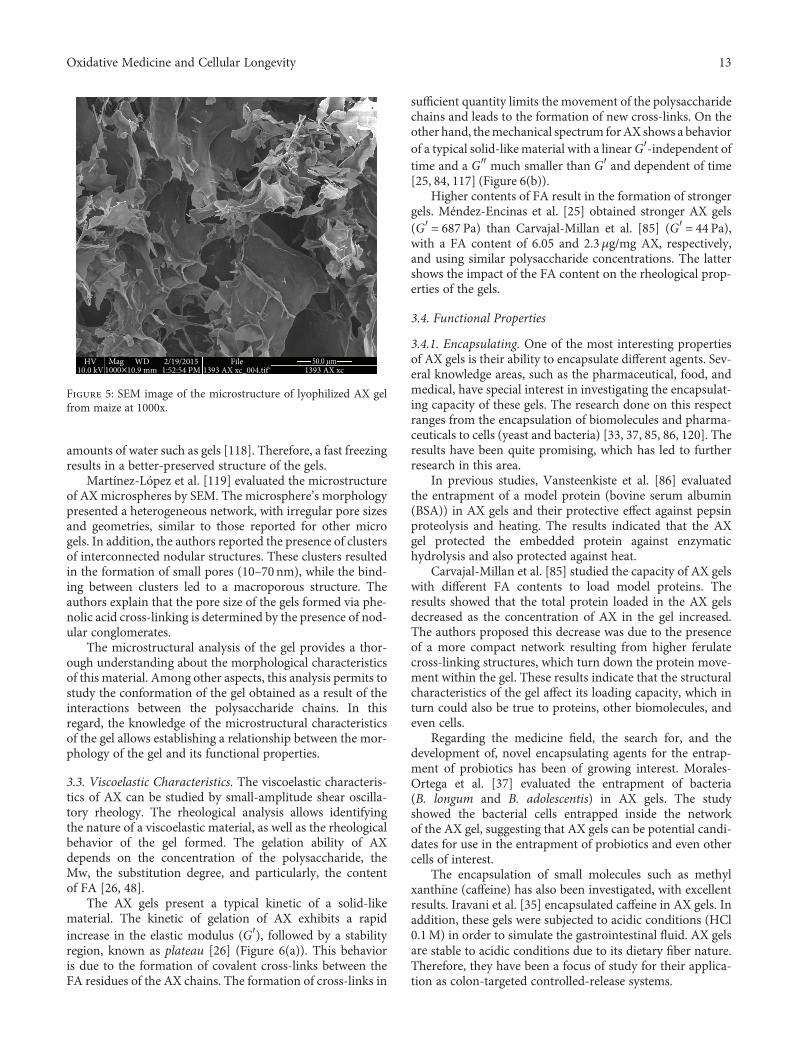
amounts of water such as gels [118]. Therefore, a fast freezingresults in a better-preserved structure of the gels.
Martínez-López et al. [119] evaluated the microstructureof AX microspheres by SEM. The microsphere’s morphologypresented a heterogeneous network, with irregular pore sizesand geometries, similar to those reported for other microgels. In addition, the authors reported the presence of clustersof interconnected nodular structures. These clusters resultedin the formation of small pores (10–70 nm), while the bind-ing between clusters led to a macroporous structure. Theauthors explain that the pore size of the gels formed via phe-nolic acid cross-linking is determined by the presence of nod-ular conglomerates.
The microstructural analysis of the gel provides a thor-ough understanding about the morphological characteristicsof this material. Among other aspects, this analysis permits tostudy the conformation of the gel obtained as a result of theinteractions between the polysaccharide chains. In thisregard, the knowledge of the microstructural characteristicsof the gel allows establishing a relationship between the mor-phology of the gel and its functional properties.
3.3. Viscoelastic Characteristics. The viscoelastic characteris-tics of AX can be studied by small-amplitude shear oscilla-tory rheology. The rheological analysis allows identifyingthe nature of a viscoelastic material, as well as the rheologicalbehavior of the gel formed. The gelation ability of AXdepends on the concentration of the polysaccharide, theMw, the substitution degree, and particularly, the contentof FA [26, 48].
The AX gels present a typical kinetic of a solid-likematerial. The kinetic of gelation of AX exhibits a rapidincrease in the elastic modulus (G′), followed by a stabilityregion, known as plateau [26] (Figure 6(a)). This behavioris due to the formation of covalent cross-links between theFA residues of the AX chains. The formation of cross-links in
sufficient quantity limits the movement of the polysaccharidechains and leads to the formation of new cross-links. On theother hand, themechanical spectrum forAX shows a behaviorof a typical solid-like material with a linearG′-independent oftime and a G″ much smaller than G′ and dependent of time[25, 84, 117] (Figure 6(b)).
Higher contents of FA result in the formation of strongergels. Méndez-Encinas et al. [25] obtained stronger AX gels(G′ = 687 Pa) than Carvajal-Millan et al. [85] (G′ = 44 Pa),with a FA content of 6.05 and 2.3μg/mg AX, respectively,and using similar polysaccharide concentrations. The lattershows the impact of the FA content on the rheological prop-erties of the gels.
3.4. Functional Properties
3.4.1. Encapsulating. One of the most interesting propertiesof AX gels is their ability to encapsulate different agents. Sev-eral knowledge areas, such as the pharmaceutical, food, andmedical, have special interest in investigating the encapsulat-ing capacity of these gels. The research done on this respectranges from the encapsulation of biomolecules and pharma-ceuticals to cells (yeast and bacteria) [33, 37, 85, 86, 120]. Theresults have been quite promising, which has led to furtherresearch in this area.
In previous studies, Vansteenkiste et al. [86] evaluatedthe entrapment of a model protein (bovine serum albumin(BSA)) in AX gels and their protective effect against pepsinproteolysis and heating. The results indicated that the AXgel protected the embedded protein against enzymatichydrolysis and also protected against heat.
Carvajal-Millan et al. [85] studied the capacity of AX gelswith different FA contents to load model proteins. Theresults showed that the total protein loaded in the AX gelsdecreased as the concentration of AX in the gel increased.The authors proposed this decrease was due to the presenceof a more compact network resulting from higher ferulatecross-linking structures, which turn down the protein move-ment within the gel. These results indicate that the structuralcharacteristics of the gel affect its loading capacity, which inturn could also be true to proteins, other biomolecules, andeven cells.
Regarding the medicine field, the search for, and thedevelopment of, novel encapsulating agents for the entrap-ment of probiotics has been of growing interest. Morales-Ortega et al. [37] evaluated the entrapment of bacteria(B. longum and B. adolescentis) in AX gels. The studyshowed the bacterial cells entrapped inside the networkof the AX gel, suggesting that AX gels can be potential candi-dates for use in the entrapment of probiotics and even othercells of interest.
The encapsulation of small molecules such as methylxanthine (caffeine) has also been investigated, with excellentresults. Iravani et al. [35] encapsulated caffeine in AX gels. Inaddition, these gels were subjected to acidic conditions (HCl0.1M) in order to simulate the gastrointestinal fluid. AX gelsare stable to acidic conditions due to its dietary fiber nature.Therefore, they have been a focus of study for their applica-tion as colon-targeted controlled-release systems.
HV10.0 kV
Mag1000×
WD10.9 mm
2/19/20151:52:54 PM
File1393 AX xc_004.tif⁎ 1393 AX xc
50.0 �휇m
Figure 5: SEM image of the microstructure of lyophilized AX gelfrom maize at 1000x.
13Oxidative Medicine and Cellular Longevity
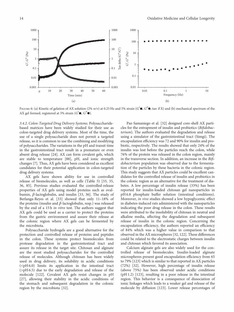
3.4.2. Colon-Targeted Drug Delivery Systems. Polysaccharide-based matrices have been widely studied for their use ascolon-targeted drug delivery systems. Most of the time, theuse of a single polysaccharide does not permit a targetedrelease, so it is common to use the combining and modifyingof polysaccharides. The variations in the pH and transit timein the gastrointestinal tract result in a premature or evenabsent drug release [24]. AX can form covalent gels, whichare stable to temperature [88], pH, and ionic strengthchanges [7]. Thus, AX gels have been considered as excellentcandidates for their potential application in colon-targeteddrug delivery systems.
AX gels have shown ability for use in controlledrelease of biomolecules, as well as cells (Table 5) [33, 35,36, 85]. Previous studies evaluated the controlled-releaseproperties of AX gels using model proteins such as oval-bumin, β-lactoglobulin, and insulin [33, 36]. The study ofBerlanga-Reyes et al. [33] showed that only 11–18% ofthe proteins (insulin and β-lactoglobulin, resp.) was releasedby the end of a 15 h in vitro test. The authors suggest thatAX gels could be used as a carrier to protect the proteinsfrom the gastric environment and assure their release atthe colonic region where AX gels can be fermented bythe microbiota.
Polysaccharide hydrogels are a good alternative for theprotection and controlled release of proteins and peptidesin the colon. These systems protect biomolecules fromprotease degradation in the gastrointestinal tract andassure its release in the target site. Chitosan and alginateare the most studied polysaccharides for the controlledrelease of molecules. Although chitosan has been widelyused in drug delivery, its solubility in acidic conditions(<pH6.0) limits its application in the intestinal tract(>pH6.5) due to the early degradation and release of themolecule [122]. Covalent AX gels resist changes in pH[27], allowing their stability in the acidic conditions ofthe stomach and subsequent degradation in the colonicregion by the microbiota [32].
Paz-Samaniego et al. [32] designed core-shell AX parti-cles for the entrapment of insulin and probiotics (Bifidobac-terium). The authors evaluated the degradation and releaseusing a simulator of the gastrointestinal tract (Simgi). Theencapsulation efficiency was 72 and 90% for insulin and pro-biotic, respectively. The results showed that only 24% of theinsulin was lost before the particles reach the colon, while76% of the protein was released in the colon region, mainlyin the transverse section. In addition, an increase in the Bifi-dobacterium population was observed due to the fermenta-tion of the particles by these bacteria in the colonic region.This study suggests that AX particles could be excellent can-didates for the controlled release of insulin and probiotics inthe colonic region as an alternative for the treatment of dia-betes. A low percentage of insulin release (33%) has beenreported for insulin-loaded chitosan gel nanoparticles inpH6.8 phosphate buffer solution (intestinal conditions).Moreover, in vivo studies showed a low hypoglycemic effectin diabetes-induced rats administered with the nanoparticlesindicating the poor drug release in the colon. These resultswere attributed to the insolubility of chitosan in neutral andalkaline media, affecting the degradation and subsequentrelease of insulin in the colonic region. Concerning theencapsulation efficiency, the authors reported an efficiencyof 84% which was a higher value in comparison to thatobserved in the AX microspheres [32, 122]. These differencescould be related to the electrostatic charges between insulinand chitosan which favored its association.
Calcium alginate gels are also widely used for the con-trolled release of biomolecules. Insulin-loaded alginatemicrospheres present good encapsulation efficiency from 65to 79% [123] which is similar to that reported in AX particles(72%) [32]. However, high percentage of insulin release(above 75%) has been observed under acidic conditions(pH1.2) [123], resulting in a poor release in the intestinalregion. This behavior is a consequence of dissociation ofionic linkages which leads to a weaker gel and release of themolecule by diffusion [123]. Lower release percentages of
0 10 20 30 40 50 600.001
0.01
0.1
1
10
100
1000 1000
0.001
0.01
0.1
1
10
100
Tan
delta
(G″/G′)
G′, G″
(Pa)
Time (min)
(a)
0.01 0.1 1 100.01
0.1
1
10
100
1000
G′, G″
(Pa)
Frequency (Hz)
(b)
Figure 6: (a) Kinetic of gelation of AX solution (2% w/v) at 0.25Hz and 5% strain (G′■, G″●, tan δX) and (b) mechanical spectrum of theAX gel formed, registered at 5% strain (G′■, G″●).
14 Oxidative Medicine and Cellular Longevity
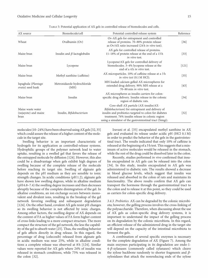
molecules (10–24%)havebeenobservedusingAXgels [32, 35]which could assure the release of a higher content of themole-cule in the target site.
Swelling behavior is an important characteristic ofhydrogels for its application as controlled-release systems.Hydrophilic groups of the polymer network lead to wateruptake, resulting in a swelled gel that favors the release ofthe entrapped molecule by diffusion [124]. However, this alsocould be a disadvantage when gels exhibit high degrees ofswelling because of the complete release of the moleculebefore reaching its target site. Swelling of alginate gelsdepends on the pH medium as they are sensible to ionicstrength changes. In acidic conditions (pH1.2), alginate gelshave shown low swelling degrees, while in alkaline medium(pH6.8–7.4) the swelling degree increases and then decreasesabruptly because of the complete disintegration of the gel. Inalkaline conditions, an ion exchange occurs between mono-valent and divalent ions, resulting in the breakup of the gelnetwork favoring swelling and subsequent degradation[124]. On the other hand, covalent AX gels resist pH changesso its swelling behavior is not affected by ionic charges.Among other factors, the swelling degree of AX depends onthe content of FA as higher values of FA form higher contentof cross-links leading to a more compact structure. The morecompact the structure of the gel network, the less the capabil-ity of the gel to absorb water [25]. Thus, the swelling behaviorof gels affects directly in drug release. In this regard, thepercentage of drug (celecoxib) released from alginate gelsin acidic medium was near 25%, while in alkaline condi-tions a complete release was observed at 8 h [124]. Similarvalues were reported for AX gels where 24% of insulin wasreleased in stomach conditions, while 75% was released inthe colon [32].
Iravani et al. [35] encapsulated methyl xanthine in AXgels and evaluated its release under acidic pH (HCl 0.1M)in order to predict the behavior of the gels in the gastrointes-tinal tract. The results indicated that only 10% of caffeine isreleased at the beginning of a 3 h test. This suggests that amin-imum of active molecules would be released in the stomach,while the rest of the drug could be dissolved later in the colon.
Recently, studies performed in vivo confirmed that insu-lin encapsulated in AX gels can be released into the colon[34]. In this study, insulin encapsulated in AX gels wasadministered to diabetic rats. The results showed a decreasein blood glucose levels, which suggest that insulin wasreleased and absorbed in the colon of rats and maintains itsfunctionality. The above results confirm that AX gels cantransport the hormone through the gastrointestinal tract tothe colon and to release it at this point, so they could be usedas carriers for colon-specific drug delivery.
3.4.3. Prebiotics. AX can be degraded by the colonic microbi-ota; however, the gelling process involves the cross-linking ofthe polysaccharide. Therefore, when discussing about the useof AX gels as colon-specific drug delivery systems, it isimportant to understand the impact of the gelling processon its degradation by the colonic microbiota. In this regard,an efficient release of the administered drug in the target sitewill depend on the capacity of the intestinal microbiota toferment the gels.
A combination of several specific enzymes is necessaryfor the complete degradation of AX (Figure 7). Among themain enzymes participating in its degradation are endo-1-4-β-xylanases (commonly called xylanases) which cleavethe xylose backbone randomly to shorter fragments and β-xylosidases that attack the nonreducing ends of the xylose
Table 5: Potential application of AX gels in controlled release of biomolecules and cells.
AX source Biomolecule/cell Potential controlled-release system Reference
Wheat Ovalbumin (Ov)Ov-AX gels for entrapment and controlledrelease of proteins. 70–88% protein releaseas Ov/AX ratio increased (24 h in vitro test).
[36]
Maize bran Insulin and β-lactoglobulinAX gels for controlled release of proteins.
11–18% of protein release at the end of a 15 hin vitro test.
[33]
Maize bran LycopeneLycopene/AX gels for controlled delivery ofbiomolecules. 3–4% lycopene release at the
end of a 4 h in vitro test.[121]
Maize bran Methyl xanthine (caffeine)AX microparticles. 10% of caffeine release at a 3 h
in vitro test (0.1M HCl).[35]
Ispaghula (Plantagoovata) seed husk
Metronidazole hydrochloride(MH)
MH-loaded calcium gelled AX microspheres forextended drug delivery. 90% MH release at a
70–80min in vitro test.[43]
Maize bran InsulinAX microspheres as insulin carriers for colon-
specific drug delivery. Insulin release in the colonicregion of diabetic rats.
[34]
Maize waste water(nejayote) and maizebran
Insulin, Bifidobacterium
Core-shell AX particle (AX-insulin/AX-Bifidobacterium) for entrapment and delivery of
insulin and probiotics targeted to colon for diabetestreatment. 76% insulin release in colonic region
using a simulator of the gastrointestinal tract (Simgi).
[32]
15Oxidative Medicine and Cellular Longevity
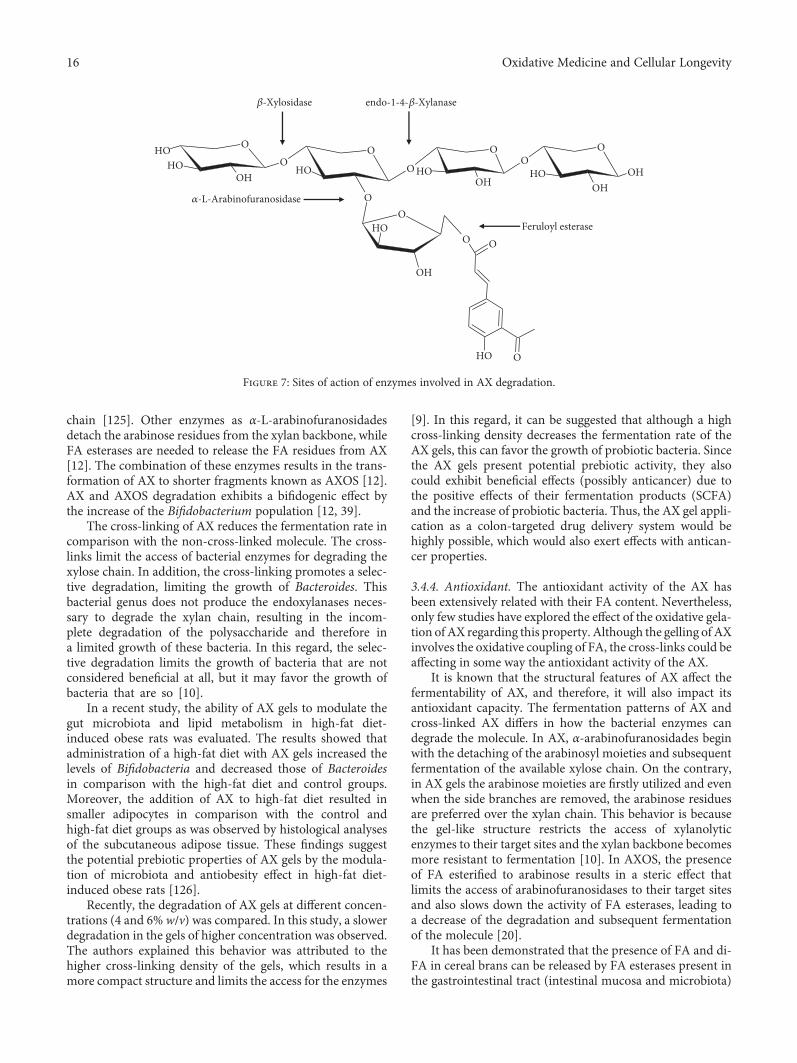
chain [125]. Other enzymes as α-L-arabinofuranosidadesdetach the arabinose residues from the xylan backbone, whileFA esterases are needed to release the FA residues from AX[12]. The combination of these enzymes results in the trans-formation of AX to shorter fragments known as AXOS [12].AX and AXOS degradation exhibits a bifidogenic effect bythe increase of the Bifidobacterium population [12, 39].
The cross-linking of AX reduces the fermentation rate incomparison with the non-cross-linked molecule. The cross-links limit the access of bacterial enzymes for degrading thexylose chain. In addition, the cross-linking promotes a selec-tive degradation, limiting the growth of Bacteroides. Thisbacterial genus does not produce the endoxylanases neces-sary to degrade the xylan chain, resulting in the incom-plete degradation of the polysaccharide and therefore ina limited growth of these bacteria. In this regard, the selec-tive degradation limits the growth of bacteria that are notconsidered beneficial at all, but it may favor the growth ofbacteria that are so [10].
In a recent study, the ability of AX gels to modulate thegut microbiota and lipid metabolism in high-fat diet-induced obese rats was evaluated. The results showed thatadministration of a high-fat diet with AX gels increased thelevels of Bifidobacteria and decreased those of Bacteroidesin comparison with the high-fat diet and control groups.Moreover, the addition of AX to high-fat diet resulted insmaller adipocytes in comparison with the control andhigh-fat diet groups as was observed by histological analysesof the subcutaneous adipose tissue. These findings suggestthe potential prebiotic properties of AX gels by the modula-tion of microbiota and antiobesity effect in high-fat diet-induced obese rats [126].
Recently, the degradation of AX gels at different concen-trations (4 and 6% w/v) was compared. In this study, a slowerdegradation in the gels of higher concentration was observed.The authors explained this behavior was attributed to thehigher cross-linking density of the gels, which results in amore compact structure and limits the access for the enzymes
[9]. In this regard, it can be suggested that although a highcross-linking density decreases the fermentation rate of theAX gels, this can favor the growth of probiotic bacteria. Sincethe AX gels present potential prebiotic activity, they alsocould exhibit beneficial effects (possibly anticancer) due tothe positive effects of their fermentation products (SCFA)and the increase of probiotic bacteria. Thus, the AX gel appli-cation as a colon-targeted drug delivery system would behighly possible, which would also exert effects with antican-cer properties.
3.4.4. Antioxidant. The antioxidant activity of the AX hasbeen extensively related with their FA content. Nevertheless,only few studies have explored the effect of the oxidative gela-tion of AX regarding this property. Although the gelling of AXinvolves the oxidative coupling of FA, the cross-links could beaffecting in some way the antioxidant activity of the AX.
It is known that the structural features of AX affect thefermentability of AX, and therefore, it will also impact itsantioxidant capacity. The fermentation patterns of AX andcross-linked AX differs in how the bacterial enzymes candegrade the molecule. In AX, α-arabinofuranosidades beginwith the detaching of the arabinosyl moieties and subsequentfermentation of the available xylose chain. On the contrary,in AX gels the arabinose moieties are firstly utilized and evenwhen the side branches are removed, the arabinose residuesare preferred over the xylan chain. This behavior is becausethe gel-like structure restricts the access of xylanolyticenzymes to their target sites and the xylan backbone becomesmore resistant to fermentation [10]. In AXOS, the presenceof FA esterified to arabinose results in a steric effect thatlimits the access of arabinofuranosidases to their target sitesand also slows down the activity of FA esterases, leading toa decrease of the degradation and subsequent fermentationof the molecule [20].
It has been demonstrated that the presence of FA and di-FA in cereal brans can be released by FA esterases present inthe gastrointestinal tract (intestinal mucosa and microbiota)
�훽-Xylosidase endo-1-4-�훽-Xylanase
Feruloyl esterase
�훼-L-Arabinofuranosidase
O
OHHO
HO
HO
O
O
OHO
O
OH
O
O
O
HOO O
O
OHHO
O
OHOHHO
Figure 7: Sites of action of enzymes involved in AX degradation.
16 Oxidative Medicine and Cellular Longevity
of humans and rats [127]. In this way, probiotics present inthe colonic microbiota, such as Lactobacillus and Bifidobac-terium, are able to produce FA esterases, enzymes requiredfor the release of the FA moieties from AX and AXOS[21, 55, 56]. A previous study showed that the presence oftwo Bifidobacterium strains (B. longum and B. adolescentis)is required for the fermentation of maize bran AX gels [9].Therefore, even if some bacteria produce the specific enzymesto release the FA moieties in AX gels, the cross-feedingbetween different bacteria producing different enzymes ishighlyneeded for the completedegradationofAXandAXgels.
The antioxidant capacity of phenolic acids depends onuptake and further metabolism [127]. Once the FA isreleased, it is then metabolized by the intestinal microbiota.The FA can be transformed into phenylpropionic acidsand/or vinyl phenol derivatives, which still exert certain anti-oxidant capacity but in a lower extent than FA. According tothis, it could be stated that the metabolism of FA by bacteriawill lead to a decrease in its antioxidant capacity as has beendemonstrated by Snelders et al. [20]. In this study, AXOSwith FA esterified to arabinose exhibited good antioxidantcapacity; however, the release and subsequent metabolizationof FA decreased its antioxidant capacity. The authors sug-gested that AXOS-bound FA will reach the colon, where FAcan be released and then metabolized by FA esterases locatedin this region [20]. In the colonic region, FA (bound or free)as well as its metabolites can exert its antioxidant activitythrough a direct interaction with the colon epithelium cellsand thus reduce colorectal cancer [128]. On the other hand,the absorption of di-FA from cereal bran in the gut and itssubsequent reach to the vascular system was demonstratedin rats [127]. These compounds also exhibit good antioxidantcapacity and may exert its beneficial effects in the colonicregion as they can interact directly with the intestinal barrier.
Previous studies have shown that not only the level of FAbut also the condition under which it appears (free, bond, ordimerized) impacts the antioxidant capacity of AX [129]. Thepresence of dimers and trimers (di-FA, tri-FA) or more com-plex ferulated structures decreases the antioxidant capacity ofAX in comparison to the FA molecule esterified to AX. Theavailability of hydroxyl groups and resonance systems ofFA vary according to the cross-linking position and areimportant factors to exert the antioxidant function of FA[87]. Thus, it is confirmed that the gelling process impactson the antioxidant capacity of the gels.
Several studies agree that the antioxidant activity of AXdecreases after the gelation process due to the formation ofdi-FA and tri-FA [8, 119]. It is well documented that the for-mation of the AX gel involves the cross-linking of FA andconsequently its oxidation. Nevertheless, the di-FA and tri-FA amounts produced during gelation do not compensatethe FA oxidized. The remnant FA can continue to react andform higher ferulated structures [84]. Trimers and tetramersof FA have been identified in the cell wall of cereal grains, andeven more complex ferulated structures are suggested [87].Since the remaining FA could react and continue to formmore complex structures, then it would be possible that suchFA could keep stabilizing the free radicals and thereforeexhibit antioxidant activity.
According to the above mentioned, the antioxidant activ-ity of AX gels could be mainly related to the FA remainingafter the gelation process. Thus, a higher FA content in AXgels may result in better antioxidant activity. AX with higherFA content (6μg/mg AX) can form gels with high cross-linking density and still exhibit antioxidant activity [8]. Inaddition, these gels preserve high amounts of FA after theirgelation [130], which would allow them to exert a greaterantioxidant activity.
The antioxidant activity of AX gels depends on the poly-saccharide structure. The content and appearance of FA inthe gel are the main factors affecting such property. AX gelswith high cross-linking density can exhibit antioxidant activ-ity. In this logical order, since the anticancer activity of AX isrelated to its antioxidant activity, the AX gels could alsoexhibit this property. However, the evaluation of the antican-cer activity of AX gels and its antioxidant property relation-ship is necessary. These findings could allow obtaining AXgels for colon-targeted drug delivery systems with protectiveeffect against colon cancer.
Ferulated AX exhibit prebiotic and antioxidant proper-ties, which confer anticancer potential to the polysaccharide.Since AX gels preserve their prebiotic and antioxidant prop-erties, they would also exert antiproliferative activity. Fur-thermore, the high cross-linking density of the gels wouldincrease such properties. In recent years, the investigationof the properties of AX gels has been the focus of numerousstudies due to its potential application as colon-targeted drugdelivery systems. In this regard, it is necessary to evaluate theeffect of gelation on the antiproliferative activity of AX.Moreover, the relationship between the structural character-istics and the functional properties of the AX gels should beinvestigated, particularly the effect of the high cross-linkingdensity of the gels on their antiproliferative activity. Highcross-linking density AX gels could be a good alternativefor the colon-targeted drug delivery in the treatment ofcolon cancer.
4. Conclusion
This review describes the functional properties of AX and AXgels and their potential application as antioxidant and anti-cancer agents. Ferulated AX have prebiotic, antioxidant,and anticancer properties which can be exploited by thepharmaceutical field. Recently, the research has focused inthe development of novel colon-targeted delivery systemsfor the treatment of colon associated diseases, such as inflam-matory bowel disease, irritable bowel disease, and particu-larly colon cancer. Due to their gelling ability, AX havegained interest as promising polysaccharides for the design-ing of drug delivery systems with bioactive properties suchas prebiotic, antioxidant, and anticancer.
The antioxidant and prebiotic activity of AX has beenpreviously demonstrated. The fermentation products of AXexhibit a prebiotic effect, providing benefits for the guthealth. Moreover, its antioxidant activity exerts a protectiveeffect against the free radicals and oxidative stress. Regardingthe gelling ability of AX, specific studies are needed to eluci-date such beneficial effects in the AX gels. The anticancer
17Oxidative Medicine and Cellular Longevity
activity of AX appears to be strongly related to its antioxidantand prebiotic properties. Future studies will be needed toevaluate the effect of gelation on the anticancer activity ofAX gels. Furthermore, this information will allow establish-ing a relationship between the anticancer activity of AX withits antioxidant and prebiotic properties.
The AX gels are promising candidates for application ascolon-targeted drug delivery systems for the treatment ofcolon cancer. However, only few studies regarding the evalu-ation of the antioxidant and prebiotic effect of AX gels havebeen conducted in vitro, and no evidence of in vivo data isavailable. Therefore, it is important to conduct research onthe performance of in vitro and in vivo studies focusing onthe effect of gelation on the biological properties (prebiotic,antioxidant, and anticancer) of AX and to elucidate the rela-tionship between the properties of the gel by itself, not only asa mere carrier.
Conflicts of Interest
The authors declare that there is no conflict of interestregarding the publication of this article.
Acknowledgments
This research was supported by “Fondo InstitucionalCONACyT-Investigación en Fronteras de la Ciencia”,México(Grant FON.INST./31/2016 to Elizabeth Carvajal-Millan).The authors are pleased to acknowledge Alma C. Campa-Mada, Karla G. Martínez-Robinson, and Alma R. Toledo-Guillén (CIAD, México).
References
[1] J. Slavin, “Why whole grains are protective: biological mech-anisms,” Proceedings of the Nutrition Society, vol. 62, no. 1,pp. 129–134, 2003.
[2] Y. Park, A. F. Subar, A. Hollenbeck, and A. Schatzkin, “Die-tary fiber intake and mortality in the NIH-AARP diet andhealth study,” Archives of Internal Medicine, vol. 171,no. 12, pp. 1061–1068, 2011.
[3] A. T. Kunzmann, H. G. Coleman, W. Y. Huang, C. M.Kitahara, M. M. Cantwell, and S. I. Berndt, “Dietary fiberintake and risk of colorectal cancer and incident and recur-rent adenoma in the prostate, lung, colorectal, and ovariancancer screening trial,” The American Journal of ClinicalNutrition, vol. 102, no. 4, pp. 881–890, 2015.
[4] J. M. Lattimer andM. D. Haub, “Effects of dietary fiber and itscomponents on metabolic health,” Nutrients, vol. 2, no. 12,pp. 1266–1289, 2010.
[5] F. E. Ayala-Soto, S. O. Serna-Saldívar, S. García-Lara, andE. Pérez-Carrillo, “Hydroxycinnamic acids, sugar composi-tion and antioxidant capacity of arabinoxylans extractedfrom different maize fiber sources,” Food Hydrocolloids,vol. 35, pp. 471–475, 2014.
[6] R. K. Durham, “Effect of hydrogen peroxide on relative vis-cosity measurements of wheat and flour suspensions,” CerealChemistry, vol. 2, no. 2, pp. 297–305, 1925.
[7] M. S. Izydorczyk and C. G. Biliaderis, “Cereal arabinoxylans:advances in structure and physicochemical properties,” Car-bohydrate Polymers, vol. 28, no. 1, pp. 33–48, 1995.
[8] R. Paz-Samaniego, M. Méndez-Encinas, J. M. Fierro-Islaset al., “Ferulated arabinoxylans recovered from low-valuemaize by-products: gelation and antioxidant capacity,” inFerulic Acid: Antioxidant Properties, Uses and PotentialHealth Benefits, W. Bryce, Ed., pp. 151–164, Nova SciencePublishers, Hauppauge, Inc, 2015.
[9] A. L. Martínez-López, E. Carvajal-Millan, V. Micard et al., “Invitro degradation of covalently cross-linked arabinoxylanhydrogels by bifidobacteria,” Carbohydrate Polymers,vol. 144, no. 1, pp. 76–82, 2016.
[10] M. J. Hopkins, H. N. Englyst, S. Macfarlane, E. Furrie, G. T.Macfarlane, and A. J. McBain, “Degradation of cross-linkedand non-cross-linked arabinoxylans by the intestinal micro-biota in children,” Applied and Environmental Microbiology,vol. 69, no. 11, pp. 6354–6360, 2003.
[11] R. Paz-Samaniego, A. Rascón-Chu, F. Brown-Bojorquezet al., “Electrospray-assisted fabrication of core-shell arabi-noxylan gel particles for insulin and probiotics entrapment,”Journal of Applied Polymer Science, vol. 135, no. 26, article46411, 2018.
[12] C. Grootaert, J. A. Delcour, C. M. Courtin, W. F. Broekaert,W. Verstraete, and T. van de Wiele, “Microbial metabolismand prebiotic potency of arabinoxylan oligosaccharides inthe human intestine,” Trends in Food Science & Technology,vol. 18, no. 2, pp. 64–71, 2007.
[13] A. M. Neyrinck, V. F. van Hée, N. Piront et al., “Wheat-derived arabinoxylan oligosaccharides with prebiotic effectincrease satietogenic gut peptides and reduce metabolicendotoxemia in diet-induced obese mice,” Nutrition & Dia-betes, vol. 2, no. 1, pp. 1–9, 2012.
[14] J. Wang, B. Sun, Y. Cao, and C. Wang, “Wheat bran feru-loyl oligosaccharides enhance the antioxidant activity ofrat plasma,” Food Chemistry, vol. 123, no. 2, pp. 472–476,2010.
[15] N. K. Badr El-Din, S. M. Abdel Fattah, D. Pan, L. Tolentino,and M. Ghoneum, “Chemopreventive activity of MGN-3/Biobran against chemical induction of glandular stomachcarcinogenesis in rats and its apoptotic effect in gastric cancercells,” Integrative Cancer Therapies, vol. 15, no. 4, pp. NP26–NP34, 2016.
[16] L. Cao, X. Liu, T. Qian et al., “Antitumor and immunomod-ulatory activity of arabinoxylans: a major constituent ofwheat bran,” International Journal of Biological Macromole-cules, vol. 48, no. 1, pp. 160–164, 2011.
[17] A. P. Femia, M. Salvadori, W. F. Broekaert et al., “Arabinox-ylan-oligosaccharides (AXOS) reduce preneoplastic lesionsin the colon of rats treated with 1,2-dimethylhydrazine(DMH),” European Journal of Nutrition, vol. 49, no. 2,pp. 127–132, 2010.
[18] W. F. Broekaert, C. M. Courtin, K. Verbeke, T. van de Wiele,W. Verstraete, and J. A. Delcour, “Prebiotic and other health-related effects of cereal-derived arabinoxylans, arabinoxylan-oligosaccharides, and xylooligosaccharides,” Critical Reviewsin Food Science and Nutrition, vol. 51, no. 2, pp. 178–194,2011.
[19] E. Noaman, N. K. Badr el-Din, M. A. Bibars, A. A. AbouMossallam, and M. Ghoneum, “Antioxidant potential byarabinoxylan rice bran, MGN-3/biobran, represents amechanism for its oncostatic effect against murine solid
18 Oxidative Medicine and Cellular Longevity
Ehrlich carcinoma,” Cancer Letters, vol. 268, no. 2, pp. 348–359, 2008.
[20] J. Snelders, H. Olaerts, E. Dornez et al., “Structural featuresand feruloylation modulate the fermentability and evolutionof antioxidant properties of arabinoxylanoligosaccharidesduring in vitro fermentation by human gut derived microbi-ota,” Journal of Functional Foods, vol. 10, pp. 1–12, 2014.
[21] D. Szwajgier and A. Jakubczyk, “Biotransformation of ferulicacid by Lactobacillus acidophilus K1 and selected Bifidobac-terium strains,” ACTA Scientiarum Polonorum TechnologiaAlimentaria, vol. 9, pp. 45–59, 2010.
[22] R. S. Govardhan Singh, P. S. Negi, and C. Radha, “Phenoliccomposition, antioxidant and antimicrobial activities of freeand bound phenolic extracts of Moringa oleifera seed flour,”Journal of Functional Foods, vol. 5, no. 4, pp. 1883–1891,2013.
[23] A. Gulbake, A. Jain, A. Jain, A. Jain, and S. K. Jain, “Insight todrug delivery aspects for colorectal cancer,”World Journal ofGastroenterology, vol. 22, no. 2, pp. 582–599, 2016.
[24] L. Zhang, Y. Sang, J. Feng, Z. Li, and A. Zhao, “Polysaccha-ride-based micro/nanocarriers for oral colon-targeted drugdelivery,” Journal of Drug Targeting, vol. 24, no. 7, pp. 579–589, 2016.
[25] M. Méndez-Encinas, E. Carvajal-Millan, M. Yadav et al.,“Gels of ferulated arabinoxylans: rheology, structuralparameters and microstructure,” in Advances in Physico-chemical Properties of Biopolymers, M. A. Masuelli and D.Renard, Eds., pp. 215–228, Bentham Science Publishers,Beijing, 2017.
[26] M. S. Izydorczyk and J. E. Dexter, “Barley β-glucans and ara-binoxylans: molecular structure, physicochemical properties,and uses in food products–a review,” Food Research Interna-tional, vol. 41, no. 9, pp. 850–868, 2008.
[27] M. S. Izydorczyk and C. G. Biliaderis, “Arabinoxylans: techo-nologically and nutrionally functional plant polysaccha-rides,” in Functional Food Carbohydrates, C. G. Biliaderisand M. S. Izydorczyk, Eds., pp. 249–283, CRC Press, BocaRaton, FL, USA, 2007.
[28] M. M. Smith and R. D. Hartley, “Occurrence and nature offerulic acid substitution of cell-wall polysaccharides in grami-naceous plants,” Carbohydrate Research, vol. 118, no. 1,pp. 65–80, 1983.
[29] M. R. Savitha Prashanth, R. R. Shruthi, and G. Muralikrishna,“Immunomodulatory activity of purified arabinoxylans fromfinger millet (Eleusine coracana, v. Indaf 15) bran,” Journal ofFood Science and Technology, vol. 52, no. 9, pp. 6049–6054,2015.
[30] A. Morales-Burgos, E. Carvajal-Millan, Y. López-Francoet al., “Syneresis in gels of highly ferulated arabinoxylans:characterization of covalent cross-linking, rheology, andmicrostructure,” Polymers, vol. 9, no. 12, 2017.
[31] M. S. Iqbal, J. Akbar, M. A. Hussain, S. Saghir, and M. Sher,“Evaluation of hot-water extracted arabinoxylans from ispa-ghula seeds as drug carriers,” Carbohydrate Polymers,vol. 83, no. 3, pp. 1218–1225, 2011.
[32] R. Paz-Samaniego, E. Carvajal-Millan, N. Sotelo-Cruz et al.,“Maize processing waste water upcycling in Mexico: recoveryof arabinoxylans for probiotic encapsulation,” Sustainability,vol. 8, no. 11, p. 1104, 2016.
[33] C.M. Berlanga-Reyes, E. Carvajal-Millán, J. Lizardi-Mendoza,A. Rascón-Chu, J. Marquez-Escalante, and A. L. Martínez-
López, “Maize arabinoxylan gels as protein deliverymatrices,”Molecules, vol. 14, no. 4, pp. 1475–1482, 2009.
[34] A. L. Martínez-López, E. Carvajal-Millan, J. Lizardi-Mendozaet al., “Matrices covalentes biodegradables para el suministrode insulina por vía oral dirigida al colon activado por lamicrobiota y proceso para su obtención,” México, Patentemexicana No. MX/a/2015/017857, 2016.
[35] S. Iravani, C. S. Fitchett, and D. M. R. Georget, “Physicalcharacterization of arabinoxylan powder and its hydrogelcontaining a methyl xanthine,” Carbohydrate Polymers,vol. 85, no. 1, pp. 201–207, 2011.
[36] E. Carvajal-Millan, S. Guilbert, J. L. Doublier, and V. Micard,“Arabinoxylan/protein gels: structural, rheological and con-trolled release properties,” Food Hydrocolloids, vol. 20, no. 1,pp. 53–61, 2006.
[37] A. Morales-Ortega, E. Carvajal-Millan, F. Brown-Bojorquezet al., “Entrapment of probiotics in water extractable arabi-noxylan gels: rheological and microstructural characteriza-tion,” Molecules, vol. 19, no. 3, pp. 3628–3637, 2014.
[38] S. Zhou, X. Liu, Y. Guo, Q. Wang, D. Peng, and L. Cao,“Comparison of the immunological activities of arabinoxy-lans from wheat bran with alkali and xylanase-aided extrac-tion,” Carbohydrate Polymers, vol. 81, no. 4, pp. 784–789,2010.
[39] A. M. Neyrinck, S. Possemiers, C. Druart et al., “Prebioticeffects of wheat arabinoxylan related to the increase in bifido-bacteria, roseburia and bacteroides/prevotella in diet-inducedobese mice,” PLoS One, vol. 6, no. 6, p. e20944, 2011.
[40] P. Lissoni, G. Messina, F. Brivio et al., “Modulation of theanticancer immunity by natural agents: inhibition of T regu-latory lymphocyte generation by arabinoxylan in patientswith locally limited or metastatic solid tumors,” Cancer Ther-apy, vol. 6, pp. 1011–1016, 2008.
[41] M. Ghoneum, “From bench to bedside: the growing use ofarabinoxylan rice bran (MGN-3/Biobran) in cancer immu-notherapy,” Austin Immunology, vol. 1, no. 2, p. 1006,2016.
[42] S. L. Ooi, D. McMullen, T. Golombick, D. Nut, and S. C. Pak,“Evidence-based review of BioBran/MGN-3 arabinoxylancompound as a complementary therapy for conventionalcancer treatment,” Integrative Cancer Therapies, vol. 17,pp. 1–14, 2017.
[43] C. J. Lee, C. S. Nah, C. S. Teng, W. W. Jun, and M. Saravanan,“Spray dried calcium gelled arabinoxylan microspheres: Anovel carrier for extended drug delivery,” Chemical Papers,vol. 69, no. 10, 2015.
[44] M. Hanif and M. Zaman, “Thiolation of arabinoxylan and itsapplication in the fabrication of controlled release mucoadhe-sive oral films,” Daru Journal of Pharmaceutical Sciences,vol. 25, no. 1, p. 6, 2017.
[45] M. Zaman, M. Hanif, and M. A. Khan, “Arabinoxylan-basedmucoadhesive oral films of tizanidine HCL designed andoptimized using central composite rotatable design,” Poly-mer-Plastics Technology and Engineering, vol. 57, no. 5,pp. 471–483, 2018.
[46] C. Antoine, S. Peyron, F. Mabille et al., “Individual contribu-tion of grain outer layers and their cell wall structure to themechanical properties of wheat bran,” Journal of Agriculturaland Food Chemistry, vol. 51, no. 7, pp. 2026–2033, 2003.
[47] C. Barron, A. Surget, and X. Rouau, “Relative amounts of tis-sues in mature wheat (Triticum aestivum L.) grain and their
19Oxidative Medicine and Cellular Longevity
carbohydrate and phenolic acid composition,” Journal ofCereal Science, vol. 45, no. 1, pp. 88–96, 2007.
[48] L. Saulnier, F. Guillon, P.-E. Sado, A.-L. Chateigner-Boutin,and X. Rouau, “Plant cell wall polysaccharides in storageorgans : xylans (Food applications),” in Elsevier ReferenceModule in Chemistry, Molecular Sciences and Chemical Engi-neering, J. Reedijk, Ed., pp. 1–32, Elsevier Inc,Waltham, 2013.
[49] M. S. Izydorczyk and C. G. Biliaderis, “Effect of molecular sizeon physical properties of wheat arabinoxylan,” Journal ofAgricultural and Food Chemistry, vol. 40, no. 4, pp. 561–568, 1992.
[50] L. B. Bindels, N. M. Delzenne, P. D. Cani, and J. Walter,“Towards a more comprehensive concept for prebiotics,”Nature Reviews Gastroenterology & Hepatology, vol. 12,no. 5, pp. 303–310, 2015.
[51] M. Vardakou, C. N. Palop, P. Christakopoulos, C. B. Faulds,M. A. Gasson, and A. Narbad, “Evaluation of the prebioticproperties of wheat arabinoxylan fractions and induction ofhydrolase activity in gut microflora,” International Journalof Food Microbiology, vol. 123, no. 1-2, pp. 166–170, 2008.
[52] S. Macfarlane, G. T. Macfarlane, and J. H. Cummings,“Review article: prebiotics in the gastrointestinal tract,” Ali-mentary Pharmacology & Therapeutics, vol. 24, no. 5,pp. 701–714, 2006.
[53] J. M. W. Wong, R. de Souza, C. W. C. Kendall, A. Emam, andD. J. A. Jenkins, “Colonic health: fermentation and shortchain fatty acids,” Journal of Clinical Gastroenterology,vol. 40, no. 3, pp. 235–243, 2006.
[54] H. C. Lee, A. M. Jenner, C. S. Low, and Y. K. Lee, “Effect of teaphenolics and their aromatic fecal bacterial metabolites onintestinal microbiota,” Research in Microbiology, vol. 157,no. 9, pp. 876–884, 2006.
[55] C. Nishizawa, T. Ohta, Y. Egashira, and H. Sanada, “Ferulicacid esterase activities of typical intestinal bacteria,” Food Sci-ence and Technology International, vol. 4, no. 1, pp. 94–97,1998.
[56] J. Ou and Z. Sun, “Feruloylated oligosaccharides: structure,metabolism and function,” Journal of Functional Foods,vol. 7, pp. 90–100, 2014.
[57] K. E. Bach Knudsen, “Microbial degradation of whole-graincomplex carbohydrates and impact on short-chain fatty acidsand health,” Advances in Nutrition, vol. 6, no. 2, pp. 206–213,2015.
[58] A. Rivière, M. Selak, D. Lantin, F. Leroy, and L. de Vuyst,“Bifidobacteria and butyrate-producing colon bacteria:importance and strategies for their stimulation in the humangut,” Frontiers in Microbiology, vol. 7, 2016.
[59] T. S. Nielsen, H. N. Lærke, P. K. Theil et al., “Diets high inresistant starch and arabinoxylan modulate digestion pro-cesses and SCFA pool size in the large intestine and faecalmicrobial composition in pigs,” British Journal of Nutri-tion, vol. 112, no. 11, pp. 1837–1849, 2014.
[60] N. Yacoubi, F. van Immerseel, R. Ducatelle, L. Rhayat,E. Bonnin, and L. Saulnier, “Water-soluble fractions obtainedby enzymatic treatment of wheat grains promote short chainfatty acids production by broiler cecal microbiota,” AnimalFeed Science and Technology, vol. 218, no. 1, pp. 110–119,2016.
[61] P. Truchado, E. Hernandez-Sanabria, B. N. Salden et al.,“Long chain arabinoxylans shift the mucosa-associatedmicrobiota in the proximal colon of the simulator of the
human intestinal microbial ecosystem (M-SHIME),” Journalof Functional Foods, vol. 32, pp. 226–237, 2017.
[62] M. Srinivasan, A. R. Sudheer, and V. P. Menon, “Ferulic acid:therapeutic potential through its antioxidant property,” Jour-nal of Clinical Biochemistry and Nutrition, vol. 40, no. 2,pp. 92–100, 2007.
[63] J.-C. Cheng, F. Dai, B. Zhou, L. Yang, and Z.-L. Liu, “Antiox-idant activity of hydroxycinnamic acid derivatives in humanlow density lipoprotein: mechanism and structure-activityrelationship,” Food Chemistry, vol. 104, no. 1, pp. 132–139,2007.
[64] J. J. Reiners Jr, R. Clift, and P. Mathieu, “Suppression of cellcycle progression by flavonoids; dependence on the arylhydrocarbon receptor,” Carcinogenesis, vol. 20, no. 8,pp. 1561–1566, 1999.
[65] C. A. Rice-Evans, N. J. Miller, and G. Paganga, “Structure-antioxidant activity relationships of flavonoids and phenolicacids,” Free Radical Biology & Medicine, vol. 20, no. 7,pp. 933–956, 1996.
[66] A. Kolakowska and G. Bartosz, “Oxidation of food compo-nents: an introduction,” in Food Oxidants and Antioxidants.Chemical, Biological and Functional Properties, G. Bartosz,Ed., pp. 1–17, CRC Press, Boca Raton, FL, USA, 2014.
[67] F. Shahidi and A. Chandrasekara, “Hydroxycinnamates andtheir in vitro and in vivo antioxidant activities,” Phytochemis-try Reviews, vol. 9, no. 1, pp. 147–170, 2010.
[68] S. Kamboj and V. Rana, “Physicochemical, rheological andantioxidant potential of corn fiber gum,” Food Hydrocolloids,vol. 39, no. 1, pp. 1–9, 2014.
[69] J. Wang, B. Sun, Y. Cao, H. Song, and Y. Tian, “Inhibitoryeffect of wheat bran feruloyl oligosaccharides on oxidativeDNA damage in human lymphocytes,” Food Chemistry,vol. 109, no. 1, pp. 129–136, 2008.
[70] R. L. Prior, X. Wu, and K. Schaich, “Standardized methodsfor the determination of antioxidant capacity and phenolicsin foods and dietary supplements,” Journal of Agriculturaland Food Chemistry, vol. 53, no. 10, pp. 4290–4302, 2005.
[71] P. Anand, A. B. Kunnumakara, C. Sundaram et al., “Cancer isa preventable disease that requires major lifestyle changes,”Pharmaceutical Research, vol. 25, no. 9, pp. 2097–2116, 2008.
[72] A. Sudhakar, “History of cancer, ancient and modern treat-ment methods,” Journal of Cancer Science & Therapy,vol. 1, no. 2, pp. i–iv, 2009.
[73] J. Qiao, Z. Liu, and Y. X. Fu, “Adapting conventional cancertreatment for immunotherapy,” Journal of Molecular Medi-cine, vol. 94, no. 5, pp. 489–495, 2016.
[74] M.Glei,T.Hofmann,K.Küster, J.Hollmann,M.G.Lindhauer,and B. L. Pool-Zobel, “Both wheat (Triticum aestivum)bran arabinoxylans and gut flora-mediated fermentationproducts protect human colon cells from genotoxic activi-ties of 4-hydroxynonenal and hydrogen peroxide,” Journalof Agricultural and Food Chemistry, vol. 54, no. 6,pp. 2088–2095, 2006.
[75] N. K. Badr el-Din, E. Noaman, and M. Ghoneum, “In vivotumor inhibitory effects of nutritional rice bran supplementMGN-3/biobran on Ehrlich carcinoma-bearing mice,” Nutri-tion and Cancer, vol. 60, no. 2, pp. 235–244, 2008.
[76] M. Ghoneum and S. Gollapudi, “Synergistic apoptotic effectof arabinoxylan rice bran (MGN-3/biobran) and curcumin(turmeric) on human multiple myeloma cell line U266in vitro,” Neoplasma, vol. 58, no. 2, pp. 118–123, 2011.
20 Oxidative Medicine and Cellular Longevity
[77] M. Ghoneum, N. K. Badr el-Din, D. A. Ali, andM. A. el-Dein,“Modified arabinoxylan from rice bran, MGN-3/biobran,sensitizes metastatic breast cancer cells to paclitaxelin vitro,” Anticancer Research, vol. 34, no. 1, pp. 81–87,2014.
[78] M. C. Figueroa-Espinoza and X. Rouau, “Oxidative cross-linking of pentosans by a fungal laccase and horseradishperoxidase: mechanism of linkage between feruloylatedarabinoxylans,” Cereal Chemistry, vol. 75, no. 2, pp. 259–265,1998.
[79] T. Geissman and H. Neukom, “On the composition of thewater-soluble wheat flour pentosans and their oxidative gela-tion,” Lebensmittel-Wissenschaft & Technologie, vol. 6, no. 1,pp. 59–62, 1973.
[80] D. M. R. Georget, A. Ng, A. C. Smith, and K. W. Waldron,“Thermal characterisation of oxidatively cross-linked Ameri-can corn bran hemicellulose,” Journal of the Science of Foodand Agriculture, vol. 79, no. 3, pp. 481–483, 1999.
[81] R. C. Hoseney and J. M. Faubion, “A mechanism of the oxi-dative gelation of wheat flour,” Cereal Chemistry, vol. 58,no. 5, pp. 421–424, 1981.
[82] M. S. Izydorczyk, C. G. Biliaderis, andW. Bushuk, “Oxidativegelation studies of water-soluble pentosans from wheat,”Journal of Cereal Science, vol. 11, no. 2, pp. 153–169, 1990.
[83] A. Ng, R. N. Greenshields, and K. W. Waldron, “Oxidativecross-linking of corn bran hemicellulose: formation of ferulicacid dehydrodimers,” Carbohydrate Research, vol. 303, no. 4,pp. 459–462, 1997.
[84] E. Carvajal-Millan, V. Landillon, M. H. Morel, X. Rouau, J. L.Doublier, and V. Micard, “Arabinoxylan gels: impact of theferuloylation degree on their structure and properties,” Bio-macromolecules, vol. 6, no. 1, pp. 309–317, 2005.
[85] E. Carvajal-Millan, S. Guilbert, M. Morel, and V. Micard,“Impact of the structure of arabinoxylan gels on their rheo-logical and protein transport properties,” Carbohydrate Poly-mers, vol. 60, no. 4, pp. 431–438, 2005.
[86] E. Vansteenkiste, C. Babot, X. Rouau, and V. Micard, “Oxida-tive gelation of feruloylated arabinoxylan as affected by pro-tein. Influence on protein enzymatic hydrolysis,” FoodHydrocolloids, vol. 18, no. 4, pp. 557–564, 2004.
[87] M. Bunzel, “Chemistry and occurrence of hydroxycinnamateoligomers,” Phytochemistry Reviews, vol. 9, no. 1, pp. 47–64,2010.
[88] E. Carvajal-Millan, B. Guigliarelli, V. Belle, X. Rouau, andV. Micard, “Storage stability of laccase induced arabinoxylangels,” Carbohydrate Polymers, vol. 59, no. 2, pp. 181–188,2005.
[89] C.M. Courtin, K. Swennen,W. F. Broekaert et al., “Effects of die-tary inclusion of xylooligo-saccharides, arabinoxylooligosaccha-rides and soluble arabinoxylan on the microbial composi-tion of caecal contents of chickens,” Journal of the Scienceof Food and Agriculture, vol. 88, no. 14, pp. 2517–2522,2008.
[90] P. van den Abbeele, P. Gérard, S. Rabot et al., “Arabinoxylansand inulin differentially modulate the mucosal and luminalgut microbiota and mucin-degradation in humanized rats,”Environmental Microbiology, vol. 13, no. 10, pp. 2667–2680,2011.
[91] B. Damen, J. Verspreet, A. Pollet, W. F. Broekaert, J. A.Delcour, and C. M. Courtin, “Prebiotic effects and intesti-nal fermentation of cereal arabinoxylans and arabinoxylan
oligosaccharides in rats depend strongly on their structuralproperties and joint presence,” Molecular Nutrition & FoodResearch, vol. 55, no. 12, pp. 1862–1874, 2011.
[92] H. Chen, Y. Fu, X. Jiang et al., “Arabinoxylan activates lipidcatabolism and alleviates liver damage in rats induced byhigh-fat diet,” Journal of the Science of Food and Agriculture,vol. 98, no. 1, pp. 253–260, 2018.
[93] A. Pérez-Martínez, J. Valentín, L. Fernández et al., “Arabi-noxylan rice bran (MGN-3/Biobran) enhances natural killercell–mediated cytotoxicity against neuroblastoma in vitroand in vivo,” Cytotherapy, vol. 17, no. 5, pp. 601–612, 2015.
[94] N. K. Badr El-Din, D. A. Ali, R. Othman, and M. Ghoneum,“Prevention of hepatocarcinogenesis in rats by arabinoxylanrice bran, MGN-3/Biobran,” in Proceedings: AACR 107thAnnual Meeting, New Orleans, LA, USA, April 2016.
[95] M. Ghoneum, “Anti-HIV activity in Vitroof MGN-3, anactivated Arabinoxylane from rice bran,” Biochemical andBiophysical Research Communications, vol. 243, no. 1,pp. 25–29, 1998.
[96] M. Ghoneum and A. Jewett, “Production of tumor necrosisfactor-alpha and interferon-gamma from human peripheralblood lymphocytes byMGN-3, a modified arabinoxylan fromrice bran, and its synergy with interleukin-2 in vitro,” CancerDetection and Prevention, vol. 24, no. 4, pp. 314–324, 2000.
[97] B. V. Dons'koi, V. P. Chernyshov, and D. V. Osypchuk,“Measurement of NK activity in whole blood by the CD69up-regulation after co-incubation with K562, comparisonwith NK cytotoxicity assays and CD107a degranulationassay,” Journal of Immunological Methods, vol. 372, no. 1-2,pp. 187–195, 2011.
[98] J. Clausen, B. Vergeiner, M. Enk, A. L. Petzer, G. Gastl, andE. Gunsilius, “Functional significance of the activation-associated receptors CD25 and CD69 on human NK-cellsand NK-like T-cells,” Immunobiology, vol. 207, no. 2,pp. 85–93, 2003.
[99] M. Ghoneum and S. Abedi, “Enhancement of natural killercell activity of aged mice by modified arabinoxylan rice bran(MGN-3/Biobran),” Journal of Pharmacy and Pharmacology,vol. 56, no. 12, pp. 1581–1588, 2004.
[100] Y. Asano, K. Kaneda, J. Hiragushi, T. Tsuchida, andK. Higashino, “The tumor-bearing state induces augmentedresponses of organ-associated lymphocytes to high-doseinterleukin-2 therapy in mice,” Cancer Immunology, Immu-notherapy, vol. 45, no. 2, pp. 63–70, 1997.
[101] D. Cholujova, J. Jakubikova, B. Czako et al., “MGN-3 arabi-noxylan rice bran modulates innate immunity in multiplemyeloma patients,” Cancer Immunology, Immunotherapy,vol. 62, no. 3, pp. 437–445, 2013.
[102] K. Ogawa, M. Takeuchi, and N. Nakamura, “Immunologicaleffects of partially hydrolyzed arabinoxylan from corn huskin mice,” Bioscience, Biotechnology, and Biochemistry,vol. 69, no. 1, pp. 19–25, 2005.
[103] H. Arase, N. Arase, and T. Saito, “Interferon gamma produc-tion by natural killer (NK) cells and NK1.1+ T cells uponNKR-P1 cross-linking,” Journal of Experimental Medicine,vol. 183, no. 5, pp. 2391–2396, 1996.
[104] D. Dieckmann, H. Plottner, S. Berchtold, T. Berger, andG. Schuler, “Ex vivo isolation and characterization ofCD4+CD25+ T cells with regulatory properties fromhuman blood,” Journal of Experimental Medicine, vol. 193,no. 11, pp. 1303–1310, 2001.
21Oxidative Medicine and Cellular Longevity
[105] H. Arase, N. Arase, and T. Saito, “Fas-mediated cytotoxicityby freshly isolated natural killer cells,” Journal of Experimen-tal Medicine, vol. 181, no. 3, pp. 1235–1238, 1995.
[106] K. Suk, Y. H. Kim, I. Chang et al., “IFNα sensitizes ME-180human cervical cancer cells to TNFα-induced apoptosis byinhibiting cytoprotective NF-κB activation,” FEBS Letters,vol. 495, no. 1-2, pp. 66–70, 2001.
[107] T. Sasagawa, M. Hlaing, and T. Akaike, “Synergistic induc-tion of apoptosis in murine hepatoma Hepa1-6 cells byIFN-γ and TNF-α,” Biochemical and Biophysical ResearchCommunications, vol. 272, no. 3, pp. 674–680, 2000.
[108] M. Ghoneum and S. Gollapudi, “Modified arabinoxylan ricebran (MGN-3/Biobran) enhances yeast-induced apoptosisin human breast cancer cells in vitro,” Anticancer Research,vol. 25, no. 2A, pp. 859–870, 2005.
[109] M. Ghoneum and S. Gollapudi, “Modified arabinoxylan ricebran (MGN-3/Biobran) sensitizes human T cell leukemiacells to death receptor (CD95)-induced apoptosis,” CancerLetters, vol. 201, no. 1, pp. 41–49, 2003.
[110] R. J. Carmody and T. G. Cotter, “Signalling apoptosis: aradical approach,” Redox Report, vol. 6, no. 2, pp. 77–90,2001.
[111] E. M. van Lieshout, M. M. Bedaf, M. Pieter, C. Ekkel,W. A. Nijhoff, and W. H. Peters, “Effects of dietary anti-carcinogens on rat gastrointestinal glutathione S-transferasetheta 1-1 levels,” Carcinogenesis, vol. 19, no. 11, pp. 2055–2057, 1998.
[112] B. Halliwell and S. Chirico, “Lipid peroxidation: its mecha-nism, measurement, and significance,” The American Journalof Clinical Nutrition, vol. 57, no. 5, pp. 715S–725S, 1993.
[113] C. Pheiffer, C. Jacobs, O. Patel, S. Ghoor, C. Muller, andJ. Louw, “Expression of UCP2 in Wistar rats varies accordingto age and the severity of obesity,” Journal of Physiology andBiochemistry, vol. 72, no. 1, pp. 25–32, 2016.
[114] T. K. L. Meyvis, S. C. de Smedt, J. Demeester, and W. E.Hennink, “Influence of the degradation mechanism ofhydrogels on their elastic and swelling properties during deg-radation,” Macromolecules, vol. 33, no. 13, pp. 4717–4725,2000.
[115] S. B. Ross-Murphy and K. P. Shatwell, “Polysaccharide strongand weak gels,” Biorheology, vol. 30, no. 3-4, pp. 217–227,1993.
[116] R. Paz-Samaniego, E. Carvajal-Millan, F. Brown-Bojorquezet al., “Gelation of arabinoxylans from maize wastewater —effect of alkaline hydrolysis conditions on the gel rheologyand microstructure,” inWaste Water Treatment Engineering,A. P. M. Samer, Ed., pp. 101–114, InTech, Houston, TX, USA,2015.
[117] A. L. Martínez-López, E. Carvajal-Millan, A. Rascón-Chu,J. Márquez-Escalante, and K. Martínez-Robinson, “Gels offerulated arabinoxylans extracted from nixtamalized andnon-nixtamalized maize bran: rheological and structuralcharacteristics,” CyTA - Journal of Food, vol. 11, Supplement1, pp. 22–28, 2013.
[118] J. A. Marquez-Escalante, E. Carvajal-Millan, Y. L. López-Franco et al., “Gels of water extractable arabinoxylans froma bread wheat variety: swelling and microstructure,” in Breed-ing and Genetic Engineering: The Biology and BiotechnologyResearch, pp. 1–12, IConcept Press Ltd, USA, 2016.
[119] A. Martínez-López, E. Carvajal-Millan, M. Miki-Yoshidaet al., “Arabinoxylan microspheres: structural and textural
characteristics,” Molecules, vol. 18, no. 4, pp. 4640–4650,2013.
[120] R. González-Estrada, M. Calderón-Santoyo, E. Carvajal-Millan et al., “Covalently cross-linked arabinoxylans filmsfor Debaryomyces hansenii entrapment,” Molecules, vol. 20,no. 6, pp. 11373–11386, 2015.
[121] A. B. Hernández-Espinoza, M. I. Piñón-Muñiz, A. Rascón-Chu, V. M. Santana-Rodríguez, and E. Carvajal-Millan,“Lycopene/arabinoxylan gels: rheological and controlledrelease characteristics,” Molecules, vol. 17, no. 3, pp. 2428–2436, 2012.
[122] A. Bayat, F. A. Dorkoosh, A. R. Dehpour et al., “Nanoparti-cles of quaternized chitosan derivatives as a carrier for colondelivery of insulin: ex vivo and in vivo studies,” InternationalJournal of Pharmaceutics, vol. 356, no. 1-2, pp. 259–266,2008.
[123] C. M. Silva, A. J. Ribeiro, I. V. Figueiredo, A. R. Gonçalves,and F. Veiga, “Alginate microspheres prepared by internalgelation: development and effect on insulin stability,” Inter-national Journal of Pharmaceutics, vol. 311, no. 1-2, pp. 1–10,2006.
[124] L. Segale, L. Giovannelli, P. Mannina, and F. Pattarino,“Calcium alginate and calcium alginate-chitosan beadscontaining celecoxib solubilized in a self-emulsifying phase,”Scientifica, vol. 2016, Article ID 5062706, 8 pages, 2016.
[125] C. M. Courtin and J. A. Delcour, “Arabinoxylans and endox-ylanases in wheat flour bread-making,” Journal of Cereal Sci-ence, vol. 35, no. 3, pp. 225–243, 2002.
[126] J. M. Fierro-Islas, Anti-Obesogenic and Prebiotic Effect of Ara-binoxilan Gels in Rats Fed with a High-Fat Diet, [M.S. Thesis],Centro de Investigación en Alimentación y Desarrollo, A.C.,2017.
[127] M. F. Andreasen, P. A. Kroon, G. Williamson, and M. T.Garcia-Conesa, “Intestinal release and uptake of phenolicantioxidant diferulic acids,” Free Radical Biology & Medi-cine, vol. 31, no. 3, pp. 304–314, 2001.
[128] B. Halliwell, K. Zhao, and M. Whiteman, “The gastrointesti-nal tract: a major site of antioxidant action?,” Free RadicalResearch, vol. 33, no. 6, pp. 819–830, 2000.
[129] J. Snelders, E. Dornez, J. A. Delcour, and C. M. Courtin,“Ferulic acid content and appearance determine the antioxi-dant capacity of arabinoxylanoligosaccharides,” Journal ofAgricultural and Food Chemistry, vol. 61, no. 42, pp. 10173–10182, 2013.
[130] M. Méndez-Encinas, Asociación arabinoxilano-proteína yefecto de la fraccción proteica en la capacidad gelificacantedel polisacárido, [M.S. thesis], Centro de Investigación enAlimentación y Desarrollo, A.C., 2015.
22 Oxidative Medicine and Cellular Longevity
Research ArticleStructural Characterization and Repair Mechanism of Gracilarialemaneiformis Sulfated Polysaccharides of Different MolecularWeights on Damaged Renal Epithelial Cells
Da Guo, Kai Yu, Xin-Yuan Sun, and Jian-Ming Ouyang
Institute of Biomineralization and Lithiasis Research, Jinan University, Guangzhou 510632, China
Correspondence should be addressed to Jian-Ming Ouyang; [email protected]
Received 1 December 2017; Revised 30 April 2018; Accepted 12 July 2018; Published 5 August 2018
Academic Editor: Amitava Das
Copyright © 2018 Da Guo et al. This is an open access article distributed under the Creative Commons Attribution License, whichpermits unrestricted use, distribution, and reproduction in any medium, provided the original work is properly cited.
Natural Gracilaria lemaneiformis sulfated polysaccharide (GLP0, molecular weight = 622 kDa) was degraded by H2O2 to obtainseven degraded fragments, namely, GLP1, GLP2, GLP3, GLP4, GLP5, GLP6, and GLP7, with molecular weights of 106, 49.6,10.5, 6.14, 5.06, 3.71, and 2.42 kDa, respectively. FT-IR and NMR results indicated that H2O2 degradation does not changethe structure of GLP polysaccharides, whereas the content of the characteristic −OSO3H group (13.46%± 0.10%) slightlyincreased than that of the natural polysaccharide (13.07%) after degradation. The repair effects of the polysaccharide fractionson oxalate-induced damaged human kidney proximal tubular epithelial cells (HK-2) were compared. When 60μg/mL of eachpolysaccharide was used to repair the damaged HK-2 cells, cell viability increased and the cell morphology was restored, asdetermined by HE staining. The amount of lactate dehydrogenase released decreased from 16.64% in the injured group to7.55%–13.87% in the repair groups. The SOD activity increased, and the amount of MDA released decreased. Moreover, themitochondrial membrane potential evidently increased. All polysaccharide fractions inhibited S phase arrest through thedecreased percentage of cells in the S phase and the increased percentage of cells in the G2/M phase. These results reveal that allGLP fractions exhibited repair effect on oxalate-induced damaged HK-2 cells. The repair ability is closely correlated with themolecular weight of the fractions. GLP2 with molecular weight of about 49.6 kDa exhibited the strongest repair effect, and GLPwith higher or lower molecular weight than 49.6 kDa showed decreased repair ability. Our results can provide references forinhibiting the formation of kidney stones and developing original anti-stone polysaccharide drugs.
1. Introduction
Seaweed polysaccharides possess an extensive range of bio-logical activities [1]. However, native seaweed polysaccha-rides cannot easily penetrate the cell membrane to exerttheir biological activity because of their large molecular sizeand poor solubility [2, 3]; as such, these polysaccharideshave limited applications. The biological activity of polysac-charides with high molecular weight may be improved bydegradation. Jo and Choi [4] performed degradation ofSargassum fulvellum polysaccharide to obtain three polysac-charide fractions with low molecular weights of 2, 23, and36 kDa; the antioxidant and anticoagulant activities increasedwith decreasing molecular weight of the polysaccharides.Zhu et al. [5] revealed that sulfated fucoidan with low
molecular weight (5–7 kDa) showed higher anticoagulanteffect than that of sulfated fucoidan with molecular weightof 120–82 kDa.
Gracilaria lemaneiformis, which belongs to Rhodophyta,Florideae, Gigarfinales, Gracilariaceae, and Gracilaria, iswidely distributed in the south coastal areas of China andcoastal areas near Japan and Korea [6] G. lemaneiformis isconsumed as food in many Asian countries and mainly usedin food industries as gelling agent [7]. G. lemaneiformispolysaccharide (GLP) mainly consists of alternating 3-linkedβ-D-galactopyranosyl agarose with −OSO3H and 4-linkedα-L-galactopyranosyl carrageenan units [6]. The 3-linkedunit belongs to the D-series, and the 4-linked unit may havethe D or L configuration, often occurring as a 3,6-anhydroga-lactopyranosyl moiety [6–8]. Many reports are available with
HindawiOxidative Medicine and Cellular LongevityVolume 2018, Article ID 7410389, 15 pageshttps://doi.org/10.1155/2018/7410389
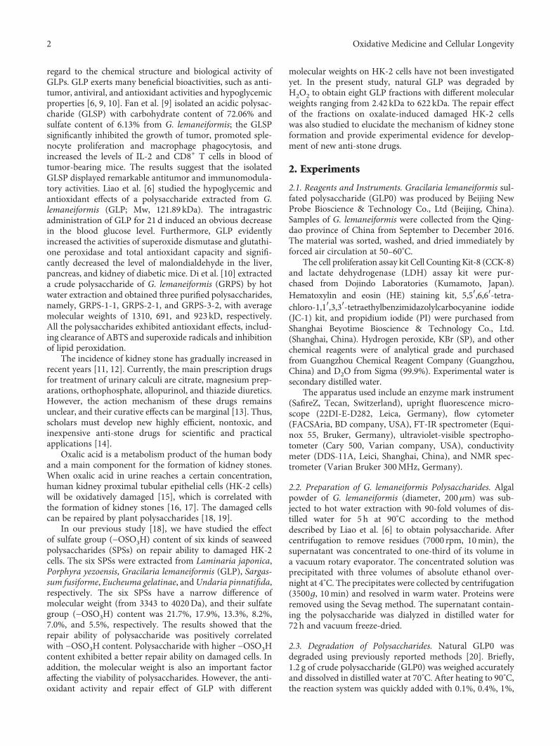
regard to the chemical structure and biological activity ofGLPs. GLP exerts many beneficial bioactivities, such as anti-tumor, antiviral, and antioxidant activities and hypoglycemicproperties [6, 9, 10]. Fan et al. [9] isolated an acidic polysac-charide (GLSP) with carbohydrate content of 72.06% andsulfate content of 6.13% from G. lemaneiformis; the GLSPsignificantly inhibited the growth of tumor, promoted sple-nocyte proliferation and macrophage phagocytosis, andincreased the levels of IL-2 and CD8+ T cells in blood oftumor-bearing mice. The results suggest that the isolatedGLSP displayed remarkable antitumor and immunomodula-tory activities. Liao et al. [6] studied the hypoglycemic andantioxidant effects of a polysaccharide extracted from G.lemaneiformis (GLP; Mw, 121.89 kDa). The intragastricadministration of GLP for 21 d induced an obvious decreasein the blood glucose level. Furthermore, GLP evidentlyincreased the activities of superoxide dismutase and glutathi-one peroxidase and total antioxidant capacity and signifi-cantly decreased the level of malondialdehyde in the liver,pancreas, and kidney of diabetic mice. Di et al. [10] extracteda crude polysaccharide of G. lemaneiformis (GRPS) by hotwater extraction and obtained three purified polysaccharides,namely, GRPS-1-1, GRPS-2-1, and GRPS-3-2, with averagemolecular weights of 1310, 691, and 923 kD, respectively.All the polysaccharides exhibited antioxidant effects, includ-ing clearance of ABTS and superoxide radicals and inhibitionof lipid peroxidation.
The incidence of kidney stone has gradually increased inrecent years [11, 12]. Currently, the main prescription drugsfor treatment of urinary calculi are citrate, magnesium prep-arations, orthophosphate, allopurinol, and thiazide diuretics.However, the action mechanism of these drugs remainsunclear, and their curative effects can be marginal [13]. Thus,scholars must develop new highly efficient, nontoxic, andinexpensive anti-stone drugs for scientific and practicalapplications [14].
Oxalic acid is a metabolism product of the human bodyand a main component for the formation of kidney stones.When oxalic acid in urine reaches a certain concentration,human kidney proximal tubular epithelial cells (HK-2 cells)will be oxidatively damaged [15], which is correlated withthe formation of kidney stones [16, 17]. The damaged cellscan be repaired by plant polysaccharides [18, 19].
In our previous study [18], we have studied the effectof sulfate group (−OSO3H) content of six kinds of seaweedpolysaccharides (SPSs) on repair ability to damaged HK-2cells. The six SPSs were extracted from Laminaria japonica,Porphyra yezoensis, Gracilaria lemaneiformis (GLP), Sargas-sum fusiforme, Eucheuma gelatinae, andUndaria pinnatifida,respectively. The six SPSs have a narrow difference ofmolecular weight (from 3343 to 4020Da), and their sulfategroup (−OSO3H) content was 21.7%, 17.9%, 13.3%, 8.2%,7.0%, and 5.5%, respectively. The results showed that therepair ability of polysaccharide was positively correlatedwith −OSO3H content. Polysaccharide with higher −OSO3Hcontent exhibited a better repair ability on damaged cells. Inaddition, the molecular weight is also an important factoraffecting the viability of polysaccharides. However, the anti-oxidant activity and repair effect of GLP with different
molecular weights on HK-2 cells have not been investigatedyet. In the present study, natural GLP was degraded byH2O2 to obtain eight GLP fractions with different molecularweights ranging from 2.42 kDa to 622 kDa. The repair effectof the fractions on oxalate-induced damaged HK-2 cellswas also studied to elucidate the mechanism of kidney stoneformation and provide experimental evidence for develop-ment of new anti-stone drugs.
2. Experiments
2.1. Reagents and Instruments. Gracilaria lemaneiformis sul-fated polysaccharide (GLP0) was produced by Beijing NewProbe Bioscience & Technology Co., Ltd (Beijing, China).Samples of G. lemaneiformis were collected from the Qing-dao province of China from September to December 2016.The material was sorted, washed, and dried immediately byforced air circulation at 50–60°C.
The cell proliferation assay kit Cell Counting Kit-8 (CCK-8)and lactate dehydrogenase (LDH) assay kit were pur-chased from Dojindo Laboratories (Kumamoto, Japan).Hematoxylin and eosin (HE) staining kit, 5,5′,6,6′-tetra-chloro-1,1′,3,3′-tetraethylbenzimidazolylcarbocyanine iodide(JC-1) kit, and propidium iodide (PI) were purchased fromShanghai Beyotime Bioscience & Technology Co., Ltd.(Shanghai, China). Hydrogen peroxide, KBr (SP), and otherchemical reagents were of analytical grade and purchasedfrom Guangzhou Chemical Reagent Company (Guangzhou,China) and D2O from Sigma (99.9%). Experimental water issecondary distilled water.
The apparatus used include an enzyme mark instrument(SafireZ, Tecan, Switzerland), upright fluorescence micro-scope (22DI-E-D282, Leica, Germany), flow cytometer(FACSAria, BD company, USA), FT-IR spectrometer (Equi-nox 55, Bruker, Germany), ultraviolet-visible spectropho-tometer (Cary 500, Varian company, USA), conductivitymeter (DDS-11A, Leici, Shanghai, China), and NMR spec-trometer (Varian Bruker 300MHz, Germany).
2.2. Preparation of G. lemaneiformis Polysaccharides. Algalpowder of G. lemaneiformis (diameter, 200μm) was sub-jected to hot water extraction with 90-fold volumes of dis-tilled water for 5 h at 90°C according to the methoddescribed by Liao et al. [6] to obtain polysaccharide. Aftercentrifugation to remove residues (7000 rpm, 10min), thesupernatant was concentrated to one-third of its volume ina vacuum rotary evaporator. The concentrated solution wasprecipitated with three volumes of absolute ethanol over-night at 4°C. The precipitates were collected by centrifugation(3500g, 10min) and resolved in warm water. Proteins wereremoved using the Sevag method. The supernatant contain-ing the polysaccharide was dialyzed in distilled water for72 h and vacuum freeze-dried.
2.3. Degradation of Polysaccharides. Natural GLP0 wasdegraded using previously reported methods [20]. Briefly,1.2 g of crude polysaccharide (GLP0) was weighed accuratelyand dissolved in distilled water at 70°C. After heating to 90°C,the reaction system was quickly added with 0.1%, 0.4%, 1%,
2 Oxidative Medicine and Cellular Longevity
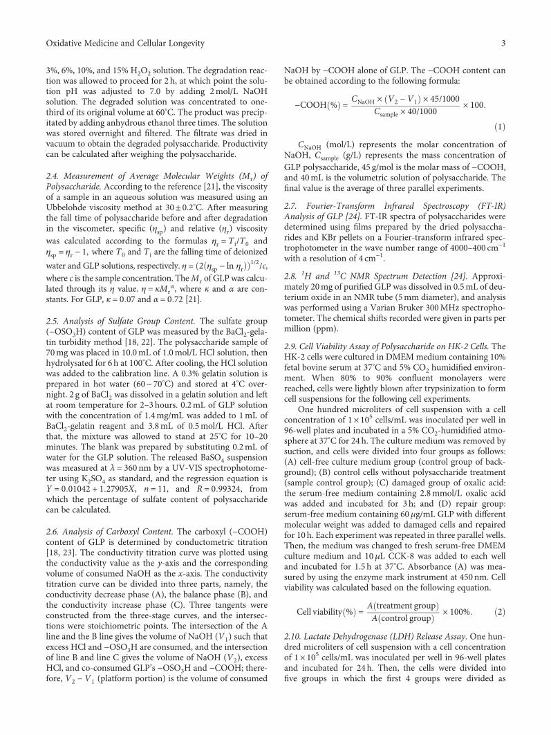
3%, 6%, 10%, and 15% H2O2 solution. The degradation reac-tion was allowed to proceed for 2 h, at which point the solu-tion pH was adjusted to 7.0 by adding 2mol/L NaOHsolution. The degraded solution was concentrated to one-third of its original volume at 60°C. The product was precip-itated by adding anhydrous ethanol three times. The solutionwas stored overnight and filtered. The filtrate was dried invacuum to obtain the degraded polysaccharide. Productivitycan be calculated after weighing the polysaccharide.
2.4. Measurement of Average Molecular Weights (Mr) ofPolysaccharide. According to the reference [21], the viscosityof a sample in an aqueous solution was measured using anUbbelohde viscosity method at 30± 0.2°C. After measuringthe fall time of polysaccharide before and after degradationin the viscometer, specific (ηsp) and relative (ηr) viscositywas calculated according to the formulas ηr = T i/T0 andηsp = ηr − 1, where T0 and T i are the falling time of deionized
water and GLP solutions, respectively. η = 2 ηsp − ln ηr1/2/c,
where c is the sample concentration. TheMr of GLP was calcu-lated through its η value. η = κMr
α, where κ and α are con-stants. For GLP, κ = 0 07 and α = 0 72 [21].
2.5. Analysis of Sulfate Group Content. The sulfate group(−OSO3H) content of GLP was measured by the BaCl2-gela-tin turbidity method [18, 22]. The polysaccharide sample of70mg was placed in 10.0mL of 1.0mol/L HCl solution, thenhydrolysated for 6 h at 100°C. After cooling, the HCl solutionwas added to the calibration line. A 0.3% gelatin solution isprepared in hot water (60~ 70°C) and stored at 4°C over-night. 2 g of BaCl2 was dissolved in a gelatin solution and leftat room temperature for 2–3hours. 0.2mL of GLP solutionwith the concentration of 1.4mg/mL was added to 1mL ofBaCl2-gelatin reagent and 3.8mL of 0.5mol/L HCl. Afterthat, the mixture was allowed to stand at 25°C for 10–20minutes. The blank was prepared by substituting 0.2mL ofwater for the GLP solution. The released BaSO4 suspensionwas measured at λ = 360 nm by a UV-VIS spectrophotome-ter using K2SO4 as standard, and the regression equation isY = 0 01042 + 1 27905X, n = 11, and R = 0 99324, fromwhich the percentage of sulfate content of polysaccharidecan be calculated.
2.6. Analysis of Carboxyl Content. The carboxyl (−COOH)content of GLP is determined by conductometric titration[18, 23]. The conductivity titration curve was plotted usingthe conductivity value as the y-axis and the correspondingvolume of consumed NaOH as the x-axis. The conductivitytitration curve can be divided into three parts, namely, theconductivity decrease phase (A), the balance phase (B), andthe conductivity increase phase (C). Three tangents wereconstructed from the three-stage curves, and the intersec-tions were stoichiometric points. The intersection of the Aline and the B line gives the volume of NaOH (V1) such thatexcess HCl and −OSO3H are consumed, and the intersectionof line B and line C gives the volume of NaOH (V2), excessHCl, and co-consumed GLP’s −OSO3H and −COOH; there-fore, V2 − V1 (platform portion) is the volume of consumed
NaOH by −COOH alone of GLP. The −COOH content canbe obtained according to the following formula:
−COOH % = CNaOH × V2 −V1 × 45/1000Csample × 40/1000 × 100
1
CNaOH (mol/L) represents the molar concentration ofNaOH, Csample (g/L) represents the mass concentration ofGLP polysaccharide, 45 g/mol is the molar mass of −COOH,and 40mL is the volumetric solution of polysaccharide. Thefinal value is the average of three parallel experiments.
2.7. Fourier-Transform Infrared Spectroscopy (FT-IR)Analysis of GLP [24]. FT-IR spectra of polysaccharides weredetermined using films prepared by the dried polysaccha-rides and KBr pellets on a Fourier-transform infrared spec-trophotometer in the wave number range of 4000–400 cm−1
with a resolution of 4 cm−1.
2.8. 1H and 13C NMR Spectrum Detection [24]. Approxi-mately 20mg of purified GLP was dissolved in 0.5mL of deu-terium oxide in an NMR tube (5mm diameter), and analysiswas performed using a Varian Bruker 300MHz spectropho-tometer. The chemical shifts recorded were given in parts permillion (ppm).
2.9. Cell Viability Assay of Polysaccharide on HK-2 Cells. TheHK-2 cells were cultured in DMEMmedium containing 10%fetal bovine serum at 37°C and 5% CO2 humidified environ-ment. When 80% to 90% confluent monolayers werereached, cells were lightly blown after trypsinization to formcell suspensions for the following cell experiments.
One hundred microliters of cell suspension with a cellconcentration of 1× 10⁵ cells/mL was inoculated per well in96-well plates and incubated in a 5% CO2-humidified atmo-sphere at 37°C for 24 h. The culture medium was removed bysuction, and cells were divided into four groups as follows:(A) cell-free culture medium group (control group of back-ground); (B) control cells without polysaccharide treatment(sample control group); (C) damaged group of oxalic acid:the serum-free medium containing 2.8mmol/L oxalic acidwas added and incubated for 3 h; and (D) repair group:serum-free medium containing 60μg/mL GLP with differentmolecular weight was added to damaged cells and repairedfor 10h. Each experiment was repeated in three parallel wells.Then, the medium was changed to fresh serum-free DMEMculture medium and 10μL CCK-8 was added to each welland incubated for 1.5 h at 37°C. Absorbance (A) was mea-sured by using the enzyme mark instrument at 450nm. Cellviability was calculated based on the following equation.
Cell viability % = A treatment groupA control group × 100% 2
2.10. Lactate Dehydrogenase (LDH) Release Assay. One hun-dred microliters of cell suspension with a cell concentrationof 1× 10⁵ cells/mL was inoculated per well in 96-well platesand incubated for 24h. Then, the cells were divided intofive groups in which the first 4 groups were divided as
3Oxidative Medicine and Cellular Longevity
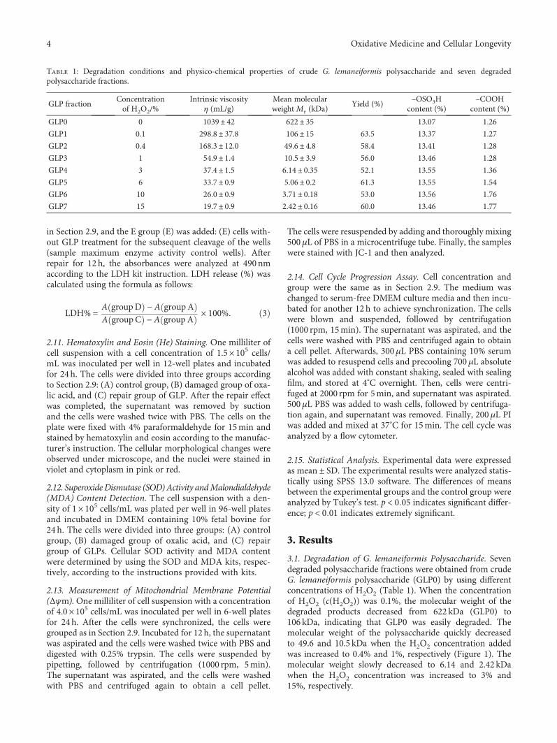
in Section 2.9, and the E group (E) was added: (E) cells with-out GLP treatment for the subsequent cleavage of the wells(sample maximum enzyme activity control wells). Afterrepair for 12 h, the absorbances were analyzed at 490nmaccording to the LDH kit instruction. LDH release (%) wascalculated using the formula as follows:
LDH% = A groupD − A groupAA groupC − A groupA × 100% 3
2.11. Hematoxylin and Eosin (He) Staining. One milliliter ofcell suspension with a cell concentration of 1.5× 10⁵ cells/mL was inoculated per well in 12-well plates and incubatedfor 24h. The cells were divided into three groups accordingto Section 2.9: (A) control group, (B) damaged group of oxa-lic acid, and (C) repair group of GLP. After the repair effectwas completed, the supernatant was removed by suctionand the cells were washed twice with PBS. The cells on theplate were fixed with 4% paraformaldehyde for 15min andstained by hematoxylin and eosin according to the manufac-turer’s instruction. The cellular morphological changes wereobserved under microscope, and the nuclei were stained inviolet and cytoplasm in pink or red.
2.12. Superoxide Dismutase (SOD)Activity andMalondialdehyde(MDA) Content Detection. The cell suspension with a den-sity of 1× 10⁵ cells/mL was plated per well in 96-well platesand incubated in DMEM containing 10% fetal bovine for24 h. The cells were divided into three groups: (A) controlgroup, (B) damaged group of oxalic acid, and (C) repairgroup of GLPs. Cellular SOD activity and MDA contentwere determined by using the SOD and MDA kits, respec-tively, according to the instructions provided with kits.
2.13. Measurement of Mitochondrial Membrane Potential(Δψm).One milliliter of cell suspension with a concentrationof 4.0× 10⁵ cells/mL was inoculated per well in 6-well platesfor 24h. After the cells were synchronized, the cells weregrouped as in Section 2.9. Incubated for 12 h, the supernatantwas aspirated and the cells were washed twice with PBS anddigested with 0.25% trypsin. The cells were suspended bypipetting, followed by centrifugation (1000 rpm, 5min).The supernatant was aspirated, and the cells were washedwith PBS and centrifuged again to obtain a cell pellet.
The cells were resuspended by adding and thoroughly mixing500μL of PBS in a microcentrifuge tube. Finally, the sampleswere stained with JC-1 and then analyzed.
2.14. Cell Cycle Progression Assay. Cell concentration andgroup were the same as in Section 2.9. The medium waschanged to serum-free DMEM culture media and then incu-bated for another 12 h to achieve synchronization. The cellswere blown and suspended, followed by centrifugation(1000 rpm, 15min). The supernatant was aspirated, and thecells were washed with PBS and centrifuged again to obtaina cell pellet. Afterwards, 300μL PBS containing 10% serumwas added to resuspend cells and precooling 700μL absolutealcohol was added with constant shaking, sealed with sealingfilm, and stored at 4°C overnight. Then, cells were centri-fuged at 2000 rpm for 5min, and supernatant was aspirated.500μL PBS was added to wash cells, followed by centrifuga-tion again, and supernatant was removed. Finally, 200μL PIwas added and mixed at 37°C for 15min. The cell cycle wasanalyzed by a flow cytometer.
2.15. Statistical Analysis. Experimental data were expressedasmean ± SD. The experimental results were analyzed statis-tically using SPSS 13.0 software. The differences of meansbetween the experimental groups and the control group wereanalyzed by Tukey’s test. p < 0 05 indicates significant differ-ence; p < 0 01 indicates extremely significant.
3. Results
3.1. Degradation of G. lemaneiformis Polysaccharide. Sevendegraded polysaccharide fractions were obtained from crudeG. lemaneiformis polysaccharide (GLP0) by using differentconcentrations of H2O2 (Table 1). When the concentrationof H2O2 (c(H2O2)) was 0.1%, the molecular weight of thedegraded products decreased from 622 kDa (GLP0) to106 kDa, indicating that GLP0 was easily degraded. Themolecular weight of the polysaccharide quickly decreasedto 49.6 and 10.5 kDa when the H2O2 concentration addedwas increased to 0.4% and 1%, respectively (Figure 1). Themolecular weight slowly decreased to 6.14 and 2.42 kDawhen the H2O2 concentration was increased to 3% and15%, respectively.
Table 1: Degradation conditions and physico-chemical properties of crude G. lemaneiformis polysaccharide and seven degradedpolysaccharide fractions.
GLP fractionConcentrationof H2O2/%
Intrinsic viscosityη (mL/g)
Mean molecularweightMr (kDa)
Yield (%)–OSO3H
content (%)–COOH
content (%)
GLP0 0 1039± 42 622± 35 13.07 1.26
GLP1 0.1 298.8± 37.8 106± 15 63.5 13.37 1.27
GLP2 0.4 168.3± 12.0 49.6± 4.8 58.4 13.41 1.28
GLP3 1 54.9± 1.4 10.5± 3.9 56.0 13.46 1.28
GLP4 3 37.4± 1.5 6.14± 0.35 52.1 13.55 1.36
GLP5 6 33.7± 0.9 5.06± 0.2 61.3 13.55 1.54
GLP6 10 26.0± 0.9 3.71± 0.18 53.0 13.56 1.76
GLP7 15 19.7± 0.9 2.42± 0.16 60.0 13.46 1.77
4 Oxidative Medicine and Cellular Longevity
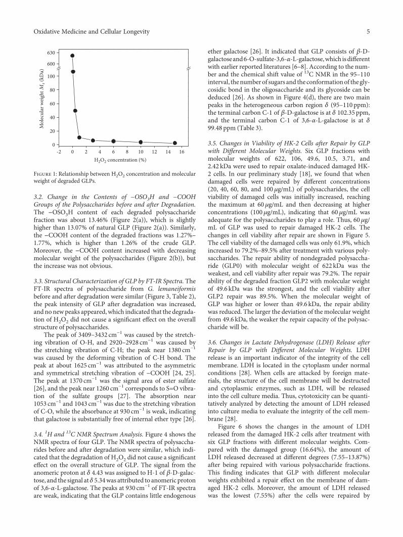
3.2. Change in the Contents of −OSO3H and −COOHGroups of the Polysaccharides before and after Degradation.The −OSO3H content of each degraded polysaccharidefraction was about 13.46% (Figure 2(a)), which is slightlyhigher than 13.07% of natural GLP (Figure 2(a)). Similarly,the −COOH content of the degraded fractions was 1.27%–1.77%, which is higher than 1.26% of the crude GLP.Moreover, the −COOH content increased with decreasingmolecular weight of the polysaccharides (Figure 2(b)), butthe increase was not obvious.
3.3. Structural Characterization of GLP by FT-IR Spectra. TheFT-IR spectra of polysaccharide from G. lemaneiformisbefore and after degradation were similar (Figure 3, Table 2),the peak intensity of GLP after degradation was increased,and nonewpeaks appeared, which indicated that the degrada-tion of H2O2 did not cause a significant effect on the overallstructure of polysaccharides.
The peak of 3409–3432 cm−1 was caused by the stretch-ing vibration of O-H, and 2920–2928 cm−1 was caused bythe stretching vibration of C-H; the peak near 1380 cm−1
was caused by the deforming vibration of C-H bond. Thepeak at about 1625 cm−1 was attributed to the asymmetricand symmetrical stretching vibration of −COOH [24, 25].The peak at 1370 cm−1 was the signal area of ester sulfate[26], and the peak near 1260 cm−1 corresponds to S=O vibra-tion of the sulfate groups [27]. The absorption near1053 cm−1 and 1043 cm−1 was due to the stretching vibrationof C-O, while the absorbance at 930 cm−1 is weak, indicatingthat galactose is substantially free of internal ether type [26].
3.4. 1H and 13C NMR Spectrum Analysis. Figure 4 shows theNMR spectra of four GLP. The NMR spectra of polysaccha-rides before and after degradation were similar, which indi-cated that the degradation of H2O2 did not cause a significanteffect on the overall structure of GLP. The signal from theanomeric proton at δ 4.43 was assigned to H-1 of β-D-galac-tose, and the signal atδ 5.34was attributed to anomeric protonof 3,6-α-L-galactose. The peaks at 930 cm−1 of FT-IR spectraare weak, indicating that the GLP contains little endogenous
ether galactose [26]. It indicated that GLP consists of β-D-galactoseand6-O-sulfate-3,6-α-L-galactose,which isdifferentwith earlier reported literatures [6–8]. According to the num-ber and the chemical shift value of 13C NMR in the 95–110interval, thenumber of sugars and the conformationof the gly-cosidic bond in the oligosaccharide and its glycoside can bededuced [26]. As shown in Figure 4(d), there are two mainpeaks in the heterogeneous carbon region δ (95–110 ppm):the terminal carbon C-1 of β-D-galactose is at δ 102.35 ppm,and the terminal carbon C-1 of 3,6-α-L-galactose is at δ99.48 ppm (Table 3).
3.5. Changes in Viability of HK-2 Cells after Repair by GLPwith Different Molecular Weights. Six GLP fractions withmolecular weights of 622, 106, 49.6, 10.5, 3.71, and2.42 kDa were used to repair oxalate-induced damaged HK-2 cells. In our preliminary study [18], we found that whendamaged cells were repaired by different concentrations(20, 40, 60, 80, and 100μg/mL) of polysaccharides, the cellviability of damaged cells was initially increased, reachingthe maximum at 60μg/mL and then decreasing at higherconcentrations (100μg/mL), indicating that 60μg/mL wasadequate for the polysaccharides to play a role. Thus, 60μg/mL of GLP was used to repair damaged HK-2 cells. Thechanges in cell viability after repair are shown in Figure 5.The cell viability of the damaged cells was only 61.9%, whichincreased to 79.2%–89.5% after treatment with various poly-saccharides. The repair ability of nondegraded polysaccha-ride (GLP0) with molecular weight of 622 kDa was theweakest, and cell viability after repair was 79.2%. The repairability of the degraded fraction GLP2 with molecular weightof 49.6 kDa was the strongest, and the cell viability afterGLP2 repair was 89.5%. When the molecular weight ofGLP was higher or lower than 49.6 kDa, the repair abilitywas reduced. The larger the deviation of the molecular weightfrom 49.6 kDa, the weaker the repair capacity of the polysac-charide will be.
3.6. Changes in Lactate Dehydrogenase (LDH) Release afterRepair by GLP with Different Molecular Weights. LDHrelease is an important indicator of the integrity of the cellmembrane. LDH is located in the cytoplasm under normalconditions [28]. When cells are attacked by foreign mate-rials, the structure of the cell membrane will be destructedand cytoplasmic enzymes, such as LDH, will be releasedinto the cell culture media. Thus, cytotoxicity can be quanti-tatively analyzed by detecting the amount of LDH releasedinto culture media to evaluate the integrity of the cell mem-brane [28].
Figure 6 shows the changes in the amount of LDHreleased from the damaged HK-2 cells after treatment withsix GLP fractions with different molecular weights. Com-pared with the damaged group (16.64%), the amount ofLDH released decreased at different degrees (7.55–13.87%)after being repaired with various polysaccharide fractions.This finding indicates that GLP with different molecularweights exhibited a repair effect on the membrane of dam-aged HK-2 cells. Moreover, the amount of LDH releasedwas the lowest (7.55%) after the cells were repaired by
-20
20
40
60
80
100
600
630
Mol
ecul
ar w
eigh
t Mr (
kDa)
H2O2 concentration (%)0 2 4 6 8 10 12 14 16
Figure 1: Relationship between H2O2 concentration and molecularweight of degraded GLPs.
5Oxidative Medicine and Cellular Longevity
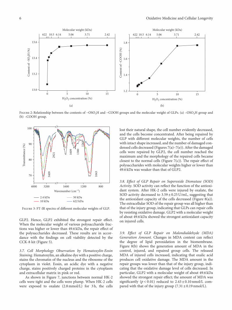
GLP2. Hence, GLP2 exhibited the strongest repair effect.When the molecular weight of various polysaccharide frac-tions was higher or lower than 49.6 kDa, the repair effect ofthe polysaccharides decreased. These results are in accor-dance with the findings on cell viability detected by theCCK-8 kit (Figure 5).
3.7. Cell Morphology Observation by Hematoxylin-EosinStaining.Hematoxylin, an alkaline dye with a positive charge,stains the chromatin of the nucleus and the ribosome of thecytoplasm in violet. Eosin, an acidic dye with a negativecharge, stains positively charged proteins in the cytoplasmand extracellular matrix in pink or red.
As shown in Figure 7, junctions between normal HK-2cells were tight and the cells were plump. When HK-2 cellswere exposed to oxalate (2.8mmol/L) for 3 h, the cells
lost their natural shape, the cell number evidently decreased,and the cells become concentrated. After being repaired byGLP with different molecular weights, the number of cellswith intact shape increased, and the number of damaged con-densed cells decreased (Figures 7(a)–7(e)). After the damagedcells were repaired by GLP2, the cell number reached themaximum and the morphology of the repaired cells becameclosest to the normal cells (Figure 7(c)). The repair effect ofpolysaccharides with molecular weights higher or lower than49.6 kDa was weaker than that of GLP2.
3.8. Effect of GLP Repair on Superoxide Dismutase (SOD)Activity. SOD activity can reflect the function of the antioxi-dant system. After HK-2 cells were injured by oxalate, theSOD activity decreased to 3.59± 0.25U/mL, suggesting thatthe antioxidant capacity of the cells decreased (Figure 8(a)).The extracellular SOD of the repair group was all higher thanthat of the injury group, indicating that GLPs can repair cellsby resisting oxidative damage. GLP2 with a molecular weightof about 49.6 kDa showed the strongest antioxidant capacityon injured cells.
3.9. Effect of GLP Repair on Malondialdehyde (MDA)Generation Amount. Changes in MDA content can reflectthe degree of lipid peroxidation in the biomembrane.Figure 8(b) shows the generation amount of MDA in thecontrol, injured, and repaired group cells. The releasedMDA of injured cells increased, indicating that oxalic acidproduces cell oxidative damage. The MDA amount in therepair groups was lower than that of the injury group, indi-cating that the oxidative damage level of cells decreased. Inparticular, GLP2 with a molecular weight of about 49.6 kDashowed the strongest repair effect; the amount of MDA wassignificantly (p < 0 01) reduced to 2.43± 0.10nmol/L com-pared with that of the injury group (7.31± 0.19 nmol/L).
13.0
13.2
13.4
13.6
Con
tent
of −
SO3H
(%)
H2O2 concentration (%)
Molecular weight (kDa)622 10.5 6.14 5.06 2.423.71
0 5 10 15
(a)
01.2
1.4
1.6
1.8
Con
tent
of −
COO
H (%
)
H2O2 concentration (%)
Molecular weight (kDa)622 3.7110.5 6.14 5.06 2.42
5 10 15
(b)
Figure 2: Relationship between the contents of −OSO3H and −COOH groups and the molecular weight of GLPs. (a) –OSO3H group and(b) –COOH group.
4000
70
80
90
100
1043
1265
1380
1609
2921
3409
Tran
smitt
ance
(%)
Wavenumber (cm−1)
2.4 kDa10 kDa
50 kDa622 kDa
800120016003200
Figure 3: FT-IR spectra of different molecular weights of GLP.
6 Oxidative Medicine and Cellular Longevity
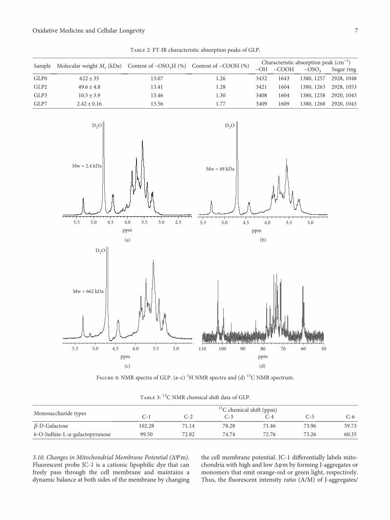
3.10. Changes in Mitochondrial Membrane Potential (ΔΨm).Fluorescent probe JC-1 is a cationic lipophilic dye that canfreely pass through the cell membrane and maintains adynamic balance at both sides of the membrane by changing
the cell membrane potential. JC-1 differentially labels mito-chondria with high and low Δψm by forming J-aggregates ormonomers that emit orange-red or green light, respectively.Thus, the fluorescent intensity ratio (A/M) of J-aggregates/
Table 2: FT-IR characteristic absorption peaks of GLP.
Sample Molecular weight Mr (kDa) Content of −OSO3H (%) Content of −COOH (%)Characteristic absorption peak (cm−1)
−OH −COOH −OSO3 Sugar ring
GLP0 622± 35 13.07 1.26 3432 1643 1380, 1257 2928, 1048
GLP2 49.6± 4.8 13.41 1.28 3421 1604 1380, 1263 2928, 1053
GLP3 10.5± 3.9 13.46 1.30 3408 1604 1380, 1258 2920, 1043
GLP7 2.42± 0.16 13.56 1.77 3409 1609 1380, 1268 2920, 1043
5.5
D2O
Mw = 2.4 kDa
ppm2.53.03.54.04.55.0
(a)
5.5
Mw = 49 kDa
D2O
ppm3.03.54.04.55.0
(b)
5.5
Mw = 662 kDa
D2O
ppm3.03.54.04.55.0
(c)
110ppm
5060708090100
(d)
Figure 4: NMR spectra of GLP. (a–c) 1H NMR spectra and (d) 13C NMR spectrum.
Table 3: 13C NMR chemical shift data of GLP.
Monosaccharide types13C chemical shift (ppm)
C-1 C-2 C-3 C-4 C-5 C-6
β-D-Galactose 102.28 71.14 78.28 71.46 73.96 59.73
6-O-Sulfate-L-α-galactopyranose 99.50 72.82 74.74 72.76 73.26 60.35
7Oxidative Medicine and Cellular Longevity
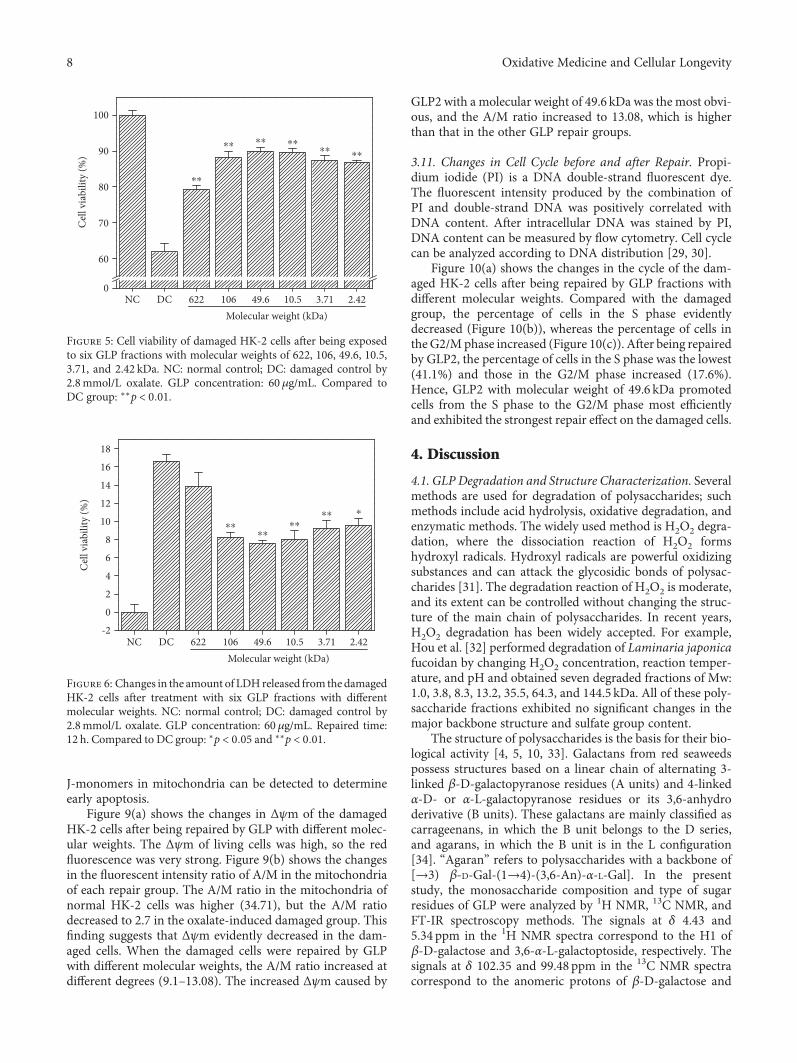
J-monomers in mitochondria can be detected to determineearly apoptosis.
Figure 9(a) shows the changes in Δψm of the damagedHK-2 cells after being repaired by GLP with different molec-ular weights. The Δψm of living cells was high, so the redfluorescence was very strong. Figure 9(b) shows the changesin the fluorescent intensity ratio of A/M in the mitochondriaof each repair group. The A/M ratio in the mitochondria ofnormal HK-2 cells was higher (34.71), but the A/M ratiodecreased to 2.7 in the oxalate-induced damaged group. Thisfinding suggests that Δψm evidently decreased in the dam-aged cells. When the damaged cells were repaired by GLPwith different molecular weights, the A/M ratio increased atdifferent degrees (9.1–13.08). The increased Δψm caused by
GLP2 with a molecular weight of 49.6 kDa was the most obvi-ous, and the A/M ratio increased to 13.08, which is higherthan that in the other GLP repair groups.
3.11. Changes in Cell Cycle before and after Repair. Propi-dium iodide (PI) is a DNA double-strand fluorescent dye.The fluorescent intensity produced by the combination ofPI and double-strand DNA was positively correlated withDNA content. After intracellular DNA was stained by PI,DNA content can be measured by flow cytometry. Cell cyclecan be analyzed according to DNA distribution [29, 30].
Figure 10(a) shows the changes in the cycle of the dam-aged HK-2 cells after being repaired by GLP fractions withdifferent molecular weights. Compared with the damagedgroup, the percentage of cells in the S phase evidentlydecreased (Figure 10(b)), whereas the percentage of cells intheG2/M phase increased (Figure 10(c)). After being repairedby GLP2, the percentage of cells in the S phase was the lowest(41.1%) and those in the G2/M phase increased (17.6%).Hence, GLP2 with molecular weight of 49.6 kDa promotedcells from the S phase to the G2/M phase most efficientlyand exhibited the strongest repair effect on the damaged cells.
4. Discussion
4.1. GLP Degradation and Structure Characterization. Severalmethods are used for degradation of polysaccharides; suchmethods include acid hydrolysis, oxidative degradation, andenzymatic methods. The widely used method is H2O2 degra-dation, where the dissociation reaction of H2O2 formshydroxyl radicals. Hydroxyl radicals are powerful oxidizingsubstances and can attack the glycosidic bonds of polysac-charides [31]. The degradation reaction of H2O2 is moderate,and its extent can be controlled without changing the struc-ture of the main chain of polysaccharides. In recent years,H2O2 degradation has been widely accepted. For example,Hou et al. [32] performed degradation of Laminaria japonicafucoidan by changing H2O2 concentration, reaction temper-ature, and pH and obtained seven degraded fractions of Mw:1.0, 3.8, 8.3, 13.2, 35.5, 64.3, and 144.5 kDa. All of these poly-saccharide fractions exhibited no significant changes in themajor backbone structure and sulfate group content.
The structure of polysaccharides is the basis for their bio-logical activity [4, 5, 10, 33]. Galactans from red seaweedspossess structures based on a linear chain of alternating 3-linked β-D-galactopyranose residues (A units) and 4-linkedα-D- or α-L-galactopyranose residues or its 3,6-anhydroderivative (B units). These galactans are mainly classified ascarrageenans, in which the B unit belongs to the D series,and agarans, in which the B unit is in the L configuration[34]. “Agaran” refers to polysaccharides with a backbone of[→3) β-D-Gal-(1→4)-(3,6-An)-α-L-Gal]. In the presentstudy, the monosaccharide composition and type of sugarresidues of GLP were analyzed by 1H NMR, 13C NMR, andFT-IR spectroscopy methods. The signals at δ 4.43 and5.34 ppm in the 1H NMR spectra correspond to the H1 ofβ-D-galactose and 3,6-α-L-galactoptoside, respectively. Thesignals at δ 102.35 and 99.48 ppm in the 13C NMR spectracorrespond to the anomeric protons of β-D-galactose and
0
60
70
80
90
100
⁎⁎⁎⁎⁎⁎⁎⁎⁎⁎
⁎⁎
Cel
l via
bilit
y (%
)
NCMolecular weight (kDa)
49.6106622DC 2.423.7110.5
Figure 5: Cell viability of damaged HK-2 cells after being exposedto six GLP fractions with molecular weights of 622, 106, 49.6, 10.5,3.71, and 2.42 kDa. NC: normal control; DC: damaged control by2.8mmol/L oxalate. GLP concentration: 60 μg/mL. Compared toDC group: ∗∗p < 0 01.
2
0
-2
4
6
12
10
8
16
14
18
Cel
l via
bilit
y (%
)
NCMolecular weight (kDa)
49.6106622DC 2.423.7110.5
⁎⁎⁎⁎
⁎
⁎⁎⁎⁎
Figure 6: Changes in the amount of LDHreleased from the damagedHK-2 cells after treatment with six GLP fractions with differentmolecular weights. NC: normal control; DC: damaged control by2.8mmol/L oxalate. GLP concentration: 60 μg/mL. Repaired time:12 h. Compared to DC group: ∗p < 0 05 and ∗∗p < 0 01.
8 Oxidative Medicine and Cellular Longevity
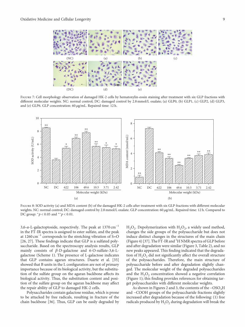
3,6-α-L-galactoptoside, respectively. The peak at 1370 cm−1
in the FT-IR spectra is assigned to ester sulfate, and the peakat 1260 cm−1 corresponds to the stretching vibration of S=O[26, 27]. These findings indicate that GLP is a sulfated poly-saccharide. Based on the spectroscopy analysis results, GLPmainly consists of β-D-galactose and 6-O-sulfate-3,6-L-galactose (Scheme 1). The presence of L-galactose indicatesthat GLP contains agaran structures. Duarte et al. [35]showed that B units in the L configuration are not of primaryimportance because of its biological activity, but the substitu-tion of the sulfate group on the agaran backbone affects itsbiological activity. Thus, the substitution content and posi-tion of the sulfate group on the agaran backbone may affectthe repair ability of GLP to damaged HK-2 cells.
Polysaccharides contain galactose residue, which is proneto be attacked by free radicals, resulting in fracture of thechain backbone [36]. Thus, GLP can be easily degraded by
H2O2. Depolymerization with H2O2, a widely used method,changes the side groups of the polysaccharide but does notinduce distinct changes in the structures of the main chain(Figure 4) [37]. The FT-IR and 1HNMR spectra ofGLP beforeand after degradation were similar (Figure 3, Table 2), and nonew peaks appeared. This finding indicated that the degrada-tion of H2O2 did not significantly affect the overall structureof the polysaccharides. Therefore, the main structure ofpolysaccharide before and after degradation slightly chan-ged. The molecular weight of the degraded polysaccharidesand the H2O2 concentration showed a negative correlation(Figure 1); this finding provides references for obtaining tar-get polysaccharides with different molecular weights.
As shown in Figures 2 and 3, the contents of the −OSO3Hand −COOH groups of the polysaccharide fractions slightlyincreased after degradation because of the following: (1) freeradicals produced by H2O2 during degradation will break the
(NC)
(DC) (e)(d)
(c)(b)(a)
Figure 7: Cell morphology observation of damaged HK-2 cells by hematoxylin-eosin staining after treatment with six GLP fractions withdifferent molecular weights. NC: normal control; DC: damaged control by 2.8mmol/L oxalate; (a) GLP0, (b) GLP1, (c) GLP2, (d) GLP3,and (e) GLP6. GLP concentration: 60 μg/mL. Repaired time: 12 h.
NC DC 622 106 49.6 10.5 3.71 2.420
2
4
6
8
10
SOD
activ
ity (U
/mL)
Molecular weight (kDa)
⁎⁎
⁎⁎
⁎⁎
⁎⁎⁎ ⁎ ⁎
(a)
NC DC 622 106 49.6 10.5 3.71 2.42Molecular weight (kDa)
0
2
4
6
8M
DA
cont
ent (
nmol
/mL)
⁎⁎
⁎⁎
⁎⁎
⁎⁎
⁎⁎⁎⁎ ⁎⁎
(b)
Figure 8: SOD activity (a) and MDA content (b) of the damaged HK-2 cells after treatment with six GLP fractions with different molecularweights. NC: normal control; DC: damaged control by 2.8mmol/L oxalate. GLP concentration: 60 μg/mL. Repaired time: 12 h. Compared toDC group: ∗p < 0 05 and ∗∗p < 0 01.
9Oxidative Medicine and Cellular Longevity
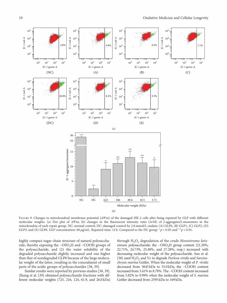
highly compact sugar chain structure of natural polysaccha-ride, thereby exposing the −OSO3H and −COOH groups ofthe polysaccharide, and (2) the water solubility of thedegraded polysaccharide slightly increased and was higherthan that of nondegraded GLP0 because of the large molecu-lar weight of the latter, resulting in the concealment of smallparts of the acidic groups of polysaccharides [38, 39].
Similar results were reported by previous studies [38, 39].Zhang et al. [39] obtained polysaccharide fractions with dif-ferent molecular weights (725, 216, 124, 61.9, and 26.0kDa)
through H2O2 degradation of the crude Monostroma latis-simum polysaccharide; the −OSO3H group content (21.20%,22.71%, 24.73%, 25.48%, and 27.28%, resp.) increased withdecreasing molecular weight of the polysaccharide. Sun et al.[38] used H2O2 and Vc to degrade Pavlova viridis and Sarcino-chrysis marina Geitler. When the molecular weight of P. viridisdecreased from 3645kDa to 55.0kDa, the −COOH contentincreased from 3.41% to 8.78%. The −COOH content increasedfrom 5.82% to 9.99% when the molecular weight of S. marinaGeitler decreased from 2595kDa to 169kDa.
2.8%
102 103 104
JC-1 green-A
JC-1
red-
A
102
103
104
105
102
103
104
105
JC-1
red-
A
JC-1
red-
A
102
103
104
105
102
103
104
105
JC-1
red-
A
JC-1
red-
A
102
103
104
105
102
103
104
105
JC-1
red-
A
102
103
104
105
JC-1
red-
A
105 102 103 104
JC-1 green-A105 102 103 104
JC-1 green-A105
102 103 104
JC-1 green-A105 102 103 104
JC-1 green-A105 102 103 104
JC-1 green-A105
102 103 104
JC-1 green-A
(NC) (A) (B)
(DC) (D) (E)
(C)
105
28.8% 8.2% 9.3%
9.9% 8.3% 7.1%
(a)
0
5
10
15
32
36
Molecular weight (kDa)
NC DC 622 106 49.6 10.5 3.71
JC-1
aggr
egat
es/m
onom
er
⁎⁎⁎
⁎⁎
⁎⁎⁎
(b)
Figure 9: Changes in mitochondrial membrane potential (ΔΨm) of the damaged HK-2 cells after being repaired by GLP with differentmolecular weights. (a) Dot plot of ΔΨm; (b) changes in the fluorescent intensity ratio (A/M) of J-aggregates/J-monomers in themitochondria of each repair group. NC: normal control; DC: damaged control by 2.8mmol/L oxalate; (A) GLP0, (B) GLP1, (C) GLP2, (D)GLP3, and (E) GLP6. GLP concentration: 60μg/mL. Repaired time: 12 h. Compared to the DC group: ∗p < 0 05 and ∗∗p < 0 01.
10 Oxidative Medicine and Cellular Longevity
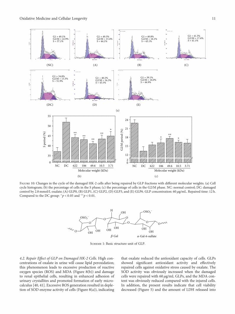
4.2. Repair Effect of GLP on Damaged HK-2 Cells. High con-centrations of oxalate in urine will cause lipid peroxidation;this phenomenon leads to excessive production of reactiveoxygen species (ROS) and MDA (Figure 8(b)) and damageto renal epithelial cells, resulting in enhanced adhesion ofurinary crystallites and promoted formation of early micro-calculus [40, 41]. Excessive ROS generation resulted in deple-tion of SOD enzyme activity of cells (Figure 8(a)), indicating
that oxalate reduced the antioxidant capacity of cells. GLPsshowed significant antioxidant activity and effectivelyrepaired cells against oxidative stress caused by oxalate. TheSOD activity was obviously increased when the damagedcells were repaired with 60μg/mL GLPs, and the MDA con-tent was obviously reduced compared with the injured cells.In addition, the present results indicate that cell viabilitydecreased (Figure 5) and the amount of LDH released into
G1 = 34.8%G2/M = 13.3%S = 51.9%
G1 = 40.1%G2/M = 22.9%S = 37.1%
G1 = 40.5%G2/M = 15.4%S = 44.1%
G1 = 40.8%G2/M = 16.1%S = 43.1%
G1 = 41.3%G2/M = 17.6%S = 41.1%
G1 = 40.2%G2/M = 16.3%S = 43.5%
G1 = 39.1%G2/M = 16.0%S = 44.9%
(NC) (A) (B)
(DC) (D) (E)
(C)
(a)
0
35
40
45
50
55
Molecular weight (kDa)NC DC 622 106 49.6 10.5 3.71
S pe
riod
(%) ⁎⁎
⁎⁎⁎⁎
⁎⁎⁎
(b)
NC DC 622 106 49.6 10.5 3.7102
12
15
18
21
24
Molecular weight (kDa)
G2/
M p
erio
d (%
)
⁎ ⁎⁎⁎
(c)
Figure 10: Changes in the cycle of the damaged HK-2 cells after being repaired by GLP fractions with different molecular weights. (a) Cellcycle histogram; (b) the percentage of cells in the S phase; (c) the percentage of cells in the G2/M phase. NC: normal control; DC: damagedcontrol by 2.8mmol/L oxalate; (A) GLP0, (B) GLP1, (C) GLP2, (D) GLP3, and (E) GLP6. GLP concentration: 60 μg/mL. Repaired time: 12 h.Compared to the DC group: ∗p < 0 05 and ∗∗p < 0 01.
O
O
OSO3−
OSO3−
O OO O O
OH OH OH OH
OHH
𝛽-Gal 𝛼-Gal-6-sulfate
OHOH
Scheme 1: Basic structure unit of GLP.
11Oxidative Medicine and Cellular Longevity
the extracellular matrix increased (Figure 6) when HK-2 cellswere damaged by oxalate. After the damaged HK-2 cells wererepaired through treatment with various fractions of GLP,the cell viability increased, the amount of LDH releaseddecreased, the cellmorphologywas improved, and thenumberof living cells increased (Figure 7). Subha and Varalakshmi[42] reported that sodium pentosan polysulfate can reduceLDH secretion in calculogenic rat urine. Meanwhile, glycos-aminoglycan can prevent the changes of cytosolic Ca2+ levelsin renal tubular epithelial cells induced by oxalic acid andrecover the cell morphology [43].
Mitochondrial membrane potential (ΔΨm) is higher innormal cells than that in damaged cells [44]. When thecells were oxidatively damaged by oxalate, the permeabilityof the mitochondrial membrane increased, which inducedCa2+ influx and depolarization of the mitochondria [45],resulting in decreased Δψm (Figure 9(b)). GLP fractions withdifferent molecular weights can be used to repair the mem-brane potential of cells. The increase in Δψm is related tothe molecular weight of GLP (Figures 8(a)–8(e)); that is,the Δψm of the cell was closest to the normal group afterbeing repaired by GLP with molecular weight of 49.6 kDa.Li et al. [46] reported that Ganoderma atrum polysaccharide(PSG-1) increased Bcl-2 protein expression in the mitochon-dria; the polysaccharide inhibited Bax translocation, cyto-chrome c release, and caspase activation, resulting in anincrease in Δψm of the cell. Sparassis crispa polysaccharidefraction with a molecular weight of 75 kDa reduced theaccumulation of reactive oxygen species, blocked Ca2+
influx, and prevented depolarization of the mitochondrialmembrane potential to protect PC12 cells against L-Glu-induced injury [45]. GLP can repair the mitochondria indamaged cells because −OSO3H and −COOH functionalgroups are rich in GLP, which can scavenge reactive oxygenspecies [24].
In damaged cells, the percentage of cells in the S phase(51.9%) increased but that in the G2/M phase decreased(Figure 10). The cell cycle was arrested in the S phase.This result could be due to the cell initiated DNA repairwhen the DNA in the cell was damaged. When DNA can-not repair by itself, the cells cannot enter the G2/M phaseand block the S phase [30]. After treatment with GLP withdifferent molecular weights, the percentage of cells in the Sphase decreased and that in the G2/M phase increased.Hence, GLP promoted cell cycle progression from the Sphase to the G2/M phase and repaired DNA replication.The repair ability on cell cycle progression was associatedwith the molecular weight of GLP (Figures 9(A)–9(E)),and GLP2 with a molecular weight of 49.6 kDa exhibitedthe strongest repair ability.
4.3. Differences in Repair Effect of Polysaccharides withDifferent Molecular Weights. The repair effect of GLP ondamaged HK-2 cells could be due to polysaccharide mole-cules, which are dispersed to the damaged cell membranegap to repair the cell or adhered to the cell membrane toblock further oxidative damage by oxalate; the dispersionand adhesion of the polysaccharide are closely correlatedwith its molecular weight [47].
(1) The high molecular weight of polysaccharides limitedtheir physical properties, such as highly compactmolecular structure, large molecular size, and lowwater solubility, resulting in decreased possibility ofmigrating to the cell membrane [32]; the polysaccha-rides cannot easily widen the cell membranes to exerttheir biological effects.
(2) For example, the biological availability of low-molecular-weight heparin is higher than that of theordinary heparin, the former thereby inhibiting ath-erosclerosis [48]. A previous study on three degradedporphyran fractions (with molecular weights of29,695, 6893, and 5876Da) reported that fractionswith low molecular weight exhibited strong DPPHradical scavenging ability and antioxidant activity[49]. Sun et al. [50] degraded the crude Porphyri-dium cruentum and obtained six degraded polysac-charide fractions, with molecular weights of 6.53,256, 606, 802.6, 903.3, and 1002 kDa; the polysaccha-ride fraction with molecular weight of 6.53 kDaexhibited the optimal effect on enhancing immuno-modulatory ability.
(3) If the polysaccharide molecular weight is too low, itcannot form an active polymer structure for the bio-logical activity. Liao et al. [6] studied the hypoglyce-mic effect of GLP with different molecular weightsin diabetic mice and discovered that GLP with highmolecular weight (such as 121.89 kDa) did not easilypass through the cell membrane to play its biologicalrole; moreover, GLP with low molecular weight (suchas 5 kDa) cannot form an active polymer structure forthe biological activity, leading to decreased bioactiv-ity. Cai et al. [51] also reported that the anticoagulantactivity of GSP-2, with molecular weight of 28 kDaand which was extracted from G. scabrabunge, islower than that of GSP-3, with a high molecularweight of 58 kDa.
(4) Polysaccharides with appropriate molecular weighthave high freedom degree and small steric hindrance;they require less energy to diffuse into cell membranebreach [47]; therefore, these polysaccharides exhibitgreat potential to be absorbed by the cell membranethrough electrostatic interactions and repair the cell.Thus, degradation of high-molecular-weight polysac-charides into their counterparts with appropriatemolecular weight can improve their biological activ-ity [32]. The appropriate molecular weight to obtainoptimal bioactivity varies among different plantpolysaccharides. For example, Meng et al. [52]reported that polysaccharides with molecular weightof 10–20 kDa exhibited higher hydroxyl radical scav-enging activity than did polysaccharide fractions withmolecular weight of 43 and 4.7 kDa.
Renal tubular cell injury plays a key role in the patho-physiological processes of renal stone diseases. Abnormallyelevated urinary oxalate induces tubular dysfunction or
12 Oxidative Medicine and Cellular Longevity
damage, thereby promoting the retention of CaOx crystals.This phenomenon is believed to be a prerequisite for theeventual formation of kidney stones [53]. Our results demon-strated that the GLP fractions can repair oxalate-induced oxi-datively damaged HK-2 cells, and their repair ability iscorrelated with the molecular weight of the fractions. Theseresults may help design structure-based drugs with specificactions on urolithiasis.
5. Conclusions
Seven fractions with molecular weights of 106, 49.6, 10.5,6.14, 5.06, 3.71, and 2.42 kDa were obtained by controllingH2O2 concentration during degradation of crude GLP.GLP consists of β-D-galactose and 6-O-sulfate-3,6-α-L-galactose. H2O2 degradation does not change the structureof GLP polysaccharides, whereas the contents of the −OSO3Hand −COOH groups of the polysaccharides slightly increasedafter degradation. Various polysaccharide fractions canrepair oxalate-induced oxidatively damaged HK-2 cells.After being repaired by polysaccharides, the cell viabilityand SOD activity increased, the amount of LDH andMDA released decreased, the cell morphology was gradu-ally restored to normal cells, the mitochondrial membranepotential increased, the percentage of cells in the S phasedecreased, and the percentage of cells in the G2/M phaseincreased. The repair ability of each polysaccharide fractionof GLP is associated with its molecular weight. GLP2 with amolecular weight of 49.6 kDa exhibited a superior repaireffect than did other degraded polysaccharide segmentsand crude polysaccharide. Moreover, polysaccharides witha molecular weight largely deviating from 49.6 kDa elicitedweak repair capacity. Our results suggested that degradedGLP fractions, especially GLP2, could be developed intonovel anti-stone polysaccharide drugs.
Conflicts of Interest
The authors declare that they have no competing interests.
Acknowledgments
This research work was granted by the National NaturalScience Foundation of China (no. 81670644 and 21701050).
References
[1] C. Faggio, M. Pagano, A. Dottore, G. Genovese, andM. Morabito, “Evaluation of anticoagulant activity of two algalpolysaccharides,” Natural Product Research, vol. 30, no. 17,pp. 1934–1937, 2016.
[2] L. Sun, J. Chu, Z. Sun, and L. Chen, “Physicochemical proper-ties, immunomodulation and antitumor activities of polysac-charide from Pavlova viridis,” Life Sciences, vol. 144,pp. 156–161, 2016.
[3] G.-J. Wu, S.-M. Shiu, M.-C. Hsieh, and G.-J. Tsai, “Anti-inflammatory activity of a sulfated polysaccharide from thebrown alga Sargassum cristaefolium,” Food Hydrocolloids,vol. 53, pp. 16–23, 2016.
[4] B. W. Jo and S.-K. Choi, “Degradation of fucoidans from Sar-gassum fulvellum and their biological activities,” CarbohydratePolymers, vol. 111, pp. 822–829, 2014.
[5] Z. Zhu, Q. Zhang, L. Chen et al., “Higher specificity of theactivity of low molecular weight fucoidan for thrombin-induced platelet aggregation,” Thrombosis Research, vol. 125,no. 5, pp. 419–426, 2010.
[6] X. Liao, L. Yang, M. Chen, J. Yu, S. Zhang, and Y. Ju, “Thehypoglycemic effect of a polysaccharide (GLP) from Gracilarialemaneiformis and its degradation products in diabetic mice,”Food & Function, vol. 6, no. 8, pp. 2542–2549, 2015.
[7] Q.-M. Liu, Y. Yang, S. J. Maleki et al., “Anti-food allergic activ-ity of sulfated polysaccharide from gracilaria lemaneiformis isdependent on immunosuppression and inhibition of p38MAPK,” Journal of Agricultural and Food Chemistry, vol. 64,no. 22, pp. 4536–4544, 2016.
[8] R. O. Silva, A. P. M. Santana, N. S. Carvalho et al., “A sulfated-polysaccharide fraction from seaweed gracilaria birdiae pre-vents naproxen-induced gastrointestinal damage in rats,”Marine Drugs, vol. 10, no. 12, pp. 2618–2633, 2012.
[9] Y. Fan, W. Wang, W. Song, H. Chen, A. Teng, and A. Liu,“Partial characterization and anti-tumor activity of an acidicpolysaccharide from Gracilaria lemaneiformis,” CarbohydratePolymers, vol. 88, no. 4, pp. 1313–1318, 2012.
[10] T. Di, G. Chen, Y. Sun, S. Ou, X. Zeng, and H. Ye, “Antioxidantand immunostimulating activities in vitro of sulfated polysac-charides isolated from Gracilaria rubra,” Journal of FunctionalFoods, vol. 28, pp. 64–75, 2017.
[11] I. Sorokin, C. Mamoulakis, K. Miyazawa, A. Rodgers, J. Talati,and Y. Lotan, “Epidemiology of stone disease across theworld,” World Journal of Urology, vol. 35, no. 9, pp. 1301–1320, 2017.
[12] L. H. R. Xu, B. Adams-Huet, J. R. Poindexter, N. M. Maalouf,O. W. Moe, and K. Sakhaee, “Temporal changes in kidneystone composition and in risk factors predisposing to stoneformation,” The Journal of Urology, vol. 197, no. 6, pp. 1465–1471, 2017.
[13] J.-M. Ouyang, M. Wang, P. Lu, and J. Tan, “Degradation ofsulfated polysaccharide extracted from algal Laminaria japon-ica and its modulation on calcium oxalate crystallization,”Materials Science and Engineering: C, vol. 30, no. 7,pp. 1022–1029, 2010.
[14] T. Alelign and B. Petros, “Kidney stone disease: an update oncurrent concepts,” Advances in Urology, vol. 2018, Article ID3068365, 12 pages, 2018.
[15] S. Unlu and E. Saglar, “Evaluation of cytogenetic and geno-toxic effects of oxalic acid by the alkaline comet assay andQRT PCR in human buccal epithelial cells,” Analytical andquantitative cytopathology and histopathology, vol. 37, no. 6,pp. 347–352, 2015.
[16] X. Y. Sun, J. M. Ouyang, and K. Yu, “Shape-dependent cellulartoxicity on renal epithelial cells and stone risk of calcium oxa-late dihydrate crystals,” Scientific Reports, vol. 7, no. 1, article7250, 2017.
[17] X. Y. Sun, K. Yu, and J. M. Ouyang, “Time-dependentsubcellular structure injuries induced by nano-/micron-sized calcium oxalate monohydrate and dihydrate crystals,”Materials Science and Engineering: C, vol. 79, pp. 445–456,2017.
[18] P. Bhadja, C.-Y. Tan, J.-M. Ouyang, and K. Yu, “Repair effectof seaweed polysaccharides with different contents of sulfate
13Oxidative Medicine and Cellular Longevity
group and molecular weights on damaged HK-2 cells,” Poly-mer, vol. 8, no. 5, p. 188, 2016.
[19] X.-T.Ma, X.-Y. Sun, K. Yu, B.-S. Gui, Q.Gui, and J.-M.Ouyang,“Effect of content of sulfate groups in seaweed polysaccha-rides on antioxidant activity and repair effect of subcellularorganelles in injured HK-2 cells,” Oxidative Medicine andCellular Longevity, vol. 2017, Article ID 2542950, 13 pages,2017.
[20] A. M. Faure, J. Werder, and L. Nystroem, “Reactive oxygenspecies responsible for beta-glucan degradation,” Food Chem-istry, vol. 141, no. 1, pp. 589–596, 2013.
[21] S. Mazumder, P. K. Ghosal, C. A. Pujol, M. J. Carlucci, E. B.Damonte, and B. Ray, “Isolation, chemical investigation andantiviral activity of polysaccharides from Gracilaria corticata(Gracilariaceae, Rhodophyta),” International Journal of Bio-logical Macromolecules, vol. 31, no. 1–3, pp. 87–95, 2002.
[22] R. Sakthivel and K. P. Devi, “Evaluation of physicochemicalproperties, proximate and nutritional composition of Graci-laria edulis collected from Palk Bay,” Food Chemistry,vol. 174, pp. 68–74, 2015.
[23] L. Luo, M. Wu, L. Xu et al., “Comparison of physicochemicalcharacteristics and anticoagulant activities of polysaccharidesfrom three sea cucumbers,” Marine Drugs, vol. 11, no. 12,pp. 399–417, 2013.
[24] S. Sudharsan, N. Subhapradha, P. Seedevi et al., “Antioxidantand anticoagulant activity of sulfated polysaccharide fromGracilaria debilis (Forsskal),” International Journal of Biologi-cal Macromolecules, vol. 81, pp. 1031–1038, 2015.
[25] J. He, Y. Xu, H. Chen, and P. Sun, “Extraction, structural char-acterization, and potential antioxidant activity of the polysac-charides from four seaweeds,” International Journal ofMolecular Sciences, vol. 17, no. 12, p. 1988, 2016.
[26] P. Salehi, Y. Dashti, F. M. Tajabadi, F. Safidkon, and R. Rabei,“Structural and compositional characteristics of a sulfatedgalactan from the red alga Gracilariopsis persica,” Carbohy-drate Polymers, vol. 83, no. 4, pp. 1570–1574, 2011.
[27] M. E. R. Duarte, M. D. Noseda, M. A. Cardoso, S. Tulio, andA. S. Cerezo, “The structure of a galactan sulfate from thered seaweed Bostrychia montagnei,” Carbohydrate Research,vol. 337, no. 12, pp. 1137–1144, 2002.
[28] F. Laffleur, J. Psenner, and W. Suchaoin, “Permeationenhancement via thiolation: in vitro and ex vivo evaluationof hyaluronic acid-cysteine ethyl ester,” Journal of Pharmaceu-tical Sciences, vol. 104, no. 7, pp. 2153–2160, 2015.
[29] F. Yu, X. Li, L. Cai et al., “Achyranthes bidentata polysaccha-rides induce chondrocyte proliferation via the promotion ofthe G1/S cell cycle transition,” Molecular Medicine Reports,vol. 7, no. 3, pp. 935–940, 2013.
[30] J. A. Seiler, C. Conti, A. Syed, M. I. Aladjem, and Y. Pommier,“The intra-S-phase checkpoint affects both DNA replicationinitiation and elongation: single-cell and -DNA fiber analyses,”Molecular and Cellular Biology, vol. 27, no. 16, pp. 5806–5818,2007.
[31] S. Wu and H. Pan, “Characteristics of maltose degradation byhydrogen peroxide,” Journal of Food Processing and Preserva-tion, vol. 40, no. 1, pp. 14–19, 2016.
[32] Y. Hou, J. Wang, W. Jin, H. Zhang, and Q. Zhang, “Degrada-tion of Laminaria japonica fucoidan by hydrogen peroxideand antioxidant activities of the degradation products of differ-ent molecular weights,” Carbohydrate Polymers, vol. 87, no. 1,pp. 153–159, 2012.
[33] X. Ji, Q. Peng, Y. Yuan, J. Shen, X. Xie, and M. Wang, “Isola-tion, structures and bioactivities of the polysaccharides fromjujube fruit (Ziziphus jujuba Mill.): a review,” Food Chemistry,vol. 227, pp. 349–357, 2017.
[34] M. Perez Recalde, D. J. Canelon, R. S. Compagnone, M. C.Matulewicz, A. S. Cerezo, and M. Ciancia, “Carrageenan andagaran structures from the red seaweed Gymnogongrus ten-uis,” Carbohydrate Polymers, vol. 136, pp. 1370–1378, 2016.
[35] M. E. Duarte, J. P. Cauduro, D. G. Noseda et al., “The structureof the agaran sulfate from Acanthophora spicifera (Rhodome-laceae, Ceramiales) and its antiviral activity. Relation betweenstructure and antiviral activity in agarans,” CarbohydrateResearch, vol. 339, no. 2, pp. 335–347, 2004.
[36] A. P. Narrainen and P. A. Lovell, “Mechanism and kinetics offree-radical degradation of xyloglucan in aqueous solution,”Polymer, vol. 51, no. 26, pp. 6115–6122, 2010.
[37] F. Tian, Y. Liu, K. Hu, and B. Y. Zhao, “The depolymerizationmechanism of chitosan by hydrogen peroxide,” Journal ofMaterials Science, vol. 38, no. 23, pp. 4709–4712, 2003.
[38] L. Sun, L. Wang, J. Li, and H. Liu, “Characterization and anti-oxidant activities of degraded polysaccharides from twomarine Chrysophyta,” Food Chemistry, vol. 160, pp. 1–7, 2014.
[39] H.-J. Zhang, W.-J. Mao, F. Fang et al., “Chemical characteris-tics and anticoagulant activities of a sulfated polysaccharideand its fragments from Monostroma latissimum,” Carbohy-drate Polymers, vol. 71, no. 3, pp. 428–434, 2008.
[40] C. Zhao, H. Yang, X. Zhu et al., “Oxalate-degrading enzymerecombined lactic acid bacteria strains reduce hyperoxaluria,”Urology, vol. 113, pp. 253.e1–253.e7, 2018.
[41] Y. Jin, K. Liu, J. Peng et al., “Rhizoma Dioscoreae Nipponicaepolysaccharides protect HUVECs from H2O2-induced injuryby regulating PPARγ factor and the NADPH oxidase/ROS–NF-κB signal pathway,” Toxicology Letters, vol. 232, no. 1,pp. 149–158, 2015.
[42] K. Subha and P. Varalakshmi, “Alterations in some risk factorsand urinary enzymes in urolithiatic rats treated with sodiumpentosan polysulphate,” Biochemistry and Molecular BiologyInternational, vol. 29, no. 2, pp. 271–280, 1993.
[43] S. Iida, M. Ishimatsu, S. Chikama et al., “Protective role of hep-arin/heparan sulfate on oxalate-induced changes in cell mor-phology and intracellular Ca2+,” Urological Research, vol. 31,no. 3, pp. 198–206, 2003.
[44] J. Zhang, Q.Wang, C. Xu et al., “MitoTEMPO prevents oxalateinduced injury in NRK-52E cells via inhibiting mitochondrialdysfunction and modulating oxidative stress,” Oxidative Med-icine and Cellular Longevity, vol. 2017, Article ID 7528090, 9pages, 2017.
[45] S. Hu, D.Wang, J. Zhang et al., “Mitochondria related pathwayis essential for polysaccharides purified from sparassis crispamediated neuro-protection against glutamate-induced toxicityin differentiated PC12 cells,” International Journal of Molecu-lar Sciences, vol. 17, no. 2, p. 133, 2016.
[46] W.-J. Li, S.-P. Nie, Y.-F. Yao et al., “Ganoderma atrumpolysaccharide ameliorates hyperglycemia-induced endothe-lial cell death via a mitochondria-ROS pathway,” Journalof Agricultural and Food Chemistry, vol. 63, no. 37,pp. 8182–8191, 2015.
[47] B. Chen, D. Bohnert, R. Ben Borgens, and Y. Cho, “Pushing thescience forward: chitosan nanoparticles and functional repairof CNS tissue after spinal cord injury,” Journal of BiologicalEngineering, vol. 7, no. 1, p. 15, 2013.
14 Oxidative Medicine and Cellular Longevity
[48] E. Holmer, K. Kurachi, and G. Soderstrom, “The molecular-weight dependence of the rate-enhancing effect of heparin onthe inhibition of thrombin, factor Xa, factor IXa, factor XIa,factor XIIa and kallikrein by antithrombin,” The BiochemicalJournal, vol. 193, no. 2, pp. 395–400, 1981.
[49] T. T. Zhao, Q. B. Zhang, H. M. Qi et al., “Degradation of por-phyran from Porphyra haitanensis and the antioxidant activi-ties of the degraded porphyrans with different molecularweight,” International Journal of Biological Macromolecules,vol. 38, no. 1, pp. 45–50, 2006.
[50] L. Sun, L. Wang, and Y. Zhou, “Immunomodulation and anti-tumor activities of different-molecular-weight polysaccharidesfrom Porphyridium cruentum,” Carbohydrate Polymers,vol. 87, no. 2, pp. 1206–1210, 2012.
[51] W. Cai, H. Xu, L. Xie et al., “Purification, characterization andin vitro anticoagulant activity of polysaccharides from Genti-ana scabra Bunge roots,” Carbohydrate Polymers, vol. 140,pp. 308–313, 2016.
[52] L. Meng, S. Sun, R. Li, Z. Shen, P. Wang, and X. Jiang, “Anti-oxidant activity of polysaccharides produced by Hirsutella sp.and relation with their chemical characteristics,” CarbohydratePolymers, vol. 117, pp. 452–457, 2015.
[53] S. R. Khan, “Hyperoxaluria-induced oxidative stress and anti-oxidants for renal protection,” Urological Research, vol. 33,no. 5, pp. 349–357, 2005.
15Oxidative Medicine and Cellular Longevity
Review ArticleTherapeutic Potential of Salviae Miltiorrhizae Radix etRhizoma against Human Diseases Based on Activation ofNrf2-Mediated Antioxidant Defense System: BioactiveConstituents and Mechanism of Action
Guo-Hui Li,1 Yan-Ru Li,2 Ping Jiao,1 Yu Zhao,2 Hui-Xin Hu,2 Hong-Xiang Lou,2
and Tao Shen 2
1Department of Pharmacy, Jinan Maternity and Child Care Hospital, Jinan, China2Key Laboratory of Chemical Biology (MOE), School of Pharmaceutical Sciences, Shandong University, Jinan, China
Correspondence should be addressed to Tao Shen; [email protected]
Received 10 January 2018; Revised 17 March 2018; Accepted 29 April 2018; Published 27 June 2018
Academic Editor: Jaideep Banerjee
Copyright © 2018 Guo-Hui Li et al. This is an open access article distributed under the Creative Commons Attribution License,which permits unrestricted use, distribution, and reproduction in any medium, provided the original work is properly cited.
Oxidative stress plays a central role in the pathogenesis of many human diseases. The nuclear factor erythroid 2-related factor 2(Nrf2) is a key transcription factor regulating the intracellular antioxidant response and is an emerging target for the preventionand therapy of oxidative stress-related diseases. Salviae Miltiorrhizae Radix et Rhizoma (SMRR) is a traditional Chinesemedicine (TCM) and is commonly used for the therapy of cardiac cerebral diseases. Cumulative evidences indicated that theextract of SMRR and its constituents, represented by lipophilic diterpenoid quinones and hydrophilic phenolic acids, werecapable of activating Nrf2 and inhibiting oxidative stress. These bioactive constituents demonstrated a therapeutic potentialagainst human diseases, exemplified by cardiovascular diseases, neurodegenerative diseases, diabetes, nephropathy, andinflammation, based on the induction of Nrf2-mediated antioxidant response and the inhibition of oxidative stress. In thepresent review, we introduced the SMRR and Nrf2 signaling pathway, summarized the constituents with an Nrf2-inducingeffect isolated from SMRR, and discussed the molecular mechanism and pharmacological functions of the SMRR extract andits constituents.
1. Introduction
Oxidative stress is defined as an imbalance of the oxi-dants/antioxidants tilting toward an oxidative status andcharacterized by the overproductions of reactive oxygenspecies (ROS) and reactive nitrogen species (RNS) com-pared with the basal state [1]. Cumulative evidences haveverified that oxidative stress impairs cellular components(e.g., lipids, proteins, and nucleic acids) and plays a centralrole in the pathogenesis of many human diseases, such ascardiovascular diseases, neurodegenerative diseases, chronicobstructive pulmonary disease (COPD), atherosclerosis,chronic kidney diseases, diabetes, and cancer [2–8]. Toeliminate excess oxidants and maintain intracellular redoxhomeostasis, cells have developed an adaptive and dynamic
antioxidant defense system, including antioxidant molecules,antioxidant enzymes, and phase II detoxifying enzymes, toprotect cells and tissues against oxidative insults.
The nuclear factor erythroid 2-related factor 2 (Nrf2)is a key transcription factor regulating the intracellularantioxidant response and plays a vital role in maintainingintracellular redox homeostasis [9]. The predominant biolog-ical function of Nrf2 is to activate the transcriptions of a widearray of cytoprotective genes that are capable of counter-acting the harmful effects caused by oxidative stress andtoxicants. Activation of the Nrf2-mediated cellular defensesystem definitely intervenes the pathogenesis of oxidativestress-induced diseases, such as cancer [10], diabetes [11],respiratory diseases [12], chronic inflammation [13], car-diovascular diseases [14], and neurodegenerative diseases
HindawiOxidative Medicine and Cellular LongevityVolume 2018, Article ID 7309073, 13 pageshttps://doi.org/10.1155/2018/7309073
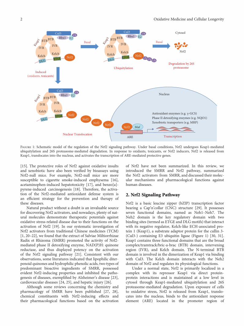
[15]. The protective roles of Nrf2 against oxidative insultsand xenobiotic have also been verified by bioassays usingNrf2-null mice. For example, Nrf2-null mice are moresusceptible to cigarette smoke-induced emphysema [16],acetaminophen-induced hepatotoxicity [17], and benzo[a]-pyrene-induced carcinogenesis [18]. Therefore, the activa-tion of the Nrf2-mediated antioxidant defense system isan efficient strategy for the prevention and therapy ofthese diseases.
Natural product without a doubt is an invaluable sourcefor discovering Nrf2 activators, and nowadays, plenty of nat-ural molecules demonstrate therapeutic potentials againstoxidative stress-related disease due to their functions on theactivation of Nrf2 [19]. In our systematic investigation ofNrf2 activators from traditional Chinese medicines (TCM)[1, 20–22], we found that the extract of Salviae MiltiorrhizaeRadix et Rhizoma (SMRR) promoted the activity of Nrf2-mediated phase II detoxifying enzyme, NAD(P)H: quinonereductase, and thus displayed potency on the activationof the Nrf2 signaling pathway [21]. Consistent with ourobservations, some literatures indicated that lipophilic diter-penoid quinones and hydrophilic phenolic acids, two types ofpredominant bioactive ingredients of SMRR, possessedevident Nrf2-inducing properties and inhibited the patho-genesis of diseases, exemplified by Alzheimer’s disease [23],cardiovascular diseases [24, 25], and hepatic injury [26].
Although some reviews concerning the chemistry andpharmacology of SMRR have been published [27, 28],chemical constituents with Nrf2-inducing effects andtheir pharmacological functions based on the activation
of Nrf2 have not been summarized. In this review, weintroduced the SMRR and Nrf2 pathway, summarizedthe Nrf2 activators from SMRR, and discussed their molec-ular mechanisms and pharmacological functions againsthuman diseases.
2. Nrf2 Signaling Pathway
Nrf2 is a basic leucine zipper (bZIP) transcription factorbearing a Cap‘n’collar (CNC) structure [29]. It possessesseven functional domains, named as Neh1–Neh7. TheNeh2 domain is the key regulatory domain with twobinding sites (termed as ETGE and DLGmotifs) that interactwith its negative regulator, Kelch-like ECH-associated pro-tein 1 (Keap1), a substrate adaptor protein for the cullin 3-(Cul3-) containing E3 ubiquitin ligase (Figure 1) [30, 31].Keap1 contains three functional domains that are the broadcomplex/tramtrack/bric-a-brac (BTB) domain, interveningregion (IVR), and Kelch domain. The N-terminal BTBdomain is involved in the dimerization of Keap1 via bindingwith Cul3. The Kelch domain interacts with the Neh2domain of Nrf2 and regulates its physiological functions.
Under a normal state, Nrf2 is primarily localized in acomplex with its repressor Keap1 via direct protein-protein interactions and is maintained at a low level incytosol through Keap1-mediated ubiquitylation and 26Sproteasome-mediated degradation. Upon exposure of cellsto oxidative stress, Nrf2 is released from Keap1, translo-cates into the nucleus, binds to the antioxidant responseelement (ARE) located in the promoter region of
IVR
Kelch
BTB
Nrf2
Cul3Rbx1 E2
UbBTB
IVR
Kelch
IVR
Kelch
BTB
Nrf2
Cul3Rbx1 E2
UbBTB
IVR
Kelch
Ub Ub Ub
IVR
Kelch
BTB
Nrf2
Cul3Rbx1 E2
UbBTB
IVR
Kelch
Nrf2Nrf2 Nrf2
ARE
MafNrf2
Induced(oxidants, toxicants)
Basal Basal
Antioxidant enzymes (e.g. γ-GCS)Phase II detoxifying enzymes (e.g. NQO1)Xenobiotic transporters (e.g. MRP)
Nucleus
Nrf2
Nuclear Translocation
UbiquitylationDegradation by 26S
proteasome
Transcription
Cytosol
DLGETGE
DLG ETGE DLG ETGE
Keap1
Keap1
Keap1
Figure 1: Schematic model of the regulation of the Nrf2 signaling pathway. Under basal conditions, Nrf2 undergoes Keap1-mediatedubiquitylation and 26S proteasome-mediated degradation. In response to oxidants, toxicants, or Nrf2 inducers, Nrf2 is released fromKeap1, translocates into the nucleus, and activates the transcription of ARE-mediated protective genes.
2 Oxidative Medicine and Cellular Longevity
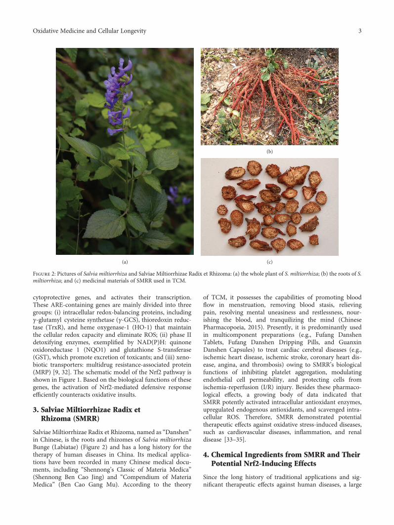
cytoprotective genes, and activates their transcription.These ARE-containing genes are mainly divided into threegroups: (i) intracellular redox-balancing proteins, includingγ-glutamyl cysteine synthetase (γ-GCS), thioredoxin reduc-tase (TrxR), and heme oxygenase-1 (HO-1) that maintainthe cellular redox capacity and eliminate ROS; (ii) phase IIdetoxifying enzymes, exemplified by NAD(P)H: quinoneoxidoreductase 1 (NQO1) and glutathione S-transferase(GST), which promote excretion of toxicants; and (iii) xeno-biotic transporters: multidrug resistance-associated protein(MRP) [9, 32]. The schematic model of the Nrf2 pathway isshown in Figure 1. Based on the biological functions of thesegenes, the activation of Nrf2-mediated defensive responseefficiently counteracts oxidative insults.
3. Salviae Miltiorrhizae Radix etRhizoma (SMRR)
Salviae Miltiorrhizae Radix et Rhizoma, named as “Danshen”in Chinese, is the roots and rhizomes of Salvia miltiorrhizaBunge (Labiatae) (Figure 2) and has a long history for thetherapy of human diseases in China. Its medical applica-tions have been recorded in many Chinese medical docu-ments, including “Shennong’s Classic of Materia Medica”(Shennong Ben Cao Jing) and “Compendium of MateriaMedica” (Ben Cao Gang Mu). According to the theory
of TCM, it possesses the capabilities of promoting bloodflow in menstruation, removing blood stasis, relievingpain, resolving mental uneasiness and restlessness, nour-ishing the blood, and tranquilizing the mind (ChinesePharmacopoeia, 2015). Presently, it is predominantly usedin multicomponent preparations (e.g., Fufang DanshenTablets, Fufang Danshen Dripping Pills, and GuanxinDanshen Capsules) to treat cardiac cerebral diseases (e.g.,ischemic heart disease, ischemic stroke, coronary heart dis-ease, angina, and thrombosis) owing to SMRR’s biologicalfunctions of inhibiting platelet aggregation, modulatingendothelial cell permeability, and protecting cells fromischemia-reperfusion (I/R) injury. Besides these pharmaco-logical effects, a growing body of data indicated thatSMRR potently activated intracellular antioxidant enzymes,upregulated endogenous antioxidants, and scavenged intra-cellular ROS. Therefore, SMRR demonstrated potentialtherapeutic effects against oxidative stress-induced diseases,such as cardiovascular diseases, inflammation, and renaldisease [33–35].
4. Chemical Ingredients from SMRR and TheirPotential Nrf2-Inducing Effects
Since the long history of traditional applications and sig-nificant therapeutic effects against human diseases, a large
(a)
(b)
(c)
Figure 2: Pictures of Salvia miltiorrhiza and Salviae Miltiorrhizae Radix et Rhizoma: (a) the whole plant of S. miltiorrhiza; (b) the roots of S.miltiorrhiza; and (c) medicinal materials of SMRR used in TCM.
3Oxidative Medicine and Cellular Longevity
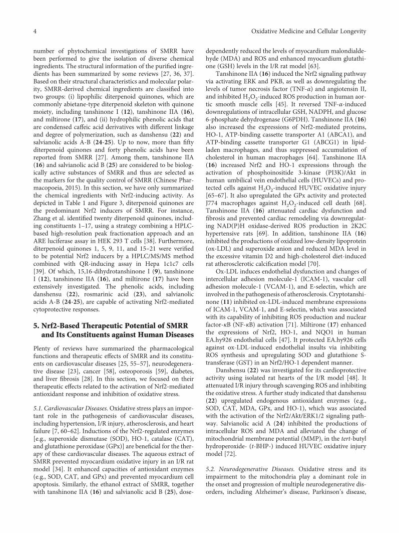
number of phytochemical investigations of SMRR havebeen performed to give the isolation of diverse chemicalingredients. The structural information of the purified ingre-dients has been summarized by some reviews [27, 36, 37].Based on their structural characteristics and molecular polar-ity, SMRR-derived chemical ingredients are classified intotwo groups: (i) lipophilic diterpenoid quinones, which arecommonly abietane-type diterpenoid skeleton with quinonemoiety, including tanshinone I (12), tanshinone IIA (16),and miltirone (17), and (ii) hydrophilic phenolic acids thatare condensed caffeic acid derivatives with different linkageand degree of polymerization, such as danshensu (22) andsalvianolic acids A-B (24-25). Up to now, more than fiftyditerpenoid quinones and forty phenolic acids have beenreported from SMRR [27]. Among them, tanshinone IIA(16) and salvianolic acid B (25) are considered to be biolog-ically active substances of SMRR and thus are selected asthe markers for the quality control of SMRR (Chinese Phar-macopoeia, 2015). In this section, we have only summarizedthe chemical ingredients with Nrf2-inducing activity. Asdepicted in Table 1 and Figure 3, diterpenoid quinones arethe predominant Nrf2 inducers of SMRR. For instance,Zhang et al. identified twenty diterpenoid quinones, includ-ing constituents 1–17, using a strategy combining a HPLC-based high-resolution peak fractionation approach and anARE luciferase assay in HEK 293 T cells [38]. Furthermore,diterpenoid quinones 1, 5, 9, 11, and 15–21 were verifiedto be potential Nrf2 inducers by a HPLC/MS/MS methodcombined with QR-inducing assay in Hepa 1c1c7 cells[39]. Of which, 15,16-dihydrotanshinone I (9), tanshinoneI (12), tanshinone IIA (16), and miltirone (17) have beenextensively investigated. The phenolic acids, includingdanshensu (22), rosmarinic acid (23), and salvianolicacids A-B (24-25), are capable of activating Nrf2-mediatedcytoprotective responses.
5. Nrf2-Based Therapeutic Potential of SMRRand Its Constituents against Human Diseases
Plenty of reviews have summarized the pharmacologicalfunctions and therapeutic effects of SMRR and its constitu-ents on cardiovascular diseases [25, 55–57], neurodegenera-tive disease [23], cancer [58], osteoporosis [59], diabetes,and liver fibrosis [28]. In this section, we focused on theirtherapeutic effects related to the activation of Nrf2-mediatedantioxidant response and inhibition of oxidative stress.
5.1. Cardiovascular Diseases.Oxidative stress plays an impor-tant role in the pathogenesis of cardiovascular diseases,including hypertension, I/R injury, atherosclerosis, and heartfailure [7, 60–62]. Inductions of the Nrf2-regulated enzymes[e.g., superoxide dismutase (SOD), HO-1, catalase (CAT),and glutathione peroxidase (GPx)] are beneficial for the ther-apy of these cardiovascular diseases. The aqueous extract ofSMRR prevented myocardium oxidative injury in an I/R ratmodel [34]. It enhanced capacities of antioxidant enzymes(e.g., SOD, CAT, and GPx) and prevented myocardium cellapoptosis. Similarly, the ethanol extract of SMRR, togetherwith tanshinone IIA (16) and salvianolic acid B (25), dose-
dependently reduced the levels of myocardium malondialde-hyde (MDA) and ROS and enhanced myocardium glutathi-one (GSH) levels in the I/R rat model [63].
Tanshinone IIA (16) induced the Nrf2 signaling pathwayvia activating ERK and PKB, as well as downregulating thelevels of tumor necrosis factor (TNF-α) and angiotensin II,and inhibited H2O2-induced ROS production in human aor-tic smooth muscle cells [45]. It reversed TNF-α-induceddownregulations of intracellular GSH, NADPH, and glucose6-phosphate dehydrogenase (G6PDH). Tanshinone IIA (16)also increased the expressions of Nrf2-mediated proteins,HO-1, ATP-binding cassette transporter A1 (ABCA1), andATP-binding cassette transporter G1 (ABCG1) in lipid-laden macrophages, and thus suppressed accumulation ofcholesterol in human macrophages [64]. Tanshinone IIA(16) increased Nrf2 and HO-1 expressions through theactivation of phosphoinositide 3-kinase (PI3K)/Akt inhuman umbilical vein endothelial cells (HUVECs) and pro-tected cells against H2O2-induced HUVEC oxidative injury[65–67]. It also upregulated the GPx activity and protectedJ774 macrophages against H2O2-induced cell death [68].Tanshinone IIA (16) attenuated cardiac dysfunction andfibrosis and prevented cardiac remodeling via downregulat-ing NAD(P)H oxidase-derived ROS production in 2K2Chypertensive rats [69]. In addition, tanshinone IIA (16)inhibited the productions of oxidized low-density lipoprotein(ox-LDL) and superoxide anion and reduced MDA level inthe excessive vitamin D2 and high-cholesterol diet-inducedrat atherosclerotic calcification model [70].
Ox-LDL induces endothelial dysfunction and changes ofintercellular adhesion molecule-1 (ICAM-1), vascular celladhesion molecule-1 (VCAM-1), and E-selectin, which areinvolved in the pathogenesis of atherosclerosis. Cryptotanshi-none (11) inhibited ox-LDL-induced membrane expressionsof ICAM-1, VCAM-1, and E-selectin, which was associatedwith its capability of inhibiting ROS production and nuclearfactor-κB (NF-κB) activation [71]. Miltirone (17) enhancedthe expressions of Nrf2, HO-1, and NQO1 in humanEA.hy926 endothelial cells [47]. It protected EA.hy926 cellsagainst ox-LDL-induced endothelial insults via inhibitingROS synthesis and upregulating SOD and glutathione S-transferase (GST) in an Nrf2/HO-1 dependent manner.
Danshensu (22) was investigated for its cardioprotectiveactivity using isolated rat hearts of the I/R model [48]. Itattenuated I/R injury through scavenging ROS and inhibitingthe oxidative stress. A further study indicated that danshensu(22) upregulated endogenous antioxidant enzymes (e.g.,SOD, CAT, MDA, GPx, and HO-1), which was associatedwith the activation of the Nrf2/Akt/ERK1/2 signaling path-way. Salvianolic acid A (24) inhibited the productions ofintracellular ROS and MDA and alleviated the change ofmitochondrial membrane potential (MMP), in the tert-butylhydroperoxide- (t-BHP-) induced HUVEC oxidative injurymodel [72].
5.2. Neurodegenerative Diseases. Oxidative stress and itsimpairment to the mitochondria play a dominant role inthe onset and progression of multiple neurodegenerative dis-orders, including Alzheimer’s disease, Parkinson’s disease,
4 Oxidative Medicine and Cellular Longevity
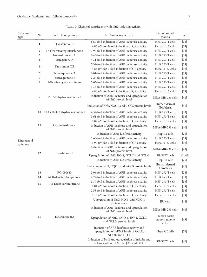
Table 1: Chemical constituents with Nrf2-inducing activity.
Structuraltype
No Name of compounds Nrf2-inducing activityCell or animal
modelsRef
Diterpenoidquinones
1 Tanshindiol B6.80-fold induction of ARE luciferase activity HEK 293 T cells [38]
4.85 μM for 2-fold induction of QR activity Hepa 1c1c7 cells [39]
2 17-Hydroxycryptotanshinone 3.97-fold induction of ARE luciferase activity HEK 293 T cells [38]
3 Isotanshinone IIA 6.45-fold induction of ARE luciferase activity HEK 293 T cells [38]
4 Trijuganone A 4.31-fold induction of ARE luciferase activity HEK 293 T cells [38]
5 Tanshinone IIB5.34-fold induction of ARE luciferase activity HEK 293 T cells [38]
4.85 μM for 2-fold induction of QR activity Hepa 1c1c7 cells [39]
6 Przewaquinone A 6.01-fold induction of ARE luciferase activity HEK 293 T cells [38]
7 Przewaquinone B 7.27-fold induction of ARE luciferase activity HEK 293 T cells [38]
8 1-Ketoisocryptotanshinone 5.65-fold induction of ARE luciferase activity HEK 293 T cells [38]
9 15,16-Dihydrotanshinone I
3.38-fold induction of ARE luciferase activity HEK 293 T cells [38]
4.80 μM for 2-fold induction of QR activity Hepa 1c1c7 cells [39]
Induction of ARE luciferase and upregulationof Nrf2 protein level
MDA-MB-231 cells [40]
Induction of Nrf2, NQO1, and γ-GCS protein levelsHuman dermalfibroblasts.
[41]
10 1,2,15,16-Tetrahydrotanshinone I 4.37-fold induction of ARE luciferase activity HEK 293 T cells [38]
11 Cryptotanshinone
3.61-fold induction of ARE luciferase activity HEK 293 T cells [38]
5.07 μM for 2-fold induction of QR activity Hepa 1c1c7 cells [39]
Induction of ARE luciferase and upregulationof Nrf2 protein level
MDA-MB-231 cells [40]
Induction of ARE-luciferase activity Hep G2 cells [26]
12 Tanshinone I
2.60-fold induction of ARE luciferase activity HEK 293 T cells [38]
5.90 μM for 2-fold induction of QR activity Hepa 1c1c7 cells [39]
Induction of ARE luciferase and upregulationof Nrf2 protein level
MDA-MB-231 cells [40]
Upregulation of Nrf2, HO-1, GCLC, and GCLM SH-SY5Y cells [42, 43]
Induction of ARE-luciferase activity Hep G2 cells [26]
Induction of Nrf2, NQO1, and γ-GCS protein levelsHuman dermal
fibroblasts[41]
13 RO-090680 3.06-fold induction of ARE luciferase activity HEK 293 T cells [38]
14 Methylenetanshinquinone 2.77-fold induction of ARE luciferase activity HEK 293 T cells [38]
15 1,2-Didehydromilitirone3.79-fold induction of ARE luciferase activity HEK 293 T cells [38]
1.04 μM for 2-fold induction of QR activity Hepa 1c1c7 cells [39]
16 Tanshinone IIA
2.58-fold induction of ARE luciferase activity HEK 293 T cells [38]
5.10 μM for 2-fold induction of QR activity Hepa 1c1c7 cells [39]
Upregulation of Nrf2, HO-1, and NQO-1protein levels
JB6 cells [44]
Induction of ARE luciferase and upregulationof Nrf2 protein level
MDA-MB-231 cells [40]
Upregulation of Nrf2, NOQ-1, HO-1, GCLC,and GCLM protein levels
Human aorticsmooth muscle
cells.[45]
Induction of ARE-luciferase activity andupregulation of mRNA levels of GCLC,
NQO1, and HO-1Hepa G2 cells [26]
Induction of Nrf2 and upregulation of mRNA andprotein levels of HO-1, NQO1, and GCLC
SH-SY5Y cells [46]
5Oxidative Medicine and Cellular Longevity
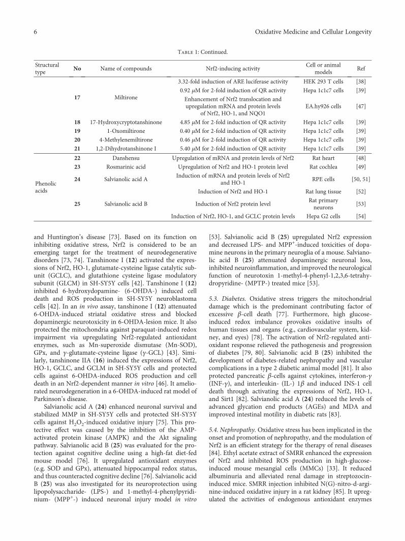
and Huntington’s disease [73]. Based on its function oninhibiting oxidative stress, Nrf2 is considered to be anemerging target for the treatment of neurodegenerativedisorders [73, 74]. Tanshinone I (12) activated the expres-sions of Nrf2, HO-1, glutamate-cysteine ligase catalytic sub-unit (GCLC), and glutathione cysteine ligase modulatorysubunit (GLCM) in SH-SY5Y cells [42]. Tanshinone I (12)inhibited 6-hydroxydopamine- (6-OHDA-) induced celldeath and ROS production in SH-SY5Y neuroblastomacells [42]. In an in vivo assay, tanshinone I (12) attenuated6-OHDA-induced striatal oxidative stress and blockeddopaminergic neurotoxicity in 6-OHDA-lesion mice. It alsoprotected the mitochondria against paraquat-induced redoximpairment via upregulating Nrf2-regulated antioxidantenzymes, such as Mn-superoxide dismutase (Mn-SOD),GPx, and γ-glutamate-cysteine ligase (γ-GCL) [43]. Simi-larly, tanshinone IIA (16) induced the expressions of Nrf2,HO-1, GCLC, and GCLM in SH-SY5Y cells and protectedcells against 6-OHDA-induced ROS production and celldeath in an Nrf2-dependent manner in vitro [46]. It amelio-rated neurodegeneration in a 6-OHDA-induced rat model ofParkinson’s disease.
Salvianolic acid A (24) enhanced neuronal survival andstabilized MMP in SH-SY5Y cells and protected SH-SY5Ycells against H2O2-induced oxidative injury [75]. This pro-tective effect was caused by the inhibition of the AMP-activated protein kinase (AMPK) and the Akt signalingpathway. Salvianolic acid B (25) was evaluated for the pro-tection against cognitive decline using a high-fat diet-fedmouse model [76]. It upregulated antioxidant enzymes(e.g. SOD and GPx), attenuated hippocampal redox status,and thus counteracted cognitive decline [76]. Salvianolic acidB (25) was also investigated for its neuroprotection usinglipopolysaccharide- (LPS-) and 1-methyl-4-phenylpyridi-nium- (MPP+-) induced neuronal injury model in vitro
[53]. Salvianolic acid B (25) upregulated Nrf2 expressionand decreased LPS- and MPP+-induced toxicities of dopa-mine neurons in the primary neuroglia of a mouse. Salviano-lic acid B (25) attenuated dopaminergic neuronal loss,inhibited neuroinflammation, and improved the neurologicalfunction of neurotoxin 1-methyl-4-phenyl-1,2,3,6-tetrahy-dropyridine- (MPTP-) treated mice [53].
5.3. Diabetes. Oxidative stress triggers the mitochondrialdamage which is the predominant contributing factor ofexcessive β-cell death [77]. Furthermore, high glucose-induced redox imbalance provokes oxidative insults ofhuman tissues and organs (e.g., cardiovascular system, kid-ney, and eyes) [78]. The activation of Nrf2-regulated anti-oxidant response relieved the pathogenesis and progressionof diabetes [79, 80]. Salvianolic acid B (25) inhibited thedevelopment of diabetes-related nephropathy and vascularcomplications in a type 2 diabetic animal model [81]. It alsoprotected pancreatic β-cells against cytokines, interferon-γ(INF-γ), and interleukin- (IL-) 1β and induced INS-1 celldeath through activating the expressions of Nrf2, HO-1,and Sirt1 [82]. Salvianolic acid A (24) reduced the levels ofadvanced glycation end products (AGEs) and MDA andimproved intestinal motility in diabetic rats [83].
5.4. Nephropathy. Oxidative stress has been implicated in theonset and promotion of nephropathy, and the modulation ofNrf2 is an efficient strategy for the therapy of renal diseases[84]. Ethyl acetate extract of SMRR enhanced the expressionof Nrf2 and inhibited ROS production in high-glucose-induced mouse mesangial cells (MMCs) [33]. It reducedalbuminuria and alleviated renal damage in streptozocin-induced mice. SMRR injection inhibited N(G)-nitro-d-argi-nine-induced oxidative injury in a rat kidney [85]. It upreg-ulated the activities of endogenous antioxidant enzymes
Table 1: Continued.
Structuraltype
No Name of compounds Nrf2-inducing activityCell or animal
modelsRef
17 Miltirone
3.32-fold induction of ARE luciferase activity HEK 293 T cells [38]
0.92 μM for 2-fold induction of QR activity Hepa 1c1c7 cells [39]
Enhancement of Nrf2 translocation andupregulation mRNA and protein levels
of Nrf2, HO-1, and NQO1EA.hy926 cells [47]
18 17-Hydroxycryptotanshinone 4.85 μM for 2-fold induction of QR activity Hepa 1c1c7 cells [39]
19 1-Oxomiltirone 0.40 μM for 2-fold induction of QR activity Hepa 1c1c7 cells [39]
20 4-Methylenemiltirone 0.46 μM for 2-fold induction of QR activity Hepa 1c1c7 cells [39]
21 1,2-Dihydrotanshinone I 5.40 μM for 2-fold induction of QR activity Hepa 1c1c7 cells [39]
Phenolicacids
22 Danshensu Upregulation of mRNA and protein levels of Nrf2 Rat heart [48]
23 Rosmarinic acid Upregulation of Nrf2 and HO-1 protein level Rat cochlea [49]
24 Salvianolic acid AInduction of mRNA and protein levels of Nrf2
and HO-1RPE cells [50, 51]
25 Salvianolic acid B
Induction of Nrf2 and HO-1 Rat lung tissue [52]
Induction of Nrf2 protein levelRat primaryneurons
[53]
Induction of Nrf2, HO-1, and GCLC protein levels Hepa G2 cells [54]
6 Oxidative Medicine and Cellular Longevity
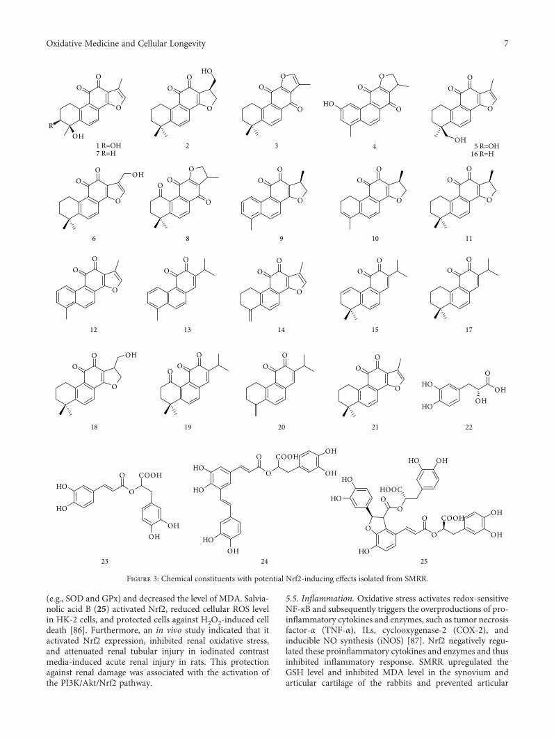
(e.g., SOD and GPx) and decreased the level of MDA. Salvia-nolic acid B (25) activated Nrf2, reduced cellular ROS levelin HK-2 cells, and protected cells against H2O2-induced celldeath [86]. Furthermore, an in vivo study indicated that itactivated Nrf2 expression, inhibited renal oxidative stress,and attenuated renal tubular injury in iodinated contrastmedia-induced acute renal injury in rats. This protectionagainst renal damage was associated with the activation ofthe PI3K/Akt/Nrf2 pathway.
5.5. Inflammation. Oxidative stress activates redox-sensitiveNF-κB and subsequently triggers the overproductions of pro-inflammatory cytokines and enzymes, such as tumor necrosisfactor-α (TNF-α), ILs, cyclooxygenase-2 (COX-2), andinducible NO synthesis (iNOS) [87]. Nrf2 negatively regu-lated these proinflammatory cytokines and enzymes and thusinhibited inflammatory response. SMRR upregulated theGSH level and inhibited MDA level in the synovium andarticular cartilage of the rabbits and prevented articular
O
OHR
OO
1 R=OH7 R=H
OO
O
3
O
O
O
HO
4
OO
O
OH5 R=OH
16 R=H
O
OO OH
6
OO
O
O
8
O
OO
9
O
OO
11
12
O
OO
O
OO
14
OO
17
OO
20
OHHO
HOO
OH
22
23 24
O
O COOHOH
OHHO
HO
HOOH
O
O COOHOH
OH
HO
O
HO
HO OO
OHHO
HOOC
O
OO
HO
2
O
OO
10
OO
13
OO
15
O
OO OH
18
OO
O
19
O
OO
21
HO
HOO
O
COOH
OHOH
25
Figure 3: Chemical constituents with potential Nrf2-inducing effects isolated from SMRR.
7Oxidative Medicine and Cellular Longevity
cartilage degeneration in rabbits with osteoarthritis [35].Extract of SMRR induced the expressions of HO-1 andNrf2 and inhibited H2O2-stimulated production of ROS inRAW 264.7 macrophages [88]. The activation of Nrf2 wasattributed to the PI3K/Akt and MEK1 signaling pathway.Tanshinone IIA (16) induced HO-1 expression in RAW264.7 macrophages and inhibited LPS-stimulated upregula-tion of COX-2 and iNOS [89]. A high-fat diet gave rise tothe decrease of Nrf2 expression, accumulation of oxidativestress, and inflammation in C57BL/6 mouse. Salvianolic acidB (25) upregulated the expressions of Nrf2, HO-1, andNQO1 and thus inhibited the expressions of NF-κB, COX-2, and iNOS in mice fed a high-fat diet [90].
5.6. Liver Diseases. As the major metabolism organ of xeno-biotics, the liver is apt to be kept in a sustained oxidativestate, which causes a variety of liver diseases, coveringfibrosis, cirrhosis, and carcinoma [91, 92]. The extract ofSMRR inhibited aflatoxin B1- (AFB1-) induced cytotoxicityin cultured primary rat hepatocytes since its capability ofinhibiting oxidative stress [93]. AFB1-induced ROS forma-tion and GSH depletion could be reverted by SMRR extracttreatment. Tanshinone IIA (16) upregulated the proteinlevel of Nrf2 and enhanced the mRNA levels of GCLC,NQO1, and HO-1 in HepG2 cells and in a C57BL/6J mouseliver [26]. Tanshinone IIA (16) alleviated acetaminophen-(APAP-) induced upregulations of serum alanine amino-transferase (ALT), aspartate aminotransferase (AST), andlactate dehydrogenase (LDH). It reverted the APAP-induced decreases of GSH, GST, GPx, SOD, and CAT ina mouse liver and thus prevented APAP-induced hepato-toxicity. Similarly, tanshinone IIA (16) upregulated the levelsof Nrf2-mediated antioxidant enzymes (e.g., HO-1, SOD,CAT, and GPx) and inhibited fibrosis in a rat modelof cirrhosis.
Salvianolic acid A (24) evidently reduced oxidative stress,evidenced by decreasing the levels of ROS and MDA andincreasing the levels of hepatic superoxide dismutase andGSH, in rat liver tissue [94]. Furthermore, salvianolic acidA (24) significantly inhibited carbon tetrachloride- (CCl4-)induced hepatotoxicity. Decreased levels of serum ALTand AST in response to CCl4 exposure were recoveredafter treatment with 24. Salvianolic acid A (24) demon-strated radioprotective effects against γ-radiation-induceddamage in human embryo liver L-02 cells, which was associ-ated with inhibitions of ROS generation and mitochondrialcytochrome C release [95]. Salvianolic acid B (25) demon-strated APAP-induced liver injury in mice [54]. It upregu-lated the expressions of Nrf2, GCLC, and HO-1 via theactivation of the PI3K and protein kinase C (PKC) pathwayin HepG2 cells.
5.7. Lung Diseases. The lung is directly and continuouslyexposed to the external oxidants and toxicants, and thusexcessive ROS are produced in the lung tissue. Theseendogenous and exogenous oxidants contribute to the path-ophysiology of lung diseases, exemplified by chronic obstruc-tive pulmonary disease and pulmonary fibrosis [96, 97].Tanshinone I (12) enhanced Nrf2-mediated expressions of
NQO1 and γ-GCS through hindering Nrf2 ubiquitinationand specifically reacting with cysteine residue at amino acid151 in Keap1 protein [40]. It protected human bronchial epi-thelial HBE cells against As(III)-induced oxidative damage inan Nrf2-dependent manner. An in vivo study using Nrf2+/+
and Nrf2−/− mice indicated that tanshinone I (12) attenuatedAs(III)-induced inflammatory lung damage in Nrf2+/+ mice,and this protective effect vanished in Nrf2−/− mice.
Salvianolic acid B (25) upregulated the expressions ofNrf2 and HO-1, enhanced GSH production, and inhib-ited cigarette smoke-induced lung pathological changesand inflammatory responses [52]. Salvianolic acid B (25)reduced the expression of ROS-generating enzyme NADPHoxidase-4 (Nox4) in lung tissue of mice [98]. It inhibitedparaquat-induced structure distortion, collagen overproduc-tion, proinflammatory cytokine release, and oxidative insults.Salvianolic acid B (25) inhibited PQ-induced activation ofthe transforming growth factor-β (TGF-β1)/Smad3 pathway,which was a key regulator of pulmonary fibrosis [99]. Thecapability of 25 in activating Nrf2 and inhibiting pulmonaryfibrosis was further confirmed by a MRC-5 human lungfibroblast cell model and a bleomycin-induced rat pulmonaryfibrosis model [100].
5.8. Ocular Diseases. Oxidative stress is involved in thepathogenesis of age-related macular degeneration [101].Salvianolic acid A (24) inhibited H2O2-induced ROS pro-duction and prevented H2O2-induced primary and trans-formed retinal pigment epithelial cell death and apoptosis[51]. Activations of Nrf2 and HO-1 by salvianolic acid A(24) are essential for this protective effect. The Nrf2-inducing effect of 24 is associated with the activations ofPI3K and the mammalian target of rapamycin (mTOR)signaling pathway [50]. It attenuated ox-LDL-induced lipi-dosis and apoptosis in the retinal pigment epithelium layerand inhibited ox-LDL-induced elevated ROS level and RPEinflammation in a rat model. These data suggested thatsalvianolic acid A (24) was a potential therapeutic agentagainst age-related macular degeneration.
5.9. Others. Because of wide distribution in the human tissuesand organs, the activation of Nrf2 is an effective method forthe therapy of many human diseases [102, 103]. Noise-induced hearing loss is associated with oxidative stress andlipoperoxidative damage. Rosmarinic acid (23) enhancedNrf2/HO-1-mediated endogenous antioxidant defense sys-tem, attenuated hearing loss, and promoted hair cell survivalin a noise-induced rat model [49]. Tanshinone I (12) and15,16-dihydrotanshinone (9) induced the expressions ofNrf2, GCLC, and NQO1 through inhibiting Nrf2 ubiquiti-nation in human Hs27 dermal fibroblasts and HaCaT kerati-nocytes [41]. These two constituents significantly suppressedsolar-simulated UV and riboflavin-sensitized ultraviolet-induced skin cell death. A solar-simulated UV-inducedhuman skin reconstruct model was established for evaluat-ing the photoprotective effect of 15,16-dihydrotanshinone(9). Treatment with 15,16-dihydrotanshinone (9) definitelyattenuated epidermal solar insult.
8 Oxidative Medicine and Cellular Longevity
6. Conclusion and Future Perspectives
Oxidative stress plays a vital role in the pathogenesis ofmany human diseases. The activation of the Nrf2-mediatedantioxidant defense system inhibits oxidative stress and thuseffectively blocks the onset and progression of oxidativestress-induced human diseases. SMRR is a traditional Chi-nese medicine that has been long used for the treatment ofcardiac cerebral diseases. Lipophilic diterpenoid quinonesand hydrophilic phenolic acids are predominant constituentsand contribute to the pharmacological functions of SMRR.These two types of constituents demonstrate potent Nrf2-inducing effects and hence are potential agents for the treat-ment of oxidative stress-related diseases. The SMRR extract,as well as the purified constituents, tanshinone I (12), tanshi-none IIA (16), salvianolic acids A (24), and B (25), has beenextensively investigated, and their inductions on the Nrf2pathway and therapeutic effects on cardiovascular diseases,neurodegenerative diseases, diabetes, nephropathy, inflam-mation, liver diseases, and lung diseases have been verifiedby multiple models in vitro and in vivo.
Although there has been great progress on SMRR and itsconstituents, important areas on their phytochemistry, phar-macology, and medical applications related to the activationof Nrf2 remain to be explored. (i) Plenty of diterpenoidquinones have been isolated from SMRR; however, exten-sive researches still focused on several active constituents,including 15,16-dihydrotanshinone I (9), tanshinone I (12),tanshinone IIA (16), and miltirone (17). Miscellaneous diter-penoid quinones should be extensively investigated, such astheir structure-activity relationship and mechanisms ofaction on the activation of Nrf2. (ii) Besides these verifiedmedical applications, SMRR and its constituents possesspotential pharmacological functions (e.g., chemopreventionon cancer and therapeutic effect on diabetic nephropathy)because of their activation of Nrf2. Thus, these unconfirmedpharmacological activities should be noted. (iii) Based on thetraditional uses, chemical constituents, and pharmacologicalfunctions of SMRR, future works on discovering new leadcompounds and developing SMRR and its constituents intonew drugs for the therapy of oxidative stress-related diseasesare significant.
Abbreviations
ABCA1: ATP-binding cassette transporter A1AFB1: Aflatoxin B1AGEs: Advanced glycation end productsALT: Alanine aminotransferaseAPAP: AcetaminophenAST: Aspartate aminotransferaseARE: Antioxidant response elementBTB: Broad complex/tramtrack/bric-a-bracCAT: CatalaseCOX-2: Cyclooxygenase-2γ-GCL: γ-Glutamate cysteine ligaseGCLC: Glutamate cysteine ligase catalytic subunitGCLM: Glutathione cysteine ligase modulatory subunitγ-GCS: γ-Glutamyl cysteine synthetase
GPx: Glutathione peroxidaseGSH: GlutathioneGST: Glutathione S-transferaseHO-1: Heme oxygenase-1ICAM-1: Intercellular adhesion molecule-1HUVECs: Human umbilical vein endothelial cellsIL: InterleukinINF-γ: Interferon-γiNOS: Inducible NO synthesisI/R: Ischemia-reperfusionIVR: Intervening regionKeap1: Kelch-like ECH-associated protein 1LDH: Lactate dehydrogenaseLPS: LipopolysaccharideMDA: MalondialdehydeMMP: Mitochondrial membrane potentialMn-SOD: Mn-superoxide dismutaseMPP+: 1-Methyl-4-phenylpyridiniumMPTP: 1-Methyl-4-phenyl-1,2,3,6-tetrahydropyridinemTOR: Mammalian target of rapamycinNF-κB: Nuclear factor-κBNQO1: NAD(P)H: quinone oxidoreductase 1Nrf2: Nuclear factor erythroid 2-related factor 26-OHDA: 6-Hydroxydopamineox-LDL: Oxidized low-density lipoproteinPKC: Protein kinase CPI3K: Phosphoinositide 3-kinaseRNS: Reactive nitrogen speciesROS: Reactive oxygen speciesSMRR: Salviae Miltiorrhizae Radix et RhizomaSOD: Superoxide dismutaset-BHP: tert-Butyl hydroperoxideTCM: Traditional Chinese medicinesTGF-β1: Transforming growth factor-βTNF-α: Tumor necrosis factor-αTrxR: Thioredoxin reductaseVCAM-1: Vascular cell adhesion molecule-1.
Conflicts of Interest
The authors declare that there is no conflict of interest.
Acknowledgments
This work was financially supported by NSF of ShandongProvince (ZR2014HM019), NNSFs of China (31470419and 81673558), and Young Scholars Program of ShandongUniversity (2015WLJH50).
References
[1] M.-X. Zhou, G.-H. Li, B. Sun et al., “Identification ofnovel Nrf2 activators from Cinnamomum chartophyllumH.W. Li and their potential application of preventing oxida-tive insults in human lung epithelial cells,” Redox Biology,vol. 14, pp. 154–163, 2018.
[2] E. Bargagli, C. Olivieri, D. Bennett, A. Prasse, J. Muller-Quer-nheim, and P. Rottoli, “Oxidative stress in the pathogenesis ofdiffuse lung diseases: a review,” Respiratory Medicine,vol. 103, no. 9, pp. 1245–1256, 2009.
9Oxidative Medicine and Cellular Longevity
[3] X. Chen, C. Guo, and J. Kong, “Oxidative stress in neurode-generative diseases,” Neural Regeneration Research, vol. 7,no. 5, pp. 376–385, 2012.
[4] D. Jay, H. Hitomi, and K. K. Griendling, “Oxidative stress anddiabetic cardiovascular complications,” Free Radical Biology& Medicine, vol. 40, no. 2, pp. 183–192, 2006.
[5] J. E. Klaunig, L. M. Kamendulis, and B. A. Hocevar,“Oxidative stress and oxidative damage in carcinogenesis,”Toxicologic Pathology, vol. 38, no. 1, pp. 96–109, 2010.
[6] A. C. Maritim, R. A. Sanders, and J. B. Watkins, “Diabetes,oxidative stress, and antioxidants: a review,” Journal ofBiochemical and Molecular Toxicology, vol. 17, no. 1,pp. 24–38, 2003.
[7] D. Harrison, K. K. Griendling, U. Landmesser, B. Hornig,and H. Drexler, “Role of oxidative stress in atherosclerosis,”The American Journal of Cardiology, vol. 91, no. 3, pp. 7–11,2003.
[8] N. D. Vaziri, “Roles of oxidative stress and antioxidanttherapy in chronic kidney disease and hypertension,” Cur-rent Opinion in Nephrology and Hypertension, vol. 13, no. 1,pp. 93–99, 2004.
[9] M. C. Jaramillo and D. D. Zhang, “The emerging role of theNrf2–Keap1 signaling pathway in cancer,” Genes & Develop-ment, vol. 27, no. 20, pp. 2179–2191, 2013.
[10] T. O. Khor, M.-T. Huang, A. Prawan et al., “Increased suscep-tibility of Nrf2 knockout mice to colitis-associated colorectalcancer,” Cancer Prevention Research, vol. 1, no. 3, pp. 187–191, 2008.
[11] B. Li, S. Liu, L. Miao, and L. Cai, “Prevention of diabetic com-plications by activation of Nrf2: diabetic cardiomyopathy andnephropathy,” Experimental Diabetes Research, vol. 2012,Article ID 216512, 7 pages, 2012.
[12] T. E. Sussan, S. Biswal et al.N. Ganguly, S. Jindal, S. Biswalet al., “Oxidative stress and respiratory diseases: the criticalrole of Nrf2,” in Studies on Respiratory Disorders, OxidativeStress in Applied Basic Research and Clinical Practice,pp. 335–348, Humana Press, New York, NY, USA, 2014.
[13] T. O. Khor, M.-T. Huang, K. H. Kwon, J. Y. Chan, B. S.Reddy, and A.-N. Kong, “Nrf2-deficient mice have anincreased susceptibility to dextran sulfate sodium–inducedcolitis,” Cancer Research, vol. 66, no. 24, pp. 11580–11584,2006.
[14] J. Li, T. Ichikawa, J. S. Janicki, and T. Cui, “Targeting the Nrf2pathway against cardiovascular disease,” Expert Opinion onTherapeutic Targets, vol. 13, no. 7, pp. 785–794, 2009.
[15] M. C. Barone, G. P. Sykiotis, and D. Bohmann, “Geneticactivation of Nrf2 signaling is sufficient to ameliorateneurodegenerative phenotypes in a Drosophila model ofParkinson’s disease,” Disease Models & Mechanisms, vol. 4,no. 5, pp. 701–707, 2011.
[16] T. Rangasamy, C. Y. Cho, R. K. Thimmulappa et al., “Geneticablation of Nrf2 enhances susceptibility to cigarette smoke–induced emphysema in mice,” The Journal of Clinical Investi-gation, vol. 114, no. 9, pp. 1248–1259, 2004.
[17] A. Enomoto, K. Itoh, E. Nagayoshi et al., “High sensitivity ofNrf2 knockout mice to acetaminophen hepatotoxicity associ-ated with decreased expression of ARE-regulated drugmetabolizing enzymes and antioxidant genes,” ToxicologicalSciences, vol. 59, no. 1, pp. 169–177, 2001.
[18] M. Ramos-Gomez, P. M. Dolan, K. Itoh, M. Yamamoto,and T. W. Kensler, “Interactive effects of nrf2 genotype
and oltipraz on benzo[a]pyrene–DNA adducts and tumoryield in mice,” Carcinogenesis, vol. 24, no. 3, pp. 461–467,2003.
[19] H. Kumar, I. S. Kim, S. V. More, B. W. Kim, and D. K. Choi,“Natural product-derived pharmacological modulators ofNrf2/ARE pathway for chronic diseases,” Natural ProductReports, vol. 31, no. 1, pp. 109–139, 2014.
[20] T. Shen, T. Jiang, M. Long et al., “A curcumin derivative thatinhibits vinyl carbamate-induced lung carcinogenesis viaactivation of the Nrf2 protective response,” Antioxidants &Redox Signaling, vol. 23, no. 8, pp. 651–664, 2015.
[21] M.-X. Zhou, X. Wei, A.-L. Li et al., “Screening of traditionalChinese medicines with therapeutic potential on chronicobstructive pulmonary disease through inhibiting oxidativestress and inflammatory response,” BMC Complementaryand Alternative Medicine, vol. 16, no. 1, p. 360, 2016.
[22] T. Shen, X.-M. Chen, B. Harder et al., “Plant extracts of thefamily Lauraceae: a potential resource for chemopreventiveagents that activate the nuclear factor-erythroid 2-related fac-tor 2/antioxidant response element pathway,” Planta Medica,vol. 80, no. 5, pp. 426–434, 2014.
[23] X.-Z. Zhang, S.-S. Qian, Y.-J. Zhang, and R.-Q. Wang, “Salviamiltiorrhiza: a source for anti-Alzheimer’s disease drugs,”Pharmaceutical Biology, vol. 54, no. 1, pp. 18–24, 2016.
[24] G. Cui, L. Shan, M. Hung et al., “A novel danshensu deriva-tive confers cardioprotection via PI3K/Akt and Nrf2 path-ways,” International Journal of Cardiology, vol. 168, no. 2,pp. 1349–1359, 2013.
[25] S. Gao, Z. Liu, H. Li, P. J. Little, P. Liu, and S. Xu, “Cardiovas-cular actions and therapeutic potential of tanshinone IIA,”Atherosclerosis, vol. 220, no. 1, pp. 3–10, 2012.
[26] W. Wang, C. Guan, X. Sun et al., “Tanshinone IIA protectsagainst acetaminophen-induced hepatotoxicity via activatingthe Nrf2 pathway,” Phytomedicine, vol. 23, no. 6, pp. 589–596, 2016.
[27] C.-Y. Su, Q.-L. Ming, K. Rahman, T. HAN, and L.-P. Qin,“Salvia miltiorrhiza: traditional medicinal uses, chemistry,and pharmacology,” Chinese Journal of Natural Medicines,vol. 13, no. 3, pp. 163–182, 2015.
[28] W.-y. Wu and Y.-p. Wang, “Pharmacological actions andtherapeutic applications of Salvia miltiorrhiza depside saltand its active components,” Acta Pharmacologica Sinica,vol. 33, no. 9, pp. 1119–1130, 2012.
[29] P. Moi, K. Chan, I. Asunis, A. Cao, and Y. W. Kan, “Iso-lation of NF-E2-related factor 2 (Nrf2), a NF-E2-like basicleucine zipper transcriptional activator that binds to thetandem NF-E2/AP1 repeat of the beta-globin locus controlregion,” Proceedings of the National Academy of Sciences ofthe United States of America, vol. 91, no. 21, pp. 9926–9930, 1994.
[30] Y. Mitsuishi, H. Motohashi, andM. Yamamoto, “The Keap1–Nrf2 system in cancers: stress response and anabolic metabo-lism,” Frontiers in Oncology, vol. 2, 2012.
[31] W. Li and A. N. Kong, “Molecular mechanisms of Nrf2-mediated antioxidant response,” Molecular Carcinogenesis,vol. 48, no. 2, pp. 91–104, 2009.
[32] A. Lau, N. F. Villeneuve, Z. Sun, P. K. Wong, and D. D.Zhang, “Dual roles of Nrf2 in cancer,” PharmacologicalResearch, vol. 58, no. 5-6, pp. 262–270, 2008.
[33] L. An, M. Zhou, F. M. M. T. Marikar et al., “Salvia miltior-rhiza lipophilic fraction attenuates oxidative stress in diabetic
10 Oxidative Medicine and Cellular Longevity
nephropathy through activation of nuclear factor erythroid2-related factor 2,” The American Journal of Chinese Medi-cine, vol. 45, no. 7, pp. 1441–1457, 2017.
[34] G. Ge, Q. Zhang, J. Ma et al., “Protective effect of Salviamiltiorrhiza aqueous extract on myocardium oxidative injuryin ischemic–reperfusion rats,” Gene, vol. 546, no. 1, pp. 97–103, 2014.
[35] B. Bai and Y. Li, “Danshen prevents articular cartilagedegeneration via antioxidation in rabbits with osteoarthri-tis,” Osteoarthritis and Cartilage, vol. 24, no. 3, pp. 514–520, 2016.
[36] H.-Y. Liu, C.-T. Jiang, J. Feng, and X. Wang, “Research prog-ress of Tanshinones,” Chinese Pharmacological Bulletin,vol. 32, pp. 1643–1647, 2016.
[37] A. R. Jassbi, S. Zare, O. Firuzi, and J. Xiao, “Bioactivephytochemicals from shoots and roots of Salvia species,”Phytochemistry Reviews, vol. 15, no. 5, pp. 829–867, 2016.
[38] H. Zhang, L.-P. Luo, H.-P. Song et al., “A high-resolutionpeak fractionation approach for streamlined screening ofnuclear-factor-E2-related factor-2 activators in Salvia mil-tiorrhiza,” Journal of Chromatography A, vol. 1326, pp. 47–55, 2014.
[39] Z. Ma, M. Zhang, and Z. Song, “Characterization of tanshi-nones with quinone reductase induction activity from RadixSalvia miltiorrhiza by liquid chromatography/tandem massspectrometry,” Rapid Communications in Mass Spectrometry,vol. 23, no. 18, pp. 2857–2866, 2009.
[40] S. Tao, Y. Zheng, A. Lau et al., “Tanshinone I activates theNrf2-dependent antioxidant response and protects againstAs(III)-induced lung inflammation in vitro and in vivo,”Antioxidants & Redox Signaling, vol. 19, no. 14, pp. 1647–1661, 2013.
[41] S. Tao, R. Justiniano, D. D. Zhang, and G. T. Wondrak,“The Nrf2-inducers tanshinone I and dihydrotanshinoneprotect human skin cells and reconstructed human skinagainst solar simulated UV,” Redox Biology, vol. 1, no. 1,pp. 532–541, 2013.
[42] X. Jing, X. Wei, M. Ren, L. Wang, X. Zhang, and H. Lou,“Neuroprotective effects of tanshinone I against 6-OHDA-induced oxidative stress in cellular and mouse model of Par-kinson’s disease through upregulating Nrf2,” NeurochemicalResearch, vol. 41, no. 4, pp. 779–786, 2016.
[43] M. R. de Oliveira, P. F. Schuck, and S. M. D. Bosco, “Tan-shinone I induces mitochondrial protection through anNrf2-dependent mechanism in paraquat-treatedhuman neu-roblastoma SH-SY5Y cells,” Molecular Neurobiology, vol. 54,no. 6, pp. 4597–4608, 2017.
[44] L. Wang, C. Zhang, Y. Guo et al., “Blocking of JB6 celltransformation by tanshinone IIA: epigenetic reactivationof Nrf2 antioxidative stress pathway,” The AAPS Journal,vol. 16, no. 6, pp. 1214–1225, 2014.
[45] H.-S. Zhang and S.-Q. Wang, “Nrf2 is involved in the effectof tanshinone IIA on intracellular redox status in humanaortic smooth muscle cells,” Biochemical Pharmacology,vol. 73, no. 9, pp. 1358–1366, 2007.
[46] X. S. Zhang, S. Ha, X. L. Wang, Y. L. Shi, S. S. Duan, andZ. A. Li, “Tanshinone IIA protects dopaminergic neuronsagainst 6-hydroxydopamine-induced neurotoxicity throughmiR-153/NF-E2-related factor 2/antioxidant response ele-ment signaling pathway,” Neuroscience, vol. 303, pp. 489–502, 2015.
[47] L. Zhang, H. Zhang, X. Li et al., “Miltirone protectshuman EA.hy926 endothelial cells from oxidized low-density lipoprotein-derived oxidative stress via a hemeoxygenase-1 and MAPK/Nrf2 dependent pathway,” Phyto-medicine, vol. 23, no. 14, pp. 1806–1813, 2016.
[48] J. Yu, L. Wang, M. Akinyi et al., “Danshensu protects iso-lated heart against ischemia reperfusion injury throughactivation of Akt/ERK1/2/Nrf2 signaling,” InternationalJournal of Clinical and Experimental Medicine, vol. 8, no. 9,pp. 14793–14804, 2015.
[49] A. R. Fetoni, F. Paciello, R. Rolesi et al., “Rosmarinic acidup-regulates the noise-activated Nrf2/HO-1 pathway andprotects against noise-induced injury in rat cochlea,” FreeRadical Biology & Medicine, vol. 85, pp. 269–281, 2015.
[50] K. Mao, W. Shu, Q. Qiu, Q. Gu, and X. Wu, “Salvianolic acida protects retinal pigment epithelium from OX-LDL-inducedinflammation in an age-related macular degenerationmodel,” Discovery Medicine, vol. 23, pp. 129–148, 2017.
[51] H. Zhang, Y. Y. Liu, Q. Jiang et al., “Salvianolic acid A pro-tects RPE cells against oxidative stress through activation ofNrf2/HO-1 signaling,” Free Radical Biology & Medicine,vol. 69, pp. 219–228, 2014.
[52] D.-F. Zhang, J. Zhang, and R. Li, “Salvianolic acid B attenu-ates lung inflammation induced by cigarette smoke in mice,”European Journal of Pharmacology, vol. 761, pp. 174–179,2015.
[53] J. Zhou, X. D. Qu, Z. Y. Li et al., “Salvianolic acid B attenuatestoxin-induced neuronal damage via Nrf2-dependent glialcells-mediated protective activity in Parkinson’s diseasemodels,” PLoS One, vol. 9, no. 7, article e101668, 2014.
[54] M. Lin, X. Zhai, G. Wang et al., “Salvianolic acid B protectsagainst acetaminophen hepatotoxicity by inducing Nrf2 andphase II detoxification gene expression via activation of thePI3K and PKC signaling pathways,” Journal of Pharmacolog-ical Sciences, vol. 127, no. 2, pp. 203–210, 2015.
[55] C.-C. Chang, Y.-C. Chang, W.-L. Hu, and Y.-C. Hung,“Oxidative stress and salvia miltiorrhiza in aging-associatedcardiovascular diseases,” Oxidative Medicine and CellularLongevity, vol. 2016, Article ID 4797102, 11 pages, 2016.
[56] W. Chen and G. Chen, “Danshen (Salvia miltiorrhiza Bunge):a prospective healing sage for cardiovascular diseases,” Cur-rent Pharmaceutical Design, vol. 23, no. 34, pp. 5125–5135,2017.
[57] Z. An, G. Yang, G. Liu, H. Zheng, C. Li, and W. Zhang,“Novel insights on the vascular protective effects of tanshi-none,” International Journal of Clinical and ExperimentalMedicine, vol. 9, pp. 20711–20718, 2016.
[58] Y. Zhang, P. Jiang, M. Ye, S. H. Kim, C. Jiang, and J. Lü,“Tanshinones: sources, pharmacokinetics and anti-canceractivities,” International Journal of Molecular Sciences,vol. 13, no. 12, pp. 13621–13666, 2012.
[59] Y. Guo, Y. Li, L. Xue et al., “Salvia miltiorrhiza: an ancientChinese herbal medicine as a source for anti-osteoporoticdrugs,” Journal of Ethnopharmacology, vol. 155, no. 3,pp. 1401–1416, 2014.
[60] R. Howden, “Nrf2 and cardiovascular defense,” OxidativeMedicine and Cellular Longevity, vol. 2013, Article ID104308, 10 pages, 2013.
[61] D. G. Harrison and M. C. Gongora, “Oxidative stress andhypertension,” Medical Clinics of North America, vol. 93,no. 3, pp. 621–635, 2009.
11Oxidative Medicine and Cellular Longevity
[62] J. L. Zweier, “Measurement of superoxide-derived free radi-cals in the reperfused heart. Evidence for a free radical mech-anism of reperfusion injury,” Journal of Biological Chemistry,vol. 263, no. 3, pp. 1353–1357, 1988.
[63] Z. Qiao, J. Ma, and H. Liu, “Evaluation of the antioxidantpotential of Salvia miltiorrhiza ethanol extract in a rat modelof ischemia-reperfusion injury,” Molecules, vol. 16, no. 12,pp. 10002–10012, 2011.
[64] Z. Liu, J. Wang, E. Huang et al., “Tanshinone IIA suppressescholesterol accumulation in human macrophages: role ofheme oxygenase-1,” Journal of Lipid Research, vol. 55, no. 2,pp. 201–213, 2014.
[65] S. Zhuang, T.-H. Cheng, N.-L. Shih et al., “Tanshinone IIAinduces heme oxygenase 1 expression and inhibits cyclicstrain-induced interleukin 8 expression in vascular endothe-lial cells,” The American Journal of Chinese Medicine,vol. 44, no. 2, pp. 377–388, 2016.
[66] P. Chan, Y.-C. Chen, L.-J. Lin et al., “Tanshinone IIA attenu-ates H2O2-induced injury in human umbilical vein endothe-lial cells,” The American Journal of Chinese Medicine, vol. 40,no. 6, pp. 1307–1319, 2012.
[67] H. Zhu, Z. Chen, Z. Ma et al., “Tanshinone IIA protects endo-thelial cells from H2O2-induced injuries via PXR activation,”Biomolecules & Therapeutics, vol. 25, no. 6, pp. 599–608,2017.
[68] Y.-I. Li, G. Elmer, and R. C. LeBoeuf, “Tanshinone IIAreduces macrophage death induced by hydrogen peroxideby upregulating glutathione peroxidase,” Life Sciences,vol. 83, no. 15-16, pp. 557–562, 2008.
[69] P. Wang, X. Wu, Y. Bao et al., “Tanshinone IIA prevents car-diac remodeling through attenuating NAD (P)H oxidase-derived reactive oxygen species production in hypertensiverats,” Pharmazie, vol. 66, no. 7, pp. 517–524, 2011.
[70] F. Tang, X. Wu, T. Wang et al., “Tanshinone II A attenuatesatherosclerotic calcification in rat model by inhibition of oxi-dative stress,” Vascular Pharmacology, vol. 46, no. 6, pp. 427–438, 2007.
[71] W. Zhao, C. Wu, and X. Chen, “Cryptotanshinone inhibitsoxidized LDL-induced adhesion molecule expression viaROS dependent NF-κB pathways,” Cell Adhesion & Migra-tion, vol. 10, no. 3, pp. 248–258, 2016.
[72] D. Jia, T. Li, X. Chen et al., “Salvianic acid A sodium pro-tects HUVEC cells against tert-butyl hydroperoxide inducedoxidative injury via mitochondria-dependent pathway,”Chemico-Biological Interactions, vol. 279, pp. 234–242,2018.
[73] D. A. Johnson and J. A. Johnson, “Nrf2—a therapeutic targetfor the treatment of neurodegenerative diseases,” Free RadicalBiology & Medicine, vol. 88, Part B, pp. 253–267, 2015.
[74] I. Buendia, P. Michalska, E. Navarro, I. Gameiro, J. Egea, andR. Leon, “Nrf2–ARE pathway: an emerging target against oxi-dative stress and neuroinflammation in neurodegenerativediseases,” Pharmacology & Therapeutics, vol. 157, pp. 84–104, 2016.
[75] H.-a. Zhang, M. Gao, L. Zhang et al., “Salvianolic acidA protects human SH-SY5Y neuroblastoma cells againstH2O2-induced injury by increasing stress tolerance ability,”Biochemical and Biophysical Research Communications,vol. 421, no. 3, pp. 479–483, 2012.
[76] S.-F. Xia, Z.-X. Xie, Y. Qiao et al., “Salvianolic acid B counter-acts cognitive decline triggered by oxidative stress in mice fed
with high-fat diets,” Journal of Functional Foods, vol. 11,pp. 278–292, 2014.
[77] E. Bhakkiyalakshmi, D. Sireesh, P. Rajaguru, R. Paulmurugan,and K. M. Ramkumar, “The emerging role of redox-sensitiveNrf2-Keap1 pathway in diabetes,” Pharmacological Research,vol. 91, pp. 104–114, 2015.
[78] F. Giacco and M. Brownlee, “Oxidative stress and dia-betic complications,” Circulation Research, vol. 107, no. 9,pp. 1058–1070, 2010.
[79] A. Uruno, Y. Furusawa, Y. Yagishita et al., “The Keap1-Nrf2system prevents onset of diabetes mellitus,” Molecular andCellular Biology, vol. 33, no. 15, pp. 2996–3010, 2013.
[80] A. Uruno, Y. Yagishita, andM. Yamamoto, “The Keap1–Nrf2system and diabetes mellitus,” Archives of Biochemistry andBiophysics, vol. 566, pp. 76–84, 2015.
[81] E. S. Kang, G. T. Lee, B. S. Kim et al., “Lithospermic acid Bameliorates the development of diabetic nephropathy inOLETF rats,” European Journal of Pharmacology, vol. 579,no. 1-3, pp. 418–425, 2008.
[82] B.-W. Lee, S. W. Chun, S. H. Kim et al., “Lithospermicacid B protects beta-cells from cytokine-induced apoptosisby alleviating apoptotic pathways and activating anti-apoptotic pathways of Nrf2–HO-1 and Sirt1,” Toxicologyand Applied Pharmacology, vol. 252, no. 1, pp. 47–54, 2011.
[83] X. Y. Yu, L. Zhang, X. Y. Yang, X. T. Li, and G. H. Du,“Salvianolic acid A improves intestinal motility in diabeticrats through antioxidant capacity and upregulation ofnNOS,” Journal of Digestive Diseases, vol. 17, no. 7, pp. 441–447, 2016.
[84] C. Zoja, A. Benigni, and G. Remuzzi, “The Nrf2 pathway inthe progression of renal disease,” Nephrology Dialysis Trans-plantation, vol. 29, Supplement 1, pp. I19–I24, 2014.
[85] Z. You, Y. Xin, Y. Liu et al., “Protective effect of SalviaMiltiorrhizae injection on N(G)-nitro-D-arginine inducednitric oxide deficient and oxidative damage in rat kidney,”Experimental and Toxicologic Pathology, vol. 64, no. 5,pp. 453–458, 2012.
[86] L. Tongqiang, L. Shaopeng, Y. Xiaofang et al., “Salvianolicacid B prevents iodinated contrast media-induced acute renalinjury in rats via the PI3K/Akt/Nrf2 pathway,” OxidativeMedicine and Cellular Longevity, vol. 2016, Article ID7079487, 11 pages, 2016.
[87] J. Kim, Y.-N. Cha, and Y.-J. Surh, “A protective role ofnuclear factor-erythroid 2-related factor-2 (Nrf2) in inflam-matory disorders,” Mutation Research/Fundamental andMolecular Mechanisms of Mutagenesis, vol. 690, no. 1-2,pp. 12–23, 2010.
[88] S. E. Lee, S. I. Jeong, H. Yang et al., “Extract of Salvia miltior-rhiza (Danshen) induces Nrf2-mediated heme oxygenase-1expression as a cytoprotective action in RAW 264.7 macro-phages,” Journal of Ethnopharmacology, vol. 139, no. 2,pp. 541–548, 2012.
[89] T.-H. Chen, Y.-T. Hsu, C.-H. Chen, S.-H. Kao, and H.-M. Lee,“Tanshinone IIA from Salvia miltiorrhiza induces hemeoxygenase-1 expression and inhibits lipopolysaccharide-induced nitric oxide expression in RAW 264.7 cells,” Mito-chondrion, vol. 7, no. 1-2, pp. 101–105, 2007.
[90] B. Wang, J. Sun, Y. Shi, and G. Le, “Salvianolic acid B inhibitshigh-fat diet-induced inflammation by activating the Nrf2pathway,” Journal of Food Science, vol. 82, no. 8, pp. 1953–1960, 2017.
12 Oxidative Medicine and Cellular Longevity
[91] S. M. Shin, J. H. Yang, and S. H. Ki, “Role of the Nrf2-ARE pathway in liver diseases,” Oxidative Medicine andCellular Longevity, vol. 2013, Article ID 763257, 9 pages,2013.
[92] L. M. Aleksunes and J. E. Manautou, “Emerging role of Nrf2in protecting against hepatic and gastrointestinal disease,”Toxicologic Pathology, vol. 35, no. 4, pp. 459–473, 2007.
[93] J. Liu, C. F. Yang, B. L. Lee, H. M. Shen, S. G. Ang, and C. N.Ong, “Effect of Salvia miltiorrhiza on aflatoxin B1-inducedoxidative stress in cultured rat hepatocytes,” Free RadicalResearch, vol. 31, no. 6, pp. 559–568, 1999.
[94] Z.-m. Wu, T. Wen, Y.-f. Tan, Y. Liu, F. Ren, and H. Wu,“Effects of salvianolic acid A on oxidative stress and liverinjury induced by carbon tetrachloride in rats,” Basic & Clin-ical Pharmacology & Toxicology, vol. 100, no. 2, pp. 115–120,2007.
[95] J. Guo, Y. Zhang, L. Zeng, J. Liu, J. Liang, and G. Guo, “Salvia-nic acid A protects L-02 cells against γ-irradiation-inducedapoptosis via the scavenging of reactive oxygen species,”Environmental Toxicology and Pharmacology, vol. 35, no. 1,pp. 117–130, 2013.
[96] J. Ciencewicki, S. Trivedi, and S. R. Kleeberger, “Oxidants andthe pathogenesis of lung diseases,” The Journal of Allergy andClinical Immunology, vol. 122, no. 3, pp. 456–468, 2008.
[97] A. Boutten, D. Goven, E. Artaud-Macari, J. Boczkowski,and M. Bonay, “NRF2 targeting: a promising therapeuticstrategy in chronic obstructive pulmonary disease,” Trendsin Molecular Medicine, vol. 17, no. 7, pp. 363–371, 2011.
[98] B. Liu, B. Cao, D. Zhang et al., “Salvianolic acid B protectsagainst paraquat-induced pulmonary injury by mediatingNrf2/Nox4 redox balance and TGF-β1/Smad3 signaling,”Toxicology and Applied Pharmacology, vol. 309, pp. 111–120, 2016.
[99] V. Della Latta, A. Cecchettini, S. Del Ry, and M. A. Morales,“Bleomycin in the setting of lung fibrosis induction: from bio-logical mechanisms to counteractions,” PharmacologicalResearch, vol. 97, pp. 122–130, 2015.
[100] M. Liu, H. Xu, L. Zhang et al., “Salvianolic acid B inhibitsmyofibroblast transdifferentiation in experimental pulmo-nary fibrosis via the up-regulation of Nrf2,” Biochemicaland Biophysical Research Communications, vol. 495, no. 1,pp. 325–331, 2018.
[101] Z. Zhao, Y. Chen, J. Wang et al., “Age-related retinopathy inNRF2-deficient mice,” PLoS One, vol. 6, no. 4, article e19456,2011.
[102] B. M. Hybertson, B. Gao, S. K. Bose, and J. M. McCord,“Oxidative stress in health and disease: the therapeuticpotential of Nrf2 activation,” Molecular Aspects of Medicine,vol. 32, no. 4-6, pp. 234–246, 2011.
[103] K. J. Trouba, H. K. Hamadeh, R. P. Amin, and D. R. Germo-lec, “Oxidative stress and its role in skin disease,” Antioxi-dants & Redox Signaling, vol. 4, no. 4, pp. 665–673, 2002.
13Oxidative Medicine and Cellular Longevity
Research ArticleAntioxidant Activity and Genotoxic Assessment of Crabwood(Andiroba, Carapa guianensis Aublet) Seed Oils
Carlos F. Araujo-Lima ,1,2 Andreia S. Fernandes,1 Erika M. Gomes,1 Larisse L. Oliveira,2,3
Andrea F. Macedo,3 Rosemar Antoniassi,4 Allan E. Wilhelm,4 Claudia A. F. Aiub ,2
and Israel Felzenszwalb 1
1Environmental Mutagenesis Laboratory, Department of Biophysics and Biometry, Rio de Janeiro State University (UERJ), Rio deJaneiro, RJ, Brazil2Genotoxicity Laboratory, Department of Genetics andMolecular Biology, Federal University of the State of Rio de Janeiro (UNIRIO),Rio de Janeiro, RJ, Brazil3Integrated Laboratory of Plant Biology, Department of Botany, Institute of Biosciences, Federal University of State of Rio de Janeiro,Rio de Janeiro, RJ, Brazil4Embrapa Food Technology, Brasília, DF, Brazil
Correspondence should be addressed to Israel Felzenszwalb; [email protected]
Received 13 December 2017; Revised 22 February 2018; Accepted 3 April 2018; Published 2 May 2018
Academic Editor: Jaideep Banerjee
Copyright © 2018 Carlos F. Araujo-Lima et al. This is an open access article distributed under the Creative Commons AttributionLicense, which permits unrestricted use, distribution, and reproduction in anymedium, provided the original work is properly cited.
The seed oil of Carapa guianensis (Aublet), a tree from the Meliaceae family commonly known as andiroba, is widelyused in Brazilian traditional medicine because of its multiple curative properties against fever and rheumatism and asan anti-inflammatory agent, antibacterial agent, and insect repellant. Since there is no consensus on the best way to obtain theC. guianensis oil and due to its ethnomedicinal properties, the aim of the present research was to evaluate the chemicalcomposition, free-radical scavenging activity, and mutagenic and genotoxicity properties of three C. guianensis oils obtained bydifferent extraction methods. The phenolic contents were evaluated by spectrophotometry. Oil 1 was obtained by pressing thedried seeds at room temperature; oil 2 was obtained by autoclaving, drying, and pressing; oil 3 was obtained by Soxhletextraction at 30–60°C using petroleum ether. The oil from each process presented differential yields, physicochemicalproperties, and phenolic contents. Oil 1 showed a higher scavenging activity against the DPPH radical when compared to oils2 and 3, suggesting a significant antioxidant activity. All oils were shown to be cytotoxic to bacteria and to CHO-K1 andRAW264.7 cells. At noncytotoxic concentrations, oil 2 presented mutagenicity to Salmonella enterica serovar Typhimuriumand induced micronuclei in both cell types. Under the same conditions, oil 3 also induced micronucleus formation. However,the present data demonstrated that oil 1, extracted without using high temperatures, was the safest for use as compared to theother two oils, not showing mutagenicity or micronucleus induction.
1. Introduction
Carapa guianensis (Aublet) is a large neotropical treebelonging to the Meliaceae family. It can be found in thenorth of South America, Central America, the Caribbean,and Sub-Saharan Africa. In Brazil, it is known as andirobaoccurring mainly in lowlands and flooded areas throughoutthe Amazon region [1, 2].
Some studies have reported several activities produced bythe seed oil from C. guianensis and used in folk medicine,
such as treating fever and rheumatism, antiallergic, analgesic,chemotherapeutic, and anti-inflammatory effects [3–7], aswell as acaricidal and insect repellent action [8–10]. It is alsoeffective against rheumatism and arthritis [1]. Furthermore,the infusion prepared with the bark and flowers of C. guia-nensis is used both as an anthelmintic and wound-healingagent in humans [11].
There is no consensus on the best way to obtain C. guia-nensis seed oil. Traditional techniques are time-consumingand may include cooking, drying in the sun, and allowing
HindawiOxidative Medicine and Cellular LongevityVolume 2018, Article ID 3246719, 11 pageshttps://doi.org/10.1155/2018/3246719
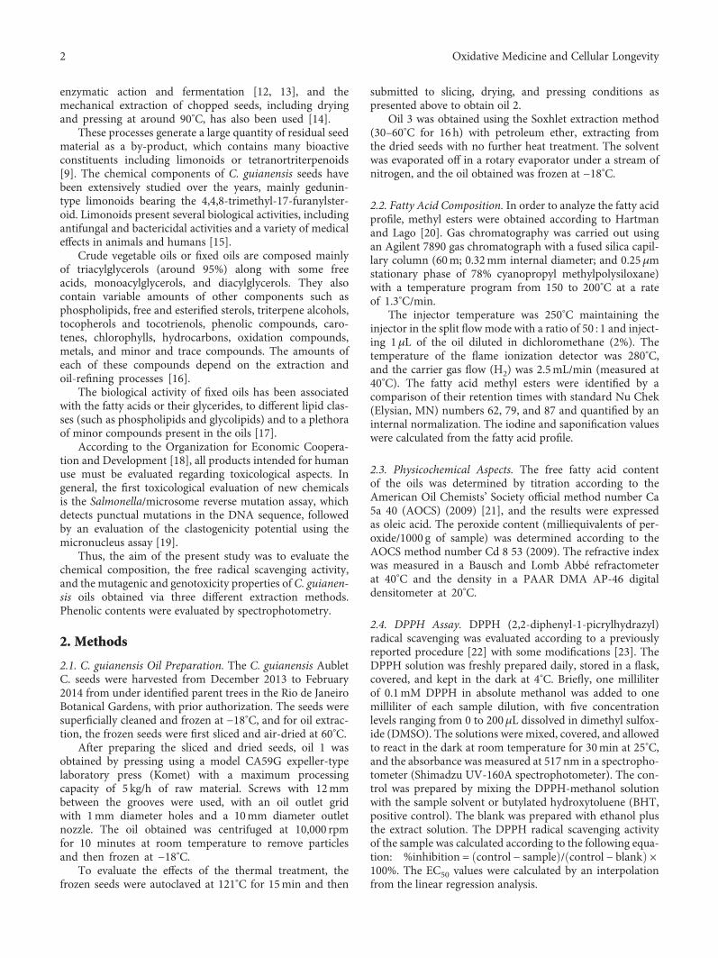
enzymatic action and fermentation [12, 13], and themechanical extraction of chopped seeds, including dryingand pressing at around 90°C, has also been used [14].
These processes generate a large quantity of residual seedmaterial as a by-product, which contains many bioactiveconstituents including limonoids or tetranortriterpenoids[9]. The chemical components of C. guianensis seeds havebeen extensively studied over the years, mainly gedunin-type limonoids bearing the 4,4,8-trimethyl-17-furanylster-oid. Limonoids present several biological activities, includingantifungal and bactericidal activities and a variety of medicaleffects in animals and humans [15].
Crude vegetable oils or fixed oils are composed mainlyof triacylglycerols (around 95%) along with some freeacids, monoacylglycerols, and diacylglycerols. They alsocontain variable amounts of other components such asphospholipids, free and esterified sterols, triterpene alcohols,tocopherols and tocotrienols, phenolic compounds, caro-tenes, chlorophylls, hydrocarbons, oxidation compounds,metals, and minor and trace compounds. The amounts ofeach of these compounds depend on the extraction andoil-refining processes [16].
The biological activity of fixed oils has been associatedwith the fatty acids or their glycerides, to different lipid clas-ses (such as phospholipids and glycolipids) and to a plethoraof minor compounds present in the oils [17].
According to the Organization for Economic Coopera-tion and Development [18], all products intended for humanuse must be evaluated regarding toxicological aspects. Ingeneral, the first toxicological evaluation of new chemicalsis the Salmonella/microsome reverse mutation assay, whichdetects punctual mutations in the DNA sequence, followedby an evaluation of the clastogenicity potential using themicronucleus assay [19].
Thus, the aim of the present study was to evaluate thechemical composition, the free radical scavenging activity,and the mutagenic and genotoxicity properties of C. guianen-sis oils obtained via three different extraction methods.Phenolic contents were evaluated by spectrophotometry.
2. Methods
2.1. C. guianensis Oil Preparation. The C. guianensis AubletC. seeds were harvested from December 2013 to February2014 from under identified parent trees in the Rio de JaneiroBotanical Gardens, with prior authorization. The seeds weresuperficially cleaned and frozen at −18°C, and for oil extrac-tion, the frozen seeds were first sliced and air-dried at 60°C.
After preparing the sliced and dried seeds, oil 1 wasobtained by pressing using a model CA59G expeller-typelaboratory press (Komet) with a maximum processingcapacity of 5 kg/h of raw material. Screws with 12mmbetween the grooves were used, with an oil outlet gridwith 1mm diameter holes and a 10mm diameter outletnozzle. The oil obtained was centrifuged at 10,000 rpmfor 10 minutes at room temperature to remove particlesand then frozen at −18°C.
To evaluate the effects of the thermal treatment, thefrozen seeds were autoclaved at 121°C for 15min and then
submitted to slicing, drying, and pressing conditions aspresented above to obtain oil 2.
Oil 3 was obtained using the Soxhlet extraction method(30–60°C for 16 h) with petroleum ether, extracting fromthe dried seeds with no further heat treatment. The solventwas evaporated off in a rotary evaporator under a stream ofnitrogen, and the oil obtained was frozen at −18°C.
2.2. Fatty Acid Composition. In order to analyze the fatty acidprofile, methyl esters were obtained according to Hartmanand Lago [20]. Gas chromatography was carried out usingan Agilent 7890 gas chromatograph with a fused silica capil-lary column (60m; 0.32mm internal diameter; and 0.25μmstationary phase of 78% cyanopropyl methylpolysiloxane)with a temperature program from 150 to 200°C at a rateof 1.3°C/min.
The injector temperature was 250°C maintaining theinjector in the split flowmode with a ratio of 50 : 1 and inject-ing 1μL of the oil diluted in dichloromethane (2%). Thetemperature of the flame ionization detector was 280°C,and the carrier gas flow (H2) was 2.5mL/min (measured at40°C). The fatty acid methyl esters were identified by acomparison of their retention times with standard Nu Chek(Elysian, MN) numbers 62, 79, and 87 and quantified by aninternal normalization. The iodine and saponification valueswere calculated from the fatty acid profile.
2.3. Physicochemical Aspects. The free fatty acid contentof the oils was determined by titration according to theAmerican Oil Chemists’ Society official method number Ca5a 40 (AOCS) (2009) [21], and the results were expressedas oleic acid. The peroxide content (milliequivalents of per-oxide/1000 g of sample) was determined according to theAOCS method number Cd 8 53 (2009). The refractive indexwas measured in a Bausch and Lomb Abbé refractometerat 40°C and the density in a PAAR DMA AP-46 digitaldensitometer at 20°C.
2.4. DPPH Assay. DPPH (2,2-diphenyl-1-picrylhydrazyl)radical scavenging was evaluated according to a previouslyreported procedure [22] with some modifications [23]. TheDPPH solution was freshly prepared daily, stored in a flask,covered, and kept in the dark at 4°C. Briefly, one milliliterof 0.1mM DPPH in absolute methanol was added to onemilliliter of each sample dilution, with five concentrationlevels ranging from 0 to 200μL dissolved in dimethyl sulfox-ide (DMSO). The solutions were mixed, covered, and allowedto react in the dark at room temperature for 30min at 25°C,and the absorbance was measured at 517nm in a spectropho-tometer (Shimadzu UV-160A spectrophotometer). The con-trol was prepared by mixing the DPPH-methanol solutionwith the sample solvent or butylated hydroxytoluene (BHT,positive control). The blank was prepared with ethanol plusthe extract solution. The DPPH radical scavenging activityof the sample was calculated according to the following equa-tion: %inhibition = control − sample / control − blank ×100%. The EC50 values were calculated by an interpolationfrom the linear regression analysis.
2 Oxidative Medicine and Cellular Longevity
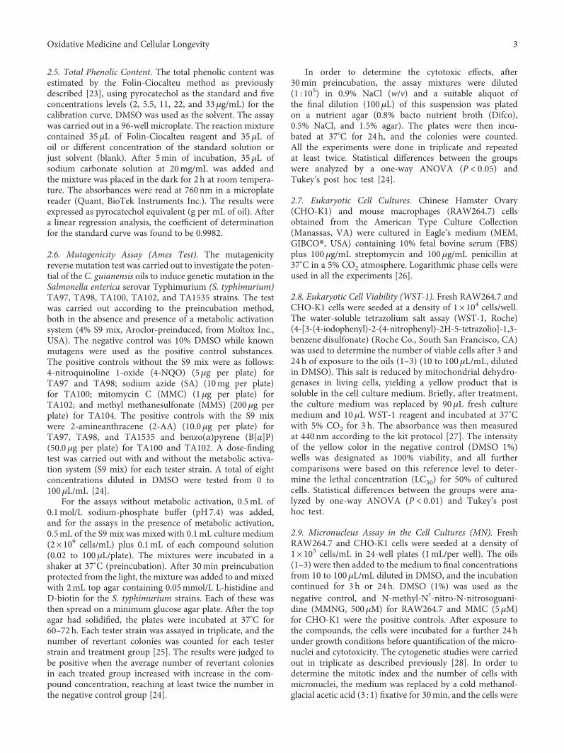
2.5. Total Phenolic Content. The total phenolic content wasestimated by the Folin-Ciocalteu method as previouslydescribed [23], using pyrocatechol as the standard and fiveconcentrations levels (2, 5.5, 11, 22, and 33μg/mL) for thecalibration curve. DMSO was used as the solvent. The assaywas carried out in a 96-well microplate. The reaction mixturecontained 35μL of Folin-Ciocalteu reagent and 35μL ofoil or different concentration of the standard solution orjust solvent (blank). After 5min of incubation, 35μL ofsodium carbonate solution at 20mg/mL was added andthe mixture was placed in the dark for 2 h at room tempera-ture. The absorbances were read at 760 nm in a microplatereader (Quant, BioTek Instruments Inc.). The results wereexpressed as pyrocatechol equivalent (g per mL of oil). Aftera linear regression analysis, the coefficient of determinationfor the standard curve was found to be 0.9982.
2.6. Mutagenicity Assay (Ames Test). The mutagenicityreverse mutation test was carried out to investigate the poten-tial of the C. guianensis oils to induce genetic mutation in theSalmonella enterica serovar Typhimurium (S. typhimurium)TA97, TA98, TA100, TA102, and TA1535 strains. The testwas carried out according to the preincubation method,both in the absence and presence of a metabolic activationsystem (4% S9 mix, Aroclor-preinduced, from Moltox Inc.,USA). The negative control was 10% DMSO while knownmutagens were used as the positive control substances.The positive controls without the S9 mix were as follows:4-nitroquinoline 1-oxide (4-NQO) (5μg per plate) forTA97 and TA98; sodium azide (SA) (10mg per plate)for TA100; mitomycin C (MMC) (1μg per plate) forTA102; and methyl methanesulfonate (MMS) (200μg perplate) for TA104. The positive controls with the S9 mixwere 2-amineanthracene (2-AA) (10.0μg per plate) forTA97, TA98, and TA1535 and benzo(α)pyrene (B[α]P)(50.0μg per plate) for TA100 and TA102. A dose-findingtest was carried out with and without the metabolic activa-tion system (S9 mix) for each tester strain. A total of eightconcentrations diluted in DMSO were tested from 0 to100μL/mL [24].
For the assays without metabolic activation, 0.5mL of0.1mol/L sodium-phosphate buffer (pH7.4) was added,and for the assays in the presence of metabolic activation,0.5mL of the S9 mix was mixed with 0.1mL culture medium(2× 10⁹ cells/mL) plus 0.1mL of each compound solution(0.02 to 100μL/plate). The mixtures were incubated in ashaker at 37°C (preincubation). After 30min preincubationprotected from the light, the mixture was added to and mixedwith 2mL top agar containing 0.05mmol/L L-histidine andD-biotin for the S. typhimurium strains. Each of these wasthen spread on a minimum glucose agar plate. After the topagar had solidified, the plates were incubated at 37°C for60–72 h. Each tester strain was assayed in triplicate, and thenumber of revertant colonies was counted for each testerstrain and treatment group [25]. The results were judged tobe positive when the average number of revertant coloniesin each treated group increased with increase in the com-pound concentration, reaching at least twice the number inthe negative control group [24].
In order to determine the cytotoxic effects, after30min preincubation, the assay mixtures were diluted(1 : 105) in 0.9% NaCl (w/v) and a suitable aliquot ofthe final dilution (100μL) of this suspension was platedon a nutrient agar (0.8% bacto nutrient broth (Difco),0.5% NaCl, and 1.5% agar). The plates were then incu-bated at 37°C for 24 h, and the colonies were counted.All the experiments were done in triplicate and repeatedat least twice. Statistical differences between the groupswere analyzed by a one-way ANOVA (P < 0 05) andTukey’s post hoc test [24].
2.7. Eukaryotic Cell Cultures. Chinese Hamster Ovary(CHO-K1) and mouse macrophages (RAW264.7) cellsobtained from the American Type Culture Collection(Manassas, VA) were cultured in Eagle’s medium (MEM,GIBCO®, USA) containing 10% fetal bovine serum (FBS)plus 100μg/mL streptomycin and 100μg/mL penicillin at37°C in a 5% CO2 atmosphere. Logarithmic phase cells wereused in all the experiments [26].
2.8. Eukaryotic Cell Viability (WST-1). Fresh RAW264.7 andCHO-K1 cells were seeded at a density of 1× 10⁴ cells/well.The water-soluble tetrazolium salt assay (WST-1, Roche)(4-[3-(4-iodophenyl)-2-(4-nitrophenyl)-2H-5-tetrazolio]-1,3-benzene disulfonate) (Roche Co., South San Francisco, CA)was used to determine the number of viable cells after 3 and24 h of exposure to the oils (1–3) (10 to 100μL/mL, dilutedin DMSO). This salt is reduced by mitochondrial dehydro-genases in living cells, yielding a yellow product that issoluble in the cell culture medium. Briefly, after treatment,the culture medium was replaced by 90μL fresh culturemedium and 10μL WST-1 reagent and incubated at 37°Cwith 5% CO2 for 3 h. The absorbance was then measuredat 440 nm according to the kit protocol [27]. The intensityof the yellow color in the negative control (DMSO 1%)wells was designated as 100% viability, and all furthercomparisons were based on this reference level to deter-mine the lethal concentration (LC50) for 50% of culturedcells. Statistical differences between the groups were ana-lyzed by one-way ANOVA (P < 0 01) and Tukey’s posthoc test.
2.9. Micronucleus Assay in the Cell Cultures (MN). FreshRAW264.7 and CHO-K1 cells were seeded at a density of1× 105 cells/mL in 24-well plates (1mL/per well). The oils(1–3) were then added to the medium to final concentrationsfrom 10 to 100μL/mL diluted in DMSO, and the incubationcontinued for 3 h or 24h. DMSO (1%) was used as thenegative control, and N-methyl-N′-nitro-N-nitrosoguani-dine (MMNG, 500μM) for RAW264.7 and MMC (5μM)for CHO-K1 were the positive controls. After exposure tothe compounds, the cells were incubated for a further 24hunder growth conditions before quantification of the micro-nuclei and cytotoxicity. The cytogenetic studies were carriedout in triplicate as described previously [28]. In order todetermine the mitotic index and the number of cells withmicronuclei, the medium was replaced by a cold methanol-glacial acetic acid (3 : 1) fixative for 30min, and the cells were
3Oxidative Medicine and Cellular Longevity
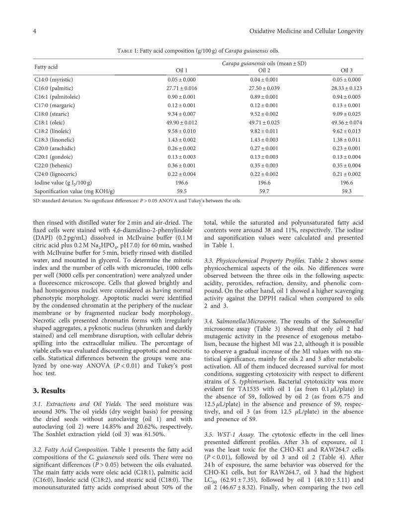
then rinsed with distilled water for 2min and air-dried. Thefixed cells were stained with 4,6-diamidino-2-phenylindole(DAPI) (0.2 pg/mL) dissolved in McIlvaine buffer (0.1Mcitric acid plus 0.2M Na2HPO4, pH7.0) for 60min, washedwith McIlvaine buffer for 5min, briefly rinsed with distilledwater, and mounted in glycerol. To determine the mitoticindex and the number of cells with micronuclei, 1000 cellsper well (3000 cells per concentration) were analyzed undera fluorescence microscope. Cells that glowed brightly andhad homogenous nuclei were considered as having normalphenotypic morphology. Apoptotic nuclei were identifiedby the condensed chromatin at the periphery of the nuclearmembrane or by fragmented nuclear body morphology.Necrotic cells presented chromatin forms with irregularlyshaped aggregates, a pyknotic nucleus (shrunken and darklystained) and cell membrane disruption, with cellular debrisspilling into the extracellular milieu. The percentage ofviable cells was evaluated discounting apoptotic and necroticcells. Statistical differences between the groups were ana-lyzed by one-way ANOVA (P < 0 01) and Tukey’s posthoc test.
3. Results
3.1. Extractions and Oil Yields. The seed moisture wasaround 30%. The oil yields (dry weight basis) for pressingthe dried seeds without autoclaving (oil 1) and withautoclaving (oil 2) were 14.85% and 20.62%, respectively.The Soxhlet extraction yield (oil 3) was 61.50%.
3.2. Fatty Acid Composition. Table 1 presents the fatty acidcompositions of the C. guianensis seed oils. There were nosignificant differences (P > 0 05) between the oils evaluated.The main fatty acids were oleic acid (C18:1), palmitic acid(C16:0), linoleic acid (C18:2), and stearic acid (C18:0). Themonounsaturated fatty acids comprised about 50% of the
total, while the saturated and polyunsaturated fatty acidcontents were around 38 and 11%, respectively. The iodineand saponification values were calculated and presentedin Table 1.
3.3. Physicochemical Property Profiles. Table 2 shows somephysicochemical aspects of the oils. No differences wereobserved between the three oils in the following aspects:acidity, peroxides, refraction, density, and phenolic com-pound. On the other hand, oil 1 showed a higher scavengingactivity against the DPPH radical when compared to oils2 and 3.
3.4. Salmonella/Microsome. The results of the Salmonella/microsome assay (Table 3) showed that only oil 2 hadmutagenic activity in the presence of exogenous metabo-lism, because the highest MI was 2.2, although it is possibleto observe a gradual increase of the MI values with no sta-tistical significance, mainly for oils 2 and 3 after metabolicactivation. All of them induced decreased survival for mostconditions, suggesting cytotoxicity with respect to differentstrains of S. typhimurium. Bacterial cytotoxicity was moreevident for TA1535 with oil 1 (as from 0.1μL/plate) inthe absence of S9, followed by oil 2 (as from 6.75 and12.5μL/plate) in the absence and presence of S9, respec-tively, and oil 3 (as from 12.5 μL/plate) in the absenceand presence of S9.
3.5. WST-1 Assay. The cytotoxic effects in the cell linespresented different profiles. After 3 h of exposure, oil 1was the least toxic for the CHO-K1 and RAW264.7 cells(P < 0 01), followed by oil 3 and oil 2 (Table 4). After24 h of exposure, the same behavior was observed for theCHO-K1 cells, but for RAW264.7, oil 3 had the highestLC50 (62.91± 7.35), followed by oil 1 (48.10± 3.11) andoil 2 (46.67± 8.32). Finally, when comparing the two cell
Table 1: Fatty acid composition (g/100 g) of Carapa guianensis oils.
Fatty acidCarapa guianensis oils (mean± SD)
Oil 1 Oil 2 Oil 3
C14:0 (myristic) 0.05± 0.000 0.04± 0.001 0.05± 0.000C16:0 (palmitic) 27.71± 0.016 27.50± 0,039 28.33± 0.123C16:1 (palmitoleic) 0.90± 0.001 0.89± 0.001 0.94± 0.005C17:0 (margaric) 0.12± 0.001 0.12± 0.001 0.13± 0.001C18:0 (stearic) 9.34± 0.007 9.52± 0.002 9.09± 0.025C18:1 (oleic) 49.90± 0.012 49.71± 0.025 49.56± 0.074C18:2 (linoleic) 9.58± 0.010 9.82± 0.011 9.62± 0.013C18:3 (linonelic) 1.43± 0.002 1.43± 0.003 1.38± 0.011C20:0 (arachidic) 0.26± 0.002 0.27± 0.001 0.23± 0.001C20:1 (gondoic) 0.13± 0.003 0.13± 0.003 0.13± 0.004C22:0 (behenic) 0.36± 0.001 0.35± 0.003 0.35± 0.004C24:0 (lignoceric) 0.22± 0.004 0.22± 0.002 0.21± 0.002Iodine value (g I2/100 g) 196.6 196.6 196.6
Saponification value (mg KOH/g) 59.5 59.7 59.3
SD: standard deviation. No significant differences: P > 0 05 ANOVA and Tukey’s between the oils.
4 Oxidative Medicine and Cellular Longevity
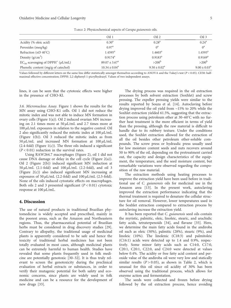
lines, it can be seen that the cytotoxic effects were higherin the presence of CHO-Kl.
3.6. Micronucleus Assay. Figure 1 shows the results for theMN assay using CHO-K1 cells. Oil 1 did not reduce themitotic index and was not able to induce MN formation inovary cells (Figure 1(a)). Oil 2 induced ovarian MN increas-ing on 2.1 times more at 50μL/mL and 2.7 times more at100μL/mL exposures in relation to the negative control. Oil2 also significantly reduced the mitotic index at 100μL/mL(Figure 1(b)). Oil 3 reduced the mitotic index as from50μL/mL and increased MN formation at 100μL/mL(2.4-fold) (Figure 1(c)). The three oils induced a significant(P < 0 01) reduction in the survival rates.
Using RAW264.7 macrophages (Figure 2), oil 1 did notcause DNA damage or delay in the cell cycle (Figure 2(a)).Oil 2 (Figure 2(b)) induced significant MN induction at50μL/mL (2.1-fold) and 100μL/mL (2.2-fold), and oil 3(Figure 2(c)) also induced significant MN increasing atexposures of 50μL/mL (2.2-fold) and 100μL/mL (2.5-fold).None of the oils induced cytostatic effects on macrophages.Both oils 2 and 3 presented significant (P < 0 01) cytotoxicresponse at 100μL/mL.
4. Discussion
The use of natural products in traditional Brazilian phy-tomedicine is widely accepted and prescribed, mainly inthe poorest areas, such as the Amazon and Northeasternregions. Thus, the pharmaceutical potential of Brazilianherbs must be considered in drug discovery studies [29].Contrary to allopathy, the traditional usage of medicinalplants is apparently considered to be safe and hence thetoxicity of traditional herbal medicines has not beentotally evaluated in most cases, although medicinal plantscan be extremely harmful to human health. Studies haverevealed that some plants frequently used in folk medi-cine are potentially genotoxic [30–32]. It is thus truly rel-evant to screen the genotoxicity during the preclinicalevaluation of herbal extracts or substances, in order toverify their mutagenic potential for both safety and eco-nomic concerns, since plants are widely used in folkmedicine and can be a resource for the development ofnew drugs [33].
The drying process was required in the oil extractionprocesses by both solvent extraction (Soxhlet) and screwpressing. The expeller pressing yields were similar to theresults reported by Souza et al. [14]. Autoclaving beforedrying improved the oil yield from ~15% to 20% while theSoxhlet extraction yielded 61.5%, suggesting that the extrac-tion process using petroleum ether at 30–60°C with no fur-ther heat treatment is the more efficient in terms of yieldthan the pressing, although the raw material is difficult tohandle due to its rubbery texture. Under the conditionsused, the Soxhlet extraction allowed for the extraction ofall the oil besides other petroleum ether-soluble com-pounds. The screw press or hydraulic press usually usedfor low moisture content seeds and nuts recovers around50 to 90% of the oil, depending on the pretreatment carriedout, the capacity and design characteristics of the equip-ment, the temperature, and the seed moisture content, butremarkable variations were observed regarding the compo-sition of the raw material.
The extraction methods using heating processes toimprove the extraction yield have been used before in tradi-tional use of C. guianensis oils for medicinal use in theAmazon area [13]. In the present work, autoclavingimproved the extraction performance indicating that thethermal treatment is required to denature the cellular struc-ture for oil removal. However, lower temperatures used inthe Soxhlet extraction compared to extraction process byautoclaving increase the extraction yield.
It has been reported that C. guianensis seed oils containthe myristic, palmitic, oleic, linoleic, stearic, and arachidicfatty acids, tetraterpenoids [34], and flavane [35]. Here,we determine the main fatty acids found in the andirobaoil such as oleic (50%), palmitic (28%), stearic (9%), andlinoleic (10%). The linolenic (C18:3) and palmitoleic(C16:1) acids were detected up to 1.4 and 0.9%, respec-tively. Some minor fatty acids such as C14:0, C17:0,C20:1, C20:1, C22:0, and C24:0 were detected at valuesbelow 0.4%. The acidity or free fatty acid content and per-oxide value of the andiroba oil were very low and staticallysimilar results (P > 0 05), as shown in Table 2, which isunusual for this oil since oil acidity of 30% has beenobserved using the traditional process, which allows forenzyme action and fermentation.
The seeds were collected and frozen before dryingfollowed by the oil extraction process, hence avoiding
Table 2: Physicochemical aspects of Carapa guianensis oils.
Oil 1 Oil 2 Oil 3
Acidity (% oleic acid) 0.30a 0.28a 0.24a
Peroxides (meq/kg) 0.97a 0a 0a
Refraction (nD 40°C) 1.4595a 1.4603a 1.4593a
Density (g/cm3) 0.9174a 0.9183a 0.9169a
EC50 scavenging of DPPH+ (μL/mL) 89.07± 3.67a >200b >200b
Phenolic content (mg/g of catechol) 10.34± 0.04a 9.50± 0.02a 9.00± 0.03a
Values followed by different letters on the same line differ statistically amongst themselves according to ANOVA and the Tukey’s test (P < 0 05). CE50: half-maximal effective concentration; DPPH: 2,2-diphenyl-1-picrylhydrazyl. Values of two independent assays.
5Oxidative Medicine and Cellular Longevity
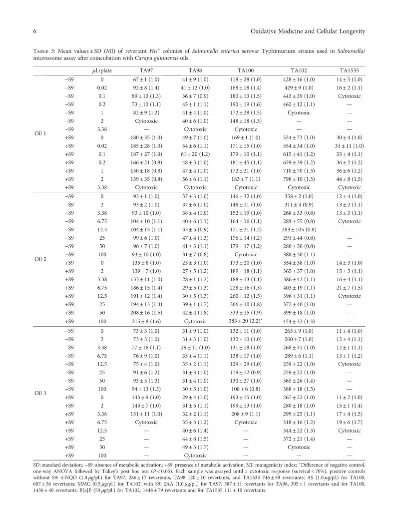
Table 3: Mean values± SD (MI) of revertant His+ colonies of Salmonella enterica serovar Typhimurium strains used in Salmonella/microssome assay after coincubation with Carapa guianensis oils.
μL/plate TA97 TA98 TA100 TA102 TA1535
Oil 1
−S9 0 67± 1 (1.0) 41± 9 (1.0) 118± 28 (1.0) 428± 16 (1.0) 14± 5 (1.0)
−S9 0.02 92± 8 (1.4) 41± 12 (1.0) 168± 18 (1.4) 429± 9 (1.0) 16± 2 (1.1)
−S9 0.1 89 ± 13 (1.3) 36± 7 (0.9) 180± 13 (1.5) 443± 39 (1.0) Cytotoxic
−S9 0.2 73 ± 10 (1.1) 45± 1 (1.1) 190± 19 (1.6) 462± 12 (1.1) —
−S9 1 82± 9 (1.2) 41± 4 (1.0) 172± 28 (1.5) Cytotoxic —
−S9 2 Cytotoxic 40± 6 (1.0) 148± 18 (1.3) — —
−S9 3.38 — Cytotoxic Cytotoxic — —
+S9 0 180± 35 (1.0) 49± 7 (1.0) 169± 1 (1.0) 534± 73 (1.0) 30± 4 (1.0)
+S9 0.02 185± 28 (1.0) 54± 6 (1.1) 171± 15 (1.0) 554± 34 (1.0) 31± 11 (1.0)
+S9 0.1 187± 27 (1.0) 61± 20 (1.2) 179± 10 (1.1) 615± 41 (1.2) 33± 4 (1.1)
+S9 0.2 166± 21 (0.9) 48± 3 (1.0) 181± 45 (1.1) 639± 39 (1.2) 36± 2 (1.2)
+S9 1 150± 18 (0.8) 47± 4 (1.0) 172± 21 (1.0) 710± 70 (1.3) 36± 6 (1.2)
+S9 2 139± 35 (0.8) 56± 6 (1.1) 183± 7 (1.1) 798± 10 (1.5) 44± 8 (1.5)
+S9 3.38 Cytotoxic Cytotoxic Cytotoxic Cytotoxic Cytotoxic
Oil 2
−S9 0 93± 1 (1.0) 37± 3 (1.0) 146± 32 (1.0) 358± 2 (1.0) 12± 4 (1.0)
−S9 2 93± 2 (1.0) 37± 6 (1.0) 148± 11 (1.0) 311± 4 (0.9) 13± 2 (1.1)
−S9 3.38 93 ± 10 (1.0) 38± 4 (1.0) 152± 19 (1.0) 268± 33 (0.8) 13± 3 (1.1)
−S9 6.75 104± 10 (1.1) 40± 6 (1.1) 164± 16 (1.1) 289± 55 (0.8) Cytotoxic
−S9 12.5 104± 15 (1.1) 33± 5 (0.9) 171± 21 (1.2) 283± 105 (0.8) —
−S9 25 99± 6 (1.0) 47± 4 (1.3) 176± 14 (1.2) 291± 44 (0.8) —
−S9 50 96± 7 (1.0) 41± 3 (1.1) 179± 17 (1.2) 280± 50 (0.8) —
−S9 100 93 ± 10 (1.0) 31± 7 (0.8) Cytotoxic 388± 50 (1.1) —
+S9 0 135± 8 (1.0) 23± 3 (1.0) 173± 20 (1.0) 354± 38 (1.0) 14± 3 (1.0)
+S9 2 139± 7 (1.0) 27± 5 (1.2) 189± 18 (1.1) 365± 37 (1.0) 15± 3 (1.1)
+S9 3.38 133± 11 (1.0) 28± 1 (1.2) 188± 13 (1.1) 386± 42 (1.1) 16± 4 (1.1)
+S9 6.75 186± 15 (1.4) 29± 5 (1.3) 228± 16 (1.3) 403± 19 (1.1) 21± 7 (1.5)
+S9 12.5 191± 12 (1.4) 30± 5 (1.3) 260± 12 (1.5) 396± 31 (1.1) Cytotoxic
+S9 25 194± 13 (1.4) 39± 1 (1.7) 306± 10 (1.8) 372± 40 (1.0) —
+S9 50 208± 16 (1.5) 42± 4 (1.8) 333± 15 (1.9) 399± 18 (1.0) —
+S9 100 215± 8 (1.6) Cytotoxic 383± 20 (2.2)∗ 454± 32 (1.3) —
Oil 3
−S9 0 73± 3 (1.0) 31± 9 (1.0) 132± 11 (1.0) 263± 9 (1.0) 11± 4 (1.0)
−S9 2 73± 3 (1.0) 31± 3 (1.0) 132± 10 (1.0) 260± 7 (1.0) 12± 4 (1.1)
−S9 3.38 77 ± 16 (1.1) 29± 11 (1.0) 131± 18 (1.0) 268± 31 (1.0) 12± 1 (1.1)
−S9 6.75 76± 9 (1.0) 33± 4 (1.1) 138± 17 (1.0) 289± 6 (1.1) 13± 1 (1.2)
−S9 12.5 75± 4 (1.0) 35± 2 (1.1) 129± 29 (1.0) 259± 22 (1.0) Cytotoxic
−S9 25 91± 6 (1.2) 31± 3 (1.0) 119± 12 (0.9) 259± 22 (1.0) —
−S9 50 93± 5 (1.3) 31± 4 (1.0) 130± 27 (1.0) 365± 26 (1.4) —
−S9 100 94 ± 13 (1.3) 30± 5 (1.0) 108± 6 (0.8) 388± 18 (1.5) —
+S9 0 145± 9 (1.0) 29± 4 (1.0) 193± 15 (1.0) 267± 22 (1.0) 11± 2 (1.0)
+S9 2 143± 7 (1.0) 31± 5 (1.1) 199± 13 (1.0) 280± 18 (1.0) 15± 1 (1.4)
+S9 3.38 151± 11 (1.0) 32± 2 (1.1) 208± 9 (1.1) 299± 25 (1.1) 17± 4 (1.5)
+S9 6.75 Cytotoxic 35± 3 (1.2) Cytotoxic 318± 16 (1.2) 19± 6 (1.7)
+S9 12.5 — 40± 6 (1.4) — 344± 22 (1.3) Cytotoxic
+S9 25 — 44± 8 (1.5) — 372± 21 (1.4) —
+S9 50 — 49± 3 (1.7) — Cytotoxic —
+S9 100 — Cytotoxic — — —
SD: standard deviation; −S9: absence of metabolic activation; +S9: presence of metabolic activation; MI: mutagenicity index; ∗Difference of negative control,one-way ANOVA followed by Tukey’s post hoc test (P < 0 05). Each sample was assayed until a cytotoxic response (survival < 70%); positive controlswithout S9: 4-NQO (1.0 μg/pl.) for TA97, 286 ± 17 revertants, TA98 120 ± 10 revertants, and TA1535 746 ± 58 revertants; AS (1.0 μg/pl.) for TA100,607 ± 56 revertants; MMC (0.5 μg/pl.) for TA102; with S9: 2AA (1.0 μg/pl.) for TA97, 587 ± 11 revertants for TA98, 305 ± 1 revertants and for TA100,1436 ± 40 revertants; B[a]P (50 μg/pl.) for TA102, 1448 ± 79 revertants and for TA1535 111 ± 10 revertants.
6 Oxidative Medicine and Cellular Longevity
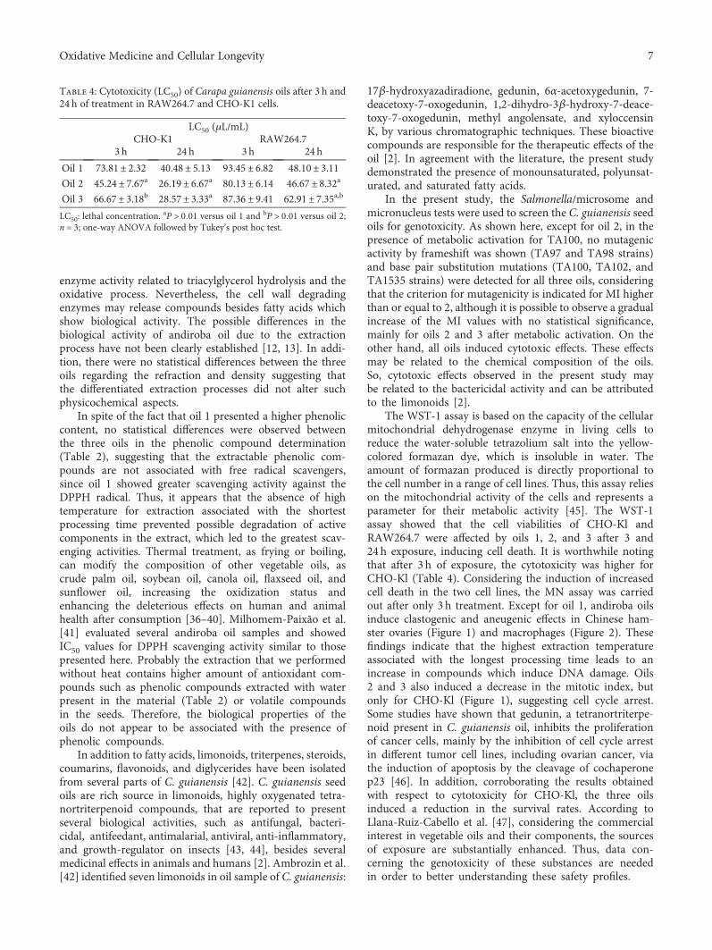
enzyme activity related to triacylglycerol hydrolysis and theoxidative process. Nevertheless, the cell wall degradingenzymes may release compounds besides fatty acids whichshow biological activity. The possible differences in thebiological activity of andiroba oil due to the extractionprocess have not been clearly established [12, 13]. In addi-tion, there were no statistical differences between the threeoils regarding the refraction and density suggesting thatthe differentiated extraction processes did not alter suchphysicochemical aspects.
In spite of the fact that oil 1 presented a higher phenoliccontent, no statistical differences were observed betweenthe three oils in the phenolic compound determination(Table 2), suggesting that the extractable phenolic com-pounds are not associated with free radical scavengers,since oil 1 showed greater scavenging activity against theDPPH radical. Thus, it appears that the absence of hightemperature for extraction associated with the shortestprocessing time prevented possible degradation of activecomponents in the extract, which led to the greatest scav-enging activities. Thermal treatment, as frying or boiling,can modify the composition of other vegetable oils, ascrude palm oil, soybean oil, canola oil, flaxseed oil, andsunflower oil, increasing the oxidization status andenhancing the deleterious effects on human and animalhealth after consumption [36–40]. Milhomem-Paixão et al.[41] evaluated several andiroba oil samples and showedIC50 values for DPPH scavenging activity similar to thosepresented here. Probably the extraction that we performedwithout heat contains higher amount of antioxidant com-pounds such as phenolic compounds extracted with waterpresent in the material (Table 2) or volatile compoundsin the seeds. Therefore, the biological properties of theoils do not appear to be associated with the presence ofphenolic compounds.
In addition to fatty acids, limonoids, triterpenes, steroids,coumarins, flavonoids, and diglycerides have been isolatedfrom several parts of C. guianensis [42]. C. guianensis seedoils are rich source in limonoids, highly oxygenated tetra-nortriterpenoid compounds, that are reported to presentseveral biological activities, such as antifungal, bacteri-cidal, antifeedant, antimalarial, antiviral, anti-inflammatory,and growth-regulator on insects [43, 44], besides severalmedicinal effects in animals and humans [2]. Ambrozin et al.[42] identified seven limonoids in oil sample of C. guianensis:
17β-hydroxyazadiradione, gedunin, 6α-acetoxygedunin, 7-deacetoxy-7-oxogedunin, 1,2-dihydro-3β-hydroxy-7-deace-toxy-7-oxogedunin, methyl angolensate, and xyloccensinK, by various chromatographic techniques. These bioactivecompounds are responsible for the therapeutic effects of theoil [2]. In agreement with the literature, the present studydemonstrated the presence of monounsaturated, polyunsat-urated, and saturated fatty acids.
In the present study, the Salmonella/microsome andmicronucleus tests were used to screen the C. guianensis seedoils for genotoxicity. As shown here, except for oil 2, in thepresence of metabolic activation for TA100, no mutagenicactivity by frameshift was shown (TA97 and TA98 strains)and base pair substitution mutations (TA100, TA102, andTA1535 strains) were detected for all three oils, consideringthat the criterion for mutagenicity is indicated for MI higherthan or equal to 2, although it is possible to observe a gradualincrease of the MI values with no statistical significance,mainly for oils 2 and 3 after metabolic activation. On theother hand, all oils induced cytotoxic effects. These effectsmay be related to the chemical composition of the oils.So, cytotoxic effects observed in the present study maybe related to the bactericidal activity and can be attributedto the limonoids [2].
The WST-1 assay is based on the capacity of the cellularmitochondrial dehydrogenase enzyme in living cells toreduce the water-soluble tetrazolium salt into the yellow-colored formazan dye, which is insoluble in water. Theamount of formazan produced is directly proportional tothe cell number in a range of cell lines. Thus, this assay relieson the mitochondrial activity of the cells and represents aparameter for their metabolic activity [45]. The WST-1assay showed that the cell viabilities of CHO-Kl andRAW264.7 were affected by oils 1, 2, and 3 after 3 and24 h exposure, inducing cell death. It is worthwhile notingthat after 3 h of exposure, the cytotoxicity was higher forCHO-Kl (Table 4). Considering the induction of increasedcell death in the two cell lines, the MN assay was carriedout after only 3 h treatment. Except for oil 1, andiroba oilsinduce clastogenic and aneugenic effects in Chinese ham-ster ovaries (Figure 1) and macrophages (Figure 2). Thesefindings indicate that the highest extraction temperatureassociated with the longest processing time leads to anincrease in compounds which induce DNA damage. Oils2 and 3 also induced a decrease in the mitotic index, butonly for CHO-Kl (Figure 1), suggesting cell cycle arrest.Some studies have shown that gedunin, a tetranortriterpe-noid present in C. guianensis oil, inhibits the proliferationof cancer cells, mainly by the inhibition of cell cycle arrestin different tumor cell lines, including ovarian cancer, viathe induction of apoptosis by the cleavage of cochaperonep23 [46]. In addition, corroborating the results obtainedwith respect to cytotoxicity for CHO-Kl, the three oilsinduced a reduction in the survival rates. According toLlana-Ruiz-Cabello et al. [47], considering the commercialinterest in vegetable oils and their components, the sourcesof exposure are substantially enhanced. Thus, data con-cerning the genotoxicity of these substances are neededin order to better understanding these safety profiles.
Table 4: Cytotoxicity (LC50) of Carapa guianensis oils after 3 h and24 h of treatment in RAW264.7 and CHO-K1 cells.
LC50 (μL/mL)CHO-K1 RAW264.7
3 h 24 h 3 h 24 h
Oil 1 73.81± 2.32 40.48± 5.13 93.45± 6.82 48.10± 3.11Oil 2 45.24± 7.67a 26.19± 6.67a 80.13± 6.14 46.67± 8.32a
Oil 3 66.67± 3.18b 28.57± 3.33a 87.36± 9.41 62.91± 7.35a,b
LC50: lethal concentration.aP > 0 01 versus oil 1 and bP > 0 01 versus oil 2;
n = 3; one-way ANOVA followed by Tukey’s post hoc test.
7Oxidative Medicine and Cellular Longevity
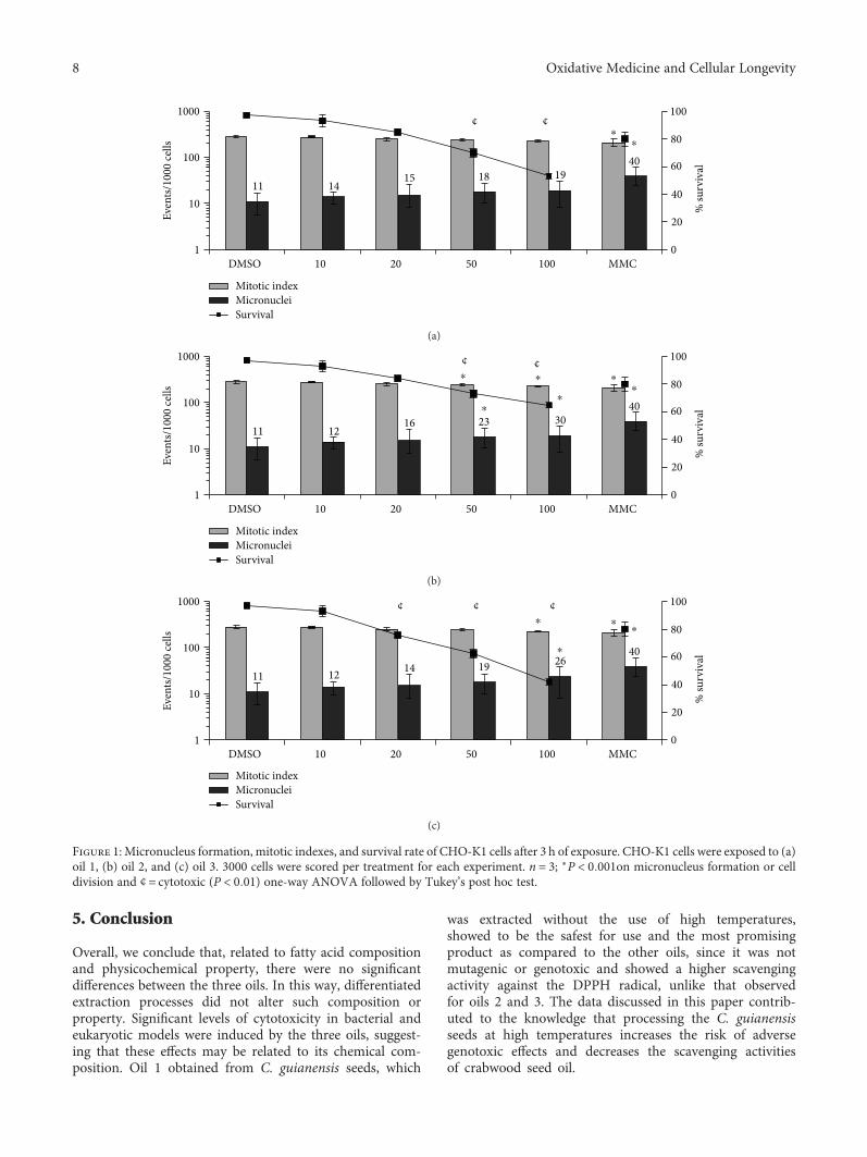
5. Conclusion
Overall, we conclude that, related to fatty acid compositionand physicochemical property, there were no significantdifferences between the three oils. In this way, differentiatedextraction processes did not alter such composition orproperty. Significant levels of cytotoxicity in bacterial andeukaryotic models were induced by the three oils, suggest-ing that these effects may be related to its chemical com-position. Oil 1 obtained from C. guianensis seeds, which
was extracted without the use of high temperatures,showed to be the safest for use and the most promisingproduct as compared to the other oils, since it was notmutagenic or genotoxic and showed a higher scavengingactivity against the DPPH radical, unlike that observedfor oils 2 and 3. The data discussed in this paper contrib-uted to the knowledge that processing the C. guianensisseeds at high temperatures increases the risk of adversegenotoxic effects and decreases the scavenging activitiesof crabwood seed oil.
DMSO
Even
ts/10
00 ce
lls
10 20 50 100 MMC
% su
rviv
al
100
80
60
40
20
0
1000
100
10
1
1115
1418
4019
¢ ¢⁎
⁎
Mitotic indexMicronucleiSurvival
(a)
DMSO
Even
ts/10
00 ce
lls
10 20 50 100 MMC
% su
rviv
al
100
80
60
40
20
0
1000
100
10
1
1116
1223
4030
¢ ¢⁎⁎
⁎
⁎
⁎
Mitotic indexMicronucleiSurvival
⁎
(b)
DMSO
Mitotic indexMicronucleiSurvival
Even
ts/10
00 ce
lls
10 20 50 100 MMC
% su
rviv
al
100
80
60
40
20
0
1000
100
10
1
111412
1940
26
¢ ¢¢
⁎⁎
⁎
⁎
(c)
Figure 1: Micronucleus formation, mitotic indexes, and survival rate of CHO-K1 cells after 3 h of exposure. CHO-K1 cells were exposed to (a)oil 1, (b) oil 2, and (c) oil 3. 3000 cells were scored per treatment for each experiment. n = 3; ∗P < 0 001on micronucleus formation or celldivision and ¢ = cytotoxic (P < 0 01) one-way ANOVA followed by Tukey’s post hoc test.
8 Oxidative Medicine and Cellular Longevity
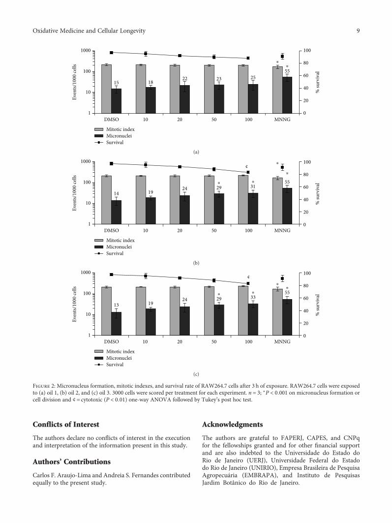
Conflicts of Interest
The authors declare no conflicts of interest in the executionand interpretation of the information present in this study.
Authors’ Contributions
Carlos F. Araujo-Lima and Andreia S. Fernandes contributedequally to the present study.
Acknowledgments
The authors are grateful to FAPERJ, CAPES, and CNPqfor the fellowships granted and for other financial supportand are also indebted to the Universidade do Estado doRio de Janeiro (UERJ), Universidade Federal do Estadodo Rio de Janeiro (UNIRIO), Empresa Brasileira de PesquisaAgropecuária (EMBRAPA), and Instituto de PesquisasJardim Botânico do Rio de Janeiro.
15 1822 23 25
55
0
20
40
60
80
100
1
10
100
1000
DMSO 10 20 50 100 MNNG
% su
rviv
al
Even
ts/10
00 ce
lls
Mitotic indexMicronucleiSurvival
⁎⁎
(a)
14 1924 29 31
55
0
20
40
60
80
100
1
10
100
1000
% su
rviv
al
Even
ts/10
00 ce
lls
DMSO 10 20 50 100 MNNG
Mitotic indexMicronucleiSurvival
⁎
⁎
⁎⁎
¢
(b)
13 1924 29 33
55
0
20
40
60
80
100
1
10
100
1000
% su
rviv
al
Even
ts/10
00 ce
lls
DMSO 10 20 50 100 MNNG
Mitotic indexMicronucleiSurvival
⁎⁎
⁎⁎
¢
(c)
Figure 2: Micronucleus formation, mitotic indexes, and survival rate of RAW264.7 cells after 3 h of exposure. RAW264.7 cells were exposedto (a) oil 1, (b) oil 2, and (c) oil 3. 3000 cells were scored per treatment for each experiment. n = 3; ∗P < 0 001 on micronucleus formation orcell division and ¢ = cytotoxic (P < 0 01) one-way ANOVA followed by Tukey’s post hoc test.
9Oxidative Medicine and Cellular Longevity
References
[1] J. H. Costa-Silva, C. R. Lima, E. J. R. Silva et al., “Acute andsubacute toxicity of the Carapa guianensis Aublet (Meliaceae)seed oil,” Journal of Ethnopharmacology, vol. 116, no. 3,pp. 495–500, 2008.
[2] M. Henriques and C. Penido, “The therapeutic properties ofCarapa guianensis,” Current Pharmaceutical Design, vol. 20,no. 6, pp. 850–856, 2014.
[3] C. Penido, K. A. Costa, R. J. Pennaforte et al., “Anti-allergiceffects of natural tetranortriterpenoids isolated from Carapaguianensis Aublet on allergen-induced vascular permeabilityand hyperalgesia,” Inflammation Research, vol. 54, no. 7,pp. 295–303, 2005.
[4] C. Penido, F. P. Conte, M. S. S. Chagas, C. A. B. Rodrigues,J. F. G. Pereira, and M. G. M. O. Henriques, “Antiinflam-matory effects of natural tetranortriterpenoids isolated fromCarapa guianensis Aublet on zymosan-induced arthritis inmice,” Inflammation Research, vol. 55, no. 11, pp. 457–464, 2006.
[5] C. M. Cazal, K. Choosang, V. G. P. Severino et al., “Evaluationof effect of triterpenes and limonoids on cell growth, cell cycleand apoptosis in human tumor cell line,” Anti-Cancer Agentsin Medicinal Chemistry, vol. 10, no. 10, pp. 769–776, 2010.
[6] F. N. de Barros, M. P. O. Farias, J. P. C. Tavares, L. C. Alves,and M. A. d. G. Faustino, “In vitro efficacy of oil from the seedof Carapa guianensis (andiroba) in the control of Felicola sub-rostratus,” Revista Brasileira de Farmacognosia, vol. 22, no. 5,pp. 1130–1133, 2012.
[7] C. E. S. Silva, O. J. dos Santos, J. M. Ribas-Filho et al., “Effect ofCarapa guianensis Aublet (andiroba) and Orbignya phalerata(babassu) in colonic healing in rats,” Revista do Colégio Brasi-leiro de Cirurgiões, vol. 42, no. 6, pp. 399–406, 2015.
[8] H. A. Miot, R. F. Batistella, K. d. A. Batista et al., “Comparativestudy of the topical effectiveness of the andiroba oil (Carapaguianensis) and DEET 50% as repellent for Aedes sp,” Revistado Instituto de Medicina Tropical de São Paulo, vol. 46, no. 5,pp. 253–256, 2004.
[9] F. A. C. de Mendonça, K. F. S. da Silva, K. K. dos Santos, K. A.L. Ribeiro Júnior, and A. E. G. Sant'Ana, “Activities of someBrazilian plants against larvae of the mosquito Aedes aegypti,”Fitoterapia, vol. 76, no. 7-8, pp. 629–636, 2005.
[10] M. P. O. Farias, D. P. Sousa, A. C. Arruda et al., “Potencialacaricida do óleo de andiroba Carapa guianensis Aubl. sobrefêmeas adultas ingurgitadas de Anocentor nitens Neumann,1897 e Rhipicephalus sanguineus Latreille, 1806,” ArquivoBrasileiro de Medicina Veterinária e Zootecnia, vol. 61, no. 4,pp. 877–882, 2009.
[11] C. O. Carvalho, A. C. S. Chagas, F. Cotinguiba et al., “Theanthelmintic effect of plant extracts on Haemonchus contortusand Strongyloides venezuelensis,” Veterinary Parasitology,vol. 183, no. 3-4, pp. 260–268, 2012.
[12] P. Shanley and M. Londres, “Andiroba Carapa guianensisAubl,” in Fruit Trees and Useful Plants in Amazonian Life.Trees and Vines, P. Shanley, Ed., Food and Agriculture Organi-zation of the United Nations (FAO), 2011, http://www.fao.org/docrep/015/i2360e/i2360e03.pdf.
[13] M. Nardi, A. C. Lira-Guedes, H. F. Albuquerque Cunha, M. C.Guedes, K. Mustin, and S. C. P. Gomes, “Artisanal extractionand traditional knowledge associated with medicinal use ofcrabwood oil (Carapa guianensis Aublet.) in a Peri-UrbanVárzea environment in the Amazon estuary,” Evidence-based
Complementary and Alternative Medicine, vol. 2016, ArticleID 5828021, 12 pages, 2016.
[14] C. R. Souza, R. M. B. Lima, C. P. Azevedo, and L. M. B. Ross,Andiroba (Carapa guianensis Aubl), Embrapa AmazôniaOcidental, Manaus, Brazil, 2006, (Embrapa AmazôniaOcidental. Documentos, 48).
[15] V. P. da Silva, R. R. Oliveira, and M. R. Figueiredo, “Isolationof limonoids from seeds of Carapa guianensis Aublet (Melia-ceae) by high-speed countercurrent chromatography,” Phyto-chemical Analysis, vol. 20, no. 1, pp. 77–81, 2009.
[16] F. D. Gunstone, “Vegetable Oils,” in Bailey's Industrial Oilsand Fats, F. Shahidi, Ed., pp. 213–267, John Wiley & SonsLtd., Chichester, UK, 6th edition, 2005.
[17] H. R. Bizzo and R. Antoniassi, “Fixed oils and antimicrobialeffects,” in Recent Progress in Medicinal Plants Vol. 33 FixedOils and Fats, J. N. Govil, Ed., pp. 273–285, StudiumPress, England and Wales, 2012.
[18] “Guidance document on revisions to OECD genetic toxicologytest guidelines,” OECD, Paris, France, Genetic toxicologyGuidance Document, August 2015.
[19] K. Mortelmans and E. Zeiger, “The Ames Salmonella/micro-some mutagenicity assay,” Mutation Research/Fundamentaland Molecular Mechanisms of Mutagenesis, vol. 455, no. 1-2,pp. 29–60, 2000.
[20] L. Hartman and R. C. Lago, “Rapid preparation of fatty acidmethyl esters from lipids,” Laboratory Practice, vol. 22, no. 6,pp. 475–6 passim, 1973.
[21] AOCS, “Official methods and recommended practices ofthe AOCS,” 2009, http://agris.fao.org/agris-search/search.do?recordID=US201300136250.
[22] X. Liu and T. Osawa, “cis Astaxanthin and especially 9-cisastaxanthin exhibits a higher antioxidant activity in vitrocompared to the all-trans isomer,” Biochemical and Bio-physical Research Communications, vol. 357, no. 1, pp. 187–193, 2007.
[23] A. S. Fernandes, J. L. Mazzei, C. G. Oliveira et al., “Protectionagainst UV-induced toxicity and lack of mutagenicity ofAntarctic Sanionia uncinata,” Toxicology, vol. 376, pp. 126–136, 2017.
[24] C. F. Araujo-Lima, R. J. M. Nunes, R. M. Carpes, C. A. F. Aiub,and I. Felzenszwalb, “Pharmacokinetic and toxicologicalevaluation of a zinc gluconate-based chemical sterilantusing in vitro and in silico approaches,” BioMed ResearchInternational, vol. 2017, Article ID 5746768, 8 pages, 2017.
[25] N. Boechat, A. S. Carvalho, K. Salomão et al., “Studies of gen-otoxicity and mutagenicity of nitroimidazoles: demystifyingthis critical relationship with the nitro group,” Memórias doInstituto Oswaldo Cruz, vol. 110, no. 4, pp. 492–499, 2015.
[26] F. S. Cardoso, C. F. Araujo-Lima, C. A. F. Aiub, andI. Felzenszwalb, “Exposure to sorbitol during lactation causesmetabolic alterations and genotoxic effects in rat offspring,”Toxicology Letters, vol. 260, pp. 36–45, 2016.
[27] E. R. A. Ferraz, C. R. Rainho, A. S. Fernandes, andI. Felzenszwalb, “Differential toxicity of an organic PM2.5extract to human lung cells cultured in three dimensions(3D) and monolayers,” Journal of Toxicology and Environmen-tal Health, Part A, vol. 79, no. 5, pp. 221–231, 2016.
[28] I. Felzenszwalb, M. R. da Costa Marques, J. L. Mazzei, andC. A. F. Aiub, “Toxicological evaluation of Euterpe edulis: apotential superfruit to be considered,” Food and ChemicalToxicology, vol. 58, pp. 536–544, 2013.
10 Oxidative Medicine and Cellular Longevity
[29] R. C. Dutra, M. M. Campos, A. R. S. Santos, and J. B. Calixto,“Medicinal plants in Brazil: pharmacological studies, drugdiscovery, challenges and perspectives,” PharmacologicalResearch, vol. 112, pp. 4–29, 2016.
[30] R. Ananthi, N. Chandra, S. T. Santhiya, and A. Ramesh,“Genotoxic and antigenotoxic effects of Hemidesmus indicusR. Br. root extract in cultured lymphocytes,” Journal of Ethno-pharmacology, vol. 127, no. 2, pp. 558–560, 2010.
[31] P. R. Melo-Reis, L. S. Bezerra, M. A. Vale, R. F. Canhête, andL. Chen-Chen, “Assessment of the mutagenic and antimuta-genic activity of Synadenium umbellatum Pax latex by micro-nucleus test in mice,” Brazilian Journal of Biology, vol. 71,no. 1, pp. 169–174, 2011.
[32] G. G. Regner, J. Gianesini, R. G. von Borowski et al., “Toxico-logical evaluation of Pterocaulon polystachyum extract: amedicinal plant with antifungal activity,” Environmental Tox-icology and Pharmacology, vol. 31, no. 1, pp. 242–249, 2011.
[33] G. Sponchiado, M. L. Adam, C. D. Silva et al., “Quantitativegenotoxicity assays for analysis of medicinal plants: a sys-tematic review,” Journal of Ethnopharmacology, vol. 178,pp. 289–296, 2016.
[34] J. H. Costa-Silva, M. M. A. Lyra, C. R. Lima et al., “A toxicolog-ical evaluation of the effect of Carapa guianensis Aublet onpregnancy in Wistar rats,” Journal of Ethnopharmacology,vol. 112, no. 1, pp. 122–126, 2007.
[35] Q. Shuhua,W. Dagang, M. Yunbao, and L. Xiaodong, “A novelflavane from Carapa guianensis,” Acta Botanica Sinica, vol. 45,no. 9, pp. 1129–1133, 2003.
[36] A. Ammouche, F. Rouaki, A. Bitam, and M. M. Bellal, “Effectof ingestion of thermally oxidized sunflower oil on the fattyacid composition and antioxidant enzymes of rat liver andbrain in development,” Annals of Nutrition & Metabolism,vol. 46, no. 6, pp. 268–275, 2002.
[37] N. Totani, S. Tateishi, T. Mori, and E. G. Hammond, “Oxida-tion of frying oils during intermittent usage,” Journal of OleoScience, vol. 61, no. 11, pp. 601–607, 2012.
[38] I. Felzenszwalb, J. L. da Costa Mazzei, S. Feitosa, C. A. FortesAiub, and D. T. de Almeida, “Toxicological assessment ofcrude palm oil (Elaeis guineensis Jacq.) used in deep frying ofakara (cowpea paste finger food),” Grasas y Aceites, vol. 65,no. 2, article e020, 2014.
[39] B. Qi, Q. Zhang, X. Sui, Z. Wang, Y. Li, and L. Jiang, “Differen-tial scanning calorimetry study—assessing the influence ofcomposition of vegetable oils on oxidation,” Food Chemistry,vol. 194, pp. 601–607, 2016.
[40] C. E. Richardson, M. Hennebelle, Y. Otoki et al., “Lipidomicanalysis of oxidized fatty acids in plant and algae oils,” Journalof Agricultural and Food Chemistry, vol. 65, no. 9, pp. 1941–1951, 2017.
[41] S. S. R. Milhomem-Paixão, M. L. Fascineli, M. M. Roll et al.,“The lipidome, genotoxicity, hematotoxicity and antioxidantproperties of andiroba oil from the Brazilian Amazon,”Genetics and Molecular Biology, vol. 39, no. 2, pp. 248–256, 2016.
[42] A. R. P. Ambrozin, A. C. Leite, F. C. Bueno et al., “Limonoidsfrom andiroba oil and Cedrela fissilis and their insecticidalactivity,” Journal of the Brazilian Chemical Society, vol. 17,no. 3, 2006.
[43] B. S. Nayak, J. Kanhai, D. M. Milne et al., “Investigation of thewound healing activity of Carapa guianensis L. (Meliaceae)bark extract in rats using excision, incision, and dead space
wound models,” Journal of Medicinal Food, vol. 13, no. 5,pp. 1141–1146, 2010.
[44] R. N. C. Miranda Júnior, M. F. Dolabela, M. N. da Silva,M. M. Póvoa, and J. G. S. Maia, “Antiplasmodial activityof the andiroba (Carapa guianensis Aubl., Meliaceae) oiland its limonoid-rich fraction,” Journal of Ethnopharmacology,vol. 142, no. 3, pp. 679–683, 2012.
[45] P. Ngamwongsatit, P. P. Banada, W. Panbangred, and A. K.Bhunia, “WST-1-based cell cytotoxicity assay as a substitutefor MTT-based assay for rapid detection of toxigenic Bacillusspecies using CHO cell line,” Journal of MicrobiologicalMethods, vol. 73, no. 3, pp. 211–215, 2008.
[46] C. A. Patwardhan, A. Fauq, L. B. Peterson, C. Miller, B. S. J.Blagg, and A. Chadli, “Gedunin inactivates the co-chaperonep23 protein causing cancer cell death by apoptosis,” Journalof Biological Chemistry, vol. 288, no. 10, pp. 7313–7325, 2013.
[47] M. Llana-Ruiz-Cabello, S. Pichardo, S. Maisanaba et al., “Invitro toxicological evaluation of essential oils and their maincompounds used in active food packaging: a review,” Foodand Chemical Toxicology, vol. 81, pp. 9–27, 2015.
11Oxidative Medicine and Cellular Longevity
Research ArticleEvaluation of the Protective Effect of Olive Leaf Extract onCisplatin-Induced Testicular Damage in Rats
Rafa S. Almeer 1 and Ahmed E. Abdel Moneim 2
1Department of Zoology, College of Science, King Saud University, Riyadh, Saudi Arabia2Department of Zoology and Entomology, Faculty of Science, Helwan University, Cairo, Egypt
Correspondence should be addressed to Rafa S. Almeer; [email protected]
Received 8 December 2017; Revised 13 March 2018; Accepted 2 April 2018; Published 26 April 2018
Academic Editor: Jaideep Banerjee
Copyright © 2018 Rafa S. Almeer and Ahmed E. Abdel Moneim. This is an open access article distributed under the CreativeCommons Attribution License, which permits unrestricted use, distribution, and reproduction in any medium, provided theoriginal work is properly cited.
In the present investigation, the effect of olive leaf extract (OLE) on testicular damage induced in rats by an intraperitoneal injectionof cisplatin (cis-diamminedichloroplatinum (CDDP)) at a dose of 5mg/kg was tested. Rats were randomly divided into 4 groups:control, CDDP, OLE, and OLE +CDDP. After 5 days of CDDP treatment, body and testicular weights, histopathological alteration,and serum male sex hormone levels were determined. In addition to the biochemical and immunohistochemical changes in thetestes, CDDP caused the disorganization of germinal epithelium and apoptosis by inducing Bax and inhibiting Bcl-2 proteinexpression. Testicular weights, catalase, serum testosterone, testicular enzymatic (including glutathione peroxidase, glutathionereductase, and superoxide dismutase) along with nonenzymatic (glutathione) antioxidants, and levels of luteinizing and follicle-stimulating hormones were significantly reduced in addition to a significant increase in testicular malondialdehyde and nitrite/nitrate levels when compared with the control group. OLE treatment markedly attenuated both biochemical andhistopathological changes. The reproductive beneficial effects of OLE were mediated, at least partly, by inducing the nuclearfactor erythroid 2-related factor 2 (Nrf2)/heme oxygenase 1 (HO-1) pathway.
1. Introduction
Cisplatin, cis-diamminedichloroplatinum (CDDP), with themolecular formula cis-[Pt(NH3)2Cl2], is widely used as astandard antineoplastic drug for treating various cancers,including bladder, lung, neck, head, and testicular cancers[1, 2]. CDDP is a DNA-alkylating molecule that exerts itsantitumor activity by inducing DNA crosslinks and DNAdouble-strand breaks; both these actions suppress DNAtranscription and replication, leading to programmed celldeath/apoptosis [3]. Furthermore, CDDP induces oxidativestress by producing reactive oxygen species (ROS) thatpromote cellular damage and necrosis through the lipidperoxidation of tissues, DNA lesions, and protein denatur-ation [4]. Although CDDP is a highly effective chemothera-peutic agent, its use is mainly limited by 2 factors:resistance development to CDDP and severe toxicity tonormal tissues, especially nephrotoxicity, neurotoxicity, andtesticular damage [5].
The mechanism underlying CDDP-induced testiculardamage includes physiological and pathohistological distur-bances resulting from oxidative stress and DNA damage[4]. Hence, several antioxidants have been examined againstCDDP-induced testicular damage. For example, arjunolicacid, a natural triterpenoid saponin isolated from the barkof Terminalia arjuna tree, significantly protected againstCDDP-induced oxidative stress and inflammation in testicu-lar tissues of rats [2]. Resveratrol administration also amelio-rated CDDP-induced epididymal oxidative stress along withtesticular damage, inhibited steroidogenesis and spermato-genesis, and restored normal testicular structure [3].
Olive (Olea europaea, Oleaceae) plant is a phytoestrogen-containing longevous tree that is socioeconomically andculturally valuable to inhabitants of the Mediterraneanregion. The leaves of this plant contain many flavonoid andpolyphenolic compounds that possess antioxidant, anti-inflammatory, anticancer, antidiabetic, gastroprotective,and wound healing properties [6, 7]. Recently, Al-Quraishy
HindawiOxidative Medicine and Cellular LongevityVolume 2018, Article ID 8487248, 11 pageshttps://doi.org/10.1155/2018/8487248
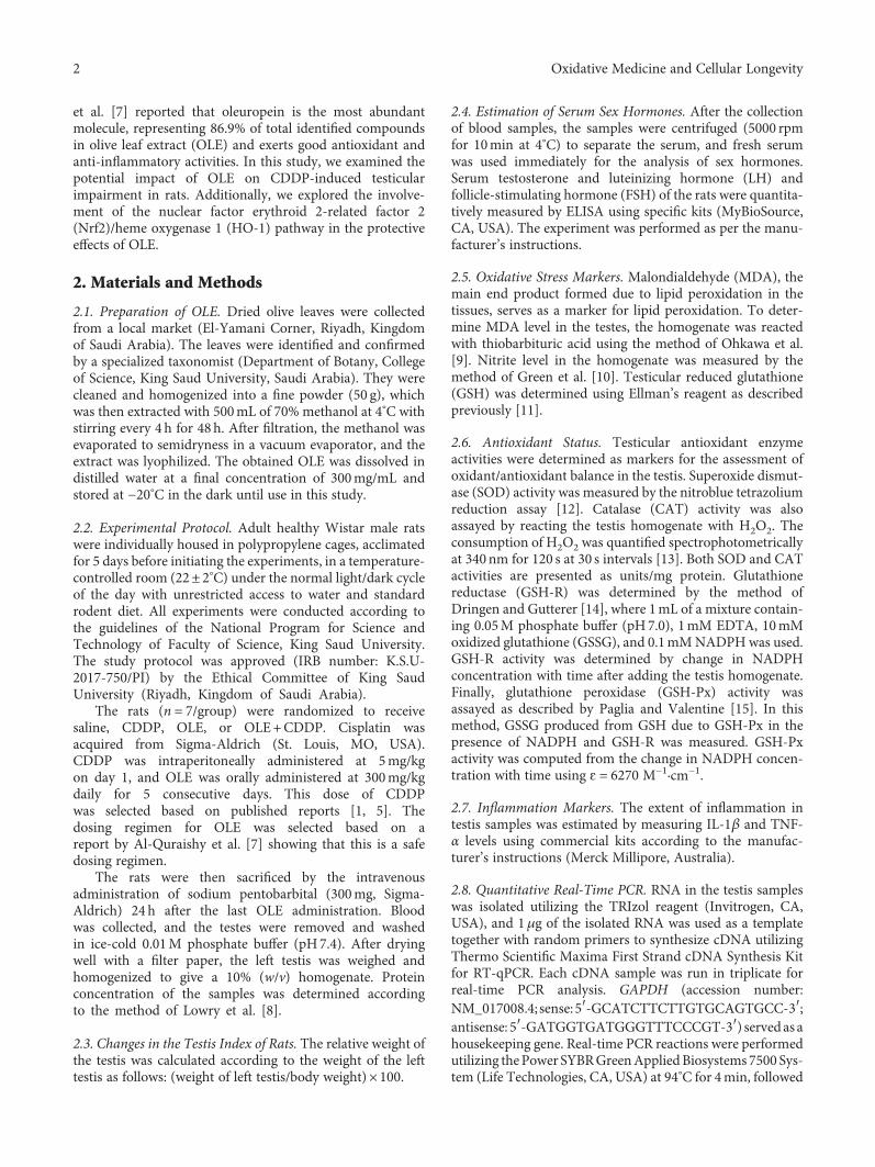
et al. [7] reported that oleuropein is the most abundantmolecule, representing 86.9% of total identified compoundsin olive leaf extract (OLE) and exerts good antioxidant andanti-inflammatory activities. In this study, we examined thepotential impact of OLE on CDDP-induced testicularimpairment in rats. Additionally, we explored the involve-ment of the nuclear factor erythroid 2-related factor 2(Nrf2)/heme oxygenase 1 (HO-1) pathway in the protectiveeffects of OLE.
2. Materials and Methods
2.1. Preparation of OLE. Dried olive leaves were collectedfrom a local market (El-Yamani Corner, Riyadh, Kingdomof Saudi Arabia). The leaves were identified and confirmedby a specialized taxonomist (Department of Botany, Collegeof Science, King Saud University, Saudi Arabia). They werecleaned and homogenized into a fine powder (50 g), whichwas then extracted with 500mL of 70% methanol at 4°C withstirring every 4 h for 48h. After filtration, the methanol wasevaporated to semidryness in a vacuum evaporator, and theextract was lyophilized. The obtained OLE was dissolved indistilled water at a final concentration of 300mg/mL andstored at −20°C in the dark until use in this study.
2.2. Experimental Protocol. Adult healthy Wistar male ratswere individually housed in polypropylene cages, acclimatedfor 5 days before initiating the experiments, in a temperature-controlled room (22± 2°C) under the normal light/dark cycleof the day with unrestricted access to water and standardrodent diet. All experiments were conducted according tothe guidelines of the National Program for Science andTechnology of Faculty of Science, King Saud University.The study protocol was approved (IRB number: K.S.U-2017-750/PI) by the Ethical Committee of King SaudUniversity (Riyadh, Kingdom of Saudi Arabia).
The rats (n = 7/group) were randomized to receivesaline, CDDP, OLE, or OLE+CDDP. Cisplatin wasacquired from Sigma-Aldrich (St. Louis, MO, USA).CDDP was intraperitoneally administered at 5mg/kgon day 1, and OLE was orally administered at 300mg/kgdaily for 5 consecutive days. This dose of CDDPwas selected based on published reports [1, 5]. Thedosing regimen for OLE was selected based on areport by Al-Quraishy et al. [7] showing that this is a safedosing regimen.
The rats were then sacrificed by the intravenousadministration of sodium pentobarbital (300mg, Sigma-Aldrich) 24 h after the last OLE administration. Bloodwas collected, and the testes were removed and washedin ice-cold 0.01M phosphate buffer (pH7.4). After dryingwell with a filter paper, the left testis was weighed andhomogenized to give a 10% (w/v) homogenate. Proteinconcentration of the samples was determined accordingto the method of Lowry et al. [8].
2.3. Changes in the Testis Index of Rats. The relative weight ofthe testis was calculated according to the weight of the lefttestis as follows: (weight of left testis/body weight)× 100.
2.4. Estimation of Serum Sex Hormones. After the collectionof blood samples, the samples were centrifuged (5000 rpmfor 10min at 4°C) to separate the serum, and fresh serumwas used immediately for the analysis of sex hormones.Serum testosterone and luteinizing hormone (LH) andfollicle-stimulating hormone (FSH) of the rats were quantita-tively measured by ELISA using specific kits (MyBioSource,CA, USA). The experiment was performed as per the manu-facturer’s instructions.
2.5. Oxidative Stress Markers. Malondialdehyde (MDA), themain end product formed due to lipid peroxidation in thetissues, serves as a marker for lipid peroxidation. To deter-mine MDA level in the testes, the homogenate was reactedwith thiobarbituric acid using the method of Ohkawa et al.[9]. Nitrite level in the homogenate was measured by themethod of Green et al. [10]. Testicular reduced glutathione(GSH) was determined using Ellman’s reagent as describedpreviously [11].
2.6. Antioxidant Status. Testicular antioxidant enzymeactivities were determined as markers for the assessment ofoxidant/antioxidant balance in the testis. Superoxide dismut-ase (SOD) activity was measured by the nitroblue tetrazoliumreduction assay [12]. Catalase (CAT) activity was alsoassayed by reacting the testis homogenate with H2O2. Theconsumption of H2O2 was quantified spectrophotometricallyat 340nm for 120 s at 30 s intervals [13]. Both SOD and CATactivities are presented as units/mg protein. Glutathionereductase (GSH-R) was determined by the method ofDringen and Gutterer [14], where 1mL of a mixture contain-ing 0.05M phosphate buffer (pH7.0), 1mM EDTA, 10mMoxidized glutathione (GSSG), and 0.1mMNADPHwas used.GSH-R activity was determined by change in NADPHconcentration with time after adding the testis homogenate.Finally, glutathione peroxidase (GSH-Px) activity wasassayed as described by Paglia and Valentine [15]. In thismethod, GSSG produced from GSH due to GSH-Px in thepresence of NADPH and GSH-R was measured. GSH-Pxactivity was computed from the change in NADPH concen-tration with time using ɛ = 6270 M−1·cm−1.
2.7. Inflammation Markers. The extent of inflammation intestis samples was estimated by measuring IL-1β and TNF-α levels using commercial kits according to the manufac-turer’s instructions (Merck Millipore, Australia).
2.8. Quantitative Real-Time PCR. RNA in the testis sampleswas isolated utilizing the TRIzol reagent (Invitrogen, CA,USA), and 1μg of the isolated RNA was used as a templatetogether with random primers to synthesize cDNA utilizingThermo Scientific Maxima First Strand cDNA Synthesis Kitfor RT-qPCR. Each cDNA sample was run in triplicate forreal-time PCR analysis. GAPDH (accession number:NM_017008.4; sense:5′-GCATCTTCTTGTGCAGTGCC-3′;antisense: 5′-GATGGTGATGGGTTTCCCGT-3′) servedas ahousekeeping gene. Real-time PCR reactions were performedutilizing thePower SYBRGreenAppliedBiosystems 7500 Sys-tem (Life Technologies, CA, USA) at 94°C for 4min, followed
2 Oxidative Medicine and Cellular Longevity
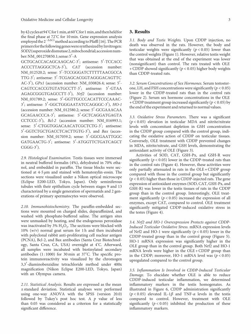
by42cyclesat94°Cfor1min,at60°Cfor1min,andthenheldforthe final phase at 72°C for 10min. Gene expression analysisemployed the2−ΔΔCtmethodaccording toPfaffl [16].ThePCRprimers forthefollowinggenesweresynthesizedbyInvitrogen:SOD2(superoxidedismutase2,mitochondrial;accessionnum-ber:NM_001270850.1; sense:5′-AGCTGCACCACAGCAAGCAC-3′; antisense: 5′-TCCACCACCCTTAGGGCTCA-3′), CAT (accession number:NM_012520.2; sense: 5′-TCCGGGATCTTTTTAACGCCATTG-3′; antisense: 5′-TCGAGCACGGTAGGGACAGTTCAC-3′), GPx1 (accession number: NM_030826.4; sense: 5′-CAGTCCACCGTGTATGCCTT-3′; antisense: 5′-GTAAAGAGCGGGTGAGCCTT-3′), Nrf2 (accession number:NM_031789.2; sense: 5′-GGTTGCCCACATTCCCAAAC-3′; antisense: 5′-GGCTGGGAATATCCAGGGC-3′), HO-1(accession number: NM_012580.2; sense: 5′-GCGAAACAAGCAGAACCCA-3′; antisense: 5′-GCTCAGGATGAGTACCTCCC-3′), Bcl-2 (accession number: NM_016993.1;sense: 5′-CTGGTGGACAACATCGCTCTG-3′; antisense:5′-GGTCTGCTGACCTCACTTGTG-3′), and Bax (acces-sion number: NM_017059.2; sense: 5′-GGCGAATTGGCGATGAACTG-3′; antisense: 5′-ATGGTTCTGATCAGCTCGGG-3′).
2.9. Histological Examination. Testis tissues were immersedin neutral buffered formalin (4%), dehydrated in 70% etha-nol, and embedded in paraffin. The tissue blocks were sec-tioned at 4-5μm and stained with hematoxylin-eosin. Thesections were visualized under a Nikon optical microscope(Eclipse E200-LED, Tokyo, Japan). Only seminiferoustubules with their epithelium cycle between stages 9 and 13characterized by a single generation of spermatids and 2 gen-erations of primary spermatocytes were observed.
2.10. Immunohistochemistry. The paraffin-embedded sec-tions were mounted on charged slides, deparaffinized, andwashed with phosphate-buffered saline. The antigen siteswere unmasked by heating, and the endogenous peroxidasewas inactivated by 3% H2O2. The sections were blocked with10% (w/v) normal goat serum for 1 h and then incubatedwith polyclonal rabbit anti-proliferating cell nuclear antigen(PCNA), Bcl-2, and Bax antibodies (Santa Cruz Biotechnol-ogy, Santa Cruz, CA, USA) overnight at 4°C. Afterward,all samples were incubated with biotinylated secondaryantibodies (1 : 1000) for 30min at 37°C. The specific pro-tein immunoreactivity was visualized by the chromogen3,3′-diaminobenzidine tetrachloride method under 400xmagnification (Nikon Eclipse E200-LED, Tokyo, Japan)with an Olympus camera.
2.11. Statistical Analysis. Results are expressed as the mean± standard deviation. Statistical analyses were performedusing one-way ANOVA with the SPSS (version 20.0)followed by Tukey’s post hoc test. A p value of lessthan 0.05 was considered as a criterion for a statisticallysignificant difference.
3. Results
3.1. Body and Testis Weights. Upon CDDP injection, nodeath was observed in the rats. However, the body andtesticular weights were significantly (p < 0 05) lower thanthe control weights (Figure 1). However, relative testis weightthat was obtained at the end of the experiment was lower(nonsignificant) than control. The rats treated with OLE+CDDP showed significantly (p < 0 05) higher body weightsthan CDDP-treated rats.
3.2. Serum Concentrations of Sex Hormones. Serum testoster-one, LH, and FSH concentrations were significantly (p < 0 05)lower in the CDDP-treated rats than in the control rats(Figure 2). Serum sex hormone concentrations in the OLE+CDDP treatment group increased significantly (p < 0 05) bythe endof the experiment and returned to normal values.
3.3. Oxidative Stress Parameters. There was a significant(p < 0 05) elevation in testicular MDA and nitrite/nitratelevels with concomitant depletion in GSH concentrationin the CDDP group compared with the control group, indi-cating the oxidative action of CDDP on testicular tissues.Conversely, OLE treatment with CDDP prevented changesin MDA, nitrite/nitrate, and GSH levels, demonstrating theantioxidant activity of OLE (Figure 3).
Activities of SOD, CAT, GSH-Px, and GSH-R weresignificantly (p < 0 05) lower in the CDDP-treated rats thanin the control rats (Figure 4). However, these activities wereonly partially attenuated in rats in the OLE+CDDP groupcompared with those in the control group but significantly(p < 0 05) higher than those in CDDP-injected rats. The geneexpression of antioxidant enzymes (SOD, CAT, GSH-Px, andGSH-R) was lower in the testis tissues of rats in the CDDPgroup than in the control group. Interestingly, OLE treat-ment significantly (p < 0 05) increased the expression of allenzymes, except CAT, compared to control. OLE treatmentsignificantly mitigated CDDP-induced oxidative stress inthe testes (Figure 4).
3.4. Nrf2 and HO-1 Overexpression Protects against CDDP-Induced Testicular Oxidative Stress. mRNA expression levelsof Nrf2 and HO-1 were significantly (p < 0 05) lower in theCDDP-treated group than in the control group (Figure 5).HO-1 mRNA expression was significantly higher in theOLE group than in the control group. Both Nrf2 and HO-1mRNA levels were higher in the OLE+CDDP group thanin the CDDP; moreover, HO-1 mRNA level was (p < 0 05)upregulated compared to the control group.
3.5. Inflammation Is Involved in CDDP-Induced TesticularDamage. To elucidate whether OLE is able to reduceCDDP-induced testicular inflammation, we determinedinflammatory markers in the testis homogenates. Asillustrated in Figure 6, CDDP administration significantly(p < 0 05) elevated IL-1β and TNF-α levels in the testiscompared to control. However, treatment with OLEsignificantly (p < 0 05) inhibited the production of theseinflammatory markers.
3Oxidative Medicine and Cellular Longevity
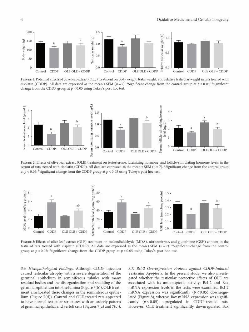
3.6. Histopathological Findings. Although CDDP injectioncaused testicular atrophy with a severe degeneration of thegerminal epithelium in seminiferous tubules with manyresidual bodies and the disorganization and shedding of thegerminal epithelium into the lumina (Figure 7(b)), OLE treat-ment ameliorated these changes in the seminiferous epithe-lium (Figure 7(d)). Control and OLE-treated rats appearedto have normal testicular structures with an orderly patternof germinal epithelial and Sertoli cells (Figures 7(a) and 7(c)).
3.7. Bcl-2 Overexpression Protects against CDDP-InducedTesticular Apoptosis. In the present study, we also investi-gated whether the testicular protective effects of OLE areassociated with its antiapoptotic activity; Bcl-2 and BaxmRNA expression levels in the testis were examined. Bcl-2mRNA expression was significantly (p < 0 05) downregu-lated (Figure 8), whereas Bax mRNA expression was signifi-cantly (p < 0 05) upregulated in CDDP-treated rats.However, OLE treatment significantly downregulated Bax
ab a
0
50
100
150
200
Bod
y w
eigh
t (g)
OLE + CDDPOLECDDPControl0.0
0.5
1.0
1.5
Testi
cula
r wei
ght (
g)
OLE + CDDPOLECDDPControl0.0
0.5
1.0
Relat
ive t
estic
ular
wei
ght (
%)
OLE + CDDPOLECDDPControl
Figure 1: Potential effects of olive leaf extract (OLE) treatment on body weight, testis weight, and relative testicular weight in rats treated withcisplatin (CDDP). All data are expressed as the mean± SEM (n = 7). aSignificant change from the control group at p < 0 05; bsignificantchange from the CDDP group at p < 0 05 using Tukey’s post hoc test.
a
a
ba
ba
b
0
1
2
3
4
Seru
m fo
llicle
-stim
ulat
ing
horm
one
leve
l (ng
/L)
0.0
0.5
1.0
1.5
Seru
m lu
tein
izin
g ho
rmon
e lev
el (n
g/L)
0
2
4
6
8
Seru
m te
stoste
rone
leve
l (pg
/mL)
OLE + CDDPOLECDDPControlCDDP OLE OLE + CDDPControl CDDP OLE OLE + CDDPControl
Figure 2: Effects of olive leaf extract (OLE) treatment on testosterone, luteinizing hormone, and follicle-stimulating hormone levels in theserum of rats treated with cisplatin (CDDP). All data are expressed as the mean± SEM (n = 7). aSignificant change from the control groupat p < 0 05; bsignificant change from the CDDP group at p < 0 05 using Tukey’s post hoc test.
a a
abb
a
b
0
2
4
6
8
MD
A le
vel (
nmol
/mg
prot
ein)
CDDP OLE OLE + CDDPControl0
20
40
60
80
Nitr
ite/n
itrat
e lev
el (�휇
mol
/mg
prot
ein)
OLE + CDDPOLECDDPControl0.0
0.1
0.2
0.3
0.4
0.5
GSH
leve
l (m
mol
/mg
prot
ein)
CDDP OLE OLE + CDDPControl
Figure 3: Effects of olive leaf extract (OLE) treatment on malondialdehyde (MDA), nitrite/nitrate, and glutathione (GSH) content in thetestis of rats treated with cisplatin (CDDP). All data are expressed as the mean± SEM (n = 7). aSignificant change from the controlgroup at p < 0 05; bsignificant change from the CDDP group at p < 0 05 using Tukey’s post hoc test.
4 Oxidative Medicine and Cellular Longevity
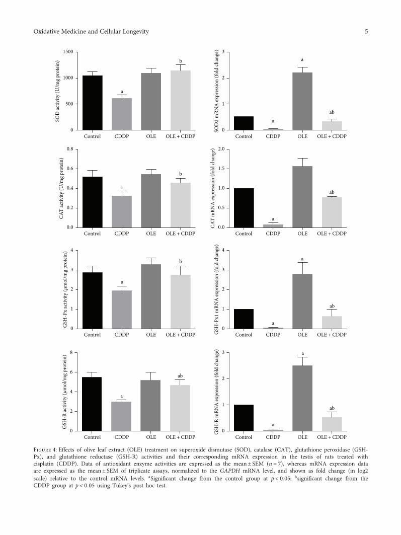
b
a
ab
a
a
b
a
ab
a
ab
a
ab
a
a
ab
a
a
a
b
CDDP OLE OLE + CDDPControl CDDP OLE OLE + CDDPControl
CDDP OLE OLE + CDDPControl CDDP OLE OLE + CDDPControl
CDDP OLE OLE + CDDPControl OLE + CDDPOLECDDPControl
OLE + CDDPOLECDDPControl CDDP OLE OLE + CDDPControl
0
500
1000
1500SO
D ac
tivity
(U/m
g pr
otei
n)
0.0
0.2
0.4
0.6
0.8
CAT
activ
ity (U
/mg
prot
ein)
0
1
2
3
4
GSH
-Px
activ
ity (�휇
mol
/mg
prot
ein)
0
2
4
6
8
GSH
-R ac
tivity
(�휇m
ol/m
g pr
otei
n)
0
1
2
3
SOD
2 m
RNA
expr
essio
n (fo
ld ch
ange
)
0.0
0.5
1.0
1.5
2.0
CAT
mRN
A ex
pres
sion
(fold
chan
ge)
0
1
2
3
4
GSH
-Px1
mRN
A ex
pres
sion
(fold
chan
ge)
0
1
2
3
GSH
-R m
RNA
expr
essio
n (fo
ld ch
ange
)
Figure 4: Effects of olive leaf extract (OLE) treatment on superoxide dismutase (SOD), catalase (CAT), glutathione peroxidase (GSH-Px), and glutathione reductase (GSH-R) activities and their corresponding mRNA expression in the testis of rats treated withcisplatin (CDDP). Data of antioxidant enzyme activities are expressed as the mean± SEM (n = 7), whereas mRNA expression dataare expressed as the mean± SEM of triplicate assays, normalized to the GAPDH mRNA level, and shown as fold change (in log2scale) relative to the control mRNA levels. aSignificant change from the control group at p < 0 05; bsignificant change from theCDDP group at p < 0 05 using Tukey’s post hoc test.
5Oxidative Medicine and Cellular Longevity
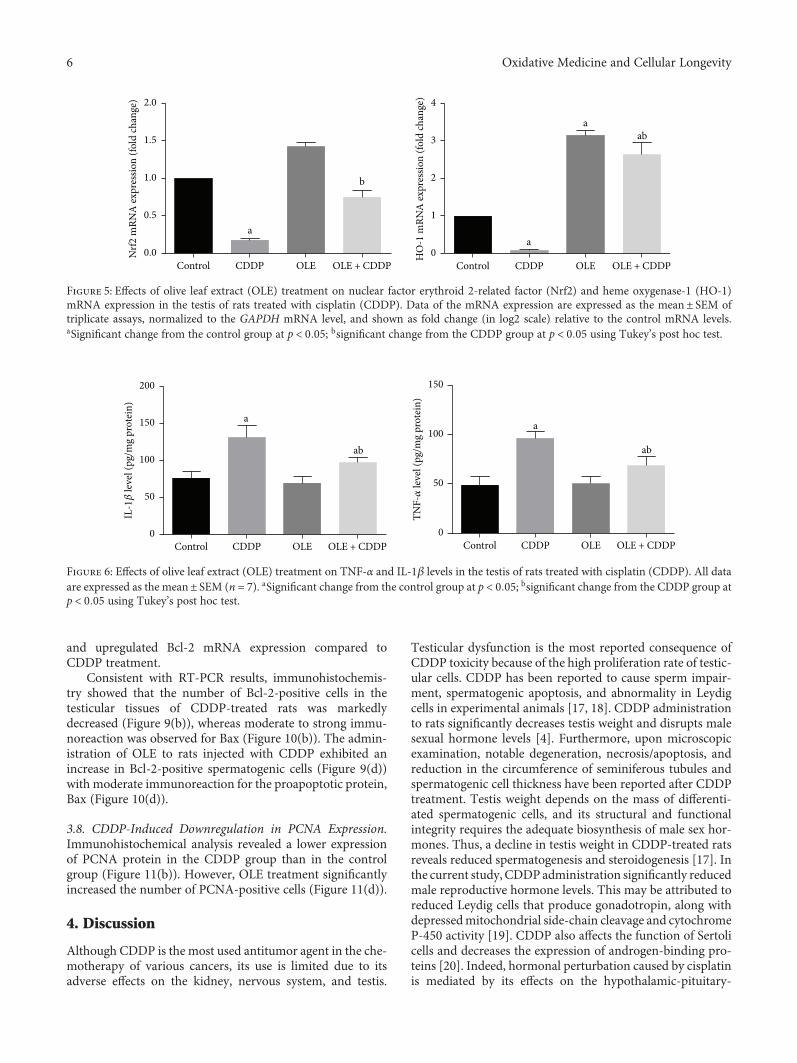
and upregulated Bcl-2 mRNA expression compared toCDDP treatment.
Consistent with RT-PCR results, immunohistochemis-try showed that the number of Bcl-2-positive cells in thetesticular tissues of CDDP-treated rats was markedlydecreased (Figure 9(b)), whereas moderate to strong immu-noreaction was observed for Bax (Figure 10(b)). The admin-istration of OLE to rats injected with CDDP exhibited anincrease in Bcl-2-positive spermatogenic cells (Figure 9(d))with moderate immunoreaction for the proapoptotic protein,Bax (Figure 10(d)).
3.8. CDDP-Induced Downregulation in PCNA Expression.Immunohistochemical analysis revealed a lower expressionof PCNA protein in the CDDP group than in the controlgroup (Figure 11(b)). However, OLE treatment significantlyincreased the number of PCNA-positive cells (Figure 11(d)).
4. Discussion
Although CDDP is the most used antitumor agent in the che-motherapy of various cancers, its use is limited due to itsadverse effects on the kidney, nervous system, and testis.
Testicular dysfunction is the most reported consequence ofCDDP toxicity because of the high proliferation rate of testic-ular cells. CDDP has been reported to cause sperm impair-ment, spermatogenic apoptosis, and abnormality in Leydigcells in experimental animals [17, 18]. CDDP administrationto rats significantly decreases testis weight and disrupts malesexual hormone levels [4]. Furthermore, upon microscopicexamination, notable degeneration, necrosis/apoptosis, andreduction in the circumference of seminiferous tubules andspermatogenic cell thickness have been reported after CDDPtreatment. Testis weight depends on the mass of differenti-ated spermatogenic cells, and its structural and functionalintegrity requires the adequate biosynthesis of male sex hor-mones. Thus, a decline in testis weight in CDDP-treated ratsreveals reduced spermatogenesis and steroidogenesis [17]. Inthe current study, CDDPadministration significantly reducedmale reproductive hormone levels. This may be attributed toreduced Leydig cells that produce gonadotropin, along withdepressedmitochondrial side-chain cleavage and cytochromeP-450 activity [19]. CDDP also affects the function of Sertolicells and decreases the expression of androgen-binding pro-teins [20]. Indeed, hormonal perturbation caused by cisplatinis mediated by its effects on the hypothalamic-pituitary-
a
b
a
aab
0.0
0.5
1.0
1.5
2.0
Nrf2
mRN
A ex
pres
sion
(fold
chan
ge)
CDDP OLE OLE + CDDPControl0
1
2
3
4
HO
-1 m
RNA
expr
essio
n (fo
ld ch
ange
)
CDDP OLE OLE + CDDPControl
Figure 5: Effects of olive leaf extract (OLE) treatment on nuclear factor erythroid 2-related factor (Nrf2) and heme oxygenase-1 (HO-1)mRNA expression in the testis of rats treated with cisplatin (CDDP). Data of the mRNA expression are expressed as the mean± SEM oftriplicate assays, normalized to the GAPDH mRNA level, and shown as fold change (in log2 scale) relative to the control mRNA levels.aSignificant change from the control group at p < 0 05; bsignificant change from the CDDP group at p < 0 05 using Tukey’s post hoc test.
ab ab
aa
0
50
100
150
200
IL-1�훽
leve
l (pg
/mg
prot
ein)
CDDP OLE OLE + CDDPControl0
50
100
150
TNF-�훼
leve
l (pg
/mg
prot
ein)
CDDP OLE OLE + CDDPControl
Figure 6: Effects of olive leaf extract (OLE) treatment on TNF-α and IL-1β levels in the testis of rats treated with cisplatin (CDDP). All dataare expressed as the mean± SEM (n = 7). aSignificant change from the control group at p < 0 05; bsignificant change from the CDDP group atp < 0 05 using Tukey’s post hoc test.
6 Oxidative Medicine and Cellular Longevity
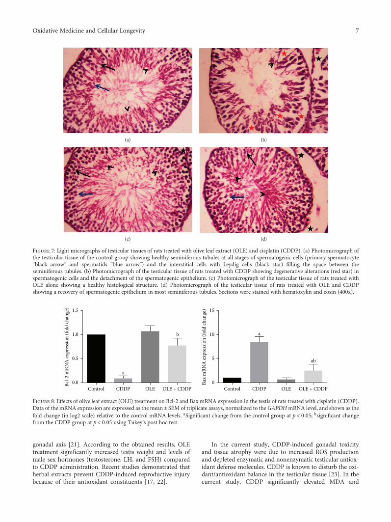
gonadal axis [21]. According to the obtained results, OLEtreatment significantly increased testis weight and levels ofmale sex hormones (testosterone, LH, and FSH) comparedto CDDP administration. Recent studies demonstrated thatherbal extracts prevent CDDP-induced reproductive injurybecause of their antioxidant constituents [17, 22].
In the current study, CDDP-induced gonadal toxicityand tissue atrophy were due to increased ROS productionand depleted enzymatic and nonenzymatic testicular antiox-idant defense molecules. CDDP is known to disturb the oxi-dant/antioxidant balance in the testicular tissue [23]. In thecurrent study, CDDP significantly elevated MDA and
(a) (b)
(c) (d)
Figure 7: Light micrographs of testicular tissues of rats treated with olive leaf extract (OLE) and cisplatin (CDDP). (a) Photomicrograph ofthe testicular tissue of the control group showing healthy seminiferous tubules at all stages of spermatogenic cells (primary spermatocyte“black arrow” and spermatids “blue arrow”) and the interstitial cells with Leydig cells (black star) filling the space between theseminiferous tubules. (b) Photomicrograph of the testicular tissue of rats treated with CDDP showing degenerative alterations (red star) inspermatogenic cells and the detachment of the spermatogenic epithelium. (c) Photomicrograph of the testicular tissue of rats treated withOLE alone showing a healthy histological structure. (d) Photomicrograph of the testicular tissue of rats treated with OLE and CDDPshowing a recovery of spermatogenic epithelium in most seminiferous tubules. Sections were stained with hematoxylin and eosin (400x).
a
b a
ab
0.0
0.5
1.0
1.5
Bcl-2
mRN
A ex
pres
sion
(fold
chan
ge)
CDDP OLE OLE + CDDPControl0
5
10
15
Bax
mRN
A ex
pres
sion
(fold
chan
ge)
CDDP OLE OLE + CDDPControl
Figure 8: Effects of olive leaf extract (OLE) treatment on Bcl-2 and Bax mRNA expression in the testis of rats treated with cisplatin (CDDP).Data of the mRNA expression are expressed as the mean± SEM of triplicate assays, normalized to the GAPDHmRNA level, and shown as thefold change (in log2 scale) relative to the control mRNA levels. aSignificant change from the control group at p < 0 05; bsignificant changefrom the CDDP group at p < 0 05 using Tukey’s post hoc test.
7Oxidative Medicine and Cellular Longevity
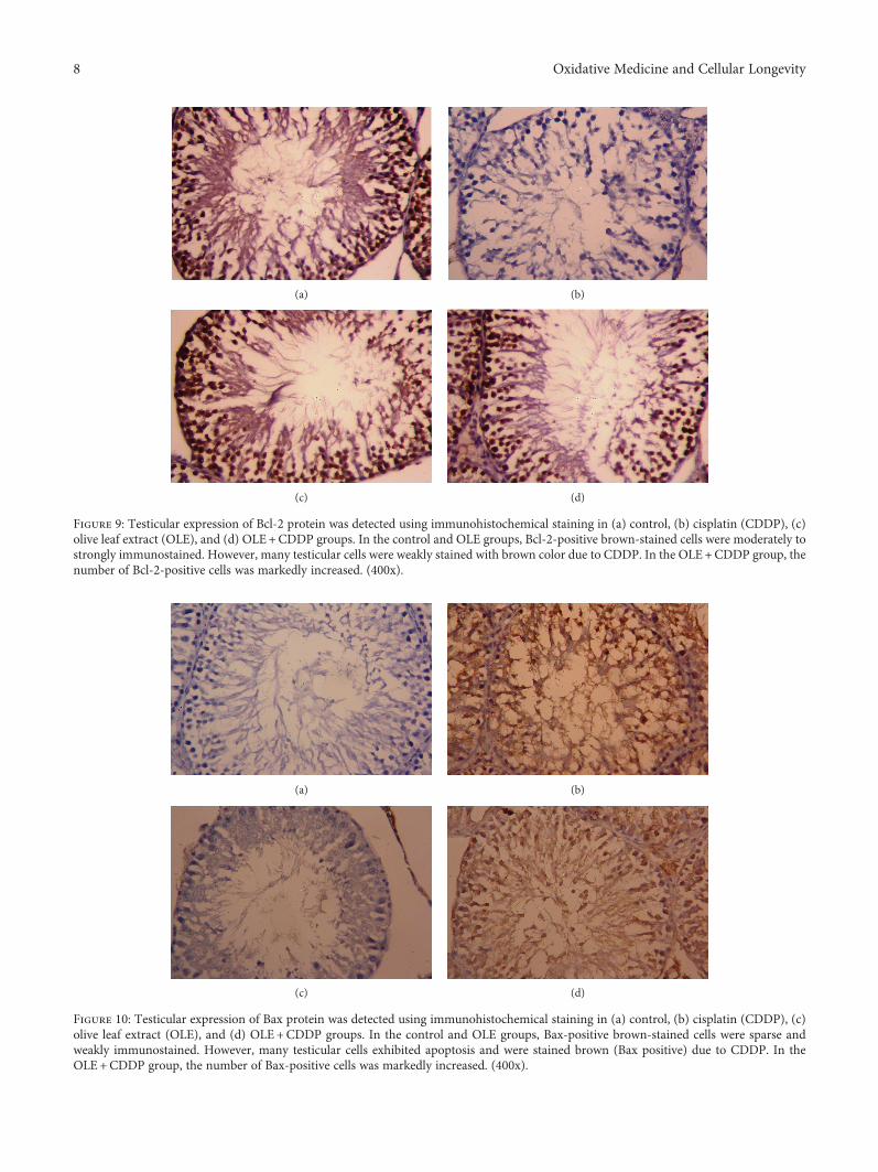
(a) (b)
(c) (d)
Figure 9: Testicular expression of Bcl-2 protein was detected using immunohistochemical staining in (a) control, (b) cisplatin (CDDP), (c)olive leaf extract (OLE), and (d) OLE+CDDP groups. In the control and OLE groups, Bcl-2-positive brown-stained cells were moderately tostrongly immunostained. However, many testicular cells were weakly stained with brown color due to CDDP. In the OLE+CDDP group, thenumber of Bcl-2-positive cells was markedly increased. (400x).
(a) (b)
(c) (d)
Figure 10: Testicular expression of Bax protein was detected using immunohistochemical staining in (a) control, (b) cisplatin (CDDP), (c)olive leaf extract (OLE), and (d) OLE+CDDP groups. In the control and OLE groups, Bax-positive brown-stained cells were sparse andweakly immunostained. However, many testicular cells exhibited apoptosis and were stained brown (Bax positive) due to CDDP. In theOLE+CDDP group, the number of Bax-positive cells was markedly increased. (400x).
8 Oxidative Medicine and Cellular Longevity
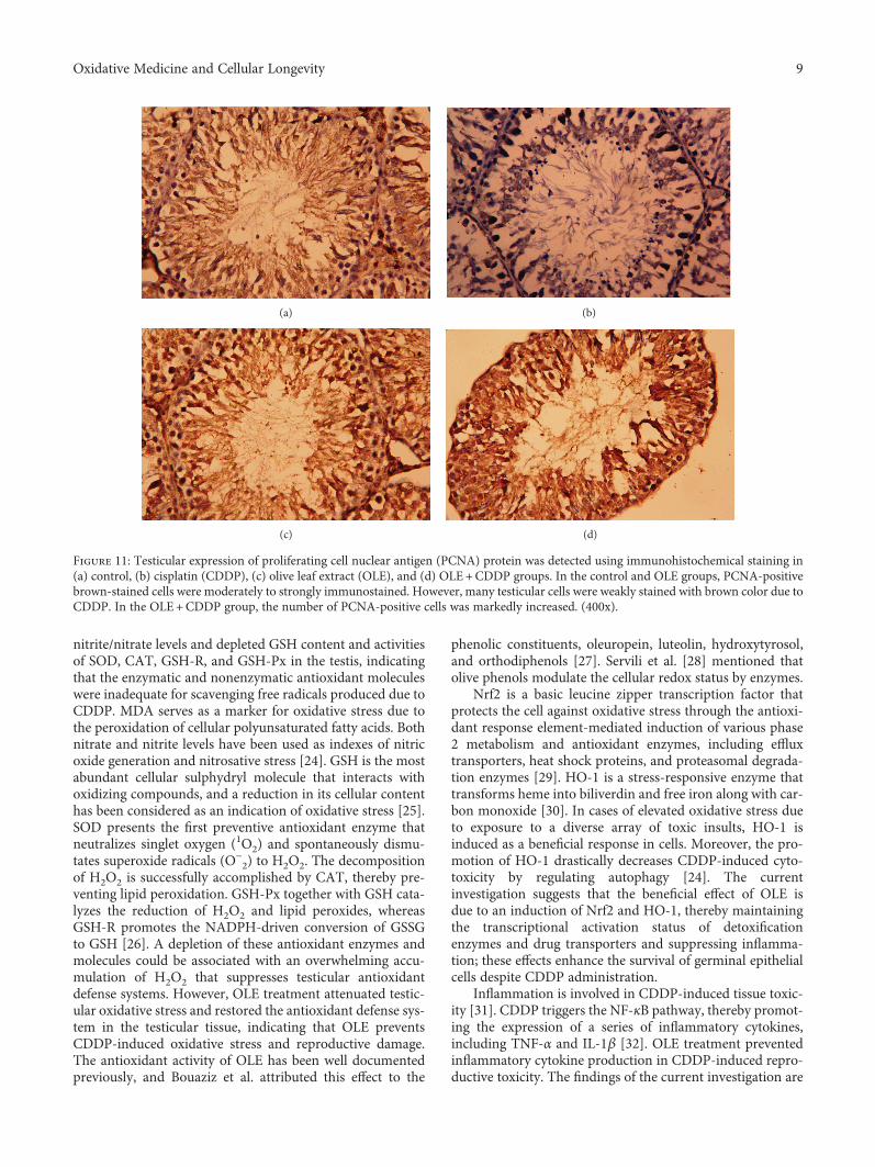
nitrite/nitrate levels and depleted GSH content and activitiesof SOD, CAT, GSH-R, and GSH-Px in the testis, indicatingthat the enzymatic and nonenzymatic antioxidant moleculeswere inadequate for scavenging free radicals produced due toCDDP. MDA serves as a marker for oxidative stress due tothe peroxidation of cellular polyunsaturated fatty acids. Bothnitrate and nitrite levels have been used as indexes of nitricoxide generation and nitrosative stress [24]. GSH is the mostabundant cellular sulphydryl molecule that interacts withoxidizing compounds, and a reduction in its cellular contenthas been considered as an indication of oxidative stress [25].SOD presents the first preventive antioxidant enzyme thatneutralizes singlet oxygen (1O2) and spontaneously dismu-tates superoxide radicals (O−
2) to H2O2. The decompositionof H2O2 is successfully accomplished by CAT, thereby pre-venting lipid peroxidation. GSH-Px together with GSH cata-lyzes the reduction of H2O2 and lipid peroxides, whereasGSH-R promotes the NADPH-driven conversion of GSSGto GSH [26]. A depletion of these antioxidant enzymes andmolecules could be associated with an overwhelming accu-mulation of H2O2 that suppresses testicular antioxidantdefense systems. However, OLE treatment attenuated testic-ular oxidative stress and restored the antioxidant defense sys-tem in the testicular tissue, indicating that OLE preventsCDDP-induced oxidative stress and reproductive damage.The antioxidant activity of OLE has been well documentedpreviously, and Bouaziz et al. attributed this effect to the
phenolic constituents, oleuropein, luteolin, hydroxytyrosol,and orthodiphenols [27]. Servili et al. [28] mentioned thatolive phenols modulate the cellular redox status by enzymes.
Nrf2 is a basic leucine zipper transcription factor thatprotects the cell against oxidative stress through the antioxi-dant response element-mediated induction of various phase2 metabolism and antioxidant enzymes, including effluxtransporters, heat shock proteins, and proteasomal degrada-tion enzymes [29]. HO-1 is a stress-responsive enzyme thattransforms heme into biliverdin and free iron along with car-bon monoxide [30]. In cases of elevated oxidative stress dueto exposure to a diverse array of toxic insults, HO-1 isinduced as a beneficial response in cells. Moreover, the pro-motion of HO-1 drastically decreases CDDP-induced cyto-toxicity by regulating autophagy [24]. The currentinvestigation suggests that the beneficial effect of OLE isdue to an induction of Nrf2 and HO-1, thereby maintainingthe transcriptional activation status of detoxificationenzymes and drug transporters and suppressing inflamma-tion; these effects enhance the survival of germinal epithelialcells despite CDDP administration.
Inflammation is involved in CDDP-induced tissue toxic-ity [31]. CDDP triggers the NF-κB pathway, thereby promot-ing the expression of a series of inflammatory cytokines,including TNF-α and IL-1β [32]. OLE treatment preventedinflammatory cytokine production in CDDP-induced repro-ductive toxicity. The findings of the current investigation are
(a) (b)
(c) (d)
Figure 11: Testicular expression of proliferating cell nuclear antigen (PCNA) protein was detected using immunohistochemical staining in(a) control, (b) cisplatin (CDDP), (c) olive leaf extract (OLE), and (d) OLE+CDDP groups. In the control and OLE groups, PCNA-positivebrown-stained cells were moderately to strongly immunostained. However, many testicular cells were weakly stained with brown color due toCDDP. In the OLE+CDDP group, the number of PCNA-positive cells was markedly increased. (400x).
9Oxidative Medicine and Cellular Longevity
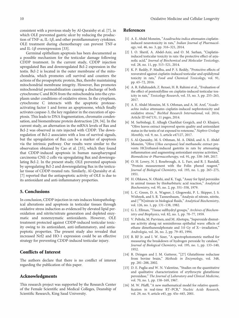
consistent with a previous study by Al-Quraishy et al. [7], inwhich OLE prevented gastric ulcer by reducing the produc-tion of TNF-α, IL-1β, and other proinflammatory cytokines.OLE treatment during chemotherapy can prevent TNF-αand IL-1β overexpression [33].
Germinal epithelium apoptosis has been documented asa possible mechanism for the testicular damage followingCDDP treatment. In the current study, CDDP injectionupregulated Bax and downregulated Bcl-2 expression in thetestis. Bcl-2 is located in the outer membrane of the mito-chondria, which promotes cell survival and counters theactions of the proapoptotic protein, Bax, thereby maintainingmitochondrial membrane integrity. However, Bax promotesmitochondrial permeabilization causing a discharge of bothcytochrome C and ROS from the mitochondria into the cyto-plasm under conditions of oxidative stress. In the cytoplasm,cytochrome C interacts with the apoptotic protease-activating factor 1 and forms an apoptosome, which finallyactivates caspase-3, the key regulator in the execution of apo-ptosis. This leads to DNA fragmentation, chromatin conden-sation, and biomembrane protein destruction [29, 34]. In thecurrent study, an alternation in the mRNA levels of Bax andBcl-2 was observed in rats injected with CDDP. The down-regulation of Bcl-2 associates with a loss of survival signals,but the upregulation of Bax can be a marker of apoptosisvia the intrinsic pathway. Our results were similar to theobservation obtained by Cao et al. [35], which they foundthat CDDP-induced apoptosis in human nasopharyngealcarcinoma CNE-2 cells via upregulating Bax and downregu-lating Bcl-2. In the present study, OLE prevented apoptosisby upregulating Bcl-2 and downregulating Bax in the testicu-lar tissue of CDDP-treated rats. Similarly, Al-Quraishy et al.[7] reported that the antiapoptotic activity of OLE is due toits antioxidant and anti-inflammatory properties.
5. Conclusions
In conclusion, CDDP injection in rats induces histopatholog-ical alterations and apoptosis in testicular tissues throughoxidative stress induction as evidenced by elevated lipid per-oxidation and nitrite/nitrate generation and depleted enzy-matic and nonenzymatic antioxidants. However, OLEtreatment protected against CDDP-induced testicular toxic-ity owing to its antioxidant, anti-inflammatory, and antia-poptotic properties. The present study also revealed thatincreased Nrf2 and HO-1 expression could be an effectivestrategy for preventing CDDP-induced testicular injury.
Conflicts of Interest
The authors declare that there is no conflict of interestregarding the publication of this paper.
Acknowledgments
This research project was supported by the Research Centerof the Female Scientific and Medical Colleges, Deanship ofScientific Research, King Saud University.
References
[1] A. E. Abdel Moneim, “Azadirachta indica attenuates cisplatin-induced neurotoxicity in rats,” Indian Journal of Pharmacol-ogy, vol. 46, no. 3, pp. 316–321, 2014.
[2] I. O. Sherif, A. Abdel-Aziz, and O. M. Sarhan, “Cisplatin-induced testicular toxicity in rats: the protective effect of arju-nolic acid,” Journal of Biochemical and Molecular Toxicology,vol. 28, no. 11, pp. 515–521, 2014.
[3] K. P. Reddy, P. Madhu, and P. S. Reddy, “Protective effects ofresveratrol against cisplatin-induced testicular and epididymaltoxicity in rats,” Food and Chemical Toxicology, vol. 91,pp. 65–72, 2016.
[4] A. R. Fallahzadeh, Z. Rezaei, H. R. Rahimi et al., “Evaluation ofthe effect of pentoxifylline on cisplatin-induced testicular tox-icity in rats,” Toxicology Research, vol. 33, no. 3, pp. 255–263,2017.
[5] A. E. Abdel Moneim, M. S. Othman, and A. M. Aref, “Azadir-achta indica attenuates cisplatin-induced nephrotoxicity andoxidative stress,” BioMed Research International, vol. 2014,Article ID 647131, 11 pages, 2014.
[6] M. Sarbishegi, E. Alhagh Charkhat Gorgich, and O. Khajavi,“Olive leaves extract improved sperm quality and antioxidantstatus in the testis of rat exposed to rotenone,”Nephro-UrologyMonthly, vol. 9, no. 3, article e47127, 2017.
[7] S. Al-Quraishy, M. S. Othman, M. A. Dkhil, and A. E. AbdelMoneim, “Olive (Olea europaea) leaf methanolic extract pre-vents HCl/ethanol-induced gastritis in rats by attenuatinginflammation and augmenting antioxidant enzyme activities,”Biomedicine & Pharmacotherapy, vol. 91, pp. 338–349, 2017.
[8] O. H. Lowry, N. J. Rosebrough, A. L. Farr, and R. J. Randall,“Protein measurement with the Folin phenol reagent,”Journal of Biological Chemistry, vol. 193, no. 1, pp. 265–275,1951.
[9] H. Ohkawa, N. Ohishi, and K. Yagi, “Assay for lipid peroxidesin animal tissues by thiobarbituric acid reaction,” AnalyticalBiochemistry, vol. 95, no. 2, pp. 351–358, 1979.
[10] L. C. Green, D. A. Wagner, J. Glogowski, P. L. Skipper, J. S.Wishnok, and S. R. Tannenbaum, “Analysis of nitrate, nitrite,and [15N]nitrate in biological fluids,” Analytical Biochemistry,vol. 126, no. 1, pp. 131–138, 1982.
[11] G. L. Ellman, “Tissue sulfhydryl groups,” Archives of Biochem-istry and Biophysics, vol. 82, no. 1, pp. 70–77, 1959.
[12] V. Peltola, M. Parvinen, andM. Ahotupa, “Superoxide dismut-ase activity along rat seminiferous epithelial wave: effects ofethane dimethanesulphonate and 3.0 Gy of X—irradiation,”Andrologia, vol. 26, no. 2, pp. 79–85, 1994.
[13] B. RF Jr. and I. W. Sizer, “A spectrophotometric method formeasuring the breakdown of hydrogen peroxide by catalase,”Journal of Biological Chemistry, vol. 195, no. 1, pp. 133–140,1952.
[14] R. Dringen and J. M. Gutterer, “[27] Glutathione reductasefrom bovine brain,” Methods in Enzymology, vol. 348,pp. 281–288, 2002.
[15] D. E. Paglia and W. N. Valentine, “Studies on the quantitativeand qualitative characterization of erythrocyte glutathioneperoxidase,” The Journal of Laboratory and Clinical Medicine,vol. 70, no. 1, pp. 158–169, 1967.
[16] M. W. Pfaffl, “A new mathematical model for relative quanti-fication in real-time RT–PCR,” Nucleic Acids Research,vol. 29, no. 9, article e45, pp. 45e–445, 2001.
10 Oxidative Medicine and Cellular Longevity
[17] A. Amin, C. Abraham, A. A. Hamza et al., “A standardizedextract of Ginkgo biloba neutralizes cisplatin-mediated repro-ductive toxicity in rats,” Journal of Biomedicine and Biotech-nology, vol. 2012, article 362049, 11 pages, 2012.
[18] A. Atessahin, E. Sahna, G. Turk et al., “Chemoprotective effectof melatonin against cisplatin-induced testicular toxicity inrats,” Journal of Pineal Research, vol. 41, no. 1, pp. 21–27, 2006.
[19] M. D. Maines, P. M. Sluss, and M. Iscan, “cis-Platinum-mediated decrease in serum testosterone is associated withdepression of luteinizing hormone receptors and cytochromeP-450scc in rat testis,” Endocrinology, vol. 126, no. 5,pp. 2398–2406, 1990.
[20] G. A. Leblanc, P. W. Kantoff, S.-F. Ng, E. Frei III, and D. J.Waxman, “Hormonal perturbations in patients with testicularcancer treated with cisplatin,” Cancer, vol. 69, no. 9, pp. 2306–2310, 1992.
[21] A. Aydiner, Y. Aytekin, and E. Topuz, “Effects of cisplatin ontesticular tissue and the Leydig cell-pituitary axis,” Oncology,vol. 54, no. 1, pp. 74–78, 1997.
[22] S. A. Adejuwon, O. M. Femi-Akinlosotu, and J. O. Omirinde,“Cisplatin-induced testicular dysfunction and its ameliorationby Launaea taraxacifolia leaf extract,” Andrologia, vol. 47,no. 5, pp. 553–559, 2015.
[23] H. Anand, M. M. Misro, S. B. Sharma, and S. Prakash, “Protec-tive effects of Eugenia jambolana extract versus N-acetyl cyste-ine against cisplatin-induced damage in rat testis,” Andrologia,vol. 47, no. 2, pp. 194–208, 2015.
[24] Y. I. Chirino and J. Pedraza-Chaverri, “Role of oxidative andnitrosative stress in cisplatin-induced nephrotoxicity,” Experi-mental and Toxicologic Pathology, vol. 61, no. 3, pp. 223–242,2009.
[25] A. A. Hamza, H. M. Elwy, and A. M. Badawi, “Fenugreek seedextract attenuates cisplatin-induced testicular damage inWistar rats,” Andrologia, vol. 48, no. 2, pp. 211–221, 2016.
[26] Y. M. Zhao, L. P. Gao, H. L. Zhang, J. X. Guo, and P. P. Guo,“Grape seed proanthocyanidin extract prevents DDP-induced testicular toxicity in rats,” Food & Function, vol. 5,no. 3, pp. 605–611, 2014.
[27] M. Bouaziz, I. Fki, H. Jemai, M. Ayadi, and S. Sayadi, “Effect ofstorage on refined and husk olive oils composition: Stabiliza-tion by addition of natural antioxidants from Chemlali oliveleaves,” Food Chemistry, vol. 108, no. 1, pp. 253–262, 2008.
[28] M. Servili, B. Sordini, S. Esposto et al., “Biological activities ofphenolic compounds of extra virgin olive oil,” Antioxidants,vol. 3, no. 1, pp. 1–23, 2014.
[29] L. M. Aleksunes, M. J. Goedken, C. E. Rockwell, J. Thomale,J. E. Manautou, and C. D. Klaassen, “Transcriptional regula-tion of renal cytoprotective genes by Nrf2 and its potentialuse as a therapeutic target to mitigate cisplatin-inducednephrotoxicity,” Journal of Pharmacology and ExperimentalTherapeutics, vol. 335, no. 1, pp. 2–12, 2010.
[30] U. Kilic, E. Kilic, Z. Tuzcu et al., “Melatonin suppressescisplatin-induced nephrotoxicity via activation of Nrf-2/HO-1 pathway,” Nutrition & Metabolism, vol. 10, no. 1, pp. 7–7,2013.
[31] X. Zhu, X. Jiang, A. Li, Z. Zhao, and S. Li, “S‐Allylmercaptocys-teine attenuates cisplatin‐induced nephrotoxicity throughsuppression of apoptosis, oxidative stress, and inflammation,”Nutrients, vol. 9, no. 2, p. 166, 2017.
[32] A. H. Eid, N. F. Abdelkader, O. M. Abd El-Raouf, H. M. Fawzy,and E.-E.-D. S. El-Denshary, “Carvedilol alleviates testicular
and spermatological damage induced by cisplatin in rats viamodulation of oxidative stress and inflammation,” Archivesof Pharmacal Research, vol. 39, no. 12, pp. 1693–1702,2016.
[33] K. M. Ahmed, “The effect of olive leaf extract in decreasing theexpression of two pro-inflammatory cytokines in patientsreceiving chemotherapy for cancer. A randomized clinicaltrial,” The Saudi Dental Journal, vol. 25, no. 4, pp. 141–147,2013.
[34] A. A. Fouad, H. O. Qutub, A. E. A. Fouad, A. M. Audeh, andW. N. Al-Melhim, “Epigallocatechin-3-gallate counters cis-platin toxicity of rat testes,” Pharmaceutical Biology, vol. 55,no. 1, pp. 1710–1714, 2016.
[35] L. H. Cao, H. T. Li, W. Q. Lin et al., “Morphine, a potentialantagonist of cisplatin cytotoxicity, inhibits cisplatin-inducedapoptosis and suppression of tumor growth in nasopharyngealcarcinoma xenografts,” Scientific Reports, vol. 6, no. 1, article18706, 2016.
11Oxidative Medicine and Cellular Longevity
Research ArticleResveratrol Inhibits ROS-Promoted Activation and Glycolysis ofPancreatic Stellate Cells via Suppression of miR-21
Bin Yan ,1 Liang Cheng ,1 Zhengdong Jiang ,1 Ke Chen ,1 Cancan Zhou ,1
Liankang Sun,1 Junyu Cao ,1 Weikun Qian ,1 Jie Li,1 Tao Shan ,2 Jianjun Lei ,1
Qingyong Ma ,1 and Jiguang Ma 3
1Department of Hepatobiliary Surgery, First Affiliated Hospital of Xi’an Jiaotong University, Xi’an 710061, China2Department of General Surgery, Second Affiliated Hospital of Xi’an Jiaotong University, Xi’an 710004, China3Department of Anesthesiology, First Affiliated Hospital of Xi’an Jiaotong University, Xi’an 710061, China
Correspondence should be addressed to Qingyong Ma; [email protected] and Jiguang Ma; [email protected]
Received 23 November 2017; Revised 28 January 2018; Accepted 15 February 2018; Published 26 April 2018
Academic Editor: Mithun Sinha
Copyright © 2018 Bin Yan et al. This is an open access article distributed under the Creative Commons Attribution License, whichpermits unrestricted use, distribution, and reproduction in any medium, provided the original work is properly cited.
Activation of pancreatic stellate cells (PSCs) initiates pancreatic fibrosis in chronic pancreatitis and furnishes a niche that enhancesthe malignancy of pancreatic cancer cells (PCCs) in pancreatic ductal adenocarcinoma (PDAC). Resveratrol (RSV), a naturalpolyphenol, exhibits potent antioxidant and anticancer effects. However, whether and how RSV influences the biologicalproperties of activated PSCs and the effects of these changes on tumor remain unknown. In the present study, we found thatRSV impeded hydrogen peroxide-driven reactive oxygen species- (ROS-) induced activation, invasion, migration, and glycolysisof PSCs. In addition, miR-21 expression in activated PSCs was downregulated after RSV treatment, whereas the PTEN proteinlevel increased. miR-21 silencing attenuated ROS-induced activation, invasion, migration, and glycolysis of PSCs, whereas theoverexpression of miR-21 rescued the responses of PSCs treated with RSV. Moreover, RSV or N-acetyl-L-cysteine (NAC)administration or miR-21 knockdown in PSCs reduced the invasion and migration of PCCs in coculture, and the effects of RSVwere partly reversed by miR-21 upregulation. Collectively, RSV inhibits PCC invasion and migration through suppression ofROS/miR-21-mediated activation and glycolysis in PSCs. Therefore, targeting miR-21-mediated glycolysis by RSV in tumorstroma may serve as a new strategy for clinical PDAC prevention or treatment.
1. Introduction
With a 7% five-year survival rate and more than 43,000estimated deaths per year in the United States, pancreaticductal adenocarcinoma (PDAC) is one of the most lethalcancers and causes serious public health issues and cancerburden [1]. PDAC is expected to become the second leadingcancer diagnosis in the US by 2030, surpassing breast, pros-tate, and colorectal cancers [2]. Aggressive growth andmetastasis, lack of early diagnosis, low rates of curativeresection, and poor responses to radiation and chemotherapycharacterize the dismal prognosis and treatment of PDAC.Modified combination regimens and novel agents areurgently needed, and the mechanisms of pharmacologicaltherapies for PDAC remain to be further elucidated.
Current therapies focus chiefly on epithelial cancer cells,which contribute to the rapid proliferation and malignancyof the tumor. However, the tumor microenvironment, whichis formed from a bulk of desmoplastic stroma comprisingimmune cells, inflammatory cells, endothelial cells, extracel-lular matrix, and, predominantly, pancreatic stellate cells(PSCs), plays an important role in tumor invasion andmetastasis [3–5]. PSC activation, which is characterized bya decrease in vitamin A-containing lipid droplets andincreased expression of α-SMA and collagen-I, is pivotal inthe development of pancreatic fibrosis and the malignantbehavior of PDAC [6, 7]. PSCs are activated by TGF-β as wellas cellular reactive oxygen species (ROS), and cancer cells cansecrete hydrogen peroxide (H2O2), which triggers oxidativestress in adjacent PSCs [8, 9]. The interaction between PSCs
HindawiOxidative Medicine and Cellular LongevityVolume 2018, Article ID 1346958, 15 pageshttps://doi.org/10.1155/2018/1346958
(cancer-associated fibroblasts) and tumor cells is mediated bydiverse secreted soluble factors such as extracellular matrixproteins, cytokines, and integrins [10], and disrupting thisconnection may provide novel approaches to cancer therapy.
According to the widely accepted Warburg effect, tumorcells favor a metabolic shift toward glycolysis even underaerobic conditions. Remarkably, cancer-associated fibro-blasts, particularly PSCs, are commonly “corrupted” andtend to increase glycolysis and autophagy to function asfactories to convert glucose to lactate and then transfer abun-dant metabolites to cancer cells. This two-compartmentmodel is defined as the “reverse Warburg effect” [11, 12].miR-21, a small single-stranded noncoding RNA classifiedas an oncogenic microRNA that can regulate gene expres-sion, is aberrantly expressed in the majority of humancancers such as pancreatic cancer [13], cervical cancer [14],and breast cancer [15]. High expression of miR-21 in thetumor stroma promotes tumor progression [16–18]. Inhi-bition of miR-21 induces apoptosis and cell cycle arrestand enhances the chemotherapeutic sensitivity of tumorsby positively modulating PTEN, PDCD4, and BCL-2 andother target genes [15, 19]. Glycolysis is also impeded bydownregulation of miR-21 in bladder cancer cells [20].However, whether aberrant miR-21 expression promotesglycolysis and the invasiveness of PSCs remains unclear.
Resveratrol (trans-3,4′,5-trihydroxystilbene, RSV), anatural polyphenol detected in grapes, berries, and peanuts,has a wide spectrum of pharmacological properties, such asantioxidant [21], anti-inflammation [22], and antitumoreffects [23]. RSV inhibits tumor growth, invasion, andepithelial-mesenchymal transition [24] and enhances che-mosensitivity [25]. RSV can impede tumor cell proliferationby reducing the phosphorylation of PI3K, Akt, ERK,FOXO3a (Ser253), and FOXO1 (Ser256). Furthermore,RSV induces apoptosis and cell cycle arrest in tumor cellsby enhancing expression of p21, p27, Bim, and cleavedcaspase-3 and by inhibiting the expression of cyclin D1[26]. There are other typical genes and pathways regulatedby RSV such as NF-κB, SIRT1-regulated pathway, and Wntsignaling pathway [27]. It is also worth noting that RSVmay exert anti-inflammation and antitumor effects by target-ing microRNAs [28]. Glycolysis of tumor cells is disrupted byRSV via the modulation of glucose consumption and glyco-lytic enzymes such as hexokinase 2 [29, 30]. However,whether and how RSV influences the invasion, migration,or glycolysis of activated PSCs and the effects of thesechanges on tumor biological properties remain unknown.
In this study, we demonstrated that RSV may inhibitROS-promoted activation, invasion, and glycolysis of PSCsvia suppression of miR-21. More importantly, impeded acti-vation and lactate secretion of PSCs attenuated pancreaticcancer cell invasion and migration in coculture.
2. Materials and Methods
2.1. Reagents. Resveratrol (>99% pure), MTT (3-(4,5-dimethyl-2-thiazolyl)-2,5-diphenyl-2-H-tetrazolium bro-mide), and NAC (N-acetyl-L-cysteine) were purchased fromSigma-Aldrich (St. Louis, MO, USA). Analytical grade 30%
H2O2 was obtained from Sinopharm Chemical Reagent Co.,Ltd. (Shanghai, China). 2′-7′-Dichloro-dihydro-fluoresceindiacetate (DCFH-DA) was obtained from Beyotime Instituteof Biotechnology (Haimen, China).
2.2. Cell Lines and Cell Culture. Human PSCs were isolatedfrom normal pancreatic tissue removed from patientsundergoing liver transplantation. These tissues wereobtained from the Department of Hepatobiliary Surgery atthe First Affiliated Hospital of Xi’an Jiaotong University.PSCs were isolated and cultured according to methodsdescribed in previous study [31]. PSCs between passages 1and 4 after isolation were used in our experiments. Thehuman pancreatic cancer cell line Panc-1 was purchasedfrom the Type Culture Collection of the Chinese Academyof Sciences (Shanghai, China) and cultured in Dulbecco’smodified Eagle medium (DMEM) containing 10% FBS(HyClone, Logan, UT, USA), 100μg/mL ampicillin, and100μg/mL streptomycin at 37°C with 5% CO2 and 95% air.The study was conducted following the Declaration ofHelsinki, and the protocol and consent forms were approvedby the relevant ethical committee of the First AffiliatedHospital of Xi’an Jiaotong University, China.
2.3. Cell Viability Assay. PSCs were plated into 96-well platesat a density of 6000 cells/well and treated with variousconcentrations (0, 12.5, 25, 50, 100, and 200μM) of RSVfor 24, 48, and 72h and different concentrations (0, 12.5,25, 37.5, 50, 62.5, 75, 87.5, 100, 150, 200, and 300μM) ofH2O2 for 24 h. Cell viability was assessed by the MTT assay.Ten microliters of 5mg/mL MTT was added to each wellafter removing the media and incubated at 37°C for 4 h.Then, 100μL of DMSO was added to each well, and the opti-cal density (OD) was measured at 490nm on a multifunctionmicroplate reader (POLARstar OPTIMA; BMG, Offenburg,Germany). The proliferation inhibition rate was calculatedaccording to the following equation: proliferation inhibitionrate = 1 −OD sample/OD control × 100%.
2.4. Transfection of miRNA Inhibitor and Mimics. Loss-of-function and gain-of-function approaches were performedusing the miR-21 inhibitor, negative inhibitor, miR-21mimics, and negative mimics, which were purchased fromGenePharma (Shanghai, China), and the sequences areprovided in Supplementary Materials Table S1. The miRNAinhibitor and mimics were transfected into PSCs usingLipofectamine 2000 according to the manufacturer’s instruc-tions, and 8h after transfection, the medium was replacedwith fresh Dulbecco’s modified Eagle medium: NutrientMixture F-12 (DMEM-F12) and cells were prepared forfurther experiments.
2.5. Western Blot Analysis. Western blot experiments havebeen described previously [25]. The antibodies used in thisstudy against glucose transporter 1 (Glut1), hexokinase 2(HK2), pyruvate kinase M2 (PKM2), and lactate dehydroge-nase A (LDHA) were from Proteintech Group (Chicago, IL).The primary antibody against α-SMA was from Sigma-Aldrich. The primary antibody against PTEN was fromAbcam (Cambridge, MA, UK).
2 Oxidative Medicine and Cellular Longevity
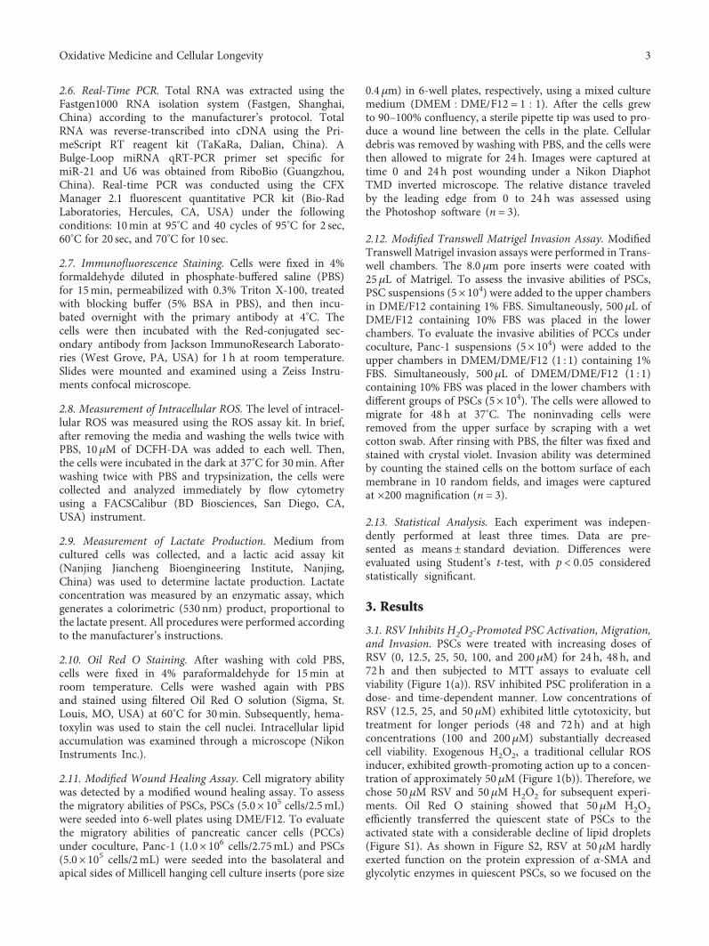
2.6. Real-Time PCR. Total RNA was extracted using theFastgen1000 RNA isolation system (Fastgen, Shanghai,China) according to the manufacturer’s protocol. TotalRNA was reverse-transcribed into cDNA using the Pri-meScript RT reagent kit (TaKaRa, Dalian, China). ABulge-Loop miRNA qRT-PCR primer set specific formiR-21 and U6 was obtained from RiboBio (Guangzhou,China). Real-time PCR was conducted using the CFXManager 2.1 fluorescent quantitative PCR kit (Bio-RadLaboratories, Hercules, CA, USA) under the followingconditions: 10min at 95°C and 40 cycles of 95°C for 2 sec,60°C for 20 sec, and 70°C for 10 sec.
2.7. Immunofluorescence Staining. Cells were fixed in 4%formaldehyde diluted in phosphate-buffered saline (PBS)for 15min, permeabilized with 0.3% Triton X-100, treatedwith blocking buffer (5% BSA in PBS), and then incu-bated overnight with the primary antibody at 4°C. Thecells were then incubated with the Red-conjugated sec-ondary antibody from Jackson ImmunoResearch Laborato-ries (West Grove, PA, USA) for 1 h at room temperature.Slides were mounted and examined using a Zeiss Instru-ments confocal microscope.
2.8. Measurement of Intracellular ROS. The level of intracel-lular ROS was measured using the ROS assay kit. In brief,after removing the media and washing the wells twice withPBS, 10μM of DCFH-DA was added to each well. Then,the cells were incubated in the dark at 37°C for 30min. Afterwashing twice with PBS and trypsinization, the cells werecollected and analyzed immediately by flow cytometryusing a FACSCalibur (BD Biosciences, San Diego, CA,USA) instrument.
2.9. Measurement of Lactate Production. Medium fromcultured cells was collected, and a lactic acid assay kit(Nanjing Jiancheng Bioengineering Institute, Nanjing,China) was used to determine lactate production. Lactateconcentration was measured by an enzymatic assay, whichgenerates a colorimetric (530 nm) product, proportional tothe lactate present. All procedures were performed accordingto the manufacturer’s instructions.
2.10. Oil Red O Staining. After washing with cold PBS,cells were fixed in 4% paraformaldehyde for 15min atroom temperature. Cells were washed again with PBSand stained using filtered Oil Red O solution (Sigma, St.Louis, MO, USA) at 60°C for 30min. Subsequently, hema-toxylin was used to stain the cell nuclei. Intracellular lipidaccumulation was examined through a microscope (NikonInstruments Inc.).
2.11. Modified Wound Healing Assay. Cell migratory abilitywas detected by a modified wound healing assay. To assessthe migratory abilities of PSCs, PSCs (5.0× 10⁵ cells/2.5mL)were seeded into 6-well plates using DME/F12. To evaluatethe migratory abilities of pancreatic cancer cells (PCCs)under coculture, Panc-1 (1.0× 10⁶ cells/2.75mL) and PSCs(5.0× 10⁵ cells/2mL) were seeded into the basolateral andapical sides of Millicell hanging cell culture inserts (pore size
0.4μm) in 6-well plates, respectively, using a mixed culturemedium (DMEM DME/F12 = 1 1). After the cells grewto 90–100% confluency, a sterile pipette tip was used to pro-duce a wound line between the cells in the plate. Cellulardebris was removed by washing with PBS, and the cells werethen allowed to migrate for 24 h. Images were captured attime 0 and 24h post wounding under a Nikon DiaphotTMD inverted microscope. The relative distance traveledby the leading edge from 0 to 24h was assessed usingthe Photoshop software (n = 3).
2.12. Modified Transwell Matrigel Invasion Assay. ModifiedTranswell Matrigel invasion assays were performed in Trans-well chambers. The 8.0μm pore inserts were coated with25μL of Matrigel. To assess the invasive abilities of PSCs,PSC suspensions (5× 10⁴) were added to the upper chambersin DME/F12 containing 1% FBS. Simultaneously, 500μL ofDME/F12 containing 10% FBS was placed in the lowerchambers. To evaluate the invasive abilities of PCCs undercoculture, Panc-1 suspensions (5× 10⁴) were added to theupper chambers in DMEM/DME/F12 (1 : 1) containing 1%FBS. Simultaneously, 500μL of DMEM/DME/F12 (1 : 1)containing 10% FBS was placed in the lower chambers withdifferent groups of PSCs (5× 10⁴). The cells were allowed tomigrate for 48 h at 37°C. The noninvading cells wereremoved from the upper surface by scraping with a wetcotton swab. After rinsing with PBS, the filter was fixed andstained with crystal violet. Invasion ability was determinedby counting the stained cells on the bottom surface of eachmembrane in 10 random fields, and images were capturedat ×200 magnification (n = 3).
2.13. Statistical Analysis. Each experiment was indepen-dently performed at least three times. Data are pre-sented as means ± standard deviation. Differences wereevaluated using Student’s t-test, with p < 0 05 consideredstatistically significant.
3. Results
3.1. RSV Inhibits H2O2-Promoted PSC Activation, Migration,and Invasion. PSCs were treated with increasing doses ofRSV (0, 12.5, 25, 50, 100, and 200μM) for 24h, 48 h, and72 h and then subjected to MTT assays to evaluate cellviability (Figure 1(a)). RSV inhibited PSC proliferation in adose- and time-dependent manner. Low concentrations ofRSV (12.5, 25, and 50μM) exhibited little cytotoxicity, buttreatment for longer periods (48 and 72 h) and at highconcentrations (100 and 200μM) substantially decreasedcell viability. Exogenous H2O2, a traditional cellular ROSinducer, exhibited growth-promoting action up to a concen-tration of approximately 50μM (Figure 1(b)). Therefore, wechose 50μM RSV and 50μM H2O2 for subsequent experi-ments. Oil Red O staining showed that 50μM H2O2efficiently transferred the quiescent state of PSCs to theactivated state with a considerable decline of lipid droplets(Figure S1). As shown in Figure S2, RSV at 50μM hardlyexerted function on the protein expression of α-SMA andglycolytic enzymes in quiescent PSCs, so we focused on the
3Oxidative Medicine and Cellular Longevity
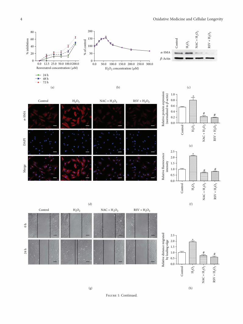
80
60
40
20% in
hibi
tion
00.0 12.5
Resveratrol concentration (�휇M)25.0 50.0 100.0200.0
24 h48 h72 h
(a)
200
150
100
50% o
f con
trol
0
H2O2 concentration (�휇M)0.0 50.0 100.0 150.0 200.0 300.0250.0
(b)
�훼-SMA
�훽-Actin
Con
trol
H2O
2
NAC
+ H
2O2
RSV
+ H
2O2
(c)
H2O2 NAC + H2O2 RSV + H2O2Control
�훼-S
MA
DA
PIM
erge
(d)
Con
trolRe
lativ
e pro
tein
expr
essio
n(n
orm
aliz
ed to
�훽-a
ctin
)
H2O
2
NAC
+ H
2O2
RSV
+ H
2O2
1.0
0.8
0.6
0.4
0.2
0.0
⁎
# #
(e)
Con
trol
Relat
ive fl
uore
scen
cein
tens
ity
H2O
2
NAC
+ H
2O2
RSV
+ H
2O2
2.5
2.0
1.5
1.0
0.5
0.0
⁎
# #
(f)
H2O2 NAC + H2O2 RSV + H2O2Control
24 h
0 h
(g)
Con
trolRe
lativ
e dist
ance
mig
rate
dby
lead
ing
edge
H2O
2
NAC
+ H
2O2
RSV
+ H
2O2
2.5
2.0
1.5
1.0
0.5
0.0
⁎
# #
(h)
Figure 1: Continued.
4 Oxidative Medicine and Cellular Longevity
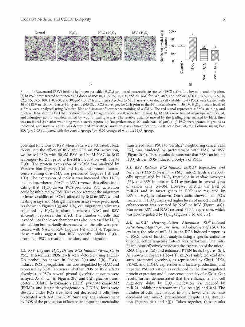
potential functions of RSV when PSCs were activated. Next,to evaluate the effects of RSV and ROS on PSC activation,we treated PSCs with 50μM RSV or 10mM NAC (a ROSscavenger) for 24 h prior to the 24h incubation with 50μMH2O2. The protein expression of α-SMA was analyzed byWestern blot (Figures 1(c) and 1(e)), and immunofluores-cence staining of α-SMA was performed (Figures 1(d) and1(f)). The expression of α-SMA was increased after H2O2incubation, whereas NAC or RSV reversed this effect, indi-cating that H2O2-driven ROS-promoted PSC activationcould be inhibited by RSV. To explore whether the migratoryor invasive ability of PSCs is affected by ROS or RSV, woundhealing assays and Matrigel invasion assays were performed.As shown in Figures 1(g) and 1(h), cell migratory ability wasenhanced by H2O2 incubation, whereas NAC and RSVefficiently repressed this effect. The number of cells thatinvaded into the lower chamber was also increased by H2O2stimulation but markedly decreased when the cells were pre-treated with NAC or RSV (Figures 1(i) and 1(j)). Together,these results suggest that RSV potently inhibits H2O2-promoted PSC activation, invasion, and migration.
3.2. RSV Impedes H2O2-Driven ROS-Induced Glycolysis inPSCs. Intracellular ROS levels were detected using DCFH-DA probes. As shown in Figures 2(a) and 2(b), H2O2-induced ROS upregulation was downregulated by NAC andrepressed by RSV. To assess whether ROS or RSV affectsglycolysis in PSCs, several pivotal glycolytic enzymes wereassayed. As shown in Figures 2(c) and 2(d), glucose trans-porter 1 (Glut1), hexokinase 2 (HK2), pyruvate kinase M2(PKM2), and lactate dehydrogenase A (LDHA) levels wereelevated under ROS treatment but decreased if cells werepretreated with NAC or RSV. Similarly, the enhancementby ROS of the production of lactate, an important metabolite
transferred from PSCs to “fertilize” neighboring cancer cells[32], was hindered by pretreatment with NAC or RSV(Figure 2(e)). These results demonstrate that RSV can inhibitH2O2-driven ROS-induced glycolysis of PSCs.
3.3. RSV Reduces ROS-Induced miR-21 Expression andIncreases PTEN Expression in PSCs.miR-21 levels are report-edly upregulated by H2O2 treatment in cardiac myocytes[33], and RSV inhibits miR-21 expression in several typesof cancer cells [34–36]. However, whether the level ofmiR-21 and its target genes in PSCs are regulated byRSV or H2O2 is unknown. Our results showed that PSCstreated with H2O2 displayed higher levels of miR-21, and thisenhancement was reversed by NAC or RSV (Figure 3(a)).Moreover, RSV and NAC restored PTEN expression, whichwas downregulated by H2O2 (Figures 3(b) and 3(c)).
3.4. miR-21 Downregulation Attenuates ROS-InducedActivation, Migration, Invasion, and Glycolysis of PSCs. Toevaluate the role of miR-21 in the ROS-induced propertiesof PSCs, loss-of-function analysis using a specific antisenseoligonucleotide targeting miR-21 was performed. The miR-21 inhibitor effectively repressed the expression of the micro-RNA (Figure 4(a)) and enhanced PTEN levels (Figure 4(b)).As shown in Figures 4(b)–4(f), miR-21 inhibited oxidativestress-promoted glycolysis, as represented by Glut1, HK2,PKM2, and LDHA expression and lactate production, andimpeded PSC activation, as evidenced by the downregulatedprotein expression and fluorescence intensity of α-SMA. Ourresults further demonstrated that the enhancement of cellmigratory ability by H2O2 incubation was reduced bymiR-21 inhibitor pretreatment (Figures 4(g) and 4(h). Thenumber of cells that invaded into the lower chamber alsodecreased with miR-21 pretreatment, despite H2O2 stimula-tion (Figures 4(i) and 4(j)). Taken together, these results
H2O2 NAC + H2O2 RSV + H2O2Control
(i)
Con
trolN
umbe
r of i
nvad
ed ce
lls(%
of c
ontro
l)
H2O
2
NAC
+ H
2O2
RSV
+ H
2O2
200
150
100
50
0
⁎
# #
(j)
Figure 1: Resveratrol (RSV) inhibits hydrogen peroxide (H2O2)-promoted pancreatic stellate cell (PSC) activation, invasion, and migration.(a, b) PSCs were treated with increasing doses of RSV (0, 12.5, 25, 50, 100, and 200 μM) for 24 h, 48 h, and 72 h or H2O2 (0, 12.5, 25, 37.5, 50,62.5, 75, 87.5, 100, 150, 200, and 300 μM) for 24 h and then subjected to MTT assays to evaluate cell viability. (c–f) PSCs were treated with50μM RSV or 10mM N-acetyl-L-cysteine (NAC), a ROS scavenger, for 24 h prior to the 24 h incubation with 50 μMH2O2. Protein levels ofα-SMA were analyzed using Western blot and immunofluorescence staining of α-SMA. The red signal represents α-SMA staining, andnuclear DNA staining by DAPI is shown in blue (magnification, ×200; scale bar: 50μm). (g, h) PSCs were treated in groups as indicated,and migratory ability was determined by wound healing assays. The relative distance moved by the leading edge marked by black lineswas measured 24 h after wounding with a sterile pipette tip (magnification, ×100; scale bar: 100μm). (i, j) PSCs were treated in groups asindicated, and invasive ability was determined by Matrigel invasion assays (magnification, ×200; scale bar: 50 μm). Column: mean; bar:SD; ∗p < 0 05 compared with the control group; #p < 0 05 compared with the H2O2 group.
5Oxidative Medicine and Cellular Longevity
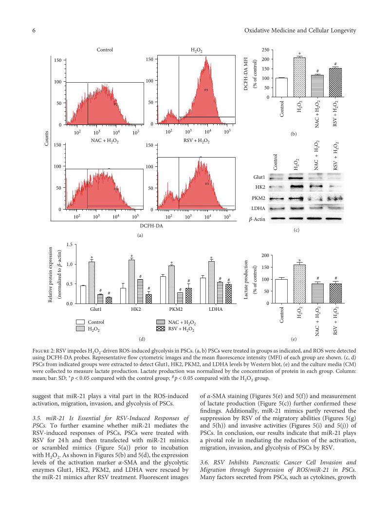
suggest that miR-21 plays a vital part in the ROS-inducedactivation, migration, invasion, and glycolysis of PSCs.
3.5. miR-21 Is Essential for RSV-Induced Responses ofPSCs. To further examine whether miR-21 mediates theRSV-induced responses of PSCs, PSCs were treated withRSV for 24 h and then transfected with miR-21 mimicsor scrambled mimics (Figure 5(a)) prior to incubationwith H2O2. As shown in Figures 5(b) and 5(d), the expressionlevels of the activation marker α-SMA and the glycolyticenzymes Glut1, HK2, PKM2, and LDHA were rescued bythe miR-21 mimics after RSV treatment. Fluorescent images
of α-SMA staining (Figures 5(e) and 5(f)) and measurementof lactate production (Figure 5(c)) further confirmed thesefindings. Additionally, miR-21 mimics partly reversed thesuppression by RSV of the migratory abilities (Figures 5(g)and 5(h)) and invasive activities (Figures 5(i) and 5(j)) ofPSCs. In conclusion, our results indicate that miR-21 playsa pivotal role in mediating the reduction of the activation,migration, invasion, and glycolysis of PSCs by RSV.
3.6. RSV Inhibits Pancreatic Cancer Cell Invasion andMigration through Suppression of ROS/miR-21 in PSCs.Many factors secreted from PSCs, such as cytokines, growth
Cou
nts
150
100
50
0
150
100
50
0
150
100
50
0
150
0
100
50
102 103 104 105 102 103 104 105
102 103 104 105102 103 104 105
DCFH-DA
Control H2O2
RSV + H2O2NAC + H2O2
(a)
Con
trol
H2O
2
RSV
+ H
2O2
NAC
+ H
2O2
DCF
H-D
A M
FI
(% o
f con
trol)
0
50
100
150
200
250 ⁎
##
(b)
Glut1
HK2
PKM2
LDHA
�훽-Actin
Con
trol
H2O
2
RSV
+ H
2O2
NAC
+ H
2O2
(c)
Relat
ive p
rote
in ex
pres
sion
(nor
mal
ized
to �훽
-act
in)
0.0
0.5
1.0
1.5
ControlH2O2 RSV + H2O2
NAC + H2O2
# #
#
#
## #
#
Glut1 HK2 PKM2 LDHA
⁎⁎
⁎⁎
(d)
Lact
ate p
rodu
ctio
n
(% o
f con
trol)
0
50
100
150
200
Con
trol
H2O
2
RSV
+ H
2O2
NAC
+ H
2O2
# #
⁎
(e)
Figure 2: RSV impedes H2O2-driven ROS-induced glycolysis in PSCs. (a, b) PSCs were treated in groups as indicated, and ROS were detectedusing DCFH-DA probes. Representative flow cytometric images and the mean fluorescence intensity (MFI) of each group are shown. (c, d)PSCs from indicated groups were extracted to detect Glut1, HK2, PKM2, and LDHA levels by Western blot, (e) and the culture media (CM)were collected to measure lactate production. Lactate production was normalized by the concentration of protein in each group. Column:mean; bar: SD; ∗p < 0 05 compared with the control group; #p < 0 05 compared with the H2O2 group.
6 Oxidative Medicine and Cellular Longevity
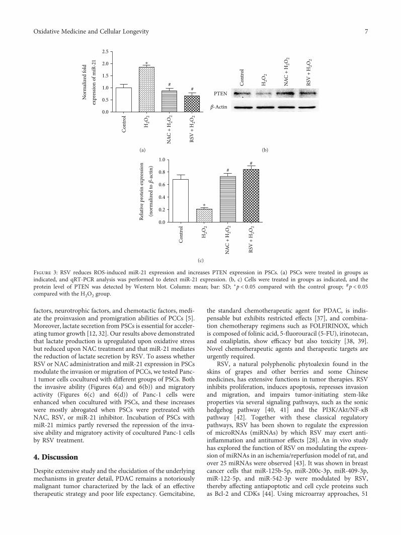
factors, neurotrophic factors, and chemotactic factors, medi-ate the proinvasion and promigration abilities of PCCs [5].Moreover, lactate secretion from PSCs is essential for acceler-ating tumor growth [12, 32]. Our results above demonstratedthat lactate production is upregulated upon oxidative stressbut reduced upon NAC treatment and that miR-21 mediatesthe reduction of lactate secretion by RSV. To assess whetherRSV or NAC administration and miR-21 expression in PSCsmodulate the invasion or migration of PCCs, we tested Panc-1 tumor cells cocultured with different groups of PSCs. Boththe invasive ability (Figures 6(a) and 6(b)) and migratoryactivity (Figures 6(c) and 6(d)) of Panc-1 cells wereenhanced when cocultured with PSCs, and these increaseswere mostly abrogated when PSCs were pretreated withNAC, RSV, or miR-21 inhibitor. Incubation of PSCs withmiR-21 mimics partly reversed the repression of the inva-sive ability and migratory activity of cocultured Panc-1 cellsby RSV treatment.
4. Discussion
Despite extensive study and the elucidation of the underlyingmechanisms in greater detail, PDAC remains a notoriouslymalignant tumor characterized by the lack of an effectivetherapeutic strategy and poor life expectancy. Gemcitabine,
the standard chemotherapeutic agent for PDAC, is indis-pensable but exhibits restricted effects [37], and combina-tion chemotherapy regimens such as FOLFIRINOX, whichis composed of folinic acid, 5-fluorouracil (5-FU), irinotecan,and oxaliplatin, show efficacy but also toxicity [38, 39].Novel chemotherapeutic agents and therapeutic targets areurgently required.
RSV, a natural polyphenolic phytoalexin found in theskins of grapes and other berries and some Chinesemedicines, has extensive functions in tumor therapies. RSVinhibits proliferation, induces apoptosis, represses invasionand migration, and impairs tumor-initiating stem-likeproperties via several signaling pathways, such as the sonichedgehog pathway [40, 41] and the PI3K/Akt/NF-κBpathway [42]. Together with these classical regulatorypathways, RSV has been shown to regulate the expressionof microRNAs (miRNAs) by which RSV may exert anti-inflammation and antitumor effects [28]. An in vivo studyhas explored the function of RSV on modulating the expres-sion of miRNAs in an ischemia/reperfusion model of rat, andover 25 miRNAs were observed [43]. It was shown in breastcancer cells that miR-125b-5p, miR-200c-3p, miR-409-3p,miR-122-5p, and miR-542-3p were modulated by RSV,thereby affecting antiapoptotic and cell cycle proteins suchas Bcl-2 and CDKs [44]. Using microarray approaches, 51
Con
trol
H2O
2
RSV
+H
2O2
NAC
+H
2O2
Nor
mal
ized
fold
expr
essio
n of
miR
-21
0.5
1.0
1.5
2.0
2.5
0.0
##
⁎
(a)
PTEN
�훽-Actin
Con
trol
H2O
2
RSV
+H
2O2
NAC
+H
2O2
(b)
Con
trol
H2O
2
RSV
+H
2O2
NAC
+H
2O2
Relat
ive p
rote
in ex
pres
sion
(nor
mal
ized
to �훽
-act
in)
0.0
0.2
0.4
0.6
0.8
1.0
##
⁎
(c)
Figure 3: RSV reduces ROS-induced miR-21 expression and increases PTEN expression in PSCs. (a) PSCs were treated in groups asindicated, and qRT-PCR analysis was performed to detect miR-21 expression. (b, c) Cells were treated in groups as indicated, and theprotein level of PTEN was detected by Western blot. Column: mean; bar: SD; ∗p < 0 05 compared with the control group; #p < 0 05compared with the H2O2 group.
7Oxidative Medicine and Cellular Longevity
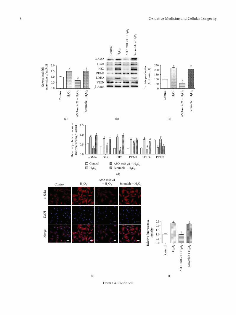
Nor
mal
ized
fold
expr
essio
n of
miR
-21
0.0
0.5
1.0
1.5
2.0
#
Con
trol
H2O
2
ASO
-miR
-21
+ H
2O2
Scra
mbl
e + H
2O2
⁎ ⁎
(a)
�훼-SMAGlut1HK2
PKM2LDHAPTEN
�훽-Actin
Con
trol
H2O
2
ASO
-miR
-21
+ H
2O2
Scra
mbl
e + H
2O2
(b)
050
100150200250
#
Lact
ate p
rodu
ctio
n(%
of c
ontro
l)
Con
trol
H2O
2
ASO
-miR
-21
+ H
2O2
Scra
mbl
e + H
2O2
⁎ ⁎
(c)
Relat
ive p
rote
in ex
pres
sion
(nor
mal
ized
to �훽
-act
in)
Glut1�훼-SMA HK2 PKM2 LDHA PTEN
ControlH2O2
ASO-miR-21 + H2O2Scramble + H2O2
0.0
0.5
1.0
1.5
##
#
##
#⁎
⁎⁎ ⁎
⁎ ⁎
⁎ ⁎ ⁎ ⁎
⁎ ⁎
(d)
Control H2O2
ASO-miR-21+ H2O2 Scramble + H2O2
Mer
geD
API
�훼-S
MA
(e)
Relat
ive fl
uore
scen
cein
tens
ity
0.00.51.01.52.02.5
#
Con
trol
H2O
2
ASO
-miR
-21
+ H
2O2
Scra
mbl
e + H
2O2
⁎⁎
(f)
Figure 4: Continued.
8 Oxidative Medicine and Cellular Longevity
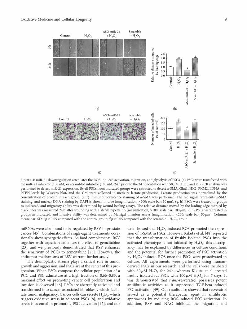
miRNAs were also found to be regulated by RSV in prostatecancer [45]. Combinations of single-agent treatments occa-sionally show synergetic effects. As food complements, RSVtogether with capsaicin enhances the effect of gemcitabine[23], and we previously demonstrated that RSV enhancesthe sensitivity of PCCs to gemcitabine [25]. However, theantitumor mechanisms of RSV warrant further study.
The desmoplastic stroma plays a critical role in tumorgrowth and aggression, and PSCs are at the center of this pro-gression. When PSCs compose the cellular population of aPCC and PSC admixture at a high fraction of 0.66–0.83, amaximal effect on promoting cancer cell proliferation andinvasion is observed [46]. PSCs are aberrantly activated andtransformed into cancer-associated fibroblasts, which facili-tate tumor malignancy. Cancer cells can secrete H2O2, whichtriggers oxidative stress in adjacent PSCs [8], and oxidativestress is essential in promoting PSC activation [47], and our
data showed that H2O2-induced ROS promoted the expres-sion of α-SMA in PSCs. However, Kikuta et al. [48] reportedthat the transformation of freshly isolated PSCs into theactivated phenotype is not initiated by H2O2; this discrep-ancy may be explained by differences in culture conditionsand the potential for further promotion of PSC activationby H2O2-induced ROS once the PSCs were preactivated inculture. All experiments were performed using human-derived PSCs in our research, and the cells were incubatedwith 50μM H2O2 for 24 h, whereas Kikuta et al. treatedfreshly isolated rat PSCs with 100μM H2O2 for 7 days. Itwas demonstrated that trans-resveratrol possesses potentantifibrotic activities as it suppressed TGF-beta-inducedPSC activation [49]. Our results also showed that resveratrolserved as a potential therapeutic agent in antifibroticapproaches by reducing ROS-induced PSC activation. Inaddition, RSV and NAC inhibited the migration and
Control H2O2
ASO-miR-21+ H2O2
Scramble+ H2O2
24 h
0 h
(g)
Relat
ive d
istan
ce m
igra
ted
by le
adin
g ed
ge
0.00.51.01.52.02.5
#
Con
trol
H2O
2
ASO
-miR
-21
+ H
2O2
Scra
mbl
e + H
2O2
⁎ ⁎
(h)
Control H2O2
ASO-miR-21+ H2O2
Scramble+ H2O2
(i)
Num
ber o
f inv
aded
cells
(% o
f con
trol)
050
100150200
#
⁎ ⁎
Con
trol
H2O
2
ASO
-miR
-21
+ H
2O2
Scra
mbl
e + H
2O2
(j)
Figure 4: miR-21 downregulation attenuates the ROS-induced activation, migration, and glycolysis of PSCs. (a) PSCs were transfected withthe miR-21 inhibitor (100 nM) or scrambled inhibitor (100 nM) 24 h prior to the 24 h incubation with 50 μMH2O2, and RT-PCR analysis wasperformed to detect miR-21 expression. (b–d) PSCs from indicated groups were extracted to detect α-SMA, Glut1, HK2, PKM2, LDHA, andPTEN levels by Western blot, and the CM were collected to measure lactate production. Lactate production was normalized by theconcentration of protein in each group. (e, f) Immunofluorescence staining of α-SMA was performed. The red signal represents α-SMAstaining, and nuclear DNA staining by DAPI is shown in blue (magnification, ×200; scale bar: 50μm). (g, h) PSCs were treated in groupsas indicated, and migratory ability was determined by wound healing assays. The relative distance moved by the leading edge marked byblack lines was measured 24 h after wounding with a sterile pipette tip (magnification, ×100; scale bar: 100μm). (i, j) PSCs were treated ingroups as indicated, and invasive ability was determined by Matrigel invasion assays (magnification, ×200; scale bar: 50μm). Column:mean; bar: SD; ∗p < 0 05 compared with the control group; #p < 0 05 compared with the scramble +H2O2 group.
9Oxidative Medicine and Cellular Longevity
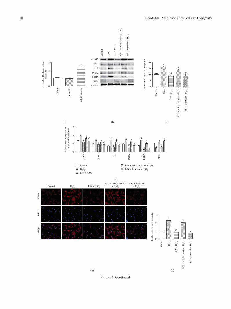
3
2
1
0Nor
mal
ized
fold
expr
essio
nof
miR
-21
Con
trol
Scra
mbl
e
miR
-21
mim
ics
⁎
(a)
�훼-SMA
Glut
Con
trol
H2O
2
RSV
+ H
2O2
RSV
+ m
iR-2
1 m
imic
s + H
2O2
RSV
+ S
cram
ble +
H2O
2
HK2
PKM2
LDHA
PTEN�훽-Actin
(b)
200
150
100
50
0
Lact
ate p
rodu
ctio
n (%
of c
ontro
l)
Con
trol
H2O
2
RSV
+ H
2O2
RSV
+ m
iR-2
1 m
imic
s + H
2O2
RSV
+ S
cram
ble +
H2O
2
⁎
# #
$
(c)
1.5
1.0
0.5
0.0Relat
ive p
rote
in ex
pres
sion
(nor
mal
ized
to �훽
-act
in)
�훼-S
MA
Glu
t1
HK2
PKM
2
LDH
A
PTEN
⁎
⁎⁎ ⁎ ⁎
⁎
##
##
####
##
##
$
$ $ $ $
$
ControlH2O2
RSV + H2O2
RSV + miR-21 mimics + H2O2
RSV + Scramble + H2O2
(d)
Control H2O2 RSV + H2O2
RSV + miR-21 mimics+ H2O2
RSV + Scramble+ H2O2
�훼-S
MA
DA
PIM
erge
(e)
3
2
1
0Relat
ive fl
uore
scen
ce in
tens
ity
Con
trol
H2O
2
RSV
+ H
2O2
RSV
+ m
iR-2
1 m
imic
s + H
2O2
RSV
+ S
cram
ble +
H2O
2
⁎
##
$
(f)
Figure 5: Continued.
10 Oxidative Medicine and Cellular Longevity
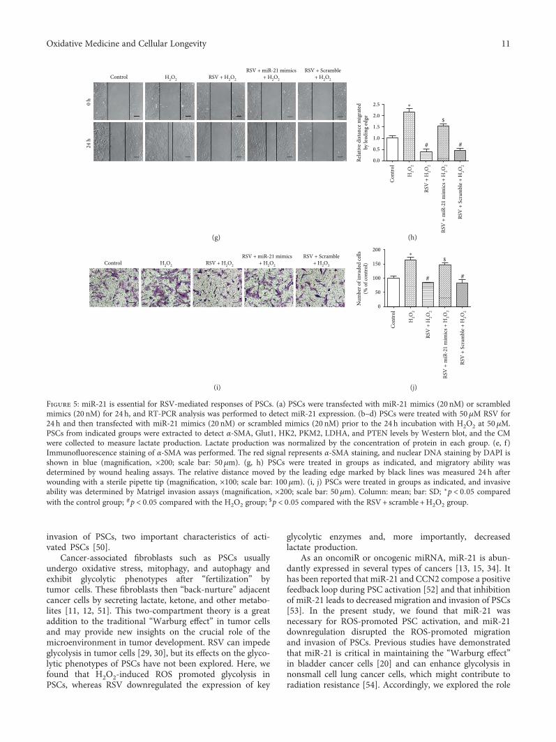
invasion of PSCs, two important characteristics of acti-vated PSCs [50].
Cancer-associated fibroblasts such as PSCs usuallyundergo oxidative stress, mitophagy, and autophagy andexhibit glycolytic phenotypes after “fertilization” bytumor cells. These fibroblasts then “back-nurture” adjacentcancer cells by secreting lactate, ketone, and other metabo-lites [11, 12, 51]. This two-compartment theory is a greataddition to the traditional “Warburg effect” in tumor cellsand may provide new insights on the crucial role of themicroenvironment in tumor development. RSV can impedeglycolysis in tumor cells [29, 30], but its effects on the glyco-lytic phenotypes of PSCs have not been explored. Here, wefound that H2O2-induced ROS promoted glycolysis inPSCs, whereas RSV downregulated the expression of key
glycolytic enzymes and, more importantly, decreasedlactate production.
As an oncomiR or oncogenic miRNA, miR-21 is abun-dantly expressed in several types of cancers [13, 15, 34]. Ithas been reported that miR-21 and CCN2 compose a positivefeedback loop during PSC activation [52] and that inhibitionof miR-21 leads to decreased migration and invasion of PSCs[53]. In the present study, we found that miR-21 wasnecessary for ROS-promoted PSC activation, and miR-21downregulation disrupted the ROS-promoted migrationand invasion of PSCs. Previous studies have demonstratedthat miR-21 is critical in maintaining the “Warburg effect”in bladder cancer cells [20] and can enhance glycolysis innonsmall cell lung cancer cells, which might contribute toradiation resistance [54]. Accordingly, we explored the role
Control H2O2 RSV + H2O2
RSV + miR-21 mimics+ H2O2
RSV + Scramble+ H2O2
0 h
24 h
(g)
1.5
2.0
2.5
0.0
0.5
1.0
Relat
ive d
istan
ce m
igra
ted
by le
adin
g ed
ge
Con
trol
H2O
2
RSV
+ H
2O2
RSV
+ m
iR-2
1 m
imic
s + H
2O2
RSV
+ S
cram
ble +
H2O
2
⁎
# #
$
(h)
Control H2O2 RSV + H2O2
RSV + miR-21 mimics+ H2O2
RSV + Scramble+ H2O2
(i)
100
150
200
0
50
Num
ber o
f inv
aded
cells
(% o
f con
trol)
Con
trol
H2O
2
RSV
+ H
2O2
RSV
+ m
iR-2
1 m
imic
s + H
2O2
RSV
+ S
cram
ble +
H2O
2
⁎
# #
$
(j)
Figure 5: miR-21 is essential for RSV-mediated responses of PSCs. (a) PSCs were transfected with miR-21 mimics (20 nM) or scrambledmimics (20 nM) for 24 h, and RT-PCR analysis was performed to detect miR-21 expression. (b–d) PSCs were treated with 50μM RSV for24 h and then transfected with miR-21 mimics (20 nM) or scrambled mimics (20 nM) prior to the 24 h incubation with H2O2 at 50μM.PSCs from indicated groups were extracted to detect α-SMA, Glut1, HK2, PKM2, LDHA, and PTEN levels by Western blot, and the CMwere collected to measure lactate production. Lactate production was normalized by the concentration of protein in each group. (e, f)Immunofluorescence staining of α-SMA was performed. The red signal represents α-SMA staining, and nuclear DNA staining by DAPI isshown in blue (magnification, ×200; scale bar: 50μm). (g, h) PSCs were treated in groups as indicated, and migratory ability wasdetermined by wound healing assays. The relative distance moved by the leading edge marked by black lines was measured 24 h afterwounding with a sterile pipette tip (magnification, ×100; scale bar: 100μm). (i, j) PSCs were treated in groups as indicated, and invasiveability was determined by Matrigel invasion assays (magnification, ×200; scale bar: 50 μm). Column: mean; bar: SD; ∗p < 0 05 comparedwith the control group; #p < 0 05 compared with the H2O2 group;
$p < 0 05 compared with the RSV+ scramble +H2O2 group.
11Oxidative Medicine and Cellular Longevity
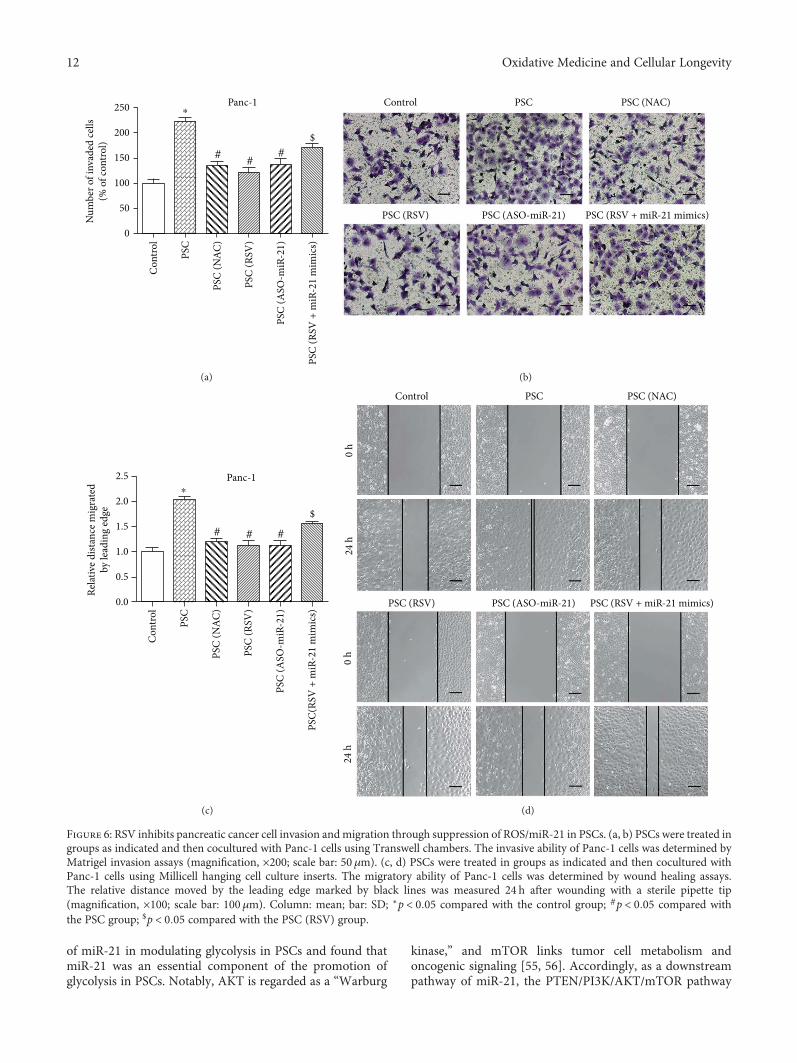
of miR-21 in modulating glycolysis in PSCs and found thatmiR-21 was an essential component of the promotion ofglycolysis in PSCs. Notably, AKT is regarded as a “Warburg
kinase,” and mTOR links tumor cell metabolism andoncogenic signaling [55, 56]. Accordingly, as a downstreampathway of miR-21, the PTEN/PI3K/AKT/mTOR pathway
250 Panc-1
200
150
100
50
0
Con
trol
PSC
PSC
(NAC
)
PSC
(RSV
)
PSC
(ASO
-miR
-21)
PSC
(RSV
+ m
iR-2
1 m
imic
s)
Num
ber o
f inv
aded
cells
(% o
f con
trol)
⁎
##
#
$
(a)
Control PSC PSC (NAC)
PSC (RSV) PSC (ASO-miR-21) PSC (RSV + miR-21 mimics)
(b)
2.5 Panc-1
2.0
1.5
1.0
0.5
0.0
Con
trol
PSC
PSC
(NAC
)
PSC
(RSV
)
PSC
(ASO
-miR
-21)
PSC(
RSV
+ m
iR-2
1 m
imic
s)
Relat
ive d
istan
ce m
igra
ted
by le
adin
g ed
ge
⁎
# # #
$
(c)
Control
0 h
24 h
0 h
24 h
PSC PSC (NAC)
PSC (RSV) PSC (ASO-miR-21) PSC (RSV + miR-21 mimics)
(d)
Figure 6: RSV inhibits pancreatic cancer cell invasion andmigration through suppression of ROS/miR-21 in PSCs. (a, b) PSCs were treated ingroups as indicated and then cocultured with Panc-1 cells using Transwell chambers. The invasive ability of Panc-1 cells was determined byMatrigel invasion assays (magnification, ×200; scale bar: 50 μm). (c, d) PSCs were treated in groups as indicated and then cocultured withPanc-1 cells using Millicell hanging cell culture inserts. The migratory ability of Panc-1 cells was determined by wound healing assays.The relative distance moved by the leading edge marked by black lines was measured 24 h after wounding with a sterile pipette tip(magnification, ×100; scale bar: 100μm). Column: mean; bar: SD; ∗p < 0 05 compared with the control group; #p < 0 05 compared withthe PSC group; $p < 0 05 compared with the PSC (RSV) group.
12 Oxidative Medicine and Cellular Longevity
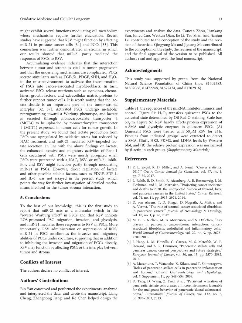
might exhibit several functions modulating cell metabolismwhose mechanisms require further elucidation. Recentstudies have suggested that RSV might function by affectingmiR-21 in prostate cancer cells [34] and PCCs [35]. Thisconnection was further demonstrated in stroma, in whichour results showed that miR-21 partly mediated theresponses of PSCs to RSV.
Accumulating evidence indicates that the interactionbetween tumor and stroma is vital in tumor progressionand that the underlying mechanisms are complicated. PCCssecrete stimulants such as TGF-β1, PDGF, SHH, and H2O2to the microenvironment to activate the transformationof PSCs into cancer-associated myofibroblasts. In turn,activated PSCs release nutrients such as cytokines, chemo-kines, growth factors, and extracellular matrix proteins tofurther support tumor cells. It is worth noting that the lac-tate shuttle is an important part of the tumor-stromainterplay [32, 57]. Activated PSCs undergo metabolicreprogramming toward a Warburg phenotype, and lactateis secreted through monocarboxylate transporter 4(MCT4) to be uploaded by monocarboxylate transporter1 (MCT1) expressed in tumor cells for tumor growth. Inthe present study, we found that lactate production fromPSCs was upregulated by oxidative stress but reduced byNAC treatment, and miR-21 mediated RSV-impeded lac-tate secretion. In line with the above findings on lactate,the enhanced invasive and migratory activities of Panc-1cells cocultured with PSCs were mostly abrogated whenPSCs were pretreated with a NAC, RSV, or miR-21 inhib-itor, and RSV might function partly through modulatingmiR-21 in PSCs. However, direct regulation of lactateand other possible soluble factors, such as PDGF, SDF-1,and IL-6, was not assayed in the present study, whichpoints the way for further investigation of detailed mecha-nisms involved in the tumor-stroma interaction.
5. Conclusions
To the best of our knowledge, this is the first study toreport that miR-21 acts as a molecular switch in the“reverse Warburg effect” in PSCs and that RSV inhibitsROS-promoted PSC migration, invasion, and glycolysis,and miR-21 mediates these responses to RSV in PSCs. Moreimportantly, RSV administration or suppression of ROS/miR-21 in PSCs ameliorates the invasive and migratoryabilities of PCCs under coculture, suggesting that in additionto inhibiting the invasion and migration of PCCs directly,RSV may function by affecting PSCs or the interplay betweentumor and stroma.
Conflicts of Interest
The authors declare no conflict of interest.
Authors’ Contributions
Bin Yan conceived and performed the experiments, analyzedand interpreted the data, and wrote the manuscript. LiangCheng, Zhengdong Jiang, and Ke Chen helped design the
experiments and analyze the data. Cancan Zhou, LiankangSun, Junyu Cao, Weikun Qian, Jie Li, Tao Shan, and JianjunLei contributed to the conception of the study and the revi-sion of the article. Qingyong Ma and Jiguang Ma contributedto the conception of the study, the revision of the manuscript,and the final approval of the version to be published. Allauthors read and approved the final manuscript.
Acknowledgments
This study was supported by grants from the NationalNatural Science Foundation of China (nos. 81402583,81502066, 81472248, 81672434, and 81702916).
Supplementary Materials
Table S1: the sequences of the miRNA inhibitor, mimics, andcontrol. Figure S1: H2O2 transfers quiescent PSCs to theactivated state determined by Oil Red O staining. Scale bar:50μm. Figure S2: RSV hardly affects protein expression ofα-SMA and glycolytic enzymes in quiescent PSCs. (A)Quiescent PSCs were treated with 50μM RSV for 24 h.Proteins from indicated groups were extracted to detectα-SMA, Glut1, HK2, PKM2, and LDHA levels by Westernblot, and (B) the relative protein expression was normalizedby β-actin in each group. (Supplementary Materials)
References
[1] R. L. Siegel, K. D. Miller, and A. Jemal, “Cancer statistics,2017,” CA: A Cancer Journal for Clinicians, vol. 67, no. 1,pp. 7–30, 2017.
[2] L. Rahib, B. D. Smith, R. Aizenberg, A. B. Rosenzweig, J. M.Fleshman, and L. M. Matrisian, “Projecting cancer incidenceand deaths to 2030: the unexpected burden of thyroid, liver,and pancreas cancers in the United States,” Cancer Research,vol. 74, no. 11, pp. 2913–2921, 2014.
[3] D. von Ahrens, T. D. Bhagat, D. Nagrath, A. Maitra, andA. Verma, “The role of stromal cancer-associated fibroblastsin pancreatic cancer,” Journal of Hematology & Oncology,vol. 10, no. 1, p. 76, 2017.
[4] M. F. B. Nielsen, M. B. Mortensen, and S. Detlefsen, “Keyplayers in pancreatic cancer-stroma interaction: cancer-associated fibroblasts, endothelial and inflammatory cells,”World Journal of Gastroenterology, vol. 22, no. 9, pp. 2678–2700, 2016.
[5] J. Haqq, L. M. Howells, G. Garcea, M. S. Metcalfe, W. P.Steward, and A. R. Dennison, “Pancreatic stellate cells andpancreas cancer: current perspectives and future strategies,”European Journal of Cancer, vol. 50, no. 15, pp. 2570–2582,2014.
[6] A. Masamune, T. Watanabe, K. Kikuta, and T. Shimosegawa,“Roles of pancreatic stellate cells in pancreatic inflammationand fibrosis,” Clinical Gastroenterology and Hepatology,vol. 7, Supplement 11, pp. S48–S54, 2009.
[7] D. Tang, D. Wang, Z. Yuan et al., “Persistent activation ofpancreatic stellate cells creates a microenvironment favorablefor the malignant behavior of pancreatic ductal adenocarci-noma,” International Journal of Cancer, vol. 132, no. 5,pp. 993–1003, 2013.
13Oxidative Medicine and Cellular Longevity
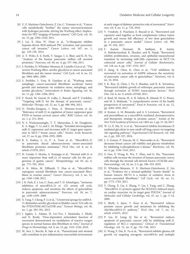
[8] U. E. Martinez-Outschoorn, Z. Lin, C. Trimmer et al., “Cancercells metabolically “fertilize” the tumor microenvironmentwith hydrogen peroxide, driving the Warburg effect: Implica-tions for PET imaging of human tumors,” Cell Cycle, vol. 10,no. 15, pp. 2504–2520, 2011.
[9] J. Lei, X. Huo, W. Duan et al., “α-Mangostin inhibitshypoxia-driven ROS-induced PSC activation and pancreaticcancer cell invasion,” Cancer Letters, vol. 347, no. 1,pp. 129–138, 2014.
[10] A. Y. Wehr, E. E. Furth, V. Sangar, I. A. Blair, and K. H. Yu,“Analysis of the human pancreatic stellate cell secretedproteome,” Pancreas, vol. 40, no. 4, pp. 557–566, 2011.
[11] S. Pavlides, D. Whitaker-Menezes, R. Castello-Cros et al., “Thereverse Warburg effect: aerobic glycolysis in cancer associatedfibroblasts and the tumor stroma,” Cell Cycle, vol. 8, no. 23,pp. 3984–4001, 2009.
[12] S. Pavlides, I. Vera, R. Gandara et al., “Warburg meetsautophagy: cancer-associated fibroblasts accelerate tumorgrowth and metastasis via oxidative stress, mitophagy, andaerobic glycolysis,” Antioxidants & Redox Signaling, vol. 16,no. 11, pp. 1264–1284, 2012.
[13] F. Sicard, M. Gayral, H. Lulka, L. Buscail, and P. Cordelier,“Targeting miR-21 for the therapy of pancreatic cancer,”Molecular Therapy, vol. 21, no. 5, pp. 986–994, 2013.
[14] O. Peralta-Zaragoza, J. Deas, A. Meneses-Acosta et al.,“Relevance of miR-21 in regulation of tumor suppressor genePTEN in human cervical cancer cells,” BMC Cancer, vol. 16,no. 1, p. 215, 2016.
[15] N. S. Wickramasinghe, T. T. Manavalan, S. M. Dougherty,K. A. Riggs, Y. Li, and C. M. Klinge, “Estradiol downregulatesmiR-21 expression and increases miR-21 target gene expres-sion in MCF-7 breast cancer cells,” Nucleic Acids Research,vol. 37, no. 8, pp. 2584–2595, 2009.
[16] B. E. Kadera, L. Li, P. A. Toste et al., “MicroRNA-21in pancreatic ductal adenocarcinoma tumor-associatedfibroblasts promotes metastasis,” PLoS One, vol. 8, no. 8,article e71978, 2013.
[17] H. Uozaki, S. Morita, A. Kumagai et al., “Stromal miR-21 ismore important than miR-21 of tumour cells for the pro-gression of gastric cancer,” Histopathology, vol. 65, no. 6,pp. 775–783, 2014.
[18] A. K. Mitra, M. Zillhardt, Y. Hua et al., “MicroRNAsreprogram normal fibroblasts into cancer-associated fibro-blasts in ovarian cancer,” Cancer Discovery, vol. 2, no. 12,pp. 1100–1108, 2012.
[19] J. K. Park, E. J. Lee, C. Esau, and T. D. Schmittgen, “Antisenseinhibition of microRNA-21 or -221 arrests cell cycle,induces apoptosis, and sensitizes the effects of gemcitabinein pancreatic adenocarcinoma,” Pancreas, vol. 38, no. 7,pp. e190–e199, 2009.
[20] X. Yang, Y. Cheng, P. Li et al., “A lentiviral sponge for miRNA-21 diminishes aerobic glycolysis in bladder cancer T24 cells viathe PTEN/PI3K/AKT/mTOR axis,” Tumour Biology, vol. 36,no. 1, pp. 383–391, 2015.
[21] J. Jagdeo, L. Adams, H. Lev-Tov, J. Sieminska, J. Michl,and N. Brody, “Dose-dependent antioxidant function ofresveratrol demonstrated via modulation of reactive oxygenspecies in normal human skin fibroblasts in vitro,” Journal ofDrugs in Dermatology, vol. 9, no. 12, pp. 1523–1526, 2010.
[22] M. Savi, L. Bocchi, R. Sala et al., “Parenchymal and stromalcells contribute to pro-inflammatory myocardial environment
at early stages of diabetes: protective role of resveratrol,”Nutri-ents, vol. 8, no. 11, p. 729, 2016.
[23] V. Vendrely, E. Peuchant, E. Buscail et al., “Resveratrol andcapsaicin used together as food complements reduce tumorgrowth and rescue full efficiency of low dose gemcitabinein a pancreatic cancer model,” Cancer Letters, vol. 390,pp. 91–102, 2017.
[24] F. Karimi Dermani, M. Saidijam, R. Amini,A. Mahdavinezhad, K. Heydari, and R. Najafi, “Resveratrolinhibits proliferation, invasion, and epithelial–mesenchymaltransition by increasing miR-200c expression in HCT-116colorectal cancer cells,” Journal of Cellular Biochemistry,vol. 118, no. 6, pp. 1547–1555, 2017.
[25] Z. Jiang, X. Chen, K. Chen et al., “YAP inhibition byresveratrol via activation of AMPK enhances the sensitivityof pancreatic cancer cells to gemcitabine,” Nutrients, vol. 8,no. 10, 2016.
[26] S. K. Roy, Q. Chen, J. Fu, S. Shankar, and R. K. Srivastava,“Resveratrol inhibits growth of orthotopic pancreatic tumorsthrough activation of FOXO transcription factors,” PLoSOne, vol. 6, no. 9, article e25166, 2011.
[27] A. Rauf, M. Imran, H. A. R. Suleria, B. Ahmad, D. G. Peters,and M. S. Mubarak, “A comprehensive review of the healthperspectives of resveratrol,” Food & Function, vol. 8, no. 12,pp. 4284–4305, 2017.
[28] A. Kumar, A. M. Rimando, and A. S. Levenson, “Resveratroland pterostilbene as a microRNA-mediated chemopreventiveand therapeutic strategy in prostate cancer,” Annals of theNewYorkAcademy of Sciences, vol. 1403, no. 1, pp. 15–26, 2017.
[29] W. Li, X. Ma, N. Li et al., “Resveratrol inhibits hexokinases IImediated glycolysis in non-small cell lung cancer via targetingAkt signaling pathway,” Experimental Cell Research, vol. 349,no. 2, pp. 320–327, 2016.
[30] L. S. Gomez, P. Zancan, M. C. Marcondes et al., “Resveratroldecreases breast cancer cell viability and glucose metabolismby inhibiting 6-phosphofructo-1-kinase,” Biochimie, vol. 95,no. 6, pp. 1336–1343, 2013.
[31] Z. Gao, X. Wang, K. Wu, Y. Zhao, and G. Hu, “Pancreaticstellate cells increase the invasion of human pancreatic cancercells through the stromal cell-derived Factor-1/CXCR4 axis,”Pancreatology, vol. 10, no. 2-3, pp. 186–193, 2010.
[32] D. Whitaker-Menezes, U. E. Martinez-Outschoorn, Z. Linet al., “Evidence for a stromal-epithelial “lactate shuttle” inhuman tumors: MCT4 is a marker of oxidative stress incancer-associated fibroblasts,” Cell Cycle, vol. 10, no. 11,pp. 1772–1783, 2011.
[33] Y. Cheng, X. Liu, S. Zhang, Y. Lin, J. Yang, and C. Zhang,“MicroRNA-21 protects against the H(2)O(2)-induced injuryon cardiac myocytes via its target gene PDCD4,” Journal ofMolecular and Cellular Cardiology, vol. 47, no. 1, pp. 5–14,2009.
[34] S. Sheth, S. Jajoo, T. Kaur et al., “Resveratrol reducesprostate cancer growth and metastasis by inhibiting theAkt/MicroRNA-21 pathway,” PLoS One, vol. 7, no. 12,article e51655, 2012.
[35] P. Liu, H. Liang, Q. Xia et al., “Resveratrol inducesapoptosis of pancreatic cancers cells by inhibiting miR-21regulation of BCL-2 expression,” Clinical & TranslationalOncology, vol. 15, no. 9, pp. 741–746, 2013.
[36] G. Wang, F. Dai, K. Yu et al., “Resveratrol inhibits glioma cellgrowth via targeting oncogenic microRNAs and multiple
14 Oxidative Medicine and Cellular Longevity
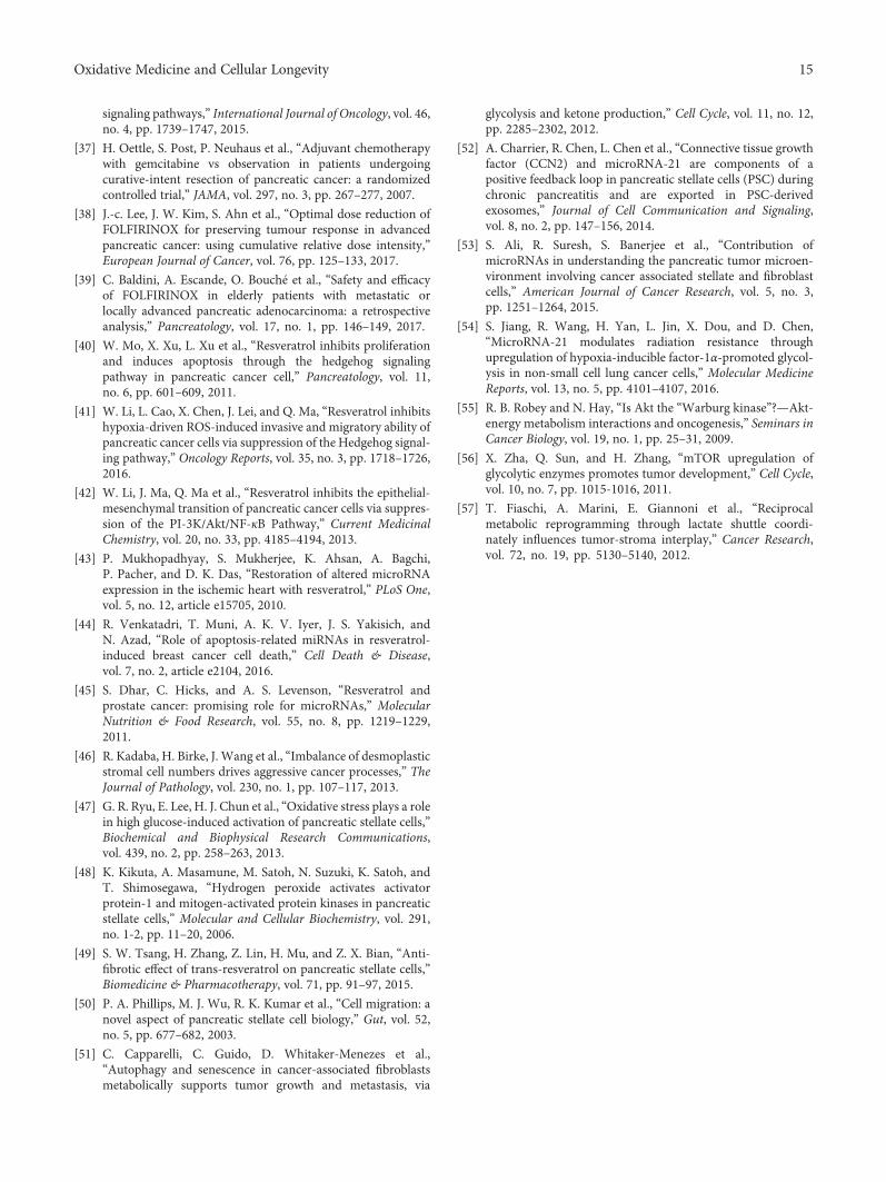
signaling pathways,” International Journal of Oncology, vol. 46,no. 4, pp. 1739–1747, 2015.
[37] H. Oettle, S. Post, P. Neuhaus et al., “Adjuvant chemotherapywith gemcitabine vs observation in patients undergoingcurative-intent resection of pancreatic cancer: a randomizedcontrolled trial,” JAMA, vol. 297, no. 3, pp. 267–277, 2007.
[38] J.-c. Lee, J. W. Kim, S. Ahn et al., “Optimal dose reduction ofFOLFIRINOX for preserving tumour response in advancedpancreatic cancer: using cumulative relative dose intensity,”European Journal of Cancer, vol. 76, pp. 125–133, 2017.
[39] C. Baldini, A. Escande, O. Bouché et al., “Safety and efficacyof FOLFIRINOX in elderly patients with metastatic orlocally advanced pancreatic adenocarcinoma: a retrospectiveanalysis,” Pancreatology, vol. 17, no. 1, pp. 146–149, 2017.
[40] W. Mo, X. Xu, L. Xu et al., “Resveratrol inhibits proliferationand induces apoptosis through the hedgehog signalingpathway in pancreatic cancer cell,” Pancreatology, vol. 11,no. 6, pp. 601–609, 2011.
[41] W. Li, L. Cao, X. Chen, J. Lei, and Q. Ma, “Resveratrol inhibitshypoxia-driven ROS-induced invasive and migratory ability ofpancreatic cancer cells via suppression of the Hedgehog signal-ing pathway,” Oncology Reports, vol. 35, no. 3, pp. 1718–1726,2016.
[42] W. Li, J. Ma, Q. Ma et al., “Resveratrol inhibits the epithelial-mesenchymal transition of pancreatic cancer cells via suppres-sion of the PI-3K/Akt/NF-κB Pathway,” Current MedicinalChemistry, vol. 20, no. 33, pp. 4185–4194, 2013.
[43] P. Mukhopadhyay, S. Mukherjee, K. Ahsan, A. Bagchi,P. Pacher, and D. K. Das, “Restoration of altered microRNAexpression in the ischemic heart with resveratrol,” PLoS One,vol. 5, no. 12, article e15705, 2010.
[44] R. Venkatadri, T. Muni, A. K. V. Iyer, J. S. Yakisich, andN. Azad, “Role of apoptosis-related miRNAs in resveratrol-induced breast cancer cell death,” Cell Death & Disease,vol. 7, no. 2, article e2104, 2016.
[45] S. Dhar, C. Hicks, and A. S. Levenson, “Resveratrol andprostate cancer: promising role for microRNAs,” MolecularNutrition & Food Research, vol. 55, no. 8, pp. 1219–1229,2011.
[46] R. Kadaba, H. Birke, J. Wang et al., “Imbalance of desmoplasticstromal cell numbers drives aggressive cancer processes,” TheJournal of Pathology, vol. 230, no. 1, pp. 107–117, 2013.
[47] G. R. Ryu, E. Lee, H. J. Chun et al., “Oxidative stress plays a rolein high glucose-induced activation of pancreatic stellate cells,”Biochemical and Biophysical Research Communications,vol. 439, no. 2, pp. 258–263, 2013.
[48] K. Kikuta, A. Masamune, M. Satoh, N. Suzuki, K. Satoh, andT. Shimosegawa, “Hydrogen peroxide activates activatorprotein-1 and mitogen-activated protein kinases in pancreaticstellate cells,” Molecular and Cellular Biochemistry, vol. 291,no. 1-2, pp. 11–20, 2006.
[49] S. W. Tsang, H. Zhang, Z. Lin, H. Mu, and Z. X. Bian, “Anti-fibrotic effect of trans-resveratrol on pancreatic stellate cells,”Biomedicine & Pharmacotherapy, vol. 71, pp. 91–97, 2015.
[50] P. A. Phillips, M. J. Wu, R. K. Kumar et al., “Cell migration: anovel aspect of pancreatic stellate cell biology,” Gut, vol. 52,no. 5, pp. 677–682, 2003.
[51] C. Capparelli, C. Guido, D. Whitaker-Menezes et al.,“Autophagy and senescence in cancer-associated fibroblastsmetabolically supports tumor growth and metastasis, via
glycolysis and ketone production,” Cell Cycle, vol. 11, no. 12,pp. 2285–2302, 2012.
[52] A. Charrier, R. Chen, L. Chen et al., “Connective tissue growthfactor (CCN2) and microRNA-21 are components of apositive feedback loop in pancreatic stellate cells (PSC) duringchronic pancreatitis and are exported in PSC-derivedexosomes,” Journal of Cell Communication and Signaling,vol. 8, no. 2, pp. 147–156, 2014.
[53] S. Ali, R. Suresh, S. Banerjee et al., “Contribution ofmicroRNAs in understanding the pancreatic tumor microen-vironment involving cancer associated stellate and fibroblastcells,” American Journal of Cancer Research, vol. 5, no. 3,pp. 1251–1264, 2015.
[54] S. Jiang, R. Wang, H. Yan, L. Jin, X. Dou, and D. Chen,“MicroRNA-21 modulates radiation resistance throughupregulation of hypoxia-inducible factor-1α-promoted glycol-ysis in non-small cell lung cancer cells,” Molecular MedicineReports, vol. 13, no. 5, pp. 4101–4107, 2016.
[55] R. B. Robey and N. Hay, “Is Akt the “Warburg kinase”?—Akt-energy metabolism interactions and oncogenesis,” Seminars inCancer Biology, vol. 19, no. 1, pp. 25–31, 2009.
[56] X. Zha, Q. Sun, and H. Zhang, “mTOR upregulation ofglycolytic enzymes promotes tumor development,” Cell Cycle,vol. 10, no. 7, pp. 1015-1016, 2011.
[57] T. Fiaschi, A. Marini, E. Giannoni et al., “Reciprocalmetabolic reprogramming through lactate shuttle coordi-nately influences tumor-stroma interplay,” Cancer Research,vol. 72, no. 19, pp. 5130–5140, 2012.
15Oxidative Medicine and Cellular Longevity
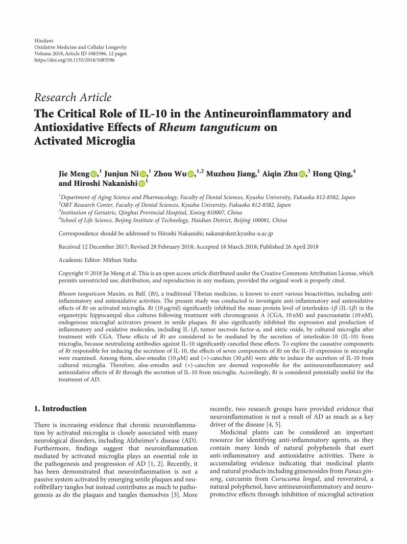
Research ArticleThe Critical Role of IL-10 in the Antineuroinflammatory andAntioxidative Effects of Rheum tanguticum onActivated Microglia
Jie Meng ,1 Junjun Ni ,1 Zhou Wu ,1,2 Muzhou Jiang,1 Aiqin Zhu ,3 Hong Qing,4
and Hiroshi Nakanishi 1
1Department of Aging Science and Pharmacology, Faculty of Dental Sciences, Kyushu University, Fukuoka 812-8582, Japan2OBT Research Center, Faculty of Dental Sciences, Kyushu University, Fukuoka 812-8582, Japan3Institution of Geriatric, Qinghai Provincial Hospital, Xining 810007, China4School of Life Science, Beijing Institute of Technology, Haidian District, Beijing 100081, China
Correspondence should be addressed to Hiroshi Nakanishi; [email protected]
Received 12 December 2017; Revised 28 February 2018; Accepted 18 March 2018; Published 26 April 2018
Academic Editor: Mithun Sinha
Copyright © 2018 Jie Meng et al. This is an open access article distributed under the Creative Commons Attribution License, whichpermits unrestricted use, distribution, and reproduction in any medium, provided the original work is properly cited.
Rheum tanguticum Maxim. ex Balf. (Rt), a traditional Tibetan medicine, is known to exert various bioactivities, including anti-inflammatory and antioxidative activities. The present study was conducted to investigate anti-inflammatory and antioxidativeeffects of Rt on activated microglia. Rt (10 μg/ml) significantly inhibited the mean protein level of interleukin-1β (IL-1β) in theorganotypic hippocampal slice cultures following treatment with chromogranin A (CGA, 10 nM) and pancreastatin (10 nM),endogenous microglial activators present in senile plaques. Rt also significantly inhibited the expression and production ofinflammatory and oxidative molecules, including IL-1β, tumor necrosis factor-α, and nitric oxide, by cultured microglia aftertreatment with CGA. These effects of Rt are considered to be mediated by the secretion of interleukin-10 (IL-10) frommicroglia, because neutralizing antibodies against IL-10 significantly canceled these effects. To explore the causative componentsof Rt responsible for inducing the secretion of IL-10, the effects of seven components of Rt on the IL-10 expression in microgliawere examined. Among them, aloe-emodin (10 μM) and (+)-catechin (30 μM) were able to induce the secretion of IL-10 fromcultured microglia. Therefore, aloe-emodin and (+)-catechin are deemed responsible for the antineuroinflammatory andantioxidative effects of Rt through the secretion of IL-10 from microglia. Accordingly, Rt is considered potentially useful for thetreatment of AD.
1. Introduction
There is increasing evidence that chronic neuroinflamma-tion by activated microglia is closely associated with manyneurological disorders, including Alzheimer’s disease (AD).Furthermore, findings suggest that neuroinflammationmediated by activated microglia plays an essential role inthe pathogenesis and progression of AD [1, 2]. Recently, ithas been demonstrated that neuroinflammation is not apassive system activated by emerging senile plaques and neu-rofibrillary tangles but instead contributes as much to patho-genesis as do the plaques and tangles themselves [3]. More
recently, two research groups have provided evidence thatneuroinflammation is not a result of AD as much as a keydriver of the disease [4, 5].
Medicinal plants can be considered an importantresource for identifying anti-inflammatory agents, as theycontain many kinds of natural polyphenols that exertanti-inflammatory and antioxidative activities. There isaccumulating evidence indicating that medicinal plantsand natural products including ginsenosides from Panax gin-seng, curcumin from Curucuma longaI, and resveratrol, anatural polyphenol, have antineuroinflammatory and neuro-protective effects through inhibition of microglial activation
HindawiOxidative Medicine and Cellular LongevityVolume 2018, Article ID 1083596, 12 pageshttps://doi.org/10.1155/2018/1083596
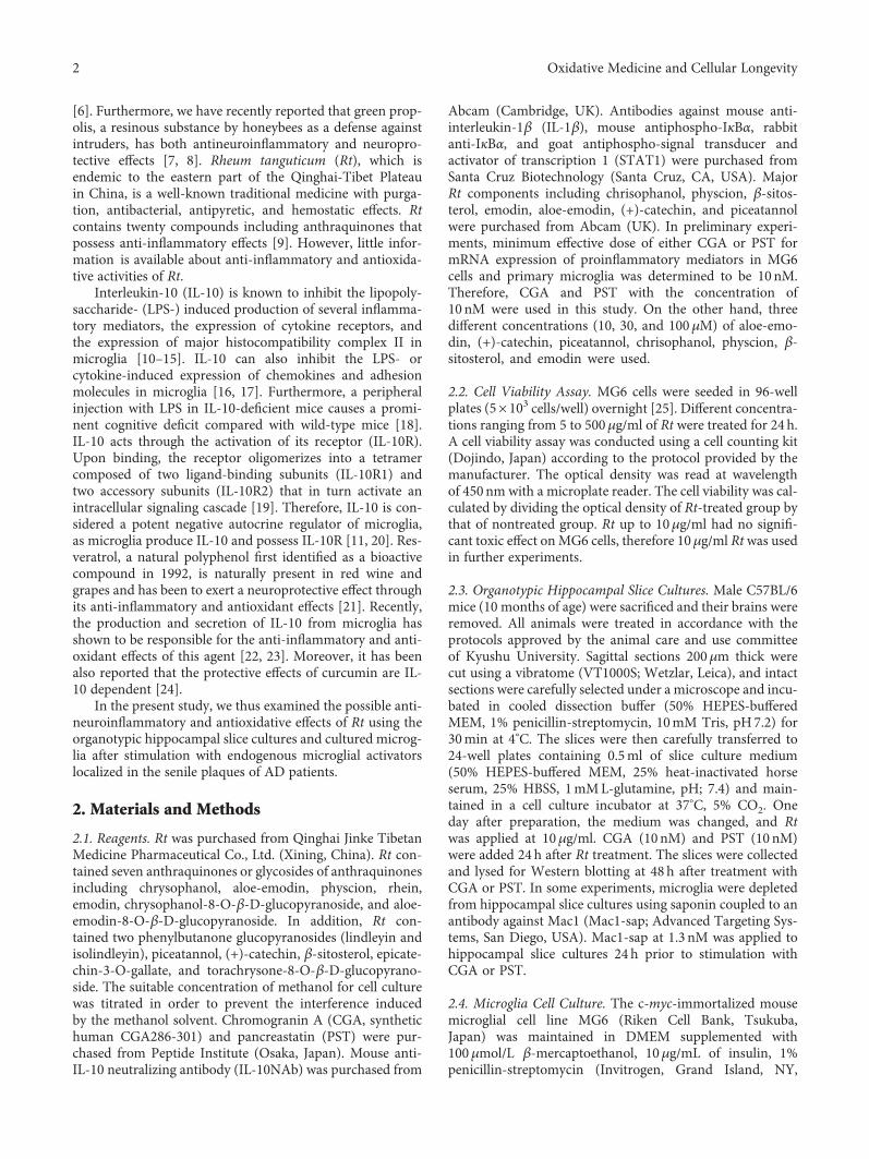
[6]. Furthermore, we have recently reported that green prop-olis, a resinous substance by honeybees as a defense againstintruders, has both antineuroinflammatory and neuropro-tective effects [7, 8]. Rheum tanguticum (Rt), which isendemic to the eastern part of the Qinghai-Tibet Plateauin China, is a well-known traditional medicine with purga-tion, antibacterial, antipyretic, and hemostatic effects. Rtcontains twenty compounds including anthraquinones thatpossess anti-inflammatory effects [9]. However, little infor-mation is available about anti-inflammatory and antioxida-tive activities of Rt.
Interleukin-10 (IL-10) is known to inhibit the lipopoly-saccharide- (LPS-) induced production of several inflamma-tory mediators, the expression of cytokine receptors, andthe expression of major histocompatibility complex II inmicroglia [10–15]. IL-10 can also inhibit the LPS- orcytokine-induced expression of chemokines and adhesionmolecules in microglia [16, 17]. Furthermore, a peripheralinjection with LPS in IL-10-deficient mice causes a promi-nent cognitive deficit compared with wild-type mice [18].IL-10 acts through the activation of its receptor (IL-10R).Upon binding, the receptor oligomerizes into a tetramercomposed of two ligand-binding subunits (IL-10R1) andtwo accessory subunits (IL-10R2) that in turn activate anintracellular signaling cascade [19]. Therefore, IL-10 is con-sidered a potent negative autocrine regulator of microglia,as microglia produce IL-10 and possess IL-10R [11, 20]. Res-veratrol, a natural polyphenol first identified as a bioactivecompound in 1992, is naturally present in red wine andgrapes and has been to exert a neuroprotective effect throughits anti-inflammatory and antioxidant effects [21]. Recently,the production and secretion of IL-10 from microglia hasshown to be responsible for the anti-inflammatory and anti-oxidant effects of this agent [22, 23]. Moreover, it has beenalso reported that the protective effects of curcumin are IL-10 dependent [24].
In the present study, we thus examined the possible anti-neuroinflammatory and antioxidative effects of Rt using theorganotypic hippocampal slice cultures and cultured microg-lia after stimulation with endogenous microglial activatorslocalized in the senile plaques of AD patients.
2. Materials and Methods
2.1. Reagents. Rt was purchased from Qinghai Jinke TibetanMedicine Pharmaceutical Co., Ltd. (Xining, China). Rt con-tained seven anthraquinones or glycosides of anthraquinonesincluding chrysophanol, aloe-emodin, physcion, rhein,emodin, chrysophanol-8-O-β-D-glucopyranoside, and aloe-emodin-8-O-β-D-glucopyranoside. In addition, Rt con-tained two phenylbutanone glucopyranosides (lindleyin andisolindleyin), piceatannol, (+)-catechin, β-sitosterol, epicate-chin-3-O-gallate, and torachrysone-8-O-β-D-glucopyrano-side. The suitable concentration of methanol for cell culturewas titrated in order to prevent the interference inducedby the methanol solvent. Chromogranin A (CGA, synthetichuman CGA286-301) and pancreastatin (PST) were pur-chased from Peptide Institute (Osaka, Japan). Mouse anti-IL-10 neutralizing antibody (IL-10NAb) was purchased from
Abcam (Cambridge, UK). Antibodies against mouse anti-interleukin-1β (IL-1β), mouse antiphospho-IκBα, rabbitanti-IκBα, and goat antiphospho-signal transducer andactivator of transcription 1 (STAT1) were purchased fromSanta Cruz Biotechnology (Santa Cruz, CA, USA). MajorRt components including chrisophanol, physcion, β-sitos-terol, emodin, aloe-emodin, (+)-catechin, and piceatannolwere purchased from Abcam (UK). In preliminary experi-ments, minimum effective dose of either CGA or PST formRNA expression of proinflammatory mediators in MG6cells and primary microglia was determined to be 10 nM.Therefore, CGA and PST with the concentration of10 nM were used in this study. On the other hand, threedifferent concentrations (10, 30, and 100μM) of aloe-emo-din, (+)-catechin, piceatannol, chrisophanol, physcion, β-sitosterol, and emodin were used.
2.2. Cell Viability Assay. MG6 cells were seeded in 96-wellplates (5× 10³ cells/well) overnight [25]. Different concentra-tions ranging from 5 to 500μg/ml of Rt were treated for 24 h.A cell viability assay was conducted using a cell counting kit(Dojindo, Japan) according to the protocol provided by themanufacturer. The optical density was read at wavelengthof 450nm with a microplate reader. The cell viability was cal-culated by dividing the optical density of Rt-treated group bythat of nontreated group. Rt up to 10μg/ml had no signifi-cant toxic effect onMG6 cells, therefore 10μg/ml Rtwas usedin further experiments.
2.3. Organotypic Hippocampal Slice Cultures. Male C57BL/6mice (10 months of age) were sacrificed and their brains wereremoved. All animals were treated in accordance with theprotocols approved by the animal care and use committeeof Kyushu University. Sagittal sections 200μm thick werecut using a vibratome (VT1000S; Wetzlar, Leica), and intactsections were carefully selected under a microscope and incu-bated in cooled dissection buffer (50% HEPES-bufferedMEM, 1% penicillin-streptomycin, 10mM Tris, pH7.2) for30min at 4°C. The slices were then carefully transferred to24-well plates containing 0.5ml of slice culture medium(50% HEPES-buffered MEM, 25% heat-inactivated horseserum, 25% HBSS, 1mML-glutamine, pH; 7.4) and main-tained in a cell culture incubator at 37°C, 5% CO2. Oneday after preparation, the medium was changed, and Rtwas applied at 10μg/ml. CGA (10nM) and PST (10 nM)were added 24 h after Rt treatment. The slices were collectedand lysed for Western blotting at 48 h after treatment withCGA or PST. In some experiments, microglia were depletedfrom hippocampal slice cultures using saponin coupled to anantibody against Mac1 (Mac1-sap; Advanced Targeting Sys-tems, San Diego, USA). Mac1-sap at 1.3 nM was applied tohippocampal slice cultures 24 h prior to stimulation withCGA or PST.
2.4. Microglia Cell Culture. The c-myc-immortalized mousemicroglial cell line MG6 (Riken Cell Bank, Tsukuba,Japan) was maintained in DMEM supplemented with100μmol/L β-mercaptoethanol, 10μg/mL of insulin, 1%penicillin-streptomycin (Invitrogen, Grand Island, NY,
2 Oxidative Medicine and Cellular Longevity
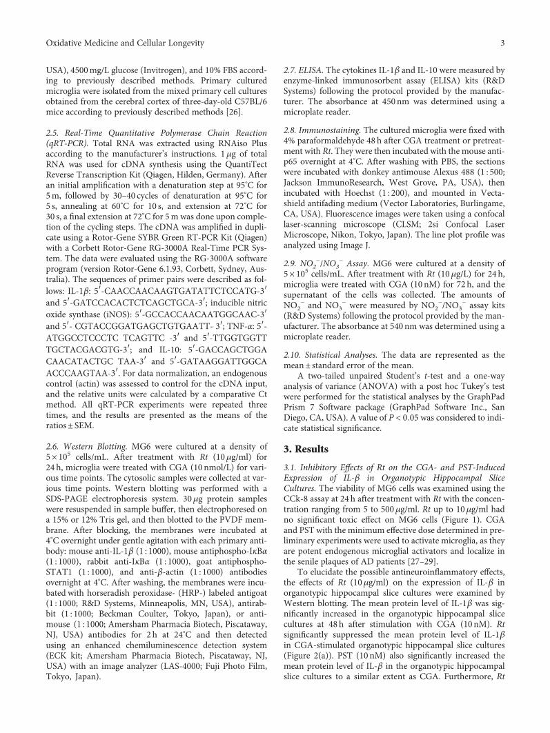
USA), 4500mg/L glucose (Invitrogen), and 10% FBS accord-ing to previously described methods. Primary culturedmicroglia were isolated from the mixed primary cell culturesobtained from the cerebral cortex of three-day-old C57BL/6mice according to previously described methods [26].
2.5. Real-Time Quantitative Polymerase Chain Reaction(qRT-PCR). Total RNA was extracted using RNAiso Plusaccording to the manufacturer’s instructions. 1μg of totalRNA was used for cDNA synthesis using the QuantiTectReverse Transcription Kit (Qiagen, Hilden, Germany). Afteran initial amplification with a denaturation step at 95°C for5m, followed by 30–40 cycles of denaturation at 95°C for5 s, annealing at 60°C for 10 s, and extension at 72°C for30 s, a final extension at 72°C for 5m was done upon comple-tion of the cycling steps. The cDNA was amplified in dupli-cate using a Rotor-Gene SYBR Green RT-PCR Kit (Qiagen)with a Corbett Rotor-Gene RG-3000A Real-Time PCR Sys-tem. The data were evaluated using the RG-3000A softwareprogram (version Rotor-Gene 6.1.93, Corbett, Sydney, Aus-tralia). The sequences of primer pairs were described as fol-lows: IL-1β: 5′-CAACCAACAAGTGATATTCTCCATG-3′and 5′-GATCCACACTCTCAGCTGCA-3′; inducible nitricoxide synthase (iNOS): 5′-GCCACCAACAATGGCAAC-3′and 5′- CGTACCGGATGAGCTGTGAATT- 3′; TNF-α: 5′-ATGGCCTCCCTC TCAGTTC -3′ and 5′-TTGGTGGTTTGCTACGACGTG-3′; and IL-10: 5′-GACCAGCTGGACAACATACTGC TAA-3′ and 5′-GATAAGGATTGGCAACCCAAGTAA-3′. For data normalization, an endogenouscontrol (actin) was assessed to control for the cDNA input,and the relative units were calculated by a comparative Ctmethod. All qRT-PCR experiments were repeated threetimes, and the results are presented as the means of theratios± SEM.
2.6. Western Blotting. MG6 were cultured at a density of5× 105 cells/mL. After treatment with Rt (10μg/ml) for24 h, microglia were treated with CGA (10 nmol/L) for vari-ous time points. The cytosolic samples were collected at var-ious time points. Western blotting was performed with aSDS-PAGE electrophoresis system. 30μg protein sampleswere resuspended in sample buffer, then electrophoresed ona 15% or 12% Tris gel, and then blotted to the PVDF mem-brane. After blocking, the membranes were incubated at4°C overnight under gentle agitation with each primary anti-body: mouse anti-IL-1β (1 : 1000), mouse antiphospho-IκBα(1 : 1000), rabbit anti-IκBα (1 : 1000), goat antiphospho-STAT1 (1 : 1000), and anti-β-actin (1 : 1000) antibodiesovernight at 4°C. After washing, the membranes were incu-bated with horseradish peroxidase- (HRP-) labeled antigoat(1 : 1000; R&D Systems, Minneapolis, MN, USA), antirab-bit (1 : 1000; Beckman Coulter, Tokyo, Japan), or anti-mouse (1 : 1000; Amersham Pharmacia Biotech, Piscataway,NJ, USA) antibodies for 2 h at 24°C and then detectedusing an enhanced chemiluminescence detection system(ECK kit; Amersham Pharmacia Biotech, Piscataway, NJ,USA) with an image analyzer (LAS-4000; Fuji Photo Film,Tokyo, Japan).
2.7. ELISA. The cytokines IL-1β and IL-10 were measured byenzyme-linked immunosorbent assay (ELISA) kits (R&DSystems) following the protocol provided by the manufac-turer. The absorbance at 450nm was determined using amicroplate reader.
2.8. Immunostaining. The cultured microglia were fixed with4% paraformaldehyde 48 h after CGA treatment or pretreat-ment with Rt. They were then incubated with the mouse anti-p65 overnight at 4°C. After washing with PBS, the sectionswere incubated with donkey antimouse Alexus 488 (1 : 500;Jackson ImmunoResearch, West Grove, PA, USA), thenincubated with Hoechst (1 : 200), and mounted in Vecta-shield antifading medium (Vector Laboratories, Burlingame,CA, USA). Fluorescence images were taken using a confocallaser-scanning microscope (CLSM; 2si Confocal LaserMicroscope, Nikon, Tokyo, Japan). The line plot profile wasanalyzed using Image J.
2.9. NO2−/NO3
− Assay. MG6 were cultured at a density of5× 105 cells/mL. After treatment with Rt (10μg/L) for 24 h,microglia were treated with CGA (10 nM) for 72 h, and thesupernatant of the cells was collected. The amounts ofNO2
− and NO3− were measured by NO2
−/NO3− assay kits
(R&D Systems) following the protocol provided by the man-ufacturer. The absorbance at 540nm was determined using amicroplate reader.
2.10. Statistical Analyses. The data are represented as themean± standard error of the mean.
A two-tailed unpaired Student’s t-test and a one-wayanalysis of variance (ANOVA) with a post hoc Tukey’s testwere performed for the statistical analyses by the GraphPadPrism 7 Software package (GraphPad Software Inc., SanDiego, CA, USA). A value of P < 0 05 was considered to indi-cate statistical significance.
3. Results
3.1. Inhibitory Effects of Rt on the CGA- and PST-InducedExpression of IL-β in Organotypic Hippocampal SliceCultures. The viability of MG6 cells was examined using theCCk-8 assay at 24 h after treatment with Rt with the concen-tration ranging from 5 to 500μg/ml. Rt up to 10μg/ml hadno significant toxic effect on MG6 cells (Figure 1). CGAand PST with the minimum effective dose determined in pre-liminary experiments were used to activate microglia, as theyare potent endogenous microglial activators and localize inthe senile plaques of AD patients [27–29].
To elucidate the possible antineuroinflammatory effects,the effects of Rt (10μg/ml) on the expression of IL-β inorganotypic hippocampal slice cultures were examined byWestern blotting. The mean protein level of IL-1β was sig-nificantly increased in the organotypic hippocampal slicecultures at 48 h after stimulation with CGA (10nM). Rtsignificantly suppressed the mean protein level of IL-1βin CGA-stimulated organotypic hippocampal slice cultures(Figure 2(a)). PST (10 nM) also significantly increased themean protein level of IL-β in the organotypic hippocampalslice cultures to a similar extent as CGA. Furthermore, Rt
3Oxidative Medicine and Cellular Longevity
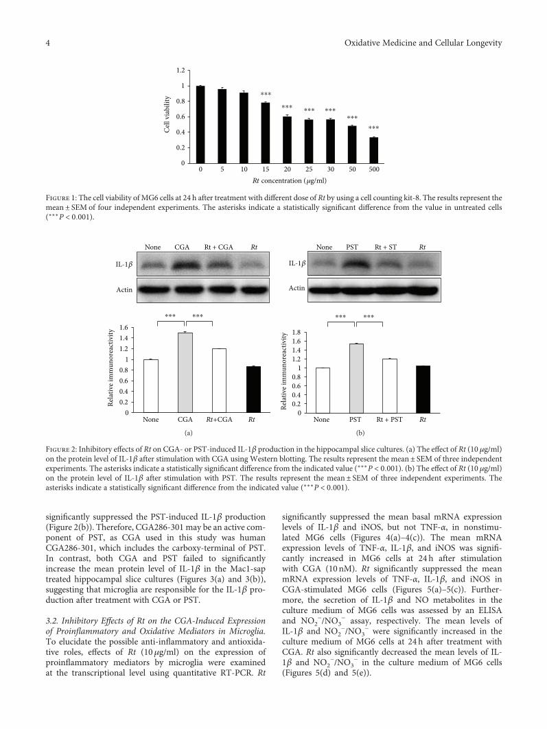
significantly suppressed the PST-induced IL-1β production(Figure 2(b)). Therefore, CGA286-301 may be an active com-ponent of PST, as CGA used in this study was humanCGA286-301, which includes the carboxy-terminal of PST.In contrast, both CGA and PST failed to significantlyincrease the mean protein level of IL-1β in the Mac1-saptreated hippocampal slice cultures (Figures 3(a) and 3(b)),suggesting that microglia are responsible for the IL-1β pro-duction after treatment with CGA or PST.
3.2. Inhibitory Effects of Rt on the CGA-Induced Expressionof Proinflammatory and Oxidative Mediators in Microglia.To elucidate the possible anti-inflammatory and antioxida-tive roles, effects of Rt (10μg/ml) on the expression ofproinflammatory mediators by microglia were examinedat the transcriptional level using quantitative RT-PCR. Rt
significantly suppressed the mean basal mRNA expressionlevels of IL-1β and iNOS, but not TNF-α, in nonstimu-lated MG6 cells (Figures 4(a)–4(c)). The mean mRNAexpression levels of TNF-α, IL-1β, and iNOS was signifi-cantly increased in MG6 cells at 24 h after stimulationwith CGA (10nM). Rt significantly suppressed the meanmRNA expression levels of TNF-α, IL-1β, and iNOS inCGA-stimulated MG6 cells (Figures 5(a)–5(c)). Further-more, the secretion of IL-1β and NO metabolites in theculture medium of MG6 cells was assessed by an ELISAand NO2
−/NO3− assay, respectively. The mean levels of
IL-1β and NO2−/NO3
− were significantly increased in theculture medium of MG6 cells at 24 h after treatment withCGA. Rt also significantly decreased the mean levels of IL-1β and NO2
−/NO3− in the culture medium of MG6 cells
(Figures 5(d) and 5(e)).
Cel
l via
bilit
y
Rt concentration (𝜇g/ml)
0
0.2
0.4
0.6
0.8
1
1.2
0 5 10 15 20 25 30 50 500
⁎⁎⁎
⁎⁎⁎ ⁎⁎⁎ ⁎⁎⁎⁎⁎⁎
⁎⁎⁎
Figure 1: The cell viability of MG6 cells at 24 h after treatment with different dose of Rt by using a cell counting kit-8. The results represent themean± SEM of four independent experiments. The asterisks indicate a statistically significant difference from the value in untreated cells(∗∗∗P < 0 001).
Rela
tive i
mm
unor
eact
ivity
None CGA Rt + CGA
IL-1𝛽
Actin
Rt
00.20.40.60.8
11.21.41.6
None CGA Rt+CGA Rt
⁎⁎⁎ ⁎⁎⁎
(a)
Rela
tive i
mm
unor
eact
ivity
PST Rt + STNone
IL-1𝛽
Actin
Rt
00.20.40.60.8
11.21.41.61.8
None PST Rt + PST Rt
⁎⁎⁎ ⁎⁎⁎
(b)
Figure 2: Inhibitory effects of Rt on CGA- or PST-induced IL-1β production in the hippocampal slice cultures. (a) The effect of Rt (10 μg/ml)on the protein level of IL-1β after stimulation with CGA using Western blotting. The results represent the mean± SEM of three independentexperiments. The asterisks indicate a statistically significant difference from the indicated value (∗∗∗P < 0 001). (b) The effect of Rt (10 μg/ml)on the protein level of IL-1β after stimulation with PST. The results represent the mean± SEM of three independent experiments. Theasterisks indicate a statistically significant difference from the indicated value (∗∗∗P < 0 001).
4 Oxidative Medicine and Cellular Longevity
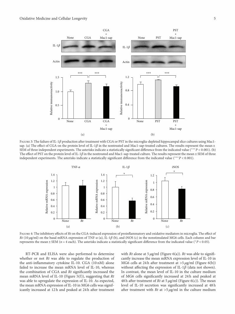
RT-PCR and ELISA were also performed to determinewhether or not Rt was able to regulate the production ofthe anti-inflammatory cytokine IL-10. CGA (10 nM) alonefailed to increase the mean mRNA level of IL-10, whereasthe combination of CGA and Rt significantly increased themean mRNA level of IL-10 (Figure 5(f)), suggesting that Rtwas able to upregulate the expression of IL-10. As expected,the mean mRNA expression of IL-10 in MG6 cells was signif-icantly increased at 12 h and peaked at 24h after treatment
with Rt alone at 5μg/ml (Figure 6(a)). Rt was able to signifi-cantly increase the mean mRNA expression level of IL-10 inMG6 cells at 24 h after treatment at >5μg/ml (Figure 6(b))without affecting the expression of IL-1β (data not shown).In contrast, the mean level of IL-10 in the culture mediumof MG6 cells significantly increased at 24h and peaked at48 h after treatment of Rt at 5μg/ml (Figure 6(c)). The meanlevel of IL-10 secretion was significantly increased at 48hafter treatment with Rt at >5μg/ml in the culture medium
IL-1𝛽
Actin
None CGA
CGA+
Mac1-sap
CGA+
Mac1-sap
None CGA
Rela
tive i
mm
unor
eact
ivity
1.5
0
1.0
0.5
⁎⁎⁎ ⁎⁎⁎
(a)
None PST
PST+
Mac1-sap
PST+
Mac1-sap
IL-1𝛽
Actin
None PST Re
lativ
e im
mun
orea
ctiv
ity
0.8
1.2
0
0.4
⁎⁎⁎ ⁎⁎⁎
(b)
Figure 3: The failure of IL-1β production after treatment with CGA or PST in the microglia-depleted hippocampal slice cultures using Mac1-sap. (a) The effect of CGA on the protein level of IL-1β in the nontreated and Mac1-sap-treated cultures. The results represent the mean±SEM of three independent experiments. The asterisks indicate a statistically significant difference from the indicated value (∗∗∗P < 0 001). (b)The effect of PST on the protein level of IL-1β in the nontreated andMac1-sap-treated culture. The results represent the mean± SEM of threeindependent experiments. The asterisks indicate a statistically significant difference from the indicated value (∗∗∗P < 0 001).
0
0.2
0.4
0.6
0.8
1
1.2
1.4
TNF-𝛼
None Rt
Relat
ive m
RNA
expr
essio
n
(a)
0
0.2
0.4
0.6
0.8
1
1.2
1.4
IL-1𝛽
None Rt
Relat
ive m
RNA
expr
essio
n
⁎
(b)
0
0.2
0.4
0.6
0.8
1
1.2
iNOS
None Rt
Relat
ive m
RNA
expr
essio
n
⁎
(c)
Figure 4: The inhibitory effects of Rt on the CGA-induced expression of proinflammatory and oxidative mediators in microglia. The effect ofRt (10 μg/ml) on the basal mRNA expression of TNF-α (a), IL-1β (b), and iNOS (c) in the nonstimulated MG6 cells. Each column and barrepresents the mean± SEM (n = 4 each). The asterisks indicate a statistically significant difference from the indicated value (∗P < 0 05).
5Oxidative Medicine and Cellular Longevity
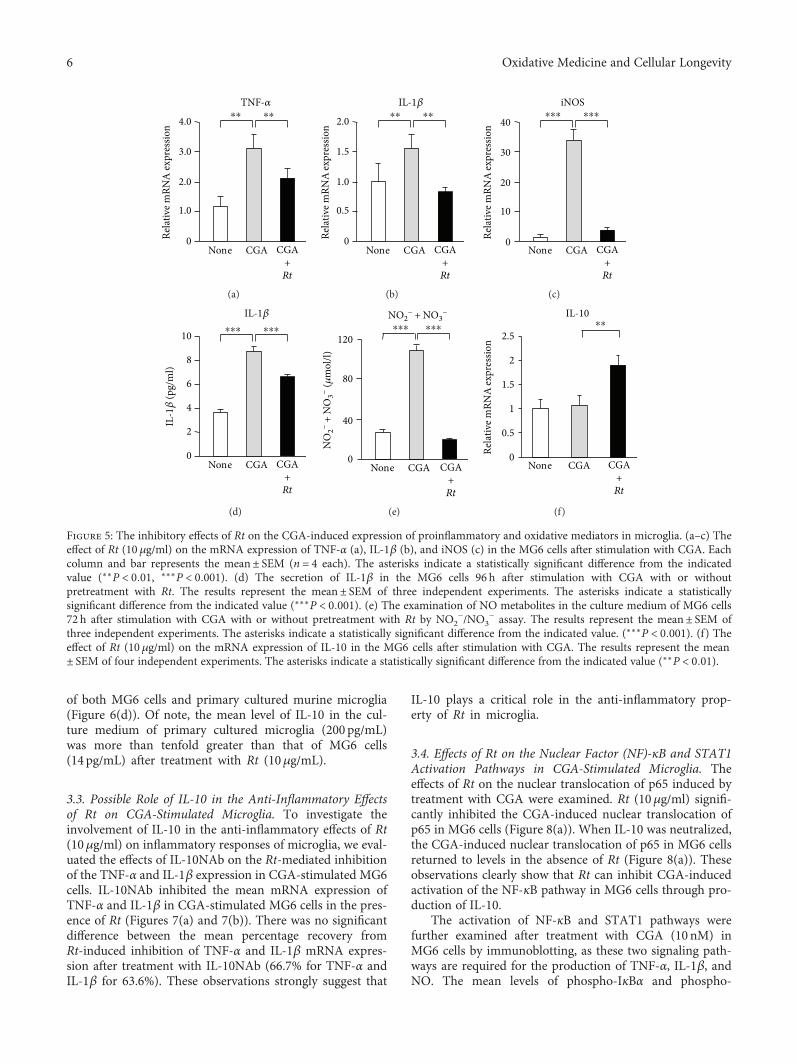
of both MG6 cells and primary cultured murine microglia(Figure 6(d)). Of note, the mean level of IL-10 in the cul-ture medium of primary cultured microglia (200 pg/mL)was more than tenfold greater than that of MG6 cells(14 pg/mL) after treatment with Rt (10μg/mL).
3.3. Possible Role of IL-10 in the Anti-Inflammatory Effectsof Rt on CGA-Stimulated Microglia. To investigate theinvolvement of IL-10 in the anti-inflammatory effects of Rt(10μg/ml) on inflammatory responses of microglia, we eval-uated the effects of IL-10NAb on the Rt-mediated inhibitionof the TNF-α and IL-1β expression in CGA-stimulated MG6cells. IL-10NAb inhibited the mean mRNA expression ofTNF-α and IL-1β in CGA-stimulated MG6 cells in the pres-ence of Rt (Figures 7(a) and 7(b)). There was no significantdifference between the mean percentage recovery fromRt-induced inhibition of TNF-α and IL-1β mRNA expres-sion after treatment with IL-10NAb (66.7% for TNF-α andIL-1β for 63.6%). These observations strongly suggest that
IL-10 plays a critical role in the anti-inflammatory prop-erty of Rt in microglia.
3.4. Effects of Rt on the Nuclear Factor (NF)-κB and STAT1Activation Pathways in CGA-Stimulated Microglia. Theeffects of Rt on the nuclear translocation of p65 induced bytreatment with CGA were examined. Rt (10μg/ml) signifi-cantly inhibited the CGA-induced nuclear translocation ofp65 in MG6 cells (Figure 8(a)). When IL-10 was neutralized,the CGA-induced nuclear translocation of p65 in MG6 cellsreturned to levels in the absence of Rt (Figure 8(a)). Theseobservations clearly show that Rt can inhibit CGA-inducedactivation of the NF-κB pathway in MG6 cells through pro-duction of IL-10.
The activation of NF-κB and STAT1 pathways werefurther examined after treatment with CGA (10 nM) inMG6 cells by immunoblotting, as these two signaling path-ways are required for the production of TNF-α, IL-1β, andNO. The mean levels of phospho-IκBα and phospho-
TNF-𝛼
Relat
ive m
RNA
expr
essio
n
0
1.0
3.0
4.0
CGANone CGA+Rt
2.0
⁎⁎ ⁎⁎
(a)
IL-1𝛽
Rela
tive m
RNA
expr
essio
n
0
0.5
1.5
2.0
CGANone CGA+Rt
1.0
⁎⁎ ⁎⁎
(b)
iNOS
Rela
tive m
RNA
expr
essio
n
0
10
30
40
CGANone CGA+Rt
20
⁎⁎⁎ ⁎⁎⁎
(c)
IL-1𝛽
IL-1𝛽
(pg/
ml)
0
2
8
10
CGANone CGA+Rt
4
6
⁎⁎⁎ ⁎⁎⁎
(d)
NO2− + NO3
−
NO
2− +
NO
3− (𝜇
mol
/l)
0
120
CGANone CGA+Rt
40
80
⁎⁎⁎ ⁎⁎⁎
(e)
IL-10
Rela
tive m
RNA
expr
essio
n
0
0.5
1
1.5
2
2.5
CGANone CGA+Rt
⁎⁎
(f)
Figure 5: The inhibitory effects of Rt on the CGA-induced expression of proinflammatory and oxidative mediators in microglia. (a–c) Theeffect of Rt (10 μg/ml) on the mRNA expression of TNF-α (a), IL-1β (b), and iNOS (c) in the MG6 cells after stimulation with CGA. Eachcolumn and bar represents the mean± SEM (n = 4 each). The asterisks indicate a statistically significant difference from the indicatedvalue (∗∗P < 0 01, ∗∗∗P < 0 001). (d) The secretion of IL-1β in the MG6 cells 96 h after stimulation with CGA with or withoutpretreatment with Rt. The results represent the mean± SEM of three independent experiments. The asterisks indicate a statisticallysignificant difference from the indicated value (∗∗∗P < 0 001). (e) The examination of NO metabolites in the culture medium of MG6 cells72 h after stimulation with CGA with or without pretreatment with Rt by NO2
−/NO3− assay. The results represent the mean± SEM of
three independent experiments. The asterisks indicate a statistically significant difference from the indicated value. (∗∗∗P < 0 001). (f) Theeffect of Rt (10 μg/ml) on the mRNA expression of IL-10 in the MG6 cells after stimulation with CGA. The results represent the mean± SEM of four independent experiments. The asterisks indicate a statistically significant difference from the indicated value (∗∗P < 0 01).
6 Oxidative Medicine and Cellular Longevity
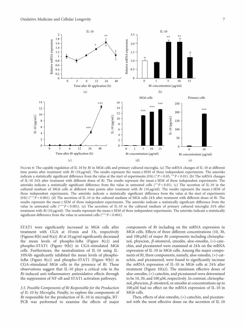
STAT1 were significantly increased in MG6 cells aftertreatment with CGA at 10min and 3h, respectively(Figures 8(b) and 9(a)). Rt at 10μg/ml significantly decreasedthe mean levels of phospho-IκBα (Figure 8(c)) andphospho-STAT1 (Figure 9(b)) in CGA-stimulated MG6cells. Furthermore, the neutralization of IL-10 using IL-10NAb significantly inhibited the mean levels of phospho-IκBα (Figure 8(c)) and phospho-STAT1 (Figure 9(b)) inCGA-stimulated MG6 cells in the presence of Rt. Theseobservations suggest that IL-10 plays a critical role in theRt-induced anti-inflammatory antioxidative effects throughthe suppression of NF-κB and STAT1 activation pathways.
3.5. Possible Components of Rt Responsible for the Productionof IL-10 by Microglia. Finally, to explore the components ofRt responsible for the production of IL-10 in microglia, RT-PCR was performed to examine the effects of major
components of Rt including on the mRNA expression inMG6 cells. Effects of three different concentrations (10, 30,and 100μM) of major Rt components including chrisopha-nol, physcion, β-sitosterol, emodin, aloe-emodin, (+)-cate-chin, and piceatannol were examined at 24 h on the mRNAexpression of IL-10 in MG6 cells. Among the major compo-nents of Rt, three components, namely, aloe-emodin, (+)-cat-echin, and piceatannol, were found to significantly increasethe mRNA expression of IL-10 in MG6 cells at 24 h aftertreatment (Figure 10(a)). The minimum effective doses ofaloe-emodin, (+)-catechin, and piceatannol were determinedto be 10, 30, and 100μM, respectively. In contrast, chrisopha-nol, physcion, β-sitosterol, or emodin at concentrations up to100μM had no effect on the mRNA expression of IL-10 inMG6 cells.
Then, effects of aloe-emodin, (+)-catechin, and piceatan-nol with the most effective doses on the secretion of IL-10
Rela
tive m
RNA
expr
essio
n
Time after Rt application (h)
IL-10
00.20.40.60.8
11.21.41.61.8
2
0 3 6 12 24 48
⁎⁎
⁎
(a)
Rela
tive m
RNA
expr
essio
n
0
1.0
1.5
2.0
3.5
0.5
0 1 5 10 15Rt concentration (𝜇g/ml)
IL-10⁎⁎ ⁎⁎
(b)
Time after Rt application (h)
IL-1
0 (p
g/m
L)
0
2
4
6
8
10
12
14
0 3 6 12 24 48
⁎⁎⁎
⁎⁎⁎
(c)
Rt concentration (𝜇g/ml)
02468
10121416
0 1 5 10 15
IL-1
0 (p
g/m
L)
MG6 cells
⁎⁎⁎⁎⁎⁎
⁎⁎⁎
(d)
0
50
100
150
200
250
0 10Rt concentration (𝜇g/ml)
IL-1
0 (p
g/m
L)
Primary cells
⁎⁎⁎
(e)
Figure 6: The capable regulation of IL-10 by Rt in MG6 cells and primary cultured microglia. (a) The mRNA changes of IL-10 at differenttime points after treatment with Rt (10 μg/ml). The results represent the mean± SEM of three independent experiments. The asterisksindicate a statistically significant difference from the value at the start of experiments (0 h) (∗P < 0 05, ∗∗P < 0 01). (b) The mRNA changesof IL-10 24 h after treatment with different doses of Rt. The results represent the mean± SEM of three independent experiments. Theasterisks indicate a statistically significant difference from the value in untreated cells (∗∗P < 0 01). (c) The secretion of IL-10 in thecultured medium of MG6 cells at different time points after treatment with Rt (10 μg/ml). The results represent the mean± SEM ofthree independent experiments. The asterisks indicate a statistically significant difference from the value at the start of experiments(0 h) (∗∗∗P < 0 001). (d) The secretion of IL-10 in the cultured medium of MG6 cells 24 h after treatment with different doses of Rt. Theresults represent the mean± SEM of three independent experiments. The asterisks indicate a statistically significant difference from thevalue in untreated cells (∗∗∗P < 0 001). (e) The secretion of IL-10 in the cultured medium of primary cultured microglia 24 h aftertreatment with Rt (10 μg/ml). The results represent the mean± SEM of three independent experiments. The asterisks indicate a statisticallysignificant difference from the value in untreated cells (∗∗∗P < 0 001).
7Oxidative Medicine and Cellular Longevity
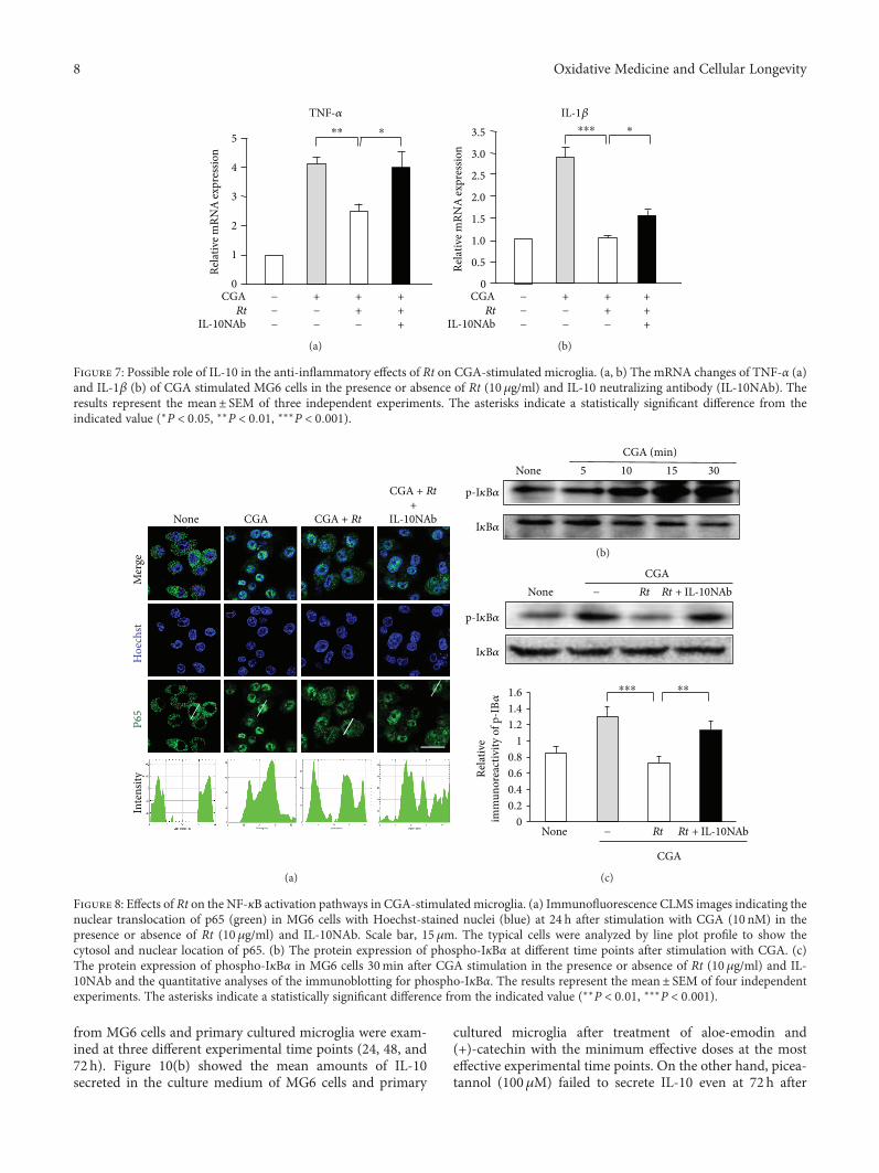
from MG6 cells and primary cultured microglia were exam-ined at three different experimental time points (24, 48, and72 h). Figure 10(b) showed the mean amounts of IL-10secreted in the culture medium of MG6 cells and primary
cultured microglia after treatment of aloe-emodin and(+)-catechin with the minimum effective doses at the mosteffective experimental time points. On the other hand, picea-tannol (100μM) failed to secrete IL-10 even at 72 h after
TNF-𝛼
Rela
tive m
RNA
expr
essio
n
0
1
2
3
4
5
CGARt
IL-10NAb
⁎⁎⁎
++−
+−−
+++
−−−
(a)
IL-1𝛽
Rela
tive m
RNA
expr
essio
n
0
1.0
1.5
2.0
2.5
3.5
++−
+−−
+++
CGARt
IL-10NAb
3.0
0.5
⁎⁎⁎⁎
−−−
(b)
Figure 7: Possible role of IL-10 in the anti-inflammatory effects of Rt on CGA-stimulated microglia. (a, b) The mRNA changes of TNF-α (a)and IL-1β (b) of CGA stimulated MG6 cells in the presence or absence of Rt (10 μg/ml) and IL-10 neutralizing antibody (IL-10NAb). Theresults represent the mean± SEM of three independent experiments. The asterisks indicate a statistically significant difference from theindicated value (∗P < 0 05, ∗∗P < 0 01, ∗∗∗P < 0 001).
P65
Hoe
chst
Mer
ge
None CGA + RtCGA
CGA + Rt+
IL-10NAb
Inte
nsity
(a)
p-I𝜅B𝛼
I𝜅B𝛼
None 3015105CGA (min)
(b)
p-I𝜅B𝛼
I𝜅B𝛼
NoneCGA
Rt− Rt + IL-10NAb
Rela
tive
imm
unor
eact
ivity
of p
-IB𝛼
00.20.40.60.8
11.21.41.6
None
CGA
Rt− Rt + IL-10NAb
⁎⁎⁎⁎⁎
(c)
Figure 8: Effects of Rt on the NF-κB activation pathways in CGA-stimulated microglia. (a) Immunofluorescence CLMS images indicating thenuclear translocation of p65 (green) in MG6 cells with Hoechst-stained nuclei (blue) at 24 h after stimulation with CGA (10 nM) in thepresence or absence of Rt (10 μg/ml) and IL-10NAb. Scale bar, 15μm. The typical cells were analyzed by line plot profile to show thecytosol and nuclear location of p65. (b) The protein expression of phospho-IκBα at different time points after stimulation with CGA. (c)The protein expression of phospho-IκBα in MG6 cells 30min after CGA stimulation in the presence or absence of Rt (10 μg/ml) and IL-10NAb and the quantitative analyses of the immunoblotting for phospho-IκBα. The results represent the mean± SEM of four independentexperiments. The asterisks indicate a statistically significant difference from the indicated value (∗∗P < 0 01, ∗∗∗P < 0 001).
8 Oxidative Medicine and Cellular Longevity
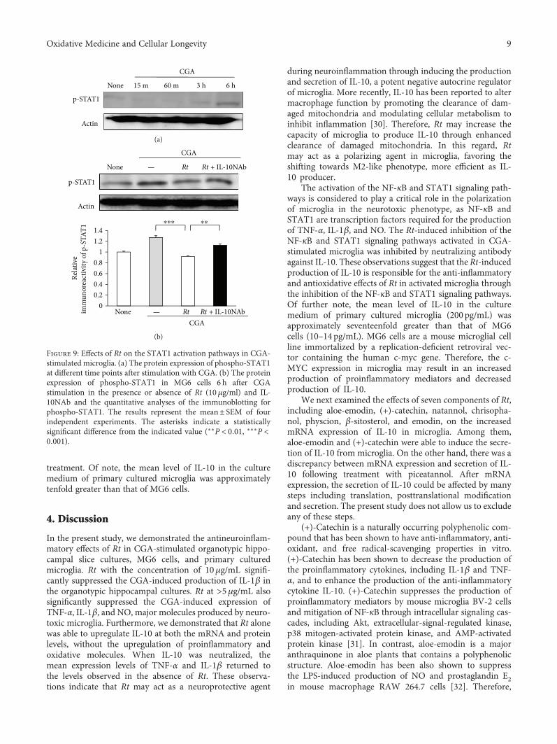
treatment. Of note, the mean level of IL-10 in the culturemedium of primary cultured microglia was approximatelytenfold greater than that of MG6 cells.
4. Discussion
In the present study, we demonstrated the antineuroinflam-matory effects of Rt in CGA-stimulated organotypic hippo-campal slice cultures, MG6 cells, and primary culturedmicroglia. Rt with the concentration of 10μg/mL signifi-cantly suppressed the CGA-induced production of IL-1β inthe organotypic hippocampal cultures. Rt at >5μg/mL alsosignificantly suppressed the CGA-induced expression ofTNF-α, IL-1β, and NO, major molecules produced by neuro-toxic microglia. Furthermore, we demonstrated that Rt alonewas able to upregulate IL-10 at both the mRNA and proteinlevels, without the upregulation of proinflammatory andoxidative molecules. When IL-10 was neutralized, themean expression levels of TNF-α and IL-1β returned tothe levels observed in the absence of Rt. These observa-tions indicate that Rt may act as a neuroprotective agent
during neuroinflammation through inducing the productionand secretion of IL-10, a potent negative autocrine regulatorof microglia. More recently, IL-10 has been reported to altermacrophage function by promoting the clearance of dam-aged mitochondria and modulating cellular metabolism toinhibit inflammation [30]. Therefore, Rt may increase thecapacity of microglia to produce IL-10 through enhancedclearance of damaged mitochondria. In this regard, Rtmay act as a polarizing agent in microglia, favoring theshifting towards M2-like phenotype, more efficient as IL-10 producer.
The activation of the NF-κB and STAT1 signaling path-ways is considered to play a critical role in the polarizationof microglia in the neurotoxic phenotype, as NF-κB andSTAT1 are transcription factors required for the productionof TNF-α, IL-1β, and NO. The Rt-induced inhibition of theNF-κB and STAT1 signaling pathways activated in CGA-stimulated microglia was inhibited by neutralizing antibodyagainst IL-10. These observations suggest that the Rt-inducedproduction of IL-10 is responsible for the anti-inflammatoryand antioxidative effects of Rt in activated microglia throughthe inhibition of the NF-κB and STAT1 signaling pathways.Of further note, the mean level of IL-10 in the culturemedium of primary cultured microglia (200 pg/mL) wasapproximately seventeenfold greater than that of MG6cells (10–14 pg/mL). MG6 cells are a mouse microglial cellline immortalized by a replication-deficient retroviral vec-tor containing the human c-myc gene. Therefore, the c-MYC expression in microglia may result in an increasedproduction of proinflammatory mediators and decreasedproduction of IL-10.
We next examined the effects of seven components of Rt,including aloe-emodin, (+)-catechin, natannol, chrisopha-nol, physcion, β-sitosterol, and emodin, on the increasedmRNA expression of IL-10 in microglia. Among them,aloe-emodin and (+)-catechin were able to induce the secre-tion of IL-10 from microglia. On the other hand, there was adiscrepancy between mRNA expression and secretion of IL-10 following treatment with piceatannol. After mRNAexpression, the secretion of IL-10 could be affected by manysteps including translation, posttranslational modificationand secretion. The present study does not allow us to excludeany of these steps.
(+)-Catechin is a naturally occurring polyphenolic com-pound that has been shown to have anti-inflammatory, anti-oxidant, and free radical-scavenging properties in vitro.(+)-Catechin has been shown to decrease the production ofthe proinflammatory cytokines, including IL-1β and TNF-α, and to enhance the production of the anti-inflammatorycytokine IL-10. (+)-Catechin suppresses the production ofproinflammatory mediators by mouse microglia BV-2 cellsand mitigation of NF-κB through intracellular signaling cas-cades, including Akt, extracellular-signal-regulated kinase,p38 mitogen-activated protein kinase, and AMP-activatedprotein kinase [31]. In contrast, aloe-emodin is a majoranthraquinone in aloe plants that contains a polyphenolicstructure. Aloe-emodin has been also shown to suppressthe LPS-induced production of NO and prostaglandin E2in mouse macrophage RAW 264.7 cells [32]. Therefore,
3 h60 m15 m 6 hNone
p-STAT1
Actin
CGA
(a)
Actin
p-STAT1
None
CGA
Rt— Rt + IL-10NAb
00.20.40.60.8
11.21.4
Rela
tive
imm
unor
eact
ivity
of p
-STA
T1
NoneCGA
Rt— Rt + IL-10NAb
⁎⁎⁎⁎⁎
(b)
Figure 9: Effects of Rt on the STAT1 activation pathways in CGA-stimulated microglia. (a) The protein expression of phospho-STAT1at different time points after stimulation with CGA. (b) The proteinexpression of phospho-STAT1 in MG6 cells 6 h after CGAstimulation in the presence or absence of Rt (10 μg/ml) and IL-10NAb and the quantitative analyses of the immunoblotting forphospho-STAT1. The results represent the mean± SEM of fourindependent experiments. The asterisks indicate a statisticallysignificant difference from the indicated value (∗∗P < 0 01, ∗∗∗P <0 001).
9Oxidative Medicine and Cellular Longevity
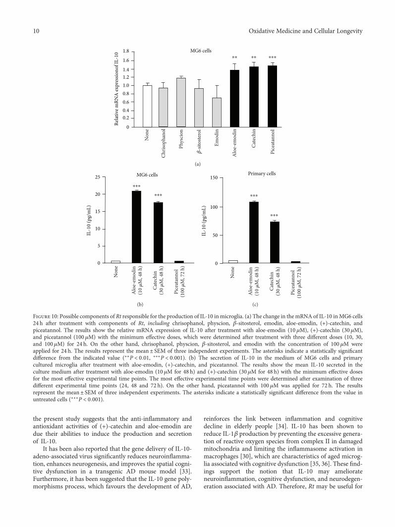
the present study suggests that the anti-inflammatory andantioxidant activities of (+)-catechin and aloe-emodin aredue their abilities to induce the production and secretionof IL-10.
It has been also reported that the gene delivery of IL-10-adeno-associated virus significantly reduces neuroinflamma-tion, enhances neurogenesis, and improves the spatial cogni-tive dysfunction in a transgenic AD mouse model [33].Furthermore, it has been suggested that the IL-10 gene poly-morphisms process, which favours the development of AD,
reinforces the link between inflammation and cognitivedecline in elderly people [34]. IL-10 has been shown toreduce IL-1β production by preventing the excessive genera-tion of reactive oxygen species from complex II in damagedmitochondria and limiting the inflammasome activation inmacrophages [30], which are characteristics of aged microg-lia associated with cognitive dysfunction [35, 36]. These find-ings support the notion that IL-10 may ameliorateneuroinflammation, cognitive dysfunction, and neurodegen-eration associated with AD. Therefore, Rt may be useful for
Relat
ive m
RNA
expr
essio
nof I
L-10
0
0.40.6
1.21.4
1.8
1.6
1.00.8
0.2
MG6 cells⁎⁎ ⁎⁎ ⁎⁎⁎
Non
e
Chris
opha
nol
Phys
cion
Emod
in
Alo
e-em
odin
Cate
chin
Pice
atan
nol
𝛽-s
itoste
rol
(a)
0
20
15
10
5
25
IL-1
0 (p
g/m
L)
MG6 cells
⁎⁎⁎
⁎⁎⁎
Non
e
Alo
e-em
odin
(10 𝜇
M, 4
8 h)
Cate
chin
(30 𝜇
M, 4
8 h)
Pice
atan
nol
(100
𝜇M
, 72
h)
(b)
0
150
100
50
Primary cells
IL-1
0 (p
g/m
L)
⁎⁎⁎
⁎⁎⁎
Non
e
Alo
e-em
odin
(10 𝜇
M, 4
8 h)
Cate
chin
(30 𝜇
M, 4
8 h)
Pice
atan
nol
(100
𝜇M
, 72
h)(c)
Figure 10: Possible components of Rt responsible for the production of IL-10 inmicroglia. (a) The change in the mRNA of IL-10 inMG6 cells24 h after treatment with components of Rt, including chrisophanol, physcion, β-sitosterol, emodin, aloe-emodin, (+)-catechin, andpiceatannol. The results show the relative mRNA expression of IL-10 after treatment with aloe-emodin (10 μM), (+)-catechin (30 μM),and piceatannol (100 μM) with the minimum effective doses, which were determined after treatment with three different doses (10, 30,and 100μM) for 24 h. On the other hand, chrisophanol, physcion, β-sitosterol, and emodin with the concentration of 100 μM wereapplied for 24 h. The results represent the mean± SEM of three independent experiments. The asterisks indicate a statistically significantdifference from the indicated value (∗∗P < 0 01, ∗∗∗P < 0 001). (b) The secretion of IL-10 in the medium of MG6 cells and primarycultured microglia after treatment with aloe-emodin, (+)-catechin, and piceatannol. The results show the mean IL-10 secreted in theculture medium after treatment with aloe-emodin (10 μM for 48 h) and (+)-catechin (30 μM for 48 h) with the minimum effective dosesfor the most effective experimental time points. The most effective experimental time points were determined after examination of threedifferent experimental time points (24, 48 and 72 h). On the other hand, piceatannol with 100 μM was applied for 72 h. The resultsrepresent the mean± SEM of three independent experiments. The asterisks indicate a statistically significant difference from the value inuntreated cells (∗∗∗P < 0 001).
10 Oxidative Medicine and Cellular Longevity
the pharmacological intervention against excessive inflam-matory and oxidative responses associated with AD byinducing the production of IL-10 by microglia, because someof active components of Rt including (+)-catechin can crossthe blood-brain barrier [37].
5. Conclusions
Rt downregulates the production of proinflammatory andoxidative mediators, including IL-1β, TNF-α, and NO, bycultured activated microglia through the production ofIL-10. Two components of Rt, aloe-emodin and (+)-cate-chin, are deemed responsible for the antineuroinflammatoryand antioxidative effects of Rt through the secretion of IL-10from microglia. Therefore, Rt may be useful for the pharma-cological intervention against excessive inflammatory andoxidative responses associated with AD by inducing the pro-duction of IL-10 by microglia.
Disclosure
Hiroshi Nakanishi present address is Department ofPharmacology, Faculty of Pharmaceutical Sciences, YasudaWomen’s University, Hiroshima 731-0153, Japan.
Conflicts of Interest
The authors declare no competing interests.
Authors’ Contributions
Jie Meng performed the most of experiments, analyzed thedata, and wrote the manuscript. Junjun Ni performed theexperiments and analyzed the data. Zhou Wu analyzedthe data and wrote the manuscript. Muzhou Jiang per-formed the experiments. Aiqin Zhu and Hong Qing pro-vided materials and supervised some of the experiments.Hiroshi Nakanishi designed and supervised the experimentsand wrote the manuscript.
Acknowledgments
This work was supported by Grants-in-Aid for ScientificResearch (no. 17K17093 to Junjun Ni, nos. 16K11478 and16H0848 to Zhou Wu, and no. 15H05015 to Hiroshi Naka-nishi), MEXT, Japan.
References
[1] P. L. McGeer, E. McGeer, J. Rogers, and J. Sibley, “Anti-inflam-matory drugs and Alzheimer disease,” The Lancet, vol. 335,no. 8696, p. 1037, 1990.
[2] P. L. McGeer and E. G. McGeer, “The amyloid cascade-inflammatory hypothesis of Alzheimer disease: implicationsfor therapy,” Acta Neuropathologica, vol. 126, no. 4, pp. 479–497, 2013.
[3] B. Zhang, C. Gaiteri, L.-G. Bodea et al., “Integrated systemsapproach identifies genetic nodes and networks in late-onsetAlzheimer’s disease,” Cell, vol. 153, no. 3, pp. 707–720, 2013.
[4] A. Olmos-Alonso, S. T. T. Schetters, S. Sri et al., “Pharmaco-logical targeting of CSF1R inhibits microglial proliferationand prevents the progression of Alzheimer’s-like pathology,”Brain, vol. 139, no. 3, pp. 891–907, 2016.
[5] E. E. Spangenberg, R. J. Lee, A. R. Najafi et al., “Eliminatingmicroglia in Alzheimer’s mice prevents neuronal loss withoutmodulating amyloid-β pathology,” Brain, vol. 139, no. 4,pp. 1265–1281, 2016.
[6] D. K. Choi, S. Koppula, and K. Suk, “Inhibitors of microglialneurotoxicity: focus on natural products,” Molecules, vol. 16,no. 2, pp. 1021–1043, 2011.
[7] Z. Wu, A. Zhu, F. Takayama et al., “Brazilian green propolissuppresses the hypoxia-induced neuroinflammatory responsesby inhibiting NF-κB activation in microglia,” Oxidative Medi-cine and Cellular Longevity, vol. 2013, Article ID 906726,10 pages, 2013.
[8] J. Ni, Z. Wu, J. Meng et al., “The neuroprotective effects of Bra-zilian green propolis on neurodegenerative damage in humanneuronal SH-SY5Y cells,” Oxidative Medicine and CellularLongevity, vol. 2017, Article ID 7984327, 13 pages, 2017.
[9] X. Peigen, H. Liyi, and W. Liwei, “Ethnopharmacologic studyof Chinese rhubarb,” Journal of Ethnopharmacology, vol. 10,no. 3, pp. 275–293, 1984.
[10] K. Frei, H. Lins, C. Schwerdel, and A. Fontana, “Antigenpresentation in the central nervous system. The inhibitoryeffect of IL-10 on MHC class II expression and productionof cytokines depends on the inducing signals and the type ofcell analyzed,” The Journal of Immunology, vol. 152, no. 6,pp. 2720–2728, 1994.
[11] T. Mizuno, M. Sawada, T. Marunouchi, and A. Suzumura,“Production of interleukin-10 by mouse glial cells in culture,”Biochemical and Biophysical Research Communications,vol. 205, no. 3, pp. 1907–1915, 1994.
[12] P. A. Lodge and S. Sriram, “Regulation of microglial activationby TGF-β, IL-10, and CSF-1,” Journal of Leukocyte Biology,vol. 60, no. 4, pp. 502–508, 1996.
[13] L. Minghetti, E. Polazzi, A. Nicolini, and G. Levi, “Oppo-site regulation of prostaglandin E2 synthesis by transforminggrowth factor-β1 and interleukin 10 in activated microglialcultures,” Journal of Neuroimmunology, vol. 82, no. 1,pp. 31–39, 1998.
[14] G. M. O'Keefe, V. T. Nguyen, and E. N. Benveniste, “Class IItransactivator and class II MHC gene expression in microglia:modulation by the cytokines TGF-β, IL-4, IL-13 and IL-10,”European Journal of Immunology, vol. 29, no. 4, pp. 1275–1285, 1999.
[15] M. Sawada, A. Suzumura, H. Hosoya, T. Marunouchi, andT. Nagatsu, “Interleukin-10 inhibits both production ofcytokines and expression of cytokine receptors in microglia,”Journal of Neurochemistry, vol. 72, no. 4, pp. 1466–1471,1999.
[16] P. Shrikant, E. Weber, T. Jilling, and E. N. Benveniste,“Intercellular adhesion molecule-1 gene expression by glialcells. Differential mechanisms of inhibition by IL-10 and IL-6,” The Journal of Immunology, vol. 155, no. 3, pp. 1489–1501, 1995.
[17] S. Hu, C. C. Chao, L. C. Ehrlich et al., “Inhibition of microglialcell RANTES production by IL-10 and TGF-β,” Journal ofLeukocyte Biology, vol. 65, no. 6, pp. 815–821, 1999.
[18] A. F. Richwine, N. L. Sparkman, R. N. Dilger, J. B. Buchanan,and R. W. Johnson, “Cognitive deficits in interleukin-10-
11Oxidative Medicine and Cellular Longevity
deficient mice after peripheral injection of lipopolysaccha-ride,” Brain, Behavior, and Immunity, vol. 23, no. 6, pp. 794–802, 2009.
[19] K. W. Moore, R. de Waal Malefyt, R. L. Coffman, andA. O'Garra, “Interleukin-10 and the interleukin-10 receptor,”Annual Review of Immunology, vol. 19, no. 1, pp. 683–765,2001.
[20] A. Ledeboer, J. J. P. Brevé, A. Wierinckx et al., “Expression andregulation of interleukin-10 and interleukin-10 receptor in ratastroglial and microglial cells,” European Journal of Neurosci-ence, vol. 16, no. 7, pp. 1175–1185, 2002.
[21] F. Zhang, J. Liu, and J.-S. Shi, “Anti-inflammatory activities ofresveratrol in the brain: role of resveratrol in microglial activa-tion,” European Journal of Pharmacology, vol. 636, no. 1–3,pp. 1–7, 2010.
[22] J. Song, S. Cheon, W. Jung, W. Lee, and J. Lee, “Resveratrolinduces the expression of interleukin-10 and brain-derivedneurotrophic factor in BV2microglia under hypoxia,” Interna-tional Journal of Molecular Sciences, vol. 15, no. 9, pp. 15512–15529, 2014.
[23] A. Cianciulli, T. Dragone, R. Calvello et al., “IL-10 plays a piv-otal role in anti-inflammatory effects of resveratrol in activatedmicroglia cells,” International Immunopharmacology, vol. 24,no. 2, pp. 369–376, 2015.
[24] C. B. Larmonier, J. K. Uno, K.-M. Lee et al., “Limited effectsof dietary curcumin on Th-1 driven colitis in IL-10 deficientmice suggest an IL-10-dependent mechanism of protection,”American Journal of Physiology-Gastrointestinal and LiverPhysiology, vol. 295, no. 5, pp. G1079–G1091, 2008.
[25] T. Takenouchi, K. Ogihara, M. Sato, and H. Kitani, “Inhibitoryeffects of U73122 and U73343 on Ca2+ influx and pore forma-tion induced by the activation of P2X7 nucleotide receptors inmouse microglial cell line,” Biochimica et Biophysica Acta(BBA) - General Subjects, vol. 1726, no. 2, pp. 177–186, 2005.
[26] D. F. Sastradipura, H. Nakanishi, T. Tsukuba et al., “Identifica-tion of cellular compartments involved in processing ofcathepsin E in primary cultures of rat microglia,” Journal ofNeurochemistry, vol. 70, no. 5, pp. 2045–2056, 1998.
[27] O. Yasuhara, I. Akiguchi, S. Nakamura et al., “Pancreastatin-like immunoreactivity in globular dystrophic neurites of senileplaques in brains of patients with Alzheimer’s disease,” Neuro-science Letters, vol. 208, no. 3, pp. 167–170, 1996.
[28] C.-M. Rangon, S. Haïk, B. A. Faucheux et al., “Differentchromogranin immunoreactivity between prion and a-betaamyloid plaque,” NeuroReport, vol. 14, no. 5, pp. 755–758,2003.
[29] Z. Wu, L. Sun, S. Hashioka et al., “Differential pathways forinterleukin-1β production activated by chromogranin A andamyloid β in microglia,” Neurobiology of Aging, vol. 34,no. 12, pp. 2715–2725, 2013.
[30] W. K. E. Ip, N. Hoshi, D. S. Shouval, S. Snapper, andR. Medzhitov, “Anti-inflammatory effect of IL-10 mediatedby metabolic reprogramming of macrophages,” Science,vol. 356, no. 6337, pp. 513–519, 2017.
[31] S. S. Syed Hussein, M. N. A. Kamarudin, and H. Abdul Kadir,“(+)-Catechin attenuates NF-κB activation through regulationof Akt, MAPK, and AMPK signaling pathways in LPS-inducedBV-2 microglial cells,” The American Journal of Chinese Med-icine, vol. 43, no. 5, pp. 927–952, 2015.
[32] M.-Y. Park, H.-J. Kwon, and M.-K. Sung, “Evaluation of aloinand aloe-emodin as anti-inflammatory agents in aloe by using
murine macrophages,” Bioscience, Biotechnology, and Bio-chemistry, vol. 73, no. 4, pp. 828–832, 2009.
[33] T. Kiyota, K. L. Ingraham, R. J. Swan, M. T. Jacobsen, S. J.Andrews, and T. Ikezu, “AAV serotype 2/1-mediated genedelivery of anti-inflammatory interleukin-10 enhances neuro-genesis and cognitive function in APP+PS1 mice,” Gene Ther-apy, vol. 19, no. 7, pp. 724–733, 2012.
[34] C. A. Magalhães, M. d. G. Carvalho, L. P. d. Sousa,P. Caramelli, and K. B. Gomes, “Alzheimer’s disease and cyto-kine IL-10 gene polymorphisms: is there an association?,”Arquivos de Neuro-Psiquiatria, vol. 75, no. 9, pp. 649–656,2017.
[35] H. Nakanishi and Z. Wu, “Microglia-aging: roles of microgliallysosome- and mitochondria-derived reactive oxygen speciesin brain aging,” Behavioural Brain Research, vol. 201, no. 1,pp. 1–7, 2009.
[36] H. Nakanishi, Y. Hayashi, and Z. Wu, “The role of microglialmtDNA damage in age-dependent prolonged LPS-inducedsickness behavior,” Neuron Glia Biology, vol. 7, no. 1, pp. 17–23, 2011.
[37] L. Wu, Q. L. Zhang, X. Y. Zhang et al., “Pharmacokinetics andblood–brain barrier penetration of (+)-catechin and (−)-epi-catechin in rats by microdialysis sampling coupled to high-performance liquid chromatography with chemiluminescencedetection,” Journal of Agricultural and Food Chemistry, vol. 60,no. 37, pp. 9377–9383, 2012.
12 Oxidative Medicine and Cellular Longevity
Research ArticleAntifibrogenic Influence of Mentha piperita L. EssentialOil against CCl4-Induced Liver Fibrosis in Rats
Hanan A. Ogaly ,1 Nadia A. Eltablawy,2 and Reham M. Abd-Elsalam3
1Department of Chemistry, College of Sciences, King Khalid University, Abha, Saudi Arabia2Biochemistry Division, National Organization for Drug Control and Research (NODCAR), Giza, Egypt3Department of Pathology, Faculty of Veterinary Medicine, Cairo University, Giza, Egypt
Correspondence should be addressed to Hanan A. Ogaly; [email protected]
Received 11 October 2017; Revised 10 January 2018; Accepted 18 January 2018; Published 19 April 2018
Academic Editor: Sudipta Saha
Copyright © 2018 Hanan A. Ogaly et al. This is an open access article distributed under the Creative Commons Attribution License,which permits unrestricted use, distribution, and reproduction in any medium, provided the original work is properly cited.
Essential oils of some aromatic plants provide an effective nonmedicinal option to control liver fibrosis.Mentha piperita L. essentialoil (MPEO) have been reported to possess protective effects against hepatotoxicity. However, its effect against liver fibrosis remainsunknown. The present study investigated the antifibrogenic potential of MPEO and its underlying mechanisms. Forty male ratsdivided into 4 groups were used: group 1 served as normal control, group 2 (liver fibrosis) received CCl4 (2.5mL/kg, IP, twiceweekly) for 8 weeks, group 3 concurrently received CCl4 plus MPEO (50mg/kg, IP, daily, from the 3rd week), and group 4received MPEO only. MPOE significantly improved the liver injury markers, lipid peroxidation (LPO), antioxidant capacity,CYP2E1 gene expressionand liver histology. Furthermore, MPOE ameliorated liver fibrosis as evidenced by the reducedexpression of desmin, α-smooth muscle actin (α-SMA), transforming growth factor-β1 (TGF-β1), and SMAD3 proteins. Inaddition, MPOE counteracted the p53 upregulation induced by CCl4 at both mRNA and protein levels. In conclusion, MPOEcould effectively attenuate hepatic fibrosis mainly through improving the redox status, suppressing p53 and subsequentlymodulating TGF-β1 and SMAD3 protein expression. These data promote the use of MPOE as a promising approach inantifibrotic therapy.
1. Introduction
Liver fibrosis and cirrhosis are the ultimate consequences ofchronic hepatic injury induced by various etiological agents[1]. The mechanism of liver fibrosis has been extensivelystudied. However, effective antifibrotic therapies are lacking[2]. The pathogenesis of hepatic fibrosis is generally basedon the activation of HSCs and excessive extracellular matrix(ECM) production [2]. Oxidative stress plays a central rolein triggering these inflammatory and fibrotic responses [3].Among the various signaling pathways involved in patho-genesis of liver fibrosis, TGF-β1/SMAD is considered themost significant signaling pathway [4]. Therefore, inhibitingTGF-β1 was found to be efficient in attenuating liver fibrosis[5, 6]. The tumor suppressor p53 is another important cellsignal primarily stimulated in response to oxidative damage
and oncogene activation [7]. Accumulating evidences sug-gested the involvement of p53 in the pathophysiology ofvarious nontumoral fibrotic liver diseases in both humanand animals [8, 9]. These data suggest that p53 regulationcould serve as an important therapeutic target for fibroticliver diseases.
Medicinal plants and their derivatives contain a widevariety of bioactive phytochemicals with a diverse pharma-cological spectrum [10, 11]. Essential oils constituentsincluding terpenes, terpenoids, phenylpropenes, and otherdegradation products have been reported to exhibit strongantioxidant and anti-inflammatory activities [12]. The effec-tiveness of some essential oils to alleviate the hepatotoxicity[13] and hepatic fibrosis [14] has been proven. Menthapiperita L. (peppermint) is one of the most popular andwidely used herbs. Pharmacological investigations have
HindawiOxidative Medicine and Cellular LongevityVolume 2018, Article ID 4039753, 15 pageshttps://doi.org/10.1155/2018/4039753
demonstrated thatM. piperita possesses analgesic, antifungal[15], antibacterial [16], antiparasitic, and immunomodula-tory activities [17]. Moreover, the hepatoprotective effectsof M. piperita leaves extract [18], oil [19, 20], or itsactive components menthol and menthone [21] have beenreported. However, to the best of our knowledge, the effectagainst hepatic fibrosis has not been reported. Therefore,the present study aimed to investigate the effects of Menthapiperita L. essential oil (MPEO) against hepatic fibrosis andto elucidate the potential underlying molecular mechanisms.
2. Material and Methods
2.1. MPOE Preparation and Characterization. Mentha piper-ita L. leaves were purchased from Harraz Drug stores (Cairo,Egypt). A voucher specimen of the studied plant material wasdeposited at Biochemistry Department, National Organiza-tion for Drug Control and Research (NODCAR), Egypt.Essential oil was extracted and characterized for its chemicalcomposition using gas-liquid chromatography coupled tomass spectrometry (GC-MS) as described by Ogaly et al. [14].
2.2. Experimental Design. Adult male rats (150–170 g) wereobtained from the breeding unit of the Research Institute ofOphthalmology (Giza, Egypt). Rats were housed underconstant temperature and 12h light/dark cycle with freeaccess to water and standard chow diet. All animal proce-dures were performed according to the protocol approvedby the Institutional Animal Care and Use Committee(IACUC), Cairo University (CU-II-F-1-18).
Forty rats were randomly divided into four groups.Group 1 (control) was given corn oil (2mL/kg, IP). Group2 (fibrosis model) was given CCl4 1 : 4 mixture with corn oil(2.5mL/kg, IP) twice weekly for eight weeks. Rats in group3 received CCl4 for two weeks to establish liver injuryand fibrosis and then treated with MPOE (50mg/kg, IP),daily from the 3rd to 8th week. Group 4 received MPOE(50mg/kg, IP) from the 3rd to 8th week. The selected dosefor MPOE (50mg/kg) was previously reported to be hepa-toprotective for rats [20].
2.3. Samples Collection. At the end of experiment, animalswere anesthetized with ethyl ether. Blood samples (3-4mL)were collected by retro-orbital puncture, and serum wasseparated by centrifugation (4000 rpm/10min). Afterward,all animals were sacrificed by cervical dislocation under ethylether anesthesia for humane reasons; the whole liver wasimmediately removed, rinsed in cold normal saline, and keptat −20°C until further analyses. Liver homogenates (10%)were prepared in 0.1M ice-cold phosphate buffered saline(pH7.4) followed by centrifugation at 14,000×g, 15minat 4°C. The separated supernatants were kept at −20°C.For histopathological examination, parts of liver were fixedin 10% neutral buffered formalin and underwent routineprocessing for paraffin embedding.
2.4. Biochemical Analyses. Serum samples were used to mea-sure liver injury markers alanine aminotransferase (ALT)and aspartate aminotransferase (AST) according to themethod of Reitman and Frankel [22]. Liver homogenates
were used for measurement of nitric oxide (NO) [23], malon-dialdehyde (MDA) as a thiobarbituric acid reactive substance(TBARS) [24], superoxide dismutase (SOD) activity [25],catalase (CAT) activity [26], reduced glutathione (GSH) level[27], and total antioxidant capacity (TAC) using commercialkits (Biodiagnostic, Cairo, Egypt). Total protein content wasmeasured according to the method of Bradford [28].
2.5. Histopathological Analyses. Seven liver samples wereharvested from each experimental group and fixed in 10%neutral buffered formalin and then processed to obtain4μm paraffin-embedded sections. The sections were stainedwith hematoxylin and eosin (H&E) and Masson’s trichrome(MT). MT staining was performed to assess collagen fibersdistribution and to determine liver fibrosis %. A numericalscoring system [29] was performed to assess the grade offibrosis, as follows: 0, no fibrosis (normal); 1, fibrousexpansion of some portal areas; 2, fibrous expansion ofmost portal areas; 3, fibrous expansion of most portalareas with occasional portal-to-portal bridging; 4, fibrousexpansion of portal areas with marked bridging (portalto portal as well as portal to central); 5, marked bridging(portal to central as well as central to central) with occasionalnodules formation; and 6, cirrhosis.
2.6. Immunohistochemical Analyses. The immunohistochem-ical (IHC) analyses were done according to the methods ofOgaly et al. [14]. Briefly, tissue sections were deparaffinizedand rehydrated. The antigen retrieval was performed bypretreating the tissue sections for 20min with citrate bufferpH 6 at microwave oven. Sections were incubated with rabbitpolyclonal antibody to α-SMA diluted 1 : 200 (ab5694;Abcam, Cambridge, UK), rabbit polyclonal antidesminantibody diluted 1 : 200 (ab15200; Abcam, Cambridge, UK),rabbit polyclonal anti-TGF-β1 antibody with concentrationof 20μg/mL (ab92486; Abcam, Cambridge, UK), rabbitpolyclonal anti-SMAD3 antibody diluted 1 : 100 (ab28379;Abcam, Cambridge, UK), and rabbit polyclonal anti-Tp53antibody diluted 1 : 100 (ab131442; Abcam, Cambridge,UK) for two hours in a humidified chamber. The tissue sec-tions were incubated with goat antirabbit IgG H&L (HRP)(ab205718; Abcam, Cambridge, UK). Finally, the slides wereincubated for 10min with 3,3′-diaminobenzidine tetrahy-drochloride (DAB; Sigma) as chromagen, counterstainedwith hematoxylin, and mounted with DPX. The imageanalyses of the stained sections were performed by LeicaQwin 500 Image Analyzer (Leica, Cambridge, England). Ineach group, seven sections were examined. The percentageof the immunopositive area (%) was calculated as mean of10 fields/section.
2.7. Gene Expression Analyses. Total RNA isolated fromliver tissues using RNeasy Mini Kit (Qiagen) was reversetranscribed and subjected to quantitative real-time RT-PCRas previously described [14]. mRNA expression levels ofTp53 and CYP2E1 genes were assessed using GAPDH geneas the reference gene. Briefly, cDNA was added to a Quanti-fast SYBR Green qPCR Master Mix (Qiagen) containing30 pg/mL of each primer (Table 1). The thermal program
2 Oxidative Medicine and Cellular Longevity
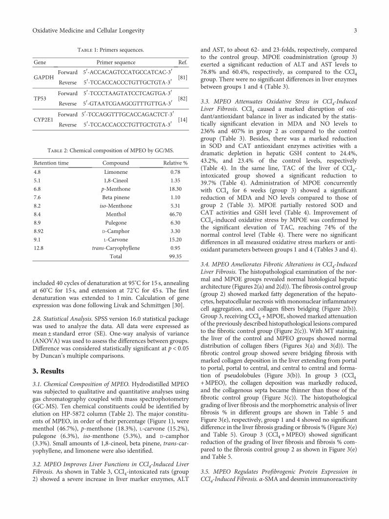
included 40 cycles of denaturation at 95°C for 15 s, annealingat 60°C for 15 s, and extension at 72°C for 45 s. The firstdenaturation was extended to 1min. Calculation of geneexpression was done following Livak and Schmittgen [30].
2.8. Statistical Analysis. SPSS version 16.0 statistical packagewas used to analyze the data. All data were expressed asmean± standard error (SE). One-way analysis of variance(ANOVA) was used to assess the differences between groups.Difference was considered statistically significant at p < 0 05by Duncan’s multiple comparisons.
3. Results
3.1. Chemical Composition of MPEO. Hydrodistilled MPEOwas subjected to qualitative and quantitative analyses usinggas chromatography coupled with mass spectrophotometry(GC-MS). Ten chemical constituents could be identified byelution on HP-5872 column (Table 2). The major constitu-ents of MPEO, in order of their percentage (Figure 1), werementhol (46.7%), p-menthone (18.3%), L-carvone (15.2%),pulegone (6.3%), iso-menthone (5.3%), and D-camphor(3.3%). Small amounts of 1,8-cineol, beta pinene, trans-car-yophyllene, and limonene were also identified.
3.2. MPEO Improves Liver Functions in CCl4-Induced LiverFibrosis. As shown in Table 3, CCl4-intoxicated rats (group2) showed a severe increase in liver marker enzymes, ALT
and AST, to about 62- and 23-folds, respectively, comparedto the control group. MPOE coadministration (group 3)exerted a significant reduction of ALT and AST levels to76.8% and 60.4%, respectively, as compared to the CCl4group. There were no significant differences in liver enzymesbetween groups 1 and 4 (Table 3).
3.3. MPEO Attenuates Oxidative Stress in CCl4-InducedLiver Fibrosis. CCl4 caused a marked disruption of oxi-dant/antioxidant balance in liver as indicated by the statis-tically significant elevation in MDA and NO levels to236% and 407% in group 2 as compared to the controlgroup (Table 3). Besides, there was a marked reductionin SOD and CAT antioxidant enzymes activities with adramatic depletion in hepatic GSH content to 24.4%,43.2%, and 23.4% of the control levels, respectively(Table 4). In the same line, TAC of the liver of CCl4-intoxicated group showed a significant reduction to39.7% (Table 4). Administration of MPOE concurrentlywith CCl4 for 6 weeks (group 3) showed a significantreduction of MDA and NO levels compared to those ofgroup 2 (Table 3). MPOE partially restored SOD andCAT activities and GSH level (Table 4). Improvement ofCCl4-induced oxidative stress by MPOE was confirmed bythe significant elevation of TAC, reaching 74% of thenormal control level (Table 4). There were no significantdifferences in all measured oxidative stress markers or anti-oxidant parameters between groups 1 and 4 (Tables 3 and 4).
3.4. MPEO Ameliorates Fibrotic Alterations in CCl4-InducedLiver Fibrosis. The histopathological examination of the nor-mal and MPOE groups revealed normal histological hepaticarchitecture (Figures 2(a) and 2(d)). The fibrosis control group(group 2) showed marked fatty degeneration of the hepato-cytes, hepatocellular necrosis with mononuclear inflammatorycell aggregation, and collagen fibers bridging (Figure 2(b)).Group 3, receiving CCl4 +MPOE, showed marked attenuationof the previously described histopathological lesions comparedto the fibrotic control group (Figure 2(c)). With MT staining,the liver of the control and MPEO groups showed normaldistribution of collagen fibers (Figures 3(a) and 3(d)). Thefibrotic control group showed severe bridging fibrosis withmarked collagen deposition in the liver extending from portalto portal, portal to central, and central to central and forma-tion of pseudolobules (Figure 3(b)). In group 3 (CCl4+MPEO), the collagen deposition was markedly reduced,and the collagenous septa became thinner than those of thefibrotic control group (Figure 3(c)). The histopathologicalgrading of liver fibrosis and the morphometric analysis of liverfibrosis % in different groups are shown in Table 5 andFigure 3(e), respectively, group 1 and 4 showed no significantdifference in the liver fibrosis grading or fibrosis % (Figure 3(e)and Table 5). Group 3 (CCl4+MPEO) showed significantreduction of the grading of liver fibrosis and fibrosis % com-pared to the fibrosis control group 2 as shown in Figure 3(e)and Table 5.
3.5. MPEO Regulates Profibrogenic Protein Expression inCCl4-Induced Fibrosis. α-SMA and desmin immunoreactivity
Table 1: Primers sequences.
Gene Primer sequence Ref.
GAPDHForward 5′-ACCACAGTCCATGCCATCAC-3′
[81]Reverse 5′-TCCACCACCCTGTTGCTGTA-3′
TP53Forward 5′-TCCCTAAGTATCCTCAGTGA-3′
[82]Reverse 5′-GTAATCGAAGCGTTTGTTGA-3′
CYP2E1Forward 5′-TCCAGGTTTGCACCAGACTCT-3′
[14]Reverse 5′-TCCACCACCCTGTTGCTGTA-3′
Table 2: Chemical composition of MPEO by GC/MS.
Retention time Compound Relative %
4.8 Limonene 0.78
5.1 1,8-Cineol 1.35
6.8 p-Menthone 18.30
7.6 Beta pinene 1.10
8.2 iso-Menthone 5.31
8.4 Menthol 46.70
8.9 Pulegone 6.30
8.92 D-Camphor 3.30
9.1 L-Carvone 15.20
12.8 trans-Caryophyllene 0.95
Total 99.35
3Oxidative Medicine and Cellular Longevity
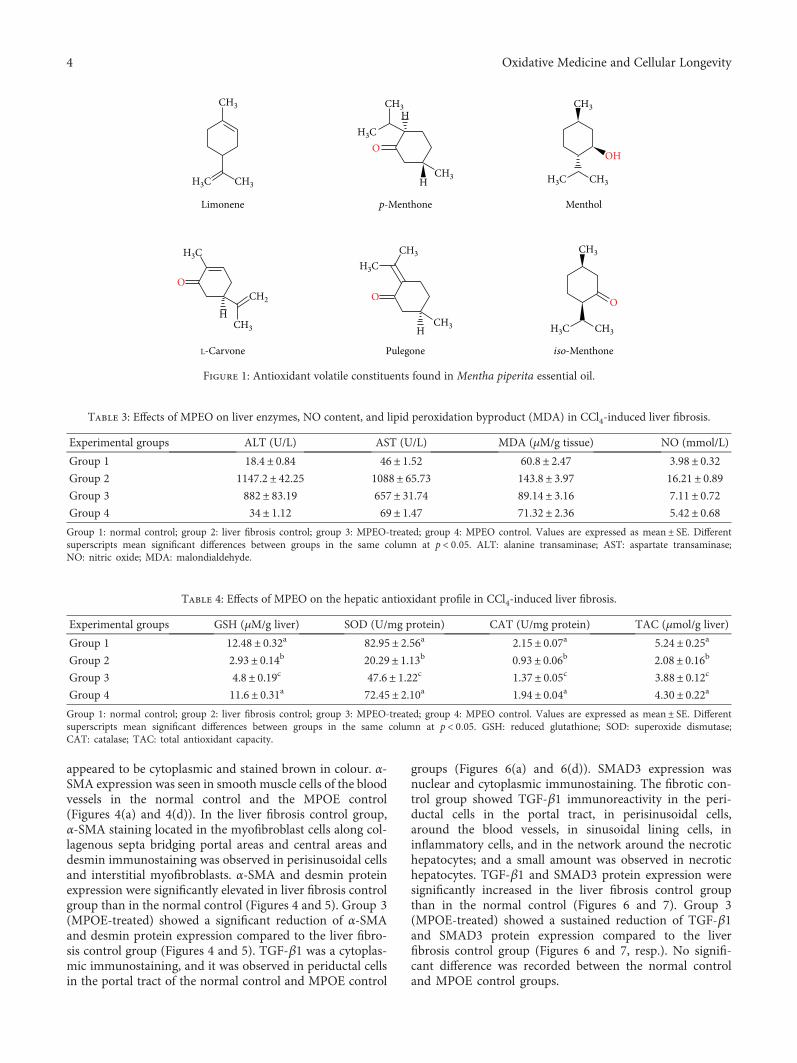
appeared to be cytoplasmic and stained brown in colour. α-SMA expression was seen in smooth muscle cells of the bloodvessels in the normal control and the MPOE control(Figures 4(a) and 4(d)). In the liver fibrosis control group,α-SMA staining located in the myofibroblast cells along col-lagenous septa bridging portal areas and central areas anddesmin immunostaining was observed in perisinusoidal cellsand interstitial myofibroblasts. α-SMA and desmin proteinexpression were significantly elevated in liver fibrosis controlgroup than in the normal control (Figures 4 and 5). Group 3(MPOE-treated) showed a significant reduction of α-SMAand desmin protein expression compared to the liver fibro-sis control group (Figures 4 and 5). TGF-β1 was a cytoplas-mic immunostaining, and it was observed in periductal cellsin the portal tract of the normal control and MPOE control
groups (Figures 6(a) and 6(d)). SMAD3 expression wasnuclear and cytoplasmic immunostaining. The fibrotic con-trol group showed TGF-β1 immunoreactivity in the peri-ductal cells in the portal tract, in perisinusoidal cells,around the blood vessels, in sinusoidal lining cells, ininflammatory cells, and in the network around the necrotichepatocytes; and a small amount was observed in necrotichepatocytes. TGF-β1 and SMAD3 protein expression weresignificantly increased in the liver fibrosis control groupthan in the normal control (Figures 6 and 7). Group 3(MPOE-treated) showed a sustained reduction of TGF-β1and SMAD3 protein expression compared to the liverfibrosis control group (Figures 6 and 7, resp.). No signifi-cant difference was recorded between the normal controland MPOE control groups.
Limonene p-Menthone Menthol
L-Carvone Pulegone iso-Menthone
CH3
CH3H3C
O
HCH3
CH2
H3C
O
H
CH3
CH3
H3C
O
CH3
CH3
H3C
CH3
O
H
HCH3
H3C
CH3
OH
CH3H3C
Figure 1: Antioxidant volatile constituents found in Mentha piperita essential oil.
Table 3: Effects of MPEO on liver enzymes, NO content, and lipid peroxidation byproduct (MDA) in CCl4-induced liver fibrosis.
Experimental groups ALT (U/L) AST (U/L) MDA (μM/g tissue) NO (mmol/L)
Group 1 18.4± 0.84 46± 1.52 60.8± 2.47 3.98± 0.32Group 2 1147.2± 42.25 1088± 65.73 143.8± 3.97 16.21± 0.89Group 3 882± 83.19 657± 31.74 89.14± 3.16 7.11± 0.72Group 4 34± 1.12 69± 1.47 71.32± 2.36 5.42± 0.68Group 1: normal control; group 2: liver fibrosis control; group 3: MPEO-treated; group 4: MPEO control. Values are expressed as mean ± SE. Differentsuperscripts mean significant differences between groups in the same column at p < 0 05. ALT: alanine transaminase; AST: aspartate transaminase;NO: nitric oxide; MDA: malondialdehyde.
Table 4: Effects of MPEO on the hepatic antioxidant profile in CCl4-induced liver fibrosis.
Experimental groups GSH (μM/g liver) SOD (U/mg protein) CAT (U/mg protein) TAC (μmol/g liver)
Group 1 12.48± 0.32a 82.95± 2.56a 2.15± 0.07a 5.24± 0.25a
Group 2 2.93± 0.14b 20.29± 1.13b 0.93± 0.06b 2.08± 0.16b
Group 3 4.8± 0.19c 47.6± 1.22c 1.37± 0.05c 3.88± 0.12c
Group 4 11.6± 0.31a 72.45± 2.10a 1.94± 0.04a 4.30± 0.22a
Group 1: normal control; group 2: liver fibrosis control; group 3: MPEO-treated; group 4: MPEO control. Values are expressed as mean ± SE. Differentsuperscripts mean significant differences between groups in the same column at p < 0 05. GSH: reduced glutathione; SOD: superoxide dismutase;CAT: catalase; TAC: total antioxidant capacity.
4 Oxidative Medicine and Cellular Longevity
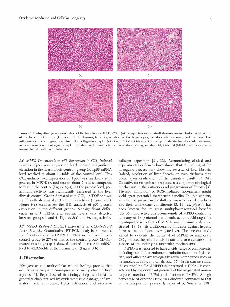
3.6. MPEO Downregulates p53 Expression in CCl4-InducedFibrosis. Tp53 gene expression level showed a significantelevation in the liver fibrosis control (group 2). Tp53 mRNAlevel reached to about 16-folds of the control level. ThisCCl4-induced overexpression of Tp53 was markedly sup-pressed in MPOE-treated rats to about 2-fold as comparedto that in the control (Figure 8(a)). At the protein level, p53immunoreactivity was significantly increased in the liverfibrosis control. Group 3 treated with CCl4 +MPOE showedsignificantly decreased p53 immunoreactivity (Figure 9(c)).Figure 9(e) summarizes the IHC analysis of p53 proteinexpression in the different groups. Nonsignificant differ-ences in p53 mRNA and protein levels were detectedbetween groups 1 and 4 (Figures 8(a) and 9), respectively.
3.7. MPEO Restored CYP2E1 Expression in CCl4-InducedLiver Fibrosis. Quantitative RT-PCR analysis showed asignificant decrease in CYP2E1 mRNA in the liver fibrosiscontrol group to 27% of that of the control group. MPOE-treated rats in group 3 showed marked increase in mRNAlevel to +2.32-folds of the normal level (Figure 8(b)).
4. Discussion
Fibrogenesis is a multicellular wound healing process thatoccurs as a frequent consequence of many chronic liverinjuries [1]. Regardless of its etiology, hepatic fibrosis isgenerally characterized by oxidative tissue damage, inflam-matory cells infiltration, HSCs activation, and excessive
collagen deposition [31, 32]. Accumulating clinical andexperimental evidences have shown that the halting of thefibrogenic process may allow the reversal of liver fibrosis.Indeed, resolution of liver fibrosis or even cirrhosis mayoccur upon eradication of the causative insult [33, 34].Oxidative stress has been proposed as a conjoint pathologicalmechanism in the initiation and progression of fibrosis [3].Thereby, inhibition of ROS-mediated fibrogenesis mightyield great potential therapeutic benefits. In this context,attention is progressively shifting towards herbal productsand their antioxidant constituents [3, 11]. M. piperita hasbeen known for its great multipharmaceutical benefits[35, 36]. The active phytocompounds of MPEO contributeto many of its profound therapeutic actions. Although thehepatoprotective effect of MPOE was previously demon-strated [18, 19], its antifibrogenic influence against hepaticfibrosis has not been investigated yet. The present studyaimed to evaluate the potential of MPOE to ameliorateCCl4-induced hepatic fibrosis in rats and to elucidate someaspects of its underlying molecular mechanisms.
MPEO was reported to have a wide range of components,including menthol, menthone, menthofuran, and methyl ace-tate, and other pharmacologically active compounds such asflavonoids, tannins, and caffeic acid [37]. In the current study,the chemical profile of MPEO, as presented in Table 2, is char-acterized by the dominant presence of the oxygenated mono-terpenes menthol (46.7%) and menthone (18.3%). A highpercentage of carvone (15%) was observed compared to thatof the composition previously reported by Sun et al. [38].
(a) (b)
(c) (d)
Figure 2: Histopathological examination of the liver tissues (H&E, ×200). (a) Group 1 (normal control) showing normal histological pictureof the liver. (b) Group 2 (fibrosis control) showing fatty degeneration of the hepatocytes, hepatocellular necrosis, and mononuclearinflammatory cells aggregation along the collagenous septa. (c) Group 3 (MPEO-treated) showing moderate hepatocellular necrosis,marked reduction of collagenous septa formation and mononuclear inflammatory cells aggregation. (d) Group 4 (MPEO control) showingnormal hepatic cellular architecture.
5Oxidative Medicine and Cellular Longevity
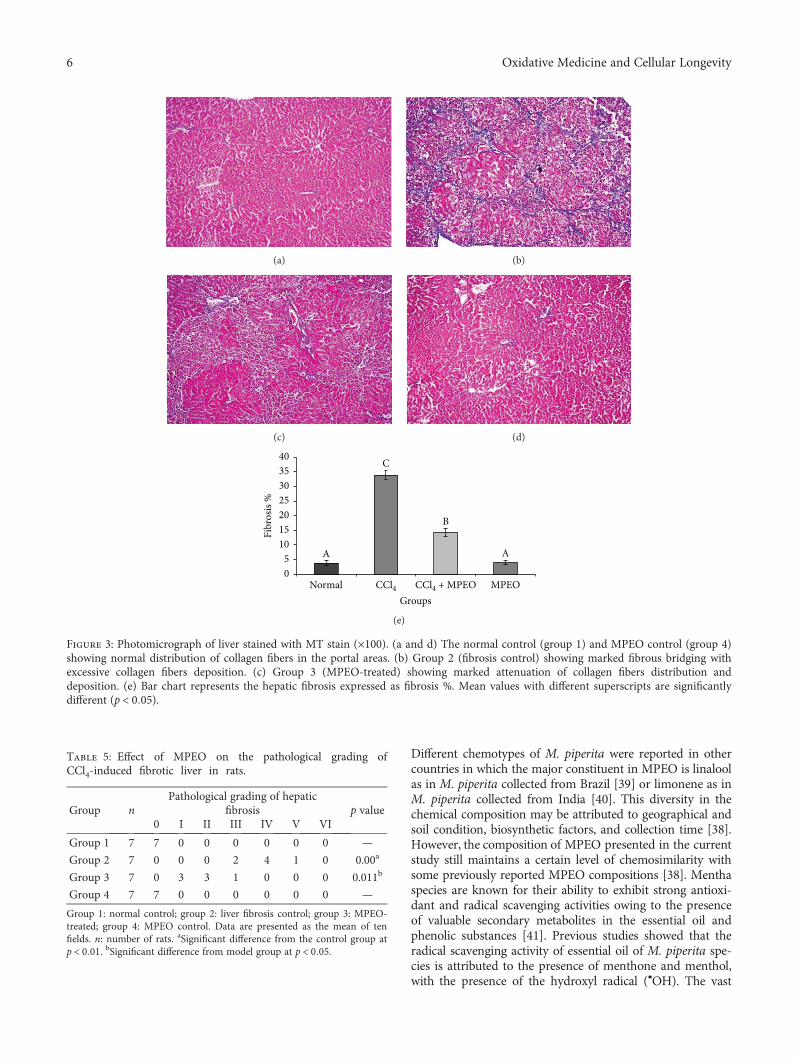
Different chemotypes of M. piperita were reported in othercountries in which the major constituent in MPEO is linaloolas in M. piperita collected from Brazil [39] or limonene as inM. piperita collected from India [40]. This diversity in thechemical composition may be attributed to geographical andsoil condition, biosynthetic factors, and collection time [38].However, the composition of MPEO presented in the currentstudy still maintains a certain level of chemosimilarity withsome previously reported MPEO compositions [38]. Menthaspecies are known for their ability to exhibit strong antioxi-dant and radical scavenging activities owing to the presenceof valuable secondary metabolites in the essential oil andphenolic substances [41]. Previous studies showed that theradical scavenging activity of essential oil of M. piperita spe-cies is attributed to the presence of menthone and menthol,with the presence of the hydroxyl radical (•OH). The vast
(a) (b)
(c) (d)
C
A
B
A
05
10152025303540
Fibr
osis
%
CCl4 CCl4 + MPEO MPEONormalGroups
(e)
Figure 3: Photomicrograph of liver stained with MT stain (×100). (a and d) The normal control (group 1) and MPEO control (group 4)showing normal distribution of collagen fibers in the portal areas. (b) Group 2 (fibrosis control) showing marked fibrous bridging withexcessive collagen fibers deposition. (c) Group 3 (MPEO-treated) showing marked attenuation of collagen fibers distribution anddeposition. (e) Bar chart represents the hepatic fibrosis expressed as fibrosis %. Mean values with different superscripts are significantlydifferent (p < 0 05).
Table 5: Effect of MPEO on the pathological grading ofCCl4-induced fibrotic liver in rats.
Group nPathological grading of hepatic
fibrosis p value0 I II III IV V VI
Group 1 7 7 0 0 0 0 0 0 —
Group 2 7 0 0 0 2 4 1 0 0.00a
Group 3 7 0 3 3 1 0 0 0 0.011b
Group 4 7 7 0 0 0 0 0 0 —
Group 1: normal control; group 2: liver fibrosis control; group 3: MPEO-treated; group 4: MPEO control. Data are presented as the mean of tenfields. n: number of rats. aSignificant difference from the control group atp < 0 01. bSignificant difference from model group at p < 0 05.
6 Oxidative Medicine and Cellular Longevity
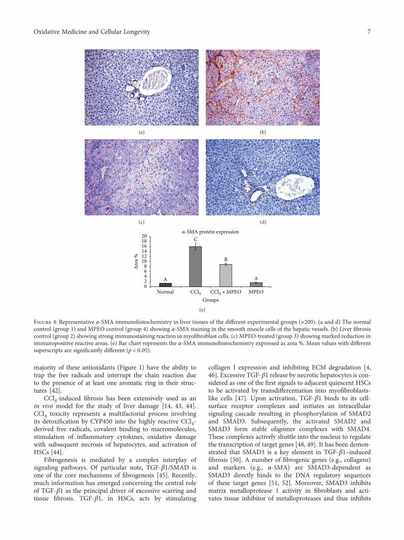
majority of these antioxidants (Figure 1) have the ability totrap the free radicals and interrupt the chain reaction dueto the presence of at least one aromatic ring in their struc-tures [42].
CCl4-induced fibrosis has been extensively used as anin vivo model for the study of liver damage [14, 43, 44].CCl4 toxicity represents a multifactorial process involvingits detoxification by CYP450 into the highly reactive CCl4-derived free radicals, covalent binding to macromolecules,stimulation of inflammatory cytokines, oxidative damagewith subsequent necrosis of hepatocytes, and activation ofHSCs [44].
Fibrogenesis is mediated by a complex interplay ofsignaling pathways. Of particular note, TGF-β1/SMAD isone of the core mechanisms of fibrogenesis [45]. Recently,much information has emerged concerning the central roleof TGF-β1 as the principal driver of excessive scarring andtissue fibrosis. TGF-β1, in HSCs, acts by stimulating
collagen I expression and inhibiting ECM degradation [4,46]. Excessive TGF-β1 release by necrotic hepatocytes is con-sidered as one of the first signals to adjacent quiescent HSCsto be activated by transdifferentiation into myofibroblasts-like cells [47]. Upon activation, TGF-β1 binds to its cell-surface receptor complexes and initiates an intracellularsignaling cascade resulting in phosphorylation of SMAD2and SMAD3. Subsequently, the activated SMAD2 andSMAD3 form stable oligomer complexes with SMAD4.These complexes actively shuttle into the nucleus to regulatethe transcription of target genes [48, 49]. It has been demon-strated that SMAD3 is a key element in TGF-β1–inducedfibrosis [50]. A number of fibrogenic genes (e.g., collagens)and markers (e.g., α-SMA) are SMAD3-dependent asSMAD3 directly binds to the DNA regulatory sequencesof these target genes [51, 52]. Moreover, SMAD3 inhibitsmatrix metalloprotease 1 activity in fibroblasts and acti-vates tissue inhibitor of metalloproteases and thus inhibits
(a) (b)
(c) (d)
𝛼-SMA protein expression
B
A
C
A02468
101214161820
Are
a %
CCl4 CCl4 + MPEO MPEONormalGroups
(e)
Figure 4: Representative α-SMA immunohistochemistry in liver tissues of the different experimental groups (×200). (a and d) The normalcontrol (group 1) and MPEO control (group 4) showing α-SMA staining in the smooth muscle cells of the hepatic vessels. (b) Liver fibrosiscontrol (group 2) showing strong immunostaining reaction in myofibroblast cells. (c) MPEO-treated (group 3) showing marked reduction inimmunopositive reactive areas. (e) Bar chart represents the α-SMA immunohistochemistry expressed as area %. Mean values with differentsuperscripts are significantly different (p < 0 05).
7Oxidative Medicine and Cellular Longevity
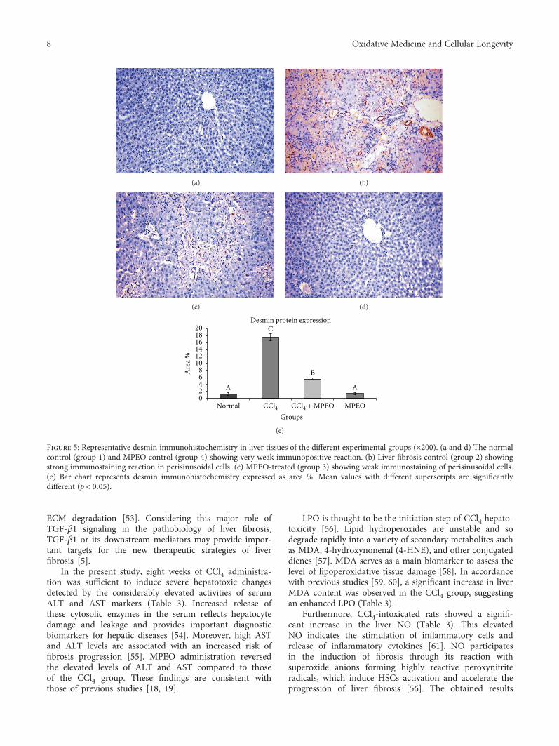
ECM degradation [53]. Considering this major role ofTGF-β1 signaling in the pathobiology of liver fibrosis,TGF-β1 or its downstream mediators may provide impor-tant targets for the new therapeutic strategies of liverfibrosis [5].
In the present study, eight weeks of CCl4 administra-tion was sufficient to induce severe hepatotoxic changesdetected by the considerably elevated activities of serumALT and AST markers (Table 3). Increased release ofthese cytosolic enzymes in the serum reflects hepatocytedamage and leakage and provides important diagnosticbiomarkers for hepatic diseases [54]. Moreover, high ASTand ALT levels are associated with an increased risk offibrosis progression [55]. MPEO administration reversedthe elevated levels of ALT and AST compared to thoseof the CCl4 group. These findings are consistent withthose of previous studies [18, 19].
LPO is thought to be the initiation step of CCl4 hepato-toxicity [56]. Lipid hydroperoxides are unstable and sodegrade rapidly into a variety of secondary metabolites suchas MDA, 4-hydroxynonenal (4-HNE), and other conjugateddienes [57]. MDA serves as a main biomarker to assess thelevel of lipoperoxidative tissue damage [58]. In accordancewith previous studies [59, 60], a significant increase in liverMDA content was observed in the CCl4 group, suggestingan enhanced LPO (Table 3).
Furthermore, CCl4-intoxicated rats showed a signifi-cant increase in the liver NO (Table 3). This elevatedNO indicates the stimulation of inflammatory cells andrelease of inflammatory cytokines [61]. NO participatesin the induction of fibrosis through its reaction withsuperoxide anions forming highly reactive peroxynitriteradicals, which induce HSCs activation and accelerate theprogression of liver fibrosis [56]. The obtained results
(a) (b)
(c) (d)
Desmin protein expression
B
A
C
A
CCl4 CCl4 + MPEO MPEONormalGroups
02468
101214161820
Are
a %
(e)
Figure 5: Representative desmin immunohistochemistry in liver tissues of the different experimental groups (×200). (a and d) The normalcontrol (group 1) and MPEO control (group 4) showing very weak immunopositive reaction. (b) Liver fibrosis control (group 2) showingstrong immunostaining reaction in perisinusoidal cells. (c) MPEO-treated (group 3) showing weak immunostaining of perisinusoidal cells.(e) Bar chart represents desmin immunohistochemistry expressed as area %. Mean values with different superscripts are significantlydifferent (p < 0 05).
8 Oxidative Medicine and Cellular Longevity
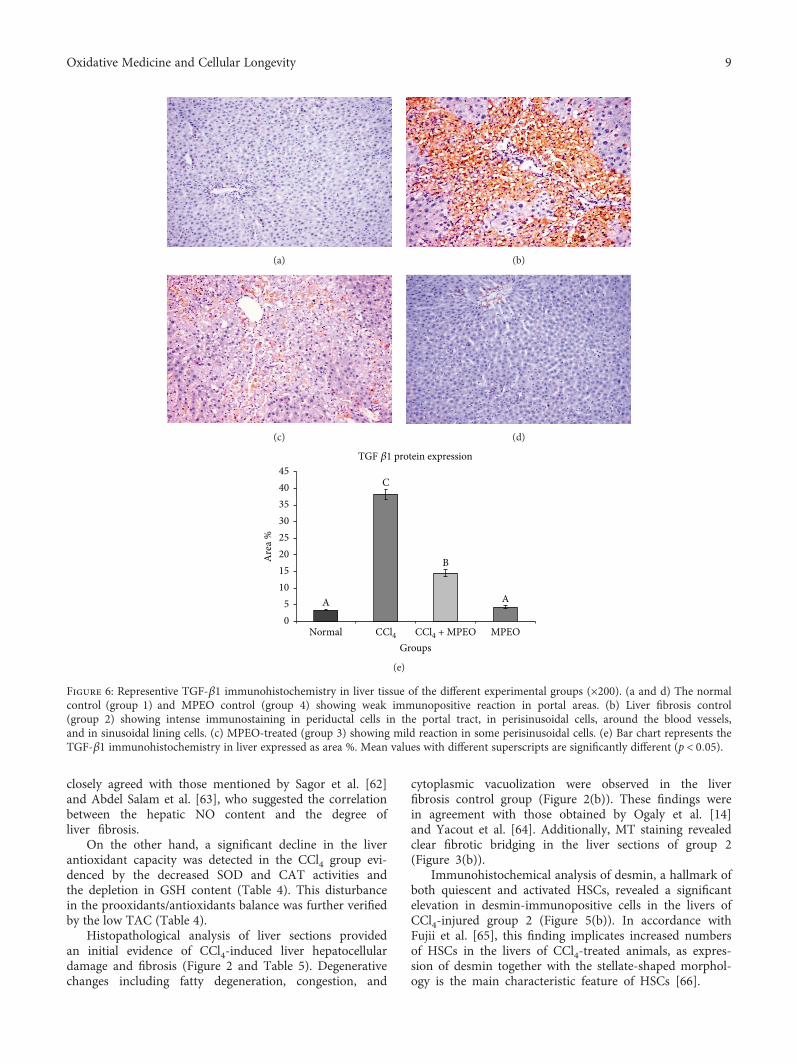
closely agreed with those mentioned by Sagor et al. [62]and Abdel Salam et al. [63], who suggested the correlationbetween the hepatic NO content and the degree ofliver fibrosis.
On the other hand, a significant decline in the liverantioxidant capacity was detected in the CCl4 group evi-denced by the decreased SOD and CAT activities andthe depletion in GSH content (Table 4). This disturbancein the prooxidants/antioxidants balance was further verifiedby the low TAC (Table 4).
Histopathological analysis of liver sections providedan initial evidence of CCl4-induced liver hepatocellulardamage and fibrosis (Figure 2 and Table 5). Degenerativechanges including fatty degeneration, congestion, and
cytoplasmic vacuolization were observed in the liverfibrosis control group (Figure 2(b)). These findings werein agreement with those obtained by Ogaly et al. [14]and Yacout et al. [64]. Additionally, MT staining revealedclear fibrotic bridging in the liver sections of group 2(Figure 3(b)).
Immunohistochemical analysis of desmin, a hallmark ofboth quiescent and activated HSCs, revealed a significantelevation in desmin-immunopositive cells in the livers ofCCl4-injured group 2 (Figure 5(b)). In accordance withFujii et al. [65], this finding implicates increased numbersof HSCs in the livers of CCl4-treated animals, as expres-sion of desmin together with the stellate-shaped morphol-ogy is the main characteristic feature of HSCs [66].
(a) (b)
(c) (d)
TGF 𝛽1 protein expression
B
A
C
A
CCl4 CCl4 + MPEO MPEONormalGroups
05
1015202530354045
Are
a %
(e)
Figure 6: Representive TGF-β1 immunohistochemistry in liver tissue of the different experimental groups (×200). (a and d) The normalcontrol (group 1) and MPEO control (group 4) showing weak immunopositive reaction in portal areas. (b) Liver fibrosis control(group 2) showing intense immunostaining in periductal cells in the portal tract, in perisinusoidal cells, around the blood vessels,and in sinusoidal lining cells. (c) MPEO-treated (group 3) showing mild reaction in some perisinusoidal cells. (e) Bar chart represents theTGF-β1 immunohistochemistry in liver expressed as area %. Mean values with different superscripts are significantly different (p < 0 05).
9Oxidative Medicine and Cellular Longevity
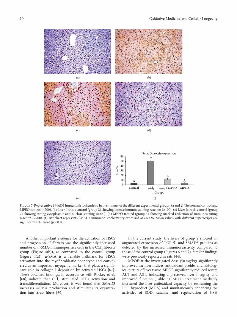
Another important evidence for the activation of HSCsand progression of fibrosis was the significantly increasednumber of α-SMA-immunopositive cells in the CCl4 fibrosisgroup (Figure 4(b)), as compared to the control group(Figure 4(a)). α-SMA is a reliable hallmark for HSCsactivation into the myofibroblastic phenotype and consid-ered as an important myogenic marker that plays a signifi-cant role in collagen I deposition by activated HSCs [67].These obtained findings, in accordance with Rockey et al.[68], indicate that CCl4 stimulated HSCs activation andtransdifferentiation. Moreover, it was found that SMAD3increases α-SMA production and stimulates its organiza-tion into stress fibers [69].
In the current study, the livers of group 2 showed anaugmented expression of TGF-β1 and SMAD3 proteins asdetected by the increased immunoreactivity compared tothose of the control group (Figures 6 and 7). Similar findingswere previously reported in rats [44].
MPOE at the investigated dose (50mg/kg) significantlyimproved the liver indices, antioxidant profile, and histolog-ical picture of liver tissue. MPOE significantly reduced serumALT and AST, indicating a preserved liver integrity andimproved function (Table 3). MPOE treatment markedlyincreased the liver antioxidant capacity by restraining theLPO byproduct (MDA) and simultaneously enhancing theactivities of SOD, catalase, and regeneration of GSH
(a) (b)
(c) (d)
(e)
Smad 3 protein expression
BA
C
A
CCl4 CCl4 + MPEO MPEONormalGroups
0102030405060
Are
a %
(f)
Figure 7: Representive SMAD3 immunohistochemistry in liver tissues of the different experimental groups. (a and e) The normal control andMPEO control (×200). (b) Liver fibrosis control (group 2) showing intense immunostaining reaction (×100). (c) Liver fibrosis control (group2) showing strong cytoplasmic and nuclear staining (×200). (d) MPEO-treated (group 3) showing marked reduction of immunostainingreaction (×200). (f) Bar chart represents SMAD3 immunohistochemistry expressed as area %. Mean values with different superscripts aresignificantly different (p < 0 05).
10 Oxidative Medicine and Cellular Longevity
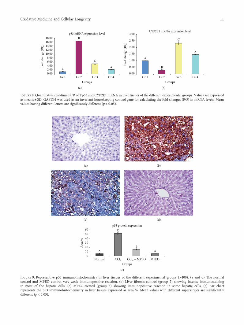
p53 mRNA expression level
A
B
C
A
Gr 2 Gr 3 Gr 4Gr 1Groups
0.002.004.006.008.00
10.0012.0014.0016.0018.00
Fold
chan
ge (R
Q)
(a)
CYP2E1 mRNA expression level
C
A
A
B
0.00
0.50
1.00
1.50
2.00
2.50
3.00
Fold
chan
ge (R
Q)
Gr 2 Gr 3 Gr 4Gr 1Groups
(b)
Figure 8: Quantitative real-time PCR of Tp53 and CYP2E1 mRNA in liver tissues of the different experimental groups. Values are expressedas means± SD. GAPDH was used as an invariant housekeeping control gene for calculating the fold changes (RQ) in mRNA levels. Meanvalues having different letters are significantly different (p < 0 05).
(a) (b)
(c) (d)
Normal CCl4 CCl4 + MPEO MPEOGroups
p53 protein expressionC
BAA
0102030405060
Are
a %
(e)
Figure 9: Representive p53 immunohistochemistry in liver tissues of the different experimental groups (×400). (a and d) The normalcontrol and MPEO control very weak immunopositive reaction. (b) Liver fibrosis control (group 2) showing intense immunostainingin most of the hepatic cells. (c) MPEO-treated (group 3) showing immunopositive reaction in some hepatic cells. (e) Bar chartrepresents the p53 immunohistochemistry in liver tissues expressed as area %. Mean values with different superscripts are significantlydifferent (p < 0 05).
11Oxidative Medicine and Cellular Longevity
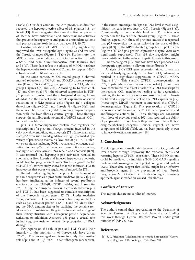
(Table 4). Our data come in line with previous studies thatreported the hepatoprotective effect of M. piperita [18] orits oil [19]. It was suggested that several active componentsof Mentha have antioxidant and antiperoxidant activitiesthat provoke the capacity of endogenous antioxidant systemsand protect against toxic hepatic damages [18, 19, 70].
Coadministration of MPOE with CCl4 significantlyimproved the liver histopathology (Figure 2) and reducedthe fibrotic changes (Figure 3, Table 5). Furthermore, theMPOE-treated group showed a marked reduction in bothα-SMA- and desmin-immunopositive cells (Figures 4(c)and 5(c)). These data reflect the efficacy of MPOE to reducethe hepatocellular toxic effects of CCl4 and to suppress HSCsactivation and proliferation as well.
In the same context, MPOE-treated group 3 showedmarked reductions in TGF-β1 and SMAD3 proteins expres-sion (Figures 6(c) and 7(c)) compared to the CCl4 fibrosisgroup (Figures 6(b) and 7(b)). According to Kanzler et al.[47] and Chen et al. [71], the observed suppression in TGF-β1 protein expression and the subsequent downregulationof SMAD3 in the livers of group 3 could explain the observedreduction of α-SMA-positive cells (Figure 4(c)), collagendeposition (Figure 3(c)), and fibrosis % (Figure 3(e)) andthe reduced fibrosis scores (Table 5) in this group in compar-ison with the liver fibrosis control group. These findingssupport the antifibrogenic potential of MPOE against CCl4-induced liver fibrosis.
p53 is a tumor-suppressor protein that regulates thetranscription of a plethora of target proteins involved in thecell cycle, differentiation, and apoptosis [72]. In normal redoxstate, p53 expression and degradation are tightly regulated by avariety of proteins to maintain a low level of p53 [73]. Differ-ent stress signals including ROS, hypoxia, and oncogene acti-vation induce p53 that becomes transcriptionally active,leading to cell cycle arrest, DNA repair, and apoptosis [8, 9].It was reported that hepatocyte p53 activation resulted inspontaneous liver fibrosis and induced hepatocyte apoptosis,in addition to upregulation of connective tissue growth factor(CTGF) [74]. In vitro study showed that p53 induces CTGF inhepatocytes that occur via regulation of microRNA [75].
Recent studies highlighted the possible involvement ofp53 in fibrogenesis as a profibrotic mediator [8, 9, 74]. p53has been implicated as an inducer of several profibroticeffectors such as TGF-β1, CTGF, α-SMA, and fibronectin[76]. During the fibrogenic process, a crosstalk between p53and TGF-β1 has been suggested to stimulate transcriptionof the fibrogenic target genes [77, 78]. Under oxidativestress, excessive ROS induces various transcription factorssuch as p53, activator protein-1 (AP-1), and NF-κB by alter-ing the DNA binding sites or by oxidizing the cysteine res-idues of such proteins resulting in conformational change oftheir tertiary structure with subsequent protein degradationactivation or inhibition. Activated p53 plays a crucial rolein inducing apoptosis to prevent the propagation of DNAdamage [72].
Few reports on the role of p53 and TGF-β1 and theirinterplay in the mechanism of fibrogenesis have arisen[75–78]. This encouraged our team to study the possiblerole of p53 and TGF-β1 in MPEO antifibrogenic mechanism.
In the current investigation, Tp53 mRNA level showed a sig-nificant increase in response to CCl4 fibrosis (Figure 8(a)).Consequently, a considerable level of p53 protein wasdetected in the livers of the fibrosis group (Figure 9). Thesefindings agreed with those of previous studies that reportedthat p53 expression increases following the onset of liverinjury [8, 9]. In the MPOE-treated group, both Tp53 mRNA(Figure 8(a)) and p53 protein expression (Figure 9(c)) weresignificantly suppressed. This p53 downregulation mighthave contributed to the reduced hepatic fibrosis in this group.
Pharmacological p53 inhibitors have been proposed as atherapeutic application to alleviate tissue fibrosis [8].
Analysis of CYP2E1 gene expression aimed to evaluatefor the detoxifying capacity of the liver. CCl4 intoxicationresulted in a significant suppression in CYP2E1 mRNA(Figure 8(b)). This specific CYP2E1 downregulation inCCl4 hepatic fibrosis was previously reported [18] and mayhave contributed to a direct attack of CYP2E1 transcript bythe reactive CCl4 metabolites leading to its degradation.Besides, the inflammatory response associated with fibrosiscould exert a suppressive effect on CYP2E1 expression [79].Interestingly, MPOE treatment counteracted this CYP2E1downregulation (Figure 8). This preservation of CYP2E1expression could be one of the MPOE hepatoprotective andantifibrogenic actions [80]. These findings are consistentwith those of previous studies [62] that reported the abilityof peppermint to modulate both phase I and phase II liverdrug-metabolizing enzymes. Besides, eugenol, an activecomponent of MPOE (Table 2), has been previously shownto induce detoxification enzymes [18].
5. Conclusion
MPEO significantly ameliorates the severity of CCl4-inducedliver fibrosis through improving the oxidative status andrestoring hepatic CYP2E1 expression. These MPEO actionscould be mediated by inhibiting TGF-β1/SMAD signalingproteins and downregulation of p53 at both gene and proteinlevels. These data suggest that MPEO might be an effectiveantifibrogenic agent in the prevention of liver fibrosisprogression. MPEO could help in developing a promisingapproach against oxidation-caused liver fibrosis.
Conflicts of Interest
The authors declare no conflict of interest.
Acknowledgments
The authors extend their appreciation to the Deanship ofScientific Research at King Khalid University for fundingthis work through General Research Project under grantnumber (G.R.P-267-38).
References
[1] S. L. Friedman, “Mechanisms of hepatic fibrogenesis,” Gastro-enterology, vol. 134, no. 6, pp. 1655–1669, 2008.
12 Oxidative Medicine and Cellular Longevity
[2] X. Zhang, X. Han, L. Yin et al., “Potent effects of dioscinagainst liver fibrosis,” Scientific Reports, vol. 5, no. 1, article9713, 2015.
[3] S. Li, H.-Y. Tan, N. Wang et al., “The role of oxidative stressand antioxidants in liver diseases,” International Journal ofMolecular Sciences, vol. 16, no. 11, pp. 26087–26124, 2015.
[4] N. C. Henderson and D. Sheppard, “Integrin-mediatedregulation of TGFβ in fibrosis,” Biochimica et Biophysica Acta(BBA) - Molecular Basis of Disease, vol. 1832, no. 7, pp. 891–896, 2013.
[5] J. Rosenbloom, F. A. Mendoza, and S. A. Jimenez, “Strategiesfor anti-fibrotic therapies,” Biochimica et Biophysica Acta(BBA) - Molecular Basis of Disease, vol. 1832, no. 7, pp. 1088–1103, 2013.
[6] F. Verrecchia and A. Mauviel, “Transforming growth factor-βand fibrosis,” World Journal of Gastroenterology, vol. 13,no. 22, pp. 3056–3062, 2007.
[7] K. H. Vousden and X. Lu, “Live or let die: the cell’s response top53,” Nature Reviews Cancer, vol. 2, no. 8, pp. 594–604,2002.
[8] T. Kodama, T. Takehara, H. Hikita et al., “Increases inp53 expression induce CTGF synthesis by mouse andhuman hepatocytes and result in liver fibrosis in mice,” TheJournal of Clinical Investigation, vol. 121, no. 8, pp. 3343–3356, 2011.
[9] A. M. Attallah, G. E. Shiha, H. Ismail, S. E. Mansy,R. El-Sherbiny, and I. El-Dosoky, “Expression of p53 pro-tein in liver and sera of patients with liver fibrosis, livercirrhosis or hepatocellular carcinoma associated with chronicHCV infection,” Clinical Biochemistry, vol. 42, no. 6,pp. 455–461, 2009.
[10] J. P. Simon and S. Evan Prince, “Natural remedies for non-steroidal anti-inflammatory drug-induced toxicity,” Journalof Applied Toxicology, vol. 37, no. 1, pp. 71–83, 2017.
[11] F. Cheung, Y. Feng, N. Wang, M. F. Yuen, Y. Tong, and V. T.Wong, “Effectiveness of Chinese herbal medicine in treatingliver fibrosis: a systematic review and meta-analysis ofrandomized controlled trials,” Chinese Medicine, vol. 7, no. 1,p. 5, 2012.
[12] T. A. Misharina, M. B. Terenina, and N. I. Krikunova, “Antiox-idant properties of essential oils,” Prikladnaia Biokhimiia iMikrobiologiia, vol. 45, no. 6, pp. 710–716, 2009.
[13] S. A. Sheweita, L. S. El-Hosseiny, and M. A. Nashashibi,“Protective effects of essential oils as natural antioxidantsagainst hepatotoxicity induced by cyclophosphamide in mice,”PLoS One, vol. 11, no. 11, article e0165667, 2016.
[14] H. A. Ogaly, N. A. Eltablawy, A. M. El-Behairy, H. El-Hindi,and R. M. Abd-Elsalam, “Hepatocyte growth factor mediatesthe antifibrogenic action of Ocimum bacilicum essential oilagainst CCl4-induced liver fibrosis in rats,” Molecules, vol. 20,no. 8, pp. 13518–13535, 2015.
[15] M. J. Saharkhiz, M. Motamedi, K. Zomorodian, K. Pakshir,R. Miri, and K. Hemyari, “Chemical composition, antifungaland antibiofilm activities of the essential oil of Menthapiperita L,” ISRN Pharmaceutics, vol. 2012, Article ID 718645,6 pages, 2012.
[16] M. B. Goudjil, S. Ladjel, S. E. Bencheikh, S. Zighmi, andD. Hamada, “Chemical composition, antibacterial andantioxidant activities of the essential oil extracted fromthe Mentha piperita of Southern Algeria,” Research Jour-nal of Phytochemistry, vol. 9, no. 2, pp. 79–87, 2015.
[17] N. N. Dejani, L. C. Souza, S. R. P. Oliveira et al., “Immunolog-ical and parasitological parameters in Schistosoma mansoni-infected mice treated with crude extract from the leavesof Mentha × piperita L,” Immunobiology, vol. 219, no. 8,pp. 627–632, 2014.
[18] A. Sharma, M. K. Sharma, and M. Kumar, “Protective effectof Mentha piperita against arsenic-induced toxicity in liverof Swiss albino mice,” Basic & Clinical Pharmacology &Toxicology, vol. 100, no. 4, pp. 249–257, 2007.
[19] A. F. Khalil, H. O. Elkatry, and H. F. El Mehairy, “Protectiveeffect of peppermint and parsley leaves oils against hepatotox-icity on experimental rats,” Annals of Agricultural Sciences,vol. 60, no. 2, pp. 353–359, 2015.
[20] A. Marjani, R. Rahmati, A. R. Mansourian, and G. Veghary,“Effect of peppermint oil on serum lipid peroxidation andhepatic enzymes after immobility stress in mice,” The OpenBiochemistry Journal, vol. 6, no. 1, pp. 51–55, 2012.
[21] M. G. Zaia, T. di Orlando Cagnazzo, K. A. Feitosa et al., “Anti-inflammatory properties of menthol and menthone in Schisto-soma mansoni infection,” Frontiers in Pharmacology, vol. 7,p. 170, 2016.
[22] S. Reitman and S. Frankel, “A colorimetric method for thedetermination of serum glutamic oxalacetic and glutamicpyruvic transaminases,” American Journal of Clinical Pathol-ogy, vol. 28, no. 1, pp. 56–63, 1957.
[23] K. M. Miranda, M. G. Espey, and D. A. Wink, “A rapid,simple spectrophotometric method for simultaneous detec-tion of nitrate and nitrite,” Nitric Oxide, vol. 5, no. 1,pp. 62–71, 2001.
[24] Z. A. Placer, L. L. Cushman, and B. C. Johnson, “Estimationof product of lipid peroxidation (malonyl dialdehyde) inbiochemical systems,” Analytical Biochemistry, vol. 16, no. 2,pp. 359–364, 1966.
[25] A. Nandi and Ι. B. Chatterjee, “Assay of superoxide dismutaseactivity in animal tissues,” Journal of Biosciences, vol. 13, no. 3,pp. 305–315, 1988.
[26] A. K. Sinha, “Colorimetric assay of catalase,” AnalyticalBiochemistry, vol. 47, no. 2, pp. 389–394, 1972.
[27] E. Beutler, O. Duron, and B. M. Kelly, “Improved methodfor the determination of blood glutathione,” The Journalof Laboratory and Clinical Medicine, vol. 61, pp. 882–888,1963.
[28] M. M. Bradford, “A rapid and sensitive method for thequantitation of microgram quantities of protein utilizing theprinciple of protein-dye binding,” Analytical Biochemistry,vol. 72, no. 1-2, pp. 248–254, 1976.
[29] K. Ishak, A. Baptista, L. Bianchi et al., “Histological gradingand staging of chronic hepatitis,” Journal of Hepatology,vol. 22, no. 6, pp. 696–699, 1995.
[30] K. J. Livak and T. D. Schmittgen, “Analysis of relative geneexpression data using real-time quantitative PCR and the2−ΔΔCT method,” Methods, vol. 25, no. 4, pp. 402–408, 2001.
[31] S. L. Devi and C. V. Anuradha, “Oxidative and nitrosativestress in experimental rat liver fibrosis: protective effect of tau-rine,” Environmental Toxicology and Pharmacology, vol. 29,no. 2, pp. 104–110, 2010.
[32] S. Li, M. Hong, H. Y. Tan, N. Wang, and Y. Feng,“Insights into the role and interdependence of oxidativestress and inflammation in liver diseases,” Oxidative Medi-cine and Cellular Longevity, vol. 2016, Article ID 4234061,21 pages, 2016.
13Oxidative Medicine and Cellular Longevity
[33] C. Trautwein, S. L. Friedman, D. Schuppan, and M. Pinzani,“Hepatic fibrosis: concept to treatment,” Journal of Hepatol-ogy, vol. 62, no. 1, Supplement 1, pp. S15–S24, 2015.
[34] P. Ramachandran, J. P. Iredale, and J. A. Fallowfield, “Res-olution of liver fibrosis: basic mechanisms and clinicalrelevance,” Seminars in Liver Disease, vol. 35, no. 2,pp. 119–131, 2015.
[35] S. M. Sharafi, I. Rasooli, P. Owlia, M. Taghizadeh, and S. D. A.Astaneh, “Protective effects of bioactive phytochemicals fromMentha piperita with multiple health potentials,” Pharmacog-nosy Magazine, vol. 6, no. 23, pp. 147–153, 2010.
[36] D. L. McKay and J. B. Blumberg, “A review of the bioactivityand potential health benefits of peppermint tea (Menthapiperita L.),” Phytotherapy Research, vol. 20, no. 8, pp. 619–633, 2006.
[37] R. Singh, M. A. M. Shushni, and A. Belkheir, “Antibacterialand antioxidant activities of Mentha piperita L,” ArabianJournal of Chemistry, vol. 8, pp. 322–328, 2011.
[38] Z. Sun, H. Wang, J. Wang, L. Zhou, and P. Yang, “Chemicalcomposition and antiinflammatory, cytotoxic and antioxidantactivities of essential oil from leaves ofMentha piperita grownin China,” PLoS One, vol. 9, no. 12, article e114767, 2014.
[39] R. da Silva Ramos, A. B. L. Rodrigues, A. L. F. Farias et al.,“Chemical composition and in vitro antioxidant, cytotoxic,antimicrobial, and larvicidal activities of the essential oil ofMentha piperita L. (Lamiaceae),” The Scientific World Journal,vol. 2017, Article ID 4927214, 8 pages, 2017.
[40] A. K. Tyagi and A. Malik, “Antimicrobial potential andchemical composition of Mentha piperita oil in liquid andvapour phase against food spoiling microorganisms,” FoodControl, vol. 22, no. 11, pp. 1707–1714, 2011.
[41] N. Ahmad, H. Fazal, I. Ahmad, and B. H. Abbasi, “Freeradical scavenging (DPPH) potential in nine Mentha spe-cies,” Toxicology and Industrial Health, vol. 28, no. 1,pp. 83–89, 2012.
[42] M. S. Brewer, “Natural antioxidants: sources, compounds,mechanisms of action, and potential applications,” Compre-hensive Reviews in Food Science and Food Safety, vol. 10,no. 4, pp. 221–247, 2011.
[43] L. Niu, X. Cui, Y. Qi et al., “Involvement of TGF-β1/Smad3signaling in carbon tetrachloride-induced acute liver injuryin mice,” PLoS One, vol. 11, no. 5, article e0156090, 2016.
[44] A. M. Abdel-Moneim, M. A. Al-Kahtani, M. A. El-Kersh, andM. A. Al-Omair, “Free radical-scavenging, anti-inflammatory/anti-fibrotic and hepatoprotective actions of taurine andsilymarin against CCl4 induced rat liver damage,” PLoS One,vol. 10, no. 12, article e0144509, 2015.
[45] W. R. Bi, C. Q. Yang, and Q. Shi, “Transforming growthfactor-β1 induced epithelial-mesenchymal transition inhepatic fibrosis,” Hepato-Gastroenterology, vol. 59, no. 118,pp. 1960–1963, 2012.
[46] Y. Inagaki and I. Okazaki, “Emerging insights into transform-ing growth factor β Smad signal in hepatic fibrogenesis,” Gut,vol. 56, no. 2, pp. 284–292, 2007.
[47] S. Kanzler, A. W. Lohse, A. Keil et al., “TGF-β1 in liver fibrosis:an inducible transgenic mouse model to study liver fibrogen-esis,” American Journal of Physiology-Gastrointestinal and LiverPhysiology, vol. 276, no. 4, Part 1, pp. G1059–G1068, 1999.
[48] X.-M. Meng, A. C. K. Chung, and H. Y. Lan, “Role of the TGF-β/BMP-7/Smad pathways in renal diseases,” Clinical Science,vol. 124, no. 4, pp. 243–254, 2013.
[49] F. Xie, Z. Zhang, H. van Dam, L. Zhang, and F. Zhou,“Regulation of TGF-β superfamily signaling by SMADmono-ubiquitination,” Cell, vol. 3, no. 4, pp. 981–993, 2014.
[50] Q. Y. Yao, B. L. Xu, J. Y. Wang, H. C. Liu, S. C. Zhang, andC. T. Tu, “Inhibition by curcumin of multiple sites of the trans-forming growth factor-beta1 signalling pathway amelioratesthe progression of liver fibrosis induced by carbon tetrachlo-ride in rats,” BMC Complementary & Alternative Medicine,vol. 12, no. 1, p. 156, 2012.
[51] G. Latella, A. Vetuschi, R. Sferra et al., “Targeted disrup-tion of Smad3 confers resistance to the development ofdimethylnitrosamine-induced hepatic fibrosis in mice,” LiverInternational, vol. 29, no. 7, pp. 997–1009, 2009.
[52] A. Masszi and A. Kapus, “Smaddening complexity: the role ofSmad3 in epithelial-myofibroblast transition,” Cells, Tissues,Organs, vol. 193, no. 1-2, pp. 41–52, 2011.
[53] M. Sato, Y. Muragaki, S. Saika, A. B. Roberts, and A. Ooshima,“Targeted disruption of TGF-β1/Smad3 signaling protectsagainst renal tubulointerstitial fibrosis induced by unilateralureteral obstruction,” The Journal of Clinical Investigation,vol. 112, no. 10, pp. 1486–1494, 2003.
[54] M. Martinot-Peignoux, N. Boyer, D. Cazals-Hatem et al.,“Prospective study on anti-hepatitis C virus-positive patientswith persistently normal serum alanine transaminase with orwithout detectable serum hepatitis C virus RNA,” Hepatology,vol. 34, no. 5, pp. 1000–1005, 2001.
[55] M. Vuda, R. D'Souza, S. Upadhya et al., “Hepatoprotectiveand antioxidant activity of aqueous extract of Hybanthusenneaspermus against CCl4-induced liver injury in rats,”Experimental and Toxicologic Pathology, vol. 64, no. 7-8,pp. 855–859, 2012.
[56] T. M. Leung, M. L. Fung, E. C. Liong, T. Y. Lau, A. A. Nanji,and G. L. Tipoe, “Role of nitric oxide in the regulation of fibro-genic factors in experimental liver fibrosis in mice,” Histologyand Histopathology, vol. 26, no. 2, pp. 201–211, 2011.
[57] S. Sundaresan and P. Subramanian, “S-Allylcysteine inhibitscirculatory lipid peroxidation and promotes antioxidantsin N-nitrosodiethylamine-induced carcinogenesis,” PolishJournal of Pharmacology, vol. 55, no. 1, pp. 37–42, 2003.
[58] M. R. Khan and D. Ahmed, “Protective effects of Digeramuricata (L.) Mart. on testis against oxidative stress of carbontetrachloride in rat,” Food and Chemical Toxicology, vol. 47,no. 6, pp. 1393–1399, 2009.
[59] H. Ghaffari, M. Venkataramana, S. C. Nayaka et al., “Hepato-protective action of Orthosiphon diffusus (Benth.) methanolactive fraction through antioxidant mechanisms: an in vivoand in vitro evaluation,” Journal of Ethnopharmacology,vol. 149, no. 3, pp. 737–744, 2013.
[60] H. Sadeghi, S. Hosseinzadeh, M. Akbartabar Touri,M. Ghavamzadeh, and M. Jafari Barmak, “Hepatoprotectiveeffect of Rosa canina fruit extract against carbon tetrachlorideinduced hepatotoxicity in rat,”Avicenna Journal of Phytomedi-cine, vol. 6, no. 2, pp. 181–188, 2016.
[61] S. Iwai, R. Karim, M. Kitano et al., “Role of oxidative DNAdamage caused by carbon tetrachloride-induced liver injur-y—enhancement of MeIQ-induced glutathione S-transferaseplacental form-positive foci in rats,” Cancer Letters, vol. 179,no. 1, pp. 15–24, 2002.
[62] A. T. Sagor, M. R. H. Chowdhury, N. Tabassum, H. Hossain,M. M. Rahman, and M. A. Alam, “Supplementation of freshucche (Momordica charantia L. var. muricata Willd) pre-vented oxidative stress, fibrosis and hepatic damage in CCl4
14 Oxidative Medicine and Cellular Longevity
treated rats,” BMC Complementary & Alternative Medicine,vol. 15, no. 1, p. 115, 2015.
[63] O. M. Abdel Salam, A. A. Sleem, and N. Shafee, “Effect oftrazodone and nefazodone on hepatic injury induced bycarbon tetrachloride,” Drug Discoveries & Therapeutics,vol. 4, no. 4, pp. 285–297, 2010.
[64] G. A. Yacout, N. M. Elguindy, and E. F. El Azab, “Hepatopro-tective effect of basil (Ocimum basilicum L.) on CCl4-inducedliver fibrosis in rats,” African Journal of Biotechnology,vol. 11, no. 90, pp. 15702–15711, 2012.
[65] T. Fujii, B. C. Fuchs, S. Yamada et al., “Mouse model of carbontetrachloride induced liver fibrosis: histopathological changesand expression of CD133 and epidermal growth factor,”BMC Gastroenterology, vol. 10, no. 1, p. 79, 2010.
[66] C. Kordes, I. Sawitza, A. Müller-Marbach et al., “CD133+
hepatic stellate cells are progenitor cells,” Biochemical andBiophysical Research Communications, vol. 352, no. 2,pp. 410–417, 2007.
[67] S. Lotersztajn, B. Julien, F. Teixeira-Clerc, P. Grenard, andA. Mallat, “Hepatic fibrosis: molecular mechanisms and drugtargets,” Annual Review of Pharmacology and Toxicology,vol. 45, no. 1, pp. 605–628, 2005.
[68] D. C. Rockey, N. Weymouth, and Z. Shi, “Smooth muscle αactin (Acta2) and myofibroblast function during hepaticwound healing,” PLoS One, vol. 8, no. 10, article e77166,2013.
[69] F. Xu, C. Liu, D. Zhou, and L. Zhang, “TGF-β/SMAD pathwayand its regulation in hepatic fibrosis,” Journal of Histochemis-try & Cytochemistry, vol. 64, no. 3, pp. 157–167, 2016.
[70] P. Kumaravelu, S. Subramaniyam, D. P. Dakshinamoorthy,and N. S. Devaraj, “The antioxidant effect of eugenol onCCl4-induced erythrocyte damage in rats,” The Journal ofNutritional Biochemistry, vol. 7, no. 1, pp. 23–28, 1996.
[71] H. B. Chen, J. G. Rud, K. Lin, and L. Xu, “Nuclear targeting oftransforming growth factor-β-activated Smad complexes,”The Journal of Biological Chemistry, vol. 280, no. 22,pp. 21329–21336, 2005.
[72] K. H. Vousden and C. Prives, “Blinded by the light: thegrowing complexity of p53,” Cell, vol. 137, no. 3, pp. 413–431, 2009.
[73] J. P. Kruse and W. Gu, “Modes of p53 regulation,” Cell,vol. 137, no. 4, pp. 609–622, 2009.
[74] X.-F. Tian, F.-J. Ji, H.-L. Zang, and H. Cao, “Activation of themiR-34a/SIRT1/p53 signaling pathway contributes to theprogress of liver fibrosis via inducing apoptosis in hepatocytesbut not in HSCs,” PLoS One, vol. 11, no. 7, article e0158657,2016.
[75] D. Pollutri, L. Gramantieri, L. Bolondi, and F. Fornari, “TP53/microRNA interplay in hepatocellular carcinoma,” Interna-tional Journal of Molecular Sciences, vol. 17, no. 12, article2029, 2016.
[76] R. Samarakoon, A. D. Dobberfuhl, C. Cooley et al., “Inductionof renal fibrotic genes by TGF-β1 requires EGFR activation,p53 and reactive oxygen species,” Cellular Signalling, vol. 25,no. 11, pp. 2198–2209, 2013.
[77] J. M. Overstreet, R. Samarakoon, K. K. Meldrum, and P. J.Higgins, “Redox control of p53 in the transcriptionalregulation of TGF-β1 target genes through SMAD coopera-tivity,” Cellular Signalling, vol. 26, no. 7, pp. 1427–1436,2014.
[78] M. Cordenonsi, S. Dupont, S. Maretto, A. Insinga,C. Imbriano, and S. Piccolo, “Links between tumor suppres-sors: p53 is required for TGF-β gene responses by cooperatingwith Smads,” Cell, vol. 113, no. 3, pp. 301–314, 2003.
[79] D. S. Riddick, C. Lee, A. Bhathena et al., “Transcriptionalsuppression of cytochrome P450 genes by endogenous andexogenous chemicals,” Drug Metabolism and Disposition,vol. 32, no. 4, pp. 367–375, 2004.
[80] G. Xie, C. C. Wong, K. W. Cheng, L. Huang, P. P.Constantinides, and B. Rigas, “Regioselective oxidation ofphospho-NSAIDs by human cytochrome P450 and flavinmonooxygenase isoforms: implications for their pharmacoki-netic properties and safety,” British Journal of Pharmacology,vol. 167, no. 1, pp. 222–232, 2012.
[81] R. E. Mostafa, A. A. S. Salama, R. F. Abdel-Rahman, andH. A. Ogaly, “Hepato-and neuro-protective influences ofbiopropolis on thioacetamide-induced acute hepatic enceph-alopathy in rats,” Canadian Journal of Physiology and Phar-macology, vol. 95, no. 5, pp. 539–547, 2016.
[82] N. A. Eltablawy and H. A. Ogaly, “Responsiveness of p53expression and genetic mutation to CCl4-induced DNAdamage in rat’s liver,” International Journal of Genomics andProteomics, vol. 5, no. 1, pp. 99–105, 2014.
15Oxidative Medicine and Cellular Longevity
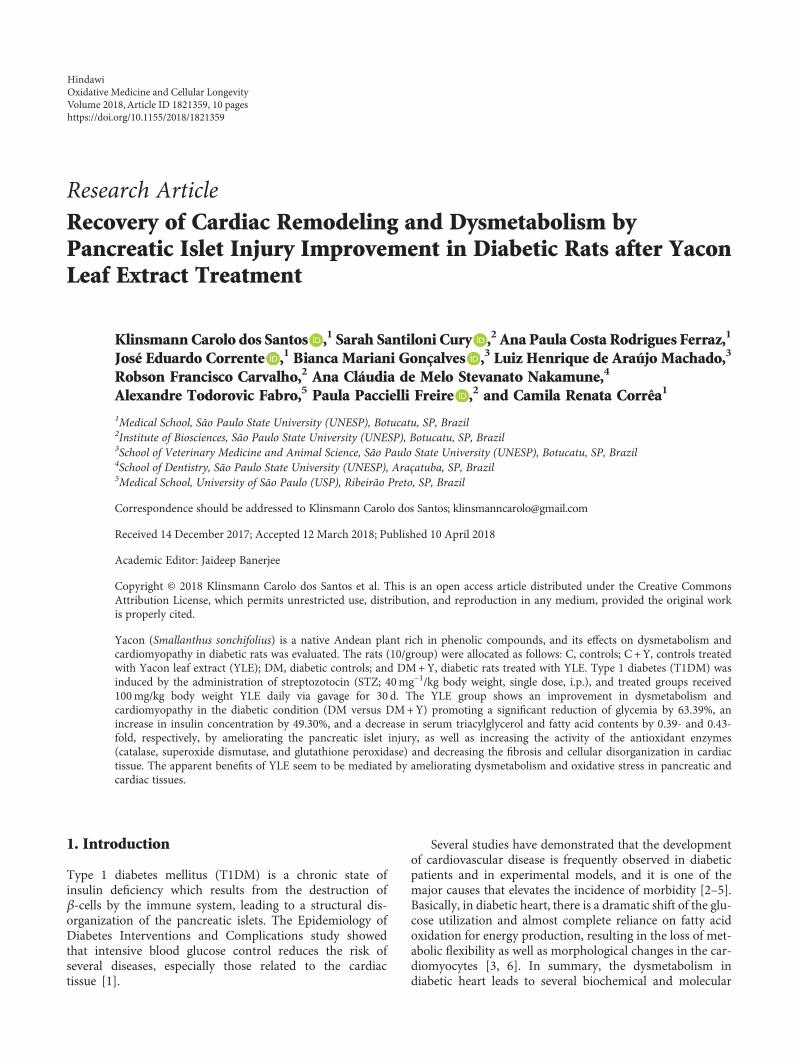
Research ArticleRecovery of Cardiac Remodeling and Dysmetabolism byPancreatic Islet Injury Improvement in Diabetic Rats after YaconLeaf Extract Treatment
Klinsmann Carolo dos Santos ,1 Sarah Santiloni Cury ,2Ana Paula Costa Rodrigues Ferraz,1
José Eduardo Corrente ,1 Bianca Mariani Gonçalves ,3 Luiz Henrique de Araújo Machado,3
Robson Francisco Carvalho,2 Ana Cláudia de Melo Stevanato Nakamune,4
Alexandre Todorovic Fabro,5 Paula Paccielli Freire ,2 and Camila Renata Corrêa1
1Medical School, São Paulo State University (UNESP), Botucatu, SP, Brazil2Institute of Biosciences, São Paulo State University (UNESP), Botucatu, SP, Brazil3School of Veterinary Medicine and Animal Science, São Paulo State University (UNESP), Botucatu, SP, Brazil4School of Dentistry, São Paulo State University (UNESP), Araçatuba, SP, Brazil5Medical School, University of São Paulo (USP), Ribeirão Preto, SP, Brazil
Correspondence should be addressed to Klinsmann Carolo dos Santos; [email protected]
Received 14 December 2017; Accepted 12 March 2018; Published 10 April 2018
Academic Editor: Jaideep Banerjee
Copyright © 2018 Klinsmann Carolo dos Santos et al. This is an open access article distributed under the Creative CommonsAttribution License, which permits unrestricted use, distribution, and reproduction in any medium, provided the original workis properly cited.
Yacon (Smallanthus sonchifolius) is a native Andean plant rich in phenolic compounds, and its effects on dysmetabolism andcardiomyopathy in diabetic rats was evaluated. The rats (10/group) were allocated as follows: C, controls; C +Y, controls treatedwith Yacon leaf extract (YLE); DM, diabetic controls; and DM+Y, diabetic rats treated with YLE. Type 1 diabetes (T1DM) wasinduced by the administration of streptozotocin (STZ; 40mg−1/kg body weight, single dose, i.p.), and treated groups received100mg/kg body weight YLE daily via gavage for 30 d. The YLE group shows an improvement in dysmetabolism andcardiomyopathy in the diabetic condition (DM versus DM+Y) promoting a significant reduction of glycemia by 63.39%, anincrease in insulin concentration by 49.30%, and a decrease in serum triacylglycerol and fatty acid contents by 0.39- and 0.43-fold, respectively, by ameliorating the pancreatic islet injury, as well as increasing the activity of the antioxidant enzymes(catalase, superoxide dismutase, and glutathione peroxidase) and decreasing the fibrosis and cellular disorganization in cardiactissue. The apparent benefits of YLE seem to be mediated by ameliorating dysmetabolism and oxidative stress in pancreatic andcardiac tissues.
1. Introduction
Type 1 diabetes mellitus (T1DM) is a chronic state ofinsulin deficiency which results from the destruction ofβ-cells by the immune system, leading to a structural dis-organization of the pancreatic islets. The Epidemiology ofDiabetes Interventions and Complications study showedthat intensive blood glucose control reduces the risk ofseveral diseases, especially those related to the cardiactissue [1].
Several studies have demonstrated that the developmentof cardiovascular disease is frequently observed in diabeticpatients and in experimental models, and it is one of themajor causes that elevates the incidence of morbidity [2–5].Basically, in diabetic heart, there is a dramatic shift of the glu-cose utilization and almost complete reliance on fatty acidoxidation for energy production, resulting in the loss of met-abolic flexibility as well as morphological changes in the car-diomyocytes [3, 6]. In summary, the dysmetabolism indiabetic heart leads to several biochemical and molecular
HindawiOxidative Medicine and Cellular LongevityVolume 2018, Article ID 1821359, 10 pageshttps://doi.org/10.1155/2018/1821359
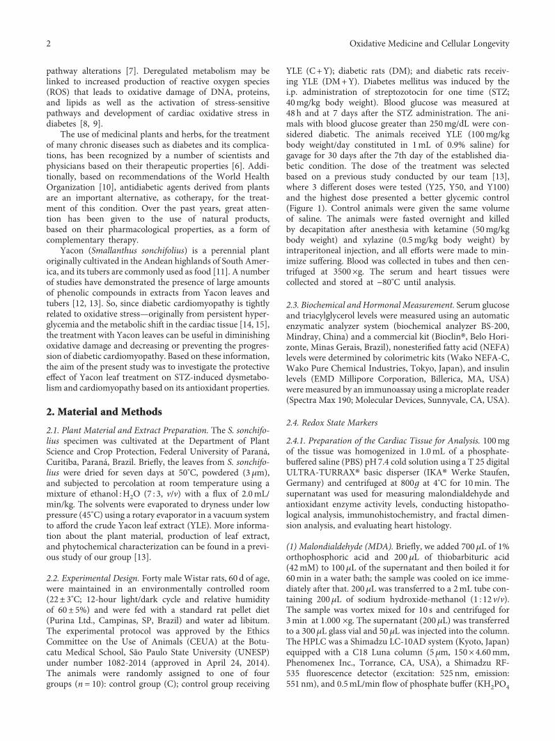
pathway alterations [7]. Deregulated metabolism may belinked to increased production of reactive oxygen species(ROS) that leads to oxidative damage of DNA, proteins,and lipids as well as the activation of stress-sensitivepathways and development of cardiac oxidative stress indiabetes [8, 9].
The use of medicinal plants and herbs, for the treatmentof many chronic diseases such as diabetes and its complica-tions, has been recognized by a number of scientists andphysicians based on their therapeutic properties [6]. Addi-tionally, based on recommendations of the World HealthOrganization [10], antidiabetic agents derived from plantsare an important alternative, as cotherapy, for the treat-ment of this condition. Over the past years, great atten-tion has been given to the use of natural products,based on their pharmacological properties, as a form ofcomplementary therapy.
Yacon (Smallanthus sonchifolius) is a perennial plantoriginally cultivated in the Andean highlands of South Amer-ica, and its tubers are commonly used as food [11]. A numberof studies have demonstrated the presence of large amountsof phenolic compounds in extracts from Yacon leaves andtubers [12, 13]. So, since diabetic cardiomyopathy is tightlyrelated to oxidative stress—originally from persistent hyper-glycemia and the metabolic shift in the cardiac tissue [14, 15],the treatment with Yacon leaves can be useful in diminishingoxidative damage and decreasing or preventing the progres-sion of diabetic cardiomyopathy. Based on these information,the aim of the present study was to investigate the protectiveeffect of Yacon leaf treatment on STZ-induced dysmetabo-lism and cardiomyopathy based on its antioxidant properties.
2. Material and Methods
2.1. Plant Material and Extract Preparation. The S. sonchifo-lius specimen was cultivated at the Department of PlantScience and Crop Protection, Federal University of Paraná,Curitiba, Paraná, Brazil. Briefly, the leaves from S. sonchifo-lius were dried for seven days at 50°C, powdered (3μm),and subjected to percolation at room temperature using amixture of ethanol :H2O (7 : 3, v/v) with a flux of 2.0mL/min/kg. The solvents were evaporated to dryness under lowpressure (45°C) using a rotary evaporator in a vacuum systemto afford the crude Yacon leaf extract (YLE). More informa-tion about the plant material, production of leaf extract,and phytochemical characterization can be found in a previ-ous study of our group [13].
2.2. Experimental Design. Forty male Wistar rats, 60 d of age,were maintained in an environmentally controlled room(22± 3°C; 12-hour light/dark cycle and relative humidityof 60± 5%) and were fed with a standard rat pellet diet(Purina Ltd., Campinas, SP, Brazil) and water ad libitum.The experimental protocol was approved by the EthicsCommittee on the Use of Animals (CEUA) at the Botu-catu Medical School, São Paulo State University (UNESP)under number 1082-2014 (approved in April 24, 2014).The animals were randomly assigned to one of fourgroups (n = 10): control group (C); control group receiving
YLE (C+Y); diabetic rats (DM); and diabetic rats receiv-ing YLE (DM+Y). Diabetes mellitus was induced by thei.p. administration of streptozotocin for one time (STZ;40mg/kg body weight). Blood glucose was measured at48 h and at 7 days after the STZ administration. The ani-mals with blood glucose greater than 250mg/dL were con-sidered diabetic. The animals received YLE (100mg/kgbody weight/day constituted in 1mL of 0.9% saline) forgavage for 30 days after the 7th day of the established dia-betic condition. The dose of the treatment was selectedbased on a previous study conducted by our team [13],where 3 different doses were tested (Y25, Y50, and Y100)and the highest dose presented a better glycemic control(Figure 1). Control animals were given the same volumeof saline. The animals were fasted overnight and killedby decapitation after anesthesia with ketamine (50mg/kgbody weight) and xylazine (0.5mg/kg body weight) byintraperitoneal injection, and all efforts were made to min-imize suffering. Blood was collected in tubes and then cen-trifuged at 3500×g. The serum and heart tissues werecollected and stored at −80°C until analysis.
2.3. Biochemical and Hormonal Measurement. Serum glucoseand triacylglycerol levels were measured using an automaticenzymatic analyzer system (biochemical analyzer BS-200,Mindray, China) and a commercial kit (Bioclin®, Belo Hori-zonte, Minas Gerais, Brazil), nonesterified fatty acid (NEFA)levels were determined by colorimetric kits (Wako NEFA-C,Wako Pure Chemical Industries, Tokyo, Japan), and insulinlevels (EMD Millipore Corporation, Billerica, MA, USA)were measured by an immunoassay using a microplate reader(Spectra Max 190; Molecular Devices, Sunnyvale, CA, USA).
2.4. Redox State Markers
2.4.1. Preparation of the Cardiac Tissue for Analysis. 100mgof the tissue was homogenized in 1.0mL of a phosphate-buffered saline (PBS) pH7.4 cold solution using a T 25 digitalULTRA-TURRAX® basic disperser (IKA® Werke Staufen,Germany) and centrifuged at 800g at 4°C for 10min. Thesupernatant was used for measuring malondialdehyde andantioxidant enzyme activity levels, conducting histopatho-logical analysis, immunohistochemistry, and fractal dimen-sion analysis, and evaluating heart histology.
(1) Malondialdehyde (MDA). Briefly, we added 700μL of 1%orthophosphoric acid and 200μL of thiobarbituric acid(42mM) to 100μL of the supernatant and then boiled it for60min in a water bath; the sample was cooled on ice imme-diately after that. 200μL was transferred to a 2mL tube con-taining 200μL of sodium hydroxide-methanol (1 : 12 v/v).The sample was vortex mixed for 10 s and centrifuged for3min at 1.000 ×g. The supernatant (200μL) was transferredto a 300μL glass vial and 50μL was injected into the column.The HPLC was a Shimadzu LC-10AD system (Kyoto, Japan)equipped with a C18 Luna column (5μm, 150× 4.60mm,Phenomenex Inc., Torrance, CA, USA), a Shimadzu RF-535 fluorescence detector (excitation: 525 nm, emission:551 nm), and 0.5mL/min flow of phosphate buffer (KH2PO4
2 Oxidative Medicine and Cellular Longevity
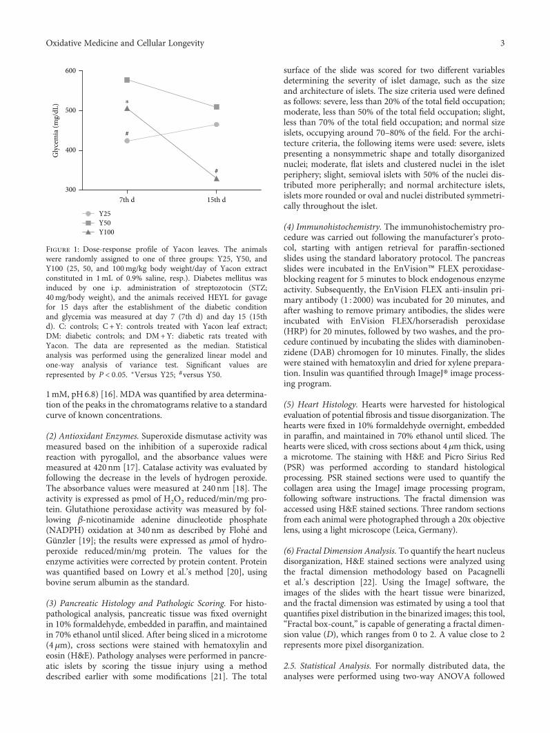
1mM, pH6.8) [16]. MDA was quantified by area determina-tion of the peaks in the chromatograms relative to a standardcurve of known concentrations.
(2) Antioxidant Enzymes. Superoxide dismutase activity wasmeasured based on the inhibition of a superoxide radicalreaction with pyrogallol, and the absorbance values weremeasured at 420nm [17]. Catalase activity was evaluated byfollowing the decrease in the levels of hydrogen peroxide.The absorbance values were measured at 240nm [18]. Theactivity is expressed as pmol of H2O2 reduced/min/mg pro-tein. Glutathione peroxidase activity was measured by fol-lowing β-nicotinamide adenine dinucleotide phosphate(NADPH) oxidation at 340nm as described by Flohé andGünzler [19]; the results were expressed as μmol of hydro-peroxide reduced/min/mg protein. The values for theenzyme activities were corrected by protein content. Proteinwas quantified based on Lowry et al.’s method [20], usingbovine serum albumin as the standard.
(3) Pancreatic Histology and Pathologic Scoring. For histo-pathological analysis, pancreatic tissue was fixed overnightin 10% formaldehyde, embedded in paraffin, and maintainedin 70% ethanol until sliced. After being sliced in a microtome(4μm), cross sections were stained with hematoxylin andeosin (H&E). Pathology analyses were performed in pancre-atic islets by scoring the tissue injury using a methoddescribed earlier with some modifications [21]. The total
surface of the slide was scored for two different variablesdetermining the severity of islet damage, such as the sizeand architecture of islets. The size criteria used were definedas follows: severe, less than 20% of the total field occupation;moderate, less than 50% of the total field occupation; slight,less than 70% of the total field occupation; and normal sizeislets, occupying around 70–80% of the field. For the archi-tecture criteria, the following items were used: severe, isletspresenting a nonsymmetric shape and totally disorganizednuclei; moderate, flat islets and clustered nuclei in the isletperiphery; slight, semioval islets with 50% of the nuclei dis-tributed more peripherally; and normal architecture islets,islets more rounded or oval and nuclei distributed symmetri-cally throughout the islet.
(4) Immunohistochemistry. The immunohistochemistry pro-cedure was carried out following the manufacturer’s proto-col, starting with antigen retrieval for paraffin-sectionedslides using the standard laboratory protocol. The pancreasslides were incubated in the EnVision™ FLEX peroxidase-blocking reagent for 5 minutes to block endogenous enzymeactivity. Subsequently, the EnVision FLEX anti-insulin pri-mary antibody (1 : 2000) was incubated for 20 minutes, andafter washing to remove primary antibodies, the slides wereincubated with EnVision FLEX/horseradish peroxidase(HRP) for 20 minutes, followed by two washes, and the pro-cedure continued by incubating the slides with diaminoben-zidene (DAB) chromogen for 10 minutes. Finally, the slideswere stained with hematoxylin and dried for xylene prepara-tion. Insulin was quantified through ImageJ® image process-ing program.
(5) Heart Histology. Hearts were harvested for histologicalevaluation of potential fibrosis and tissue disorganization. Thehearts were fixed in 10% formaldehyde overnight, embeddedin paraffin, and maintained in 70% ethanol until sliced. Thehearts were sliced, with cross sections about 4μm thick, usinga microtome. The staining with H&E and Picro Sirius Red(PSR) was performed according to standard histologicalprocessing. PSR stained sections were used to quantify thecollagen area using the ImageJ image processing program,following software instructions. The fractal dimension wasaccessed using H&E stained sections. Three random sectionsfrom each animal were photographed through a 20x objectivelens, using a light microscope (Leica, Germany).
(6) Fractal Dimension Analysis. To quantify the heart nucleusdisorganization, H&E stained sections were analyzed usingthe fractal dimension methodology based on Pacagnelliet al.’s description [22]. Using the ImageJ software, theimages of the slides with the heart tissue were binarized,and the fractal dimension was estimated by using a tool thatquantifies pixel distribution in the binarized images; this tool,“Fractal box-count,” is capable of generating a fractal dimen-sion value (D), which ranges from 0 to 2. A value close to 2represents more pixel disorganization.
2.5. Statistical Analysis. For normally distributed data, theanalyses were performed using two-way ANOVA followed
600
500
400
Gly
cem
ia (m
g/dL
)
3007th d
Y25
#
⁎
#
Y50Y100
15th d
Figure 1: Dose-response profile of Yacon leaves. The animalswere randomly assigned to one of three groups: Y25, Y50, andY100 (25, 50, and 100mg/kg body weight/day of Yacon extractconstituted in 1mL of 0.9% saline, resp.). Diabetes mellitus wasinduced by one i.p. administration of streptozotocin (STZ;40mg/body weight), and the animals received HEYL for gavagefor 15 days after the establishment of the diabetic conditionand glycemia was measured at day 7 (7th d) and day 15 (15thd). C: controls; C +Y: controls treated with Yacon leaf extract;DM: diabetic controls; and DM+Y: diabetic rats treated withYacon. The data are represented as the median. Statisticalanalysis was performed using the generalized linear model andone-way analysis of variance test. Significant values arerepresented by P < 0 05. ∗Versus Y25; #versus Y50.
3Oxidative Medicine and Cellular Longevity
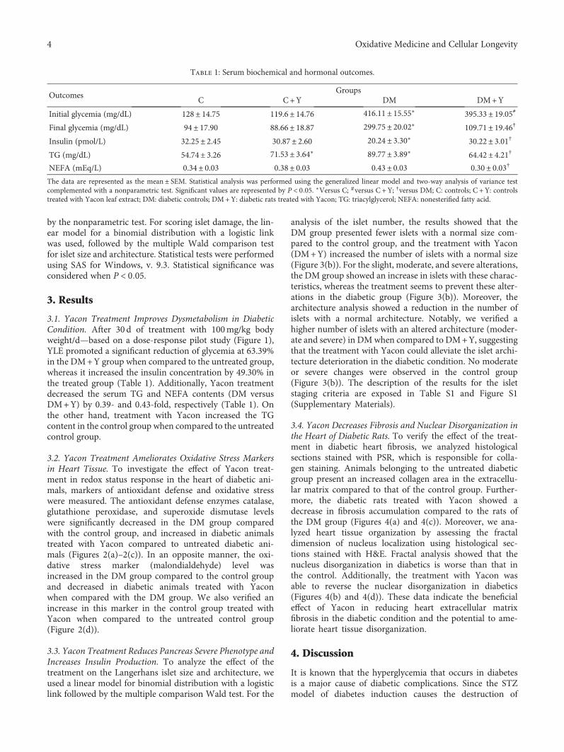
by the nonparametric test. For scoring islet damage, the lin-ear model for a binomial distribution with a logistic linkwas used, followed by the multiple Wald comparison testfor islet size and architecture. Statistical tests were performedusing SAS for Windows, v. 9.3. Statistical significance wasconsidered when P < 0 05.
3. Results
3.1. Yacon Treatment Improves Dysmetabolism in DiabeticCondition. After 30 d of treatment with 100mg/kg bodyweight/d—based on a dose-response pilot study (Figure 1),YLE promoted a significant reduction of glycemia at 63.39%in the DM+Y group when compared to the untreated group,whereas it increased the insulin concentration by 49.30% inthe treated group (Table 1). Additionally, Yacon treatmentdecreased the serum TG and NEFA contents (DM versusDM+Y) by 0.39- and 0.43-fold, respectively (Table 1). Onthe other hand, treatment with Yacon increased the TGcontent in the control group when compared to the untreatedcontrol group.
3.2. Yacon Treatment Ameliorates Oxidative Stress Markersin Heart Tissue. To investigate the effect of Yacon treat-ment in redox status response in the heart of diabetic ani-mals, markers of antioxidant defense and oxidative stresswere measured. The antioxidant defense enzymes catalase,glutathione peroxidase, and superoxide dismutase levelswere significantly decreased in the DM group comparedwith the control group, and increased in diabetic animalstreated with Yacon compared to untreated diabetic ani-mals (Figures 2(a)–2(c)). In an opposite manner, the oxi-dative stress marker (malondialdehyde) level wasincreased in the DM group compared to the control groupand decreased in diabetic animals treated with Yaconwhen compared with the DM group. We also verified anincrease in this marker in the control group treated withYacon when compared to the untreated control group(Figure 2(d)).
3.3. Yacon Treatment Reduces Pancreas Severe Phenotype andIncreases Insulin Production. To analyze the effect of thetreatment on the Langerhans islet size and architecture, weused a linear model for binomial distribution with a logisticlink followed by the multiple comparison Wald test. For the
analysis of the islet number, the results showed that theDM group presented fewer islets with a normal size com-pared to the control group, and the treatment with Yacon(DM+Y) increased the number of islets with a normal size(Figure 3(b)). For the slight, moderate, and severe alterations,the DM group showed an increase in islets with these charac-teristics, whereas the treatment seems to prevent these alter-ations in the diabetic group (Figure 3(b)). Moreover, thearchitecture analysis showed a reduction in the number ofislets with a normal architecture. Notably, we verified ahigher number of islets with an altered architecture (moder-ate and severe) in DMwhen compared to DM+Y, suggestingthat the treatment with Yacon could alleviate the islet archi-tecture deterioration in the diabetic condition. No moderateor severe changes were observed in the control group(Figure 3(b)). The description of the results for the isletstaging criteria are exposed in Table S1 and Figure S1(Supplementary Materials).
3.4. Yacon Decreases Fibrosis and Nuclear Disorganization inthe Heart of Diabetic Rats. To verify the effect of the treat-ment in diabetic heart fibrosis, we analyzed histologicalsections stained with PSR, which is responsible for colla-gen staining. Animals belonging to the untreated diabeticgroup present an increased collagen area in the extracellu-lar matrix compared to that of the control group. Further-more, the diabetic rats treated with Yacon showed adecrease in fibrosis accumulation compared to the rats ofthe DM group (Figures 4(a) and 4(c)). Moreover, we ana-lyzed heart tissue organization by assessing the fractaldimension of nucleus localization using histological sec-tions stained with H&E. Fractal analysis showed that thenucleus disorganization in diabetics is worse than that inthe control. Additionally, the treatment with Yacon wasable to reverse the nuclear disorganization in diabetics(Figures 4(b) and 4(d)). These data indicate the beneficialeffect of Yacon in reducing heart extracellular matrixfibrosis in the diabetic condition and the potential to ame-liorate heart tissue disorganization.
4. Discussion
It is known that the hyperglycemia that occurs in diabetesis a major cause of diabetic complications. Since the STZmodel of diabetes induction causes the destruction of
Table 1: Serum biochemical and hormonal outcomes.
OutcomesGroups
C C+Y DM DM+Y
Initial glycemia (mg/dL) 128± 14.75 119.6± 14.76 416.11± 15.55∗ 395.33± 19.05#
Final glycemia (mg/dL) 94± 17.90 88.66± 18.87 299.75± 20.02∗ 109.71± 19.46†
Insulin (pmol/L) 32.25± 2.45 30.87± 2.60 20.24± 3.30∗ 30.22± 3.01†
TG (mg/dL) 54.74± 3.26 71.53± 3.64∗ 89.77± 3.89∗ 64.42± 4.21†
NEFA (mEq/L) 0.34± 0.03 0.38± 0.03 0.43± 0.03 0.30± 0.03†
The data are represented as the mean ± SEM. Statistical analysis was performed using the generalized linear model and two-way analysis of variance testcomplemented with a nonparametric test. Significant values are represented by P < 0 05. ∗Versus C; #versus C + Y; †versus DM; C: controls; C + Y: controlstreated with Yacon leaf extract; DM: diabetic controls; DM+Y: diabetic rats treated with Yacon; TG: triacylglycerol; NEFA: nonesterified fatty acid.
4 Oxidative Medicine and Cellular Longevity
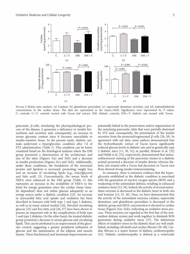
pancreatic β-cells, simulating the physiopathological pro-cess of the disease, it generates a deficiency in insulin bio-synthesis and secretion and, consequently, an increase inserum glycemia content since it becomes unavailable toinsulin-sensitive tissue. In the present study, diabetic ani-mals underwent a hyperglycemic condition after 7 d ofSTZ administration (Table 1). This condition can be bettervisualized based on the histological analyses where the DMgroup presented a deterioration of the architecture andsize of the islets (Figures 3(a) and 3(b)) and a decreasein insulin production (Figures 3(c) and 3(d)). Additionally,under these conditions, the breakdown of the structuralprotein and lipolysis is increased, promoting weight lossand an increase of circulating lipids (e.g., triacylglyceroland fatty acid) [3]. Concomitantly, the serum levels ofNEFA were enhanced in the DM group (Table 1); thisrepresents an increase in the availability of NEFA to theheart for energy generation since the cardiac tissue (insu-lin dependent) does not utilize glucose adequately as anenergy source under a diabetic condition [23]. An increasein myocardial fatty acid uptake and oxidation has beendescribed in humans with both type 1 and type 2 diabetes,as well as in many animal models [24]. Elevated circulatingglucose [25] and free fatty acid [26, 27] have been shown topossess an important role in the complications of both type1 and type 2 diabetes. On the other hand, the treated diabeticgroup presented a decrease in serum glucose and circulatinglipids. This can be attributed to an improvement of the glyce-mic control, suggesting a greater peripheral utilization ofglucose and the maintenance of the adipose and muscletissues. These biochemical and hormonal improvements are
potentially linked to the preservation and/or regeneration ofthe remaining pancreatic islets that were partially destroyedby STZ and, consequently, the potentiation of the insulinsecretion from the protected/regenerated β-cells [28, 29]. Inagreement with our data, some authors demonstrated thatthe hydroethanolic extract of Yacon leaves significantlyreduced glucose levels in diabetic rats and in genetically type2 diabetic mice [11, 30, 31]; in parallel, Honoré et al. [32]and Habib et al. [33], respectively, demonstrated that immu-nofluorescent staining of the pancreatic tissues in a diabeticanimal presented a decrease of insulin density whereas dia-betic rats treated with a Yacon leaf decoction or Yacon rootflour showed strong insulin immunostaining.
In summary, there is extensive evidence that the hyper-glycemia established in the diabetic condition is associatedwith the generation of reactive oxygen species (ROS) and aweakening of the antioxidant defense, resulting in enhancedoxidative stress [13, 34]. Indeed, the activity of several antiox-idant enzymes is decreased in the diabetic heart in both ratsand humans [13, 35, 36]. Here, we have demonstrated thatthe activity of the antioxidant enzymes catalase, superoxidedismutase, and glutathione peroxidase is decreased in thediabetic group and MDA concentration is elevated in cardiactissue (Figures 2(a)–2(d)), indicating an oxidative stress pro-cess. These enzymes are regarded as the first line of the anti-oxidant defense system and work together to diminish ROSgeneration during oxidative stress [37]. The deleteriouseffects of oxidative stress in the diabetic heart are well estab-lished, including cell death and cardiac fibrosis [38–40]. Car-diac fibrosis is a major feature of diabetic cardiomyopathy[41]. Diabetic cardiomyopathy is defined as a ventricular
4P = 0.018
C C + Y DM DM + Y
3
2
1
0
Cata
lase
(pm
ol/m
g pr
otei
n/m
in)
(a)
P = 0.021 P = 0.047
C C + Y DM DM + Y
4
3
2
1
0
Glu
tath
ione
per
oxid
ase
(�휇m
ol/m
g pr
otei
n/m
in)
(b)
P < 0.001P < 0.001
C C + Y DM DM + Y
120
60
0
Supe
roxi
de d
ismut
ase
(U/m
g pr
otei
n/m
in)
(c)
P < 0.001
C C + Y DM DM + Y
P < 0.001P = 0.016140
70
0
Mal
ondi
alde
hyde
(nm
ol/m
g pr
otei
n)(d)
Figure 2: Redox state markers. (a) Catalase; (b) glutathione peroxidase; (c) superoxide dismutase activities; and (d) malondialdehydeconcentration in the cardiac tissue. The data are represented as the mean± SEM. Significance were represented by P values.C: controls; C +Y: controls treated with Yacon leaf extract; DM: diabetic controls; DM+Y: diabetic rats treated with Yacon.
5Oxidative Medicine and Cellular Longevity
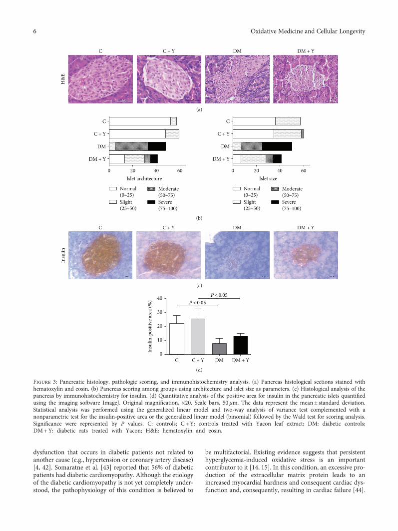
dysfunction that occurs in diabetic patients not related toanother cause (e.g., hypertension or coronary artery disease)[4, 42]. Somaratne et al. [43] reported that 56% of diabeticpatients had diabetic cardiomyopathy. Although the etiologyof the diabetic cardiomyopathy is not yet completely under-stood, the pathophysiology of this condition is believed to
be multifactorial. Existing evidence suggests that persistenthyperglycemia-induced oxidative stress is an importantcontributor to it [14, 15]. In this condition, an excessive pro-duction of the extracellular matrix protein leads to anincreased myocardial hardness and consequent cardiac dys-function and, consequently, resulting in cardiac failure [44].
C
H&
E
C + Y DM DM + Y
(a)
C
C + Y
DM DM
DM + Y
0 20Islet architecture
40 60 0 20Islet size
40 60
Normal(0–25)Slight(25–50)
Moderate(50–75)Severe(75–100)
C
C + Y
DM + Y
Normal(0–25)Slight(25–50)
Moderate(50–75)Severe(75–100)
(b)
C
Insu
lin
C + Y DM DM + Y
(c)
40
30
20
10
0Insu
lin-p
ositi
ve ar
ea (%
)
C C + Y
P < 0.05P < 0.05
DM DM + Y
(d)
Figure 3: Pancreatic histology, pathologic scoring, and immunohistochemistry analysis. (a) Pancreas histological sections stained withhematoxylin and eosin. (b) Pancreas scoring among groups using architecture and islet size as parameters. (c) Histological analysis of thepancreas by immunohistochemistry for insulin. (d) Quantitative analysis of the positive area for insulin in the pancreatic islets quantifiedusing the imaging software ImageJ. Original magnification, ×20. Scale bars, 50μm. The data represent the mean± standard deviation.Statistical analysis was performed using the generalized linear model and two-way analysis of variance test complemented with anonparametric test for the insulin-positive area or the generalized linear model (binomial) followed by the Wald test for scoring analysis.Significance were represented by P values. C: controls; C +Y: controls treated with Yacon leaf extract; DM: diabetic controls;DM+Y: diabetic rats treated with Yacon; H&E: hematoxylin and eosin.
6 Oxidative Medicine and Cellular Longevity
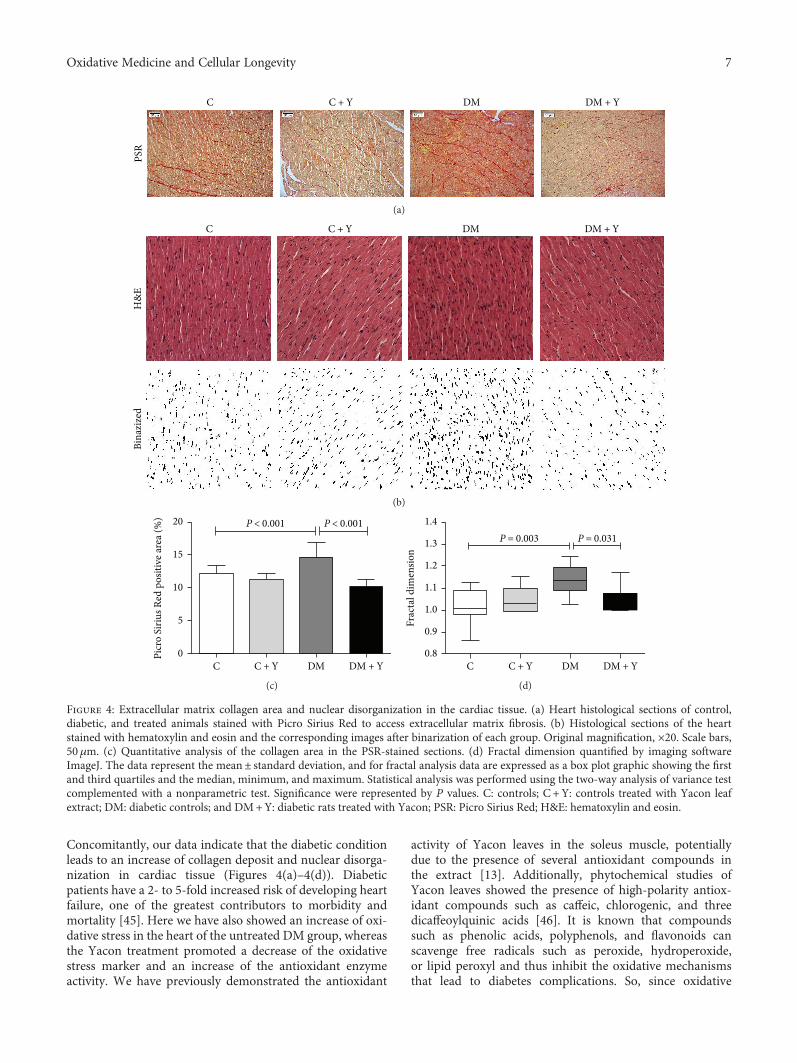
Concomitantly, our data indicate that the diabetic conditionleads to an increase of collagen deposit and nuclear disorga-nization in cardiac tissue (Figures 4(a)–4(d)). Diabeticpatients have a 2- to 5-fold increased risk of developing heartfailure, one of the greatest contributors to morbidity andmortality [45]. Here we have also showed an increase of oxi-dative stress in the heart of the untreated DM group, whereasthe Yacon treatment promoted a decrease of the oxidativestress marker and an increase of the antioxidant enzymeactivity. We have previously demonstrated the antioxidant
activity of Yacon leaves in the soleus muscle, potentiallydue to the presence of several antioxidant compounds inthe extract [13]. Additionally, phytochemical studies ofYacon leaves showed the presence of high-polarity antiox-idant compounds such as caffeic, chlorogenic, and threedicaffeoylquinic acids [46]. It is known that compoundssuch as phenolic acids, polyphenols, and flavonoids canscavenge free radicals such as peroxide, hydroperoxide,or lipid peroxyl and thus inhibit the oxidative mechanismsthat lead to diabetes complications. So, since oxidative
PSR
C C + Y DM DM + Y
(a)
Bina
zize
dH
&E
C C + Y DM DM + Y
(b)
20
15
10
5
0Picr
o Si
rius R
ed p
ositi
ve ar
ea (%
)
C C + Y DM DM + Y
P < 0.001 P < 0.001
(c)
1.4
1.3
1.2
1.1
1.0
0.9
0.8
Frac
tal d
imen
sion
C C + Y DM DM + Y
P = 0.003 P = 0.031
(d)
Figure 4: Extracellular matrix collagen area and nuclear disorganization in the cardiac tissue. (a) Heart histological sections of control,diabetic, and treated animals stained with Picro Sirius Red to access extracellular matrix fibrosis. (b) Histological sections of the heartstained with hematoxylin and eosin and the corresponding images after binarization of each group. Original magnification, ×20. Scale bars,50μm. (c) Quantitative analysis of the collagen area in the PSR-stained sections. (d) Fractal dimension quantified by imaging softwareImageJ. The data represent the mean± standard deviation, and for fractal analysis data are expressed as a box plot graphic showing the firstand third quartiles and the median, minimum, and maximum. Statistical analysis was performed using the two-way analysis of variance testcomplemented with a nonparametric test. Significance were represented by P values. C: controls; C +Y: controls treated with Yacon leafextract; DM: diabetic controls; and DM+Y: diabetic rats treated with Yacon; PSR: Picro Sirius Red; H&E: hematoxylin and eosin.
7Oxidative Medicine and Cellular Longevity
stress is related to the cardiac remodeling and fibrosisthrough several mechanisms [38, 39, 47], this could bethe mechanism by which DM+Y presented an improve-ment of the cardiac alterations mentioned above. Giventhe fundamental role of oxidative stress in the pathogene-sis of diabetes and diabetic cardiomyopathy, there is grow-ing interest in the use of antioxidants as a complementarytherapeutic approach to prevent/treat these conditions.Numerous studies demonstrated that ameliorating oxidativestress through antioxidant treatment might be an effectivestrategy for reducing diabetic cardiomyopathy [48, 49].
The important findings of this study are the cardio-and pancreatic protective effects of Yacon treatment inexperimental STZ-induced diabetic cardiomyopathy andpancreatic islet dysfunction in terms of the preservationof the Langerhans islet architecture and insulin productionas well as an inhibition of collagen content accumulationand enhancement of antioxidant enzyme activities in car-diac tissue. Summarizing, our results demonstrated thatSTZ administration successfully induced diabetes and dia-betic cardiomyopathy as indicated by the fractal analysis,indicating cellular disorganization, as well as an increasein collagen deposition in heart tissue and a decrease ofinsulin production and preservation of the architectureof the pancreatic β-cells. Interestingly, these cardiac andpancreatic abnormalities were improved by the adminis-tration of the Yacon extract. Furthermore, exploring thepotential therapeutic effects of plants and herbal medi-cine could contribute to the detection of new targetsand treatments. Our findings, even as a basic researchmodel, provide information that can guide future studiesaimed at elucidating new therapeutic alternatives fordiabetic complications.
Conflicts of Interest
The authors declare that there are no conflicts of interestregarding the publication of this paper.
Acknowledgments
The authors thank the São Paulo Research Foundation(FAPESP Grant no. 2018/05103-6) for financial supportand the Coordination of Improvement of Higher EducationPersonnel (CAPES) for scholar stipend.
Supplementary Materials
Table S1: analysis of the islets by scores. Description of theresults for the islet staging criteria (normal, slight, moderate,and severe) obtained after the linear model for a binomialdistribution with a logistic link followed by the multipleWald comparison test for islet size and architecture. C: con-trols; C+Y: controls treated with Yacon leaf extract; DM:diabetic controls; and DM+Y: diabetic rats treated withYacon; ND: no difference; NT: not tested. The linear modelfor a binomial distribution followed by the multiple Waldcomparison test analyzed statistical significance. Figure S1:analysis of the islets by scores. Description of the results forthe islet staging criteria (normal, slight, moderate, and
severe) obtained after the linear model for a binomial distri-bution with a logistic link followed by the multiple Waldcomparison test for islet size and architecture. Blue linesrepresent statistical significance while red lines mean nodifference (ND) or not tested (NT) among the groups.(Supplementary Materials)
References
[1] M. Copenhaver and R. P. Hoffman, “Type 1 diabetes: whereare we in 2017?,” Translational Pedriatrics, vol. 6, no. 4,pp. 359–364, 2017.
[2] T. Miki, S. Yuda, H. Kouzu, and T. Miura, “Diabetic cardiomy-opathy: pathophysiology and clinical features,” Heart FailureReviews, vol. 18, no. 2, pp. 149–166, 2013.
[3] M. Bayeva, K. T. Sawicki, and H. Ardehali, “Taking diabetes toheart—deregulation of myocardial lipid metabolism in dia-betic cardiomyopathy,” Journal of the American Heart Associ-ation, vol. 2, no. 6, article e000433, 2013.
[4] I. Falcão-Pires and A. F. Leite-moreira, “Diabetic cardiomyop-athy: understanding the molecular and cellular basis to prog-ress in diagnosis and treatment,” Heart Failure Reviews,vol. 17, no. 3, pp. 325–344, 2012.
[5] L. A. Suzuki, M. Poot, R. G. Gerrity, and K. E. Bornfeldt, “Dia-betes accelerates smooth muscle accumulation in lesions ofatherosclerosis: lack of direct growth-promoting effects of highglucose levels,” Diabetes, vol. 50, no. 4, pp. 851–860, 2001.
[6] J. Tian, Y. Zhao, Y. Liu, Y. Liu, K. Chen, and S. Lyu, “Rolesand mechanisms of herbal medicine for diabetic cardiomy-opathy: current status and perspective,” Oxidative Medicineand Cellular Longevity, vol. 2017, Article ID 8214541, 15pages, 2017.
[7] U. Varma, P. Koutsifeli, V. L. Benson, K. M. Mellor, and L. M.D. Delbridge, “Molecular mechanisms of cardiac pathology indiabetes—experimental insights,” Biochimica et BiophysicaActa (BBA) - Molecular Basis of Disease, 2017.
[8] H. Tsutsui, S. Kinugawa, S. Matsushima, and T. Yokota, “Oxi-dative stress in cardiac and skeletal muscle dysfunction associ-ated with diabetes mellitus,” Journal of Clinical Biochemistryand Nutrition, vol. 48, no. 1, pp. 68–71, 2011.
[9] T. V. Fiorentino, A. Prioletta, P. Zuo, and F. Folli, “Hypergly-cemia-induced oxidative stress and its role in diabetes mellitusrelated cardiovascular diseases,” Current PharmaceuticalDesign, vol. 19, no. 32, pp. 5695–5703, 2013.
[10] World Health Organization (WHO), Traditional MedicineStrategy 2005:1–6, http://www.wpro.who.int/health_technology/book_who_traditional_medicine_strategy_2002_2005.pdf.
[11] S. Baroni, F. Suzuki-Kemmelmeier, S. M. Caparroz-Assef,R. K. N. Cuman, and C. A. Bersani-Amado, “Effect of crudeextracts of leaves of Smallanthus sonchifolius (yacon) onglycemia in diabetic rats,” Revista Brasileira de CiênciasFarmacêuticas, vol. 44, no. 3, pp. 521–530, 2008.
[12] B. Simonovska, I. Vovk, S. Andrenšek, K. Valentová, andJ. Ulrichová, “Investigation of phenolic acids in yacon(Smallanthus sonchifolius) leaves and tubers,” Journal ofChromatography A, vol. 1016, no. 1, pp. 89–98, 2003.
[13] K. C. Dos Santos, B. G. Bueno, L. F. Pereira et al., “Yacon(Smallanthus sonchifolius) leaf extract attenuates hyperglyce-mia and skeletal muscle oxidative stress and inflammationin diabetic rats,” Evidence-Based Complement and Alterna-tive Medicine, vol. 2017, article 6418048, 9 pages, 2017.
8 Oxidative Medicine and Cellular Longevity
[14] S. Boudina and E. D. Abel, “Diabetic cardiomyopathy, causesand effects,” Reviews in Endocrine and Metabolic Disorders,vol. 11, no. 1, pp. 31–39, 2010.
[15] J. S. Dobrin, D. Lebeche, and G. L. L. Place, “Diabetic cardio-myopathy: signaling defects and therapeutic approaches,”Expert Review of Cardiovascular Therapy, vol. 8, no. 3,pp. 373–391, 2010.
[16] D. T. Pierine, M. E. L. Navarro, I. O. Minatel et al., “Lycopenesupplementation reduces TNF-α via RAGE in the kidney ofobese rats,” Nutrition & Diabetes, vol. 4, no. 11, article e142,2014.
[17] S. L. Marklund, “Product of extracellular-superoxide dismut-ase catalysis,” FEBS Letters, vol. 184, no. 2, pp. 237–239, 1985.
[18] H. Aebi, “[13] Catalase in vitro,” Methods in Enzymology,vol. 105, pp. 121–126, 1984.
[19] L. Flohé and W. A. Günzler, “[12] Assays of glutathioneperoxidase,” Methods in Enzymology, vol. 105, pp. 114–120, 1984.
[20] O. H. Lowry, N. J. Rosebrough, A. L. Farr, and R. J. Randall,“Protein measurement with the Folin phenol reagent,” TheJournal of Biological Chemistry, vol. 193, no. 1, pp. 265–275,1951.
[21] R. Jain, D. M. Tartar, R. K. Gregg et al., “Innocuous IFNγinduced by adjuvant-free antigen restores normoglycemiain NOD mice through inhibition of IL-17 production,”Journal of Experimental Medicine, vol. 205, no. 1, pp. 207–218, 2008.
[22] F. L. Pacagnelli, A. K. D. A. Sabela, T. B. Mariano et al., “Fractaldimension in quantifying experimental-pulmonary-hyperten-sion-induced cardiac dysfunction in rats,” Arquivos Brasileirosde Cardiologia, vol. 107, no. 1, pp. 33–39, 2016.
[23] C.M.Galhardi, Y. S. Diniz, L. A. Faine et al., “Toxicity of copperintake: lipid profile, oxidative stress and susceptibility to renaldysfunction,” Food and Chemical Toxicology, vol. 42, no. 12,pp. 2053–2060, 2004.
[24] E. D. Abel, S. E. Litwin, and G. Sweeney, “Cardiac remodelingin obesity,” Physiological Reviews, vol. 88, no. 2, pp. 389–419,2008.
[25] D. M. Nathan and for the DCCT/EDIC Research Group, “Thediabetes control and complications trial/epidemiology of dia-betes interventions and complications study at 30 years: over-view,” Diabetes Care, vol. 37, no. 1, pp. 9–16, 2014.
[26] H. C. Chiu, A. Kovacs, D. A. Ford et al., “A novel mouse modelof lipotoxic cardiomyopathy,” The Journal of Clinical Investi-gation, vol. 107, no. 7, pp. 813–822, 2001.
[27] H. Yagyu, G. Chen, M. Yokoyama et al., “Lipoprotein lipase(LpL) on the surface of cardiomyocytes increases lipid uptakeand produces a cardiomyopathy,” The Journal of ClinicalInvestigation, vol. 111, no. 3, pp. 419–426, 2003.
[28] M. J. Aybar, A. N. Sánchez Riera, A. Grau, and S. S. Sánchez,“Hypoglycemic effect of the water extract of Smallantus sonch-ifolius (yacon) leaves in normal and diabetic rats,” Journal ofEthnopharmacology, vol. 74, no. 2, pp. 125–132, 2001.
[29] K. Valentová, A. Lebeda, I. Dolezalová et al., “The biolog-ical and chemical variability of yacon,” Journal of Agricul-tural and Food Chemistry, vol. 54, no. 4, pp. 1347–1352,2006.
[30] T. M. Miura, Y. Itoh, T. Kaneko et al., “Corosolic acid inducesGLUT4 translocation in genetically type 2 diabetic mice,”Biological and Pharmaceutical Bulletin, vol. 27, no. 7,pp. 1103–1105, 2004.
[31] T. M. Miura, “Antidiabetic activity of Fuscoporia oblique andSamallanthus sonchifolius in genetically type 2 diabetic mice,”Journal of Traditional Medicine, vol. 24, no. 2, pp. 47–50, 2007.
[32] S. M. Honoré, W. M. Cabrera, S. B. Genta, and S. S. Sánchez,“Protective effect of yacon leaves decoction against earlynephropathy in experimental diabetic rats,” Food and Chemi-cal Toxicology, vol. 50, no. 5, pp. 1704–1715, 2012.
[33] N. C. Habib, S. M. Honoré, S. B. Genta, and S. S. Sánchez,“Hypolipidemic effect of Smallanthus sonchifolius (yacon)roots on diabetic rats: biochemical approach,” Chemico-Bio-logical Interactions, vol. 194, no. 1, pp. 31–39, 2011.
[34] P. T. Yeh, H. W. Huang, C. M. Yang, W. S. Yang, andC. H. Yang, “Astaxanthin inhibits expression of retinal oxi-dative stress and inflammatory mediators in streptozotocin-induced diabetic rats,” PloS One, vol. 11, no. 1, articlee0146438, 2016.
[35] J. S. Johansen, A. K. Harris, D. J. Rychly, and A. Ergul, “Oxida-tive stress and the use of antioxidants in diabetes: linking basicscience to clinical practice,” Cardiovascular Diabetology, vol. 4,no. 1, p. 5, 2005.
[36] K. C. dos Santos, C. P. Braga, P. O. Barbanera, F. Seiva, A. F.Junior, and A. Ange, “Cardiac energy metabolism and oxida-tive stress biomarkers in diabetic rat treated with resveratrol,”PLoS One, vol. 9, no. 7, article e102775, 2014.
[37] J. M. Matés, C. Pérez-Gómez, and I. Núñez de Castro, “Antiox-idant enzymes and human diseases,” Clinical Biochemistry,vol. 32, no. 8, pp. 595–603, 1999.
[38] A. Aneja, W. H. W. Tang, S. Bansilal, M. J. Garcia, and M. E.Farkouh, “Diabetic cardiomyopathy: insights into pathogene-sis, diagnostic challenges, and therapeutic options,” The Amer-ican Journal of Medicine, vol. 121, no. 9, pp. 748–757, 2008.
[39] Z. Y. Fang, J. B. Prins, and T. H. Marwick, “Diabetic cardiomy-opathy: evidence, mechanisms, and therapeutic implications,”Endocrine Reviews, vol. 25, no. 4, pp. 543–567, 2004.
[40] D. Cai, M. Yuan, D. F. Frantz et al., “Local and systemic insulinresistance resulting from hepatic activation of IKK-β and NF-κB,” Nature Medicine, vol. 11, no. 2, pp. 183–190, 2005.
[41] B. Li, Z. Zheng, Y. Wei et al., “Therapeutic effects ofneuregulin-1 in diabetic cardiomyopathy rats,” CardiovascularDiabetology, vol. 10, no. 1, p. 69, 2011.
[42] S. Boudina and E. D. Abel, “Diabetic cardiomyopathy revis-ited,” Circulation, vol. 115, no. 25, pp. 3213–3223, 2007.
[43] J. B. Somaratne, G. A. Whalley, K. K. Poppe et al., “Screeningfor left ventricular hypertrophy in patients with type 2 diabetesmellitus in the community,” Cardiovascular Diabetology,vol. 10, no. 1, p. 29, 2011.
[44] C. Li, L. Lv, H. Li, and D. Yu, “Cardiac fibrosis and dysfunctionin experimental diabetic cardiomyopathy are ameliorated byalpha-lipoic acid,” Cardiovascular Diabetology, vol. 11, no. 1,pp. 73–10, 2012.
[45] D. S. H. Bell, “Heart failure: the frequent, forgotten, and oftenfatal complication of diabetes,” Diabetes Care, vol. 26, no. 8,pp. 2433–2441, 2003.
[46] S. B. Genta, W. M. Cabrera, M. I. Mercado, A. Grau, C. A.Catalán, and S. S. Sánchez, “Hypoglycemic activity of leaforganic extracts from Smallanthus sonchifolius: constituentsof the most active fractions,” Chemico-Biological Interac-tions, vol. 185, no. 2, pp. 143–152, 2010.
[47] L. Cai, Y. Wang, G. Zhou et al., “Attenuation by metallothio-nein of early cardiac cell death via suppression of mitochon-drial oxidative stress results in a prevention of diabetic
9Oxidative Medicine and Cellular Longevity
cardiomyopathy,” Journal of the American College of Cardiol-ogy, vol. 48, no. 8, pp. 1688–1697, 2006.
[48] M. A. Haidara, H. Z. Yassin, M. Rateb, H. Ammar, and M. A.Zorkani, “Role of oxidative stress in development of cardiovas-cular complications in diabetes mellitus,” Current VascularPharmacology, vol. 4, no. 3, pp. 215–227, 2006.
[49] V. Kain, S. Kumar, and S. L. Sitasawad, “Azelnidipine preventscardiac dysfunction in streptozotocin-diabetic rats by reducingintracellular calcium accumulation, oxidative stress and apo-ptosis,” Cardiovascular Diabetology, vol. 10, no. 1, p. 97, 2011.
10 Oxidative Medicine and Cellular Longevity
Research ArticleThe Encapsulation of Lycopene in Nanoliposomes Enhances ItsProtective Potential in Methotrexate-Induced KidneyInjury Model
Nenad Stojiljkovic ,1 Sonja Ilic ,1 Vladimir Jakovljevic ,2,3 Nikola Stojanovic ,4
Slavica Stojnev ,5 Hristina Kocic,6 Marko Stojanovic ,4 and Gordana Kocic 7
1Department of Physiology, Faculty of Medicine, University of Niš, Bulevar Zorana Ðinđića 81, 18000 Niš, Serbia2Department of Physiology, Faculty of Medical Sciences, University of Kragujevac, Svetozara Markovica 69, 34000 Kragujevac, Serbia3Department of Human Pathology, I.M. Sechenov First Moscow State Medical University, Moscow, Russia4Faculty of Medicine, University of Niš, Bulevar Zorana Ðinđića 81, 18000 Niš, Serbia5Department of Pathology, Faculty of Medicine, University of Niš, Bulevar Zorana Ðinđića 81, 18000 Niš, Serbia6Faculty of Medicine, University of Maribor, Magdalenski trg 5, SI-2000 Maribor, Slovenia7Department of Biochemistry, Faculty of Medicine, University of Niš, Bulevar Zorana Ðinđića 81, 18000 Niš, Serbia
Correspondence should be addressed to Vladimir Jakovljevic; [email protected]
Received 3 November 2017; Accepted 1 January 2018; Published 13 March 2018
Academic Editor: Mithun Sinha
Copyright © 2018 Nenad Stojiljkovic et al. This is an open access article distributed under the Creative Commons AttributionLicense, which permits unrestricted use, distribution, and reproduction in any medium, provided the original work isproperly cited.
Methotrexate is an antimetabolic drug with a myriad of serious side effects including nephrotoxicity, which presumably occurs dueto oxidative tissue damage. Here, we evaluated the potential protective effect of lycopene, a potent antioxidant carotenoid, given intwo different pharmaceutical forms in methotrexate-induced kidney damage in rats. Serum biochemical (urea and creatinine) andtissue oxidative damage markers and histopathological kidney changes were evaluated after systemic administration of bothlycopene dissolved in corn oil and lycopene encapsulated in nanoliposomes. Similar to previous studies, single dose ofmethotrexate induced severe functional and morphological alterations of kidneys with cell desquamation, tubular vacuolation,and focal necrosis, which were followed by serum urea and creatinine increase and disturbances of tissue antioxidant status.Application of both forms of lycopene concomitantly with methotrexate ameliorated changes in serum urea and creatinine andoxidative damage markers and markedly reversed structural changes of kidney tissue. Moreover, animals that received lycopenein nanoliposome-encapsulated form showed higher degree of recovery than those treated with free lycopene form. The findingsof this study indicate that treatment with nanoliposome-encapsulated lycopene comparing to lycopene in standard vehicle hasan advantage as it more efficiently reduces methotrexate-induced kidney dysfunction.
1. Introduction
Methotrexate (MTX) is antimetabolic drug which is oftenused for the treatment of several autoimmune disorders suchas rheumatoid arthritis, psoriasis, and various malignanttumors including lymphoblastic leukemia, lymphoma, osteo-sarcoma, breast cancer, and head and neck cancer [1]. Clini-cal use of MTX is limited due to its side effects that includebone marrow suppression, hepatotoxicity, nephrotoxicity,pulmonary fibrosis, and gastrointestinal mucosal damage.
Since MTX is primarily excreted by kidneys [2], severity ofnephrotoxicity depends on both the dose and frequency ofMTX administration [3]. Similar to other agents with neph-rotoxic effect, MTX-induced renal impairment is clinicallyfollowed by hematuria and increased levels of serum creati-nine and urea in humans. These effects can be faithfullyreproduced in experimental animals after single dose ofMTX [4, 5].
Lycopene, a red-colored carotenoid, found mainly intomatoes but also in other fruits and vegetables (watermelons,
HindawiOxidative Medicine and Cellular LongevityVolume 2018, Article ID 2627917, 11 pageshttps://doi.org/10.1155/2018/2627917
Momordica cochinchinensis, Spreng fruit, papayas, etc.),possesses a strong antioxidant activity. It protects cells againstdamage caused by free radicals with its reactive oxygen species(ROS) scavenging properties [6, 7]. It is proven to be morepotent than similar antioxidants, like β-carotene and vitaminE [6, 8], largely due to its several conjugated double bonds.Besides the numerous beneficial properties (anti-inflamma-tory, anticancer, etc.), investigations revealed that lycopenehas protective effect in animalmodels of nephrotoxicity [8, 9].
Nanoliposomes are bilayer lipid vesicles which can beencapsulated with various bioactive agents includingmedicaments, pharmaceuticals, nutritional supplements,antioxidants, polynucleotides, and polypeptides [10]. Nanoli-posomes have the potential to increase solubility andbioavailability, in vitro and in vivo stability, improve time-controlled drug releasing, minimize concentrations requiredfor optimum therapeutic efficacy, enable cell-specific target-ing, and decrease adverse effects of drugs on healthy cellsand tissues [11, 12]. In the case of lycopene, formulation withnanoliposomes could prevent its rapid interaction withhighly reactive compounds, like plasma proteins or metalions, allowing it to reach the targeted damaged tissue. More-over, using nanoliposomes as carriers to incorporatelycopene into membranes might be an initial step in cellprevention as it is proposed that damage of cell membraneby ROS causes the onset of number of pathological eventsleading to oxidative injury [13].
Since, to the best of our knowledge, no study exists whichshows potential of using lycopene in this new pharmaceuticalformulation to treat drug-induced tissue injury, we aimed atevaluating characteristics of the nanoliposome encapsulationand efficacy of this preparation in preventing/amelioratingmethotrexate-induced kidney injury. To achieve this, we firstmeasured in vitro properties of encapsulated lycopene andthen compared its efficacy with free form lycopene throughthe evaluation of changes in serum and tissue oxidativestress markers and quantification of kidney tissue damagesinduced by MTX.
2. Materials and Methods
2.1. Drugs and Chemicals. Methotrexate was obtained fromEBEWE Pharma (Ges.m.b.H.NFG.KG, Austria), while keta-mine (Ketamidor 10%) was purchased from Richter Pharma(AG, Wels, Austria). Lycopene was purchased from Sigma-Aldrich (St. Louis, USA) and all other used chemicals wereobtained from either Sigma-Aldrich (St. Louis, USA) or CarlRoth (Karlsruhe, Germany).
2.2. Experimental Protocol In Vitro
2.2.1. Nanoliposomes Encapsulation with Lycopene. The 10%solution of phospholipid nanoparticles, in a form ofnanospheres, purchased from Nattermann Phospholipids(Germany), was encapsulated by lycopene at the concentra-tion of 4mg/ml. Encapsulated nanoparticles were isolatedby centrifugation at 6500g for 30min at 4°C according tothe method of Kocic et al. [13].
2.2.2. Efficacy of Lycopene Encapsulation. The efficacy ofencapsulation was determined based on a method previouslydescribed in great detail [14]. Briefly, after mixing thelycopene-loaded liposomes and petroleum ether (3ml), themixture was further vortexed for 3min followed by a centrifu-gation (2000 rpm, 5min). The upper layer was separated andits absorbance was measured at 470nm (V-1800 Shimadzuspectrophotometer). The encapsulation efficacy (%) wascalculated as (amount of incorporated lycopene)/(initialamount of added lycopene)× 100.
2.2.3. Sustained Release of Encapsulated Lycopene. The mix-ture of lycopene nanoparticles with phosphate buffer salinewas incubated at 37°C for 24 h from which the aliquots weretaken in order to measure the release of lycopene from nano-liposomes. At the defined time points (0, 1, 2, 4, 8, 12, and24 h), the aliquots were taken and diluted in petroleum etherin order to measure their absorbance at 470nm and quantifythe amount of free lycopene present. The content of lycopenewas calculated using a standard curve.
2.2.4. pH Dependent Stability of Encapsulated Lycopene. Thestability of free and encapsulated lycopene in different pHbuffers (6.5, 7.4, 8.0, and 9.0) was studied following previ-ously described method [15]. The mixture containing eitherfree or encapsulated lycopene and buffer solution with differ-ent pH values (1 : 10, v/v) was incubated at 37°C, from whichthe aliquots were taken after 0, 20, 40, 60, 120, and 180minutes. The residual rate of lycopene present in the mixtureat designated time point was determined spectrophotometri-cally at 470nm using a standard curve for lycopene. Residualrate is the residual amount of lycopene expressed in percent-ages in relation to the initial amount of lycopene.
2.2.5. Metal Ion Chelating Properties of EncapsulatedLycopene. The stability of formed lycopene nanoliposomesin the presence of different metal ions (K+, Ca2+, Mg2+,Al3+, or Cu2+) was evaluated according to the methoddescribed by Chen et al. [15]. The free/encapsulated lycopenewas incubated with 1mmol/l of metal solution for 2 h at 37°C.After the incubation period, the mixture was centrifuged inorder to separate the formed lycopene-metal chelate. Theobtained supernatant was mixed with petroleum ether, andthe absorbance of the solution was measured at 470 nm inorder to determine the residual rate of lycopene, where resid-ual rate is the residual amount of lycopene expressed inpercentages in relation to the initial amount of lycopene.
2.2.6. Susceptibility of Encapsulated Lycopene to H2O2. Aftercentrifugated nanoparticles were resuspended and both lyco-pene encapsulated and native nanoparticles as well as freelycopene were exposed to H2O2 for 30min at 37°C [13].The intensity of lipid peroxidation was determined using col-orimetric method that involves thiobarbituric acid as areagent [16].
2.3. Experimental Protocol In Vivo
2.3.1. Animals and Housing. Forty-eight male Wistar rats(200–250 g) were divided into 8 groups of 6 animals and
2 Oxidative Medicine and Cellular Longevity
maintained under standard laboratory conditions at theVivarium of the Institute of Biomedical Research, MedicalFaculty, Niš, Serbia. Laboratory was kept under standardtemperature (22°C) and humidity (60%), with equal durationof light/dark cycle. During the entire experiment, animals hadfree access to food andwater. All experiments were conductedat the Institute of Biomedical Research, Medical Faculty, Niš,Serbia, and are in accordance with all ethical regulations ofEuropean Union (EU Directive of 2010; 2010/63/EU) andRepublic of Serbia (332-07-00073/2017-05/1).
2.3.2. Animal Treatment. Treatment protocol included 8groups of 6 rats treated daily by an intraperitoneal injection(i.p.), as following:
(i) Control (C) group: the animals were given corn oil(0.2ml/day) for 10 days.
(ii) Nanoliposomes (NL) group: the animals weregiven empty nanoliposomes (10ml/kg) for 10 days.
(iii) Lycopene (LYC) group: the animals were givenlycopene dissolved in corn oil (6mg/kg) for 10 days.
(iv) Encapsulated nanoliposomes (ENL) group: the ani-mals were given encapsulated lycopene (6mg/kg)for 10 days.
(v) Methotrexate (MTX) group: the animals weregiven MTX (20mg/kg) at day 1 and corn oil(0.2ml/day) for 10 days.
(vi) Methotrexate-nanoliposomes (MTX-NL) group:the animals were given MTX (20mg/kg) at day 1and empty nanoliposomes (10ml/kg) for 10 days.
(vii) Methotrexate-lycopene (MTX-LYC) group: theanimals were given MTX (20mg/kg) at day 1 andlycopene dissolved in corn oil (6mg/kg) for 10 days.
(viii) Methotrexate-encapsulated nanoliposomes (MTX-ENL) group: the animals were given MTX(20mg/kg) at day 1 and encapsulated lycopene(6mg/kg) for 10 days.
Twenty-four hours after administration of the last dose,all animals were sacrificed by ketamine overdose. For bio-chemical analysis, blood was taken from the aorta and thekidneys were removed postmortem for tissue biochemical(frozen and stored at −80°C) and histopathological (fixatedin 10% buffered formalin) studies. Kidney tissue homogenate(10% w/v) was made in ice-cold distilled water and wascentrifugated at 14000 rpm for 10min (at 4°C) in order toobtain clear supernatant for further analysis.
2.4. Biochemical Analysis
2.4.1. Serum Biochemical Parameters Estimation. Thecollected blood was allowed to clot at room temperatureand afterwards centrifugated in order to obtain serum.Kidney functional state was estimated using serum creatinineand urea levels that were assayed by Olympus AU680®Chemistry-Immuno Analyzer.
2.4.2. Determination of Protein Concentration. Proteinconcentration in kidney tissue homogenates was measuredaccording to Lowry’s method, using bovine serum as stan-dard, as described previously [17].
2.4.3. Malondialdehyde Levels Determination. The determi-nation of the extent of tissue lipid peroxidation was basedon the amount of formed malondialdehyde (MDA) esti-mated by a spectrophotometric method (532nm), MultiskanAscent (Labsystems, Finland) using thiobarbituric acidreaction [16]. The amount of tissue MDA was expressed asnmol/mg of kidney tissue proteins.
2.4.4. Catalase Activity Determination. Tissue catalase (CAT)activity was determined using hydrogen peroxide (H2O2) assubstrate, where after the incubation period the reactionwas stopped by the addition of ammonium molybdate [18].The absorbance of the reaction was measured at 405nmand the results were expressed as IU/mg of proteins.
2.4.5. Advanced Oxidized Proteins Determination. The con-centration of advanced oxidized proteins products (AOPP)in renal tissue homogenates was determined by a spectro-photometric method previously described in detail [17].The method is based on the reaction of AOPP with potas-sium iodide (KI) in an acidic medium. The intensity of thereaction was measured immediately at 340 nm, and the con-centrations of AOPP were expressed as μmol/mg of proteins.
2.4.6. Myeloperoxidase Activity Determination.Myeloperoxi-dase (MPO) activity was determined in kidney tissue homog-enates using o-phenylenediamine activated with H2O2 asdescribed previously [19]. Shortly after the incubation, thereaction was stopped with H2SO4 solution, and optical densi-ties (OD) were determined at 540nm using a microplatereader. The results are expressed as OD/mg of proteins.
2.4.7. Tissue Nitrite Concentration Determination. The con-centration of nitrites present in tissue homogenates was mea-sured using a Griess reagent [20]. The mixture consisting oftissue homogenate and Griess reagent was incubated at roomtemperature for 10min, and the absorbance of each samplewas measured at 540nm using a microplate reader. Thenitrite concentrations were calculated using a standard curveof sodium nitrite.
2.5. Histopathological Analysis. After fixation of kidneysamples in buffered formaldehyde solution (10%, w/v), thetissues were routinely processed and embedded in paraffin.Embedded tissue was cut into 4-5μm thick sections, stainedwith hematoxylin and eosin (H&E) and periodic acid-Schiff(PAS) as described previously [21] and analyzed usingOlympus BH2 light microscope. The extent of kidney tissuedamage was evaluated based on morphological changes insections stained with H&E. The PAS staining allowed a betterinsight into glomerular and tubular structures appearanceand the detection of PAS-positive tubular casts. Semiquanti-tative scoring system for tubular, interstitial, and glomerularchanges was used in order to evaluate the extent of kidney
3Oxidative Medicine and Cellular Longevity
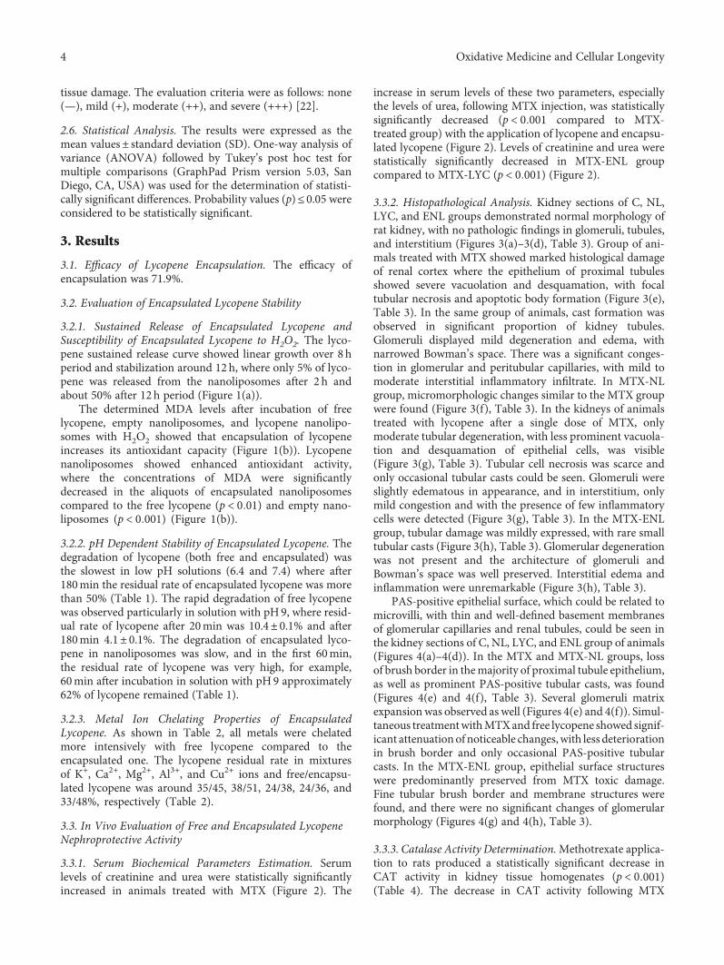
tissue damage. The evaluation criteria were as follows: none(—), mild (+), moderate (++), and severe (+++) [22].
2.6. Statistical Analysis. The results were expressed as themean values± standard deviation (SD). One-way analysis ofvariance (ANOVA) followed by Tukey’s post hoc test formultiple comparisons (GraphPad Prism version 5.03, SanDiego, CA, USA) was used for the determination of statisti-cally significant differences. Probability values (p)≤ 0.05 wereconsidered to be statistically significant.
3. Results
3.1. Efficacy of Lycopene Encapsulation. The efficacy ofencapsulation was 71.9%.
3.2. Evaluation of Encapsulated Lycopene Stability
3.2.1. Sustained Release of Encapsulated Lycopene andSusceptibility of Encapsulated Lycopene to H2O2. The lyco-pene sustained release curve showed linear growth over 8 hperiod and stabilization around 12 h, where only 5% of lyco-pene was released from the nanoliposomes after 2 h andabout 50% after 12h period (Figure 1(a)).
The determined MDA levels after incubation of freelycopene, empty nanoliposomes, and lycopene nanolipo-somes with H2O2 showed that encapsulation of lycopeneincreases its antioxidant capacity (Figure 1(b)). Lycopenenanoliposomes showed enhanced antioxidant activity,where the concentrations of MDA were significantlydecreased in the aliquots of encapsulated nanoliposomescompared to the free lycopene (p < 0 01) and empty nano-liposomes (p < 0 001) (Figure 1(b)).
3.2.2. pH Dependent Stability of Encapsulated Lycopene. Thedegradation of lycopene (both free and encapsulated) wasthe slowest in low pH solutions (6.4 and 7.4) where after180min the residual rate of encapsulated lycopene was morethan 50% (Table 1). The rapid degradation of free lycopenewas observed particularly in solution with pH9, where resid-ual rate of lycopene after 20min was 10.4± 0.1% and after180min 4.1± 0.1%. The degradation of encapsulated lyco-pene in nanoliposomes was slow, and in the first 60min,the residual rate of lycopene was very high, for example,60min after incubation in solution with pH9 approximately62% of lycopene remained (Table 1).
3.2.3. Metal Ion Chelating Properties of EncapsulatedLycopene. As shown in Table 2, all metals were chelatedmore intensively with free lycopene compared to theencapsulated one. The lycopene residual rate in mixturesof K+, Ca2+, Mg2+, Al3+, and Cu2+ ions and free/encapsu-lated lycopene was around 35/45, 38/51, 24/38, 24/36, and33/48%, respectively (Table 2).
3.3. In Vivo Evaluation of Free and Encapsulated LycopeneNephroprotective Activity
3.3.1. Serum Biochemical Parameters Estimation. Serumlevels of creatinine and urea were statistically significantlyincreased in animals treated with MTX (Figure 2). The
increase in serum levels of these two parameters, especiallythe levels of urea, following MTX injection, was statisticallysignificantly decreased (p < 0 001 compared to MTX-treated group) with the application of lycopene and encapsu-lated lycopene (Figure 2). Levels of creatinine and urea werestatistically significantly decreased in MTX-ENL groupcompared to MTX-LYC (p < 0 001) (Figure 2).
3.3.2. Histopathological Analysis. Kidney sections of C, NL,LYC, and ENL groups demonstrated normal morphology ofrat kidney, with no pathologic findings in glomeruli, tubules,and interstitium (Figures 3(a)–3(d), Table 3). Group of ani-mals treated with MTX showed marked histological damageof renal cortex where the epithelium of proximal tubulesshowed severe vacuolation and desquamation, with focaltubular necrosis and apoptotic body formation (Figure 3(e),Table 3). In the same group of animals, cast formation wasobserved in significant proportion of kidney tubules.Glomeruli displayed mild degeneration and edema, withnarrowed Bowman’s space. There was a significant conges-tion in glomerular and peritubular capillaries, with mild tomoderate interstitial inflammatory infiltrate. In MTX-NLgroup, micromorphologic changes similar to the MTX groupwere found (Figure 3(f), Table 3). In the kidneys of animalstreated with lycopene after a single dose of MTX, onlymoderate tubular degeneration, with less prominent vacuola-tion and desquamation of epithelial cells, was visible(Figure 3(g), Table 3). Tubular cell necrosis was scarce andonly occasional tubular casts could be seen. Glomeruli wereslightly edematous in appearance, and in interstitium, onlymild congestion and with the presence of few inflammatorycells were detected (Figure 3(g), Table 3). In the MTX-ENLgroup, tubular damage was mildly expressed, with rare smalltubular casts (Figure 3(h), Table 3). Glomerular degenerationwas not present and the architecture of glomeruli andBowman’s space was well preserved. Interstitial edema andinflammation were unremarkable (Figure 3(h), Table 3).
PAS-positive epithelial surface, which could be related tomicrovilli, with thin and well-defined basement membranesof glomerular capillaries and renal tubules, could be seen inthe kidney sections of C, NL, LYC, and ENL group of animals(Figures 4(a)–4(d)). In the MTX and MTX-NL groups, lossof brush border in themajority of proximal tubule epithelium,as well as prominent PAS-positive tubular casts, was found(Figures 4(e) and 4(f), Table 3). Several glomeruli matrixexpansionwas observed as well (Figures 4(e) and 4(f)). Simul-taneous treatmentwithMTXand free lycopene showed signif-icant attenuation of noticeable changes,with less deteriorationin brush border and only occasional PAS-positive tubularcasts. In the MTX-ENL group, epithelial surface structureswere predominantly preserved from MTX toxic damage.Fine tubular brush border and membrane structures werefound, and there were no significant changes of glomerularmorphology (Figures 4(g) and 4(h), Table 3).
3.3.3. Catalase Activity Determination.Methotrexate applica-tion to rats produced a statistically significant decrease inCAT activity in kidney tissue homogenates (p < 0 001)(Table 4). The decrease in CAT activity following MTX
4 Oxidative Medicine and Cellular Longevity
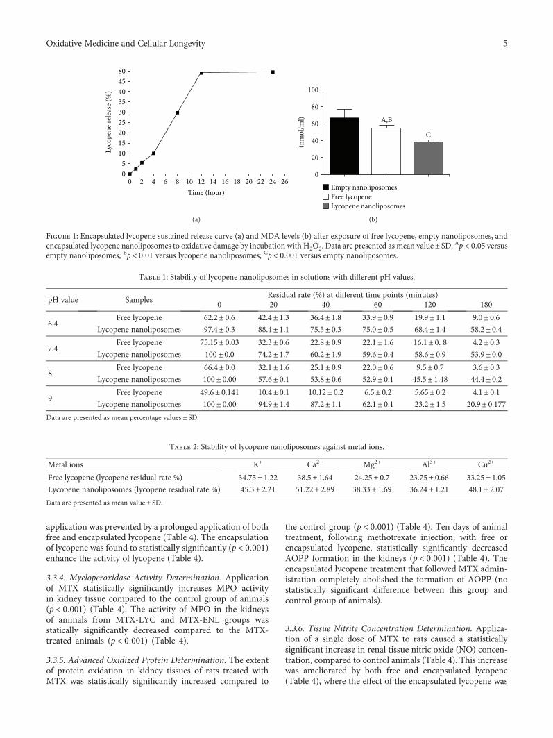
application was prevented by a prolonged application of bothfree and encapsulated lycopene (Table 4). The encapsulationof lycopene was found to statistically significantly (p < 0 001)enhance the activity of lycopene (Table 4).
3.3.4. Myeloperoxidase Activity Determination. Applicationof MTX statistically significantly increases MPO activityin kidney tissue compared to the control group of animals(p < 0 001) (Table 4). The activity of MPO in the kidneysof animals from MTX-LYC and MTX-ENL groups wasstatically significantly decreased compared to the MTX-treated animals (p < 0 001) (Table 4).
3.3.5. Advanced Oxidized Protein Determination. The extentof protein oxidation in kidney tissues of rats treated withMTX was statistically significantly increased compared to
the control group (p < 0 001) (Table 4). Ten days of animaltreatment, following methotrexate injection, with free orencapsulated lycopene, statistically significantly decreasedAOPP formation in the kidneys (p < 0 001) (Table 4). Theencapsulated lycopene treatment that followed MTX admin-istration completely abolished the formation of AOPP (nostatistically significant difference between this group andcontrol group of animals).
3.3.6. Tissue Nitrite Concentration Determination. Applica-tion of a single dose of MTX to rats caused a statisticallysignificant increase in renal tissue nitric oxide (NO) concen-tration, compared to control animals (Table 4). This increasewas ameliorated by both free and encapsulated lycopene(Table 4), where the effect of the encapsulated lycopene was
804540353025201510
50
0 2 4 6 8 10 12 14Time (hour)
Lyco
pene
rele
ase (
%)
16 18 20 22 24 26
(a)
C
100
A,B
80
(nm
ol/m
l) 60
40
20
0
Empty nanoliposomesFree lycopeneLycopene nanoliposomes
(b)
Figure 1: Encapsulated lycopene sustained release curve (a) and MDA levels (b) after exposure of free lycopene, empty nanoliposomes, andencapsulated lycopene nanoliposomes to oxidative damage by incubation with H2O2. Data are presented as mean value± SD. Ap < 0 05 versusempty nanoliposomes; Bp < 0 01 versus lycopene nanoliposomes; Cp < 0 001 versus empty nanoliposomes.
Table 1: Stability of lycopene nanoliposomes in solutions with different pH values.
pH value SamplesResidual rate (%) at different time points (minutes)
0 20 40 60 120 180
6.4Free lycopene 62.2± 0.6 42.4± 1.3 36.4± 1.8 33.9± 0.9 19.9± 1.1 9.0± 0.6
Lycopene nanoliposomes 97.4± 0.3 88.4± 1.1 75.5± 0.3 75.0± 0.5 68.4± 1.4 58.2± 0.4
7.4Free lycopene 75.15± 0.03 32.3± 0.6 22.8± 0.9 22.1± 1.6 16.1± 0. 8 4.2± 0.3
Lycopene nanoliposomes 100± 0.0 74.2± 1.7 60.2± 1.9 59.6± 0.4 58.6± 0.9 53.9± 0.0
8Free lycopene 66.4± 0.0 32.1± 1.6 25.1± 0.9 22.0± 0.6 9.5± 0.7 3.6± 0.3
Lycopene nanoliposomes 100± 0.00 57.6± 0.1 53.8± 0.6 52.9± 0.1 45.5± 1.48 44.4± 0.2
9Free lycopene 49.6± 0.141 10.4± 0.1 10.12± 0.2 6.5± 0.2 5.65± 0.2 4.1± 0.1
Lycopene nanoliposomes 100± 0.00 94.9± 1.4 87.2± 1.1 62.1± 0.1 23.2± 1.5 20.9± 0.177Data are presented as mean percentage values ± SD.
Table 2: Stability of lycopene nanoliposomes against metal ions.
Metal ions K+ Ca2+ Mg2+ Al3+ Cu2+
Free lycopene (lycopene residual rate %) 34.75± 1.22 38.5± 1.64 24.25± 0.7 23.75± 0.66 33.25± 1.05Lycopene nanoliposomes (lycopene residual rate %) 45.3± 2.21 51.22± 2.89 38.33± 1.69 36.24± 1.21 48.1± 2.07Data are presented as mean value ± SD.
5Oxidative Medicine and Cellular Longevity
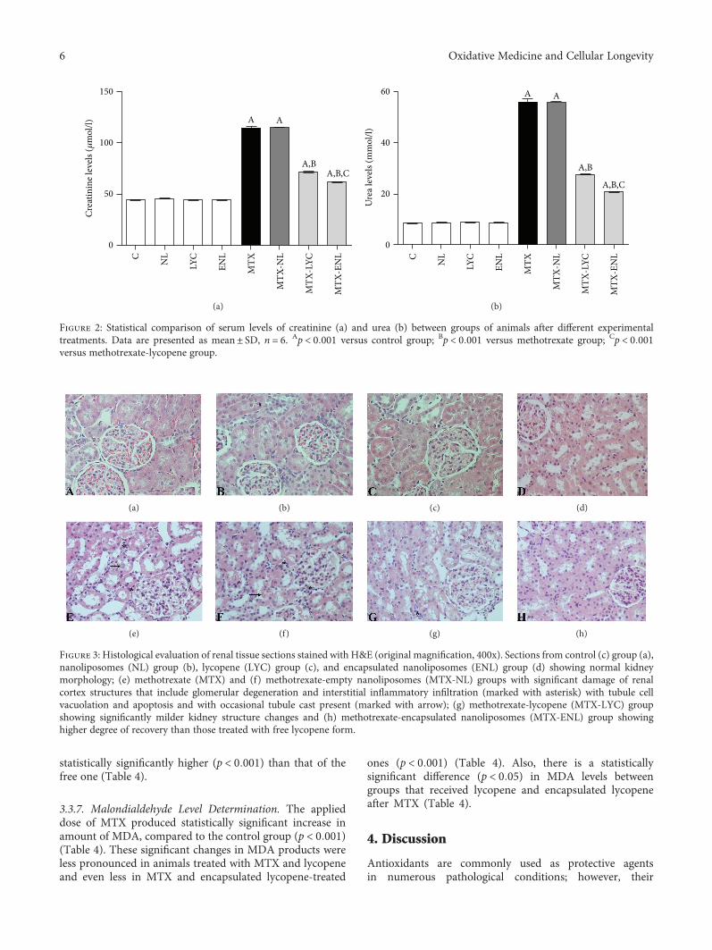
statistically significantly higher (p < 0 001) than that of thefree one (Table 4).
3.3.7. Malondialdehyde Level Determination. The applieddose of MTX produced statistically significant increase inamount of MDA, compared to the control group (p < 0 001)(Table 4). These significant changes in MDA products wereless pronounced in animals treated with MTX and lycopeneand even less in MTX and encapsulated lycopene-treated
ones (p < 0 001) (Table 4). Also, there is a statisticallysignificant difference (p < 0 05) in MDA levels betweengroups that received lycopene and encapsulated lycopeneafter MTX (Table 4).
4. Discussion
Antioxidants are commonly used as protective agentsin numerous pathological conditions; however, their
C NL
LYC
ENL
MTX
MTX
-NL
MTX
-LYC
MTX
-EN
L
A A
A,BA,B,C
100
150
50
0
Crea
tinin
e lev
els (�휇
mol
/l)
(a)
C NL
LYC
ENL
MTX
MTX
-NL
MTX
-LYC
MTX
-EN
L
A A
A,B
A,B,C
60
40
20
0
Ure
a lev
els (m
mol
/l)
(b)
Figure 2: Statistical comparison of serum levels of creatinine (a) and urea (b) between groups of animals after different experimentaltreatments. Data are presented as mean± SD, n = 6. Ap < 0 001 versus control group; Bp < 0 001 versus methotrexate group; Cp < 0 001versus methotrexate-lycopene group.
(a) (b) (c) (d)
(e) (f) (g) (h)
Figure 3: Histological evaluation of renal tissue sections stained with H&E (original magnification, 400x). Sections from control (c) group (a),nanoliposomes (NL) group (b), lycopene (LYC) group (c), and encapsulated nanoliposomes (ENL) group (d) showing normal kidneymorphology; (e) methotrexate (MTX) and (f) methotrexate-empty nanoliposomes (MTX-NL) groups with significant damage of renalcortex structures that include glomerular degeneration and interstitial inflammatory infiltration (marked with asterisk) with tubule cellvacuolation and apoptosis and with occasional tubule cast present (marked with arrow); (g) methotrexate-lycopene (MTX-LYC) groupshowing significantly milder kidney structure changes and (h) methotrexate-encapsulated nanoliposomes (MTX-ENL) group showinghigher degree of recovery than those treated with free lycopene form.
6 Oxidative Medicine and Cellular Longevity
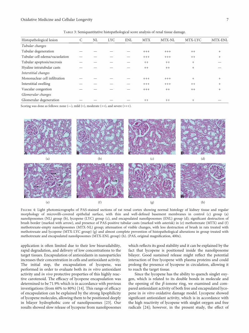
application is often limited due to their low bioavailability,rapid degradation, and delivery of low concentrations to thetarget tissues. Encapsulation of antioxidants in nanoparticlesincreases their concentration in cells and antioxidant activity.The initial step, the encapsulation of lycopene, wasperformed in order to evaluate both its in vitro antioxidantactivity and in vivo protective properties of this highly reac-tive carotenoid. The efficacy of lycopene encapsulation wasdetermined to be 71.9% which is in accordance with previousinvestigations (from 60% to 80%) [14]. This range of efficacyof encapsulation can be explained by the strong lipophilicityof lycopene molecules, allowing them to be positioned deeplyin bilayer hydrophobic core of nanoliposomes [23]. Ourresults showed slow release of lycopene from nanoliposomes
which reflects its good stability and it can be explained by thefact that lycopene is positioned inside the nanoliposomebilayer. Good sustained release might reflect the potentialinteraction of free lycopene with plasma proteins and couldprolong the presence of lycopene in circulation, allowing itto reach the target tissue.
Since the lycopene has the ability to quench singlet oxy-gen, which is related to its double bonds in molecule andthe opening of the β-ionone ring, we examined and com-pared antioxidant activity of both free and encapsulated lyco-pene in in vitro oxidative damage model. Lycopene showedsignificant antioxidant activity, which is in accordance withthe high reactivity of lycopene with singlet oxygen and freeradicals [24]; however, in the present study, the effect of
Table 3: Semiquantitative histopathological score analysis of renal tissue damage.
Histopathological lesion C NL LYC ENL MTX MTX-NL MTX-LYC MTX-ENL
Tubular changes
Tubular degeneration — — — — +++ +++ ++ +
Tubular cell edema/vacuolation — — — — +++ +++ ++ +
Tubular apoptosis/necrosis — — — — ++ ++ + —
Hyaline intratubular casts — — — — ++ ++ + —
Interstitial changes
Mononuclear cell infiltration — — — — +++ +++ + +
Interstitial swelling — — — — +++ +++ ++ +
Vascular congestion — — — — +++ ++ ++ +
Glomerular changes
Glomerular degeneration — — — — ++ ++ + —
Scoring was done as follows: none (—), mild (+), moderate (++), and severe (+++).
(a) (b) (c) (d)
(e) (f) (g) (h)
Figure 4: Light photomicrographs of PAS-stained sections of rat renal cortex showing normal histology of kidney tissue and regularmorphology of microvilli-covered epithelial surface, with thin and well-defined basement membranes in control (c) group (a)nanoliposomes (NL) group (b), lycopene (LYC) group (c), and encapsulated nanoliposomes (ENL) group (d); significant destruction ofbrush border (marked with arrow), and presence of PAS-positive tubular casts (marked with asterisk) in (e) methotrexate (MTX) and (f)methotrexate-empty nanoliposomes (MTX-NL) group; attenuation of visible changes, with less destruction of brush in rats treated withmethotrexate and lycopene (MTX-LYC group) (g) and almost complete prevention of histopathological alterations in group treated withmethotrexate and encapsulated nanoliposomes (MTX-ENL group) (h). (PAS, original magnification, 400x).
7Oxidative Medicine and Cellular Longevity
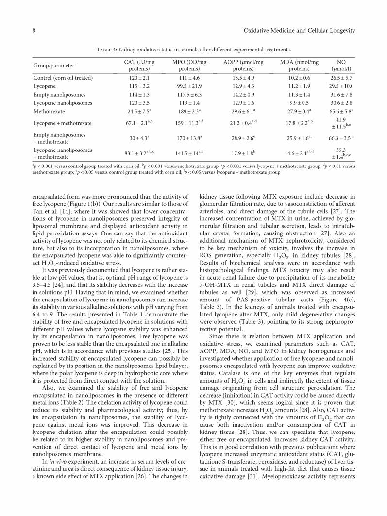
encapsulated form was more pronounced than the activity offree lycopene (Figure 1(b)). Our results are similar to those ofTan et al. [14], where it was showed that lower concentra-tions of lycopene in nanoliposomes preserved integrity ofliposomal membrane and displayed antioxidant activity inlipid peroxidation assays. One can say that the antioxidantactivity of lycopene was not only related to its chemical struc-ture, but also to its incorporation in nanoliposomes, wherethe encapsulated lycopene was able to significantly counter-act H2O2-induced oxidative stress.
It was previously documented that lycopene is rather sta-ble at low pH values, that is, optimal pH range of lycopene is3.5–4.5 [24], and that its stability decreases with the increasein solutions pH. Having that in mind, we examined whetherthe encapsulation of lycopene in nanoliposomes can increaseits stability in various alkaline solutions with pH varying from6.4 to 9. The results presented in Table 1 demonstrate thestability of free and encapsulated lycopene in solutions withdifferent pH values where lycopene stability was enhancedby its encapsulation in nanoliposomes. Free lycopene wasproven to be less stable than the encapsulated one in alkalinepH, which is in accordance with previous studies [25]. Thisincreased stability of encapsulated lycopene can possibly beexplained by its position in the nanoliposomes lipid bilayer,where the polar lycopene is deep in hydrophobic core whereit is protected from direct contact with the solution.
Also, we examined the stability of free and lycopeneencapsulated in nanoliposomes in the presence of differentmetal ions (Table 2). The chelation activity of lycopene couldreduce its stability and pharmacological activity; thus, byits encapsulation in nanoliposomes, the stability of lyco-pene against metal ions was improved. This decrease inlycopene chelation after the encapsulation could possiblybe related to its higher stability in nanoliposomes and pre-vention of direct contact of lycopene and metal ions bynanoliposomes membrane.
In in vivo experiment, an increase in serum levels of cre-atinine and urea is direct consequence of kidney tissue injury,a known side effect of MTX application [26]. The changes in
kidney tissue following MTX exposure include decrease inglomerular filtration rate, due to vasoconstriction of afferentarterioles, and direct damage of the tubule cells [27]. Theincreased concentration of MTX in urine, achieved by glo-merular filtration and tubular secretion, leads to intratub-ular crystal formation, causing obstruction [27]. Also anadditional mechanism of MTX nephrotoxicity, consideredto be key mechanism of toxicity, involves the increase inROS generation, especially H2O2, in kidney tubules [28].Results of biochemical analysis were in accordance withhistopathological findings. MTX toxicity may also resultin acute renal failure due to precipitation of its metabolite7-OH-MTX in renal tubules and MTX direct damage oftubules as well [29], which was observed as increasedamount of PAS-positive tubular casts (Figure 4(e),Table 3). In the kidneys of animals treated with encapsu-lated lycopene after MTX, only mild degenerative changeswere observed (Table 3), pointing to its strong nephropro-tective potential.
Since there is relation between MTX application andoxidative stress, we examined parameters such as CAT,AOPP, MDA, NO, and MPO in kidney homogenates andinvestigated whether application of free lycopene and nanoli-posomes encapsulated with lycopene can improve oxidativestatus. Catalase is one of the key enzymes that regulateamounts of H2O2 in cells and indirectly the extent of tissuedamage originating from cell structure peroxidation. Thedecrease (inhibition) in CAT activity could be caused directlyby MTX [30], which seems logical since it is proven thatmethotrexate increases H2O2 amounts [28]. Also, CAT activ-ity is tightly connected with the amounts of H2O2 that cancause both inactivation and/or consumption of CAT inkidney tissue [28]. Thus, we can speculate that lycopene,either free or encapsulated, increases kidney CAT activity.This is in good correlation with previous publications wherelycopene increased enzymatic antioxidant status (CAT, glu-tathione S-transferase, peroxidase, and reductase) of liver tis-sue in animals treated with high-fat diet that causes tissueoxidative damage [31]. Myeloperoxidase activity represents
Table 4: Kidney oxidative status in animals after different experimental treatments.
Group/parameterCAT (IU/mgproteins)
MPO (OD/mgproteins)
AOPP (μmol/mgproteins)
MDA (nmol/mgproteins)
NO(μmol/l)
Control (corn oil treated) 120± 2.1 111± 4.6 13.5± 4.9 10.2± 0.6 26.5± 5.7Lycopene 115± 3.2 99.5± 21.9 12.9± 4.3 11.2± 1.9 29.5± 10.0Empty nanoliposomes 114± 1.3 117.5± 6.3 14.2± 0.9 11.3± 1.4 31.6± 7.8Lycopene nanoliposomes 120± 3.5 119± 1.4 12.9± 1.6 9.9± 0.5 30.6± 2.8Methotrexate 24.5± 7.5a 189± 2.3a 29.6± 6.1a 27.9± 0.4a 65.6± 5.8a
Lycopene +methotrexate 67.1± 2.1a,b 159± 11.3a,d 21.2± 0.4a,d 17.8± 2.2a,b 41.9± 11.5b,e
Empty nanoliposomes+methotrexate
30± 4.3a 170± 13.8a 28.9± 2.6e 25.9± 1.6a, 66.3± 3.5 a
Lycopene nanoliposomes+methotrexate
83.1± 3.2a,b,c 141.5± 14a,b 17.9± 1.8b 14.6± 2.4a,b,f 39.3± 1.4b,c,e
ap < 0 001 versus control group treated with corn oil; bp < 0 001 versus methotrexate group; cp < 0 001 versus lycopene +methotrexate group; dp < 0 01 versusmethotrexate group; ep < 0 05 versus control group treated with corn oil; fp < 0 05 versus lycopene +methotrexate group
8 Oxidative Medicine and Cellular Longevity
a sensitive marker of tissue infiltration with neutrophils, dueto a good correlation between its activity and histopatholog-ical finding [32]. Methotrexate is also known to induce thereleases of free radicals, MPO among them, fromstimulated polymorphonuclear neutrophils causing cellularoxidative damage [28, 32]. Generated products of MPO,chlorinating oxidants arriving from chlorides and H2O2, areable to cause cell damage by reacting with amino acids,proteins, carbohydrates, lipids, and nucleobases [32]. Theapplication of both forms of lycopene statistically significantdecreases MPO activity (Table 4), which is in accordancewith previous studies where it is shown that lycopenedecreases MPO activity in the damaged kidney [33, 34] andmyocardial [35] and pancreatic [36] tissues.
Nitric oxide represents an important cell mediator that isinvolved in numerous both physiological and pathophysio-logical processes [37]. One of the theories explaining MTXnephrotoxicity involves NO signaling and increase of induc-ible nitric oxide synthase (iNOS), which is barely detectablein normal renal tissue [38]. Nitric oxide is also involved inmacrophage activation, and during the inflammatory pro-cesses in the kidneys, macrophages produce large amountsof NO playing an important role in renal tissue pathology[39]. Previous studies evaluated the effects of free lycopene(10mg/kg, orally applied) on MTX-induced kidney dam-age and consequential NO increase [38]. However, nostatistically significant effects on NO concentrations weredetected. On the other hand, in the model of unilateralureteral obstruction-induced kidney damage, lycopenewas proven to reduce tissue NO levels [33], similarly tothe findings of the present study (Table 4). The differencein the obtained results may lay in the different routes oflycopene administration (i.p. and per os), and these resultscould possibly be explained through high reactivity of lyco-pene, whose activity significantly decreases when appliedorally due to its interaction with numerous compoundsleading to its isomerization, oxidation, and/or degradation[14]. In the present study, this was potentially avoided bythe application of lycopene via i.p. route and by its encapsu-lation with nanoliposomes.
The formation of AOPP is believed to be one of the ini-tial steps in cell oxidative stress that further signals the devel-opment of renal diseases [40]. Activated MPO in the tissuegenerates hypochlorous acid which mediates protein modifi-cations causing tyrosine chlorination and formation of chlo-ramines and carbonyls [32]. The accumulation of AOPP inrenal cortex, especially in glomerulus, is known to decreasethe Bowman’s spaces and thus causing the impairments inkidney function [41]. Such pathological findings, as well asincrease in AOPP concentration, were detected in animalstreated with MTX (Figure 4(e) and Table 4). In addition toROS-mediated protein damage during exposure to MTX,generated ROS damages unsaturated lipids as well [42],making MDA products a sensitive marker of lipid peroxida-tion. The formation of MDA could be connected with dam-aging activity of both peroxynitrite, from superoxide anionand nitric oxide, and hypochlorous acid [28, 32]. Since theactivity of MPO and NO concentrations is statistically signif-icantly decreased by both forms of lycopene, it is not
surprising that the MDA and AOPP amounts are statisticallysignificantly decreased in these groups as well (Table 4).
This study primarily aimed at comparing efficacy of twodifferent pharmaceutical forms of lycopene on MTX-induced toxicity at kidney tissue level. However, it is limitedin defining potential differences of their sites of action atparticular kidney tissue cell targets. Previous in vitro studiesrevealed that lycopene can accumulate in epithelial cellsand affect different protein synthesis depending on concen-tration applied [43]. The present study should warrant futurework focusing on more detailed action of nanoliposome-encapsulated lycopene at the cellular level.
5. Conclusion
Lycopene is presumed to protect cells from oxidative damageby stabilization of the membrane and/or scavenging free rad-icals generated within the tissue [44]. In this study, wedemonstrated that nanoliposome-encapsulated lycopenerepresents a protected form of this highly reactive carotenoidwith enhanced activity. The methotrexate-induced nephro-toxicity model allowed us to investigate and compare theefficacy of the free and encapsulated lycopene in preventingreactive oxygen species-induced kidney tissue damage, whichis considered to be a key mechanism of methotrexate toxicity.Our results demonstrated that encapsulated lycopene showedstronger antioxidant activity than free one. Since lycopeneis widely used as a dietary supplement, encapsulation oflycopene should be taken as a potential “shelter” whenconsidering lycopene formulation. Also, in this form, itshould be recommended for simultaneous application withmethotrexate therapy.
Conflicts of Interest
The authors declare that there is no conflict of interestregarding the publication of this article.
Acknowledgments
This work was funded by the project of the Faculty of Medi-cine, University of Niš, Serbia (Project no. INT-MFN-13).
References
[1] B. C. Widemann, F. M. Balis, B. Kempf-Bielack et al., “High-dose methotrexate-induced nephrotoxicity in patients withosteosarcoma,” Cancer, vol. 100, no. 10, pp. 2222–2232, 2004.
[2] S. Kivity, Y. Zafrir, R. Loebstein, R. Pauzner, M. Mouallem,and H. Mayan, “Clinical characteristics and risk factors forlow dose methotrexate toxicity: a cohort of 28 patients,” Auto-immunity Reviews, vol. 13, no. 11, pp. 1109–1113, 2014.
[3] N. Jahovic, G. Sener, H. Cevik, Y. Ersoy, S. Arbak, and B. C.Yegen, “Amelioration of methotrexate-induced enteritis bymelatonin in rats,” Cell Biochemistry & Function, vol. 22,no. 3, pp. 169–178, 2004.
[4] I. Asvadi, B. Hajipour, A. Asvadi, N. A. Asl, L. Roshangar, andA. Khodadadi, “Protective effect of pentoxyfilline in renal tox-icity after methotrexate administration,” European Review for
9Oxidative Medicine and Cellular Longevity
Medical and Pharmacological Sciences, vol. 15, no. 9, pp. 1003–1009, 2011.
[5] N. Vardi, H. Parlakpinar, B. Ates, A. Cetin, and A. Otlu, “Theprotective effects of Prunus armeniaca L (apricot) againstmethotrexate-induced oxidative damage and apoptosis in ratkidney,” Journal of Physiology and Biochemistry, vol. 69,no. 3, pp. 371–381, 2013.
[6] S. S. Palabiyik, P. Erkekoglu, N. D. Zeybek et al., “Protectiveeffect of lycopene against ochratoxin A induced renal oxidativestress and apoptosis in rats,” Experimental and ToxicologicPathology, vol. 65, no. 6, pp. 853–861, 2013.
[7] W. Stahl and H. Sies, “Antioxidant activity of carotenoids,”Molecular Aspects of Medicine, vol. 24, no. 6, pp. 345–351,2003.
[8] A. Atessahin, S. Yilmaz, I. Karahan, A. O. Ceribasi, andA. Karaoglu, “Effects of lycopene against cisplatin-inducednephrotoxicity and oxidative stress in rats,” Toxicology,vol. 212, no. 2-3, pp. 116–123, 2005.
[9] A. Dogukan, M. Tuzcu, C. A. Agca et al., “A tomato lycopenecomplex protects the kidney from cisplatin-induced injury viaaffecting oxidative stress as well as Bax, Bcl-2, and HSPsexpression,” Nutrition and Cancer, vol. 63, no. 3, pp. 427–434,2011.
[10] S. K. Sahoo, S. Parveen, and J. J. Panda, “The present andfuture of nanotechnology in human health care,” Nanomedi-cine: Nanotechnology, Biology, and Medicine, vol. 3, no. 1,pp. 20–31, 2007.
[11] G. A. Hughes, “Nanostructure-mediated drug delivery,”Nano-medicine: Nanotechnology, Biology, and Medicine, vol. 1, no. 1,pp. 22–30, 2005.
[12] M. R. Mozafari, “Bioactive entrapment and targeting usingnanocarrier technologies: an introduction,” in NanocarrierTechnologies: Frontiers of Nanotherapy, M. R. Mozafari, Ed.,pp. 1–16, Springer, Netherlands, 2005.
[13] G. Kocic, K. Tomovic, H. Kocic et al., “Antioxidative, mem-brane protective and antiapoptotic effects of melatonin, insilico study of physico-chemical profile and efficiency of nano-liposome delivery compared to betaine,” RSC Advances, vol. 7,no. 3, pp. 1271–1281, 2017.
[14] C. Tan, J. Xue, S. Abbas, B. Feng, X. Zhang, and S. Xia, “Lipo-some as a delivery system for carotenoids: comparative antiox-idant activity of carotenoids as measured by ferric reducingantioxidant power, DPPH assay and lipid peroxidation,” Jour-nal of Agricultural and Food Chemistry, vol. 62, no. 28,pp. 6726–6735, 2014.
[15] X. Chen, L. Q. Zou, J. Niu, W. Liu, S. F. Peng, and C. M. Liu,“The stability, sustained release and cellular antioxidant activ-ity of curcumin nanoliposomes,” Molecules, vol. 20, no. 8,pp. 14293–14311, 2015.
[16] J. A. Buege and S. D. Aust, “Microsomal lipid peroxidation,”Methods in Enzymology, vol. 52, pp. 302–310, 1978.
[17] S. Ilic,N. Stojiljkovic, S.Veljkovic et al., “Morphometric studyofstructural kidney damages caused by cisplatin in rats. Effects ofquercetin,”ActaMicroscopica, vol. 25, no. 4, pp. 128–137, 2016.
[18] L. Goth, “A simple method for determination of serum cata-lase activity and revision of reference range,” Clinica ChimicaActa, vol. 196, no. 2-3, pp. 143–151, 1991.
[19] S. S. Jacob, P. Shastry, and P. R. Sudhakaran, “Influenceof non-enzymatically glycated collagen on monocyte–mac-rophage differentiation,” Atherosclerosis, vol. 159, no. 2,pp. 333–341, 2001.
[20] N. S. Radulovic, P. J. Randjelovic, N. M. Stojanovic, N. D.Cakic, G. A. Bogdanovic, and A. V. Zivanovic, “Aboriginalbush foods: a major phloroglucinol from Crimson Bottlebrushflowers (Callistemon citrinus, Myrtaceae) displays strong anti-nociceptive and antiinflammatory activity,” Food ResearchInternational, vol. 77, Part 2, pp. 280–289, 2015.
[21] N. Stojiljkovic, M. Stoiljkovic, P. Randjelovic, S. Veljkovic, andD. Mihailovic, “Cytoprotective effect of vitamin C againstgentamicin-induced acute kidney injury in rats,” Experimentaland Toxicologic Pathology, vol. 64, no. 1-2, pp. 69–74, 2012.
[22] P. Randjelovic, S. Veljkovic, N. Stojiljkovic et al., “Salicylic acidattenuates gentamicin-induced nephrotoxicity in rats,” TheScientific World Journal, vol. 2012, Article ID 390613, 6 pages,2012.
[23] M. van de Ven, M. Kattenberg, G. van Ginkel, and Y. K.Levine, “Study of the orientational ordering of carotenoids inlipid bilayers by resonance-Raman spectroscopy,” BiophysicalJournal, vol. 45, no. 6, pp. 1203–1209, 1984.
[24] T. Anguelova and J. Warthesen, “Degradation of lycopene, α-carotene, and β-carotene during lipid peroxidation,” Journalof Food Science, vol. 65, no. 1, pp. 71–75, 2000.
[25] M. F. Salama, E. I. Seliem, K. F. Mahmoud, and A. A. Amin,“Physiochemical characterization and oxidative stability ofencapsulated nano lycopene pigments extracted by CO2 fluidextraction,” International Journal of Current Microbiologyand Applied Sciences, vol. 4, no. 3, pp. 307–320, 2015.
[26] R. Singh, R. Shah, C. Turner, O. Regueira, and T. L. Vasylyeva,“N-acetylcysteine renoprotection in methotrexate inducednephrotoxicity and its effects on B-cell lymphoma,” IndianJournal of Medical and Paediatric Oncology, vol. 36, no. 4,pp. 243–248, 2015.
[27] M. A. Perazella, “Crystal-induced acute renal failure,” TheAmerican Journal of Medicine, vol. 106, no. 4, pp. 459–465,1999.
[28] W. Ahmed, A. Zaki, and T. Nabil, “Prevention ofmethotrexate-induced nephrotoxicity by concomitant admin-istration of garlic aqueous extract in rat,” Turkish Journal ofMedical Sciences, vol. 45, no. 3, pp. 507–516, 2015.
[29] B. C. Widemann and P. C. Adamson, “Understanding andmanaging methotrexate nephrotoxicity,” The Oncologist,vol. 11, no. 6, pp. 694–703, 2006.
[30] C. L. Coleshowers, O. O. Oguntibeju, M. Ukpong, and E. J.Truter, “Effects of methotrexate on antioxidant enzyme statusin a rodent model,” Medical Technology SA, vol. 24, no. 1,pp. 5–9, 2010.
[31] S. K. Choi and J. S. Seo, “Lycopene supplementation sup-presses oxidative stress induced by a high fat diet in ger-bils,” Nutrition Research and Practice, vol. 7, no. 1,pp. 26–33, 2013.
[32] V. K. Kolli, P. Abraham, B. Isaac, and D. Selvakumar, “Neutro-phil infiltration and oxidative stress may play a critical role inmethotrexate-induced renal damage,” Chemotherapy, vol. 55,no. 2, pp. 83–90, 2009.
[33] H. I. Atilgan, A. Aydin, M. Sadic et al., “The protective effect oflycopene on kidney against experimentally induced unilateralureteral obstruction,” Acta Medica Mediterranea, vol. 32,pp. 1631–1636, 2016.
[34] M. Sadic, H. I. Atilgan, A. Aydin et al., “Scintigraphic, histo-pathologic, and biochemical evaluation of lycopene effects onrenal ischemia reperfusion injury in rats,” Serbian Archives ofMedicine, vol. 145, no. 3-4, pp. 153–158, 2017.
10 Oxidative Medicine and Cellular Longevity
[35] U. Aman, P. Vaibhav, and R. Balaraman, “Tomato lycopeneattenuates myocardial infarction induced by isoproterenol:electrocardiographic, biochemical and anti-apoptotic study,”Asian Pacific Journal of Tropical Biomedicine, vol. 2, no. 5,pp. 345–351, 2012.
[36] E. Ozkan, C. Akyüz, E. Dulundu et al., “Protective effects oflycopene on cerulein-induced experimental acute pancreatitisin rats,” The Journal of Surgical Research, vol. 176, no. 1,pp. 232–238, 2012.
[37] Y. C. Hou, A. Janczuk, and P. G. Wang, “Current trends in thedevelopment of nitric oxide donors,” Current PharmaceuticalDesign, vol. 5, no. 6, pp. 417–441, 1999.
[38] E. Oguz, S. Kocarslan, S. Tabur, H. Sezen, Z. Yilmaz, andN. Aksoy, “Effects of lycopene alone or combined with melato-nin on methotrexate-induced nephrotoxicity in rats,” AsianPacific Journal of Cancer Prevention, vol. 16, no. 14,pp. 6061–6066, 2015.
[39] U. Rolle, H. Shima, and P. Puri, “Nitric oxide, enhanced bymacrophage-colony stimulating factor, mediates renal damagein reflux nephropathy,” Kidney International, vol. 62, no. 2,pp. 507–513, 2002.
[40] S. Yamagishi and T. Matsui, “Advanced glycation end prod-ucts, oxidative stress and diabetic nephropathy,” OxidativeMedicine and Cellular Longevity, vol. 3, no. 2, pp. 101–108,2010.
[41] Y. Liu, “Advanced oxidation protein products: a causative linkbetween oxidative stress and podocyte depletion,” KidneyInternational, vol. 76, no. 11, pp. 1125–1127, 2009.
[42] E. Uz, F. Oktem, H. R. Yilmaz, E. Uzar, and F. Ozguner, “Theactivities of purine-catabolizing enzymes and the level of nitricoxide in rat kidneys subjected to methotrexate: protectiveeffect of caffeic acid phenethyl ester,” Molecular and CellularBiochemistry, vol. 277, no. 1-2, pp. 165–170, 2005.
[43] H. L. Hantz, L. F. Young, and K. R. Martin, “Physiologicallyattainable concentrations of lycopene induce mitochondrialapoptosis in LNCaP human prostate cancer cells,” Experimen-tal Biology and Medicine, vol. 230, no. 3, pp. 171–179, 2016.
[44] G. Krishnamoorthy, K. Selvakumar, P. Venkataraman,P. Elumalai, and J. Arunakaran, “Lycopene supplementationprevents reactive oxygen species mediated apoptosis in Sertolicells of adult albino rats exposed to polychlorinated biphe-nyls,” Interdisciplinary Toxicology, vol. 6, no. 2, pp. 83–92,2013.
11Oxidative Medicine and Cellular Longevity
Copyright © 2022 FDOKUMEN