The effect of CD14 C159T polymorphism on in vitro IgE synthesis and cytokine production by PBMC from...
10
ORIGINAL ARTICLE EPIDEMIOLOGY AND GENETICS The effect of CD14 C159T polymorphism on in vitro IgE synthesis and cytokine production by PBMC from children with asthma C. Sackesen 1 , E. Birben 1 , O. U. Soyer 1 , U. M. Sahiner 1 , T. S. Yavuz 1 , E. Civelek 1 , E. Karabulut 2 , M. Akdis 3 , C. A. Akdis 3 & O. Kalayci 1 1 Pediatric Allergy and Asthma Unit; 2 Department of Biostatistics, Hacettepe University School of Medicine, Hacettepe, Ankara, Turkey; 3 Swiss Institute of Allergy and Asthma Research (SIAF), University of Zurich, Davos, Switzerland To cite this article: Sackesen C, Birben E, Soyer OU, Sahiner UM, Yavuz TS, Civelek E, Karabulut E, Akdis M, Akdis CA, Kalayci O. The effect of CD14 C159T polymorphism on in vitro IgE synthesis and cytokine production by PBMC from children with asthma. Allergy 2011; 66: 48–57. Asthma is a complex genetic disorder that involves inter- actions between genetic and environmental factors. Within this network of complex interactions, a strong association between total IgE levels and asthma has long been recognized (1), and eventually led to the development of a humanized monoclonal anti-IgE antibody for the treatment of severe asthma (2). Because of the significant role of IgE in allergies and asthma, there has been an increasing interest in the environmental and immunological factors such as lipopoly- saccharide (LPS) that may regulate IgE expression. Within the general context of hygiene hypothesis, several studies have shown that endotoxin exposure may protect against atopy (3) and that the effect can already be Keywords asthma; CD14; gene; IgE; lipopolysaccharide; polymorphism. Correspondence Cansin Sackesen and Omer Kalayci, Pediatric Allergy and Asthma Unit, Hacettepe University School of Medicine, Hacettepe 06100, Ankara, Turkey. Tel.: +90 312 305 1700 Fax: +90 312 311 2357 E-mail: [email protected], [email protected] Accepted for publication 20 May 2010 DOI:10.1111/j.1398-9995.2010.02428.x Edited by: Stephan Weidinger Abstract Background: Even though the genotype at the promoter region of the CD14 mole- cule is known to affect the atopic phenotypes, the cellular and molecular basis of this association is largely unknown. Objective: To investigate the effect of lipopolysaccharide (LPS) on IgE production and cytokine profile by peripheral blood mononuclear cells (PBMC) obtained from asthmatic children with the TT and the CC genotypes at position )159 of the CD14 gene. Methods: Peripheral blood mononuclear cells from asthmatic children with alterna- tive genotypes at CD14 C159T locus were stimulated with 2 and 200 ng/ml LPS in vitro. The IgE, IgG and, IgM response was determined by ELISA and Ig e-germ- line, IgG, and IgM transcription by real-time PCR. A cluster of cytokines was mea- sured by cytometric bead array. Results: Asthmatic children with the TT genotype but not those with the CC geno- type responded with increased IgE synthesis and germline transcription to LPS stim- ulation. There were no genotype-related differences in IgG and IgM. TT but not the CC genotype was associated with significantly increased interleukin (IL)-4/IL-12 and IL-4/interferon-gamma (IFN-c) ratios in the culture supernatant. There were no genotype-related differences in IL-1b, IL-7, IL-10, IL-13, IL-17A, granulocyte colony stimulating factor, granulocyte macrophage colony stimulating factor, mono- cyte chemotactic protein, and tumor necrosis factor alpha. Conclusion: Peripheral blood mononuclear cells from asthmatic children with the TT genotype at position )159 of the CD14 gene make more IgE than those with the CC genotype following LPS stimulation because of increased germline transcrip- tion and have an augmented Th2 cytokine profile. Abbreviations BSA, bovine serum albumin; G-CSF, granulocyte colony stimulating factor; GM-CSF, granulocyte macrophage colony stimulating factor; IFN-c, interferon-gamma; IL, interleukin; LPS, lipopolysaccharide; MCP, monocyte chemotactic protein; MIP-1a, macrophage inflammatory protein 1 alpha; TNF-a, tumor necrosis factor alpha. Allergy 48 Allergy 66 (2011) 48–57 ª 2010 John Wiley & Sons A/S
-
Upload
independent -
Category
Documents
-
view
0 -
download
0
Transcript of The effect of CD14 C159T polymorphism on in vitro IgE synthesis and cytokine production by PBMC from...

ORIGINAL ARTICLE EPIDEMIOLOGY AND GENETICS
The effect of CD14 C159T polymorphism on in vitro IgEsynthesis and cytokine production by PBMC from childrenwith asthmaC. Sackesen1, E. Birben1, O. U. Soyer1, U. M. Sahiner1, T. S. Yavuz1, E. Civelek1, E. Karabulut2,M. Akdis3, C. A. Akdis3 & O. Kalayci1
1Pediatric Allergy and Asthma Unit; 2Department of Biostatistics, Hacettepe University School of Medicine, Hacettepe, Ankara, Turkey;3Swiss Institute of Allergy and Asthma Research (SIAF), University of Zurich, Davos, Switzerland
To cite this article: Sackesen C, Birben E, Soyer OU, Sahiner UM, Yavuz TS, Civelek E, Karabulut E, Akdis M, Akdis CA, Kalayci O. The effect of CD14 C159T
polymorphism on in vitro IgE synthesis and cytokine production by PBMC from children with asthma. Allergy 2011; 66: 48–57.
Asthma is a complex genetic disorder that involves inter-
actions between genetic and environmental factors. Within
this network of complex interactions, a strong association
between total IgE levels and asthma has long been recognized
(1), and eventually led to the development of a humanized
monoclonal anti-IgE antibody for the treatment of severe
asthma (2). Because of the significant role of IgE in allergies
and asthma, there has been an increasing interest in the
environmental and immunological factors such as lipopoly-
saccharide (LPS) that may regulate IgE expression.
Within the general context of hygiene hypothesis, several
studies have shown that endotoxin exposure may protect
against atopy (3) and that the effect can already be
Keywords
asthma; CD14; gene; IgE;
lipopolysaccharide; polymorphism.
Correspondence
Cansin Sackesen and Omer Kalayci,
Pediatric Allergy and Asthma Unit,
Hacettepe University School of Medicine,
Hacettepe 06100, Ankara, Turkey.
Tel.: +90 312 305 1700
Fax: +90 312 311 2357
E-mail: [email protected],
Accepted for publication 20 May 2010
DOI:10.1111/j.1398-9995.2010.02428.x
Edited by: Stephan Weidinger
Abstract
Background: Even though the genotype at the promoter region of the CD14 mole-
cule is known to affect the atopic phenotypes, the cellular and molecular basis of
this association is largely unknown.
Objective: To investigate the effect of lipopolysaccharide (LPS) on IgE production
and cytokine profile by peripheral blood mononuclear cells (PBMC) obtained from
asthmatic children with the TT and the CC genotypes at position )159 of the CD14
gene.
Methods: Peripheral blood mononuclear cells from asthmatic children with alterna-
tive genotypes at CD14 C159T locus were stimulated with 2 and 200 ng/ml LPS
in vitro. The IgE, IgG and, IgM response was determined by ELISA and Ig �e-germ-
line, IgG, and IgM transcription by real-time PCR. A cluster of cytokines was mea-
sured by cytometric bead array.
Results: Asthmatic children with the TT genotype but not those with the CC geno-
type responded with increased IgE synthesis and germline transcription to LPS stim-
ulation. There were no genotype-related differences in IgG and IgM. TT but not the
CC genotype was associated with significantly increased interleukin (IL)-4/IL-12
and IL-4/interferon-gamma (IFN-c) ratios in the culture supernatant. There were
no genotype-related differences in IL-1b, IL-7, IL-10, IL-13, IL-17A, granulocyte
colony stimulating factor, granulocyte macrophage colony stimulating factor, mono-
cyte chemotactic protein, and tumor necrosis factor alpha.
Conclusion: Peripheral blood mononuclear cells from asthmatic children with the
TT genotype at position )159 of the CD14 gene make more IgE than those with
the CC genotype following LPS stimulation because of increased germline transcrip-
tion and have an augmented Th2 cytokine profile.
Abbreviations
BSA, bovine serum albumin; G-CSF, granulocyte colony stimulating
factor; GM-CSF, granulocyte macrophage colony stimulating factor;
IFN-c, interferon-gamma; IL, interleukin; LPS, lipopolysaccharide;
MCP, monocyte chemotactic protein; MIP-1a, macrophage
inflammatory protein 1 alpha; TNF-a, tumor necrosis factor alpha.
Allergy
48 Allergy 66 (2011) 48–57 ª 2010 John Wiley & Sons A/S

programmed in utero (4). In addition to the timing and dose
of endotoxin exposure (5–8), the genetic variants of the
molecules participating in the endotoxin signaling pathway
may strongly influence the host response to endotoxin
(9–14).
A C–T polymorphism at position )159 in the promoter of
CD14 (C159T) (rs2569190 A/G) changes the affinity of Sp
family of transcription factors and thus modulates the cellu-
lar response to endotoxin (9). Following the pioneering study
by Baldini et al. (10) that showed that the C allele is associ-
ated with higher IgE and lower soluble CD14 levels, either
the T or the C allele was found to be associated with atopic
outcomes in various populations (10, 11, 13), whereas some
studies have shown no association at all (12, 14, 15). In fact,
a more recent analysis found a significant heterogeneity
among published studies (12).
To partly account for these observed discrepancies, recent
data have suggested that the response to LPS by alternative
genotypes can be greatly influenced by its concentration in
the environment and that the genotype stratified IgE response
is a function of the strength of the environmental exposure
(16, 17).
Even though there is accumulating evidence that endotoxin
may modulate the IgE response, this information has been
obtained basically from population studies and epidemiologi-
cal observations. The laboratory evidence for this observation
has been missing. Specifically, it is unknown whether PBMC
from individuals with the TT and CC genotypes at position
)159 of the CD14 gene actually synthesize different amounts
of IgE upon exposure to LPS and more importantly whether
the response to various concentrations of LPS differs between
the two genotypes.
During the development of allergic disease, effector Th2
cells produce interleukin (IL)-4, IL-5, IL-9, and IL-13 that
play important roles in allergic inflammation (18, 19). The
relationship between the cytokines that are operative in IgE
synthesis and CD14 )159 genotype has not been extensively
studied.
Even though the genetic polymorphism at the CD14
C159T locus apparently affects the IgE response, the infor-
mation is gathered basically from epidemiological studies,
and direct laboratory evidence for this association is lacking.
Moreover, the cellular and molecular mechanisms of this
relationship are largely unknown. We have previously shown
that the CC genotype is associated with higher IgE in a
population of Turkish children with asthma (20). Therefore,
we recruited children with asthma from the same population
with the TT and CC genotypes at position )159 of the CD14
gene and stimulated the cells with increasing doses of LPS
and compared IgE, IgM, IgG1, and IgG4 synthesis and Th1,
Th2, and Th17 cytokines between the two groups.
Methods
Study population
Children aged 6–18 who were diagnosed with asthma and
who were previously genotyped at the CD14 )159 locus were
recruited from the Pediatric Allergy and Asthma Unit of
Hacettepe University, School of Medicine, Ankara, Turkey.
These children belonged to a cohort of children with asthma
who were previously genotyped for another study in our
department (20). Of 613 children who were genotyped (20),
we selected children with asthma in stable condition, who
have had no disease exacerbation, respiratory infections, or
received systemic corticosteroids within the last 4 weeks who
presented to our department between October 2007 and April
2008. All children had evidence of reversible airway obstruc-
tion as defined by at least a 12% improvement in FEV1
following bronchodilator administration.
Spirometric measurements, total IgE, and eosinophil
counts were obtained, and skin testing was performed as
described previously (20). All study procedures were carried
out in accordance with a protocol previously approved by
the Ethics Committee of Hacettepe University. All parents
provided written informed consent for the study procedures.
Even though the parents of these children had initially signed
an informed consent for DNA donation, a new Ethics Com-
mittee Approval has been obtained, and the parents of all
children signed a new informed consent for the current study,
and the children provided their assets to allow phlebotomy
once more and do tissue culture experiments.
Cell preparation and culture
Peripheral blood mononuclear cells (PBMC) were isolated
from heparinized venous blood by density-gradient centrifu-
gation on Histopaque 1077 (Sigma, St Louis, MO, USA)
washed three times in Phosphate buffered saline (PBS)
(Sigma) and resuspended in Iscove’s Modifies Dulbecco’s
Medium (IMDM + l-Glutamin; Gibco, Grand Island, NY,
USA) containing 10% fetal bovine serum (FBS) (Hyclone,
Logan, UT, USA), 36 mM sodium bicarbonate, 1% ITS +
Premix (containing 12.5 mg/ml recombinant human insulin,
12.5 mg/ml human transferrin, 12.5 lg/ml selenous acid and
10.7 mg/ml linoleic acid; BD Biosciences, Bedford, MA,
USA), Penicillin (100 U/ml) (Sigma) and Streptomycin
(100 mg/ml) (Sigma), as previously described (21).
In 48-well plates, 1 · 106 PBMC were cultured in complete
medium in the absence and presence of LPS (2 and 200 ng/
ml), (Escherichia coli, 055:B5; Fluka, Buchs, Switzerland) in
500 ll complete medium. Supernatants were collected at 48 h
for cytokine determination, RNA was extracted for epsilon
germline determination at day 5, and supernatants were
collected at day 12 for immunoglobulin determinations (22,
23). All media used in the experiments were free of LPS
contamination.
Immunoglobulin measurements
IgE was measured by ELISA. Briefly, ELISA plates were
coated overnight at 4�C with anti-IgE (Polyclonal rabbit
anti-human IgE antibody, DakoCytomation, Denmark).
Plates were blocked with 3% FBS + 3% bovine serum albu-
min (BSA) in PBS, washed, and standards and samples were
added in appropriate dilutions for 2 h. After washing,
Sackesen et al. CD14 gene variants and IgE synthesis
Allergy 66 (2011) 48–57 ª 2010 John Wiley & Sons A/S 49
HRP-conjugated goat anti-human IgE antibody (Biosource,
Camarillo, CA, USA) was added for 2 h. TMB (3,3¢,5,5¢-tetramethyl-benzidine) substrate solution was used to obtain
coloration, and the reaction was stopped with 10% H2SO4.
Optical density was measured at 450 nm. IgG1 and IgG4 and
IgM determinations were carried out with the cytometric
bead array (Bio-Plex; Bio-Rad Laboratories Inc., Hercules,
CA, USA) according to the manufacturer’s protocol.
Ig �e-germline transcription
Total RNA was extracted from the PBMCs using RNeasy
mini kit (Qiagen, Valencia, CA, USA) according to the man-
ufacturer’s instructions. Approximately 500 ng total RNA
was reverse transcribed by ImProm-II Reverse Transcription
System (Promega Corporation, Madison, WI, USA) into
complimentary DNA (cDNA). The PCR primers and probes
were designed based on the sequences reported in GenBank
with the Primer Express software version 1.2 (Applied Bio-
systems, Foster City, CA, USA). A reference gene screening
has been carried out, and elongation factor (EF)-1a was used
as an endogenous control. Primers used for relative quantifi-
cation are EF-1 a f 5¢-CTGAACCATCCAGGCCAAAT-3¢,EF-1 a r 5¢-GCCGTGTGGCAATCCAAT-3¢, IgE f 5¢ACA-
CATCCACAGGCACCAAA-3¢, IgE r 5¢TTGCAGCAGCG-
GGTCAA-3¢, IgG1 f 5¢-CTCTCAGCCAGGACCAGGGA-3¢,IgG1 r 5¢-GGTGGGCATGTGTGAGTTTTG-3¢, IgG4 f 5¢-ACCATGGTCACCGTCTCCTCA-3¢, IgG4 r 5¢-GGGACC-
ATATTTGGACTC-3¢. The prepared cDNAs were amplified
by using iTaq SYBR Green Supermix with ROX (Bio-Rad,
Basel, Switzerland), according to the manufacturer’s recom-
mendations in an ABI PRISM 7900 Sequence Detection
System (Applied Biosystems) in triplicates. Relative quantifi-
cation and calculation of the range of confidence was per-
formed by using the comparative DDCT method (24).
Determination of cytokine concentrations
Concentrations of IL-1b, IL-2, IL-4, IL-5, IL-7, IL-10, IL-12,IL-13, IL-17A, granulocyte colony stimulating factor
(G-CSF), granulocyte macrophage colony stimulating factor
(GM-CSF), monocyte chemotactic protein (MCP), macro-
phage inflammatory protein 1 alpha (MIP-1a), interferon-
gamma (IFN-c), and tumor necrosis factor alpha (TNF-a)were determined by cytometric bead array (Bio-Plex; Bio-
Rad Laboratories Inc.) according to the manufacturer’s
protocol.
All measurements were carried out in duplicate with a
<10% variation between the two measurements.
Statistical analyses
Statistical analyses were carried out with spss 15 for
Windows (SPSS Inc, Chicago, IL, USA) and prism 5 for
Windows (graphpad Software, Inc., San Diego, CA, USA).
All quantitative variables, including age, eosinophil counts,
immunoglobulin and cytokine levels, and FEV1, showed
non-normal distribution and failed to normalize after various
transformation methods. Therefore, data are given as median
and interquartile range, and all statistical comparisons were
carried out using Mann–Whitney U-test or anova on ranks
as appropriate. For all analyses, a P-value <0.05 was consid-
ered significant. For pair-wise comparisons of the non-
normally distributed data, Mann–Whitney U-test with
Bonferroni correction was used.
We have performed multiple linear regression analysis to
establish the factors that were associated with IgE in culture
supernatants after stimulation with LPS 200 ng/ml. Square
root transformation was carried out for IgE levels in the cul-
ture supernatant. Five of the IgE values were below the
detection limit, and we have taken those values as zero.
Because logarithmic transformation of zero yields infinity, we
preferred square root transformation to log transformation.
Using transformed IgE values, we constructed a regression
model. With this regression model, we calculated the residu-
als, which are the difference between observed values and
values predicted in the regression. Then, we constructed a
box plot as well as a histogram to see whether residual distri-
butions are skewed. Both graphs showed that the distribution
of the residuals after the square root transformation was not
skewed.
In this regression model, our primary endpoint was the
effect of CD14 genotype on IgE levels in the culture superna-
tant. We considered the following as covariates in the regres-
sion model: age, gender, age of onset, skin test positivity,
plasma total IgE levels, and eosinophil counts. The model
was constructed using backward elimination. To ensure that
the assumptions for linear regression analysis are met, we
checked for the normality of residuals by Q-Q plots and
Shapiro–Wilk test and in addition we constructed a scatter
plot of residuals versus predicted values.
Results
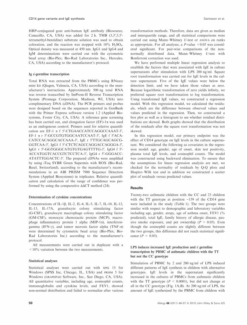
Twenty-two asthmatic children with the CC and 23 children
with the TT genotype at position )159 of the CD14 gene
were included in the study (Table 1). The two groups were
similar with respect to demographic and laboratory variables
including age, gender, atopy, age of asthma onset, FEV1 (%
predicted), total IgE, family history of allergic disease, pas-
sive smoke exposure, and pet ownership (P > 0.05). Even
though the eosinophil counts are slightly different between
the two groups, this difference did not reach statistical signifi-
cance (P > 0.05).
LPS induces increased IgE production and �e germline
transcription by PBMC of asthmatic children with the TT
but not the CC genotype
Stimulation of PBMC by 2 and 200 ng/ml of LPS induced
different patterns of IgE synthesis in children with alternative
genotypes. IgE levels in the supernatant significantly
increased in the cultures of PBMCs from asthmatic children
with the TT genotype (P < 0.0001), but did not change at
all in the CC genotype (Fig. 1A,B). At 200 ng/ml of LPS, the
amount of IgE synthesized by the PBMC from children with
CD14 gene variants and IgE synthesis Sackesen et al.
50 Allergy 66 (2011) 48–57 ª 2010 John Wiley & Sons A/S
the TT genotype was significantly higher than that synthe-
sized by the CC genotype (P = 0.029) (Fig. 1C). Analyzing
the data according to the atopic status demonstrated that
LPS induced IgE synthesis only in children with the TT
but not CC genotype regardless of their atopic status (Fig. 2
A–D).
Multiple linear regression analysis showed that at 200 ng/
ml the TT genotype was significantly associated with the IgE
levels in the culture supernatant independent of other covari-
ates (b = 0.644 SE = 0.239, P = 0.01). In this model,
plasma total IgE had a small but significant effect, as well
(b = 0.001 SE = 0.0002, P = 0.001).
To determine whether the increased IgE concentration in
culture supernatants is because of increased transcription, we
measured e-germline transcription using quantitative real-time
PCR. There were individuals who responded with elevated
IgE transcription in both groups. However, the difference
between the groups reached significance only in the TT group
(Fig. 3A,B).
Stimulation of PBMC by LPS did not result in any
changes in IgG1, IgG4, and IgM concentrations in the cul-
ture supernatant in either group but resulted in similar induc-
tions of IgG1 and IgG4 mRNA expression in PBMCs from
both the CC and TT individuals (data not shown).
LPS induces higher production of Th2 cytokines by PBMC
from asthmatic children with the TT genotype
Among the multiple cytokines that were analyzed, IL-2, IL-4,
and IL-13 showed significant increases upon stimulation with
2 and 200 ng/ml LPS in children with both the TT and CC
genotypes (Fig. 4A–C). However, as can be seen in Fig. 4,
the differences in IL-2 and IL-4 were more striking in the TT
genotype. Moreover, IL-4/IL-12 and IL-4/IFN-c ratios
increased significantly only in the TT group (P = 0.041 and
0.05, respectively) (Fig. 5A,B); but not in the CC group
(P > 0.05 for both). There was a highly significant difference
in IL-4/IL-12 and IL-4/IFN-c ratios between unstimulated
samples and 200 ng/ml samples only in the TT group (Fig. 5
A,B) (P = 0.006 and P = 0.001, respectively).
Of the other cytokines that were measured, IL-1b, IL-7,
IL-10, GM-CSF, G-CSF, IFN-c, and TNF increased simi-
larly in both CC and TT groups; and IL-17, MCP, MIP-1adid not change in either group (data not shown). IL-5 was
present in very small quantities in both groups. Stratification
of the data according to the atopic status did not have any
significant effect on the results.
Table 1 Demographic and laboratory characteristics of the study
population*
CD14 )159 CC CD14 )159 TT
N 22 23
Age 11.2 (9.2–14.6) 10.5 (9.5–11.6)
Gender
Boys 11 12
Girls 11 11
Skin test positivity 11 11
Total IgE (kU/l) 110.5 (50.3–309.8) 109.0 (65.0–230.0)
Eosinophil count 292.5 (187.5–399.8) 186.0 (129.0–335.0)
FEV1% 95.5 (88.8–106.3) 100.0 (90.0–108.0)
Age of onset of asthma 6 (4–10) 4 (2.5–7)
Anti-inflammatory
treatment
7 (31.8%) 12 (52.2%)
Family history of atopic
disease
7 7
Passive smoke exposure 5 5
Pet ownership 2 3
*P > 0.05 for all the variables between the two groups.
CD14 –159 CC Genotype
0
2
4
6
8
10
12
us 2 ng/ml 200 ng/ml
LPS
NS
A
LPS
0
2
4
6
8
10
12
us 200 ng/ml
CC CC TTTT
NS
P = 0.029CD14 –159 CC and TT Genotype C
CD14 –159 TT Genotype
10
12
0
2
4
6
8
us 2 ng/ml 200 ng/ml
LPS
P < 0.0001
NS
P < 0.001
NS
B
Figure 1 Increased IgE response to lipopolysaccharide (LPS) by
cells from asthmatic children with the TT genotype at CD14 )159
position. Peripheral blood mononuclear cells were obtained from
asthmatic children with the CC (A) and the TT (B) genotypes and
stimulated with indicated doses of LPS for 12 days. Cells from chil-
dren with the TT genotype synthesized significantly higher amounts
of IgE compared to CC genotype upon stimulation with 200 ng/ml
of LPS (C). us, unstimulated.
Sackesen et al. CD14 gene variants and IgE synthesis
Allergy 66 (2011) 48–57 ª 2010 John Wiley & Sons A/S 51

Discussion
Our study shows that PBMC from children with asthma who
are homozygous for the T allele at )159 position of the
CD14 gene respond with increased in vitro IgE synthesis to
increasing doses of LPS, whereas those who are homozygous
for the C allele do not. This differential cellular IgE response
may be associated with a cytokine profile favoring the Th2
response in PBMC with the TT genotype as indicated by
increased IL-4/IL-12 and IL-4/IFN-c in this group.
Even though the association between the CD14 C159T
genotype and total IgE levels has been the subject of numer-
ous investigations in many different populations, the cellular
and molecular basis of this association is largely unknown.
In this study, we provide direct evidence for the first time
that under controlled conditions of the in vitro environment,
PBMCs from individuals with the TT genotype mount
an increased IgE synthesis. Importantly, this finding was
strongly supported by our linear regression analysis, which
showed that a higher IgE in culture supernatant was indepen-
dently and significantly associated with the TT genotype. The
observation that there was a significant increase in �e-germline
transcription only in the group with the TT genotype sug-
gests that the major mechanism underlying the difference in
the protein concentration may be increased transcriptional
rate.
We have previously shown that Turkish asthmatic children
with the CC genotype have higher levels of circulating IgE in
their plasma (20). Our current study, however, shows that the
PBMC from asthmatic children with the TT genotype
respond with higher IgE synthesis to increasing doses of LPS.
In addition, the difference between the two genotypes
becomes significant at 200 ng/ml of LPS concentration. This
observation is in line with our previous findings that PBMC
from the individuals with the TT genotype secrete higher
level of sCD14 into the cultures supernatant (22). Appar-
ently, there is a difference between the in vivo plasma
IgE-genotype relationship and in vitro IgE synthesis-genotype
relationship. It seems that the C allele is a risk factor for
higher IgE in vivo; and the T allele becomes a risk factor for
increased IgE synthesis upon stimulation with high doses of
LPS in vitro. We believe that the explanation to this seem-
ingly contradictory observation may lie in the gene-environ-
ment effect that suggests that the genetic influences can be
directly and significantly influenced by environmental expo-
sures, which is, in this case, LPS concentration. From a prac-
tical point of view, this may be important in preventive
strategies as protecting against environmental endotoxin
exposure may be helpful in some genotypes but not others.
Apparently, the measurement of environmental endotoxin
exposure in this cohort would be very helpful in determining
the genotype-related changes in total IgE as a function of
endotoxin exposure. Unfortunately, however, the level of
endotoxin exposure has not been measured in our patients.
The results of the studies that have investigated the associ-
ation between CD14 )159 genotype and atopic outcomes can
IgE
ng/
ml
us 2 ng/ml 200 ng/ml
LPS
us 2 ng/ml 200 ng/ml
LPS
CD14 –159 CC Atopic CD14 –159 CC Non-atopic
IgE
ng/
ml
us 2 ng/ml 200 ng/ml
LPS
CD14 –159 TT Atopic CD14 –159 TT Non-atopic
NS
P = 0.012
NS
NS
NS
P = 0.003
P = 0.005
P = 0.007
0
5
10
15
0
5
10
15
0
5
10
15
us 2 ng/ml 200 ng/ml
LPS
0
5
10
15
IgE
ng/
ml
IgE
ng/
ml
A B
C D
Figure 2 Lipopolysaccharide (LPS) induces IgE production from
both atopic and nonatopic children with asthma. Peripheral blood
mononuclear cells were obtained from atopic with the TT genotype
and non-atopic asthmatic children with the CC (A and B) and the TT
(C and D) genotypes at the CD14 )159 locus and stimulated with
indicated doses of LPS for 12 days.
CD14 gene variants and IgE synthesis Sackesen et al.
52 Allergy 66 (2011) 48–57 ª 2010 John Wiley & Sons A/S
be evaluated in three distinct categories. Kedda et al. and
Ober et al. have shown that the presence of the T allele is
associated with atopy (9–13), whereas others have shown that
it is the C allele that is associated with atopic phenotypes
such as sensitization to molds (25), and more positive aller-
gen-specific IgE tests (26) or total IgE (20). In addition, there
are other studies that have shown no association between this
specific genotype and atopic phenotypes (14, 15). Within this
complexity, when the results of the asthma and endotoxin
study (6) were stratified according to the endotoxin exposure
(27), it was shown that the C allele was associated with a
higher risk of allergic sensitization at low levels of endotoxin
exposure and lower risk at high levels of exposure. This
observation was later substantiated by two independent
studies (16, 17). Our data provide strong in vitro evidence to
support this observation, as only the cells from individuals
with the TT genotype have responded with increasing IgE
synthesis to high doses of LPS. These findings suggest that
the combination of environmental factors and genetic deter-
minants may have different stimulatory effects on the same
population depending on the CD14 genotype (28). It is not
exactly possible to make a one to one comparison between
the environmental exposure studies, which measures the
relative amount of LPS per unit area or per gram of dust
with the pure LPS stimulation used under the highly con-
trolled environment in the test tube. In addition, like all
other in vitro studies and unlike environmental exposures,
our study cannot provide any data regarding long-term con-
tinuous exposure.
The results of our study show that LPS induced IgE syn-
thesis only in children with the TT but not CC genotype
regardless of their atopic status. However, as shown in
Fig. 2, there appears to be more IgE variability in the atopic
children, indicating that the CD14 )159 polymorphism may
be less important in altering IgE levels as a result of LPS
exposure in atopic children. Alternatively, the number of
samples may be too small to say anything conclusive about
this.
Various studies searching the association of the CD14
genotype on the atopic status in asthmatics and nonasth-
matics have shown that the effects of CD14 genotype on
atopic phenotypes can be influenced by many factors. For
instance in their pioneering study, Baldini et al. (10)
showed that the effects of CD14 genotype on total IgE
levels can only be observed in skin test-positive individuals
but not in skin test-negative individuals. Similarly, O’Don-
nell et al. (29) have shown that the influence of CD14
)159C on the atopic phenotype may be age specific, exert-
ing an effect during mid childhood, which is no longer
apparent by early adulthood. Therefore, these observations
including ours imply that the genotypic effects on CD14
can depend on other co-existing entities such as asthma,
atopy, or the age.
In this study, to provide some explanation for the cellular
basis of our observation, we measured a variety of cytokines
in culture supernatants. LPS increased IL-2 and IL-4 produc-
tion by PBMC in the culture supernatants from both geno-
types. However, cells from individuals with the TT genotype
had a stronger response. In addition, the increase in the
Th-2/Th-1 cytokine ratios such as IL-4/IFN- c and IL-4/
IL-12 was significant only in the TT group. These results
have strong implications regarding the differential regulation
of IgE in alternative genotypes. IL-2 was identified as a
T-cell growth factor that is produced by T cells following
activation by mitogens or antigens (30, 31) and was shown
to stimulate the growth and differentiation of many cell types
including B cells and monocytes/macrophages (32, 33). On
the other hand, IL-4 is the most important factor leading to
IgE switch in B cells, whereas IL-12 and IFN-c are the
major Th-1 cytokines and counteract the actions of IL-4 on
IgE synthesis both at the transcriptional and at the receptor
level (34). Th1 cells also efficiently contribute to the effector
2 ng/ml 200 ng/ml0
5
10
200
400
1000
2000
3000
us
IgE
ε g
erm
line
expr
essi
on
LPS
CD14 –159 TT Genotype
CD14 –159 CC Genotype
0
5
10
200
400
1000
2000
3000
2 ng/ml 200 ng/mlus
LPS
IgE
ε g
erm
line
expr
essi
onNS
P = 0.038
P = 0.001
P = 0.006
A
B
´´
Figure 3 Increased IgE class switch in response to lipopolysaccha-
ride (LPS) in CD14 )159 TT genotype. Total RNA was extracted on
day 5 and relative �e germline expression quantified with real-time
PCR. For each individual, measurements after LPS stimulations
were expressed as fold increase relative to the measurement from
unstimulated cells.
Sackesen et al. CD14 gene variants and IgE synthesis
Allergy 66 (2011) 48–57 ª 2010 John Wiley & Sons A/S 53

phase in allergic diseases (35) or dampen allergic inflamma-
tion depending on specific disease model and stage of the
inflammation (36). They play a role in apoptosis of the epi-
thelium in asthma and atopic dermatitis (35), and predomi-
nant Th2 profile in atopic diseases might be a result of the
increased tendency to activation-induced cell death of high
IFN-c-producing Th1 cells (37). Taken together, our data
suggest that the PBMC from children with asthma carrying
the TT genotype may have an increased T-cell response
(IL-2) followed by an exaggerated Th-2/Th-1 (IL-4/IFN-c and
IL-4/IL-12), which eventually lead to higher IgE synthesis.
However, the molecular mechanisms underlying the geno-
type-related differential cytokine expression remain to be dis-
covered. It was previously shown that the transcriptional
activity of the different alleles at the CD14 C/T locus may
depend on the relative expression of various Sp family tran-
scription factors (9). In addition, LPS stimulation activates a
variety of intracellular molecular cascades culminating in the
activation of a variety of transcription factors such as IRF3,
IRF7, AP1 and NF-jB (38). It is possible that this particular
genotype at the CD14 gene may result in a preferential acti-
vation of one transcription factor over the other and thus
result in altered ratios of Th1/Th2 cytokine leading to altered
IgE synthesis.Even though our data may suggest that IL-17
responses in children with asthma are not regulated by the
CD14 promoter )159 genotypes, this statement should be
approached with caution because of the low power of the
study because of its small sample size. Alternatively, this lack
of difference might be because of the presence of TGF-B in
the serum, which may have modulated the IL-10 and IL-17
responses.
Similar to the relationship between CD14 )159 genotype
and IgE, the data on cytokines are also conflicting. For
example, Bottcher et al. have shown that the levels of LPS-
induced IL-12(p70) were higher in Swedish subjects with TT
genotype than subjects carrying the C allele (39). Reijmerink
et al., (40) on the other hand, have failed to show an associa-
tion between CD14 )159 genotype and IL-12 and IL-10
0
10
20
30
40
us 2 ng/ml 200 ng/ml
LPS
0
10
20
30
40
50
us 2 ng/ml 200 ng/ml
LPS
200
400
600
800
0us 2 ng/ml 200 ng/ml
LPS
CD14 –159 CC
CD14 –159 TT
us-LPS 2*-LPS 200*
us-LPS 2
us-LPS 200
CC 0.004 0.002 0.001
TT < 0.0001 < 0.0001 < 0.0001
us-LPS 2-LPS 200
us-LPS 2
us LPS 200
CC 0.013 NS 0.003
TT 0.013 0.011 < 0.0001
us-LPS 2-LPS 200
us-LPS 2
us-LPS 200
CC < 0.0001 0.001 < 0.0001
TT 0.003 0.001 0.009
A
B
C
Figure 4 Cytokine concentrations in the peripheral blood mononu-
clear cells culture supernatants with alternative CD14 genotypes.
Culture supernatants were collected after 48 h of stimulation with
indicated doses of endotoxin. (*) denotes the concentration of
lipopolysaccharide in the cell culture medium. The comparisons are
between the different lipopolysaccharide concentrations within the
same genotype.
CD14 gene variants and IgE synthesis Sackesen et al.
54 Allergy 66 (2011) 48–57 ª 2010 John Wiley & Sons A/S

cytokines in a limited number of Dutch subjects with asthma.
The reasons for the observed discrepancies remain to be
determined.
Investigation into other immunoglobulin isotypes showed
that there were no genotype-related differences in IgG1 and
IgG4 protein concentrations suggesting that the genotype-
specific effect that we observed is, in fact, specific for IgE.
The increase in the transcription of IgG1 and IgG4 mRNA
without any accompanying increases in the concentration of
these proteins may be because of a couple of reasons. First
of all, our experiments were designed specifically to investi-
gate the IgE regulation; and therefore, the experimental time
points were ideal for IgE measurement in the culture super-
natant and for measurement of �e-germline transcription but
not for other immunoglobulin isotypes. Secondly, secreted
IgG may have been captured by the Fc receptors that are
abundantly present on the surface of many cells that are
present in the PBMC mixture. Alternatively, the high basal
rate of these immunoglobulins resulting from spontaneous
secretion may have prevented a difference from becoming
apparent.
As can be seen in the figures related to the measurement
of IgE protein and �e-germline transcription, there is still var-
iance among individuals of the same genotype. Obviously,
the genetic variants of other molecules participating in the
endotoxin response pathway or the haplotypes conferred by
other polymorphisms on the CD14 gene might also signifi-
cantly influence the results of our analysis. Alternatively,
blocking experiments using monoclonal antibodies against
CD14 could be helpful in determining the specificity of the
CD14 genotype-related IgE response. These can be the sub-
jects of another study. It should also be noted our study is
basically aimed at delineating the molecular mechanisms
underlying the endotoxin response and has a small sample
size. Therefore, these findings should be interpreted with cau-
tion and need to be replicated in a larger and independent
sample.
In conclusion, our study provides strong in vitro evidence
that the cellular response to LPS is a function of both LPS
concentration and the genotype at the CD14 C159T locus,
which may significantly affect the cytokine production and
important atopic phenotypes such as IgE.
Acknowledgments
This study is supported by Hacettepe University Scientific
Research Fund grant #0202101020, The Scientific and Tech-
nological research Council of Turkey (TUBITAK) grant
#108S356 and Swiss National Science Foundation grant
# 32-112306 (MA), # 32-105865 (CAA).
0
1
2
3
CD14 –159 TT CD14 –159 CC
IL-4
/IL-1
2
us 2 ng/ml 200 ng/ml
LPS
0
1
2
3
IL-4
/IL-1
2
us 2 ng/ml 200 ng/ml
LPS
0.00
0.02
0.04
0.06
0.08
IL-4
/INF
-γ
0.00
0.02
0.04
0.06
0.08
IL-4
/INF
-γ
NS
NS
P = 0.006
P = 0.041
NS
P = 0.001
P = 0.05NS
us 2 ng/ml 200 ng/ml
LPS
us 2 ng/ml 200 ng/ml
LPS
A
B
Figure 5 IL-4/IL-12 (A) and IL-4/IFN-c (B) ratios in peripheral
blood mononuclear cells culture supernatants following lipopolysac-
charide (LPS) stimulation from children with asthma homozygous
at position )159 of the CD14 gene. The comparisons are
between the different LPS concentration within the same
genotype.
Sackesen et al. CD14 gene variants and IgE synthesis
Allergy 66 (2011) 48–57 ª 2010 John Wiley & Sons A/S 55
References
1. Burrows B, Martinez FD, Halonen M, Bar-
bee RA, Cline MG. Association of asthma
with serum IgE levels and skin-test reactivity
to allergens. N Engl J Med 1989;320:271–
277.
2. Busse WW. Anti-immunoglobulin E (oma-
lizumab) therapy in allergic asthma. Am J
Respir Crit Care Med 2001;164(8 Pt 2):S12–
S17.
3. Von Hertzen LC, Haahtela T. Asthma and
atopy – the price of affluence? Allergy
2004;59:124–137.
4. van Gool CJ, Thijs C, Dagnelie PC, Hen-
quet CJ, van Houwelingen AC, Schrander J
et al. Determinants of neonatal IgE level:
parity, maternal age, birth season and peri-
natal essential fatty acid status in infants of
atopic mothers. Allergy 2004;59:961–968.
5. Gereda JE, Leung DY, Thatayatikom A,
Streib JE, Price MR, Klinnert MD et al.
Relation between house-dust endotoxin
exposure, type 1 T-cell development, and
allergen sensitisation in infants at high risk
of asthma. Lancet 2000;355:1680–1683.
6. Braun-Fahrlander C, Riedler J, Herz U,
Eder W, Waser M, Grize L et al. Environ-
mental exposure to endotoxin and its rela-
tion to asthma in school-age children.
N Engl J Med 2002;347:869–877.
7. Park JH, Gold DR, Spiegelman DL, Burge
HA, Milton DK. House dust endotoxin and
wheeze in the first year of life. Am J Respir
Crit Care Med 2001;163:322–328.
8. Tulic MK, Wale JL, Holt PG, Sly PD.
Modification of the inflammatory response
to allergen challenge after exposure to bacte-
rial lipopolysaccharide. Am J Respir Cell
Mol Biol 2000;22:604–612.
9. LeVan TD, Bloom JW, Bailey TJ, Karp CL,
Halonen M, Martinez FD et al. A common
single nucleotide polymorphism in the CD14
promoter decreases the affinity of Sp protein
binding and enhances transcriptional activ-
ity. J Immunol 2001;167:5838–5844.
10. Baldini M, Lohman IC, Halonen M, Erick-
son RP, Holt PG, Martinez FD. A Polymor-
phism* in the 5¢ flanking region of the
CD14 gene is associated with circulating
soluble CD14 levels and with total serum
immunoglobulin E. Am J Respir Cell Mol
Biol 1999;20:976–983.
11. Koppelman GH, Reijmerink NE, Colin
Stine O, Howard TD, Whittaker PA,
Meyers DA et al. Association of a promoter
polymorphism of the CD14 gene and atopy.
Am J Respir Crit Care Med 2001;163:965–
969.
12. Kedda MA, Lose F, Duffy D, Bell E,
Thompson PJ, Upham J. The CD14 C-159T
polymorphism is not associated with asthma
or asthma severity in an Australian adult
population. Thorax 2005;60:211–214.
13. Ober C, Tsalenko A, Parry R, Cox NJ. A
second-generation genomewide screen for
asthma-susceptibility alleles in a founder
population. Am J Hum Genet 2000;67:1154–
1162.
14. Kabesch M, Hasemann K, Schickinger V,
Tzotcheva I, Bohnert A, Carr D et al. A
promoter polymorphism in the CD14 gene is
associated with elevated levels of soluble
CD14 but not with IgE or atopic diseases.
Allergy 2004;59:520–525.
15. Sengler C, Haider A, Sommerfeld C, Lau S,
Baldini M, Martinez F et al. Evaluation of
the CD14 C-159 T polymorphism in the
German Multicenter Allergy Study cohort.
Clin Exp Allergy 2003;33:166–169.
16. Simpson A, John SL, Jury F, Niven R,
Woodcock A, Ollier WE et al. Endotoxin
exposure, CD14, and allergic disease: an
interaction between genes and the environ-
ment. Am J Respir Crit Care Med 2006;
174:386–392.
17. Williams LK, McPhee RA, Ownby DR, Pet-
erson EL, James M, Zoratti EM et al. Gene-
environment interactions with CD14 C-260T
and their relationship to total serum IgE
levels in adults. J Allergy Clin Immunol
2006;118:851–857.
18. Chatila TA, Li N, Garcia-Lloret M, Kim
HJ, Nel AE. T-cell effector pathways in
allergic diseases: transcriptional mechanisms
and therapeutic targets. J Allergy Clin
Immunol 2008;121:812–823; quiz 824–5.
19. Akdis CA, Akdis M. Mechanisms and treat-
ment of allergic disease in the big picture of
regulatory T cells. J Allergy Clin Immunol
2009;123:735–746; quiz 747–8.
20. Sackesen C, Karaaslan C, Keskin O, Tokol
N, Tahan F, Civelek E et al. The effect of
polymorphisms at the CD14 promoter and
the TLR4 gene on asthma phenotypes in
Turkish children with asthma. Allergy
2005;60:1485–1492.
21. Claassen JL, Levine AD, Buckley RH. A
cell culture system that enhances mononu-
clear cell IgE synthesis induced by recombi-
nant human interleukin-4. J Immunol
Methods 1990;126:213–222.
22. Keskin O, Birben E, Sackesen C, Soyer OU,
Alyamac E, Karaaslan C et al. The effect of
CD14-c159T genotypes on the cytokine
response to endotoxin by peripheral blood
mononuclear cells from asthmatic children.
Ann Allergy Asthma Immunol 2006;97:321–
328.
23. Meiler F, Klunker S, Zimmermann M,
Akdis CA, Akdis M. Distinct regulation of
IgE, IgG4 and IgA by T regulatory cells and
toll-like receptors. Allergy 2008;63:1455–
1463.
24. Kunzmann S, Wohlfahrt JG, Itoh S, Asao
H, Komada M, Akdis CA et al. SARA and
Hgs attenuate susceptibility to TGF-beta1-
mediated T cell suppression. FASEB J
2003;17:194–202.
25. Buckova D, Holla LI, Znojil V, Vasku A.
Polymorphisms of the CD14 gene and atopic
phenotypes in Czech patients with IgE-
mediated allergy. J Hum Genet
2006;51:977–983.
26. Takeuchi K, Suzuki S, Yagawa M, Yuta A,
Majima Y. A CD14 gene polymorphism is
associated with the IgE level for Dermato-
phagoides pteronyssinus. Acta Otolaryngol
2005;125:966–971.
27. Eder W, Klimecki W, Yu L, von Mutius E,
Riedler J, Braun-Fahrlander C et al.
Opposite effects of CD 14/-260 on serum
IgE levels in children raised in different
environments. J Allergy Clin Immunol
2005;116:601–607.
28. Vercelli D. Learning from discrepancies:
CD14 polymorphisms, atopy and the endo-
toxin switch. Clin Exp Allergy 2003;33:153–
155.
29. O’Donnell AR, Toelle BG, Marks GB, Hay-
den CM, Laing IA, Peat JK et al. Age-spe-
cific relationship between CD14 and atopy
in a cohort assessed from age 8 to 25 years.
Am J Respir Crit Care Med 2004;169:615–
622.
30. Mookerjee BK, Pauly JL. Mitogenic effect
of interleukin-2 on unstimulated human
T cells: an editorial review. J Clin Lab Anal
1990;4:138–149.
31. Melchers F, Andersson J, Corbel C, Leptin
M, Lernhardt W, Gerhard W et al. Regula-
tion of B lymphocyte replication and matu-
ration. J Cell Biochem 1982;19:315–332.
32. Smith KA. Interleukin-2: inception, impact,
and implications. Science 1988;240:1169–
1176.
33. Sule NS, Nerurkar RP, Kamath S. Interleu-
kin-2 as a therapeutic agent. J Assoc Physi-
cians India 2001;49:897–900.
34. Geha RS, Jabara HH, Brodeur SR. The reg-
ulation of immunoglobulin E class-switch
recombination. Nat Rev Immunol 2003;3:
721–732.
35. Trautmann A, Schmid-Grendelmeier P,
Kruger K, Crameri R, Akdis M, Akkaya A
et al. T cells and eosinophils cooperate in
the induction of bronchial epithelial cell
apoptosis in asthma. J Allergy Clin Immunol
2002;109:329–337.
36. Finotto S, Neurath MF, Glickman JN, Qin
S, Lehr HA, Green FH et al. Development
of spontaneous airway changes consistent
with human asthma in mice lacking T-bet.
Science 2002;295:336–338.
37. Akkoc T, de Koning PJ, Ruckert B, Barlan
I, Akdis M, Akdis CA. Increased activation-
induced cell death of high IFN-gamma-pro-
ducing T(H)1 cells as a mechanism of T(H)2
CD14 gene variants and IgE synthesis Sackesen et al.
56 Allergy 66 (2011) 48–57 ª 2010 John Wiley & Sons A/S

predominance in atopic diseases. J Allergy
Clin Immunol 2008;121:652–658.e1.
38. Kawai T, Akira S. TLR signaling. Semin
Immunol 2007;19:24–32.
39. Fageras Bottcher M, Hmani-Aifa M, Lind-
strom A, Jenmalm MC, Mai XM, Nilsson L
et al. A TLR4 polymorphism is associated
with asthma and reduced lipopolysaccha-
ride-induced interleukin-12(p70) responses in
Swedish children. J Allergy Clin Immunol
2004;114:561–567.
40. Reijmerink NE, Hylkema MN, Postma DS,
Bruinenberg M, Kauffman HF, Koppelman
GH. Confounding effect of atopy on func-
tional effects of the CD14/-159 promoter
polymorphism. J Allergy Clin Immunol
2006;117:219; author reply 220.
Sackesen et al. CD14 gene variants and IgE synthesis
Allergy 66 (2011) 48–57 ª 2010 John Wiley & Sons A/S 57