Protease-Activated Receptor-2 Activation Contributes to House Dust Mite-Induced IgE Responses in...
9
Protease-Activated Receptor-2 Activation Contributes to House Dust Mite-Induced IgE Responses in Mice Sijranke Post 1,2 , Irene H. Heijink 1,2,3 , Arjen H. Petersen 1 , Harold G. de Bruin 1,2 , Antoon J. M. van Oosterhout 1,2 , Martijn C. Nawijn 1,2 * 1 Lab. Allergology & Pulmonary Diseases, Department of Pathology & Medical Biology, University Medical Center Groningen, University of Groningen, Groningen, The Netherlands, 2 GRIAC Research Institute, University Medical Center Groningen, University of Groningen, Groningen, The Netherlands, 3 Department of Pulmonology, University Medical Center Groningen, University of Groningen, Groningen, The Netherlands Abstract Aeroallergens such as house dust mite (HDM), cockroach, and grass or tree pollen are innocuous substances that can induce allergic sensitization upon inhalation. The serine proteases present in these allergens are thought to activate the protease- activated receptor (PAR)-2, on the airway epithelium, thereby potentially inducing allergic sensitization at the expense of inhalation tolerance. We hypothesized that the proteolytic activity of allergens may play an important factor in the allergenicity to house dust mite and is essential to overcome airway tolerance. Here, we aimed to investigate the role of PAR-2 activation in allergic sensitization and HDM-induced allergic airway inflammation. In our study, Par-2 deficient mice were treated with two different HDM extracts containing high and low serine protease activities twice a week for a period of 5 weeks. We determined airway inflammation through quantification of percentages of mononuclear cells, eosinophils and neutrophils in the bronchial alveolar lavage fluid and measured total IgE and HDM-specific IgE and IgG1 levels in serum. Furthermore, Th2 and pro-inflammatory cytokines including IL-5, IL-13, Eotaxin-1, IL-17, KC, Chemokine (C-C motif) ligand 17 (CCL17) and thymic stromal lymphopoietin (TSLP), were measured in lung tissue homogenates. We observed that independent of the serine protease content, HDM was able to induce elevated levels of eosinophils and neutrophils in the airways of both wild-type (WT) and Par-2 deficient mice. Furthermore, we show that induction of pro-inflammatory cytokines by HDM exposure is independent of Par-2 activation. In contrast, serine protease activity of HDM does contribute to enhanced levels of total IgE, but not HDM-specific IgE. We conclude that, while Par-2 activation contributes to the development of IgE responses, it is largely dispensable for the HDM-induced induction of pro-inflammatory cytokines and airway inflammation in an experimental mouse model of HDM-driven allergic airway disease. Citation: Post S, Heijink IH, Petersen AH, de Bruin HG, van Oosterhout AJM, et al. (2014) Protease-Activated Receptor-2 Activation Contributes to House Dust Mite-Induced IgE Responses in Mice. PLoS ONE 9(3): e91206. doi:10.1371/journal.pone.0091206 Editor: Michael B. Fessler, National Institute of Environmental Health Sciences, United States of America Received February 13, 2013; Accepted February 10, 2014; Published March 20, 2014 Copyright: ß 2014 Post et al. This is an open-access article distributed under the terms of the Creative Commons Attribution License, which permits unrestricted use, distribution, and reproduction in any medium, provided the original author and source are credited. Funding: This study was supported by grants from the Dutch Lung Foundation (NAF 3.2.07.019) and the Foundation Dutch Asthma Control (Stichting Astma bestrijding SAB2011/016). The funders had no role in study design, data collection and analysis, decision to publish, or preparation of the manuscript. Competing Interests: The authors have declared that no competing interests exist. * E-mail: [email protected] Introduction Allergic asthma is a chronic inflammatory pulmonary disease that is characterized by airway hyperreactivity (AHR), airway remodeling, eosinophillic and T helper 2 (Th2) cell infiltration into the airways and an allergen-specific IgE response [1]. Inhaled allergens are in first contact with the airway epithelium, which functions as a barrier (towards the inhaled environment) and is an important part of the innate immune system [2]. The airway epithelial response to allergens is considered to be one of the key drivers of airway inflammation in asthma [3]. The aeroallergen House dust mite (HDM) has most commonly been associated with the development of allergic sensitization and asthma [4,5]. The allergenicity of HDM has largely been attributed to its protease activity, a feature shared by many allergens, including fungi and cockroach [6,7]. The airway epithelium expresses several so-called pattern recognition receptors (PRRs), which in mouse models were found to be critical for the activation of the airway epithelium by HDM and the induction of an innate immune response [8,9]. One of the PRRs activated by proteases is protease-activated receptor (PAR)-2, which is expressed by airway epithelium [10] and is up- regulated in the airways asthma patients [11]. PAR-2 is activated by serine proteases present in HDM [12], which stimulate the release of pro-inflammatory cytokines and chemokines including IL-6, IL-8, GM-CSF and TSLP in cultured airway epithelial cells [13,14]. In mouse studies, inhalation of ovalbumin (OVA) in the presence of a PAR-2 agonist peptide (PAR-2 ap) induced allergic sensitization at the expense of inhalation tolerance [15]. In addition, Par-2 deficient mice showed diminished infiltration of eosinophils and diminished levels of IgE, combined with reduced AHR in the classical OVA-driven experimental asthma model compared to wild-type (Wt) mice [16]. These experiments show that activation of Par-2 may contribute to allergic sensitization through the airways, airway inflammation and AHR upon allergen re-challenge in parenterally sensitized mice. However, no data are available on the relevance for PAR-2 activation in the allergic sensitization driven by HDM, which - unlike the model allergen OVA - harbors endogenous protease activity [17]. Here, we aimed to investigate the role of Par-2 activation in HDM- driven allergic airway inflammation and the induction of an IgE PLOS ONE | www.plosone.org 1 March 2014 | Volume 9 | Issue 3 | e91206
Transcript of Protease-Activated Receptor-2 Activation Contributes to House Dust Mite-Induced IgE Responses in...

Protease-Activated Receptor-2 Activation Contributes toHouse Dust Mite-Induced IgE Responses in MiceSijranke Post1,2, Irene H. Heijink1,2,3, Arjen H. Petersen1, Harold G. de Bruin1,2, Antoon J. M. van
Oosterhout1,2, Martijn C. Nawijn1,2*
1 Lab. Allergology & Pulmonary Diseases, Department of Pathology & Medical Biology, University Medical Center Groningen, University of Groningen, Groningen, The
Netherlands, 2 GRIAC Research Institute, University Medical Center Groningen, University of Groningen, Groningen, The Netherlands, 3 Department of Pulmonology,
University Medical Center Groningen, University of Groningen, Groningen, The Netherlands
Abstract
Aeroallergens such as house dust mite (HDM), cockroach, and grass or tree pollen are innocuous substances that can induceallergic sensitization upon inhalation. The serine proteases present in these allergens are thought to activate the protease-activated receptor (PAR)-2, on the airway epithelium, thereby potentially inducing allergic sensitization at the expense ofinhalation tolerance. We hypothesized that the proteolytic activity of allergens may play an important factor in theallergenicity to house dust mite and is essential to overcome airway tolerance. Here, we aimed to investigate the role ofPAR-2 activation in allergic sensitization and HDM-induced allergic airway inflammation. In our study, Par-2 deficient micewere treated with two different HDM extracts containing high and low serine protease activities twice a week for a period of5 weeks. We determined airway inflammation through quantification of percentages of mononuclear cells, eosinophils andneutrophils in the bronchial alveolar lavage fluid and measured total IgE and HDM-specific IgE and IgG1 levels in serum.Furthermore, Th2 and pro-inflammatory cytokines including IL-5, IL-13, Eotaxin-1, IL-17, KC, Chemokine (C-C motif) ligand 17(CCL17) and thymic stromal lymphopoietin (TSLP), were measured in lung tissue homogenates. We observed thatindependent of the serine protease content, HDM was able to induce elevated levels of eosinophils and neutrophils in theairways of both wild-type (WT) and Par-2 deficient mice. Furthermore, we show that induction of pro-inflammatorycytokines by HDM exposure is independent of Par-2 activation. In contrast, serine protease activity of HDM does contributeto enhanced levels of total IgE, but not HDM-specific IgE. We conclude that, while Par-2 activation contributes to thedevelopment of IgE responses, it is largely dispensable for the HDM-induced induction of pro-inflammatory cytokines andairway inflammation in an experimental mouse model of HDM-driven allergic airway disease.
Citation: Post S, Heijink IH, Petersen AH, de Bruin HG, van Oosterhout AJM, et al. (2014) Protease-Activated Receptor-2 Activation Contributes to House DustMite-Induced IgE Responses in Mice. PLoS ONE 9(3): e91206. doi:10.1371/journal.pone.0091206
Editor: Michael B. Fessler, National Institute of Environmental Health Sciences, United States of America
Received February 13, 2013; Accepted February 10, 2014; Published March 20, 2014
Copyright: � 2014 Post et al. This is an open-access article distributed under the terms of the Creative Commons Attribution License, which permits unrestricteduse, distribution, and reproduction in any medium, provided the original author and source are credited.
Funding: This study was supported by grants from the Dutch Lung Foundation (NAF 3.2.07.019) and the Foundation Dutch Asthma Control (Stichting Astmabestrijding SAB2011/016). The funders had no role in study design, data collection and analysis, decision to publish, or preparation of the manuscript.
Competing Interests: The authors have declared that no competing interests exist.
* E-mail: [email protected]
Introduction
Allergic asthma is a chronic inflammatory pulmonary disease
that is characterized by airway hyperreactivity (AHR), airway
remodeling, eosinophillic and T helper 2 (Th2) cell infiltration into
the airways and an allergen-specific IgE response [1]. Inhaled
allergens are in first contact with the airway epithelium, which
functions as a barrier (towards the inhaled environment) and is an
important part of the innate immune system [2]. The airway
epithelial response to allergens is considered to be one of the key
drivers of airway inflammation in asthma [3]. The aeroallergen
House dust mite (HDM) has most commonly been associated with
the development of allergic sensitization and asthma [4,5]. The
allergenicity of HDM has largely been attributed to its protease
activity, a feature shared by many allergens, including fungi and
cockroach [6,7]. The airway epithelium expresses several so-called
pattern recognition receptors (PRRs), which in mouse models were
found to be critical for the activation of the airway epithelium by
HDM and the induction of an innate immune response [8,9]. One
of the PRRs activated by proteases is protease-activated receptor
(PAR)-2, which is expressed by airway epithelium [10] and is up-
regulated in the airways asthma patients [11]. PAR-2 is activated
by serine proteases present in HDM [12], which stimulate the
release of pro-inflammatory cytokines and chemokines including
IL-6, IL-8, GM-CSF and TSLP in cultured airway epithelial cells
[13,14].
In mouse studies, inhalation of ovalbumin (OVA) in the
presence of a PAR-2 agonist peptide (PAR-2 ap) induced allergic
sensitization at the expense of inhalation tolerance [15]. In
addition, Par-2 deficient mice showed diminished infiltration of
eosinophils and diminished levels of IgE, combined with reduced
AHR in the classical OVA-driven experimental asthma model
compared to wild-type (Wt) mice [16]. These experiments show
that activation of Par-2 may contribute to allergic sensitization
through the airways, airway inflammation and AHR upon
allergen re-challenge in parenterally sensitized mice. However,
no data are available on the relevance for PAR-2 activation in the
allergic sensitization driven by HDM, which - unlike the model
allergen OVA - harbors endogenous protease activity [17]. Here,
we aimed to investigate the role of Par-2 activation in HDM-
driven allergic airway inflammation and the induction of an IgE
PLOS ONE | www.plosone.org 1 March 2014 | Volume 9 | Issue 3 | e91206
response. To this end, we exposed Par-2 deficient mice to two
HDM extracts with low and high serine protease activity [17]. We
found that both HDM extracts induced airway inflammation and
elevated levels of pro-inflammatory cytokines in lung tissue of Par-
2 deficient mice. In addition, exposure to the HDM extract with
the high, but not the low level of serine protease activity, increased
total but not HDM-specific IgE responses in Par-2 deficient mice.
These results indicate that Par-2 activation is largely dispensable
for the induction of airway inflammation by HDM and contributes
to the induction of an IgE response through activation by serine
proteases.
Materials and Methods
Experimental AnimalsPar-2 deficient mice (B6.Cg-F2rl1tm1Mslb/J) and Wt (C57BL/
6J) mice were purchased from Jackson Laboratory (Bar Harbor,
Me., USA). Mice were kept under specific pathogen-free
conditions and maintained on a 12-hour light-dark cycle, with
food and water ad libitum. All animal experiments were evaluated
and approved of by The Institutional Animal Care and Use
Committee of the University of Groningen (The Netherlands).
HDM sensitization protocolThe biochemical component content in the HDM extracts as
well as the administration protocols have been previously
described [17]. Briefly, the low serine protease (2.761 V/ml,
[17]) containing Greer HDM extract (Greer Laboratories, Lenoir,
NC, USA) and the high serine protease (2221.9644.8 V/ml, [17])
containing Citeq HDM extract kindly provided by Citeq Biologics
(Citeq Biologics, Groningen, the Netherlands) were dissolved in
sterile phosphate-buffered saline (PBS; 2.5 mg total weight/ml)
and administered intranasally (20 ml), twice per week for a total of
5 weeks. Twenty-four hours after the last HDM administration,
mice were anesthetized with isoflurane/oxygen (Nicholas Piramal
India Ltd., London, UK), lungs were lavaged with PBS (UMCG
Pharmacy), blood was collected for serum isolation and individual
lung lobes were snap frozen in liquid Nitrogen and stored at
280uC until further analysis.
Collection and measurement of the bronchoalveolarlavage fluid
Briefly, bronchoalveolar lavage (BAL) was performed using
PBS, containing 5% bovine serum albumin (BSA) and a mix of
protease and phosphatase inhibitors (1 complete mini tablet/
10 mL; Roche, Mannheim, Germany). The trachea was cannu-
lated and lungs were lavaged four times with 1 ml PBS. BAL fluid
(BALF) cells were pooled and counted using a coulter counter.
Cytospin preparations were made and stained with Diff-Quick
(Merz & Dade, Dudingen, Switzerland) and evaluated in a blinded
fashion. Cells were distinguished into mononuclear cells, neutro-
phils, and eosinophils by standard morphology. Per cytospin
preparation, 300 cells were counted.
Cytokine assay in mouse lung tissueLevels of IL-5 and IL-13 in homogenized lung tissue lysates
were determined by ELISA, according to the manufacturer’s
instructions (BD Pharmingen, San Diego, CA). Levels of eotaxin-
1, KC, CCL17, TSLP, IFNc and IL-17 were determined in
homogenized lung tissue lysates using Duoset ELISA Develop-
ment Kit (R&D Systems, Minneapolis, MN), according to the
manufacturer’s guidelines.
Measurement of Total- and HDM-specific IgE and HDM-specific IgG1 levels in mouse serum
Total IgE levels and HDM-specific IgE levels were determined
as previously described [17]. Briefly, for the total-IgE measure-
ment a NUNC MaxiSorp flat-bottom 96-well plate (Sigma, St
Louis, MO) was coated with 1 mg/ml anti-mouse IgE (BD
Pharmingen) in PBS overnight at 4uC. Next day, the plates were
washed three times with wash buffer (PBS containing 0.05%
Tween 20 (Sigma, St Louis, MO, USA)) and blocked for 1 hour
with ELISA buffer (50 mM Tris(hydroxymethyl)aminomethane
(Merck KGaA, Darmstadt, Germany), 136.9 mM NaCl (Merck
KGaA, Darmstadt, Germany), 2 mM Ethylenediaminetetraacetic
acid (EDTA; Sigma), 0.5% albumin from bovine serum (BSA;
Sigma) and 0.05% Tween, dissolved in 1000 ml H2O, pH 7.2)
additionally containing 1% BSA. Serum samples and standard
(purified mouse IgEk control) were incubated at room tempera-
ture for 2 hours. After the plate was washed three times, samples
were labeled with 0.5 mg/ml biotin-anti-mouse IgE (BD Pharmin-
gen,) and incubated for 2 hours. Next, plates were washed three
times and incubated with horseradish-peroxidase (1/10000) for
1 hour.
For the HDM-IgE measurement a NUNC MaxiSorp flat-
bottom 96-well plate (Sigma, St Louis, MO) was coated with 2 mg/
ml anti-mouse IgE (BD Pharmingen) in PBS overnight at 4uC.
Next day the plates were washed three times with wash buffer and
blocked for 1 hour with PBS containing 1% BSA. Serum samples
were incubated at room temperature for 2 hours. After the plate
was washed three times, samples were labeled with biotin-
conjugated HDM (1/200) and incubated for 2 hours. Next, plates
were washed three times and incubated with horseradish-
peroxidase (1/300) for 1 hour.
For the HDM-specific IgG1 ELISA, a NUNC MaxiSorp flat-
bottom 96-well plate (Sigma, St Louis, MO) was coated with
10 mg/ml HDM (Greer) overnight at 4uC. The plate was washed
three times and blocked for 1 hour with ELISA buffer. Serum
samples were incubated at room temperature for 2 hours, where
after washed three times. Then, samples were labeled for 1 hour
with 0.5 mg/ml biotinylated-IgG1 followed by horseradish-perox-
idase incubated for 30 minutes.
For all three ELISA’s, after the last label step, plates were
washed three times and incubated with 0.4 mg/ml o-Phenylene-
diamine dihydrochloride (OPD; Sigma) for about 20 min, where
after the reaction was stopped with 4M H2SO4. The plate was
read at 490 nm.
ImmunohistochemistryLungs were treated as previously described (17). Briefly, lungs
were inflated with TissueTek O.C.T. Compound (Sakura Finetek
Europe B.V, Zouterwoude, The Netherlands), and fixed in 10%
Formalin for 24-hours, embedded in paraffin and cut in 3 mm-
thick sections. Lung sections were deparaffinised in xylene,
dehydrated in ethanol and washed in PBS. Antigen retrieval was
performed by heating lung sections to the boiling point in 10 mM
Tris/1 mM EDTA at pH 9.0 for 15 minutes. Sections were then
washed with PBS and blocked with PBS containing 30% H2O2
for 30 min. Lung sections were immunostained with mouse-anti-
Par-2 (SAM11; 1/50; Santa Cruz Biotechnology Inc., Heidelberg,
Germany) for 1 hour, followed by incubation with the the
secondary Ab (1/100; Rabbit-anti-mouse-PO; DAKO, Glostrup,
Denmark), and the tertiary Ab (1/100; Goat-anti-rabbit-PO;
DAKO). The immunostains were developed by using 3-amino-9-
ethylcarbazole (AEC) substrate and mounted with a glass slide
using Kaiser’s glycerine (Life Technologies Europe BV, Bleiswijk,
the Netherlands). Slides were examined and images were acquired
PAR-2 Contributes to HDM-Induced IgE
PLOS ONE | www.plosone.org 2 March 2014 | Volume 9 | Issue 3 | e91206
by a microscope (Olympus BX53) attached to a Color digital
camera (Zeiss) using the Axiovision System (Zeiss).
Statistical analysisStatistical significance was determined using the Mann-
Whitney-U test and P values,0.05 were considered significant.
Results
Par-2 deficiency does not affect HDM-induced airwayinflammation
We aimed to test whether activation of the Par-2 by the
endogenous serine protease activity of HDM extracts is required
for allergic sensitization and the induction of airway inflammation.
To this end, we exposed Par-2 deficient or Wt mice twice a week
to PBS or HDM extracts with either a low serine protease content
(Greer) or a high serine protease content (Citeq) for 5 weeks. We
studied inflammatory cells in the BALF 24 hours after the last
exposure. Treatment of Par-2 deficient mice and Wt controls with
both HDM extracts did not significantly increase total number of
inflammatory cells in BALF (Figure 1A and B). Nevertheless, both
HDM treatments induced a significant increase in the eosinophilic
cell fraction in both Par-2 deficient mice and Wt type controls
(Figure 1E and F). Unexpectedly, we observed that the Citeq
extract (with high serine protease activity) induced a significantly
stronger increase in the percentage of eosinophils in BALF in the
Par-2 deficient mice than in the Wt mice (Figure 1F). Only the
Citeq HDM extract induced a significant increase in the fraction
of neutrophils in BAL fluid, with again no differences between Wt
or Par-2 deficient mice (Figure 1G and H).
Furthermore, in Wt mice of the same genetic background as the
Par-2 deficient mice (C57Bl/6J) we investigated AHR in response
to the Greer HDM extract, which we previously found capable to
induce AHR in mice on a BALB/c genetic background [17]. In
these experiments, the HDM-treated C57Bl/6J mice did not show
significant differences in airway resistance in response to a dilution
series of metacholine compared to PBS-exposed control mice (See
Figure S1). Given this observation, we did not further analyze
AHR in the Par-2 deficient mice. To test whether this difference in
HDM-induced AHR between the two strains was dependent on
the level of Par-2 expression, we stained lung sections obtained
from BALB/c and C57BL/6 mice exposed to PBS and BALB/c
mice exposed Citeq and Greer HDM for Par-2 by immunohis-
tochemsitry. We did not observe differences in PAR-2 staining
between the bronchial epithelium of PBS exposed C57BL/6 or
BALB/c mice (Figure S2A and B), nor did we observe differences
in PAR2 staining intensity or pattern between the PBS and Greer
or Citeq HDM exposed BALB/c mice (Figure S2A, C and D).
In summary, the HDM-induced eosinophilic airway inflamma-
tion was not attenuated, in the Par-2 deficient mice, with even a
stronger response in airway eosinophilia induced by the extract
with the highest serine protease activity.
High protease levels are required for allergic sensitizationTo assess the induction of allergic sensitization after HDM
exposure in Wt and Par-2 deficient mice, we first investigated the
IgE response. In agreement with our previously reported data
Figure 1. Par-2-deficiency does not influence the inflammatory response after HDM exposure. Total cell counts and mononuclear,eosinophil and neutrophil fractions in BALF from Wt or Par-2 deficient mice after chronic exposure to PBS or HDM (Greer/Citeq). BALF cells werecounted 24 hours after the last PBS/HDM exposure. Total cell count after PBS and (A) HDM Greer exposure or (B) HDM Citeq exposure, Mononuclearcell count (%) after PBS and (C) HDM Greer exposure or (D) HDM Citeq exposure, Eosinophil cell count (%) after PBS and (E) HDM Greer exposure or (F)HDM Citeq exposure, Neutrophil cell count (%) after PBS and (G) HDM Greer exposure or (H) HDM Citeq exposure. Median levels are shown (n = 7–8mice per group). *p,0.05 and **p,0.01 between PBS and HDM exposed mice or differences between Wt and Par-2 deficient mice.doi:10.1371/journal.pone.0091206.g001
PAR-2 Contributes to HDM-Induced IgE
PLOS ONE | www.plosone.org 3 March 2014 | Volume 9 | Issue 3 | e91206
obtained in mice with a BALB/c genetic background [17], we
observed the induction of an IgE response, both for total IgE levels
and for HDM-specific IgE, only in mice exposed to the high serine
protease containing Citeq HDM extract, but not in those exposed
to the Greer HDM (Figure 2A–D). Interestingly, we observed that
these Citeq HDM-induced total IgE levels were significantly lower
in Par-2 deficient mice compared to the Wt mice (Figure 2B).
Remarkably, this difference was not observed in the HDM-specific
IgE response. Next, we investigated the HDM-specific IgG1
response, which showed that both HDM extracts induced
significant levels of HDM-specific IgG1 in both Wt and Par-2
deficient mice (Figure 2E–F), although this induction failed to
reach statistical significance in Citeq HDM-treated Par-2 deficient
mice (Figure 2F).
These results confirm our previous findings in the BALB/c
genetic background mice [17], indicating that the high serine
protease content in HDM plays a role in the development of the
total and HDM-specific IgE response, but not in the HDM-
specific IgG1 response. Interestingly, Par-2 deficiency affects the
total, but not the HDM-specific IgE response.
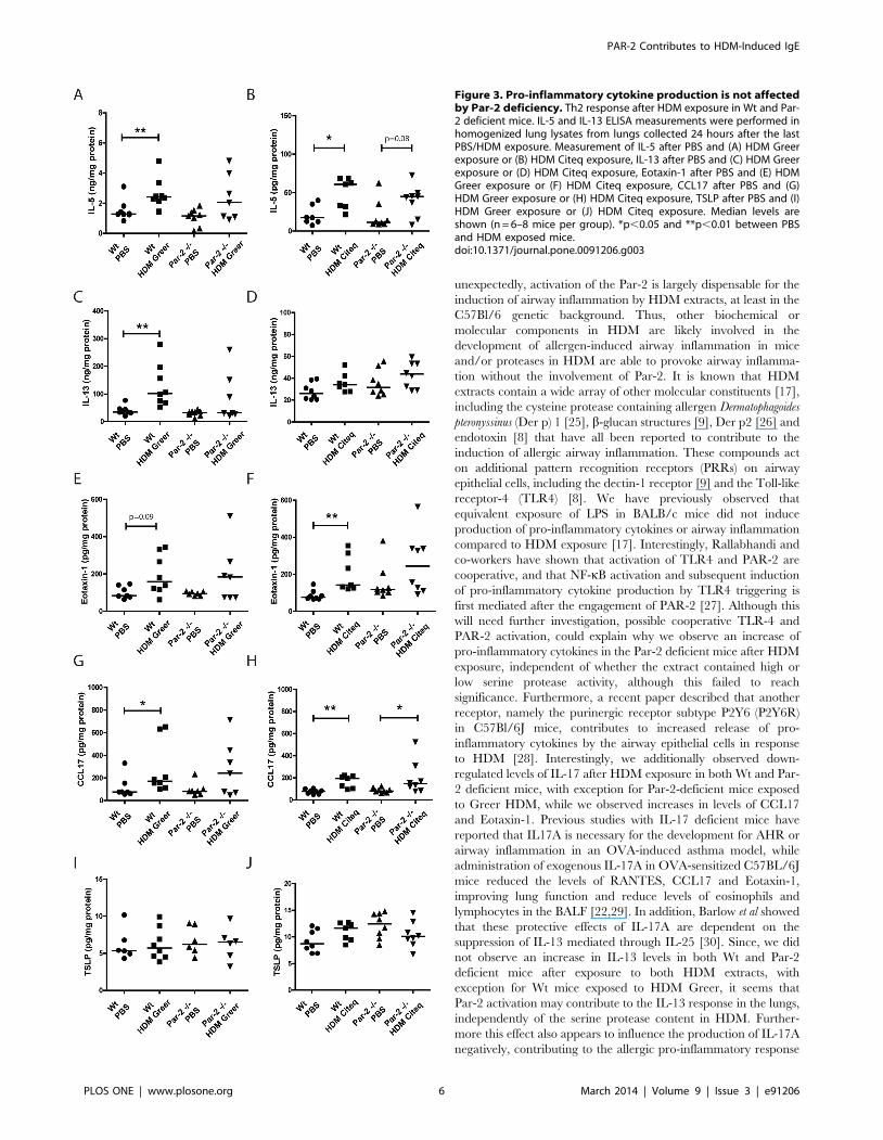
Pro-inflammatory cytokine production in the lung is notaffected by PAR-2 deficiency
Next we assessed the effect of the high and low serine protease
extract on levels of Th2 cytokines in the lungs of Wt and Par-2
deficient mice. Both HDM extracts significantly increased levels of
IL-5 in the lungs compared to PBS-exposure in Wt mice
(Figure 3A–B). In the Par-2 deficient mice, both HDM extracts
failed to induce a statistical significant increase in IL-5 levels,
although a trend (p = 0.08) was observed for the high serine
protease-containing HDM extract (Figure 3B). Furthermore,
HDM-induced IL-5 levels were not significantly different between
the Wt and Par-2 deficient mice. With respect to IL-13, we only
observed a significant increase in Greer HDM-treated Wt mice
compared to PBS-exposed mice, with no effect of the Citeq HDM
treatment and no differences between Wt or Par-2 deficient mice
(Figure 3C–D).
Since IL-5 and eotaxin-1 are both important for the recruitment
of eosinophils [18], we also investigated the eotaxin-1 production
in both Wt and Par-2 deficient mice after HDM exposure. Only a
significant increase in eotaxin-1 levels of was observed in Wt mice
exposed to Citeq HDM, while a trend (p = 0.09) towards
significance was observed in mice exposed to the Greer HDM
extract (Figure 3E–F). Neither extracts were able to induce
significant increase of Eotaxin-1 levels in the Par-2 deficient mice
(Figure 3E–F), although levels were not significantly lower in the
Par-2 deficient compared to the Wt mice.
Next we investigated the expression of the pro-allergic factors
CCL17 and TSLP, which attract Th2 cells and promote Th2 cell
differentiation, respectively [19,20]. Here, we found that both
HDM extracts induced an increase in CCL17 levels in both Wt
and Par-2 deficient mice compared to PBS-exposed mice, with no
significant differences between Wt and Par-2 deficient, although
this failed to reach statistical significance for the Greer HDM
treatment in Par-2 deficient mice (Figure 3G–H). In contrast, the
HDM extracts did not induce an increase in TSLP levels in both
the Par-2 deficient and Wt groups (Figure 3I–J).
As we observed induction neutrophillic airway infiltration in
Citeq HDM-treated mice (Figure 1H), we also analyzed the levels
of KC, a known chemo-attractant for neutrophils [21]. Both
HDM extracts induced elevated levels of KC in lung tissue of both
Par-2 deficient and Wt control mice compared to the PBS-exposed
controls. Although this failed to reach statistical significance in the
Par-2 deficient mice (Figure 4A–B), we did not observe significant
differences between Par-2 deficient and Wt mice. The differences
between the datasets obtained from Citeq and Greer extract
exposure may stem from differences in ELISA values due to a
different day of analysis.
Subsequently, we investigated levels of IL-17, which is known as
a negative regulator for allergic asthma, but positively associated
with neutrophillic airway inflammation [22]. Interestingly, HDM
treatment generally seemed to down-regulate IL-17 levels,
although significance was only reached in Par-2 deficient mice
exposed to Citeq HDM (Figure 4D), while a trend (p = 0.08) was
observed for the Greer HDM exposure in Wt mice (Figure 4C).
Again, we did not observe significant differences between Par-2
deficient and Wt mice.
In comparison, we additionally investigated the Th1 response
by analyzing the levels of IFNc in HDM-treated Par-2 deficient
mice and control littermates. In both HDM models, we did not
observe an altered level of IFNc in lung tissue homogenate from
wild-type mice, irrespective of the source of the HDM (Figure 4E–
F). Also in Par-2 deficient mice, no significant induction of IFNc is
observed for either Greer or Citeq HDM treated mice (Figure 4E–
F), although a trend towards increased IFNc levels is observed in
the PAR2-deficient mice treated with Citeq HDM compared to
PBS treated controls (Figure 4F).
Overall, these results show that the HDM-induced pro-
inflammatory cytokine response in mice is not markedly affected
by Par-2 deficiency.
Discussion
In this study, we investigated the role of Par-2 activation in
HDM-induced allergic sensitization and airway inflammation in
mice. We show that HDM treatment initiates the influx of
eosinophils and neutrophils into the airways, independent of the
serine protease levels present in the HDM extract and with no
difference between Wt and Par-2 deficient mice [17]. Further-
more, our findings suggest that Par-2 activation may contribute to
the IL-13 response in the lungs, although here too, the differences
in serine protease activity between the two HDM extracts do not
seem to be relevant for the response. In contrast, the level of serine
protease activity in HDM does seem to play a role in the
development of the total and HDM-specific IgE responses,
whereas the presence of Par-2 receptor is only required for
optimal induction of total IgE response. Overall, our findings
indicate that HDM-induced IgE responses are associated with the
protease activity of the HDM extract, while only the total IgE
response depends on the activation of the Par-2 receptor. In
contrast, the HDM-induced airway inflammation and induction of
pro-inflammatory cytokines is independent on Par-2 receptor
activation.
The role of PAR-2 in mediating the production of pro-
inflammatory cytokines and airway inflammation has extensively
been investigated, using both specific small-peptide agonists of the
receptor as well as by using Par-2 over-expressing or deficient mice
[15,16]. Schmidlin et al were one of the first to show that over-
expression of Par-2 in FVB mice leads to exacerbation of airway
inflammation in the airways, while deletion of Par-2 in C57BL/6J
mice diminishes the inflammation in the airways after OVA
exposure [16]. Furthermore, using an OVA tolerance model in
BALB/c mice, Ebeling et al. showed that administration of an
PAR-2 ap simultaneously with OVA administration induced
allergic sensitization instead of inhalation tolerance [15], which is
induced by OVA when applied through the airways [23]. We
addressed the role of Par-2 in mediating airway inflammation
using protease-containing HDM, that in contrast to OVA induces
PAR-2 Contributes to HDM-Induced IgE
PLOS ONE | www.plosone.org 4 March 2014 | Volume 9 | Issue 3 | e91206

allergic sensitization when applied through the airways [17,24].
Exposure of the Par-2 deficient mice to HDM extracts containing
either high or low serine protease activity resulted in airway
inflammation, as indicated by the elevated levels of eosinophils and
neutrophils in the BALF, with exception for the neutrophil levels
after HDM Greer exposure. These results indicate that,
Figure 2. The protease content in HDM is responsible for the IgE but not the IgG1 response. Allergic sensitization response after HDMexposure in Wt and Par-2 deficient mice. Total IgE, HDM-specific IgE and HDM-specific IgG1 ELISA measurements were performed in serum collected24 hours after the last PBS/HDM exposure. Measurements of total IgE after PBS and (A) HDM Greer exposure or (B) HDM Citeq exposure, HDM-IgEafter PBS and (C) HDM Greer exposure or (D) HDM Citeq exposure, HDM-IgG1 after PBS and (E) HDM Greer exposure or (F) HDM Citeq exposure.Median levels are shown (n = 6–8 mice per group). *p,0.05, **p,0.01 and ***p,0.001 between PBS and HDM exposed mice or between Wt and Par-2 deficient mice.doi:10.1371/journal.pone.0091206.g002
PAR-2 Contributes to HDM-Induced IgE
PLOS ONE | www.plosone.org 5 March 2014 | Volume 9 | Issue 3 | e91206
unexpectedly, activation of the Par-2 is largely dispensable for the
induction of airway inflammation by HDM extracts, at least in the
C57Bl/6 genetic background. Thus, other biochemical or
molecular components in HDM are likely involved in the
development of allergen-induced airway inflammation in mice
and/or proteases in HDM are able to provoke airway inflamma-
tion without the involvement of Par-2. It is known that HDM
extracts contain a wide array of other molecular constituents [17],
including the cysteine protease containing allergen Dermatophagoides
pteronyssinus (Der p) 1 [25], b-glucan structures [9], Der p2 [26] and
endotoxin [8] that have all been reported to contribute to the
induction of allergic airway inflammation. These compounds act
on additional pattern recognition receptors (PRRs) on airway
epithelial cells, including the dectin-1 receptor [9] and the Toll-like
receptor-4 (TLR4) [8]. We have previously observed that
equivalent exposure of LPS in BALB/c mice did not induce
production of pro-inflammatory cytokines or airway inflammation
compared to HDM exposure [17]. Interestingly, Rallabhandi and
co-workers have shown that activation of TLR4 and PAR-2 are
cooperative, and that NF-kB activation and subsequent induction
of pro-inflammatory cytokine production by TLR4 triggering is
first mediated after the engagement of PAR-2 [27]. Although this
will need further investigation, possible cooperative TLR-4 and
PAR-2 activation, could explain why we observe an increase of
pro-inflammatory cytokines in the Par-2 deficient mice after HDM
exposure, independent of whether the extract contained high or
low serine protease activity, although this failed to reach
significance. Furthermore, a recent paper described that another
receptor, namely the purinergic receptor subtype P2Y6 (P2Y6R)
in C57Bl/6J mice, contributes to increased release of pro-
inflammatory cytokines by the airway epithelial cells in response
to HDM [28]. Interestingly, we additionally observed down-
regulated levels of IL-17 after HDM exposure in both Wt and Par-
2 deficient mice, with exception for Par-2-deficient mice exposed
to Greer HDM, while we observed increases in levels of CCL17
and Eotaxin-1. Previous studies with IL-17 deficient mice have
reported that IL17A is necessary for the development for AHR or
airway inflammation in an OVA-induced asthma model, while
administration of exogenous IL-17A in OVA-sensitized C57BL/6J
mice reduced the levels of RANTES, CCL17 and Eotaxin-1,
improving lung function and reduce levels of eosinophils and
lymphocytes in the BALF [22,29]. In addition, Barlow et al showed
that these protective effects of IL-17A are dependent on the
suppression of IL-13 mediated through IL-25 [30]. Since, we did
not observe an increase in IL-13 levels in both Wt and Par-2
deficient mice after exposure to both HDM extracts, with
exception for Wt mice exposed to HDM Greer, it seems that
Par-2 activation may contribute to the IL-13 response in the lungs,
independently of the serine protease content in HDM. Further-
more this effect also appears to influence the production of IL-17A
negatively, contributing to the allergic pro-inflammatory response
Figure 3. Pro-inflammatory cytokine production is not affectedby Par-2 deficiency. Th2 response after HDM exposure in Wt and Par-2 deficient mice. IL-5 and IL-13 ELISA measurements were performed inhomogenized lung lysates from lungs collected 24 hours after the lastPBS/HDM exposure. Measurement of IL-5 after PBS and (A) HDM Greerexposure or (B) HDM Citeq exposure, IL-13 after PBS and (C) HDM Greerexposure or (D) HDM Citeq exposure, Eotaxin-1 after PBS and (E) HDMGreer exposure or (F) HDM Citeq exposure, CCL17 after PBS and (G)HDM Greer exposure or (H) HDM Citeq exposure, TSLP after PBS and (I)HDM Greer exposure or (J) HDM Citeq exposure. Median levels areshown (n = 6–8 mice per group). *p,0.05 and **p,0.01 between PBSand HDM exposed mice.doi:10.1371/journal.pone.0091206.g003
PAR-2 Contributes to HDM-Induced IgE
PLOS ONE | www.plosone.org 6 March 2014 | Volume 9 | Issue 3 | e91206

Figure 4. Par-2 deficiency affects IL-17 but not KC or IFNc production after HDM exposure. ELISA measurements were performed inhomogenized lung lysates from lungs collected 24 hours after the last PBS/HDM exposure. Measurements of KC after PBS and (A) HDM Greerexposure or (B) HDM Citeq exposure, IL-17 after PBS and (C) HDM Greer exposure or (D) HDM Citeq exposure, and IFNc after PBS and (E) HDM Greerexposure or (F) HDM Citeq exposure. Median levels are shown (n = 6–8 mice per group). *p,0.05 and **p,0.01 between PBS and HDM exposedmice.doi:10.1371/journal.pone.0091206.g004
PAR-2 Contributes to HDM-Induced IgE
PLOS ONE | www.plosone.org 7 March 2014 | Volume 9 | Issue 3 | e91206

to HDM. Consequently, in contrast to the previous in vitro studies
[13,14], we conclude that Par-2 activation merely contributes to
the induction of pro-inflammatory cytokines in vivo, instead of
being critically required for the response.
In addition to the role of Par-2 in the HDM-induced airway
inflammation, we also studied the role of Par-2 activation in the
HDM-induced IgE and IgG1 responses. Here, we found that both
specific and non-specific IgE responses were induced by the HDM
extract with the high protease activity only, while Par-2 deficiency
reduced the total, but not the HDM-specific-IgE response,
suggesting that (serine protease-dependent) Par-2 activation is
only involved in the HDM-induced total IgE response. In a
previous study of Gough et al, treatment of Der p1 with an
irreversible cysteine protease-specific inhibitor (E-64) reduced the
IgE-eliciting activity of the allergen without affecting the
production of IgG in vivo [24]. In line with these results, we have
previously reported that the IgE-inducing high serine protease
containing Citeq HDM extract also contains a high amount of
cysteine protease activity compared to the Greer extract [17], and
we speculate that cysteine proteases may be involved in the HDM-
induced IgE responses. In addition, the same study also
demonstrated, that mice exposed to an equivalent amount of
LPS had no significant effect on IgE levels [17], excluding a role
for HDM endotoxins to provoke the observed allergic sensitization
response.
For interpretation of our data, it is relevant to consider that
cytokine levels in BAL and lung tissue were assessed 24 hours after
the last HDM challenge. While we have previously successfully
measured HDM-induced cytokine and chemokine responses in
lung tissue at this time-point [17], it has been shown that the
highest levels after HDM exposure are found 4–8 hours after the
last exposure [31]. This indicates that our time-point of analysis
might not have been the most optimal for detecting differences in
cytokine and chemokine levels in lung tissue or BAL between Par-
2 deficient mice and controls. Moreover, we have used C57Bl/6
mice for our analyses. The role of the Par-2 in airway
inflammation induced by proteolytically active allergen extracts
has previously extensively been investigated in Par-2 deficient mice
on both the C57Bl/6J and the BALB/c background, using
cockroach fecal remnants (GC frass) as aeroallergen, of which
allergenicity is attributed to its serine protease content [32–34].
These studies showed that allergic airway inflammation after GC
frass exposure is in part mediated via Par-2 in BALB/c but is
independent of Par-2 in C57Bl/6J mice [34]. Notwithstanding,
also on the BALB/c genetic background, GC frass induced a
significant response in Par-2 deficient mice, indicating that GC
frass also on the BALB/c background induces an in part Par-2
independent airway inflammation.
The discrepancy between C57Bl/6J and BALB/c in the
response to mucosally administered aeroallergen extracts might
be due to a difference between the two strains in activation of
airway epithelial NF-kB. A recent study by Alcorn et al has
demonstrated that BALB/c and C57Bl/6J mice differ markedly in
their activation of NF-kB in the airway epithelium upon allergen
challenge in the classical OVA-driven mouse model of allergic
airway inflammation [35]. Here, OVA inhalation in parenterally
sensitized mice activates the NF-kB pathway in the airway
epithelium of the BALB/c strain but not of the C57Bl/6J strain,
which was associated with reduced expression of NF-kB sensitive
cytokines induced in the BAL by the OVA challenges in the
C57Bl/6J background, most notably TNFa and KC [35]. These
data might well be very relevant for the difference in the Par-2
dependence of the response to GC frass between the two strains,
since GC frass has been found to induce NF-kB activation [34].
For the data presented here on HDM-induced allergic airway
inflammation, it is relevant to note that the role of airway epithelial
NF-kB activation for HDM-induced responses was recently found
to limited for the classical parameters of allergic airway
inflammation: airway epithelial-specific inhibition of NF-kB only
partially affected airway eosinophilia, and had no effect on levels of
IL-13, HDM-specific IgE and IgG1 and mucus metaplasia after
HDM exposure [36]. Therefore, although we have not performed
experiments on HDM-induced responses in PAR-2 deficient mice
on a BALB/c background that would be more sensitive to airway
epithelial NF-kB activation, we propose that our data do clearly
indicate that part of the HDM-induced response is independent on
Par-2 activation.
Overall, our study shows that Par-2 activation through HDM
contributes to the induction of total, but not allergen-specific IgE
responses while the production of pro-inflammatory cytokines and
the development of airway inflammation is at least in part
independent on the activation of Par-2.
Supporting Information
Figure S1 C57Bl/6J mice do not show significantdifferences in airway resistance after PBS or HDMexposure. Airway hyperreactivity was measured by Flexivent in
response to metacholine in Wt C57Bl/6J mice after PBS and
HDM Greer exposure. Absolute mean values (6SEM) are shown.
(TIF)
Figure S2 HDM exposure has no effect on Par-2expression. PAR-2 staining of histological lung sections obtained
from (A) BALB/c and (B) C57BL/6J mice exposed to PBS and
BALB/c mice exposed to (C) Greer HDM extract and (D) Citeq
HDM extract twice a week for a period of 5 weeks. Representative
pictures are shown. Magnification 406. Red arrows indicate highly
PAR-2 expressing cells.
(TIF)
Acknowledgments
We thank Citeq Biologics for the kind gift of their HDM extract.
Author Contributions
Conceived and designed the experiments: SP IHH AJMO MCN.
Performed the experiments: SP AP HGdB. Analyzed the data: SP IHH
MCN. Wrote the paper: SP IHH AJMO MCN.
References
1. Wenzel SE (2012) Asthma phenotypes: The evolution from clinical to molecular
approaches. Nat Med 18: 716–725.
2. Lambrecht BN, Hammad H (2012) The airway epithelium in asthma. Nat Med
18: 684–692.
3. Holgate ST (2011) The sentinel role of the airway epithelium in asthma
pathogenesis. Immunol Rev 242: 205–219.
4. Nelson RP Jr, DiNicolo R, Fernandez-Caldas E, Seleznick MJ, Lockey RF, et al.
(1996) Allergen-specific IgE levels and mite allergen exposure in children with
acute asthma first seen in an emergency department and in nonasthmatic control
subjects. J Allergy Clin Immunol 98: 258–263.
5. Gregory LG, Lloyd CM (2011) Orchestrating house dust mite-associated allergy
in the lung. Trends Immunol 32: 402–411.
6. Boitano S, Flynn AN, Sherwood CL, Schulz SM, Hoffman J, et al. (2011)
Alternaria alternata serine proteases induce lung inflammation and airway
epithelial cell activation via PAR2. Am J Physiol Lung Cell Mol Physiol 300:
L605–14.
7. Jeong SK, Kim HJ, Youm JK, Ahn SK, Choi EH, et al. (2008) Mite and
cockroach allergens activate protease-activated receptor 2 and delay epidermal
permeability barrier recovery. J Invest Dermatol 128: 1930–1939.
PAR-2 Contributes to HDM-Induced IgE
PLOS ONE | www.plosone.org 8 March 2014 | Volume 9 | Issue 3 | e91206
8. Hammad H, Chieppa M, Perros F, Willart MA, Germain RN, et al. (2009)
House dust mite allergen induces asthma via toll-like receptor 4 triggering ofairway structural cells. Nat Med 15: 410–416.
9. Nathan AT, Peterson EA, Chakir J, Wills-Karp M (2009) Innate immune
responses of airway epithelium to house dust mite are mediated through beta-glucan-dependent pathways. J Allergy Clin Immunol 123: 612–618.
10. Cocks TM, Fong B, Chow JM, Anderson GP, Frauman AG, et al. (1999) Aprotective role for protease-activated receptors in the airways. Nature 398: 156–
160.
11. Knight DA, Lim S, Scaffidi AK, Roche N, Chung KF, et al. (2001) Protease-activated receptors in human airways: Upregulation of PAR-2 in respiratory
epithelium from patients with asthma. J Allergy Clin Immunol 108: 797–803.12. Ebeling C, Forsythe P, Ng J, Gordon JR, Hollenberg M, et al. (2005) Proteinase-
activated receptor 2 activation in the airways enhances antigen-mediated airwayinflammation and airway hyperresponsiveness through different pathways.
J Allergy Clin Immunol 115: 623–630.
13. Kauffman HF, Tamm M, Timmerman JA, Borger P (2006) House dust mitemajor allergens der p 1 and der p 5 activate human airway-derived epithelial
cells by protease-dependent and protease-independent mechanisms. Clin MolAllergy 4: 5.
14. Asokananthan N, Graham PT, Stewart DJ, Bakker AJ, Eidne KA, et al. (2002)
House dust mite allergens induce proinflammatory cytokines from respiratoryepithelial cells: The cysteine protease allergen, der p 1, activates protease-
activated receptor (PAR)-2 and inactivates PAR-1. J Immunol 169: 4572–4578.15. Ebeling C, Lam T, Gordon JR, Hollenberg MD, Vliagoftis H (2007) Proteinase-
activated receptor-2 promotes allergic sensitization to an inhaled antigenthrough a TNF-mediated pathway. J Immunol 179: 2910–2917.
16. Schmidlin F, Amadesi S, Dabbagh K, Lewis DE, Knott P, et al. (2002) Protease-
activated receptor 2 mediates eosinophil infiltration and hyperreactivity inallergic inflammation of the airway. J Immunol 169: 5315–5321.
17. Post S, Nawijn MC, Hackett TL, Baranowska M, Gras R, et al. (2012) Thecomposition of house dust mite is critical for mucosal barrier dysfunction and
allergic sensitisation. Thorax 67: 488–495.
18. Walsh ER, Sahu N, Kearley J, Benjamin E, Kang BH, et al. (2008) Strain-specific requirement for eosinophils in the recruitment of T cells to the lung
during the development of allergic asthma. J Exp Med 205: 1285–1292.19. Heijink IH, Kies PM, Kauffman HF, Postma DS, van Oosterhout AJ, et al.
(2007) Down-regulation of E-cadherin in human bronchial epithelial cells leadsto epidermal growth factor receptor-dependent Th2 cell-promoting activity.
J Immunol 178: 7678–7685.
20. Ying S, O’Connor B, Ratoff J, Meng Q, Mallett K, et al. (2005) Thymic stromallymphopoietin expression is increased in asthmatic airways and correlates with
expression of Th2-attracting chemokines and disease severity. J Immunol 174:8183–8190.
21. Arizmendi NG, Abel M, Puttagunta L, Asaduzzaman M, Davidson C, et al.
(2011) Mucosal exposure to cockroach extract induces allergic sensitization andallergic airway inflammation. Allergy Asthma Clin Immunol 7: 22.
22. Schnyder-Candrian S, Togbe D, Couillin I, Mercier I, Brombacher F, et al.
(2006) Interleukin-17 is a negative regulator of established allergic asthma. J ExpMed 203: 2715–2725.
23. Holt PG, Batty JE, Turner KJ (1981) Inhibition of specific IgE responses in mice
by pre-exposure to inhaled antigen. Immunology 42: 409–417.24. Gough L, Campbell E, Bayley D, Van Heeke G, Shakib F (2003) Proteolytic
activity of the house dust mite allergen der p 1 enhances allergenicity in a mouseinhalation model. Clin Exp Allergy 33: 1159–1163.
25. Adam E, Hansen KK, Astudillo Fernandez O, Coulon L, Bex F, et al. (2006)
The house dust mite allergen der p 1, unlike der p 3, stimulates the expression ofinterleukin-8 in human airway epithelial cells via a proteinase-activated receptor-
2-independent mechanism. J Biol Chem 281: 6910–6923.26. Osterlund C, Gronlund H, Polovic N, Sundstrom S, Gafvelin G, et al. (2009)
The non-proteolytic house dust mite allergen der p 2 induce NF-kappaB andMAPK dependent activation of bronchial epithelial cells. Clin Exp Allergy 39:
1199–1208.
27. Rallabhandi P, Nhu QM, Toshchakov VY, Piao W, Medvedev AE, et al. (2008)Analysis of proteinase-activated receptor 2 and TLR4 signal transduction: A
novel paradigm for receptor cooperativity. J Biol Chem 283: 24314–24325.28. Vieira RP, Muller T, Grimm M, von Gernler V, Vetter B, et al. (2011)
Purinergic receptor type 6 contributes to airway inflammation and remodeling in
experimental allergic airway inflammation. Am J Respir Crit Care Med 184:215–223.
29. Nakae S, Komiyama Y, Nambu A, Sudo K, Iwase M, et al. (2002) Antigen-specific T cell sensitization is impaired in IL-17-deficient mice, causing
suppression of allergic cellular and humoral responses. Immunity 17: 375–387.30. Barlow JL, Flynn RJ, Ballantyne SJ, McKenzie AN (2011) Reciprocal expression
of IL-25 and IL-17A is important for allergic airways hyperreactivity. Clin Exp
Allergy 41: 1447–1455.31. Gregory LG, Causton B, Murdoch JR, Mathie SA, O’Donnell V, et al. (2009)
Inhaled house dust mite induces pulmonary T helper 2 cytokine production.Clin Exp Allergy 39: 1597–1610.
32. Lewkowich IP, Day SB, Ledford JR, Zhou P, Dienger K, et al. (2011) Protease-
activated receptor 2 activation of myeloid dendritic cells regulates allergic airwayinflammation. Respir Res 12: 122.
33. Day SB, Ledford JR, Zhou P, Lewkowich IP, Page K (2012) German cockroachproteases and protease-activated receptor-2 regulate chemokine production and
dendritic cell recruitment. J Innate Immun 4: 100–110.34. Page K, Ledford JR, Zhou P, Dienger K, Wills-Karp M (2010) Mucosal
sensitization to german cockroach involves protease-activated receptor-2. Respir
Res 11: 62.35. Alcorn JF, Ckless K, Brown AL, Guala AS, Kolls JK, et al. (2010) Strain-
dependent activation of NF-kappaB in the airway epithelium and its role inallergic airway inflammation. Am J Physiol Lung Cell Mol Physiol 298: L57–66.
36. Tully JE, Hoffman SM, Lahue KG, Nolin JD, Anathy V, et al. (2013) Epithelial
NF-kappaB orchestrates house dust mite-induced airway inflammation,hyperresponsiveness, and fibrotic remodeling. J Immunol 191: 5811–5821.
PAR-2 Contributes to HDM-Induced IgE
PLOS ONE | www.plosone.org 9 March 2014 | Volume 9 | Issue 3 | e91206