Haploinsufficiency of c-Met in cd44 / Mice Identifies a Collaboration of CD44 and c-Met In Vivo
Upload
jagiellonianCategory
view
2download
0
Haploinsufficiency at the human IFNGR2 locuscontributes to mycobacterial disease
Xiao-Fei Kong1, Guillaume Vogt1,2,3,{, Yuval Itan1,{, Anna Macura-Biegun4,{, Anna Szaflarska4,
Danuta Kowalczyk4, Ariane Chapgier2,3, Avinash Abhyankar1, Dieter Furthner5, Claudia
Djambas Khayat6, Satoshi Okada1, Vanessa L. Bryant1, Dusan Bogunovic1, Alexandra Kreins1,
Marcela Moncada-Velez1,7, Melanie Migaud2, Sulaiman Al-Ajaji8, Saleh Al-Muhsen9,
Steven M. Holland10, Laurent Abel2,3, Capucine Picard2,3,12,13, Damien Chaussabel11, Jacinta
Bustamante2,3,13, Jean-Laurent Casanova1,2,3,12,∗,{ and Stephanie Boisson-Dupuis1,2,3,{
1St Giles Laboratory of Human Genetics of Infectious Diseases, Rockefeller Branch, The Rockefeller University,
New York, NY 10065, USA 2Laboratory of Human Genetics of Infectious Diseases, Necker Branch, U980, Institut
National de la Sante et de la Recherche Medicale (INSERM), 75015 Paris, France 3University Paris Descartes, Paris
Cite Sorbonne, Necker Medical School, 75015 Paris, France 4Department of Clinical Immunology, Jagiellonian
University Medical College, Wielicka 265, 30-663 Cracow, Poland 5Landes Frauen-&Kinderklinik Linz/Women-
&Children’s Hospital Linz, Krankenhausstrasse 26-30, Linz 4020, Austria 6Department of Pediatrics, Hotel Dieu de
France Hospital, PO Box16-6830, Beirut, Lebanon 7Group of Primary Immunodeficiencies, School of Medicine,
University of Antioquia, Medellin, Colombia 8Department of Pediatrics, King Abdulaziz Medical City, MC-1510, Riyadh
11426, Saudi Arabia 9Novel Primary Immunodeficiency and Infectious Diseases Program, Department of Pediatrics,
College of Medicine, King Saud University, Riyadh 11451, Saudi Arabia 10Laboratory of Clinical Infectious Diseases,
National Institutes of Allergy and Infectious Diseases, National Institutes of Health, Bethesda, MD 20892, USA11Benaroya Research Institute, Seattle, WA 98101-2795, USA 12Pediatric Immunology-Hematology Unit and 13Study
Center of Primary Immunodeficiencies, Assistance Publique-Hopitaux de Paris (AP-HP), Necker Hospital, 75015
Paris, France
Received September 10, 2012; Revised November 6, 2012; Accepted November 13, 2012
Mendelian susceptibility to mycobacterial diseases (MSMD) is a rare syndrome, the known genetic etiologiesof which impair the production of, or the response to interferon-gamma (IFN-g). We report here a patient (P1)with MSMD whose cells display mildly impaired responses to IFN-g, at levels, however, similar to those fromMSMD patients with autosomal recessive (AR) partial IFN-gR2 or STAT1 deficiency. Whole-exome sequen-cing (WES) and Sanger sequencing revealed only one candidate variation for both MSMD-causing and IFN-g-related genes. P1 carried a heterozygous frame-shift IFNGR2 mutation inherited from her father. Weshow that the mutant allele is intrinsically loss-of-function and not dominant-negative, suggesting haploin-sufficiency at the IFNGR2 locus. We also show that Epstein-Barr virus transformed B lymphocyte cellsfrom 10 heterozygous relatives of patients with AR complete IFN-gR2 deficiency respond poorly to IFN-g,in some cases as poorly as the cells of P1. Naive CD41 T cells and memory IL-4-producing T cells fromthese individuals also responded poorly to IFN-g, whereas monocytes and monocyte-derived macrophages(MDMs) did not. This is consistent with the lower levels of expression of IFN-gR2 in lymphoid than in myeloidcells. Overall, MSMD in this patient is probably due to autosomal dominant (AD) IFN-gR2 deficiency, resulting
†These authors contributed equally to this work.
∗To whom correspondence should be addressed at: St Giles Laboratory of Human Genetics of Infectious Diseases, Rockefeller Branch, The RockefellerUniversity, 1230 York Avenue, PO Box163, New York, NY 10065, USA. Tel: +1 2123277331; Fax: +1 2123277330; Email: [email protected]
‡These authors contributed equally to this work.
# The Author 2012. Published by Oxford University Press. All rights reserved.For Permissions, please email: [email protected]
Human Molecular Genetics, 2013, Vol. 22, No. 4 769–781doi:10.1093/hmg/dds484Advance Access published on November 16, 2012
at Rockefeller U
niversity Library on D
ecember 19, 2013
http://hmg.oxfordjournals.org/
Dow
nloaded from

from haploinsufficiency, at least in lymphoid cells. The clinical penetrance of AD IFN-gR2 deficiency is incom-plete, possibly due, at least partly, to the variability of cellular responses to IFN-g in these individuals.
INTRODUCTION
Mendelian susceptibility to mycobacterial diseases (MSMD,MIM209950) is a rare condition characterized by the occur-rence of clinical disease caused by poorly virulent mycobac-teria, such as BCG vaccines or environmental mycobacteria,in otherwise healthy children (1–4). The patients are also vul-nerable to the more virulent Mycobacterium tuberculosis (5,6).Non-typhoidal Salmonella infections are also seen in MSMDpatients, but infections due to other intra-macrophagic patho-gens are less common (3,7). Mendelian mutations in six auto-somal (IFNGR1, IFNGR2, STAT1, IRF8, IL12B and IL12RB1)and two X-linked (CYBB and NEMO) genes have been discov-ered which account for about half of the patients with MSMD(7–9). They collectively define up to 15 different genetic eti-ologies, based on the mode of inheritance (dominant or reces-sive), functional impairment (complete or partial defect) andbiochemical features (depending on both the mutation andthe protein domain affected). We also recently identified, bygenome-wide linkage analysis combined with whole-exomesequencing (WES), patients with MSMD and AR ISG15 defi-ciency, thereby revealing an unexpected key role of ISG15 asan interferon-gamma (IFN-g)-inducing cytokine (10). Allthese defects impair the production of or the responseto IFN-g directly or indirectly, indicating that IFN-g is essen-tial for the confinement of most mycobacterial species in
natura (11).The inherited defects of IL12RB1 (12,13), IL12B (14) and
ISG15 (10) in patients with MSMD are autosomal recessive(AR) traits characterized by an impairment of IFN-g produc-tion by T or natural killer (NK) cells. The encoded proteinsof all three genes are indispensable for an adequate IL-12-(IL12B, IL12RB1) or ISG15-dependent (ISG15) induction ofIFN-g. An X-linked NEMO defect can lead to weak IL-12 in-duction, with indirect effects on IFN-g production (15). Thereare 11 types of disorders in which responses to IFN-g areimpaired, including four different IFN-gR1 defects (16–20),leading to clinical MSMD of different severities. Dominantpartial STAT1 defects result in the impairment of the IFN-gresponse through various mechanisms, including phosphoryl-ation and DNA binding (21,22). Autosomal dominant (AD)IRF-8 defects probably confer susceptibility to MSMDthrough low CD11c+CD1c+ dendritic cell count and anIFN-g-induced pathway (9). MSMD-CYBB defects are alsolinked to the IFN-g pathway as CYBB is an IFN-g-induciblecomponent of the oxidative burst (8). IFN-gR2 plays an im-portant role in mediating the IFN-g response and both reces-sive complete and partial IFN-gR2 defects have beenreported to cause MSMD (23–27).
Inherited IFN-gR2 deficiency was first identified in 1998 in achild with MSMD (24). Eleven patients from nine kindreds andeight IFNGR2 mutations have now been reported (Fig. 1A),with two forms of IFN-gR2 deficiency, defined as partial andcomplete (23–28). Nine patients from seven kindreds display
complete IFN-gR2 deficiency with abolished cellularresponses to IFN-g, in terms of STAT1 phosphorylation,gamma-activated factor (GAF) activation and the inductionof GAF-dependent target genes (24–28). Two forms of com-plete IFN-gR2 deficiency have been reported on the basis ofprotein expression at the cell surface. In some patients, a pre-mature stop codon in the region encoding the extracellulardomain results in a lack of receptor expression at the cellsurface (24). In other patients, the defect is characterized bythe detectable protein expression of non-functional receptorsat the cell surface (26,27). Finally, two patients have beenshown to display partial IFN-gR2 deficiency, with impairedbut not abolished responses to IFN-g (23, Moncada-Velez,M. et al., submitted). All these patients display AR IFN-gR2deficiency. However, an IFNGR2 null allele has been shownto exert negative dominance at the cellular level in a healthyindividual (25), raising the possibility that there may be symp-tomatic patients with AD IFN-gR2 deficiency. We report herethe first MSMD patient with AD IFN-gR2 deficiency. Theunderlying mechanism involves haploinsufficiency at theIFNGR2 locus, as all heterozygous relatives of patients withcomplete IFN-gR2 deficiency respond poorly to IFN-g.z
RESULTS
Impaired response to IFN-g in EBV-B cells from a patientwith MSMD
We investigated a 15-year-old Polish girl (P1) with MSMD byfirst studying the response of her Epstein-Barr virus trans-formed B lymphocyte (EBV-B) cells to IFN-g. Upon stimula-tion with IFN-g, the patient’s EBV-B cells displayed weakerSTAT1 phosphorylation than a healthy control as measuredby western blotting with a specific phosphotyrosine-701STAT1 antibody (Fig. 2A). GAF DNA-binding activity wasalso impaired, as shown by electrophoretic mobility shiftassay (EMSA) (Fig. 2B and Supplementary Material,Fig. S1), but this activity was not entirely abolished. In con-trast, the patient’s cells responded normally to IFN-a (asshown by western blotting and EMSA). Dose–responseexperiments further showed that the defect was more pro-nounced at higher than at lower concentrations of IFN-g(Fig. 2C). We then compared IFN-g-induced GAF DNA-binding activities in P1 with those in other reported humanpartial and complete defects in the IFN-g pathway, due tomono- or bi-allelic mutations in IFNGR1 (16–20), IFNGR2(24–27, Moncada-Velez, M. et al., submitted) and STAT1(21,22,29,30). P1’s IFN-g response was similar to that forcells from patients with partial recessive IFN-gR2 deficiency(23) and partial recessive STAT1 deficiency (particularly,the K201N/K201N mutant) (30) (Fig. 2D and SupplementaryMaterial, Fig. S2). Collectively, these results suggest that theimpaired GAF-DNA-binding activity observed in EBV-Bcells stimulated by IFN-g is responsible for MSMD in P1.
770 Human Molecular Genetics, 2013, Vol. 22, No. 4
at Rockefeller U
niversity Library on D
ecember 19, 2013
http://hmg.oxfordjournals.org/
Dow
nloaded from

A heterozygous IFNGR2 mutation in P1
We, therefore, sequenced the exons and flanking intronregions of IFNGR1, IFNGR2 and STAT1 by Sanger sequen-cing. We identified a heterozygous 186delC mutation inIFNGR2 exon 2 (Fig. 1C). No other mutation was found inthe three genes. We nevertheless subjected the genomicDNA of P1 and P1’s father to WES (Supplementary Material,Table S1), but did not identify any other new variation particu-larly in the 15 genes known to be involved in the IFN-g
signaling pathway or the nine genes known to cause MSMD(Supplementary Material, Tables S2 and 3). The patient washeterozygous for IFNGR2, having a mutant allele and a WTallele. The mutant allele was not found, by sequencing, in200 controls from Poland and was also absent from publicdatabases (dbSNP, 1000genome) and our own in-house data-base of 700 exomes. The mutation is predicted to generate apremature stop codon at position 82, upstream from thesegment encoding the transmembrane domain. The patient’sfather carried the same mutation in IFNGR2, whereas her
Figure 1. Pedigree and mutations of IFNGR2. (A) Schematic presentation of the mutations identified in the IFNGR2 gene. The leader sequence (L, 1–22),extracellular domain (EC, 23–248), transmembrane domain (TM, 249–272) and intracellular domain (IC, 273–337) are indicated. Mutations marked in redcause complete AR IFN-gR2 deficiency with no detectable expression of IFN-gR2 at the cell surface. The mutations marked in blue cause complete ARIFN-gR2 deficiency with detectable surface expression of a non-functional IFN-gR2. With the antibody now available, the cells of patients carrying the382–387dup and 663del27 mutations (26) were shown to have impaired, but detectable IFN-gR2 expression at the surface of Epstein-Barr virus transformedB lymphocyte (EBV-B) cells. The mutation marked in purple cause partial AR IFN-gR2 deficiency. The mutation marked in green is the heterozygous mutationreported here. (B) Family pedigree of P1. (C) Electropherogram showing the heterozygous IFNGR2 186delC mutation found in P1. (D) Pedigree of the familieswith IFN-gR2 deficiency recruited for this study. The IFNGR2 genotype of the heterozygous individuals included is shown in red.
Human Molecular Genetics, 2013, Vol. 22, No. 4 771
at Rockefeller U
niversity Library on D
ecember 19, 2013
http://hmg.oxfordjournals.org/
Dow
nloaded from

Figure 2. P1’s IFN-g response and exploration of a possible dominant-negative effect. EBV-B cells from a healthy control, P1, P1’s father and mother were stimulated with IFN-g or IFN-a and subjected towestern blot analysis for STAT1, Tyr701 phosphorylated STAT1 (A) or to EMSA for the assessment of GAF DNA-binding activities (B). (C) The GAF DNA-binding activities of EBV-B cells stimulatedwith various concentrations of IFN-g were compared between a healthy control, P1 and P1’s father. (D) GAF DNA-binding activities in the EBV-B cells were compared between a healthy control, indi-viduals heterozygous for IFNGR1 or STAT1, P1 and patients with various IFNGR1, IFNGR2 and STAT1 mutations resulting in partial or complete deficiency, after stimulation with IFN-g. The genotype ofeach cell line is written for the corresponding gene. (E) SV40-transformed fibroblasts from a healthy control were transfected with various plasmids encoding the WT or a mutant form of IFN-gR2. Thevarious mutations included 186delC, 663del27 and 791delG, 382–87dup. Cells were also transfected with an IFNGR1 818del4 or STAT1 L706S expression plasmid as a positive control, for the detection ofdominant-negative effects on IFN-g signaling.
77
2H
um
an
Mo
lecula
rG
enetics,
20
13
,V
ol.
22
,N
o.
4
at Rockefeller University Library on December 19, 2013 http://hmg.oxfordjournals.org/ Downloaded from

mother had two wild-type (WT) alleles (Fig. 1B). Since causalgenetic lesions have been identified as far as 500 kb upstreamor downstream from other morbid genes (31), we assessed theexpression of the two IFNGR2 alleles by RT-PCR. Both thealleles appeared to be spliced and expressed with a similar ef-ficiency in P1 and her father (data not shown), arguing againstcompound heterozygosity for two morbid alleles at theIFNGR2 locus in P1. However, these data from EBV-B cellsdo not rule out the remote but finite possibility of a second,subtle regulatory mutation affecting IFNGR2 expressionin vivo in relevant primary cells, such as T cells and macro-phages, which express IFN-gR, and are critical for immunityagainst mycobacteria.
The patient displays AD IFN-gR2 deficiency butthe IFNGR2 allele is not dominant-negative
We tested the hypothesis of AD IFN-gR2 deficiency in thispatient further, by assessing the response to IFN-g of EBV-Bcells from P1 in more depth and comparing this response withthat of her parents. EBV-B cells from P1 and her father dis-played similar low levels of GAF DNA-binding activity andSTAT1 Tyr701 phosphorylation upon IFN-g stimulation,whereas such activity was normal in EBV-B cells from thepatient’s mother (Fig. 2A and B). In contrast, the IFN-a re-sponse was similar in all three cell lines, strongly suggestingthat the heterozygous IFNGR2 mutation is exclusively respon-sible for the inherited cellular phenotype of impaired responseto IFN-g in P1 and her father. We then investigated whetherthis heterozygous IFNGR2 allele caused AD IFN-gR2 defi-ciency by negative dominance or haploinsufficiency. We firsttested the hypothesis of a dominant-negative effect accountingfor the cellular phenotype in cells heterozygous for the 186delCIFNGR2 allele, consistent with a previous report showing thatthe 791delG mutation was dominant-negative at the cellularlevel in vitro (the two heterozygous individuals were clinicallyhealthy) (25). SV40-transformed fibroblasts from a healthycontrol were transfected with either a WT or a mutantIFNGR2 allele (the mutants tested were 186delC, 663del27(27), 791delG (25) and 382–387dup (26)), and two previouslyreported dominant-negative alleles of IFNGR1 818del4 (19)and STAT1 L706S (22). They were then stimulated withIFN-g. Consistent with previous reports, a dominant-negativeeffect was observed in fibroblasts transfected with IFNGR1818del4, IFNGR2 791delG and STAT1 L706S alleles;however, we observed no dominant-negative effect on fibro-blasts transfected with the other IFNGR2 mutant alleles, includ-ing 186delC (this report), 663del27 (27) and 382–387dup (26)(Fig. 2E). Thus, the heterozygous IFNGR2 186delC allele in P1and her father is responsible for the impaired response to IFN-g,as suggested by the strict familial cosegregation of the IFNGR2genotype and IFN-g responsiveness. However, this impairmentis not caused by a dominant-negative effect of the mutant allele.Instead, it probably results from haploinsufficiency.
Levels of IFN-gR2 expression at the cell surface are lowin EBV-B cells
We tested the hypothesis that the IFNGR2 mutation is actingby haploinsufficiency, by expanding our studies to include
the heterozygous relatives of six patients with completeIFN-gR2 deficiency (24–27), with the heterozygous relativesof patients with complete IFN-gR1 deficiency serving as con-trols (16,33). We first investigated the cell-surface expressionof IFN-gR2 in the EBV-B cells heterozygous for aloss-of-function IFNGR2 allele, referred to below asIFNGR2 WT/2. We designated the heterozygous individualsWT/E+ or WT/E2, on the basis of the intrinsic expression(E+) or lack of expression (E2) of the mutant IFN-gR2protein at the cell surface. Different types of mutations havedifferent consequences for the expression of the encoded re-ceptor at the cell surface. Some mutations prevent the cell-surface expression of the mutated receptor (186delC,218delAA, Y235X and 791delG, Fig. 3A) (25), whereasothers allow the expression of a non-functional receptor atthe cell surface (T168N, 663del27 and 382–87dup, Fig. 3A)(26,27). We showed, by flow cytometry, that EBV-B cellsfrom WT/E2 individuals had significantly lower levels ofIFN-gR2 expression at the cell surface than EBV-B cellsfrom WT/WT controls (P ¼ 0.007, Fig. 3C). In contrast, nosignificant difference was observed between the WT/WT andWT/E+ groups. Moreover, the levels of IFN-gR1 expressionwere similar between heterozygous IFNGR1 WT/2 and het-erozygous IFNGR2 WT/2 EBV-B cells, in which they werenormal (data not shown). We analyzed 12 cell lines carryingseven different IFNGR2 mutations and obtained results thatwere both reproducible and consistent with no outliers(Fig. 3C). The levels of IFN-gR2 expression were consistentlylower in the WT/E2 EBV-B cells, as shown by the mean,median, delta mean or delta median fluorescence intensity ofallophycocyanin (APC)-labeled IFN-gR2 antibody. Thesedata indicate that heterozygosity for null IFNGR2 alleles isassociated with lower than normal levels of expression ofthe encoded receptor on the surface of EBV-B cells. Thesedata further suggest that there may be haploinsufficiency atthe IFNGR2 locus, in terms of IFN-g responsiveness, for het-erozygous cells other than those of P1 and her father.
Dominance due to IFNGR2 haploinsufficiency in EBV-Bcells
We tested the hypothesis of haploinsufficiency at the IFNGR2locus, by evaluating Tyr701 STAT1 phosphorylation by flowcytometry in EBV-B cells from 12 individuals heterozygousfor null IFNGR2 alleles (186delC, 218delAA, Y235X,791delG, T168N, 663del27 and 382–87dup) (25–27). STAT1Tyr701 phosphorylation levels were significantly lower in allheterozygous IFNGR2 individuals (WT/2) than in WT indivi-duals, after stimulation with IFN-g, but not IFN-a (Fig. 4A). Wealso found that GAF DNA-binding activity levels were signifi-cantly lower in heterozygous cells than in cells from healthycontrols (Fig. 4B). The mean GAF activity in heterozygoteswas �40% that of controls. In contrast, EBV-B cells with a het-erozygous loss-of-function mutation in either STAT1 orIFNGR1 had normal responses, suggesting a gene-specificgene dosage effect for IFNGR2, but not for IFNGR1 andSTAT1 (21,30) (Fig. 4A and B, data not shown). Consistentwith haploinsufficiency, we found that EBV-B cells from P1displayed a more pronounced defect at high concentrations ofIFN-g, requiring the presence of larger numbers of functional
Human Molecular Genetics, 2013, Vol. 22, No. 4 773
at Rockefeller U
niversity Library on D
ecember 19, 2013
http://hmg.oxfordjournals.org/
Dow
nloaded from

receptors (Fig. 2C). We also compared the responses of hetero-zygous mutant cells. There were some inter-individual andinter-experimental differences, but we consistently found thatall cell lines heterozygous for a null IFNGR2 allele had animpaired response to IFN-g (Fig. 4C and Supplementary Mater-ial, Fig. S2), and that P1’s response was in the lower range forthis group. Finally, as expected, the GAF DNA-binding activityin response to IFN-a was normal in IFNGR2 heterozygotes,suggesting that the impairment of GAF DNA-binding activityis specific to the IFN-g pathway. Overall, these results strongly
suggest that haploinsufficiency at the IFNGR2 locus in EBV-Bcells constitutes a general mechanism for impaired IFN-g sig-naling in the cells heterozygous for a null allele and is not spe-cific to P1 and her father.
Impaired ISGs induction due to IFNGR2haploinsufficiency
We defined the molecular consequences of AD IFN-gR2 defi-ciency, more precisely, by carrying out a whole-transcriptome
Figure 3. IFN-gR2 expression in heterozygous IFNGR2 cells. (A) Schematic diagram of IFN-gR2 structures. In WT/WT healthy controls, the protein is encodedby two WT alleles. WT/E2 individuals have a heterozygous loss-of-function mutation preventing the expression of the receptor on the cell surface. WT/E+individuals have a loss-of-function mutation changing the structure of IFN-gR2, but with protein detectable on the cell surface. (B) Monocytes from a healthycontrol, an individual carrying a heterozygous IFNGR2 218delAA mutation and an individual carrying a heterozygous IFNGR1 202–1G.T mutation were incu-bated with an antibody recognizing the extracellular domain of IFN-gR1 or IFN-gR2. The red plot corresponds to the isotype control. The blue plot correspondsto the specific antibody for IFN-gR1 and IFN-gR2. (C) Delta mean fluorescence intensity (DMFI) of IFN-gR2 in EBV-B cells was compared between healthycontrols, WT/E2, WT/E+ and 218delAA/218delAA patients. The solid red circles indicate the DMFI of the parent with a heterozygous 791delG mutation. Stu-dent’s t-test was used to assess the significance of differences between two groups. Each experiment was carried out at least three times.
774 Human Molecular Genetics, 2013, Vol. 22, No. 4
at Rockefeller U
niversity Library on D
ecember 19, 2013
http://hmg.oxfordjournals.org/
Dow
nloaded from

Figure 4. Haploinsufficiency in EBV-B cells from heterozygous IFNGR2 individuals. (A) Tyr701 STAT1 phosphorylation was quantified by FACS in EBV-Bcells after stimulation with 105 IU/ml IFN-g or IFN-a for 30 min. These experiments were carried out twice. (B) GAF DNA-binding activities in EBV-B cellswere compared between healthy controls, heterozygous IFNGR1 individuals and heterozygous IFNGR2 individuals following stimulation with various concen-trations of IFN-g or IFN-a, as indicated. The dot indicates the relative value of GAF DNA-binding activity for a given cell with respect to the same healthycontrol. The solid red circles and squares indicate the values for the parent with a heterozygous 791delG mutation. The solid green circles and squares indicatethe values for P1. (C) P1’s GAF DNA-binding activity was compared with that of the other individuals heterozygous for IFNGR2.
Human Molecular Genetics, 2013, Vol. 22, No. 4 775
at Rockefeller U
niversity Library on D
ecember 19, 2013
http://hmg.oxfordjournals.org/
Dow
nloaded from
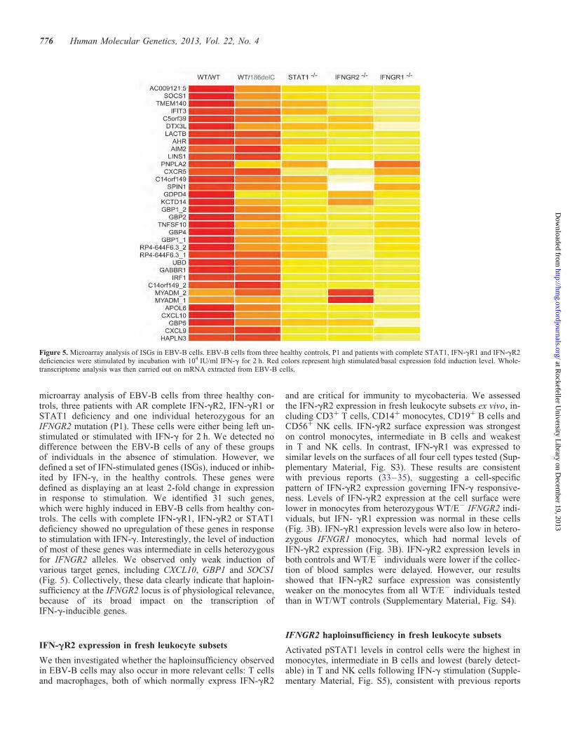
microarray analysis of EBV-B cells from three healthy con-trols, three patients with AR complete IFN-gR2, IFN-gR1 orSTAT1 deficiency and one individual heterozygous for anIFNGR2 mutation (P1). These cells were either being left un-stimulated or stimulated with IFN-g for 2 h. We detected nodifference between the EBV-B cells of any of these groupsof individuals in the absence of stimulation. However, wedefined a set of IFN-stimulated genes (ISGs), induced or inhib-ited by IFN-g, in the healthy controls. These genes weredefined as displaying an at least 2-fold change in expressionin response to stimulation. We identified 31 such genes,which were highly induced in EBV-B cells from healthy con-trols. The cells with complete IFN-gR1, IFN-gR2 or STAT1deficiency showed no upregulation of these genes in responseto stimulation with IFN-g. Interestingly, the level of inductionof most of these genes was intermediate in cells heterozygousfor IFNGR2 alleles. We observed only weak induction ofvarious target genes, including CXCL10, GBP1 and SOCS1(Fig. 5). Collectively, these data clearly indicate that haploin-sufficiency at the IFNGR2 locus is of physiological relevance,because of its broad impact on the transcription ofIFN-g-inducible genes.
IFN-gR2 expression in fresh leukocyte subsets
We then investigated whether the haploinsufficiency observedin EBV-B cells may also occur in more relevant cells: T cellsand macrophages, both of which normally express IFN-gR2
and are critical for immunity to mycobacteria. We assessedthe IFN-gR2 expression in fresh leukocyte subsets ex vivo, in-cluding CD3+ T cells, CD14+ monocytes, CD19+ B cells andCD56+ NK cells. IFN-gR2 surface expression was strongeston control monocytes, intermediate in B cells and weakestin T and NK cells. In contrast, IFN-gR1 was expressed tosimilar levels on the surfaces of all four cell types tested (Sup-plementary Material, Fig. S3). These results are consistentwith previous reports (33–35), suggesting a cell-specificpattern of IFN-gR2 expression governing IFN-g responsive-ness. Levels of IFN-gR2 expression at the cell surface werelower in monocytes from heterozygous WT/E2 IFNGR2 indi-viduals, but IFN- gR1 expression was normal in these cells(Fig. 3B). IFN-gR1 expression levels were also low in hetero-zygous IFNGR1 monocytes, which had normal levels ofIFN-gR2 expression (Fig. 3B). IFN-gR2 expression levels inboth controls and WT/E2 individuals were lower if the collec-tion of blood samples were delayed. However, our resultsshowed that IFN-gR2 surface expression was consistentlyweaker on the monocytes from all WT/E2 individuals testedthan in WT/WT controls (Supplementary Material, Fig. S4).
IFNGR2 haploinsufficiency in fresh leukocyte subsets
Activated pSTAT1 levels in control cells were the highest inmonocytes, intermediate in B cells and lowest (barely detect-able) in T and NK cells following IFN-g stimulation (Supple-mentary Material, Fig. S5), consistent with previous reports
Figure 5. Microarray analysis of ISGs in EBV-B cells. EBV-B cells from three healthy controls, P1 and patients with complete STAT1, IFN-gR1 and IFN-gR2deficiencies were stimulated by incubation with 104 IU/ml IFN-g for 2 h. Red colors represent high stimulated/basal expression fold induction level. Whole-transcriptome analysis was then carried out on mRNA extracted from EBV-B cells.
776 Human Molecular Genetics, 2013, Vol. 22, No. 4
at Rockefeller U
niversity Library on D
ecember 19, 2013
http://hmg.oxfordjournals.org/
Dow
nloaded from

(34,35). We then investigated the occurrence of haploinsuffi-ciency in T cells and monocytes from IFNGR2 heterozygousindividuals. We studied naive CD4+ T cells and differentiatedIFN-g-producing (‘Th1’) and IL-4-producing (‘Th2’) CD4+ Tcells. Naive T cells and IL-4-producing T cells responded toIFN-g in terms of STAT1 phosphorylation, whereasIFN-g-producing T cells did not (data not shown), consistentwith previous reports that IFN-g-producing T cells lackIFN-gR2 expression and are unresponsive to IFN-g (36–39).The levels of pSTAT1 in IFNGR2 heterozygous naive andIL-4-producing CD4+ T cells were lower than those inhealthy controls (Fig. 6A and B). We then tested monocytesand monocyte-derived macrophages (MDMs) by stimulationwith macrophage colony-stimulating factor + interleukins-4(M-CSF + IL-4) or granulocyte colony-stimulating factor +tumor necrosis factor-a (GM-CSF + TNF-a). Control mono-cytes generated similar amounts of pSTAT1 in response tostimulation with IFN-a and IFN-g. Control MDMs inducedto differentiate with M-CSF + IL-4 responded more stronglyto IFN-g and produced larger amounts of IFN-gR2 thanmacrophages induced to differentiate with GM-CSF +TNF-a (data not shown). Following stimulation with IFN-g,pSTAT1 levels in monocytes and MDMs were no lower inIFNGR2 heterozygotes than in controls (Fig. 6C and D).Thus, IFNGR2 haploinsufficiency seems to be restricted to
EBV-B cell lines and freshly analyzed T cells and does notaffect monocytes and MDMs, at least as assessed by STAT1phosphorylation in response to stimulation with IFN-g. Unsur-prisingly, IFNGR2 haploinsufficiency seems to affect princi-pally the cell types that normally express low levels ofIFN-gR2 at their surface.
DISCUSSION
This report identifies AD IFN-gR2 deficiency as a new geneticetiology of MSMD. We observed haploinsufficiency at thehuman IFNGR2 locus, at least in some lymphoid cells (bothEBV-B cell lines and primary T cells), and in all heterozygousindividuals tested. Under the conditions tested, this finding isspecific to IFNGR2, as there was no detectable haploinsuffi-ciency for the IFNGR1 and STAT1 loci. Haploinsufficiencyand dominant negativity are the two independent, mutually ex-clusive mechanisms that can account for dominant phenotypesin diploid cells heterozygous for a mutant allele. Haploinsuffi-ciency at the IFNGR2 locus can be explained biochemically,because the assembly and expression of the membrane-boundIFN-g receptor requires IFN-gR2 expression which is a limit-ing factor. Unlike IFN-gR1, which is ubiquitously and uni-formly expressed, IFN-gR2 is expressed under tight control
Figure 6. Haploinsufficiency in the primary immune cells from heterozygous IFNGR2 individuals. (A) Naive CD4+ T cells, (B) IL-4-producing T helper cells,(C) monocytes (D) MDMs generated in the presence of either M-CSF + IL-4 or GM-CSF + TNF-a were stimulated by incubation with IFN-g, IFN-a or IL-27for 15 min. Western blots were carried out to evaluate STAT1 phosphorylation. These experiments were carried out at least twice.
Human Molecular Genetics, 2013, Vol. 22, No. 4 777
at Rockefeller U
niversity Library on D
ecember 19, 2013
http://hmg.oxfordjournals.org/
Dow
nloaded from

and serves as a key regulator of IFN-g responsiveness and sig-naling in the mouse model and in human subjects (36,39,40).IFN-gR2 had been described as a tuning factor preventing theantiproliferative effect of IFN-g in subsets of T helper cells(36). Recent biochemical studies of IFN-a receptors havealso shown that different levels of cell-surface receptors arerequired for different biological functions (41). For IFN-a,small numbers of receptor binding sites are sufficient tomediate antiviral responses, but not to mediate antiprolifera-tive effects (41,42). Consistent with this, our results showthat the cellular penetrance of IFNGR2 haploinsufficiencydepends on the level of receptor expression at the cellsurface. Cellular penetrance is complete in lymphocytes, in-cluding T cells, which normally express small numbers ofIFN-gR2. Conversely, heterozygosity for IFNGR2 nullalleles does not seem to affect the response of cells that nor-mally express larger numbers of these receptors, such as pha-gocytes. This finding suggests that T cells play a role in thepathogenesis of MSMD in patients with various mutationsaffecting the IFN-g response pathway, including patientswith AD IFN-gR2 deficiency, such as P1.
Clinically, only one of the sixteen known IFNGR2 hetero-zygous individuals tested displayed MSMD. The clinical pene-trance of IFNGR2 haploinsufficiency is therefore low, at leastfor the clinical phenotype of MSMD. This is consistent withour previous demonstration of a correlation between the clin-ical severity of MSMD and the level of responsiveness toIFN-g, as the cellular phenotype seen in IFNGR2 heterozy-gotes is milder than that of most other defects in the IFN-g re-sponse pathway (11,16–20,22,25–27). It is similar to that ofAR partial IFN-gR2 (R114C/R114C) or STAT1 deficiency(K201N/K201N). Moreover, the cells of P1 consistently pre-sented one of the weakest responses of any of the cells hetero-zygous for IFNGR2 null alleles tested. The reasons for thisremain unclear, as WES identified no other potential variantfrom the IFN-g signaling pathway in P1. Moreover, our bio-chemical investigation of known factors in the IFN-g signalingpathway revealed no abnormal expression (data not shown).Incomplete clinical penetrance has already been observed inthe IFN-g signaling pathway, particularly for AD STAT1 de-ficiency (for which the clinical penetrance of MSMD is low)(21,22,43,44). In addition, independently of the IFN-g signal-ing pathway, other mutations may contribute to MSMD in P1.For example, we found a heterozygous ISG15 rare variant inP1 but not in her father. We recently showed that at leastfour individuals heterozygous for ISG15 loss-of-functionalleles have no obvious clinical phenotype in terms ofMSMD (10). However, heterozygous ISG15 mutation mayplay as a modifier in the context of another genetic lesion,such as AD IFN-gR2 deficiency, by reducing the amount ofIFN-g production. In any case, as previously suggested,MSMD is more likely to occur in individuals carryingdominant-negative IFNGR2 alleles, whose cellular responseto IFN-g is impaired (25). It is now tempting to speculatethat IFNGR2 haploinsufficiency may also, and perhaps morecommonly, underlie clinical tuberculosis in children living inareas of endemic disease, including those who did notdevelop clinical disease upon BCG vaccination (5,6,45,46).
MATERIALS AND METHODS
Ethics statement
All members of the families agreed to participate in this study,which was approved by the ethics committees of the hospitalsconcerned.
Case report
P1 is a Polish girl born to non-consanguineous parents in 1990.She was vaccinated by BCG injection into the left deltoid atbirth and presented with enlarged and purulent homolateral ax-illary lymph nodes at the age of 2 months. She had no fever,no enlargement of the liver or spleen and no obvious abnor-mality on chest X-ray. Complete blood count and detailed T,B and NK immunophenotyping results were normal. P1 wasthen treated with lymphadectomy and local streptomycin.Histological examination of the excised lymph nodesshowed cellular infiltration and granulomatosa mimicking tu-berculosis lymphadenitis (type II granuloma). Acid-fast stain-ing revealed the presence of positively stained bacilli. Culturesof pus from the axillary region yielded no pathogens. The in-fection resolved over a period of 4 months, with no oral anti-mycobacterial therapy. With the exception of this BCGdisease, P1 had developed no other severe infections by herlast follow-up visit at the age of 15 years (Fig. 1B). She hadhad mild respiratory tract infections about twice yearly anddeveloped mumps at the age of 5 years and chicken pox atthe age of 7 years, both of which ran a mild clinical course.Other vaccinations, including those with combined diphtheria,tetanus and poliovirus, attenuated rubella and mumps vaccine,hepatitis B virus (HBV) and flu, were uneventful. P1 wasfound to have protective antibodies against HBV. No specificfamily history for related MSMD was found. P1’s maternalgrandmother had hay fever. The parents of P1 remainedhealthy, although her father had heart arrhythmia.
We also retrospectively recruited 10 heterozygous relativesof previously reported and new MSMD patients homozygousfor null IFNGR2 mutations (Fig. 1D; a detailed clinicalreport will be published elsewhere). One new patient (P2)with complete IFN-gR2 deficiency is homozygous for the218delAA mutation (Fig. 1D). She is 6 years old, lives inFrance and was born to a consanguineous family from Paki-stan. She suffered from BCG disease at 11 months of ageand had a high serum IFN-g (53 pg/ml) level. Two patients(P3 and P4) were born to a consanguineous family fromSaudi Arabia. P3, a boy, suffered from severe BCG-osisshortly after birth and died. His sister (P4) was diagnosedwith a recessive IFNGR2 Y235X mutation after birth. Shewas not vaccinated with BCG and has remained healthy.None of the 10 heterozygous relatives had ever displayedany detectable mycobacterial phenotype remotely reminiscentof MSMD, not even self-healing BCG-itis. Five of ten rela-tives had never been vaccinated with BCG and never hadMSMD.
778 Human Molecular Genetics, 2013, Vol. 22, No. 4
at Rockefeller U
niversity Library on D
ecember 19, 2013
http://hmg.oxfordjournals.org/
Dow
nloaded from

Cell culture and stimulation, DNA extraction, PCR,microarray transcriptome analysis and sequencing
Genomic DNA was extracted from fresh blood cells, as previ-ously described (20). Primers and PCR conditions are avail-able upon request. Sequencing was carried out on an ABI3130× (Applied Biosystems) sequencer. Peripheral bloodmononuclear cells (PBMCs) were isolated with Ficoll as pre-viously described (8). EBV-transformed B lymphocytes(EBV-B cells) were cultured as previously described (47).EBV-B cells, monocytes, MDMs, naive T cells and Th2cells were stimulated with the indicated doses of IFN-g(Imukin, Boehringer Ingelheim), IFN-a 2b (IntronA, ScheringPlough) and IL-27 (R&D). Electroporation and Lipofectaminmethods were used to transfect EBV-B cells and fibroblasts,respectively, with a WT construct, with or without mutantIFNGR2 plasmids (pcDNA3.1-IFNGR2), as previouslyreported (27). We incubated five million EBV-B cells withor without 104IU/ml IFN-g for 2 h and then subjected themto microarray analysis as previously described (48). The pro-cedure of WES and analysis were carried out as previouslydescribed (49,50). Briefly, variant calls were made withGATK UnifiedGenotyper. All calls with a read coverage≤10× and a Phred-scaled SNP quality of ≤30 wereremoved from consideration.
Electrophoretic mobility shift assay (EMSA) and westernblotting
EMSA was carried out as previously described (17–19).Briefly, cells were stimulated by incubation for 20 min withIFN-g or IFN-a, at the indicated doses. We incubated 10 mgof nuclear extract with a 32P-labeled (a-dATP) GAS (fromthe FCGR1 promoter) probe and subjected the mixture to elec-trophoresis in a polyacrylamide gel. The GAF DNA-bindingactivity was assessed with ImageQuant (Amersham Bio-science). We used antibodies recognizing STAT1 and tyrosine701-phosphorylated STAT1 for western blot analysis, as pre-viously described (30).
Flow cytometry analysis
We used fluorescence-activated cell sorting (FACS) to assessIFN-gR2 expression on the surface of EBV-B cells, PBMCsand monocytes. Briefly, 0.5 to 1 million PBMCs or EBV-Bcells were first blocked by incubation with 10 ml of FcR block-ing reagent in 1% SVF + 1× PBS for 10 min. We then added125 ng of anti-IFN-gR2-APC (FAB773A, R&D systems) andincubated the cells for 30 min on ice before washing themtwice with 1% SVF + 1× PBS. Goat IgG control APC(IC108A, R&D systems) served as the isotype control.Tyr-701-phosphorylated STAT1 levels were assessed as previ-ously described. Antibodies directed against CD3 (560366,BD), CD14 (557742, BD), CD19 (555413, BD) and CD56(557699, BD) conjugated with different fluorochromes wereused to distinguish between the various cell populations inPBMCs, and dead cells were excluded by Aqua Live/Deadstaining (Invitrogen). Tyr-701-phosphorylated STAT1 levelswere assessed by activating cells by incubation with IFN-gor IFN-a for 30 min and incubating with an antibody specific
for Tyr 701-phosphorylated STAT1. Flurokon signals weremeasured with a BD LSR II and analyzed with Flowjo (TreeStar, Inc.).
Differentiation of macrophage and CD41 T cellsand statistics
We isolated CD14+ monocytes from PBMCs using a positiveselection kit from Miltenyi Biotec (8). Briefly, 0.5 millionmonocytes were incubated in 24-well plates with 0.5 ng/mlGM-CSF (215-MC-050, R&D systems) and 0.5 ng/ml TNF-a(210-TA-050), or with 50 ng/ml M-CSF (216-MC-025, R&Dsystems) and 50 ng/ml IL-4 (204-IL-050, R&D systems), toinduce macrophage differentiation, as previously described(8,51). CD4+ naive T cells were obtained using MiltenyiMACS naive CD4+ T cell isolation kit II (No.130-094-131).In total, 0.5 million naive T cells in 1 ml RPMI-1640 supple-mented with 10% human plasma were allowed to differentiatein each of the wells of 24-well plates, as previously described(52), with 5 mg/ml anti-CD3 antibody, 750 ng/ml anti-CD28antibody and 20 IU/ml rIL-2, with either 1 ng/ml IL-12 or50 ng/ml IL-4, for Th1 and Th2 cell differentiation, respective-ly. The cells were stimulated with IFN-g or IL-27 on the fourthday after differentiation. ANOVA was used to assess the differ-ence between multiple groups, and Student’s t-tests were thencarried out to assess the significance of differences in quantita-tive variables between two groups.
SUPPLEMENTARY MATERIAL
Supplementary Material is available at HMG online.
ACKNOWLEDGEMENTS
We thank Bertrand Boisson and Minji Byun for helpful discus-sions and critical reading. We thank Yelena Nemirovskaya,Eric Anderson, Tiffany Nivare, Tatiana Kochetkov, LucileJanniere, Martine Courat, Devon Davenport, Jacqueline Roseand Jacqueline Feinberg for technical and secretarial assist-ance and all members of the Laboratory of Human Geneticsof Infectious Diseases for helpful discussions.
Conflict of Interest statement: None declared.
FUNDING
X.F.K. was supported by the Stony Wold-Herbert Fund,Choh-Hao Li Memorial Fund Scholar award and the ShanghaiEducational Development Foundation, Y.I. is supported bythe AXA Research Fund, V.L.B. is supported by the StonyWold-Herbert Fund and A.K. is supported by the FondationMedicale Medische Stichting Mathilde E. Horlait-Dapens.The Laboratory of Human Genetics of Infectious Diseases issupported in part by grants from the St Giles Foundation,the Jeffrey Modell Foundation, The Rockefeller UniversityCenter for Clinical and Translational Science grant number8UL1TR000043 from the National Center for ResearchResources and the National Center for Advancing Sciences(NCATS), National Institutes of Health, the National Institute
Human Molecular Genetics, 2013, Vol. 22, No. 4 779
at Rockefeller U
niversity Library on D
ecember 19, 2013
http://hmg.oxfordjournals.org/
Dow
nloaded from

of Allergy and Infectious Diseases grant number5R01AI089970-02, The Rockefeller University and the Euro-pean Research Council (ERC). J.L.C. received a research grantfrom the Jeffrey Modell Foundation in collaboration withTalecris BioTherapeutics. The funder was not involved inthe study design; collection, analysis and interpretation ofdata; writing of the paper or decision to submit for publication.
REFERENCES
1. Alcais, A., Abel, L. and Casanova, J.L. (2009) Human genetics ofinfectious diseases: between proof of principle and paradigm. J. Clin.Invest., 119, 2506–2514.
2. Casanova, J.L. and Abel, L. (2002) Genetic dissection of immunity tomycobacteria: the human model. Annu. Rev. Immunol., 20, 581–620.
3. Casanova, J.L. and Abel, L. (2004) The human model: a genetic dissectionof immunity to infection in natural conditions. Nat. Rev. Immunol., 4,55–66.
4. Fortin, A., Abel, L., Casanova, J.L. and Gros, P. (2007) Host genetics ofmycobacterial diseases in mice and men: forward genetic studies ofBCG-osis and tuberculosis. Annu. Rev. Genomics Hum. Genet., 8,163–192.
5. Alcais, A., Fieschi, C., Abel, L. and Casanova, J.L. (2005) Tuberculosis inchildren and adults: two distinct genetic diseases. J. Exp. Med., 202,1617–1621.
6. Boisson-Dupuis, S., El Baghdadi, J., Parvaneh, N., Bousfiha, A.,Bustamante, J., Feinberg, J., Samarina, A., Grant, A.V., Janniere, L., ElHafidi, N. et al. (2011) IL-12Rbeta1 deficiency in two of fifty childrenwith severe tuberculosis from Iran, Morocco, and Turkey. PLoS One, 6,e18524.
7. Filipe-Santos, O., Bustamante, J., Chapgier, A., Vogt, G., deBeaucoudrey, L., Feinberg, J., Jouanguy, E., Boisson-Dupuis, S., Fieschi,C., Picard, C. et al. (2006) Inborn errors of IL-12/23- andIFN-gamma-mediated immunity: molecular, cellular, and clinicalfeatures. Semin. Immunol., 18, 347–361.
8. Bustamante, J., Arias, A.A., Vogt, G., Picard, C., Galicia, L.B., Prando,C., Grant, A.V., Marchal, C.C., Hubeau, M., Chapgier, A. et al. (2011)Germline CYBB mutations that selectively affect macrophages inkindreds with X-linked predisposition to tuberculous mycobacterialdisease. Nat. Immunol., 12, 213–221.
9. Hambleton, S., Salem, S., Bustamante, J., Bigley, V., Boisson-Dupuis, S.,Azevedo, J., Fortin, A., Haniffa, M., Ceron-Gutierrez, L., Bacon, C.M.et al. (2011) IRF8 mutations and human dendritic-cell immunodeficiency.N. Engl. J. Med., 365, 127–138.
10. Bogunovic, D., Byun, M., Durfee, L.A., Abhyankar, A., Sanal, O.,Mansouri, D., Salem, S., Radovanovic, I., Grant, A.V., Adimi, P. et al.(2012) Mycobacterial disease and impaired IFN-gamma immunity inhumans with inherited ISG15 deficiency. Science, 337, 1684–1688.
11. Dupuis, S., Doffinger, R., Picard, C., Fieschi, C., Altare, F., Jouanguy, E.,Abel, L. and Casanova, J.L. (2000) Human interferon-gamma-mediatedimmunity is a genetically controlled continuous trait that determines theoutcome of mycobacterial invasion. Immunol. Rev., 178, 129–137.
12. Altare, F., Durandy, A., Lammas, D., Emile, J.F., Lamhamedi, S., LeDeist, F., Drysdale, P., Jouanguy, E., Doffinger, R., Bernaudin, F. et al.(1998) Impairment of mycobacterial immunity in human interleukin-12receptor deficiency. Science, 280, 1432–1435.
13. de Jong, R., Altare, F., Haagen, I.A., Elferink, D.G., Boer, T., van BredaVriesman, P.J., Kabel, P.J., Draaisma, J.M., van Dissel, J.T., Kroon, F.P.et al. (1998) Severe mycobacterial and Salmonella infections ininterleukin-12 receptor-deficient patients. Science, 280, 1435–1438.
14. Picard, C., Fieschi, C., Altare, F., Al-Jumaah, S., Al-Hajjar, S., Feinberg,J., Dupuis, S., Soudais, C., Al-Mohsen, I.Z., Genin, E. et al. (2002)Inherited interleukin-12 deficiency: IL12B genotype and clinicalphenotype of 13 patients from six kindreds. Am. J. Hum. Genet., 70,336–348.
15. Filipe-Santos, O., Bustamante, J., Haverkamp, M.H., Vinolo, E., Ku, C.L.,Puel, A., Frucht, D.M., Christel, K., von Bernuth, H., Jouanguy, E. et al.(2006) X-linked susceptibility to mycobacteria is caused by mutations inNEMO impairing CD40-dependent IL-12 production. J. Exp. Med., 203,1745–1759.
16. Jouanguy, E., Altare, F., Lamhamedi, S., Revy, P., Emile, J.F., Newport,M., Levin, M., Blanche, S., Seboun, E., Fischer, A. et al. (1996)Interferon-gamma-receptor deficiency in an infant with fatal bacilleCalmette-Guerin infection. N. Engl. J. Med., 335, 1956–1961.
17. Jouanguy, E., Dupuis, S., Pallier, A., Doffinger, R., Fondaneche, M.C.,Fieschi, C., Lamhamedi-Cherradi, S., Altare, F., Emile, J.F., Lutz, P. et al.
(2000) In a novel form of IFN-gamma receptor 1 deficiency, cell surfacereceptors fail to bind IFN-gamma. J. Clin. Invest., 105, 1429–1436.
18. Jouanguy, E., Lamhamedi-Cherradi, S., Altare, F., Fondaneche, M.C.,Tuerlinckx, D., Blanche, S., Emile, J.F., Gaillard, J.L., Schreiber, R.,Levin, M. et al. (1997) Partial interferon-gamma receptor 1 deficiency in achild with tuberculoid bacillus Calmette-Guerin infection and a siblingwith clinical tuberculosis. J. Clin. Invest., 100, 2658–2664.
19. Jouanguy, E., Lamhamedi-Cherradi, S., Lammas, D., Dorman, S.E.,Fondaneche, M.C., Dupuis, S., Doffinger, R., Altare, F., Girdlestone, J.,Emile, J.F. et al. (1999) A human IFNGR1 small deletion hotspotassociated with dominant susceptibility to mycobacterial infection. Nat.
Genet., 21, 370–378.20. Kong, X.F., Vogt, G., Chapgier, A., Lamaze, C., Bustamante, J., Prando,
C., Fortin, A., Puel, A., Feinberg, J., Zhang, X.X. et al. (2010) A novelform of cell type-specific partial IFN-gammaR1 deficiency caused by agerm line mutation of the IFNGR1 initiation codon. Hum. Mol. Genet., 19,434–444.
21. Chapgier, A., Boisson-Dupuis, S., Jouanguy, E., Vogt, G., Feinberg, J.,Prochnicka-Chalufour, A., Casrouge, A., Yang, K., Soudais, C., Fieschi,C. et al. (2006) Novel STAT1 alleles in otherwise healthy patients withmycobacterial disease. PLoS Genet., 2, e131.
22. Dupuis, S., Dargemont, C., Fieschi, C., Thomassin, N., Rosenzweig, S.,Harris, J., Holland, S.M., Schreiber, R.D. and Casanova, J.L. (2001)Impairment of mycobacterial but not viral immunity by a germline humanSTAT1 mutation. Science, 293, 300–303.
23. Doffinger, R., Jouanguy, E., Dupuis, S., Fondaneche, M.C., Stephan, J.L.,Emile, J.F., Lamhamedi-Cherradi, S., Altare, F., Pallier, A.,Barcenas-Morales, G. et al. (2000) Partial interferon-gamma receptorsignaling chain deficiency in a patient with bacille Calmette-Guerin andMycobacterium abscessus infection. J. Infect. Dis., 181, 379–384.
24. Dorman, S.E. and Holland, S.M. (1998) Mutation in thesignal-transducing chain of the interferon-gamma receptor andsusceptibility to mycobacterial infection. J. Clin. Invest., 101, 2364–2369.
25. Rosenzweig, S.D., Dorman, S.E., Uzel, G., Shaw, S., Scurlock, A., Brown,M.R., Buckley, R.H. and Holland, S.M. (2004) A novel mutation inIFN-gamma receptor 2 with dominant negative activity: biologicalconsequences of homozygous and heterozygous states. J. Immunol., 173,4000–4008.
26. Vogt, G., Bustamante, J., Chapgier, A., Feinberg, J., Boisson Dupuis, S.,Picard, C., Mahlaoui, N., Gineau, L., Alcais, A., Lamaze, C. et al. (2008)Complementation of a pathogenic IFNGR2 misfolding mutation withmodifiers of N-glycosylation. J. Exp. Med., 205, 1729–1737.
27. Vogt, G., Chapgier, A., Yang, K., Chuzhanova, N., Feinberg, J., Fieschi,C., Boisson-Dupuis, S., Alcais, A., Filipe-Santos, O., Bustamante, J. et al.
(2005) Gains of glycosylation comprise an unexpectedly large group ofpathogenic mutations. Nat. Genet., 37, 692–700.
28. Toyoda, H., Ido, M., Nakanishi, K., Nakano, T., Kamiya, H., Matsumine,A., Uchida, A., Mizutani, H., de Beaucoudrey, L., Vogt, G. et al. (2010)Multiple cutaneous squamous cell carcinomas in a patient with interferongamma receptor 2 (IFN gamma R2) deficiency. J. Med. Genet., 47,631–634.
29. Chapgier, A., Kong, X.F., Boisson-Dupuis, S., Jouanguy, E., Averbuch,D., Feinberg, J., Zhang, S.Y., Bustamante, J., Vogt, G., Lejeune, J. et al.
(2009) A partial form of recessive STAT1 deficiency in humans. J. Clin.
Invest., 119, 1502–1514.30. Kong, X.F., Ciancanelli, M., Al-Hajjar, S., Alsina, L., Zumwalt, T.,
Bustamante, J., Feinberg, J., Audry, M., Prando, C., Bryant, V. et al.
(2010) A novel form of human STAT1 deficiency impairing early but notlate responses to interferons. Blood, 116, 5895–5906.
31. D’Haene, B., Attanasio, C., Beysen, D., Dostie, J., Lemire, E., Bouchard,P., Field, M., Jones, K., Lorenz, B., Menten, B. et al. (2009)Disease-causing 7.4 kb cis-regulatory deletion disrupting conservednon-coding sequences and their interaction with the FOXL2 promotor:implications for mutation screening. PLoS Genet., 5, e1000522.
32. Dorman, S.E., Picard, C., Lammas, D., Heyne, K., van Dissel, J.T.,Baretto, R., Rosenzweig, S.D., Newport, M., Levin, M., Roesler, J. et al.
780 Human Molecular Genetics, 2013, Vol. 22, No. 4
at Rockefeller U
niversity Library on D
ecember 19, 2013
http://hmg.oxfordjournals.org/
Dow
nloaded from

(2004) Clinical features of dominant and recessive interferon gammareceptor 1 deficiencies. Lancet, 364, 2113–2121.
33. Bach, E.A., Aguet, M. and Schreiber, R.D. (1997) The IFN gammareceptor: a paradigm for cytokine receptor signaling. Annu. Rev.
Immunol., 15, 563–591.
34. Bernabei, P., Coccia, E.M., Rigamonti, L., Bosticardo, M., Forni, G.,Pestka, S., Krause, C.D., Battistini, A. and Novelli, F. (2001)Interferon-gamma receptor 2 expression as the deciding factor inhuman T, B, and myeloid cell proliferation or death. J. Leukoc. Biol., 70,950–960.
35. van Boxel-Dezaire, A.H. and Stark, G.R. (2007) Cell type-specificsignaling in response to interferon-gamma. Curr. Top. Microbiol.
Immunol., 316, 119–154.
36. Bach, E.A., Szabo, S.J., Dighe, A.S., Ashkenazi, A., Aguet, M., Murphy,K.M. and Schreiber, R.D. (1995) Ligand-induced autoregulation ofIFN-gamma receptor beta chain expression in T helper cell subsets.Science, 270, 1215–1218.
37. Bach, E.A., Tanner, J.W., Marsters, S., Ashkenazi, A., Aguet, M., Shaw,A.S. and Schreiber, R.D. (1996) Ligand-induced assembly and activationof the gamma interferon receptor in intact cells. Mol. Cell. Biol., 16,3214–3221.
38. Kotenko, S.V., Izotova, L.S., Pollack, B.P., Mariano, T.M., Donnelly,R.J., Muthukumaran, G., Cook, J.R., Garotta, G., Silvennoinen, O., Ihle,J.N. et al. (1995) Interaction between the components of the interferongamma receptor complex. J. Biol. Chem., 270, 20915–20921.
39. Pernis, A., Gupta, S., Gollob, K.J., Garfein, E., Coffman, R.L., Schindler,C. and Rothman, P. (1995) Lack of interferon gamma receptor beta chainand the prevention of interferon gamma signaling in TH1 cells. Science,269, 245–247.
40. Gajewski, T.F., Goldwasser, E. and Fitch, F.W. (1988) Anti-proliferativeeffect of IFN-gamma in immune regulation. II. IFN-gamma inhibits theproliferation of murine bone marrow cells stimulated with IL-3, IL-4, orgranulocyte-macrophage colony-stimulating factor. J. Immunol., 141,2635–2642.
41. Levin, D., Harari, D. and Schreiber, G. (2011) Stochastic receptorexpression determines cell fate upon interferon treatment. Mol. Cell. Biol.,31, 3252–3266.
42. Thomas, C., Moraga, I., Levin, D., Krutzik, P.O., Podoplelova, Y., Trejo,A., Lee, C., Yarden, G., Vleck, S.E., Glenn, J.S. et al. (2011) Structurallinkage between ligand discrimination and receptor activation by type Iinterferons. Cell, 146, 621–632.
43. Sampaio, E.P., Bax, H.I., Hsu, A.P., Kristosturyan, E., Pechacek, J.,Chandrasekaran, P., Paulson, M.L., Dias, D.L., Spalding, C., Uzel, G.et al. (2012) A novel STAT1 mutation associated with disseminatedmycobacterial disease. J. Clin. Immunol, 32, 681–689.
44. Tsumura, M., Okada, S., Sakai, H., Yasunaga, S., Ohtsubo, M., Murata,T., Obata, H., Yasumi, T., Kong, X.F., Abhyankar, A. et al. (2012)Dominant-negative STAT1 SH2 domain mutations in unrelated patientswith mendelian susceptibility to mycobacterial disease. Hum. Mutat, 33,1377–1387.
45. Alcais, A., Quintana-Murci, L., Thaler, D.S., Schurr, E., Abel, L. andCasanova, J.L. (2010) Life-threatening infectious diseases of childhood:single-gene inborn errors of immunity? Ann. N. Y. Acad. Sci., 1214,18–33.
46. Tabarsi, P., Marjani, M., Mansouri, N., Farnia, P., Boisson-Dupuis, S.,Bustamante, J., Abel, L., Adimi, P., Casanova, J.L. and Mansouri, D.(2011) Lethal tuberculosis in a previously healthy adult with IL-12receptor deficiency. J. Clin. Immunol., 31, 537–539.
47. Kong, X.F., Zhang, X.X., Gong, Q.M., Gao, J., Zhang, S.Y., Wang, L.,Xu, J., Han, Y., Jin, G.D., Jiang, J.H. et al. (2007) MxA induction maypredict sustained virologic responses of chronic hepatitis B patients withIFN-alpha treatment. J. Interferon Cytokine Res., 27, 809–818.
48. Sancho-Shimizu, V., Perez de Diego, R., Lorenzo, L., Halwani, R.,Alangari, A., Israelsson, E., Fabrega, S., Cardon, A., Maluenda, J.,Tatematsu, M. et al. (2011) Herpes simplex encephalitis in children withautosomal recessive and dominant TRIF deficiency. J. Clin. Invest., 121,4889–4902.
49. Bolze, A., Byun, M., McDonald, D., Morgan, N.V., Abhyankar, A.,Premkumar, L., Puel, A., Bacon, C.M., Rieux-Laucat, F., Pang, K. et al.(2010) Whole-exome-sequencing-based discovery of human FADDdeficiency. Am. J. Hum. Genet., 87, 873–881.
50. Byun, M., Abhyankar, A., Lelarge, V., Plancoulaine, S., Palanduz, A.,Telhan, L., Boisson, B., Picard, C., Dewell, S., Zhao, C. et al. (2010)Whole-exome sequencing-based discovery of STIM1 deficiency in a childwith fatal classic Kaposi sarcoma. J. Exp. Med., 207, 2307–2312.
51. Vogt, G. and Nathan, C. (2011) In vitro differentiation of humanmacrophages with enhanced antimycobacterial activity. J. Clin. Invest.,121, 3889–3901.
52. Ma, C.S., Hare, N.J., Nichols, K.E., Dupre, L., Andolfi, G., Roncarolo,M.G., Adelstein, S., Hodgkin, P.D. and Tangye, S.G. (2005) Impairedhumoral immunity in X-linked lymphoproliferative disease is associatedwith defective IL-10 production by CD4+ T cells. J. Clin. Invest., 115,1049–1059.
Human Molecular Genetics, 2013, Vol. 22, No. 4 781
at Rockefeller U
niversity Library on D
ecember 19, 2013
http://hmg.oxfordjournals.org/
Dow
nloaded from
Copyright © 2022 FDOKUMEN