Characterisation of an engineered trastuzumab IgE antibody and effector cell mechanisms targeting...
16
Cancer Immunol Immunother (2009) 58:915–930 DOI 10.1007/s00262-008-0607-1 123 ORIGINAL ARTICLE Characterisation of an engineered trastuzumab IgE antibody and eVector cell mechanisms targeting HER2/neu-positive tumour cells Panagiotis Karagiannis · Josef Singer · James Hunt · Samuel K. E. Gan · Sarah M. Rudman · Diana Mechtcheriakova · Regina Knittelfelder · Tracy R. Daniels · Philip S. Hobson · Andrew J. Beavil · James Spicer · Frank O. Nestle · Manuel L. Penichet · Hannah J. Gould · Erika Jensen-Jarolim · Sophia N. Karagiannis Received: 20 June 2008 / Accepted: 26 September 2008 / Published online: 22 October 2008 © Springer-Verlag 2008 Abstract Trastuzumab (Herceptin ® ), a humanized IgG1 antibody raised against the human epidermal growth factor receptor 2 (HER2/neu), is the main antibody in clinical use against breast cancer. Pre-clinical evidence and clinical studies indicate that trastuzumab employs several anti- tumour mechanisms that most likely contribute to enhanced survival of patients with HER2/neu-positive breast carcino- mas. New strategies are aimed at improving antibody-based therapeutics like trastuzumab, e.g. by enhancing antibody- mediated eVector function mechanisms. Based on our previous Wndings that a chimaeric ovarian tumour antigen- speciWc IgE antibody showed greater eYcacy in tumour cell killing, compared to the corresponding IgG1 antibody, we have produced an IgE homologue of trastuzumab. Trast- uzumab IgE was engineered with the same light- and heavy-chain variable-regions as trastuzumab, but with an epsilon in place of the gamma-1 heavy-chain constant region. We describe the physical characterisation and ligand binding properties of the trastuzumab IgE and eluci- date its potential anti-tumour activities in functional assays. Both trastuzumab and trastuzumab IgE can activate mono- cytic cells to kill tumour cells, but they operate by diVerent mechanisms: trastuzumab functions in antibody-dependent cell-mediated phagocytosis (ADCP), whereas trastuzumab IgE functions in antibody-dependent cell-mediated cytotox- icity (ADCC). Trastuzumab IgE, incubated with mast cells Panagiotis Karagiannis and Josef Singer contributed equally to this work. Electronic supplementary material The online version of this article (doi:10.1007/s00262-008-0607-1) contains supplementary material, which is available to authorized users. P. Karagiannis · F. O. Nestle · S. N. Karagiannis (&) Cutaneous Medicine and Immunotherapy Unit, St. John’s Institute of Dermatology, Division of Genetics and Molecular Medicine, King’s College London School of Medicine, 9th Floor, Guy’s Tower, Guy’s Hospital, London SE1 9RT, UK e-mail: [email protected] J. Hunt · S. K. E. Gan · P. S. Hobson · A. J. Beavil · H. J. Gould Randall Division of Cell and Molecular Biophysics, King’s College London, 3rd Floor, New Hunt’s House, Guy’s Campus, London SE1 1UL, UK S. M. Rudman · J. Spicer Department of Academic Oncology, King’s College London, 3rd Floor, Bermondsey Wing, Guy’s Hospital, London SE1 9RT, UK T. R. Daniels · M. L. Penichet Division of Surgical Oncology, Department of Surgery, David GeVen School of Medicine, University of California, Los Angeles, USA M. L. Penichet Departments of Microbiology, Immunology, and Molecular Genetics, David GeVen School of Medicine, University of California, Los Angeles, USA M. L. Penichet The Jonsson Comprehensive Cancer Center, David GeVen School of Medicine, University of California, Los Angeles, USA P. Karagiannis · J. Singer · D. Mechtcheriakova · R. Knittelfelder · E. Jensen-Jarolim IPP, Department of Pathophysiology, Center of Physiology, Pathophysiology and Immunology, Medical University Vienna, Waehringer G. 18, 1090 Vienna, Austria
Transcript of Characterisation of an engineered trastuzumab IgE antibody and effector cell mechanisms targeting...

Cancer Immunol Immunother (2009) 58:915–930
DOI 10.1007/s00262-008-0607-1ORIGINAL ARTICLE
Characterisation of an engineered trastuzumab IgE antibody and eVector cell mechanisms targeting HER2/neu-positive tumour cells
Panagiotis Karagiannis · Josef Singer · James Hunt · Samuel K. E. Gan · Sarah M. Rudman · Diana Mechtcheriakova · Regina Knittelfelder · Tracy R. Daniels · Philip S. Hobson · Andrew J. Beavil · James Spicer · Frank O. Nestle · Manuel L. Penichet · Hannah J. Gould · Erika Jensen-Jarolim · Sophia N. Karagiannis
Received: 20 June 2008 / Accepted: 26 September 2008 / Published online: 22 October 2008© Springer-Verlag 2008
Abstract Trastuzumab (Herceptin®), a humanized IgG1antibody raised against the human epidermal growth factorreceptor 2 (HER2/neu), is the main antibody in clinical useagainst breast cancer. Pre-clinical evidence and clinicalstudies indicate that trastuzumab employs several anti-tumour mechanisms that most likely contribute to enhancedsurvival of patients with HER2/neu-positive breast carcino-mas. New strategies are aimed at improving antibody-basedtherapeutics like trastuzumab, e.g. by enhancing antibody-mediated eVector function mechanisms. Based on our
previous Wndings that a chimaeric ovarian tumour antigen-speciWc IgE antibody showed greater eYcacy in tumourcell killing, compared to the corresponding IgG1 antibody,we have produced an IgE homologue of trastuzumab. Trast-uzumab IgE was engineered with the same light- andheavy-chain variable-regions as trastuzumab, but with anepsilon in place of the gamma-1 heavy-chain constantregion. We describe the physical characterisation andligand binding properties of the trastuzumab IgE and eluci-date its potential anti-tumour activities in functional assays.Both trastuzumab and trastuzumab IgE can activate mono-cytic cells to kill tumour cells, but they operate by diVerentmechanisms: trastuzumab functions in antibody-dependentcell-mediated phagocytosis (ADCP), whereas trastuzumabIgE functions in antibody-dependent cell-mediated cytotox-icity (ADCC). Trastuzumab IgE, incubated with mast cells
Panagiotis Karagiannis and Josef Singer contributed equally to this work.
Electronic supplementary material The online version of this article (doi:10.1007/s00262-008-0607-1) contains supplementary material, which is available to authorized users.
P. Karagiannis · F. O. Nestle · S. N. Karagiannis (&)Cutaneous Medicine and Immunotherapy Unit, St. John’s Institute of Dermatology, Division of Genetics and Molecular Medicine, King’s College London School of Medicine, 9th Floor, Guy’s Tower, Guy’s Hospital, London SE1 9RT, UKe-mail: [email protected]
J. Hunt · S. K. E. Gan · P. S. Hobson · A. J. Beavil · H. J. GouldRandall Division of Cell and Molecular Biophysics, King’s College London, 3rd Floor, New Hunt’s House, Guy’s Campus, London SE1 1UL, UK
S. M. Rudman · J. SpicerDepartment of Academic Oncology, King’s College London, 3rd Floor, Bermondsey Wing, Guy’s Hospital, London SE1 9RT, UK
T. R. Daniels · M. L. PenichetDivision of Surgical Oncology, Department of Surgery, David GeVen School of Medicine, University of California, Los Angeles, USA
M. L. PenichetDepartments of Microbiology, Immunology, and Molecular Genetics, David GeVen School of Medicine, University of California, Los Angeles, USA
M. L. PenichetThe Jonsson Comprehensive Cancer Center, David GeVen School of Medicine, University of California, Los Angeles, USA
P. Karagiannis · J. Singer · D. Mechtcheriakova · R. Knittelfelder · E. Jensen-JarolimIPP, Department of Pathophysiology, Center of Physiology, Pathophysiology and Immunology, Medical University Vienna, Waehringer G. 18, 1090 Vienna, Austria
123

916 Cancer Immunol Immunother (2009) 58:915–930
and HER2/neu-expressing tumour cells, triggers mast celldegranulation, recruiting against cancer cells a potentimmune response, characteristic of allergic reactions.Finally, in viability assays both antibodies mediate compa-rable levels of tumour cell growth arrest. These functionalcharacteristics of trastuzumab IgE, some distinct from thoseof trastuzumab, indicate its potential to complement orimprove upon the existing clinical beneWts of trastuzumab.
Keywords HER2/neu · Trastuzumab · IgE · Monocytes · Mast cells · Tumour immunity
AbbreviationsHER2/neu Human epidermal growth factor receptor 2ADCC Antibody-dependent cell-mediated
cytotoxicityADCP Antibody-dependent cell-mediated
phagocytosisFBP Folate binding proteinsFc�RI� Soluble Fc�RI �-chainECDHER2 HER2 protein extracellular domainCM Complete mediumPI Propidium iodideCFSE Carboxy-Xuorescein diacetate, succinimidyl
esterNIP 4-Hydroxy-3-nitro-phenacetylPI3K Phosphoinositide 3-kinaseTGF-a Tumour growth factor �VEGF Vascular endothelial growth factorTNF-� Tumour necrosis factor-�MTS 3-(4,5-Dimethylthiazol-2-yl)-5-(3-carboxy-
methoxyphenyl)-2-(4-sulfophenyl)-2H-tetrazolium inner salt)
PMS Phenazine methosulfate
Introduction
HER2/neu (c-erb-B2) is a 185 kDa protein that belongs tothe human epidermal growth factor receptor family. Itsfunctions include engendering cell signalling and regulat-ing cell growth, proliferation, diVerentiation and motility[1]. Approximately 30% of breast carcinomas as well asother cancers, such as those of the ovary, endometrium,bladder, prostate and lung, over-express HER2/neu, whilstits expression in normal tissues is low [2]. Its expression inbreast cancer is thought to play a vital role in the pathogen-esis of breast tumours and is linked to poor clinical out-comes [3]. This antigen is now a validated target for cancertherapeutics.
Trastuzumab (Herceptin®) is an IgG1 antibody raisedagainst the extracellular domain (ECD) of HER2/neu and isthe main antibody in clinical use for the treatment of
HER2-positive breast cancers [4–6]. Trastuzumab wasapproved by the FDA in 1998 for the treatment of meta-static HER2/neu over-expressing breast cancer and is nowalso used as adjuvant therapy for early breast cancers. Thesuccess of trastuzumab in breast cancer therapy hasrenewed interest in antibody therapies and provoked furtherresearch into the development of therapeutic antibodies.However, only a subset of patients treated with trastuzumabshow signiWcant responses and thus there is scope for addi-tional modalities designed to improve clinical outcomes [7].
Trastuzumab is thought to exert anti-tumour eVects by anumber of mechanisms. The best-deWned mechanism is theblocking of the hetero-dimerization of HER2/neu receptorswith other HER family members (HER1, HER3) on thesurface of breast cancer cells thereby switching oV vitaltumour cell growth signals [8, 9]. Trastuzumab inhibitsmetalloproteinase activity and interferes with signalling viaphosphoinositide 3-kinase (PI3 K) pathways, promotingapoptosis and cell cycle arrest during the G1 phase.Another mechanism is thought to be blocking angiogenesisby inducing expression of anti-angiogenic factors such asthrombospondin-1 and suppression of pro-angiogenic fac-tors such as TGF-�, VEGF, angiopoietin-1, and plasmino-gen-activator inhibitor-1 [10]. Clinical and pre-clinicalstudies suggest that trastuzumab may also enlist immuneeVector cells to attack and kill tumour cells by cytotoxicity(ADCC) and phagocytosis (ADCP), and by augmentingchemotherapy-induced cytotoxicity [11–14]. Studies arenow focusing on strategies aimed at improving the signiW-cant but circumscribed success of trastuzumab. Theseinclude optimising antigen speciWcity or aYnity andenhancing antibody-mediated eVector cell functions tar-geted against tumour cells.
Although there are Wve antibody classes in man, eachwith distinctive functions in the immune system, trast-uzumab, but essentially all monoclonal antibodies approvedfor clinical use are IgG1 s. Antibodies of the IgG classfunction most eVectively in the circulation [15]. There aremany reasons why IgE antibodies might be more eVectiveagainst tumours that develop in tissues and are thereforeinaccessible to IgGs [16]. The concentration of IgE in theserum of normal individuals is minute (<150 ng/mL, 1/10,000 the concentration of IgG) because IgE is seques-tered in solid tissues, where it is bound with high-aYnity toreceptors on its eVector and antigen-presenting cells [17].The aYnity of IgE for Fc�RI (Ka »1011 M¡1) is 102–105
times higher than those of IgGs for their receptors, makingIgE the only antibody strongly retained by eVector cells inthe absence of antigen [17, 18]. The half-life of IgE in tis-sues (measured in the skin »2 weeks) is longer than that ofIgG (2–3 days) [18, 19]. IgE saturates Fc�RI at nM concen-trations, but only 10% of the receptors need be occupied byIgE and antigen for full mast cell activation and eVector cell
123

Cancer Immunol Immunother (2009) 58:915–930 917
recruitment to the site of antigen challenge in tissues [17,20]. IgE antibodies on the surface of tissue mast cells arecross-linked by antigens to induce the release of histamines,leukotrienes, proteases, and, importantly, Th2 cell-typecytokines (IL-3, IL-4, IL-5, IL-6, IL-9, IL-13, TNF-�) atthe site of antigen challenge. This results in activation ofthe resident immune eVector cells, but also elicits furtherrecruitment and persistence of an inXammatory cell inWl-trate, comprising Th2 cells, monocytes, eosinophils andbasophils, from the circulation, which enhances and main-tains the local immune response [17]. IgE antibodiesdirected against a tumour-associated antigen would there-fore trigger an immediate local eVector cell responseagainst tumour cells and stimulate a cascade of inXamma-tion targeted to the tumour cells in situ. These activitiescould possibly be highly eVective in immune rejection oftumours embedded in solid tissues.
Several studies support the ideas IgE antibodies may behighly eVective tools in cancer therapy [21–28]. We havepreviously shown that an antibody of the IgE class is supe-rior to the corresponding IgG1 antibody against folate bind-ing protein (FBP) in prolonging survival of mice in twoxenograft models of ovarian cancer [29–32]. Ours and otherstudies [23, 27, 33–35] have contributed to the suggestionthat the antibody class may inXuence the nature as well asthe potency of the immune responses elicited againsttumour cells.
In order to examine the mechanisms by which an IgEversion of trastuzumab may act in tumour cell killing, wehave engineered a humanised trastuzumab IgE. Here, wereport the physical characterisation and functional proper-ties of the engineered trastuzumab IgE, and show that theseproperties are distinct from those of trastuzumab (IgG1).Our data suggests that trastuzumab IgE may potentiatetumour killing by mechanisms and pathways that might behighly eVective in cancer therapy.
Materials and methods
Antibodies and reagents
Chimaeric mAbs MOv18 IgE and MOv18 IgG (IgG1 iso-type) against the human folate binding protein (FBP), NIPIgE speciWc for the hapten 4-hydroxy-3-nitro-phenacetyl(NIP) and the recombinant IgE receptor Fc�RI alpha (sFc�-RI�) were prepared as before [29, 36, 37]. ECDHER2, thesoluble human HER2 protein comprising the HER2/neuextracellular domain (ECD) (90 kDa) was prepared as pre-viously described [38]. Trastuzumab (Herceptin®) wasfrom Genentech (San Francisco, CA, USA), goat anti-human IgE-FITC was from VECTOR Laboratories Ltd.(Peterborough, UK) and anti-CD89-PE and anti-CD33-
APC mAbs were from BD Biosciences (Oxford, UK). Anti-bodies to Fc� and Fc� receptors, human IgG isotype-matched control and goat anti-mouse-Ig-FITC Abs werefrom Dako (Glostrup, Denmark). PI, CFSE, and tissue cul-ture reagents were from Invitrogen (Paisley, UK).
Generation of trastuzumab IgE antibody
The cDNA derived from the protein sequences of the heavyand light chains of the trastuzumab variable regions wassynthesised (Gene Art AG, Regensburg, Germany) basedon the published protein sequence of trastuzumab (source:http://www.pdb.org; 1n8z) [39]. This cDNA was thencloned into two vectors based on a pTT vector backbone,one containing the epsilon heavy chain of IgE (humighae2,accession no: L00022; Kenten et al. 1982), the other con-taining the human kappa light chain constant region cDNA(IGKC, accession no: BC110394) [40, 41] (Fig. 1). For fullamino acid sequences for trastuzumab IgE see Supplemen-tary Table I (Supplementary Data). For transfection intocompatible HEK293 cells, vector DNA was produced usingthe HiSpeed Plasmid Maxi Kit® (Qiagen®), according tothe manufacturer’s instructions. HEK293 cells were har-vested and seeded at 4 £ 105 cells/mL and allowed toadhere before being transfected with 1 �g of DNA with theaid of 2 �g of PEI (Polyethenylenimine, MW: 25 kDa;Polysciences Inc., Warrington, PA, USA) per 4 £ 107 cells[40]. Supernatants were harvested 2–4 weeks later and anti-bodies were puriWed by aYnity chromatography as previ-ously described [29]. Antibody purity was conWrmed byHPLC analysis.
Fig. 1 Schematic representation of the design of trastuzumab IgEantibody. To engineer trastuzumab IgE, the variable heavy and lightchains of trastuzumab (IgG1, left; regions indicated with stars) wereinserted into the epsilon heavy chain regions of IgE and the epsilonheavy chain was combined with the kappa light chain to produce thecorresponding trastuzumab IgE antibody (right). The resulting engi-neered IgE molecule should recognise the HER2/neu antigen and IgEreceptors (see Supplementary Table I, Supplementary Data for fullsequence). Glycosylation sites are depicted by black circles
123

918 Cancer Immunol Immunother (2009) 58:915–930
Kinetic assays of antibody binding to HER2 and Fc�RI
Kinetic studies were performed using surface plasmon res-onance (SPR) analysis to examine the speciWcities andbinding aYnities of trastuzumab (IgG1) and trastuzumabIgE antibodies to the soluble ECDHER2 protein and to thesoluble high-aYnity IgE receptor alpha subunit (sFc�RI�),each immobilised on the biosensor surface. Kinetics oftrastuzumab IgE binding to immobilised Fc�RI� on the bio-sensor surface were compared to the well-characterised chi-maeric antibody NIP IgE. All experiments were performedat 24°C on a Biacore 3000 instrument (Biacore Int. SA,Switzerland). Methods and kinetic analysis have beendescribed previously [36, 42, 43]. In these experiments,antibodies were tested at a concentration range of 125–7.8nM, coupling density was typically restricted to 200RU,Xow rate 20 �L/min, and exposure time to analyte 360 s.
Cell culture
The human monocytic cell line U937 [44] (kindly providedby Prof. J.-P. Kinet, Harvard University, Boston, MA, USA)was grown in RPMI 1640 medium, 10% FCS, 2 mM l-gluta-mine, penicillin (5,000 U/mL) and streptomycin (100 �g/mL). The murine colon adenocarcinoma cell lines CT26 [45,46] and CT26-HER2/neuHer2+ transfected with the HER2/neu antigen [47] were grown in Iscove’s medium (IMDM),5% FCS, 2 mM l-glutamine, penicillin (5,000 U/mL) andstreptomycin (100 �g/mL). The human breast adenocarci-noma cell line SKBR3 (ATCC, No. HTB-30), that naturallyexpresses the HER2/neu antigen, was grown in DMEM,10% FCS, 2 mM l-glutamine, penicillin (5,000 U/mL) andstreptomycin (100 �g/mL). The rat basophilic leukaemiamast cell line RBL-SX38 [48] (Prof. J.-P. Kinet, HarvardUniversity, Boston, MA, USA) expresses the human form ofthe Fc�RI receptor and was maintained in MEM supple-mented with 10% FCS, 250 �g/mL Geneticin, 2 mM l-gluta-mine, penicillin (5,000 U/mL) and streptomycin (100 �g/mL). All cells were maintained at 37°C in 5% CO2.
Flow cytometric assessments of antibody binding to receptors on cells
For Xow cytometric assessments of antibody binding to thetumour-associated antigens FBP and HER2/neu and to Fc�(IgE) and Fc� (IgG) receptors on receptor-expressing cells,cells were incubated with 0.5 �g/mL mAbs for 30 min at4°C, followed by two washes in FACS buVer (PBS, 5%normal goat serum). Cells were then given anti-human IgE-FITC or anti-human IgG-FITC (10 �g/mL) for 30 min at4°C, washed in FACS buVer and Wxed in 1% paraformalde-hyde-FACS buVer prior to acquisition and analysis on adual laser FACSCalibur™ (BD Biosciences). For quantita-
tive assessments of Fc� and Fc� cell surface receptors,2 £ 105 U937 cells/sample were stained with mouse mAbsby indirect immunoXuorescence using the QIFIKIT®
(Dako). U937 cells, setup beads and calibration beads weregiven goat anti-mouse IgG-FITC, followed by two washesin FACS buVer and analysis by Xow cytometry on aFACSAria II Xow system (BD Biosciences). The numbersof receptors per cell were calculated against Xuorescent cal-ibrating bead standards using linear regression.
Flow cytometric ADCC/ADCP assay
Treatment of tumour cells
We employed our previously described novel three-colourXow cytometric assay to simultaneously measure tumour cellcytotoxicity (ADCC) and phagocytosis (ADCP) of HER2/neu-positive tumour cells by human eVector cells [30, 31, 49].CT26-HER2/neu or SKBR3 cells were stained 24 h prior toassays with 7.5 �M CFSE in PBS for 10 min at 37°C, washedin RPMI 1640 medium, 10% FCS, 2 mM L-glutamine, andreturned to normal culture conditions. The following day,CFSE-labelled tumour cells were washed, mixed withunstained eVector cells at E:T ratio of 2:1 with or without anti-bodies, followed by incubation for 2.5 h at 37°C. Antibodieswere tested at concentrations of 0.05, 0.5 and 5 �g/mL.
Three-colour Xow cytometric assay setup
CFSE-labelled tumour cells were detected in FL1 (530/30 nm band pass Wlter), PE-labelled monocytic eVectorcells in FL2 (582/42 nm band pass Wlter) and PI+ dead cellsin FL3 (670 nm LP band pass Wlter) channels, whilst con-trol samples were set for compensation adjustmentsbetween Xuorochromes. Two dual colour Xow cytometricdot plots were generated to calculate ADCC and ADCP aspreviously described [30, 31, 49]. BrieXy, one dot plotdepicted CFSE + tumour cells and PI + cells, allowingquantitation of tumour targets killed externally by eVectorcells (ADCC, cytotoxicity) (CFSE +/PI + cells). Thesecond dot plot depicted CFSE + tumour cells andCD89-PE + eVector cells in order to quantitate totalCFSE + tumour cells and the number of tumour cellspresent within PE + eVector cells, depicting phagocytosis(ADCP) by eVector cells (CFSE +/PE + cells) [49]. Thisdot plot would also indicate any non-speciWc uptake ofCFSE Xuorescence by PE + U937 eVector cells.
Confocal imaging of cell contact and antibody-mediated tumour cell phagocytosis
U937 monocytes, which served as eVector cells, wereincubated on Lab-Tec II glass chamber slides (SLS Ltd,
123

Cancer Immunol Immunother (2009) 58:915–930 919
Manchester, UK) with CFSE-labelled CT26-HER2/neutumour cells at an original E:T ratio of 2:1. Treatmentswere performed as above. Mixed cell cultures were incu-bated for 3 h with MOv18 IgG, MOv18 IgE, trastuzumab(IgG1) or trastuzumab IgE antibodies. At the end of theincubations, cells were then given anti-CD33-APC for40 min at 4°C, to label monocytic cells. Cells were thenwashed, Wxed in 4% paraformaldehyde-FACS buVer andmounted with Xuorescence preserver (Dako). Fluores-cence microscopy was performed on a Zeiss Axiovert200 confocal microscope (63 £ oil immersion objec-tive). Acquisition and analysis was performed withUltraView software (PerkinElmer, Waltham, MA,USA).
In vitro degranulation assays
The ability of the engineered trastuzumab IgE to triggerdegranulation was measured in vitro using the rat baso-philic mast cell line RBL-SX38. This cell line expressesthe human form of the Fc�RI receptor as a ���2 tetramer,the form naturally expressed on the surface of human mastcells [48, 50, 51]. For degranulation experiments cellswere plated at 2 £ 104 per well in 100 �L in 96 well Xat-bottomed tissue culture plates and incubated overnight at37°C in a humidiWed CO2 incubator. The following day,cells were sensitised with IgE diluted in culture medium at100 ng/mL, incubated for two hours at 37°C and washedtwice with HBSS, 1% BSA (wash buVer). Cell degranulationwas triggered for 30 min either with 100 �L of anti-humanIgE polyclonal rabbit mAb (Dako) (Wnal concentration:100 ng/mL), or HER2/neu-expressing CT26 cells added atdiVerent concentrations (1 £ 103 to 5 £ 105 per well) inwash buVer at 37ºC. Degranulation was terminated byplacing the cells on ice and the supernatants removedfor quantiWcation of mediator release. Control supernatantswere either from individual or mixed cell populationsalone treated with no antigen for background release, orwash buVer plus 0.1% Triton-X-100 (Tx) for total release.Degranulation was measured by quantiWcation of �-hexos-aminidase release, assayed using a Xuorogenic substrate(4-methylumbelliferyl-N-acetyl-�-D-glucosaminide) pre-pared according to a standard protocol [51, 52]. Superna-tants were incubated in black 96 well plates with an equalvolume of substrate at 37°C for 2 h and quenched with0.5 M Tris. Fluorescence was measured using a ThermoFluoskan II Wtted with a 350 nm excitation and a 450 nmemission Wlters. All measurements were made in triplicatefor each concentration and release was expressed as a per-centage of total content determined by treatment with 0.1%Triton-X-100 solution in HBSS, 1% BSA. Backgroundrelease, subtracted from all values, was always <10% oftotal release.
Cell viability assay
Tumour cell viability was analysed by the MTS assay(tetrazolium compound 3-(4,5-dimethylthiazol-2-yl)-5-(3-carboxymethoxyphenyl)-2-(4-sulfophenyl)-2H-tetrazo-lium inner salt) using phenazine methosulfate (PMS) as areducing agent (CellTiter 96® AQueous One Solution CellProliferation Assay kit, Promega, Southampton, UK). Cellswere seeded in 96-well plates at 4 £ 104 cells per well andallowed to adhere overnight under standard culture condi-tions prior to assays. Cells were exposed to 0.5 �g/mLtrastuzumab, trastuzumab IgE, MOv18 IgG or MOv18 IgEantibodies over a period of 4, 24, and 48 h. Control groupsreceived media alone, or 0.9% v/v Triton-X-100 for 30 minprior to addition of MTS. Following treatments, MTS/PMSsolution prepared according to the manufacturer’s instruc-tions were added at 20 �L per well and cells were incubatedfor a further 1 h prior to recording absorbance at 490 nmwith a 96-well plate reader. The quantity of formazan prod-uct as measured by the amount of 490 nm absorbance isdirectly proportional to the number of living cells in cul-ture.
Data handling and statistical analysis
In surface plasmon resonance assays, mean values §SDwere calculated from three measurements. Flow cytometryexperiments of receptor binding and blocking wererepeated at least three times. In vitro ADCC/ADCP assayswere performed in triplicate and data are shown as meanADCC § SD and ADCP § SD of a number (n) of indepen-dent experiments (see Supplementary Table II in Supple-mentary Data). Statistical analyses of in vitro ADCC/ADCP assays and microscopic measurements of eVector-tumour cell interactions were performed by means of theunpaired two-tailed Student’s t test, and signiWcance wasaccepted at P < 0.05.
Results
Binding of trastuzumab IgE to antigen and Fc�RI
We have compared the kinetics of binding of the engi-neered trastuzumab IgE and trastuzumab (IgG1) to HER2/neu ECDHER2 immobilised on a biosensor surface bysurface plasmon resonance (Fig. 2a; Table 1). The twoantibodies exhibited similar rates of association anddissociation from their complexes with ECDHER2 (ka, kd,mean § SD, Table 1). These data demonstrate that trast-uzumab IgE exhibits the expected interaction withECDHER2, and the calculated aYnity values of both trast-uzumab IgE and IgG1 (Ka of 1010) are similar to those
123

920 Cancer Immunol Immunother (2009) 58:915–930
previously reported for the IgG1 [6, 53]. Comparison ofsensograms representing binding of trastuzumab IgE andthe chimaeric NIP IgE to the immobilised soluble alpha-chain of Fc�RI (sFc�RI�) demonstrates that both antibodiesbind to the high-aYnity IgE receptor with the expectedkinetics and aYnity (ka, kd, mean § SD; Fig. 2b; Table 1).In particular, both antibodies demonstrate the documentedslow dissociation rate that is characteristic of the complexof IgE with Fc�RI (Fig. 2b) [36, 42, 43]. As expected, trast-uzumab (IgG1) did not bind to the IgE receptor (Fig. 2b,right panel).
Trastuzumab interaction with receptors on monocytic eVector and tumour cells
Flow cytometric assessments of trastuzumab and trast-uzumab IgE interactions with HER2/neu and Ig (Fc� andFc�) receptors on the surface of cells served two purposes.The Wrst was to conWrm that the antibodies recognise theirnative receptors as presented on cell surfaces. The second
was to explore the mechanisms employed by trastuzumaband trastuzumab IgE together with human monocytic cellsto target and kill HER2/neu receptor-expressing tumourcells. For this, we analysed the interactions of these anti-bodies with U937 monocytic cells, which served as eVectorcells, and with the CT26 cells transfected to express thehuman HER2/neu receptor on the cell surface (CT26-HER2/neu) [46], used as target cells.
The expression of IgG receptors Fc�RI, Fc�RII andFc�RIII and of IgE receptors Fc�RI and CD23 on U937cells were also measured (Table 2). U937 monocytesexpress Fc�RI (»12,000 molecules per cell) and Fc�RII(»19,000 molecules per cell) but very low levels of Fc�RIII(»700 molecules per cell). In agreement with our previ-ously published data [30], we measured approximately22,000 molecules of Fc�RI are expressed per cell, whilstthe expression of CD23 was low (»2,200 molecules/cell).
Consistent with the abundant expression of Fc� receptorson the surface of U937 cells, trastuzumab bound to Fc�receptors expressed on the surface of 99.8% of U937 mono-
Fig. 2 Comparative SPR analy-sis of trastuzumab IgE and trast-uzumab (IgG1) kinetics of binding to immobilised HER2 receptor ECDHER2 (a) and to im-mobilised Fc�RI� (b). Data were recorded using a Biacore 3000 (Xow rate 20 �L/min). Antibod-ies were tested at concentrations ranging from 125 to 7.8 nM. All values derived from the Wtting procedures are given in Table 1
Table 1 Calculated kinetic values of trastuzumab IgE binding to HER2 and Fc�RI
Kinetic parameters and aYnity constants derived from the SPR analysis of NIP IgE and trastuzumab IgE binding to immobilised sFc�RI� and trast-uzumab (IgG1) and trastuzumab IgE binding to immobilised ECDHER2. Both IgEs and trastuzumab were analysed using a 1:1 model of associationfrom which association and dissociation constants were derived for each component (shown § SD for at least Wve determinations in the concen-tration range 125–7.8 nM)
Constant ECDHER2 surface sFc�RI� surface
Trastuzumab IgE Trastuzumab (IgG1) Trastuzumab IgE NIP IgE
ka (M¡1 s¡1) (3.5 § 1.4) £ 105 (7.3 § 3.5) £ 105 (4.8 § 2.3) £ 105 (2.3 § 1.1) £ 105
kd (s¡1) (2.4 § 0.3) £ 10¡5 (1.8 § 0.9) £ 10¡5 (2.3 § 0.5) £ 10¡5 (2.6 § 0.5) £ 10¡5
Ka (M¡1) 1.5 £ 1010 4.2 £ 1010 2.1 £ 1010 1.0 £ 1010
123

Cancer Immunol Immunother (2009) 58:915–930 921
cytic cells (Fig. 3a, upper left), as did the chimaeric antibodyMOv18 IgG, speciWc for the ovarian tumour antigen FBP,used as positive control (Fig. 3a, upper right). Trastuzumabalso bound to 79.8% of CT26-HER2/neu cells (Fig. 3a, bot-tom left), whilst only background binding of the MOv18IgG was detected (5.2%) (Fig. 3a, bottom right). These dataconWrm the speciWcity of the antibody for the human HER2/neu antigen expressed on the surface of tumour cells as wellas to the Fc� receptors expressed by monocytic cells.
Trastuzumab-mediated killing of tumour cells
We employed our previously developed three-colour Xowcytometric assay to simultaneously measure trastuzumabADCC and ADCP of HER2/neu-expressing tumour cells[49] (Fig. 3b, c). The CT26-HER2/neu cells were used astumour targets and human U937 monocytes were employedto provide eVector cells. The green-Xuorescent dye CFSEwas used to stain live CT26-HER2/neu cells prior to incu-bation with U937 monocytes and, after the incubation,U937 cells were stained with anti-CD89-PE and dead cellswith propidium iodide (PI). Two colour Xow cytometric dotplots show that after 2.5 h in culture, U937 cells mixed withtrastuzumab and CT26-HER2/neu mediated little ADCCabove that seen with samples incubated with the MOv18IgG (5.3 vs. 4.8%; Fig. 3b, top panels, top right boxes fordouble positive CFSE +/PI + cells). Two-colour Xow cyto-metric dot plots also showed that incubation with trast-uzumab induced appreciable tumour cell ADCP comparedto control MOv18 IgG (30.6 vs. 4.9%; Fig. 3b, bottom pan-els; top right boxes for double positive CFSE +/PE + cells).Flow cytometric ADCC/ADCP assay measurements con-Wrmed that trastuzumab at an optimal concentration of0.5 �g/mL mediated signiWcant levels of ADCP of CT26-HER2/neu tumour cells by monocytic cells. Levels ofADCP increased »10% above those of the MOv18 IgG andno antibody controls (Fig. 3c and Supplementary Table II,Supplementary Data). Statistically signiWcant levels ofADCC were not measured with any of the conditions testedhere (Supplementary Table II, Supplementary Data). Thesedata suggest that trastuzumab-IgG1 mediates ADCP, butnot ADCC of tumour cells by monocytic cells.
Cell viability assays demonstrated that CT26-HER2/neutumour cells were susceptible to the anti-proliferative eVectsof trastuzumab (IgG1). These eVects were detected after24 h (89 vs. 96.6% cell viability for trastuzumab andMOv18 IgG, respectively) and 48 h incubation (87.5 vs.104.5% cell viability for trastuzumab and MOv18 IgG,respectively). No cell growth arrest was detected with trast-uzumab after exposure to antibody for 4 h (104.6 and 100%viability for trastuzumab and MOv18 IgG, respectively(Supplementary Fig. 1, Supplementary Data). This con-Wrmed that tumour cell death measured with the ADCC/ADCP assays after 3 h exposure to antibodies was not theresult of receptor hetero-dimerisation blocking by trast-uzumab alone. Thus, the rapid cell death detected by theADCC/ADCP assay was most likely mediated by Fc� recep-tors on U937 monocytes in combination with trastuzumab.
The tumour-targeting and phagocytic activities oftrastuzumab measured in the ADCC/ADCP assays wereconWrmed by confocal microscopical imaging (Fig. 3d).CT26-HER2/neu cells were pre-labelled with CFSE (green),incubated with U937 cells at and E:T ratio of 2:1, combinedwith antibodies, incubated for 3 h on glass chamber slides, andU937 cells were labelled with anti-CD33-APC mAb (red).In samples incubated with trastuzumab, enhanced contactbetween CT26-HER2/neu tumour cells (green) and U937monocytic cells (red) was evident, and in many instances twoor more monocytic cells were observed in contact with a sin-gle tumour cell (Fig. 3d, left, white arrows). We also observedphagocytosis of tumour cells, clearly visible in the mergedimage of the green tumour cells inside the red U937 cells:most monocytic cells in contact with tumour cells appeared tocontain tumour cell material (Fig. 3d, left; white arrows). Incontrast to these observations, neither contact nor phagocytosiswere observed in samples given MOv18 IgG examined after3 h in the same culture conditions (Fig. 3d, right). To conWrmthe microscopic observations, the frequency of interactionsbetween eVector and target cells were measured (Table 3).Incubation with trastuzumab led to enhanced contact (24.5%of tumour cells) between tumour and U937 monocytes after3 h compared to 7.9% contact observed in samples incubatedwith MOv18 IgG. Most of the U937 monocytes in contactwith tumour cells contained tumour cell material (20.4% oftumour cells), suggesting tumour cell phagocytosis, rarelyseen with MOv18 IgG (3.5%). Findings from microscopicobservations and measurements of cell-cell interactions are inagreement with our ADCC/ADCP assays suggesting thattrastuzumab mediated tumour cell killing by phagocytosis.
Trastuzumab IgE interaction with HER2/neu and IgE receptors on monocytic eVector cells and tumour cells
We analysed the interactions of trastuzumab IgE with itsFc� receptors on U937 monocytic cells and with human
Table 2 QuantiWcation of IgE and IgG Receptors on U937 Mono-cytes
Surface antigen Number of molecules per cell (mean § SD) (n = 9)
CD23 2.2 £ 103 § 1.3 £ 103
Fc�RI 21.8 £ 103 § 4.8 £ 103
Fc�RI 12.1 £ 103 § 3.9 £ 103
Fc�RII 19.0 £ 103 § 3.9 £ 103
Fc�RIII 0.7 £ 103 § 0.4 £ 103
123

922 Cancer Immunol Immunother (2009) 58:915–930
HER2/neu on the surface of CT26-HER2/neu cells. Trast-uzumab IgE bound to Fc� receptors expressed on the sur-face of U937 monocytic cells (53.6% of cells) (Fig. 4a,
upper left), in a manner indistinguishable from the positivecontrol MOv18 IgE (60.6% of cells) (Fig. 4a, upper right).Lower levels of trastuzumab IgE binding to Fc� receptors,
Fig. 3 a Flow cytometric analysis of binding to Fc� receptor-express-ing U937 monocytic cells for trastuzumab (IgG1) (top left) and the chi-maeric MOv18 IgG antibody against the ovarian cancer antigen FBP(top right). Flow cytometric analysis of binding to the HER2/neu anti-gen on the surface of CT26-HER2/neu cells for trastuzumab (bottomleft) and the chimaeric MOv18 IgG antibody against FBP (bottomright) (Monoclonal antibodies: solid lines; secondary antibody con-trols: grey histograms). b Trastuzumab-mediated killing of CT26-HER2/neu tumour cells by U937 monocytes: two-colour Xow cytomet-ric dot plots detected no ADCC but appreciable ADCP after 2.5 h inculture. CFSE-labelled tumour cells (x-axis) and dead tumour cells la-belled with Propidium Iodide (PI) (y-axis), double-positive cells depicttumour cells killed by ADCC (CFSE+/PI+, upper right boxes, valuesare % ADCC) (top). CFSE-labelled tumour cells phagocytosed byU937 cells labelled with CD89-PE mAb (y-axis, upper left), doublepositive cells, CFSE+/PE+ (upper right boxes, values are % ADCP)
(bottom). c QuantiWcation of trastuzumab-mediated CT26-HER2/neutumour cell killing by U937 monocytes after 2.5 h using the ADCC/ADCP assay. Cytotoxicity: black bars; phagocytosis: white bars. Re-sults are means § SD of six independent experiments. SigniWcance ofvalues compared to samples given MOv18 IgG (top) or no antibody(bottom) by the Student’s t test: n/sP > 0.05, *P < 0.05, **P < 0.005,***P < 0.0005. d Representative confocal Xuorescence images ofCT26-HER2/neu-U937 interactions potentiated by trastuzumab.CFSE-stained CT26-HER2/neu tumour cells (green) and CD33-APClabelled U937 cells (red) combined at an original E:T ratio of 2:1 andincubated for 3 h in culture. U937 cells (red) given trastuzumab IgG(left) showed enhanced contact with tumour cells (green) and phago-cytosis of tumour cells (green CFSE inside U937 monocytes, whitearrows). Neither eVector-target cell contact nor phagocytosis wasobserved when cells were incubated with control MOv18 IgG antibody(right). Original magniWcation £63 (scale bar 15 �m)
123

Cancer Immunol Immunother (2009) 58:915–930 923
compared to binding of trastuzumab binding to Fc� recep-tors, are consistent with lower expression of IgE receptorson U937 cells (Table 2) and our previous Wndings thatMOv18 IgE bound to only a fraction of Fc�RI receptors onthese cells [30]. Trastuzumab IgE also recognised theHER2/neu receptor since it bound to 89.3% of CT26-HER2/neu cells (Fig. 4a, bottom left), similarly, to trast-uzumab (91.5% cells, Fig. 4a, bottom middle), whilst onlybackground binding of trastuzumab IgE was detected inuntransfected CT26 cells, which do not express the HER2/neu receptor (5.2%) (Fig. 4a, bottom right). These data con-Wrm the speciWcity of the engineered IgE antibody for thehuman HER2/neu antigen as well as for the Fc� receptorsexpressed on the surface of the monocytic cells.
Assessments of the functional properties of trastuzumab IgE
Monocytic cells and IgE-mediated tumour cell killing
We wished to assess whether our engineered trastuzumabIgE was biologically active and thus sought to characteriseits biological properties using two functional assays. Onerelated to the ability of this antibody to mediate tumour celltargeting and killing by human eVector cells and wasassessed using our three-colour Xow cytometric ADCC/ADCP assay. As done above for trastuzumab, the CT26-HER2/neu cells were used as tumour targets and humanU937 monocytes were employed to provide eVector cells.Using this method, we observed that incubation of CT26-HER2/neu and U937 cells with trastuzumab IgE was asso-ciated with increased tumour cell death by cytotoxicity(ADCC) (Fig. 4b, c). This was evident by the increasedpopulation of CFSE +/PI + tumour cells in samples incu-bated with trastuzumab IgE (Fig. 4b, upper right, top right
boxes for CFSE +/PI + tumour cells), compared to thosewith the control MOv18 IgE (18.7 vs. 4.6%; Fig. 4b, upperleft). In contrast to trastuzumab, the neither the trastuzumabnor the MOv18 IgE enhanced the phagocytosis of tumourcells, as seen in the double positive CFSE +/PE + cell pop-ulation (4.7 vs. 4.3%; Fig. 4b, bottom panel, top right boxesfor CFSE +/PE + tumour cells). These results also con-Wrmed that phagocytic killing by trastuzumab and U937monocytes, measured by the ADCC/ADCP assays (Fig. 3),were a result of Fc� receptor functions of this antibodyrather than non-speciWc uptake of killed tumour cells.
Therefore, Xow cytometric ADCC/ADCP assay mea-surements conWrmed that trastuzumab IgE, at an optimalconcentration of 0.5 �g/mL mediated signiWcant levels ofADCC of CT26-HER2/neu cells by the monocytic cells(Fig. 4c). Levels of ADCC increased by »10% above thoseof the MOv18 IgE, tested at the same concentrations, andthe no antibody controls (Fig. 4c and Supplementary TableII, Supplementary Data). Statistically signiWcant levels ofADCP were not measured in this assay system (P > 0.05;Supplementary Table II, Supplementary Data). These datasuggest that trastuzumab IgE mediates tumour killing by amechanism diVerent from trastuzumab, directing mono-cytes to act in ADCC instead of ADCP against the tumourcells.
Interestingly, the antibody concentration (0.5 �g/mL)required to achieve maximum tumour cell killing by mono-cytes in these assays was the same for IgG and IgE (Fig. 3c,Fig. 4c and Supplementary Table II, Supplementary Data).Furthermore, when compared directly in ADCC/ADCPassays, the two IgE antibodies mediated similar levels oftotal tumour cell killing (35% of tumour cells by IgE vs.36% of tumour cells by IgG; Table 4). Our data show thatIgE is as eVective as IgG in recruiting monocytes to killtumour cells in vitro and warrant further studies to comparethe anti-tumour eVector cell functions of these antibodiesin vivo.
Trastuzumab IgE-mediated interactions of monocytic and tumour cells
The tumour-targeting activities of trastuzumab IgE mea-sured in the ADCC/ADCP assays were studied by confocalmicroscopical imaging (Fig. 4d). CT26-HER2/neu cellswere pre-labelled with CFSE (green), incubated with U937cells, combined with antibodies for 3 h on glass chamberslides, and U937 cells were labelled with anti-CD33-APC(red). In samples incubated with trastuzumab IgE, contactbetween CT26-HER2/neu tumour cells and U937 mono-cytic cells, was clearly evident, and two or more monocyticcells were frequently observed in contact with or in closeproximity to a single tumour cell (Fig. 4d, left; whitearrows). However, in contrast to our observations with
Table 3 Microscopic Measurements of CT26-HER2/neu: U937 CellInteractions
Data were collected by counting eVector: target cell contact or targetcell phagocytosis per microscopic Weld at a magniWcation of 40£ andpercentage values were calculated. Mean values were calculated fromten microscopic Welds for each condition and are shown § SD. Stu-dent’s t test was used to generate signiWcance of values compared tosamples given the corresponding MOv18n/s P > 0.05; * P < 0.05; ** P < 0.005; *** P < 0.0005
Antibody E:T contact CT26-HER2/neu phagocytosis
(mean § SD) (%) (mean § SD) (%)
Trastuzumab (IgG1) 24.5 § 10.5** 20.43 § 9.2***
MOv18 IgG 7.9 § 11.2 3.5 § 5.9
Trastuzumab IgE 29.2 § 13.9*** 4.7 § 5.8 n/s
MOv18 IgE 5.1 § 5.2 2.2 § 5.1
123

924 Cancer Immunol Immunother (2009) 58:915–930
Fig. 4 a Flow cytometric analysis of binding to IgE receptor-expressingU937 monocytic cells for trastuzumab IgE (top left) and the chimaericMOv18 IgE antibody against the ovarian cancer antigen FBP (top right).Flow cytometric analysis of trastuzumab IgE (bottom left) and trastuzumab(IgG1) antibody (bottom middle) binding to the HER2/neu antigen on thesurface of HER2/neu-expressing CT26-HER2/neu cells, and lack of trast-uzumab IgE binding to HER2/neu-negative CT26 tumour cells (bottomright). (Monoclonal antibodies: solid lines; secondary antibody controls:grey histograms). b Trastuzumab IgE-mediated killing of CT26-HER2/neu tumour cells by U937 monocytes: two-colour Xow cytometric dotplots detected ADCC but no ADCP after 2.5 h in culture. CFSE-labelledtumour cells (x-axis) and dead tumour cells labelled with Propidium Iodide(PI) (y-axis), double-positive cells depict tumour cells killed by ADCC(CFSE+/PI+, upper right boxes, values are % ADCC) (top). CFSE-labelledtumour cells phagocytosed by U937 cells labelled with CD89-PE mAb(y-axis) are double positive CFSE+/PE+ cells (upper right boxes, values
are % ADCP) (bottom). c Quantitation of trastuzumab IgE-mediatedCT26-HER2/neu tumour cell killing by U937 monocytes after 2.5 h usingthe ADCC/ADCP assay, at diVerent antibody concentrations. Cytotoxic-ity: black bars; phagocytosis: white bars. Results are mean § SD of Wveindependent experiments. SigniWcance of values compared to samples giv-en MOv18 IgE (top) or no antibody (bottom) by the Student’s t test:n/sP > 0.05, *P < 0.05, **P < 0.005, ***P < 0.0005. d Typical confocalXuorescence images of CT26-HER2/neu-U937 interactions potentiated bytrastuzumab IgE. CFSE-stained CT26-HER2/neu tumour cells (green)and anti-CD33-APC labelled U937 cells (red) combined at an original E:Tratio of 2:1 and incubated for 3 h in culture. U937 cells (red) given trast-uzumab IgE (left) exhibited enhanced contact with tumour cells (green)but no phagocytosis of tumour cells was observed (no green CFSE Xuo-rescence detected inside U937 cells; white arrows). No eVector-target cellcontact was observed when cells were incubated with control MOv18 IgEantibody (right). Original magniWcation £63 (scale bar 15 �m)
123

Cancer Immunol Immunother (2009) 58:915–930 925
trastuzumab, trastuzumab IgE did not appear to enhancephagocytosis, since no green tumour material was observedinside the red U937 cells (Fig. 4d, left). As expected, rarecontact was observed with the control antibody MOv18IgE, examined in the same culture conditions (Fig. 4d,right). Measurements of interactions between eVector andtarget cells (Table 3) showed that trastuzumab IgE medi-ated enhanced eVector: target cell contact (29.2% of tumourcells) after 3 h, compared to 5.1% contact in samples givenMOv18 IgG. A very small proportion of the U937 mono-cytes in contact with tumour cells contained tumour cellmaterial (4.7% of tumour cells), suggesting little tumourcell phagocytosis was mediated by trastuzumab IgE. Micro-scopic observations and measurements of eVector: tumourcell interactions are in agreement with our ADCC/ADCPassays and are consistent with a role for trastuzumab IgE inmediating tumour cell death by cytotoxicity.
Trastuzumab IgE activity in mast cell degranulation assays
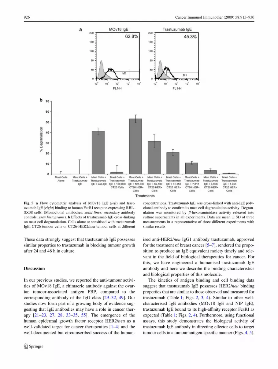
The second functional assay examined the capacity of trast-uzumab IgE to stimulate mast cells by way of the high-aYnity IgE receptor Fc�RI, and trigger their degranulation.This is a test of the potency of IgE to activate these eVectorcells, and occurs only following cross-linking of Fc�RI-bound IgE by multivalent antigen, by anti-human IgE poly-clonal antibodies or by antigen-expressing target cells. Weused a previously established system, designed to evaluatethe functional activities of IgE antibodies [48]. The assayutilises the rat basophilic mast cell line RBL-SX38, trans-fected to express all four subunits (���2) of human Fc�RI[48]. Flow cytometric evaluations conWrmed that trast-uzumab IgE bound to cell surface Fc�RI of the RBL-SX38cells (45.3% of cells), similar to the previously character-ised chimaeric MOv18 IgE (Fig. 5a). To assess the abilityof trastuzumab IgE to cause degranulation of RBL-SX38cells, we measured �-hexosaminidase release upon cellstimulation and cross-linking by tumour cells (Fig. 5b).
Mast cells alone and mast cells stimulated with trastuzumabIgE in the absence of cross-linking by anti-IgE antibody,triggered minimal mast cell degranulation [54]. Mast cellsstimulated with trastuzumab IgE in the presence of anti-IgEantibody triggered a strong degranulation response(»40%), compared to negligible degranulation measuredwith controls.
We also examined whether the engineered trastuzumabIgE cross-linked by HER2/neu–expressing tumour cellswas capable of stimulating mast cells in an antigen-dependentmanner. Trastuzumab IgE induced strong degranulation ofRBL-SX38 cells following stimulation with CT26-HER2/neu cells, which express HER2/neu, whilst untransfectedCT26, which do not express HER2/neu, did not potentiate�-hexosaminidase release (Fig. 5b). Furthermore, CT26-HER2/neu tumour cells with trastuzumab IgE potentiatedsigniWcant �-hexosaminidase release that was decreasedproportionally to the decreasing number of tumour cells persample. These results clearly demonstrate the functionalactivity of trastuzumab IgE-CT26-HER2/neu cells intriggering mast cell activation and mediator release, andconWrm that the IgE possesses biological activities thatcould be speciWcally directed against HER2/neu-expressingtumour cells in cancer patients.
Trastuzumab IgE targeting of SKBR3 breast cancer cells
ADCC/ADCP assays using the human breast adenocarci-noma cell line SKBR3, which naturally express the HER2/neu antigen, conWrm that trastuzumab and trastuzumab IgEcan focus U937 eVector cells to kill SKBR3 cells (Fig. 6).As with CT26-HER2/neu cells (Figs. 3, 4), trastuzumabacted in ADCP of tumour cells (Fig. 6a) whilst trastuzumabIgE killed by ADCC (Fig. 3b). These Wndings demonstratethe functional properties of trastuzumab IgE focusing eVec-tor cell functions against native HER2/neu-expressingbreast tumour cells.
Since trastuzumab (IgG1) can potentiate anti-tumoureVects by blocking hetero-dimerisation of HER2/neu recep-tors with other HER family members on the surface ofbreast cancer cells, switching oV tumour cell growth signals[8, 9], the anti-proliferative properties of the engineeredtrastuzumab IgE were examined using the MTS cell viabil-ity assay (Fig. 6c). Neither trastuzumab nor trastuzumabIgE had any anti-tumour growth eVects after 4 h incubationwith SKBR3 cells (99.3 and 98.6% viability for IgG andIgE, respectively). Decreased tumour cell viability wasmeasured after 24 h (88.3 and 83.6% for IgG and IgE,respectively) and more prominent eVects were measuredafter 48 h exposure of tumour cells to the antibodies (72.2and 64.0% for IgG and IgE, respectively). Control MOv18IgG and MOv18 IgE antibodies did not aVect SKBR3 cellviability (mean values ranging from 93.0 to 103.8% viability).
Table 4 % Total tumour cell death by U937 monocytes and antibodies
SigniWcance comparing trastuzumab (IgG1) and trastuzumab IgE tosamples given no antibody (left brackets), MOv18 IgG/IgE (middlebrackets) and signiWcance comparing trastuzumab (IgG1) to trast-uzumab IgE (right bracket)
Comparisons by the Student’s t test: (n/s)P > 0.05; (*)P < 0.05;(**)P < 0.005; (***)P < 0.0005
Antibody Tumour cell death § SD (n = 6) (%)
No ab 12.5 § 3.1
MOv18 IgG 18.5 § 6.6
Trastuzumab (IgG1) 0.5 �g/mL 36.1 § 2.2 (***)(***)
MOv18 IgE 19.8 § 3.2
Trastuzumab IgE 0.5 �g/mL 34.8 § 3.5 (***)(***)(n/s)
123
926 Cancer Immunol Immunother (2009) 58:915–930
These data strongly suggest that trastuzumab IgE possessessimilar properties to trastuzumab in blocking tumour growthafter 24 and 48 h in culture.
Discussion
In our previous studies, we reported the anti-tumour activi-ties of MOv18 IgE, a chimaeric antibody against the ovar-ian tumour-associated antigen FBP, compared to thecorresponding antibody of the IgG class [29–32, 49]. Ourstudies now form part of a growing body of evidence sug-gesting that IgE antibodies may have a role in cancer ther-apy [21–23, 27, 28, 33–35, 55]. The emergence of thehuman epidermal growth factor receptor HER2/neu as awell-validated target for cancer therapeutics [1–4] and thewell-documented but circumscribed success of the human-
ised anti-HER2/neu IgG1 antibody trastuzumab, approvedfor the treatment of breast cancer [5–7], rendered the propo-sition to produce an IgE equivalent moiety timely and rele-vant in the Weld of biological therapeutics for cancer. Forthis, we have engineered a humanised trastuzumab IgEantibody and here we describe the binding characteristicsand biological properties of this molecule.
The kinetics of antigen binding and cell binding datasuggest that trastuzumab IgE possesses HER2/neu bindingproperties that are similar to those observed and measured fortrastuzumab (Table 1; Figs. 2, 3, 4). Similar to other well-characterised IgE antibodies (MOv18 IgE and NIP IgE),trastuzumab IgE bound to its high-aYnity receptor Fc�RI asexpected (Table 1; Figs. 2, 4). Furthermore, using functionalassays, this study demonstrates the biological activity oftrastuzumab IgE antibody in directing eVector cells to targettumour cells in a tumour antigen-speciWc manner (Figs. 4, 5).
Fig. 5 a Flow cytometric analysis of MOv18 IgE (left) and trast-uzumab IgE (right) binding to human Fc�RI receptor-expressing RBL-SX38 cells. (Monoclonal antibodies: solid lines; secondary antibodycontrols: grey histograms). b EVects of trastuzumab IgE cross-linkingon mast cell degranulation. Cells alone or sensitised with trastuzumabIgE, CT26 tumour cells or CT26-HER2/neu tumour cells at diVerent
concentrations. Trastuzumab IgE was cross-linked with anti-IgE poly-clonal antibody to conWrm its mast cell degranulation activity. Degran-ulation was monitored by �-hexosaminidase activity released intoculture supernatants in all experiments. Data are mean § SD of threemeasurements in a representative of three diVerent experiments withsimilar results
123

Cancer Immunol Immunother (2009) 58:915–930 927
Using our ADCC/ADCP assays and microscopicimages, we made a number of observations relating to theeVector functions of trastuzumab (IgG1) and trastuzumabIgE. Trastuzumab triggered appreciable levels of mono-cyte-mediated tumour cell phagocytosis by human mono-cytic cells (Figs. 3b, c, 6a), also clearly observed inconfocal images by the presence of tumour cell materialingested by monocytic cells in contact with tumour cells(Fig. 3d; Table 3). Whilst the signalling and tumour growtharrest activities of trastuzumab and their role in its clinicaleYcacy have been the subject of many studies, the anti-tumour mechanisms relating to eVector cell functions oftrastuzumab have been less extensively investigated [7, 8,10–14]. ADCC is a known anti-tumour mechanism oftrastuzumab [11, 13, 14]. ADCP is not a widely describedfunction attributed to trastuzumab, but our data are consis-tent with ADCP as a potential anti-tumour mechanism fortrastuzumab with human macrophages, as reported byLazar et al. [12].
Surprisingly, no trastuzumab-dependent monocyte-mediated cytotoxicity (ADCC) of tumour cells was detectedabove that seen in the controls in our ADCC/ADCP assays.Using our ADCC/ADCP assay to simultaneously measurethe contributions of ADCC and ADCP [30, 31, 49], wedetected only ADCP killing of tumour cells by trast-uzumab. There are several possible explanations for the dis-crepancy between the present results and earlier reports. (1)Previous assays on the eVector cell functions of trast-uzumab, in combination with either unfractionated or puri-Wed human eVector cell populations, did not assess thecontributions of ADCP in the death of tumour cells [11–14]. It is therefore possible that ADCP-mediated tumourcell death may have been scored as ADCC if the assaysmeasured total loss of tumour cells; (2) U937 monocytesexpress Fc�RI and Fc�RII, but very low levels of Fc�RIII(Table 2), and thus it is possible that the absence of appreciablelevels of Fc�RIII may result in low levels of ADCC [18];(3) The previously reported tumour cell trastuzumab ADCC
Fig. 6 a Quantitation of trastuzumab (IgG1)-mediated tumour cell killing of SKBR3 human breast tumour cells by U937 monocytes after 2.5 h using the ADCC/ADCP assay. b Quantitation of trastuzumab IgE-mediated tumour cell killing of SKBR3 human breast tumour cells by U937 monocytes after 2.5 h using the ADCC/ADCP assay. Cytotoxicity: black bars; phagocytosis: white bars. Results are means § SD of 5 independent experiments. SigniWcance of values compared to samples given MOv18 IgG/IgE (top) or no antibody (bottom) by the Student’s t test: n/sP > 0.05, *P < 0.05, **P < 0.005, ***P < 0.0005. c Cell viability assays (MTS) demonstrating levels of suscepti-bility of SKBR3 breast cancer cells to trastuzumab (IgG1), trastuzumab IgE, MOv18 IgG and MOv18 IgE antibodies at 4, 24 and 48 h in culture. Each data point represents mean % cell viability § SD (n = 4)
123

928 Cancer Immunol Immunother (2009) 58:915–930
may have been mediated by other IgG receptor-bearingeVector cells in peripheral blood lymphocytes, such as NKcells and neutrophils, as previously shown [11–14].
When compared to the FBP-speciWc MOv18 IgE anti-body in the same assay, trastuzumab IgE activated mono-cytic eVector cells to exhibit signiWcantly enhanced contactwith tumour cells and to eVectively kill HER2/neu-express-ing tumour cells (Fig. 4d; Table 3). Trastuzumab IgEdirected monocytes to kill HER2/neu-transfected tumourcells and also tumour cells naturally-expressing the HER2/neu antigen by ADCC (cytotoxicity), a mechanism clearlydiVerent from that of the anti-tumour mechanism employedby trastuzumab in the same assay system (Figs. 3, 4, 6).This may reXect the speciWc binding of trastuzumab IgE tothe high-aYnity receptor Fc�RI. Evidence presented in ourprevious studies on the mechanism of MOv18 IgE mono-cyte-mediated tumour cell killing, suggested that binding ofIgE to its high-aYnity receptor Fc�RI on monocytes trig-gered tumour cell death by a cytotoxic mechanism, whilstbinding to the low-aYnity receptor CD23 resulted intumour cell death by ADCP. U937 monocytes express lowlevels of CD23 (»2,200 molecules/cell, Table 2) and, asobserved in our studies with MOv18 IgE, these receptorlevels are too low to mediate ADCP of tumour cells [30].The function of trastuzumab IgE eVected through CD23remains to be explored and may reveal additional tumourcell killing properties of the trastuzumab IgE.
Our assays clearly indicated that the levels of tumourcell killing mediated by trastuzumab IgE were equivalent tothose by trastuzumab (IgG) at the same optimal doses andin the same assay system (Figs. 3, 4, Table 4). This was truedespite the relatively low levels of IgE binding on the sur-face of U937 cells, compared to IgG (Figs. 3a, 4a), inagreement with previous Wndings [30] suggesting thepotency of IgE-mediated eVector functions [20–23, 27, 28,35, 56]. IgE has much higher aYnity for Fc�RI(Ka = 1010 M¡1) than IgG1 has for any of its three Fc�receptors, especially for Fc�RIII (Ka = 105 M¡1), the mainreceptor associated with tumour cell killing [17, 18, 57].The relatively high aYnities of the IgE-Fc�RI interactionmay compensate for the lower binding of IgE on U937monocytes, resulting in comparable levels of tumour cellkilling. These data clearly indicate the biological functionof trastuzumab IgE in focusing monocytic cells to killHER2/neu-expressing tumour cells as eVectively as, butthrough a mechanism diVerent from, trastuzumab.
The antibody concentration (0.5 �g/mL) required toachieve maximum tumour cell killing by ADCC/ADCPwas the same for IgG and IgE in our in vitro assays (Figs. 3,4). In addition, our cell viability data clearly show thattrastuzumab IgE, at concentrations found optimal for eVec-tor cell responses (0.5 �g/mL), maintains the ability tomediate tumour cell growth arrest over a period of 48 h in
culture and at levels similar to those measured for trast-uzumab (Fig. 6c). The concentration found optimal for thein vitro eVector cell functions of trastuzumab IgE was ten-fold lower than our previously reported optimal concentra-tions required for MOv18 IgG and IgE-mediated (5 �g/mL)killing of ovarian tumour cells in equivalent in vitro assays[30–32]. This may suggest that lower levels of trastuzumabIgE may be required for in vivo activities compared tothose used for MOv18 IgG and MOv18 IgE studies. TheIgE responses measured and observed in our assays supportthe conclusion that trastuzumab IgE functions with similarpotency, but through mechanisms diVerent from those oftrastuzumab in vitro, warranting further exploration of thisengineered antibody in more clinically relevant models. Itis possible, however, that trastuzumab IgE may be moreeVective than trastuzumab (IgG1) in vivo for the reasonscited in the Introduction or that a combination of trast-uzumab and trastuzumab IgE could have potential synergis-tic anti-tumour eVects.
We also assessed the potential of the engineered trast-uzumab IgE to activate other potent IgE receptor-bearingeVector cells. For this, we have utilised a functional assaythat exempliWes the unique properties of IgE to generateand enhance immune eVector functions that can be targetedagainst cancer cells. Trastuzumab IgE bound to the humanFc�RI ���2 receptor on the surface of mast cells can becross-linked by HER2/neu-expressing tumour cells to trig-ger mast cell degranulation in an antigen-speciWc manner(Fig. 5). Tissue mast cell degranulation is a known biologi-cal property of the IgE antibody class. Mediators releasedby degranulated mast cells initiate and potentiate eVectorcell recruitment to the site of tumour antigen challenge,which can be expected to lead to activation and stimulationof recruited and locally present eVector cells to act in theADCC and ADCP of tumour cells. Mast cell activation byIgE may thus serve as a potential trigger of a strong, local,tumour antigen-speciWc IgE immune response againstcancer.
This is the Wrst study describing the properties of anengineered trastuzumab IgE. Based on the considerable evi-dence pointing to a number of advantages that IgE mayhave in tumour cell surveillance and killing, compared toIgG, our work points to the importance and value of furtherresearch to investigate the eYcacy and mechanisms ofaction of tumour antigen-speciWc antibodies of diVerentclasses, in particular IgE and may help to realise the fullpotential of antibodies for immunotherapy of cancer.
Acknowledgments This work was generously supported by the theNational Institute for Health Research (NIHR) Biomedical ResearchCentre (BRC) at Guy’s and St. Thomas’ NHS Foundation Trust/King’s College London, United Kingdom; the Austrian ScienceFund (FWF) (P-18238-B13); the European Molecular BiologyOrganisation (EMBO) (fellowship ASTF258.00-2008); Hans und
123

Cancer Immunol Immunother (2009) 58:915–930 929
Blanca Moser Stiftung (AP00326OFF), Austria; NIH/NCI R01 sup-plement CA107023-02S1, Susan G. Komen Breast Cancer Founda-tion grant (BCTR0706771) and the 2007–2008 University ofCalifornia Cancer Research Coordinating Committee seed grant,USA. We thank Dr. Rebecca Beavil, Dr. Pooja Takhar and Mr. Rich-ard Brunner for their helpful comments and Ms. Kate Kirwan forexpert assistance with the Wgures. We are grateful to Dr. Jean-PierreKinet and to Dr. Silvana Canevari for the generous provision ofadvice and materials.
References
1. Rubin I, Yarden Y (2001) The basic biology of HER2. Ann Oncol12(Suppl 1):S3–S8
2. Kaptain S, Tan LK, Chen B (2001) Her-2/neu and breast cancer.Diagn Mol Pathol 10:139–152
3. Yarden Y (2001) Biology of HER2 and its importance in breastcancer. Oncology 61(Suppl 2):1–13
4. Landgraf R (2007) HER2 therapy. HER2 (ERBB2): functionaldiversity from structurally conserved building blocks. Breast Can-cer Res 9:202
5. Hortobagyi GN (2005) Trastuzumab in the treatment of breast can-cer. N Engl J Med 353:1734–1736
6. Carter P, Presta L, Gorman CM, Ridgway JB, Henner D, WongWL, Rowland AM, Kotts C, Carver ME, Shepard HM (1992)Humanization of an anti-p185HER2 antibody for human cancertherapy. Proc Natl Acad Sci USA 89:4285–4289
7. Gonzalez-Angulo AM, Morales-Vasquez F, Hortobagyi GN(2007) Overview of resistance to systemic therapy in patients withbreast cancer. Adv Exp Med Biol 608:1–22
8. Hudis CA (2007) Trastuzumab—mechanism of action and use inclinical practice. N Engl J Med 357:39–51
9. Lee-HoeXich ST, Crocker L, Yao E, Pham T, Munroe X, HoeXichKP, Sliwkowski MX, Stern HM (2008) A central role for HER3 inHER2-ampliWed breast cancer: implications for targeted therapy.Cancer Res 68:5878–5887
10. Izumi Y, Xu L, di Tomaso E, Fukumura D, Jain RK (2002)Tumour biology: herceptin acts as an anti-angiogenic cocktail.Nature 416:279–280
11. Barok M, Isola J, Palyi-Krekk Z, Nagy P, Juhasz I, Vereb G,Kauraniemi P, Kapanen A, Tanner M, Szollosi J (2007) Trast-uzumab causes antibody-dependent cellular cytotoxicity-mediated growth inhibition of submacroscopic JIMT—1 breastcancer xenografts despite intrinsic drug resistance. Mol CancerTher 6:2065–2072
12. Lazar GA, Dang W, Karki S, Vafa O, Peng JS, Hyun L, Chan C,Chung HS, Eivazi A, Yoder SC et al (2006) Engineered antibodyFc variants with enhanced eVector function. Proc Natl Acad Sci US A 103:4005–4010
13. Gennari R, Menard S, Fagnoni F, Ponchio L, Scelsi M, TagliabueE, Castiglioni F, Villani L, Magalotti C, Gibelli N et al (2004) Pilotstudy of the mechanism of action of preoperative trastuzumab inpatients with primary operable breast tumors overexpressingHER2. Clin Cancer Res 10:5650–5655
14. Sliwkowski MX, Lofgren JA, Lewis GD, Hotaling TE, FendlyBM, Fox JA (1999) Nonclinical studies addressing the mechanismof action of trastuzumab (Herceptin). Semin Oncol 26:60–70
15. Riethmuller G, Johnson JP (1992) Monoclonal antibodies in thedetection and therapy of micrometastatic epithelial cancers. CurrOpin Immunol 4:647–655
16. Gould HJ, Takhar P, Harries HE, Durham SR, Corrigan CJ (2006)Germinal-centre reactions in allergic inXammation. Trends Immu-nol 27:446–452
17. Gould HJ, Sutton BJ, Beavil AJ, Beavil RL, McCloskey N, CokerHA, Fear D, Smurthwaite L (2003) The biology of IGE and thebasis of allergic disease. Annu Rev Immunol 21:579–628
18. Clynes RA, Towers TL, Presta LG, Ravetch JV (2000) InhibitoryFc receptors modulate in vivo cytoxicity against tumor targets. NatMed 6:443–446
19. Geha RS, Helm B, Gould H (1985) Inhibition of the Prausnitz-Kustner reaction by an immunoglobulin epsilon-chain fragmentsynthesised in E. coli. Nature 315:577–578
20. Gonzalez-Espinosa C, Odom S, Olivera A, Hobson JP, MartinezME, Oliveira-Dos-Santos A, Barra L, Spiegel S, Penninger JM,Rivera J (2003) Preferential signalling and induction of allergy-promoting lymphokines upon weak stimulation of the high aYnityIgE receptor on mast cells. J Exp Med 197:1453–1465
21. Riemer AB, Untersmayr E, Knittelfelder R, Duschl A, Pehamber-ger H, Zielinski CC, Scheiner O, Jensen-Jarolim E (2007) Activeinduction of tumor-speciWc IgE antibodies by oral mimotope vac-cination. Cancer Res 67:3406–3411
22. Jensen-Jarolim E, Achatz G, Turner MC, Karagiannis S, LegrandF, Capron M, Penichet ML, Rodriguez JA, Siccardi AG, Vangeli-sta L et al (2008) AllergoOncology: the role of IgE-mediated aller-gy in cancer. Allergy 63:1255–1266
23. Reali E, Greiner JW, Corti A, Gould HJ, Bottazzoli F, Paganelli G,Schlom J, Siccardi AG (2001) IgEs targeted on tumor cells: thera-peutic activity and potential in the design of tumor vaccines. Can-cer Res 61:5517–5522
24. Turner MC, Chen Y, Krewski D, Ghadirian P (2006) An overviewof the association between allergy and cancer. Int J Cancer118:3124–3132
25. Turner MC, Chen Y, Krewski D, Ghadirian P, Thun MJ, Calle EE(2005) Cancer mortality among US men and women with asthmaand hay fever. Am J Epidemiol 162:212–221
26. Nagy E, Berczi I, Sehon AH (1991) Growth inhibition of murinemammary carcinoma by monoclonal IgE antibodies speciWc for themammary tumor virus. Cancer Immunol Immunother 34:63–69
27. Kershaw MH, Darcy PK, Trapani JA, MacGregor D, Smyth MJ(1998) Tumor-speciWc IgE-mediated inhibition of human colou-rectal carcinoma xenograft growth. Oncol Res 10:133–142
28. Riemer AB, Klinger M, Wagner S, Bernhaus A, Mazzucchelli L,Pehamberger H, Scheiner O, Zielinski CC, Jensen-Jarolim E(2004) Generation of peptide mimics of the epitope recognized bytrastuzumab on the oncogenic protein Her-2/neu. J Immunol173:394–401
29. Gould HJ, Mackay GA, Karagiannis SN, O’Toole CM, Marsh PJ,Daniel BE, Coney LR, Zurawski VR Jr, Joseph M, Capron M et al(1999) Comparison of IgE and IgG antibody-dependent cytotoxic-ity in vitro and in a SCID mouse xenograft model of ovarian car-cinoma. Eur J Immunol 29:3527–3537
30. Karagiannis SN, Bracher MG, Beavil RL, Beavil AJ, Hunt J,McCloskey N, Thompson RG, East N, Burke F, Sutton BJ et al(2008) Role of IgE receptors in IgE antibody-dependent cytotox-icity and phagocytosis of ovarian tumor cells by human monocyticcells. Cancer Immunol Immunother 57:247–263
31. Karagiannis SN, Bracher MG, Hunt J, McCloskey N, Beavil RL,Beavil AJ, Fear DJ, Thompson RG, East N, Burke F et al (2007)IgE-antibody-dependent immunotherapy of solid tumors: cyto-toxic and phagocytic mechanisms of eradication of ovarian cancercells. J Immunol 179:2832–2843
32. Karagiannis SN, Wang Q, East N, Burke F, RiVard S, BracherMG, Thompson RG, Durham SR, Schwartz LB, Balkwill FR et al(2003) Activity of human monocytes in IgE antibody-dependentsurveillance and killing of ovarian tumor cells. Eur J Immunol33:1030–1040
33. Luiten RM, Fleuren GJ, Warnaar SO, Litvinov SV (1996) Target-speciWc activation of mast cells by immunoglobulin E reactive
123

930 Cancer Immunol Immunother (2009) 58:915–930
with a renal cell carcinoma-associated antigen. Lab Invest 74:467–475
34. Luiten RM, Warnaar SO, Schuurman J, Pasmans SG, Latour S,Daeron M, Fleuren GJ, Litvinov SV (1997) Chimeric immuno-globulin E reactive with tumor-associated antigen activates humanFc epsilon RI bearing cells. Hum Antibodies 8:169–180
35. Teng MW, Kershaw MH, Jackson JT, Smyth MJ, Darcy PK(2006) Adoptive transfer of chimeric FcepsilonRI gene-modiWedhuman T cells for cancer immunotherapy. Hum Gene Ther17:1134–1143
36. Cook JP, Henry AJ, McDonnell JM, Owens RJ, Sutton BJ, GouldHJ (1997) IdentiWcation of contact residues in the IgE binding siteof human FcepsilonRIalpha. Biochemistry 36:15579–15588
37. Neuberger MS, Williams GT, Mitchell EB, Jouhal SS, FlanaganJG, Rabbitts TH (1985) A hapten-speciWc chimaeric IgE antibodywith human physiological eVector function. Nature 314:268–270
38. Dela Cruz JS, Lau SY, Ramirez EM, De Giovanni C, Forni G,Morrison SL, Penichet ML (2003) Protein vaccination with theHER2/neu extracellular domain plus anti-HER2/neu antibody-cytokine fusion proteins induces a protective anti-HER2/neuimmune response in mice. Vaccine 21:1317–1326
39. Cho HS, Mason K, Ramyar KX, Stanley AM, Gabelli SB, DenneyDW Jr, Leahy DJ (2003) Structure of the extracellular region ofHER2 alone and in complex with the Herceptin Fab. Nature421:756–760
40. Durocher Y, Perret S, Kamen A (2002) High-level and high-throughput recombinant protein production by transient transfec-tion of suspension-growing human 293-EBNA1 cells. NucleicAcids Res 30:E9
41. Gan SK, Hunt J, Beavil AJ, Marsh PJ, Harries HE (2008) Thedesign and optimisation of a transient expression system for therapid expression of human immunoglobulin E. Example cited inGB patent application 61/060,239
42. McDonnell JM, Calvert R, Beavil RL, Beavil AJ, Henry AJ,Sutton BJ, Gould HJ, Cowburn D (2001) The structure of the IgECepsilon2 domain and its role in stabilizing the complex with itshigh-aYnity receptor FcepsilonRIalpha. Nat Struct Biol 8:437–441
43. Henry AJ, Cook JP, McDonnell JM, Mackay GA, Shi J, Sutton BJ,Gould HJ (1997) Participation of the N-terminal region of Cepsi-lon3 in the binding of human IgE to its high-aYnity receptor Fcep-silonRI. Biochemistry 36:15568–15578
44. Sundstrom C, Nilsson K (1976) Establishment and characteriza-tion of a human histiocytic lymphoma cell line (U-937). Int J Can-cer 17:565–577
45. Corbett TH, Griswold DP Jr, Roberts BJ, Peckham JC, SchabelFM Jr (1975) Tumor induction relationships in development of
transplantable cancers of the colon in mice for chemotherapy as-says, with a note on carcinogen structure. Cancer Res 35:2434–2439
46. Griswold DP, Corbett TH (1975) A colon tumor model for anti-cancer agent evaluation. Cancer 36:2441–2444
47. Penichet ML, Challita PM, Shin SU, Sampogna SL, RosenblattJD, Morrison SL (1999) In vivo properties of three human HER2/neu-expressing murine cell lines in immunocompetent mice. LabAnim Sci 49:179–188
48. Wiegand TW, Williams PB, Dreskin SC, Jouvin MH, Kinet JP,Tasset D (1996) High-aYnity oligonucleotide ligands to humanIgE inhibit binding to Fc epsilon receptor I. J Immunol 157:221–230
49. Bracher M, Gould HJ, Sutton BJ, Dombrowicz D, Karagiannis SN(2007) Three-colour Xow cytometric method to measure antibody-dependent tumour cell killing by cytotoxicity and phagocytosis.J Immunol Methods 323:160–171
50. Bodinier M, Brossard C, Triballeau S, Morisset M, Guerin-Marc-hand C, Pineau F, de Coppet P, Moneret-Vautrin DA, Blank U,Denery-Papini S (2008) Evaluation of an in vitro mast cell degran-ulation test in the context of food allergy to wheat. Int Arch Aller-gy Immunol 146:307–320
51. Linko-Lopponen S, Makinen M (1985) A microtiter plate assayfor N-acetyl-beta-D-glucosaminidase using a Xuorogenic sub-strate. Anal Biochem 148:50–53
52. Casal JA, Chabas A, Tutor JC (2003) Thermodynamic determina-tion of beta-hexosaminidase isoenzymes in mononuclear and poly-morphonuclear leukocyte populations. Am J Med Genet A116A:229–233
53. Gerstner RB, Carter P, Lowman HB (2002) Sequence plasticity inthe antigen-binding site of a therapeutic anti-HER2 antibody.J Mol Biol 321:851–862
54. Posner RG, Geng D, Haymore S, Bogert J, Pecht I, Licht A, Sav-age PB (2007) Trivalent antigens for degranulation of mast cells.Org Lett 9:3551–3554
55. Riemer AB, Jensen-Jarolim E (2007) Mimotope vaccines: epitopemimics induce anti-cancer antibodies. Immunol Lett 113:1–5
56. Bramswig KH, Knittelfelder R, Gruber S, Untersmayr E, RiemerAB, Szalai K, Horvat R, Kammerer R, Zimmermann W, ZielinskiCC et al (2007) Immunization with mimotopes prevents growth ofcarcinoembryonic antigen positive tumors in BALB/c mice. ClinCancer Res 13:6501–6508
57. Ravetch JV, Kinet JP (1991) Fc receptors. Annu Rev Immunol9:457–492
123