Autoantibody to p185 erbB2/neu oncoprotein by vaccination with xenogenic DNA
9
ORIGINAL ARTICLE Antonio Concetti Augusto Amici Cristina Petrelli Alberto Tibaldi Mauro Provinciali Franco Maria Venanzi Received: 29 August 1996 / Accepted: 31 October 1996 AbstractmThe passive transfer of antibodies and vaccina- tion procedures against p185, the erbB2/neu oncoprotein, are approaches being explored for treatment of human breast cancer. We now report the possibility of using the erbB2/neu gene as an immunogen. This study demonstrates that intramuscular or intradermal injections of rat neuNT full-length DNA into mice generate anti-p185 autoantibo- dies. Anti-p185 polyclonals were also shown to bind the homologous human receptor ErbB2 and to stain specimens of breast adenocarcinoma from both neu-transgenic mice and humans. Further, in vitro assays demonstrated that anti- p185 IgG (probably dependent on CD4 + Th1) were able to inhibit human SKBR3 tumour cell growth and to mediate their lysis by natural killer cells. The continuous presence of circulating neu autoantibodies in mice did not cause any discernible toxic effects on normal tissues expressing low levels of self-antigen, even after 1 year. The experiments reported here raise the possibility that boosting anti-ErbB2 immunity by DNA vaccination will not induce harmful autoimmunity in humans. Key wordsmDNA immunization p185 erbB2/neu Autoantibodies Breast cancer The c-erbB2/neu protooncogene encodes a tyrosine kinase growth factor receptor (p185), which appears to play a crucial role in the pathogenesis of many human cancers (i. e. mammary, ovary and lung adenocarcinoma) [33]. The receptor, detectable in small amounts in differentiated epithelial/glandular tissues or during placentation and or- ganogenesis [23, 22], is overexpressed in about 30% of invasive breast carcinomas and in approximately 70% of in situ ductal carcinomas [33, 19]. Experimental evidence indicates that p185 overexpression contributes to trans- formation and to tumour progression and suggests that the prognostic significance of oncogene expression (i. e. a worse clinical outcome in most subsets of patients) arises from the aggressive phenotype it confers on malig- nant cells [30]. The extracellular domain of the erbB2/neu receptor represents an attractive target for antibody-based therapy. First, such a treatment may be able to interfere with the expression and/or functions of this oncoprotein, thus inhib- iting tumour growth. Further, since the overexpressed oncoprotein appears stable and homogeneously distributed within the primary tumour and its metastasis [24], such an approach is likely to target the whole tumour. The potential use of anti-p185 monoclonal antibodies (mAb) as tumour inhibitors was originally demonstrated in neu-transformed cells, where antibody binding induced down-regulation of p185 and reversal of the transformed phenotype [14]. Moreover, anti-p185 mAb exerted a cyto- static effect on neu-transformed cells in a syngenic rat background [12] or in a transgenic model of mouse breast cancer [21]. Further, some anti-p185 monoclonals, which individually do not show biological activity, gain tumour- growth-inhibitory properties when admixed [18], or when coupled with radionuclides [6] or with drugs [26]. Thus, these experiments suggest that erbB2/neu-specific antibod- ies (either alone or in combination with other agents) may prove to be of some benefit in the treatment of breast cancer. This work is dedicated to the memory of Professor Pier Luigi Gariboldi and was supported by grants from MURST 60% (F. M. V. and A. C.) and from Associazione Italiana per la Ricerca sul Cancro (F. M. V.) A. C., A. A. and F. M. V. contributed equally to the work A. Concetti ( ) A. Amici ( ) C. Petrelli F. M. Venanzi ( ) Laboratory of Genetic Immunization, Department of Biology M. C. A., via F. Camerini, I-62032 Camerino (MC), Italy Fax: +39 737/636216 e-mail: [email protected] A. Tibaldi M. Provinciali Immunology Center, Research Department, I. N. R. C. A., Ancona, Italy Cancer Immunol Immunother (1996) 43: 307 – 315 Springer-Verlag 1996
Transcript of Autoantibody to p185 erbB2/neu oncoprotein by vaccination with xenogenic DNA

ORIGINAL ARTICLE
Antonio Concetti ? Augusto Amici ? Cristina PetrelliAlberto Tibaldi ? Mauro ProvincialiFranco Maria Venanzi
Autoantibody to p185erbB2/neu oncoprotein
by vaccination with xenogenic DNA
Received: 29 August 1996 / Accepted: 31 October 1996
AbstractmThe passive transfer of antibodies and vaccina-tion procedures against p185, theerbB2/neu oncoprotein,are approaches being explored for treatment of humanbreast cancer. We now report the possibility of using theerbB2/neugene as an immunogen. This study demonstratesthat intramuscular or intradermal injections of rat neuNTfull-length DNA into mice generate anti-p185 autoantibo-dies. Anti-p185 polyclonals were also shown to bind thehomologous human receptor ErbB2 and to stain specimensof breast adenocarcinoma from bothneu-transgenic miceand humans. Further, in vitro assays demonstrated that anti-p185 IgG (probably dependent on CD4+ Th1) were able toinhibit human SKBR3 tumour cell growth and to mediatetheir lysis by natural killer cells. The continuous presenceof circulatingneuautoantibodies in mice did not cause anydiscernible toxic effects on normal tissues expressing lowlevels of self-antigen, even after 1 year.
The experiments reported here raise the possibility thatboosting anti-ErbB2 immunity by DNA vaccination will notinduce harmful autoimmunity in humans.
Key wordsmDNA immunization? p185erbB2/neu?
Autoantibodies? Breast cancer
Introduction
The c-erbB2/neuprotooncogene encodes a tyrosine kinasegrowth factor receptor (p185), which appears to play acrucial role in the pathogenesis of many human cancers (i. e.mammary, ovary and lung adenocarcinoma) [33]. Thereceptor, detectable in small amounts in differentiatedepithelial/glandular tissues or during placentation and or-ganogenesis [23, 22], is overexpressed in about 30% ofinvasive breast carcinomas and in approximately 70% of insitu ductal carcinomas [33, 19]. Experimental evidenceindicates that p185 overexpression contributes to trans-formation and to tumour progression and suggests thatthe prognostic significance of oncogene expression(i. e. a worse clinical outcome in most subsets of patients)arises from the aggressive phenotype it confers on malig-nant cells [30].
The extracellular domain of theerbB2/neu receptorrepresents an attractive target for antibody-based therapy.First, such a treatment may be able to interfere with theexpression and/or functions of this oncoprotein, thus inhib-iting tumour growth. Further, since the overexpressedoncoprotein appears stable and homogeneously distributedwithin the primary tumour and its metastasis [24], such anapproach is likely to target the whole tumour.
The potential use of anti-p185 monoclonal antibodies(mAb) as tumour inhibitors was originally demonstrated inneu-transformed cells, where antibody binding induceddown-regulation of p185 and reversal of the transformedphenotype [14]. Moreover, anti-p185 mAb exerted a cyto-static effect onneu-transformed cells in a syngenic ratbackground [12] or in a transgenic model of mouse breastcancer [21]. Further, some anti-p185 monoclonals, whichindividually do not show biological activity, gain tumour-growth-inhibitory properties when admixed [18], or whencoupled with radionuclides [6] or with drugs [26]. Thus,these experiments suggest thaterbB2/neu-specific antibod-ies (either alone or in combination with other agents) mayprove to be of some benefit in the treatment of breastcancer.
This work is dedicated to the memory of Professor Pier Luigi Gariboldiand was supported by grants from MURST 60% (F. M. V. and A. C.)and from Associazione Italiana per la Ricerca sul Cancro (F. M. V.)A. C., A. A. and F. M. V. contributed equally to the work
A. Concetti ( ) ? A. Amici ( ) ? C. Petrelli ? F. M. Venanzi ( )Laboratory of Genetic Immunization, Department of Biology M. C. A.,via F. Camerini, I-62032 Camerino (MC), ItalyFax: +39737/636216e-mail: [email protected]
A. Tibaldi ? M. ProvincialiImmunology Center, Research Department, I. N. R. C. A.,Ancona, Italy
Cancer Immunol Immunother (1996) 43: 307–315 Springer-Verlag 1996

However an antibody-based immunotherapy faces majorobstacles for practical clinical purposes: i) the need forsetting experimental criteria for selection of suitable mAb(e. g. different antibodies to the same protooncogene mayhave opposite effects on tumour growth) [18]; ii) thepotential antigenicity of murine monoclonals in humans;iii) the amounts of antibodies needed for treatments, theduration of which is ill-defined. In adjuvant therapy,months or perhaps years have to be considered.
Anti-ErbB2 active immunization (vaccination) shouldovercome these problems. Recently, in fact, vaccinationwith mixtures of syntheticerbB2/neu peptides has beenproposed [3, 9].
As another possibility to achieve anti-erbB2/neu immu-nity, we now propose a DNA-based vaccination that isdependent upon inoculation of naked DNA sequencesdirectly into host target tissues without the use of a deliveryvehicle [10].
In order to circumvent tolerance, which may preventinduction of an immunoresponse to self-p185, we trans-fected skeletal muscles or dermal layers of mice withplasmids carrying the cDNA encoding rat activatedneuoncogene (neuNT) [1]. We report, for the first time, on thepossibility of using a DNA vaccine against an oncoproteinand describe the humoral anti-p185 immunoresponse eli-cited.
Materials and methods
Cell cultures and transfections
SKBR3 human breast carcinoma and A431 human epidermoid carci-noma cell lines were obtained from Dr. P. G. Natali (Istituto ReginaElena, IRE, Rome). Transfected mouse fibroblasts (3T3 NHI) over-expressing either human ErbB2 (T28.1) [7] or a chimeric receptorprotein containing the extracellular domain (ECD) of epidermal growthfactor receptor (EGFR) and the intracellular domain (ICD) of ErbB2(T19.7) [7] were obtained from Dr. O. Segatto (IRE). NIH 3T3 cellswere transfected in our laboratory with pCMVneuNT (see below)complexed with calcium phosphate and selected wth G418 accordingto a published protocol [1]. G418-resistent colonies were picked upafter 2 weeks and grown into cell lines (TC.22).
Cellular lysates and tissues extraction
RIPA buffer (0.6 ml) containing freshly prepared inhibitors (100µg/mlphenylmethylsulphonyl fluoride, 30µl/ml aprotinin and 100 mMsodium orthovanadate) was added to 100-cm2 plates. The lysateswere passed several times through a syringe fitted with a 21-guageneedle, incubated for 30 min on ice and microfuged for 20 min at 4°C;supernatants were collected. Mouse kidneys were removed and cut intovery small pieces with a razor blade. Thin frozen tissue was thawedand lysed by adding 3 ml ice-cold RIPA buffer and inhibitors on ice for30 min. The lysates were centrifuged at 15000g for 30 min at 4°C.Clear supernatants were ultrafiltered through an Amicon cell (model8200) using XM100A membranes (cut-off4100000 kDa). Retainedsolution was stored at –80°C.
Anti-erbB2/neu antobodies
Two monoclonal antibodies (W6-100 and W6-800 IgG2a isotype)reactive towards the extracellular domain of human ErbB2 were
obtained from Dr. P. G. Natali. Rabbit anti-ErbB2/Neu polyclonalIgG (K-15), specific for the intracellular domain of the human androdent oncogene receptor, were purchased from Santa Cruz Biotech-nology USA.
Plasmid DNA
Expression plasmid coding for rat neuNT under the control of simianvirus 40 (SV40) early promoter, pSV2neuNT [1] was obtained fromProf. N. Ranzani (Universita` di Pavia). To construct a plasmid with astronger promoter (pCMVneuNT) the full-length DNA for neuNT fromthe pSV2neuNT plasmid was excised by combinedHindIII, SalIdigestions and cloned in pcDNA3 (inVitrogen), which contains thecytomegalovirus (CMV) early promoter/enhancer. As control DNA“antigen”, thegagprotein p24 of feline immunodeficiency virus (FIV)[34] was cloned in a pcDNA3 vector. The resulting plasmid wasdesignated as pCMV-p24. Large-scale preparations of plasmid DNAwere carried out by a Quiagen kit, according to the manufacturer’sinstructions. For experimental use, the DNAwas reconstituted in sterilesaline (1 mg/ml) and stored in aliquots at –20°C for direct injections.
Vaccination protocols
Outbred (Balb/c, Swiss CD1) or inbred (Balb/c H2d) mice of both sexeswere anaesthesized with a nembutal equitesinain solution (0.3 ml/100 gbody weight). After exposure of the femoral quadriceps, the muscleswere injected with 100 ml DNA solution (100–10µg) delivered withan insulin syringe. The animals were immunized with three adminis-trations carried out at biweekly intervals. The same immunizationschedule was used for Wistar rats. Intradermal injections (i. d.) at1–2 cm distal from the tail base were performed with a 1-ml syringeand a 28-guage needle delivering various amounts of free DNA in atotal volume of 100µl. The schedule was the same as that used for i. m.immunization.
Immunotechniques
ELISA
Serum antibody responses to the rat p185 were determined in solid-phase enzyme-linked immunosorbent assay by using a cell lysate ofTC.22 cells (from approximately 105 cells in 0.05 M carbonate buffer,pH 9.6) to coat the microtitres wells. After 8 h of incubation at 4°C,the solutions were aspirated and dilutions of test sera were added to thewells. Following 1 h treatment at 37°C, the wells were washed threetimes in phosphate-buffered saline containing 0.05% Tween-20 (PBST)then incubated with horseradish-peroxidase(HRP)-labelled goat anti-(mouse Ig/IgG) (Sigma) at a dilution of 1 :3000. After incubation withthe second antibody (45 min at 37°C), the wells were again washedthree times with PBST, and colour-developed by using 3,3,5,59-tetra-methylbenzidine (Sigma). Absorbance was read at 450 nm in a multi-channel reader (Titertek Multiscan, Labsystem). ELISA titres againstTC.22 cells were reported as the last dilution that gave an absorbancereading at least 0.05 unit above background binding absorbance.
Western and dot blotting
Total cells or tissues lysates (2×10⁶ cells or 1/10 whole protein extractfrom mouse kidney) were electrophoresed in polyacrylamide/sodiumdodecyl sulphate gels (PAGE-SDS; 7% acrylamide). The proteins weretransferred to nitrocellulose membranes using an electroblotting appa-ratus as described [17]. After non-specific binding had been blocked inPBST-Blotto, the strips were incubated for 2 h at room temperaturewith primary antibodies (routinely mouse sera diluted 1 :50). Follow-ing washes with PBST, membranes were incubated for 1 h withalkaline phosphatase or HRP-conjugated goat anti-(mouse Ig/IgG)diluted 1 :3000 in Blotto. Staining was developed either by nitrobluetetrazolium/5-Bromo-4-chloro-3-indolyl Phosphate or 4-chloro-1-naphthol chromogenic reagents.
308

Confocal laser microscopy
SKBR3 cells were grown on coverslips and fixed either with a mixtureof cold methanol/acetone (1 :1, v :v) (permeabilized cells) or with 2%paraformaldheyde/PBS (non-permeabilized cells). The samples werewashed then incubated with anti-p185 antisera (1/30 final dilution) andthe antigen/antibody reaction was demonstrated using sheep anti-(mouse IgG) antibodies conjugated with fluorescein isothiocyanate(FITC, Boringher) (1/60 dilution). The mounted slides were examinedunder the laser confocal microscope (BioRad MRC 800K) with a tripleargon laser and a 60 X oil-immersion objective.
FACS scanning
The immunoreactivity of anti-p185 antibodies towards p170EGFR wasanalysed by FACS scanning of viable A431 tumour cells. The cells,after incubation with the second FITC-conjugated anti-(mouse Ig)antibody, were analysed using a Becton-Dickinson FACS (argonlaser excitation light, 488 nm; emitted light, 525 nm).
Tissue samples and immunohistochemical studies
Mouse kidneys were fixed in Carnoy solution, whereas mouse andhuman breast cancer specimens were formalin-fixed and paraffin-embedded. Tissue sections of 4–5µm were dewaxed by xylol andwashed in alcohol. After rinsing with PBS, the sections were incubatedfor 4 h at room temperature with mouse antisera (1/30 dilution) or inhuman breast cancer W6-100 monoclonals (5µg/ml) (positive control).Bound primary antibodies were labelled either using biotinylated goatanti-(mouse Ig/IgG) and streptavidin peroxidase or alternatively withgoat anti-(mouse Ig/IgG) conjugated with alkaline phosphatase. Toavoid the possibility of the heterogeneous distribution of p185 withinthe tissues leading to false-negatives, at least two non-consecutivesections of different specimens were analysed.
IgG purification through protein-A–Superose
The separation of IgG2a, IgG2b and IgG3 from IgG1 antibodies wasperformed by protein-A–Superose on a fast protein liquid chromato-graphy apparatus (Pharmacia, Uppsala), according to the manufac-turer’s instructions.
Cell proliferation and cytotoxicity assays
The antiproliferative effect of anti-neu IgG was determined after acontinuous 7-day incubation with SKRR3 tumour cells. The cells wereplated in 24-well plates (103 cells/plate) and incubated, on day 0, eitherwith 30 µl (approx. 300µg total IgG) of antisera from immunizedanimals heated at 55°C for 30 min or with W6-100 and W8-100 mAb(30 µg each). All samples were in triplicate. After 1 week, the cellswere removed and the number of viable cells determined by haemo-cytometer.
Antibody-mediated cell-dependent cytotoxicity (ADCC) assayswere performed as follows: fluorescent-labelled (carboxy-fluoresceindiacetate) SKBR3 or A431 tumour cells (5×10³) in 25 µl wereincubated for 1 h at various concentrations of protein-A–Superose-purified mouse IgG (10–20µg/ml) or of W6–100 Ab (0.1–10µg/ml)and then for 5 h with mouse splenocyte effector cells (effector to targetcells ratio from 25:1 to 200 :1). Lysis was determined by fluorimetricanalysis using Fluoroskan-2 (Flow Instruments).
Results
In vivo production of rat Neu antigen
Unlike the injection of proteins/peptides or killed viruses,the antigen produced by DNA injection is made within thesomatic cells of the host. In order to evaluate the ability ofpSV2neuNT DNA to express the antigen in vivo, mice weresacrificed 2 days after a single injection of plasmid. Quad-riceps muscles were removed and extracted for total pro-teins (molecular mass4100 kDa). Aliquots were thenspotted on nitrocellulose filters and assayed in immuno-dot-blot for the presence of p185 protein by using mono-specific rabbit polyclonal K15 anti-erbB2/neu IgG.
The results reported in Fig. 1 show that the pSV2neuNTvector was able to direct the synthesis of the p185 moleculeinside the tissue fibres. No staining was observed insamples obtained from muscles injected with control plas-mids.
309
Fig. 2mEnzyme-linked immunosorbent assay (ELISA) of TC.22 cellslysates by pSV2neuNT and pCMVneuNT antisera (two animals/group).Each serum dilution was analysed in triplicate. Final ELISA titresdetermined as described in Materials and methods. Control serumderived from a mouse immunized with pCMV-p24
Fig. 1mImmunodot blots with anti-erbB2/neu polylconal antibodies(K15) to test the reactivity against different amounts of whole muscleextracts. Total extract: proteins from one muscle in 2 ml phosphate-buffered saline.a 0.05 ml; b 0.1 ml; c 0.25 ml whole muscle extract.Lanes 1:extract from an untreated mouse;2, 3 extract obtained fromtwo independent mice injected with 100µg pSV2 neuNT. Staining withgoat anti-(rabbit Ig/IgG) conjugated with horseradish peroxidase(HRP).V void
B cell responses in vaccinated rodents
Balb/c or Swiss mice were immunized either with controlplasmids (pCDNA3, pCMV-p24) or with equal doses ofeither pSV2neuNT or pCMVneuNT according to the sche-dule (see Materials and methods). The production andspecificity of anti-p185 antibodies was evaluated byELISA and Western blotting procedures using cellularlysates of pCMVneuNT-transfected fibroblasts (TC.22) asthe source of antigen. As controls, preimmune sera from thesame vaccinated animals or rabbit K15 antibodies wereused.
As shown in Fig. 2 ELISA assay indicated thatpSV2neuNT injections reproducibly induced higher anti-
body responses (ELISA titres: pSV2neuNT = 1:500 com-pared to pCMVneuNT = 1:150).
Further, Western blot analysis (Fig. 3) confirmed thatIgG from pSV2neuNT-treated outbred mice specificallyrecognized the rat antigen, and that similar monospecificantibodies were elicited either by immunization of inbredBalb/C (H2d) mice or by intradermal injection of the sameamounts of pSV2neuNT plasmid (100µg).
Immunization with neuNT plasmid represents a classicalxenogenic immunization (a rat protein in mouse host). Toverify whether anti-p185 immunity could also be generatedin an entirely syngenic system, we inoculated pSV2neuNTDNA into Wistar rats.
Although previous studies (results not shown) had in-dicated that muscular extracts from rats injected withpSV2neuNT contained reactive anti-K15 antibody proteins,no anti-receptor immunoglobulins were detectable in trea-ted animals even on increasing the quantity of inoculatedvector up to 0.8 mg. On the other hand, rats immunizedwith the homologous human receptor cDNA (100µgpSV2erbB2) produced anti-ErbB2 antibodies. Taken to-gether these results are in agreement with other studies[9, 2] and reinforce the concept that rodents are fullytolerant towards self-Neu antigen.
Mouse anti-p185 autoantibodies recognize the humanreceptor
Given that immunization of mice with the rat gene inducesthe production of anti-(rat receptor) antibodies, we investi-gated whether the same antibodies were also able to bindboth the mouse receptor (self-antigen) and the homologoushuman variant ErbB2 (89% homology).
The immunoreactivity of circulating mouse IgG againstp185 self-antigen was first shown by histochemical means.
310
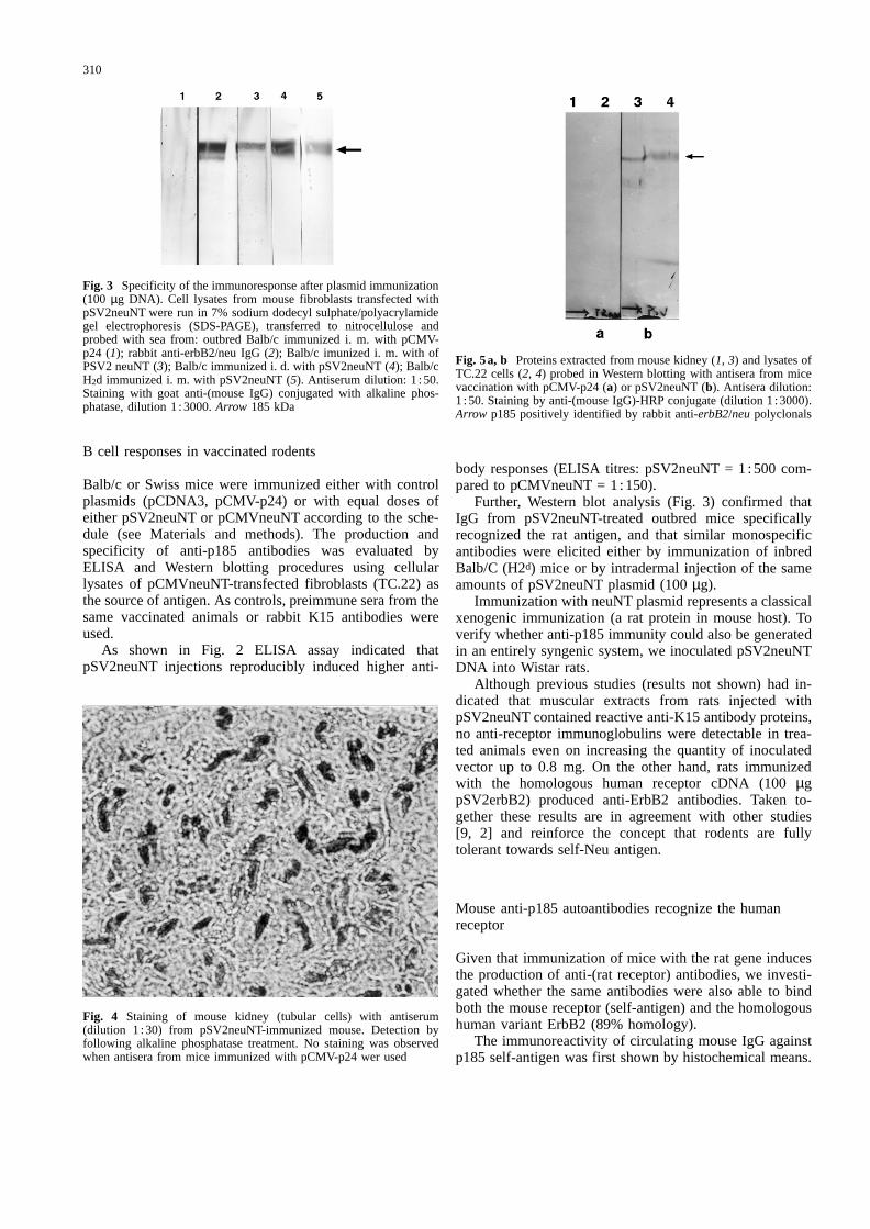
Fig. 4mStaining of mouse kidney (tubular cells) with antiserum(dilution 1 :30) from pSV2neuNT-immunized mouse. Detection byfollowing alkaline phosphatase treatment. No staining was observedwhen antisera from mice immunized with pCMV-p24 wer used
Fig. 3mSpecificity of the immunoresponse after plasmid immunization(100 µg DNA). Cell lysates from mouse fibroblasts transfected withpSV2neuNT were run in 7% sodium dodecyl sulphate/polyacrylamidegel electrophoresis (SDS-PAGE), transferred to nitrocellulose andprobed with sea from: outbred Balb/c immunized i. m. with pCMV-p24 (1); rabbit anti-erbB2/neu IgG (2); Balb/c imunized i. m. with ofPSV2 neuNT (3); Balb/c immunized i. d. with pSV2neuNT (4); Balb/cH2d immunized i. m. with pSV2neuNT (5). Antiserum dilution: 1 :50.Staining with goat anti-(mouse IgG) conjugated with alkaline phos-phatase, dilution 1 :3000.Arrow 185 kDa
Fig. 5a, bmProteins extracted from mouse kidney (1, 3) and lysates ofTC.22 cells (2, 4) probed in Western blotting with antisera from micevaccination with pCMV-p24 (a) or pSV2neuNT (b). Antisera dilution:1 :50. Staining by anti-(mouse IgG)-HRP conjugate (dilution 1 :3000).Arrow p185 positively identified by rabbit anti-erbB2/neupolyclonals

Both in the mouse and rat, theneu protooncogene isphysiologically expressed in epithelial/glandular tissuessuch as lung, liver, kidney and gut, and in the basal layerof the epidermis and placenta [23, 33]. In our experiments,we treated mouse kidney with pre-immune or immune serafrom rodents immunized with pSV2neuNT or irrelevantplasmids. The staining patterns (Fig. 4) clearly indicated aselective reactivity of IgG from immunized pSV2neuNTmice towards kidney tubular cells. No reactivity wasdetected for preimmune or pcDNA3 sera. The identity ofmurine p185 as the target antigen was verified by Westernblotting of kidney extracts (Fig. 5).
The possibility that mouse anti-p185 antibodies alsorecognized the human homologous antigen was evaluatedby immunofluorescence confocal microscopy of overex-pressing p185erbB2 SKBR3 cells. As shown in Fig. 6, thenon-permeabilized cells displayed a strong fluorecencedistributed in a diffuse way on the plasma membrane. Onthe other hand, the permeabilized ones presented a well-
defined fluorescence in the perinuclear region. Very similarpictures were obtained by Debrin et al. [11] by staining ofB104-1-1 cells (aneu-gene-transfected cell line) with anti-neu monoclonal Ab.
To confirm the reactivity of murine anti-p185 IgG withthe human receptor we performed Western blots with celllysates obtained from SKBR3, T28.1 (3T3 fibroblaststransfected with a retroviral vector forerbB2) or T19.7cells (NR6 fibroblasts transfected with a chimeric geneexpressing EGFR-ECD and ErbB2-ICD). The resultsshown in Fig. 7 demonstrate the presence of anti-p185antibodies in the sera of pSV2neuNT-immunized mice.
Since no binding of anti-p185 to overexpressingp170EGFR A431 cells was detected using FACS (resultsnot shown), it is likely that the crossreactivity ofanti-p185 antibodies observed with T19.7 cell lysate wasdue to the intracellular portion (ErbB2) of the chimericreceptor.
Staining of mammary tumors
The finding that anti-p185 IgG could interact with rat neuand human erbB2 oncoprotein suggested the possibility ofusing these antibodies to detect the overexpression ofErbB2/Neu in transgenic FVB/neu (strain 202) mice [15]and human breast cancer specimens. Figure 8 reports theimmunohistochemical staianing of the plasma membrane ofthe tumour cells. No staianing was observed when antiserafrom mice immunized with an irrelevant plasmid wereused.
In vitro effects of polyclonals on human cancer cells
In order to test the effects of polyclonals in vitro, we usedthe cell proliferation assay in culture and ADCC, onSKBR3 human breast cancer tumour cells, as described inMaterials and methods. The experiments were performed
311
Fig. 6a–cmConfocal laser microscopy showing the reactivity of anti-p185 antibodies against SKBR3 cells.a, b Cells fixed and permabil-ized with cold methanol/alcohol; orc fixed with 2% paraformaldehydewithout permeabilization.a pcDNA3 control serum;b andc antiserumfrom pSV2neuNT-vaccinated mice
Fig. 7a, bmProteins from lysates of SKBR3, T28.1 and T19.7 cells (1,2 and 4 respectively) and of untransformed fibroblasts (3) wereseparated by 7% SDS-PAGE and transferred to nitrocellulose. Thestrips were then blotted with antisera from mice immunized either withpCMV-p24 (a) or pSV2neuNT (b). Staining detection with alkaline-phosphatase conjugated goat anti-(mouse Ig) IgG

with whole sera or with samples of protein-A–Sepharose-fractionated IgG. As shown in Fig. 9 both growth inhibitionand antibody-dependent lysis of target cells were observedonly when the protein-A-retained fractions were used(IgG2a, IgG2b, IgG3).
Discussion
It is well known that individuals or animals should beimmunologically tolerant to the administration of ErbB2 orNeu self-receptor as whole proteins [9, 2]. The tolerancemechanisms could involve T or B cells, or both. It isgenerally held that T cell tolerance is much more profoundand sustained than that of B cells [4]. Since early studies[36] showed that foreign homologous molecules can beused to break tolerance towards both intracellular andextracellular antigens, it was expected that the toleranceof erbB2/neu B cells could also be broken by molecularmimicry.
For this purpose, we utilized a DNA plasmid encodingthe cDNA of rat neuNT, driven by the SV40 early promoter
312
Fig. 8a, bmDistribution of overexpressed p185 oncoprotein in humanmammary adenocarcinoma (a) and in mammary nodular carcinomafrom neu transgenic mice, strain 202 (b). Both samples were fromformalin-fixed tissues. Detection by antisera from pSV2neuNT-vacci-nated mice and peroxidase/aviding-biotin (a) or alkaline peroxidase (b)staining. (a) Arrowheadspoint to cell membranes
Fig. 9amSKBR3 cell growth in the presence of antisera from DNA-vaccinated mice or monoclonal antibody. SKBR3 tumour cells incu-bated with: 1 antiserum from mouse vaccinated with pcDNA3;2–5 equal amounts of antisera obtained from four different micevaccinated with pSV2neuNT;6–8 protein-A-depleted antibodiesfrom pSV2neuNT mice (2, 3, 5 respectively);9 SKBR3 tumour cellsincubated with W6-100 mAb.b Antibody-dependent cell cytotoxicityby protein-A-fractionated IgG from vaccinated mice and by anti-ErbB2mAb (see Materials and methods for details). Samples: mAb W6-100(0.5 µg/ml); IgG (from PSV2neuNT-immunized mice) eluted fromprotein A column (15µg/ml); IgG not retained in protein A column(50 µg/ml). E (Effector cells) mouse splenocytes;T (target cells)SKBR3 or A431 cancer cells (5×10³)

and enhancer (pSV2neuNT). Following immunization withthe plasmid, almost all animals (90%) responded to en-dogenously synthesized protein by producing antibodiesthat reacted in Western blotting against rat antigen. Incontrast, mice injected with null (pCDNA3) or with irrele-vant plasmid (pCMVp24) did not respond. The elicitationof such an immunoresponse was not limited to outbredmouse populations but, contrary to a previous report [2], itwas also effective in the Balb/c H2d strain.
Despite the reported fairly low efficency of intramus-cular naked DNA uptake [10],neugene transfer in musclesis adequate for the purpose of DNA-based immunization.Indeed, ELISA tests consistently showed antibody titres(1 :500) that are considered to confer a strong immunity inhumans. Comparable results were obtained in independentexperiments by using either lower amounts of PSV2neuNTplasmid (30 µg) or a different route of administration(intradermal injections). Interestingly, similar anti-Neutitres have been obtained by immunizing mice with ratprotein [9].
It is of some importance that the weaker pSV2 promoterdemonstrates, compared to the widely used CMV promoter,a higher efficency in the generation of anti-Neu antibodies.This result is not completely unexpected since such differ-ences have already been observed during DNA vaccinationagainst the rabies virus glycoprotein [38]. Moreover, plas-mid vectors expressing larger amounts of gene products donot necessarily induce a stronger immunoresponse to theencoded antigens [32].
We observed that anti-(rat p185) IgG were widely cross-reactive, being able to bind both mouse and human recep-tor. An antibody (autoantibody) response was detectedtowards whole mouse protein (immunohistochemistry ofkidney and Western analysis of kidney proteins), andtowards both the extracellular and the intracellular domainsof human ErbB2 (confocal microscopy of non-permeabi-lized SKBR3 and Western blotting of chimeric T19.7). Onthe other hand, polyclonals did not significantly react withEGFR, a member of the same family of growth factorreceptors, as judged by FACS scans of viable A431 cells.
These results suggest that cross-reactive antibodies eli-cited by DNA vaccination recognize epitopes that arehighly conserved in the course of evolution, and thatsome of them are of a sequential nature. In this respect itmay be of interest that, in a large panel of monoclonals sofar isolated from mice immunized with ErbB2-overexpres-sing human cells, all were conformational and only 1 out of28 crossreacted with the rodent receptor [P. G. Natali, IRE,Rome, and S. Menard, Istituto Nazionale Tumori (INT)Milan; personal communications].
Although anti-p185 polyclonals have not been tested yetfor their ability to restrict tumour growth in vivo (e. g.xenotransplantation of tumour cells in athymic mice), it isnoteworthy that the antibodies were able to inhibit theanchorage-dependent growth of human cancer SKBR3cells, and to mediate their lysis in ADCC assay. The findingthat inhibitory and ADCC-mediating antibodies were bothretained on a protein A matrix (then depleted of IgG1isotype) suggests that DNA vaccination induces an antibody
response (IgG2a, IgG2b and IgG3 isotypes) driven by Th1cells.
Accordingly, when stimulated with mitogen (concanav-alin A), we found that splenocytes from pSV2-neuNT-vaccinated mice displayed a threefold increase ininterferon γ production relative to that from lymphocytesof untreated mice (results not shown). Thus, like viral andbacterial sequences [5, 37], injections of an oncogeniccDNA may elicit cellular and humoral immune responsesthat have a T helper cell type 1 bias.
It is necessary to confirm the inhibitory action ofpolyclonals on other tumour cell lines expressing variouslevels of erbB2/neu protein. So far we have not analysedthe effects of polyclonals on receptor protein (i. e. turnover,phosphotyrosine contents). Polylconals may well containIgG populations that can activate the receptor; however,it has been reported that, while a single mAb may inhibitor accelerate tumour growth oferbB2-transformed fibro-blasts, the inhibitory effect is dominant in an antibodymixture [18].
It has been shown that some patients with breast canceroverexpressing the oncoprotein, have antibodies and T cellimmunity to the ErbB2 antigen [8, 31]. Although thecorrelation between anti-ErbB2 immunity and cancer prog-nosis is still unknown, preliminary results on patient anti-Erb2 IgG indicate that the humoral response does notinterfere with tumour progression (Dr. S. Pupa, INT, un-published results).
The finding that patients have cancer in the face of ameasurable humoral immunoresponse to the tumour antigenmakes it useful to compare human autoantibodies and anti-ErbB2 polyclonals obtained by plasmid immunization. Thereason for this is twofold: (a) experimentally inducedautoantibodies in animals models often resemble in speci-ficities those found in the sera of autoimmune patients [27];(b) DNA vaccination against mouse receptor may subvertneo-developmental processes. Indeed, our own early studies[35] showed that anti-neu DNA vaccination can induceresorption of embryos and a small number of offspringduring allogenic pregnancy.
Although these results are to be regarded as preliminary,we speculate that pregnancy loss may be (at least partially)related to anti-p185 antibodies targetting the receptor ex-pressed on placental trophoblasts [22]. The hypothesis issupported by the following observations: (i) antibodiesfrom vaccinated mice, eluted by acid treatments fromblood-free abortive placenta, were able to inhibit SKBR3cell growth; (ii) serum antibodies from the same animalswere able to stain trophoblast layers (manuscript in prep-aration); (iii) it has been reported [28] that administrationof EGFR antiserum decreases the number and size of theoffspring in mice.
Although anti-p185 polyclonals proved to mediate bothcytotoxicity and growth inhibition of tumour cells, as wellas to impair pregnancy in mice, we observed no autoim-mune toxicity in any treataed animals during 1 year offollow-up.
Specifically, haematoxylin/eosin-stained histologicalsections (kidney, mammary lactating gland, intestine, lung
313

etc.) from vaccinated mice were indistinguishable fromthose obtained from untreated mice. Furthermore, coupledwith the absence of inflammation or injury, we found noincrease in the amounts of circulating anti-DNA IgG, IgMantibodies.
It is now thought that boosting autoimmunity (co-acti-vation of CD4+ Th1 and CD8+ cytotoxic cells) against self/mutated self/tumour antigens should be a prerequisite forsuccessful immunotherapeutic intervention in cancer [29].
In our model the induction of autoreactive B cells in theabsence of T cells specific for the self-antigen is unlikely toactivate autoimmune spirals [20]. Thus, rat cDNA in miceelicited autoreactive B cells by triggering T cells specificfor rat epitopes. It should be stressed, however, that provi-sion of a T cell bypass may result in the generation ofautoreactiveerbB2/neu T cells, since such cells are nottotally deleted or anergized in rodents and humans [8, 9].As a consequence, in the presence of self-protein (sheddedduring tumour growth or administered for co-immuniza-tion), autoreactive B cells may present cryptic determinantsto autoreactive CD4+ lymphocytes [26]. Moreover, suchepitopes could be displayed to “ignorant” T cells followingeither binding of (self)antigen/antibody complexes to anti-gen-presenting cells [25] or alterations of self-antigenprocessing induced by cytokines released from Th1 cellsresponding to a foreign homologous antigen [14].
DNA-based vaccines offer several advantages over othersystems: (i) plasmid DNA is not an infective agent and it isapparently not integrated into the host genome; (ii) it lacksthe anamnestic responses observed with recombinantviruses; (iii) there is no need of adjuvant to obtain a strong,long-lasting immune response; (iv) plasmids are rapidlyproduced and purified and the sequence of the codedantigen easily controlled. Further, DNA-mediated immuni-zation, inducing the host cell to manufacture potentially allthe epitopes of a given antigen, could by-pass the hosthaplotype limitation.
The generation of an immunoresponse against the wholeerbB2/neureceptor in mice reported here, strongly supportsthe possibility that a xenogenic immunization (i. e. by amurine gene) would be capable of eliciting similar safeimmunoresponses in outbred human populations.
Whether DNA vaccination against theerbB2/neuonco-protein can offer any therapeutic benefit can only beanswered by clinical trials.
AcknowledgementsmWe thank P. G. Natali and O. Segatto formonoclonal antibodies and transformed cell lines and N. Ranzani forthe generous gift of neuNT plasmid. We also thank P. L. Lollini(Istituto di Cancerologia, Universita` di Bologna), P. Musiani (Anato-mia Patologica Universita` di Chieti) and M. Cardarelli (Servizio diAnatomia Patologica, Ospedale di Macerata) for specimens of trans-genic and human breast cancer. We are grateful to P. G. Natali,O. Segatto, S. Menard and A. Lanzavecchia (Basel Institute for Im-munology) for critical reading of the manuscript.
References
1. Bargmann CI, Hung MC, Weinberg RA (1986) Multiple indepen-dent activation of theneu oncogene by a point mutation alteringthe transmembrane domain of p185. Cell 45: 649
2. Bernards R, Destree A, McKenzie S, Gordon E, Weinberg RA,Panicali D (1987) Effective tumor immunotherapy directed againstan oncogene-encoded product using a vaccina virus vector. ProcNatl Acad Sci USA 84: 6854
3. Cheever MA, Disis ML, Bernhard H, Gralow JR, Hand SL,Huseby ES, Qin HL, Takahashi M, Chen W (1995) Immunity tooncogenic proteins. Immunol Rev 145: 33
4. Chiller J, Habicht G, Weigle WO (1971) Kinetic differences inunresponsiveness of thymus and bone marrow cells. Science171: 813
5. Davis HL, Whalen RG (1995) DNA-based immunization: In:Dixon G (ed) Molecular and cell biology of human gene ther-apeutics. Chapman & Hall, London, p 368
6. De Santes K, Slamon D, Anderson SK, Shepard M, Fendly B,Maneval D, Press O (1992) Radiolabeled antibody targetting of theHER-2/neu oncoprotein. Cancer Res 52: 1916
7. Di Fiore PP, Pierce JH, Kraus MH, Segatto O, King CR, Aaron-son SA (1987)erbB-2is a potent oncogene when overexpressed inNIH/3T3 cells. Science 237: 178
8. Disis ML, Calenoff E, Mc Lauglhin G, Murphy AE, Chen W,Groner B, Jeschke M, Lydon N, McGlynn E, Livingstone RB,Moe R, Cheever MA (1994) Existent T-cell and antibody immu-nity to HER-2/neu protein in patients with breast cancer. CancerRes 16: 54
9. Disis ML, Gralow JR, Bernhard H, Hand SL, Rubin WD,Cheever MA (1996) Peptide-based, but not whole protein, vac-cines elicit immunity to HER-2/neu, an oncogenic self-protein.J Immunol 156: 3151
10. Donnelly JJ, Ulmer JB, Liu MA (1994) Immunization withpolynucleotides. A novel approach to vaccination. Immunologist2/1: 20
11. Drebin JA, Stern DF, Link VC, Weinberg RA, Greene MI (1984)Monoclonal antibodies identify a cell-surface antigen associatedwith an activated cellular oncogene. Nature 312: 545
12. Drebin JA, Link VC, Stern DF, Weiberg RA, Greene MI (1985)Downregulation of an oncogene protein product and reversion ofthe transformed phenotype by monoclonal antibodies. Cell 41: 695
13. Drebin JA, Link VC, Weiberg RA, Greene MI (1986) Inhibition oftumor growth by a monoclonal antibody with an oncogeneencoded tumor antigen. Proc Natl Acad Sci USA 83: 9129
14. Elson JC, Barker RN, Thompson SJ, Williams NA (1995) Im-munologically ignorant autoreactive T cells, epitope spreading andrepertoire limitation. Immunol Today 16: 71
15. Guy CT, Webster MA, Schaller M, Parson TJ, Cardiff RD,Muller WJ (1992) Expression of theneu protooncogene in themammary epithelium of transgenic mice induces metastatic dis-ease. Proc Natl Acad Sci USA 89: 10578
16. Hancock MC, Langton BC, Chan T, Toy P, Monahan JJ,Mischak RP, Shawver LK (1991) A monoclonal antibody againstthe c-erbB2 protein enhances the cytotoxicity ofcis-diamminedichloroplatinum against human breast and ovarian tumor celllines. Cancer Res 51: 4575
17. Harlow E, Lane D (1988) Antibodies, a laboratory manual. HarlowE, Lane D, Cold Spring Harbor Laboratory, p 479
18. Hurwitz E, Stankovski I, Sela M, Yarden Y (1995) Suppressionand promotion of tumor growth by monoclonal antibodies toErbB-2 differentially correlate with cellular uptake. Proc NatlAcad Sci USA 92: 3353
19. Hynes NE, Stern DF (1994) The biology of erbB2/neu/HER-2 andits role in cancer. Biochim Biophys Acta 1198: 165
20. Jevnikar AM, Grusby MJ, Glimcher LH (1994) Prevention ofnephritis in major histocompatibility complex class II deficientMRL-lpr mice. J Exp Med 179: 1137
314

21. Katsumata M, Okudaira T, Samanta A, Clark DP, Debrin JA,Jolicoeur P, Greene MI (1995) Prevention of breast tumor devel-opment in vivo by downregulation of the p185neu receptor. NatMed 1: 644
22. Knezevic V, Spaventi R, Poljak L, Slade N, Svajger A, Pavelic K(1994) p185 neu is expressed in yolk sac during rat postimplanta-tion development. J Anat 181: 1985
23. Kokay Y, Cohen JA, Debrin JA, Greene MI (1987) Stage andtissue-specific expression of the neu oncogene in rat development.Proc Natl Acad Sci USA 84: 8498
24. Lacroix H, Iglehart JD, Skinner MA, Kraus MH (1989) Over-expression of erbB-2 or EGF receptor proteins present in earlystage mammary carcinoma is detected simultaneously in matchedprimary tumors and regional metastases. Oncogene 4: 145
25. Lanzavecchia A (1995) How can cryptic epitopes trigger auto-immunity? J Exp Med 181: 1945
26. Lin RH, Mamula MJ, Hardin JA, Janeway CA (1991) Induction ofautoreactive B cells allows priming of autoreactive T cells. J ExpMed 173: 1433
27. Mamula MJ (1995) Lupus autoimmunity: from peptides to par-ticles. Immunol Rev 144: 301
28. Miettinen PJ, Berger JE, Meneses J, Phung Y, Pedersen RA,Werb Z, Derynck R (1995) Epithelial immaturity and multiorganfailure in mice lacking epidermal growth factor receptor. Nature376: 337
29. Nanda NK, Sercaz EE (1995) Induction of anti-selfimmunity tocure cancer. Cell 82: 13
30. Park JW, Stagg R, Lewis GD, Carter P, Maneval D, Slamon DJ,Jaffe H, Shepard HM (1992) In: Dickson RB, Lippmann ME (eds)Genes, oncogenes and hormones: advances in cellular and mole-cular biology of breast cancer. Kluver, Boston, p 193
31. Pupa SM, Menard S, Andreola S, et al. (1993) Antibody responseagainst the c-erbB2 oncoprotein in breast carcinoma patients.Cancer Res 53: 5864
32. Sato Y, Roman M, Tighe H, Lee D, Corr M, Nguyen M-D,Silverman, GJ, Lotz M, Carson DA, Raz E (1996) Immunostimo-latory DNA sequences necessary for effective intradermal geneimmunization. Science 273: 352
33. Slamon DJ, Godolphin W, Jones LA, Holt A, Wong SG, Keith DE,Levin WJ, Stuart SG, Udove J, Ullrich A, Press MF (1989) Studiesof the HER-2/neu protooncogene in human breast and ovarycancer. Science 244: 707
34. Talbott RL, Spranger EE, Lovelance KM, et al (1989) Nucleotidesequence and genomic organisation of feline immunodeficiencyvirus. Proc Natl Acad Sci, USA 86: 5743
35. Venanzi FM, Petrelli C, Concetti A, Amici A (1995) neu/HER-2cDNA vaccination and pregnancy loss. DNA vaccines: a new erain vaccinology. Ann Acad Sci [New York]. Liu MA, HillemanMR, Kurt R (eds), vol. 772, p 274
36. Weigle WO (1962) Termination of acquired immunological toler-ance to protein antigens following immunization with alteredprotein antigens. J Exp Med 116: 913
37. Whalen RG, Leclerc C, Deriaud E, Schirmbeck R, Reimann J,Davis H (1995) DNA-mediated immunization to the hepatitis Bsurface antigen: activation and entrainment of the immune re-sponse. DNA vaccines: a new era in vaccinology. Ann Acad Sci[New York]. Liu MA, Hilleman MR, Kurt R (eds), vol. 772: p 64
38. Xiang ZQ, Spitalnik S, Tram M, Wunner WH, Cheng J, Ertl HC(1994) Virology 199: 132
315