Evaluation of T7 and lambda phage display systems for survey of autoantibody profiles in cancer...
15
This article appeared in a journal published by Elsevier. The attached copy is furnished to the author for internal non-commercial research and education use, including for instruction at the authors institution and sharing with colleagues. Other uses, including reproduction and distribution, or selling or licensing copies, or posting to personal, institutional or third party websites are prohibited. In most cases authors are permitted to post their version of the article (e.g. in Word or Tex form) to their personal website or institutional repository. Authors requiring further information regarding Elsevier’s archiving and manuscript policies are encouraged to visit: http://www.elsevier.com/copyright
-
Upload
independent -
Category
Documents
-
view
0 -
download
0
Transcript of Evaluation of T7 and lambda phage display systems for survey of autoantibody profiles in cancer...

This article appeared in a journal published by Elsevier. The attachedcopy is furnished to the author for internal non-commercial researchand education use, including for instruction at the authors institution
and sharing with colleagues.
Other uses, including reproduction and distribution, or selling orlicensing copies, or posting to personal, institutional or third party
websites are prohibited.
In most cases authors are permitted to post their version of thearticle (e.g. in Word or Tex form) to their personal website orinstitutional repository. Authors requiring further information
regarding Elsevier’s archiving and manuscript policies areencouraged to visit:
http://www.elsevier.com/copyright

Author's personal copy
Research paper
Evaluation of T7 and lambda phage display systems for survey ofautoantibody profiles in cancer patients
Zane Kalnina a, Karīna Siliņa a, Irēna Meistere a, Pawel Zayakin a, Alexander Rivosh a,Artūrs Ābols a, Mārcis Leja b, Olga Minenkova c, Dirk Schadendorf d, Aija Linē a,⁎
a Biomedical Research and Study Centre of Latvia, Riga, Latviab Faculty of Medicine, University of Latvia, Riga, Latvia
c Kenton Labs, c/o Sigma-Tau, Pomezia, Italyd German Cancer Research Center, Skin Cancer Unit, Heidelberg, Germany
Received 3 January 2008; received in revised form 28 January 2008; accepted 29 January 2008Available online 21 February 2008
Abstract
In the current studywe attempted to evaluate the suitability of T7 Select 10-3b andλKM8 phage display systems for the identification ofantigens eliciting B cell responses in cancer patients and the production of phage-displayed antigen microarrays that could be exploited forthe monitoring of autoantibody profiles. Members of 15 tumour-associated antigen (TAA) families were cloned into both phage displayvectors and the TAA mini-libraries were immunoscreened with 22 melanoma patients' sera resulting in the detection of reactivity againstmembers of 5 antigen families in both systems, yet with variable sensitivity. T7 phage display system showed greater sensitivity for thedetection of antibodies against members of CTAG,MAGEA andGAGE families, both systems showed equal performance in detecting thereactivity againstMAGECandSSX2while onlyλKM8allowed the detection of anti-CTAGE5 antibodies. The biological properties of bothphages turned out to be equally suitable for the production of antigenmicroarrays however in linewith the plaque assay the sensitivity for thedetection of various autoantibodies differed between the vectors. However, presumably due to the higher variability of the backgroundsignals in the microarray assay, it turned out to have comparable, in some cases even slightly lower sensitivity than the plaque assay.
Next, we explored the repertoire of antigens that could be identified by screening T7 phage-displayed testis cDNA library withsera from melanoma patients. From the 243 antigens identified, only 24 represented known genes translated in their natural readingframe and included known TAAs like Annexin XI-A and a novel potential CT antigen SPAG8. Another 12 were uncharacterisedgenes but the remaining clones contained DNA fragments in non-natural reading frames that most likely represent mimotopes,nevertheless, they may turn out to be valid biomarkers.© 2008 Elsevier B.V. All rights reserved.
Keywords: Autoantibodies; Phage display; Antigen microarray; SEREX; Melanoma antigens
1. Introduction
Circulating autoantibodies against tumour-derived pro-teins have been observed in themost if not all cancer patientshence they may serve as biomarkers for the screening,diagnosis, prognosis or monitoring of cancer. In fact,autoantibodies against a number of CT antigens, including
Journal of Immunological Methods 334 (2008) 37–50www.elsevier.com/locate/jim
Abbreviations: SEREX, serological identification of antigens byrecombinant expression cloning; TAA, tumour-associated antigens; pfu,plaque forming units; CT, cancer-testis; UTR, untranslated region, ORF,open reading frame.⁎ Corresponding author. Biomedical Research and Study Centre of
Latvia, Ratsupites Str 1, LV-1067, Riga, Latvia. Tel.: +371 7808208,fax: +371 7442407.
E-mail address: [email protected] (A. Linē).
0022-1759/$ - see front matter © 2008 Elsevier B.V. All rights reserved.doi:10.1016/j.jim.2008.01.022

Author's personal copy
NY-ESO-1, SSX2, MAGEA1 and 3 (Stockert et al., 1998)and several members of CTAGE family (Usener et al.,2003), and melanocyte differentiation antigens such asRAB38 (Zippelius et al., 2007) have been detectedexclusively in the sera from cancer patients, thus suggestingthey may serve as highly specific diagnostic markers.Autoantibodies against another set of antigens such asGLEA2, PHF3 and endostatin have been shown to correlatewith the clinical outcome in glioblastoma and breast cancerpatients, respectively (Pallasch et al., 2005; Bachelot et al.,2006). Moreover, appearance of anti-p53 autoantibodiesmay predict subsequent development of cancer (Li et al.,2005), while their prognostic significance still remains asubject of debate (Soussi, 2000). However, so far the clinicalutility of tumour-associated autoantibodies has beenhampered by the low frequencies to each particular antigenand the heterogeneity of antibody repertoires in cancerpatients. This could be overcome by developing high-throughput techniques that would allow to identify acomprehensive set of immunogenic proteins in a giventype of cancer and subsequently analyse the occurrence ofantibodies against them in large sets of sera from patientswith cancer, autoimmune disorders and healthy donors.
SEREX (serological identification of tumour antigensby recombinant expression cloning) is a widely usedtechnique for the identification of immunogenic proteins incancer patients that is based on the construction of cDNAexpression libraries from tumour tissues, expression of therecombinant proteins in E. coli and immunoscreening ofthe libraries with sera from cancer patients (Sahin et al.,1995). The application of this technique to a variety oftumour entities has led to the identification of more than2000 genes encoding potential tumour antigens, most ofwhich are deposited in the Cancer Immunome database(http://www2.licr.org/CancerImmunomeDB). However,this approach is extremely time-consuming and labour-intensive and therefore is generally not applicable for theprofiling autoantibody responses in multiple serumsamples. Recently, several laboratories have exploitedvarious phage display strategies, including M13 filamen-tous phage (Sioud and Hansen, 2001; Somers et al., 2002),pJuFo system (Fossa et al., 2004), T7 phage (Hansen et al.,2001; Zhong et al., 2004) and lambda phage (Minenkovaet al., 2003; Pavoni et al., 2004) for the construction ofcDNA expression libraries and successfully applied themfor the isolation of cDNAs encoding tumour-associatedantigens. A feature that makes the phage display-basedstrategies particularly attractive for the searching ofantigens is that tumour-derived cDNAs are expressed asfusion proteins with one of the phage coat proteins (orlinked via a Jun–Fos interaction in case of pJuFo system)and exposed on the surface of the phage thus allowing the
selection of serum-reactive phage clones by biopanning,which is much faster as well as more cost and labour-effective approach than the conventional immunoscreen-ing.Moreover, the recombinant phage particles can directlybe printed onto glass slides to produce antigen microarraysthat allow monitoring the antibody responses againsthundreds to thousands of antigens simultaneously using amicrolitre of serum (Fernandez-Madrid et al., 1999;Cekaite et al., 2004; Zhong et al., 2005; Wang et al.,2005; Chatterjee et al., 2006). However, themain drawbackof the phage display-based systems for the expression ofcDNA libraries is the bias in the repertoire of proteins thatcan be displayed on the surface of the phage resulting fromthe biological peculiarities of a given phage species. Forexample, the assembly of M13 phage takes place in theperiplasm of E. coli, therefore only those fusion proteinsthat can be exported through the bacterial inner membranewill be displayed on the phage capsid (Hufton et al., 1999).Capsids of lytic phages such as T7 and lambda phages areassembled in the cytoplasm therefore the repertoires ofcDNA libraries do not have the biological constraintssimilar toM13 phage (Krumpe et al., 2006). The capabilityof a protein to be displayed on the surface of a lytic phagedepends mostly on its impact on the assembly process,where the length, biochemical properties, hydrophilicityand hydrophobicity as well as folding characteristics playthe major role. Moreover, the copy number of recombinantproteins per phage particle may vary in these systemsallowing the assembly of chimerical capsids, thus affectingthe sensitivity of the assay.
In the current study we assessed the capability of T7Select 10-3b and λKM8 phages to display a range ofclinically relevant tumour antigens by cloning themembers of 15 TAA families into these display vectors,compared the sensitivity of the systems for the detectionof autoantibodies by immunoscreening of the TAA mini-libraries with sera from 22 melanoma patients, andevaluated the suitability of these phages for the productionof antigen microarrays. Moreover, in order to characterisethe repertoire ofmelanoma associated antigens that can beidentified using T7 Select 10-3b system, we constructedT7 phage-displayed testis cDNA library and screened itwith sera from 9 melanoma patients resulting in theidentification of 243 different sero-reactive phage clones.
2. Materials and methods
2.1. Serum samples
Serum samples from melanoma patients (stage II–IV)were collected at the Skin Cancer Unit, German CancerResearch Center and Latvian Oncology Center after the
38 Z. Kalnina et al. / Journal of Immunological Methods 334 (2008) 37–50

Author's personal copy
patients' informed consent was obtained in accordancewith the regulations of local ethics committees. Thesamples were aliquoted and stored at −70 °C.
2.2. Construction of phage-displayed cDNA expressionlibraries
2.2.1. T7 and lambda phage-displayed tumour-associated antigen (TAA) mini-libraries
Either the entire ORFs or the antigenic regionspredicted using an algorithm developed byWelling et al.(1985) of 15 tumour antigens or antigen families wereamplified by PCR using High Fidelity PCR enzyme mix(Fermentas) and cDNA from testis or melanoma tissueas a template. The antigens and the regions chosen forcloning are listed in Table 1, and the sequences of PCRprimers are available on request. All reverse primerscontained NotI site and forward primers contained eitherSpeI or SalI sites for cloning into λKM8 or T7 Select10-3b vectors, respectively. For the amplification ofgenes from multigene families degenerated primerswere designed to amplify as many as possible membersof the same gene family. If more than one region of agene was amplified, PCR products were combined priorto ligation in the vector. PCR products were digestedwith NotI and SpeI or SalI (Fermentas) and purifiedusing GFX PCR DNA and Gel Band Purification kit(Amersham Biosciences). λKM8 (Pavoni et al., 2004)vector DNA was digested with SpeI and NotI but T7
Select 10-3b vector (Novagen) was digested with SalIand NotI, treated with shrimp alkaline phosphatase(Fermentas) followed by phenol/chloroform extractionand ethanol precipitation. Each digested PCR product(5 ng) was ligated into λKM8 or T7 Select 10-3b vector(100 ng), and 1/5 of each ligation mixture was subjectedto in vitro packaging using 5 µl of Gigapack III GoldPackaging extract (Stratagene) or T7 Select Packagingextract (Novagen), respectively, resulting in mini-libraries of 1–5×10⁵ pfu for lambda phage and 0.5–5×10⁴ pfu for T7 phage. The obtained TAA mini-libraries were amplified once using E. coli BB4 cells orIPTG-induced BLT5615 cells, respectively. The lysateswere centrifuged to remove cell debris and stored at+4 °C or as glycerol stocks at −80 °C.
2.2.2. T7 phage-displayed testis cDNA expressionlibrary
Testis cDNA library was constructed using T7 Select10-3b vector and OrientExpress cDNA library construc-tion system (Novagen) according to the manufacturer'sinstructions. Briefly, mRNAwas isolated from 150 µg oftestis total RNA (Ambion) using Dynabeads mRNApurification kit (Invitrogen) and converted to cDNAusing HindIII Random primers (5′-TTNNNNNN-3′).Then cDNAwas ligated to directional EcoRI and HindIIIlinkers, digested with the corresponding restrictionenzymes and ligated into pre-digested T7 Select 10-3bvector followed by in vitro packaging resulting in a
Table 1Antigens comprising TAA mini-libraries
Antigen family Number ofmRNAs a
Regions cloned(nt positions/NCBI RefSeq No)
Different genes found by sequencing5 random clones
T7 Select 10-3B λKM8
CTAG1B (NY-ESO) 2 86–400; 338–628/NM_001327 CTAG1B, CTAG2 CTAG1B, CTAG2MAGEA 10 209–618; 590–1137/NM_005362 MAGEA1, 2, 3, 4, 9 MAGEA1, 3, 4, 6SSX 10 58–401; 58–617/NM_005636 SSX2 SSX1, 2BAGE 1 195–330/NM_001187 BAGE BAGEGAGE 8 84–410/NM_001472 GAGE2, 7, 8 GAGE1, 8MAGEB 4 98–516/NM_002364 MAGEB2,4 MAGEB2MAGEC 2 931–1453/NM_016249 MAGEC1, 2 MAGEC1, 2SPANX 6 57–347/NM_022661 SPANXA2, SPANXE SPANXDLDHC 1 245–785/NM_017448 LDHC LDHCCT45 4 246–798/NM_001017417 CT45-1 CT45-1THEG 1 778–1176/NM_016585 THEG THEGCTAGE 6 130–1070; 311–1070; 1052–1616;
1589–2354/203354; 11–235/NM_022663CTAGE1, 3, 5, 5Δex7 CTAGE1, 5
MTA1 1 1394–1772/NM_004689 MTA1 MTA1TYR 1 494–710/NM_000372 TYR TYRMLANA 1 54–408/NM_005511 MLANA MLANA
a Number of known members of the antigen family that theoretically could be amplified with the selected primers.
39Z. Kalnina et al. / Journal of Immunological Methods 334 (2008) 37–50

Author's personal copy
library of 8×10⁶ pfu. The library was amplified once inIPTG-induced BLT5615 cells.
2.3. Selection of serum-reactive phage clones
2.3.1. Immunoscreening of phage-displayed TAA mini-libraries
For screening of T7 phage-displayed TAA mini-libraries, ~103 pfu from each library was spotted ongridded LB/carbenicillin agar plates pre-coated with LBtop agarose containing BLT5615 cells grown in LBsupplemented with 1×M9 salts, 0.4% glucose, 1 mMMgSO4 and carbenicillin (50 µg/ml) to OD600=0.5 andinduced with IPTG for 30 min. After ~2 h incubation at37 °Cwhen the plaques reached ~1mm in diameter, plateswere overlaid with Protan nitrocellulose (NC) filters(Schleicher & Schuell) and incubated for 1 h at 37 °C.For screening of lambda phage-displayed TAA mini-libraries, BB4 cells grown in LB supplemented with 0.2%maltose and 10 mM MgSO4 were plated on NZY agarplates, infected with ~103 pfu from each mini-library andthe plates were incubated at 37 °C until visible plaquesappeared (6–8 h). The filters were blocked with 5% (w/vol) milk powder in TBS, 0.05% Tween 20 for 1 h, andthen incubated overnight with 1/200 diluted patients'serum preabsorbed with E.coli-phage lysate. The serum-reactive clones were detected by incubating the filters withalkaline phosphatase conjugated anti-human IgG, Fcγspecific secondary antibody (Pierce) and NBT/BCIP(Fermentas). Ten serum-reactive clones from each mini-library were subjected to secondary screenings andsubcloned to monoclonality.
2.3.2. Biopanning of T7 phage-displayed testis cDNAlibrary
Approximately 5×10¹⁰ pfu from T7 phage-displayedtestis cDNA library were incubated overnight with 2 µl ofpatient's serum that had been preabsorbed with BLT5615and T7 phage lysate coupled with BrCN-activated sephar-ose. A hundred microlitre of Protein G coated magneticbeads (Pierce) were washed twice with blocking solution(5% milk powder in TBS, 0.05% Tween 20), added to thephage–serum mixture and incubated for 2 h at RT underagitation. The beads were washed 10 times with 1 ml TBS,0.05% Tween and the bound phages were either amplifiedand subjected to the second round of biopanning or titratedand used for immunoscreening as described above.
2.3.3. Identification of cDNAsThe inserts of serum-reactive phages were amplified
by 35-cycle PCRusing primers flanking the insert (T7-Up2:5′-CTTCGCCCAGAAGCTGCA-3′, T7-Down: 5′-
AACCCCTCAAGACCCGTTTA-3′, λKM8-Up: 5′-CAATCTGTGTGGGCACTCG-3′, λKM8-Down: 5′-CGGCTGGTAATGGGTAAAGG-3′) and 1 µl of phagesolution as a template. PCR products were purified anddirectly sequenced using ABI Prism BigDye Terminatorv3.1 cycle sequencing kit and 3130 Genetic Analyser(Applied Biosystems). DNA sequences were analysedusing BLAST tool at www.ncbi.nlm.nih.gov, Translatetool at www.expasy.org and compared against sequencesavailable at Cancer Immunome Database (www2.licr.org/CancerImmunomeDB).
2.4. Production of GST fusion proteins and Westernblot analysis
To confirm the recognition of the phage-displayedproteins, GST fusion proteins were produced and usedfor standard Western blot analysis. cDNA inserts ofserum-reactive clones were amplified by PCR andcloned into a prokaryotic GST expression vector pGEX-4T-1 (Amersham Biosciences) to produce either thenatural products of these genes or the out-of-framepeptides displayed by the serum-reactive clones.
Purified recombinant proteins (500 ng) were resus-pended in Laemmli sample buffer, denatured for 5 min at100 °C and separated by SDS/polyacrilamide gelelectrophoresis (PAGE) on two gels simultaneouslyand transferred onto Protan NC membranes (Schleicher& Schuell). The filters were blocked for 1 h in 5% milkpowder in TBS, 0.05% Tween 20 and incubated with 1/20000 diluted HRP conjugated goat anti-GST antibody(Amersham Biosciences) or 1/200 diluted patient's serafollowed by incubation with HRP conjugated goat anti-human IgG antibody 1/10000 (Sigma) and detectedusing ECL Plus Western Blotting Detection Reagents(Amersham Biosciences).
To determine the copy number of fusion proteins per T7phage particle, phages were amplified in E. coli BLT5615cells, the phage particles were precipitated from the lysateswith 3.5% PEG 8000, 0.4MNaCl, titrated and ~109 pfu ofeach phage were used for Western blot analysis with 1/5000 diluted rabbit HRP conjugated anti-T7 tag antibody(ABcam) recognising the N-terminus of T7 coat protein10B.To ascertain that unincorporated recombinant proteinsare not co-precipitated with the phage particles, NaCl/PEGprecipitated phage solution containing ~1010 pfu wasfiltrated through 100000 NMWL filter units (Millipore)and the flow-through fraction was run in parallel with thephage samples as a negative control. For quantitycalibration a standard curve was constructed from 5 serial3-fold dilutions of one phage clone. The images wereanalysed usingGelWorks software (Ultra-Violet Products).
40 Z. Kalnina et al. / Journal of Immunological Methods 334 (2008) 37–50

Author's personal copy
2.5. Production of antigen microarrays
Nine corresponding T7 and lambda phage clonesisolated by immunoscreening from TAA mini-librariesand four non-recombinant phage clones were grown inBLT5615 and BB4 cells, respectively, until completelysis. The lysates were clarified by centrifugation andarrayed in 5 replicates onto nitrocellulose-coated 16-padFAST slides (Whatman) using a QArray Mini micro-arrayer (Genetix) to generate T7 and lambda phage-displayed antigen microarrays. The microarrays wereblocked in 5% (w/vol) milk powder in TBS, 0.05%Tween 20 for 1 h, and then incubated with 1/200 dilutedpatients' sera (preabsorbed with E.coli-phage lysates)for 2 h at room temperature. The slides were rinsed withTBS and washed 4 times in TBS, 0.5% Tween 20 for15 min each and then incubated with anti-T7 tail fiber(Novagen) or anti-gpV (λ tail protein) monoclonalantibody (kindly provided by Dr. Mauruzio Cianfriglia)at a dilution of 1/10000 or 1/1500, respectively todetermine the amount of phages in each spot. After 3washes in TBS, 0.5% Tween 20 for 10 min, themicroarrays were incubated with Cy5 labelled goat anti-human IgG antibody (1/1500) and Cy3 labelled goatanti-mouse IgG antibody (1/3000) (Jackson ImmunoR-esearch) for 1 h, then washed thrice in TBS, 0.5% Tween20, rinsed with distilled water and dried by centrifuga-tion. The microarrays were read using AQuire scanner
(Genetix) and the images were analysed using GenetixQScan software. For each spot the mean Cy5 and Cy3signals were background subtracted, averaged betweenreplicates, and the Cy5/Cy3 ratios were calculated foreach antigen and normalised by that of non-recombinantphages. A cut-off value for defining serum-reactiveantigens was set as N3 SDs above the mean ratio for allthe spots.
3. Results
3.1. Construction of lambda and T7 phage-displayedTAA mini-libraries
Members of 12 CTantigen families and 3 other tumourantigens were cloned into lambda KM8 and T7 Select 10-3b phage display vectors to produce TAAmini-libraries. InλKM8 vector cDNAs are fused to the N-terminus of thecoat protein gpD and are separated by a flexible GS linkerwhile in T7 phage they are fused to the C-terminus of thecoat protein 10B. The antigen families and individual genesas well as the protein regions chosen for cloning are listedin Table 1. Complete ORFs were amplified for shortertranscripts, while for genes with long ORFs the potentialantigenic regions were predicted using an algorithmdeveloped byWelling et al. (1985) and chosen for cloning.In the case of tyrosinase the immunodominant region thathas been shown to react with sera from melanoma patients
Table 2Antigens identified by immunoscreening lambda and T7 phage-displayed TAA mini-libraries and individual sera recognising the respective antigens
TAA mini-library Antigen Reactive sera a
T7 Select 10-3B λKM8
CTAG CTAG1B MA001643, MA000703, MA000161, MA000445,MA00SK, MA00AM, MA00550, MA000513
MA001643, MA000703,MA000161, MA000445
CTAG2 MA001643, MA000703, MA000161, MA000445,MA00SK, MA00AM, MA00550
MA001643, MA000703,MA000445
CTAG1B-ORF2 UKRV-Mel31 –MAGEA MAGEA1 MA000513 MA000513
MAGEA2 MA000513 MA000513MAGEA3 MA000513 MA000513MAGEA12 MA000513 –MAGEA2 antisense MA00WF –
MAGEC MAGEC1 MA000703 MA000703MAGEC2 MA000703 –
SSX SSX2 MA000951 MA000951CTAGE CTAGE5 MA000273, MA000525
CTAGE pseudogene MA000525, MA000273, MA001816, MA001111 –CTAGE1 antisense MA001404 –
GAGE GAGE3-7 subgroup MA000445, MA001111, MA001404 MA000445GAGE1, 8 MA000445 MA000445GAGE2-ORF2 MA00GG –GAGE7-ORF2 MA001643 –
a Sera are ranked by the signal intensities in immunoscreening.
41Z. Kalnina et al. / Journal of Immunological Methods 334 (2008) 37–50

Author's personal copy
but not with sera from healthy donors (Lucchese et al.,2005) was chosen. For the amplification of genes frommultigene families, degenerated PCR primers capable ofamplifying as many members of these families as possiblewere designed. The cloning of the respective PCR productsinto λKM8 and T7 Select 10-3b vectors resulted in TAAmini-libraries in the size of 1–5×10⁵ pfu and 0.5–5×10⁴ pfu, respectively. The complexity of the mini-libraries and the insertion of cDNAs in the correct readingframe relative to the phage coat proteins were determinedby sequencing 5 random clones from each mini-library(Table 1).
3.2. Comparison of serum reactivity against TAAdisplayedon lambda and T7 phages
Approximately 2×10³ pfu from each of the 15lambdas and T7 phage-displayed TAA mini-librarieswere immunoscreened with sera from 22 melanomapatients. Ten sero-reactive phage clones from each sero-reactive TAA mini-library were purified and identifiedby sequencing their cDNA inserts. Generally, relativelygood concordance in the recognition of antigensdisplayed on lambda and T7 phages was observed —the reactivity against antigens representing CTAG (NY-ESO-1), MAGEA, MAGEC, SSX and GAGE familieswas detected in both expression systems (yet withdifferent sensitivity) whereas no reactivity againstBAGE, MAGEB, SPANX, LDHC, CT45, THEG,MTA1, TYR and MLANA was observed in any ofthem (Table 2).
In both systems, phage capsids are composed of wild-type capsid proteins and hybrid proteins, however the
display density and its regulation are different. T7 Select 10-3b system has been shown to display 5–15 copies ofrecombinant protein per phage but the rest of the wild-type10A capsid protein (415 in total) is provided by a plasmid ina complementing host (BLT5615) (Rosenberg et al., 1996).In the λKM8 system, wild-type copies of protein D areprovided by a copy of the respective gene in the genome ofthe phage and the capsid was shown to be composed of~50% recombinant proteins at least in the case of scFvantibody display (Vaccaro et al., 2006). Since it has beensuggested that the display density on T7 phage is variableand depends on the size of the hybrid coat protein (Zucconiet al., 2001), we tried to determine the copy number offusion proteins per phage particle in order to verify that thelack of reactivity against the mentioned antigens is not dueto the failure to display the respective antigens. IndividualT7 phage clones encoding 11 different antigens wereamplified, the phage particles were precipitated from thebacterial lysates with PEG/NaCl and subjected to Westernblot analysis using antibody against the N-terminus of T7coat protein 10B that should recognise both, the fusionprotein and the non-recombinant coat protein. Five 3-folddilutions of the clone encoding MLANA were used toconstruct a standard curve and the quantity of each bandwascalculated using GelWorks software. The fusion proteins ofthe corresponding length were detected in all phage clonesanalysed, however the copy number per phage variedmarkedly being thehighest in phages expressingN-terminusof CTAG1B (~17%) and lowest in phages expressing N-terminus of CTAGE5 (~1%) (Fig. 1).
Concerning the sensitivity, T7 Select phage displaysystem showed a greater sensitivity in detecting the re-activity against members of CTAG, MAGEA and GAGE
Fig. 1. Copy number of TAA fusion proteins per T7 phage particle determined by Western blotting using antibody against N-terminus of the coatprotein 10A/10B. The lower band represents wt protein 10A, arrows indicate the fusion proteins of the respective sizes. Quantification of wt/fusionprotein ratio was done by construction of a standard curve from a five 3-fold dilutions of one phage on separate gels (not shown) and the intensities ofbands were calculated using GelWorks software.
42 Z. Kalnina et al. / Journal of Immunological Methods 334 (2008) 37–50

Author's personal copy
antigen families both in terms of signal intensities and thenumber of positive sera. Both systems showed equalperformance in detection of reactivity against MAGEC andSSX2 while only λKM8 allowed the detection of anti-CTAGE5 antibodies.
Eight sera reacted with CTAG1B (7 with CTAG2)when these antigens were displayed on T7 phage but only4 of them reacted with CTAG1B (3 with CTAG2) whenexpressed by λKM8. Since the signal intensities for thosefour sera that did not recognise λ phage-displayedantigens were low, this inconsistency most likely resultedfrom more efficient display of this particular antigen onT7 phage (in fact, display density of CTAG1B was thehighest among all the phages analysed), and not from theinability of lambda phage to display CTAG antigens.
An opposite situation was observed in the case ofCTAGE5, where 2 sera recognised its N-terminal fragmentwhen it was displayed on lambda phage but did not reactwith the corresponding T7 phage. At the same time, 4 serareacted with T7 phage clones expressing 56 aa polypeptidederived from CTAGE pseudogene containing a stop codon.Western blot analysis showed that T7 phages display ~30copies of the polypeptide encoded by the pseudogene butonly 4 copies of CTAGE5 hence suggesting that very lowdisplay density did not allow to detect the presence of serumantibodies against CTAGE5. Alternatively, there could beconformational differences of this antigen when displayedon T7 resulting in poor accessibility of the epitope.
Moreover, 5 sera reacted with T7 phage clonesexpressing CTAG1B, GAGE2 and GAGE7 ORF 2peptides and MAGEA2 and CTAGE1 antisense pep-
tides. All these cDNAs contain stop codons whentranslated as fusion proteins with the coat protein 10B.Since cDNAs are expressed as N-terminal fusionproteins to gpD in λKM8 system, such peptides perdefinition cannot be displayed on lambda phage. UKRV-Mel-31 serum reacted with 3 different CTAG1B-encoding phage clones containing frame-shifting dele-tions (most likely resulting from PCR errors) resulting inthe expression of CTAG1B-ORF2 peptide, which is wellknown CD4+ and CD8+ T cell antigen whose immuno-genicity is supposed to be associated with the translationof CTAG1B alternative ORF in cancer cells (Slager et al.,2003). Whether or not MAGEA2, CTAGE1, GAGE2and 7 are also translated in alternative ORFs in cancersand therefore could be considered as tumour specificantigens remains to be determined. The variable displaydensity and the detection of reactivity against the clonesexpressing out-of-frame peptides could result in falsenegative and positive calls, respectively, that provides adisadvantage of T7 phage display system in studiesaiming to determine the presence of antibodies againstdefinite antigens, therefore λKM8 would be the expres-sion system of choice for these kinds of studies. At thesame time, the out-of-frame peptides might turn out to benovel tumour specific antigens or valid biomarkers.
3.3. Suitability of T7 and lambda phages for the productionof antigen microarrays
In order to assess the suitability of both phage displaysystems for the production of antigen microarrays, nine
Fig. 2. T7 and lambda phage-displayed antigen microarrays. Nine recombinant T7 and lambda phage clones expressing corresponding TAAs and fournon-recombinant phages were amplified, spotted on FAST slides in quintuplicate and tested with sera from melanoma patients (detected with Cy5labelled secondary antibody) and with monoclonal antibody against tail protein of T7 or lambda phage (detected with Cy3 labelled secondaryantibody) to quantify the amount of phages in each spot. Only partial array images are shown.
43Z. Kalnina et al. / Journal of Immunological Methods 334 (2008) 37–50

Author's personal copy
T7 and lambda phage clones encoding correspondingTAAs and 4 non-recombinant phages were amplifiedand spotted on FAST slides, and the microarrays weretested with 7 serum samples used for the immunoscre-ening of the TAA mini-libraries (Fig. 2). The variationbetween replicates was less than 10% for both displaysystems. Similarly to plaque immunoscreening, T7 Selectsystem showed a higher sensitivity for the detection ofautoantibodies against CTAG1B and MAGEA1, bothsystems detected anti-SSX2 and GAGE antibodies withcomparable signal-to-noise ratios but anti-CTAGE5 anti-bodieswere detected only inλKM8 system (yet only in oneout of two CTAGE-positive serum samples) (Table 3). Inmost of the cases, the antigens that were found to be serum-positive by the immunoscreening of TAA mini-librarieswere defined as positive using the selected cut-off value(N3 SDs above the average of all spots in the array) inmicroarray screening. However, although T7 phage clonesexpressing CTAG1B C-terminus and GAGE1 wereidentified by plaque immunoscreening using MA001643andMA000445 sera, respectively, the signal intensities forthese clones using the respective sera did not reach thedefined cut-off value. Similarly, lambda phage clonesexpressing CTAG1B and CTAG2 C-termini and CTAGE5also were called sero-negative against the sera that wereused to isolate these clones from TAA mini-libraries.Nonetheless, lowering the cut-off value most likely wouldresult in false-positive calls due to the variability in thebackground signal intensities of serum non-reactive clones.At the same time MA000951 serum was defined asCTAG1B positive by microarray screening while noreactivity against CTAG1B was detected by plaque assay.Hence the microarray screening and plaque immunoscre-ening have comparable yet not identical sensitivity for thedetection of autoantibodies.
3.4. Selection of sero-reactive clones from T7 phage-displayed testis cDNA library
As the ultimate goal of our study is to identify acomprehensive set of antigens that could be further used forprofiling autoantibody repertoires in patients' sera, we nextdecided to explore the repertoire of antigens that could beidentified using T7 Select phage display system. Since thegerm cell transcriptome shares many characteristics withcancers, we used testis as a source of RNA for theconstruction of T7 phage-displayed cDNA expressionlibrary and screened it with sera from 9melanoma patients.The selection of serum-reactive clones was based on thebiopanning using protein G coated magnetic beadsfollowed by the immunoscreening of the enriched library.Commonly, 4 to 5 rounds of biopanning are performed inorder to achieve sufficient enrichment with the phage ofinterest (Somers et al., 2002; Willats, 2002). We reasonedthat in an experiment where polyclonal antibodies withdifferent titres and affinities are used to select potentialantigens from a library of phages expressing fusion proteinsof different sizes and biochemical properties, performingmultiple rounds of biopanningmay result in the enrichmentof more viable phages recognised by high-titre antibodiesand the under-representation of phages whose infectivity orviability is affected by the fusion protein they express.Therefore, initially we assessed the repertoire of antigensselected after the first and the second round of biopanningusing two different serum samples. Approximately5×1010 pfu were used for biopanning with MA002079and LGP-Mel 150 sera that resulted in the recovery of~5×10⁵ pfu in both cases. Approx. 8×10³ pfu of theenriched libraries were subjected to immunoscreening thatresulted in the detection of ~80 and 60 serum-reactiveclones with MA002079 and LGP-Mel 150 sera,
Table 3Detection of autoantibodies against T7 and lambda phage-displayed TAAs using microarrays
MA001643 MA000513 MA000445 MA000951 MA000161 MA000273 MA000525
T7 λ T7 λ T7 λ T7 Λ T7 λ T7 λ T7 λ
CTAG1B, N-ter 37.9 17.5 28.8 3.9 8.2 5.1 2.1 1.4 39.3 2.8 0.9 1.5 1.9 0.9CTAG2, C-ter 58.8 3.2 64.4 1.5 9.8 2.1 1.3 1.0 43.2 1.6 1.4 1.6 1.4 0.9SSX2, N-ter 2.3 1.2 2.9 1.0 2.5 1.7 3.5 1.9 3.5 1.1 1.1 1.6 1.6 0.9GAGE3-7 2.2 1.2 3.0 1.0 5.2 4.8 1.5 1.4 3.4 1.4 1.2 1.6 1.4 1.0CTAGE5 2.0 1.2 2.5 2.1 1.7 1.8 1.2 1.1 3.3 1.4 0.9 2.5 1.3 1.2CTAG1B, C-ter 4.6 2.7 3.0 0.9 2.3 2.8 1.6 1.2 1.1 1.6 0.9 1.6 1.5 0.9SSX2, FL 2.3 1.0 2.8 0.6 2.4 1.2 4.3 2.1 1.8 0.8 1.0 1.4 1.2 0.9MAGEA1 2.3 1.2 119.7 2.7 1.7 1.7 1.6 1.4 1.6 1.3 1.2 1.7 1.4 1.0GAGE1 2.2 1.3 2.8 1.3 3.0 5.7 1.6 1.2 2.9 1.7 0.8 1.5 1.5 1.1
Cy5/Cy3 ratios for each antigen were normalised by that of non-recombinant phages and the cut-off value for serum-positive clones was set as N3SDs above the average of all the spots (marked with bold).
44 Z. Kalnina et al. / Journal of Immunological Methods 334 (2008) 37–50
Author's personal copy
respectively. The remaining phages were amplified andsubjected to the second round of biopanning followed bythe immunoscreening with the respective serum. Althougha higher enrichment with serum-reactive clones (N130positive clones per 8×10³ pfu) was achieved, a consider-able reduction in the diversity of the antigen repertoire wasobserved. The representation of different antigens amongclones detected in biopan 1 and 2 fell from 73 to 10% and92 to 50% when screened with MA002079 and LGP-Mel150 sera, respectively. Therefore we chose to use a singleround of biopanning throughout the study.
Next, serum-reactive clones were selected from T7phage-displayed testis cDNA library using two moreindividual sera and a pool of 5 melanoma patients' sera.Approximately 104 pfu from the enriched libraries wereimmunoscreened and that resulted in the detection of40–150 reactive clones per serum. In total 436 serum-
reactive phage clones were isolated, purified via severalrounds of immunoscreening and their cDNA insertswere amplified by PCR and sequenced.
3.5. Sequence analysis and characterisation of theidentified antigens
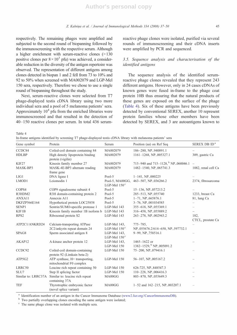
The sequence analysis of the identified serum-reactive phage clones revealed that they represent 243different antigens. However, only in 24 cases cDNAs ofknown genes were fused in-frame to the phage coatprotein 10B thus ensuring that the natural products ofthese genes are exposed on the surface of the phage(Table 4). Six of these antigens have been previouslydetected by conventional SEREX, another 10 representprotein families whose other members have beendetected by SEREX, and 3 are autoantigens known to
Table 4In-frame antigens identified by screening T7 phage-displayed testis cDNA library with melanoma patients' sera
Gene symbol Protein Serum Position (aa) on Ref Seq SEREX DB ID a
CCDC84 Coiled-coil domain containing 84 MA002079 186–280, NP_940891.1 –HDLBP High density lipoprotein binding
protein (vigilin)MA002079 1161–1268, NP_005327.1 309, gastric Ca
KIF27 Kinesin family member 27 MA002079 715–940 and 715–1128, b NP_060046.1 –MASK-BP3 MASK-4E-BP3 alternate reading
frame geneMA002079 1482–1540, NP_065741.3 1082, renal cell Ca
LIG1 DNA ligase I Pool-5 1–141, NP_000225 –LMOD1 Leiomodin 1 Pool-5, MA00GG,
LGP-Mel 150 c463–507, NP_036266.2 2374, fibrosarcoma
COPS4 COP9 signalosome subunit 4 Pool-5 15–136, NP_057213.2 –R3HDM2 R3H domain-containing protein 2 Pool-5 285–513, NP_055740 1233, breast CaANXA11 Annexin A11 Pool-5 1–71, NP_665876.1 81, lung CaDKFZP566E164 Hypothetical protein LOC25858 Pool-5 3–76, NP_001034585 –SENP1 Sentrin/SUMO-specific protease 1 LGP-Mel 143 355–618, NP_055369.1 –KIF1B Kinesin family member 1B isoform b LGP-Mel 143 316–458, NP_055889.2 –RPS2 Ribosomal protein S2 LGP-Mel 143 263–278, NP_002943.2 182,
CTCL, prostate CaATP2C1/ANKRD24 Calcium-transporting ATPase
2C2/ankyrin repeat domain 24LGP-Mel 143,LGP-Mel 150 c
775–785,NP_055676.2/614–650, NP_597732.1
–
SPAG8 Sperm associated antigen 8 LGP-Mel 143,LGP-Mel 150 c
9–99, NP_758516.1 –
AKAP12 A-kinase anchor protein 12 LGP-Mel 143,LGP-Mel 150
1465–1622 or1382–1529, b NP_005091.2
–
CCDC92 Coiled-coil domain-containingprotein 92 (Limkain beta-2)
LGP-Mel 150 75–200, NP_079416.1 –
ATP5G2 ATP synthase, H+ transporting,mitochondrial F0 complex
LGP-Mel 150 56–107, NP_005167.2 –
LRRC50 Leucine rich repeat containing 50 LGP-Mel 150 626-725, NP_848547.3 –SLU7 Step II splicing factor LGP-Mel 150 110–228, NP_006416.3 –Similar to: LRRC37A Similar to: leucine rich repeat
containing 37AMA00GG 803–870, NP_055649.3 –
TEF Thyrotrophic embryonic factor(novel splice variant)
MA00GG 1–52 and 162–215, NP_003207.1 –
a Identification number of an antigen in the Cancer Immunome Database (www2.licr.org//CancerImmunomeDB).b Two partially overlapping clones encoding the same antigen were isolated;c The same phage clone was isolated with multiple sera.
45Z. Kalnina et al. / Journal of Immunological Methods 334 (2008) 37–50

Author's personal copy
induce autoantibody production in autoimmune dis-orders (LMOD1, AKAP12) or infertility (SPAG8),while no immune responses against 4 antigens —SENP1, TEF, SLU7 and DKFZP566E164 have beenreported before. One of the clones contained a hybridcDNA generated by in-frame fusion of ATP2C1 andANKRD24 genes; however no evidence for the expres-sion of this fusion mRNAs in the testis tissue was found,suggesting that most likely the clone arose as a cloningartefact. Of the remaining 219 clones, 12 represent novelsplice variants of known genes or uncharacterised genesfor which the natural ORFs have not been determined,143 represent 5′ or 3′ UTRs or cDNAs fused to 10B in adifferent reading frame, 52 clones represent intergenicDNA with no corresponding ESTs, 3 clones encode forribosomal RNA genes and 9 clones contain mitochon-drial DNA.
The antibody reactivity against two in-frame antigens(SPAG8 and LRRC50) and two out-of-frame peptides(encoded by GOLGA5 and LAMC1) was confirmed byWestern blot analysis. The proteins encoded by thecorresponding serum-reactive clones were expressed asGST fusion proteins (for GOLGA5 and LAMC1 both—the natural products of these genes and 5′-3′ frame 2peptide of GOLGA5 and 3′-5′ frame 3 peptide ofLAMC1 were produced), purified and used for Westernblot analysis with anti-GST antibody (not shown) and
LGP-Mel 150 serum (Fig. 3A). This confirmed thereactivity against SPAG8 and LRRC50 encoded antigensand GOLGA5 and LAMC1 out-of-frame peptides.
We further hypothesised that the relatively highproportion of out-of-frame peptides among the serum-reactive clones identified by screening T7 phage-displayed library could be associated with the higherdisplay density of these peptides than natural ORFs due totheir shorter size (the size of the out-of-frame peptidesranged from 2 to 56 aa, average 21 aa, while in-frameproteins ranged from10 to 414 aa, average 120 aa). To testthis, a panel of 8 serum-reactive clones (4 in-frame, 4 out-of-frame antigens) and an empty T7 phage was assembledand subjected to Western blot analysis with anti-T7 10Bantibody and LGP-Mel 150 serum (Fig. 3B) as describedabove. The copy number of fusion proteins per phageparticle ranged from 17 to 42 (4–10%). No correlationbetween the copy number and (1) the size of the fusionprotein, (2) the size of the insert and (3) signal intensity inWestern blot and plaque immunoscreening with patient'sserum was observed. This shows that the detection ofserum reactivity against out-of-frame peptides is anintrinsic feature of T7 Select 10-3b phage display systemthat is not associated with the variations in the copynumber of recombinant proteins per phage. The serumreactivity was detected against all fusion proteins, exceptfor the AKAP12, hence suggesting that the anti-AKAP12
Fig. 3. Western blot analysis of serum reactivity against selected antigens. (A) In-frame and out-of-frame peptides encoded by the selected serum-reactive phages were expressed as GST C-terminal fusion proteins and tested for the reactivity with LGP-Mel 150 serum and anti-GST antibody toconfirm the expression of the fusion protein and to determine its size (not shown). The arrows indicate the location of GST fusion proteins.(B) Lysates of ~109 pfu of the selected phage particles were separated by SDS/PAGE and tested with anti-10B antibody to determine the copy numberof recombinant proteins per phage particle (quantification was done as described in Fig. 1) with patient's serum used for the immunoselection.
46 Z. Kalnina et al. / Journal of Immunological Methods 334 (2008) 37–50

Author's personal copy
antibodies recognise a conformational epitope that isabsent on denatured protein, while all the other antigenscarry linear epitopes.
4. Discussion
In the current study we attempted to evaluate thesuitability of T7 Select 10-3b and λKM8 phage displaysystems for monitoring autoantibody responses against arange of clinically relevant tumour antigens. This showedthat both systems are capable of displaying members of allantigen families that were cloned and a relatively goodconcordance in the detection of autoantibodies by plaqueimmunoscreening was observed — reactivity against 5antigen families was detected using both display systemswhile no reactivity against the remaining 9 antigen familieswas detected. As the display of the non-reactive antigens atleast on the T7 phages was confirmed by Western blotanalysis, these are likely to represent true negative calls.However, the sensitivity of the detection of autoantibodiesagainst various antigens seems to differ between the displaysystems— T7 Select system exhibited a higher sensitivity(in terms of signal intensities and the number of reactivesera) for the detection of autoantibodies against themembers of CTAG,MAGEA and GAGE antigen families.In line with the immunoscreening results, T7 phage displaysystem turned out to be more sensitive for the detection ofantibodies against CTAG1B/CTAG2 and MAGEA anti-gens in microarray screening also. This was an unexpectedfinding, since lambda phage has been shown to be able toassemble capsids consisting of up to ~50% N-terminalfusion protein D (Vaccaro et al., 2006), while T7 phagecapsids can incorporate only 1.2–3.6% recombinantproteins according to the manufacturer's description(Novagen) or 1–17.0% according to our results. Thissuggests that the N-terminus of the recombinant protein Don lambda phage may be spatially less accessible forantibodies than C-terminus of 10B on T7 phage. In fact, theanalysis of the crystal structure of gpD demonstrated thatthe N-termini up to Ser 15 are disordered and are locatednear the three-fold axis of gpD trimer on the side that bindsto the capsid surface and hence at least partially may behidden under the gpD trimer (Yang et al., 2000).
At the same time, the reactivity against CTAGE5 couldbe detected only when it was displayed on lambda phagebut not on T7 phage. Since the copy number of fusionproteins on CTAGE5 encoding T7 phages was the lowestamong all the phages analysed (~1%), most likely, it didnot reach the detection limit thus preventing the serumreactivity it to be detected. Hence, the very variabledisplay density of T7 Select system confers a risk of falsenegative calls due to an insufficient copy number and
furthermore suggests that the signal intensity in plaqueassay ormicroarraysmay depend not only on the antibodytitre but also on the copy number of recombinant proteins.However, this disadvantage could be overcome byconstructing a novel vector that would allow monitoringthe copy number of fusion proteins per phage particle.
Next, we applied T7 Select 10-3b phage display-basedSEREX approach to search for antigens eliciting immuneresponses in melanoma patients that resulted in the iden-tification of 436 serum-reactive clones representing 243different antigens. However, only 24 of them representedknown genes translated in their natural reading frames andanother 12 were novel splice variants or uncharacterisedgenes (with at least two ESTs confirming that thesesequences are transcribed) with unknown protein se-quences. Six of the in-frame antigens have been previouslydetected by conventional SEREX and another 10 representprotein families whose other members have been detectedby SEREX but no immune responses to 4 antigens havebeen reported before thus demonstrating that the repertoireof antigens identified by T7 phage display-based SEREXapproach overlaps with conventional SEREX, at the sametime may allow the detection of novel antigens.
Some of them have been previously shown to have adiagnostic value or may play a significant role in cancerprogression. For example, the presence of autoantibodiesagainst Annexin XI-A could significantly discriminatebetween breast cancer and non-cancer control sera andtheir frequency was higher in patients with ductalcarcinoma in situ than in invasive ductal carcinoma(Fernandez-Madrid et al., 1999). Another of the identifiedantigens, sperm-associated antigen 8 (SPAG8), so far wasimplicated in a rare form of female infertility, where anti-SPAG8 antibodies have been shown to cause spermagglutination (Zhang et al., 2000). This protein was shownto be predominantly expressed on the acrosome of spermand functionally involved in the acrosome reaction andsperm binding to the zona pellucida (Cheng et al., 2007).Very recently it was found to be overexpressed in HPV18infected cervical cancer cells (Vazquez-Ortiz et al., 2007).At the same time, a closely related protein, SPAG1, wasshown to be expressed at high levels in a large proportionof pancreatic ductal adenocarcinomas and contribute tocancer cell motility. Hence, SPAG may represent a novelCTantigen family, however more detailed analysis of theirexpression in normal and cancerous tissues is required.Moreover, considering its localisation on cell surface atleast on spermatozoa, it seems to be an attractive target forantibody-based therapeutical approaches.
The remaining 219 clones contained fragments ofintergenicDNA (52), mtDNA (9), rRNA (3), 5′or 3′UTRsor cDNAs (143) cloned out-of-frame relatively to the coat
47Z. Kalnina et al. / Journal of Immunological Methods 334 (2008) 37–50

Author's personal copy
protein 10B. It cannot be excluded that some of the 143clones encoding UTRs or cDNAs in alternative ORFsindeed represent cancer-specific antigens generated byframe-shiftingmutations, defects in pre-mRNA splicing oraberrations in translational controls in cancer cells, asevidenced by the detection of serum-reactive clonesexpressing CTAG1B-ORF2 peptide by screening TAAmini-libraries. Nevertheless, most likely the majority ofthese clones, particularly those 52 clones containingintergenic regions with no evidence of expression, displaypeptides that are not naturally expressed and therefore canbe considered as mimotopes. A similar proportion of in-frame and out-of-frame antigens has been found byChatterjee et al. (2006) in a study where serum-reactiveclones were selected from T7 phage-displayed ovariancancer cDNA library. Out-of-frame peptides, yet in smallernumber, also have been detected by using pJuFo phagedisplay system (Fossa et al., 2004) and lambda phagesurface display (Minenkova et al., 2003). The nature of theantigens they mimic is not known and we believe —cannot be unambiguously determined by BLAST searchthrough protein databases because the exact epitopesequences are not known and, moreover, they maymimic protein as well as non-protein antigens of variouspathogens not only cancer cells. Consequently, the proba-bility of finding cancer-associated biomarkers among themshould be lower than among the in-frame antigens.
Experience of our and other groups (personal commu-nication Dr. G. Li; Nottingham and Dr. S. Eichmuller,Heidelberg) has shown that approximately 1/3 of theserum-reactive clones identified by conventional SEREXalso contain cDNAs fused to β-galactosidase gene in anon-natural reading frame. Although it is possible that thenatural products of these genes could be produced bymeans of using alternative ribosomal binding sites, to ourknowledge it has never been experimentally confirmed.Therefore we assume that a considerable fraction of theseantigens also representsmimotopes, though their incidenceis much lower than in T7 phage display-based SEREXapproach. We reasoned, the identification of so highpercentage of mimotopes using T7 phage could be causedby a higher display density of the out-of-frame peptidesand subsequent detection of low affinity antibodies due toa higher valence of the epitope carrier. However, theanalysis of the display density on the selected phageparticles did not confirm this hypothesis, hence either theoverrepresentation of the phages expressing shorterpeptides in the amplified libraries or the folding propertiesand accessibility of the C-terminus of the coat protein 10Bare likely to be responsible for this phenomenon.
The application of phage display technology to cancerserology offers time-, labour- and cost-effective alter-
native to conventional SEREX. Furthermore, it gives anopportunity to produce antigen chips by printing therecombinant phage particles on microarray slides thusallowing avoidance of production and purification ofrecombinant proteins that would not be feasible for all theantigens identified. This in turn enables the analysis of thewhole autoantibody profile in patients' sera that wouldallow establishing the significance of the autoantibodyprofiles, not individual autoantibodies, as biomarkers forthe early detection and prognosis of cancer and predictionof response to immunotherapy. So far, T7 phage,presumably due to its favourable biological properties(fast growing, chemically resistant and easy to obtainhigh-titre stocks) and availability of good commercialantibodies against the phage tail protein, has been thevector of choice for the production of antigen microarrays(Fernandez-Madrid et al., 1999; Zhong et al., 2005;Wanget al., 2005; Chatterjee et al., 2006). However, the variablecopy number and the display of very high percentage ofmimotopes constitute the two main drawbacks forexploiting T7 Select system for the analysis of autoanti-body profiles. Here we demonstrated that the lambdaphage is equally suitable for the production of phage-displayed antigen microarrays — it turned out to bepossible to obtain high-titre phage stocks without anyconcentration or purification steps, the phage capsidappeared to be sufficiently stable and in the most cases thedisplayed proteins retained their capacity to be recognisedby antibodies, however it was less sensitive than T7 phagefor the detection of antibodies against several antigens.Nevertheless, as the lambda phage is capable to assemblecapsids with N-terminal gpD fusion proteins thatefficiently diminish display of out-of-frame peptides, itcould be a preferential display system for the studiesaiming to define novel potential therapeutic targets or toassess the presence of autoantibodies against knowntumour antigens.
In conclusion, the exploitation of phage display-basedapproaches for the identification of tumour antigensprovides a time- and labour-effective alternative to theconventional SEREX allowing the identification of adiverse antigen repertoire that partially overlaps withSEREX. Moreover, both T7 Select and λKM8 displaysystems are equally suitable for the production of antigenmicroarrays allowing the monitoring of autoantibodyprofiles, however they differ in the sensitivity of thedetection of antibodies against various antigens.
Acknowledgments
We are thankful to Dr. Mauruzio Cianfriglia forproviding anti-gpV monoclonal antibody and to Dr.
48 Z. Kalnina et al. / Journal of Immunological Methods 334 (2008) 37–50

Author's personal copy
Aivars Stengrēvics for providing a part of the collectionof sera from melanoma patients. We appreciate the helpof Antje Sucker and Dr. Stefan Eichmüller for selectingappropriate serum samples.
This study was supported by EU 6th FrameworkProgram ENACT (LSHC-CT-2004-503306) and a grantfrom Latvian State Research Program No. 07-VP-2, andfellowships from ESF.
References
Bachelot, T., Ratel, D., Menetrier-Caux, C., Wion, D., Blay, J.Y.,Berger, F., 2006. Autoantibodies to endostatin in patients withbreast cancer: correlation to endostatin levels and clinical outcome.Br. J. Cancer 94, 1066.
Cekaite, L., Haug, O., Myklebost, O., Aldrin, M., Ostenstad, B.,Holden, M., Frigessi, A., Hovig, E., Sioud, M., 2004. Analysis ofthe humoral immune response to immunoselected phage-displayedpeptides by a microarray-based method. Proteomics. 4, 2572.
Chatterjee, M., Mohapatra, S., Ionan, A., Bawa, G., li-Fehmi, R.,Wang, X., Nowak, J., Ye, B., Nahhas, F.A., Lu, K., Witkin, S.S.,Fishman, D., Munkarah, A., Morris, R., Levin, N.K., Shirley, N.N.,Tromp, G., Abrams, J., Draghici, S., Tainsky, M.A., 2006. Diag-nostic markers of ovarian cancer by high-throughput antigen cloningand detection on arrays. Cancer Res. 66, 1181.
Cheng, G.Y., Shi, J.L., Wang, M., Hu, Y.Q., Liu, C.M., Wang, Y.F.,Xu, C., 2007. Inhibition of mouse acrosome reaction and sperm-zona pellucida binding by anti-human sperm membrane protein 1antibody. Asian J. Androl. 9, 23.
Fernandez-Madrid, F., VandeVord, P.J., Yang, X., Karvonen, R.L.,Simpson, P.M., Kraut, M.J., Granda, J.L., Tomkiel, J.E., 1999.Antinuclear antibodies as potential markers of lung cancer. Clin.Cancer Res. 5, 1393.
Fossa, A., Alsoe, L., Crameri, R., Funderud, S., Gaudernack, G.,Smeland, E.B., 2004. Serological cloning of cancer/testis antigensexpressed in prostate cancer using cDNA phage surface display.Cancer Immunol. Immunother. 53, 431.
Hansen, M.H., Ostenstad, B., Sioud, M., 2001. Identification ofimmunogenic antigens using a phage-displayed cDNA library froman invasive ductal breast carcinoma tumour. Int. J. Oncol. 19, 1303.
Hufton, S.E., Moerkerk, P.T., Meulemans, E.V., de Bruine, A., Arends,J.W., Hoogenboom, H.R., 1999. Phage display of cDNArepertoires: the pVI display system and its applications for theselection of immunogenic ligands. J. Immunol. Methods 231, 39.
Krumpe, L.R., Atkinson, A.J., Smythers, G.W., Kandel, A.,Schumacher, K.M., McMahon, J.B., Makowski, L., Mori, T.,2006. T7 lytic phage-displayed peptide libraries exhibit lesssequence bias than M13 filamentous phage-displayed peptidelibraries. Proteomics 6, 4210.
Li, Y., Karjalainen, A., Koskinen, H., Hemminki, K., Vainio, H.,Shnaidman, M., Ying, Z., Pukkala, E., Brandt-Rauf, P.W., 2005.p53 autoantibodies predict subsequent development of cancer. Int.J. Cancer 114, 157.
Lucchese, A., Willers, J., Mittelman, A., Kanduc, D., Dummer, R.,2005. Proteomic scan for tyrosinase peptide antigenic pattern invitiligo and melanoma: role of sequence similarity and HLA-DR1affinity. J. Immunol. 175, 7009.
Minenkova, O., Pucci, A., Pavoni, E., De, T.A., Fortugno, P., Gargano,N., Cianfriglia, M., Barca, S., De, P.S., Martignetti, A., Felici, F.,
Cortese, R., Monaci, P., 2003. Identification of tumor-associatedantigens by screening phage-displayed human cDNA libraries withsera from tumor patients. Int. J. Cancer 106, 534.
Pallasch, C.P., Struss, A.K., Munnia, A., Konig, J., Steudel, W.I.,Fischer, U., Meese, E., 2005. Autoantibodies against GLEA2 andPHF3 in glioblastoma: tumor-associated autoantibodies correlatedwith prolonged survival. Int. J. Cancer 117, 456.
Pavoni, E., Vaccaro, P., Pucci, A., Monteriu, G., Beghetto, E., Barca,S., Dupuis, M.L., De Pasquale, C.A., Lugini, A., Cianfriglia, M.,Cortesi, E., Felici, F., Minenkova, O., 2004. Identification of apanel of tumor-associated antigens from breast carcinoma celllines, solid tumors and testis cDNA libraries displayed on lambdaphage. BMC Cancer 4, 78.
Rosenberg, A., Griffin, K., Studier, W.F., McCormick, M., Berg, J.,Novy, R., Mierendorf, R., 1996. T7 Select Phage Display System:a powerful new protein display system based on bacteriophage T7.Innovations 1–6.
Sahin, U., Tureci, O., Schmitt, H., Cochlovius, B., Johannes, T.,Schmits, R., Stenner, F., Luo, G., Schobert, I., Pfreundschuh, M.,1995. Human neoplasms elicit multiple specific immune responsesin the autologous host. Proc. Natl. Acad. Sci. U. S. A 92, 11810.
Sioud, M., Hansen, M.H., 2001. Profiling the immune response inpatients with breast cancer by phage-displayed cDNA libraries.Eur. J. Immunol. 31, 716.
Slager, E.H., Borghi, M., van der Minne, C.E., Aarnoudse, C.A.,Havenga, M.J., Schrier, P.I., Osanto, S., Griffioen, M., 2003. CD4+Th2 cell recognition of HLA-DR-restricted epitopes derived fromCAMEL: a tumor antigen translated in an alternative open readingframe. J. Immunol. 170, 1490.
Somers, V.A., Brandwijk, R.J., Joosten, B.,Moerkerk, P.T., Arends, J.W.,Menheere, P., Pieterse, W.O., Claessen, A., Scheper, R.J., Hoogen-boom,H.R., Hufton, S.E., 2002. A panel of candidate tumor antigensin colorectal cancer revealed by the serological selection of a phagedisplayed cDNA expression library. J. Immunol. 169, 2772.
Soussi, T., 2000. p53 Antibodies in the sera of patients with varioustypes of cancer: a review. Cancer Res. 60, 1777.
Stockert, E., Jager, E., Chen, Y.T., Scanlan, M.J., Gout, I., Karbach, J.,Arand, M., Knuth, A., Old, L.J., 1998. A survey of the humoralimmune response of cancer patients to a panel of human tumorantigens. J. Exp. Med. 187, 1349.
Usener, D., Schadendorf, D., Koch, J., Dubel, S., Eichmuller, S., 2003.cTAGE: a cutaneous T cell lymphoma associated antigen familywith tumor-specific splicing. J. Invest. Dermatol. 121, 198.
Vaccaro, P., Pavoni, E., Monteriu, G., Andrea, P., Felici, F., Minenkova,O., 2006. Efficient display of scFv antibodies on bacteriophagelambda. J. Immunol. Methods 310, 149.
Vazquez-Ortiz, G., Garcia, J.A., Ciudad, C.J., Noe, V., Penuelas, S.,Lopez-Romero, R., Mendoza-Lorenzo, P., Pina-Sanchez, P., Sal-cedo, M., 2007. Differentially expressed genes between high-riskhuman papillomavirus types in human cervical cancer cells. Int. J.Gynecol. Cancer 17, 484.
Wang, X., Yu, J., Sreekumar, A., Varambally, S., Shen, R., Giacherio, D.,Mehra, R., Montie, J.E., Pienta, K.J., Sanda, M.G., Kantoff, P.W.,Rubin, M.A., Wei, J.T., Ghosh, D., Chinnaiyan, A.M., 2005.Autoantibody signatures in prostate cancer. N. Engl. J.Med. 353, 1224.
Welling, G.W., Weijer, W.J., van der Zee, R., Welling-Wester, S., 1985.Prediction of sequential antigenic regions in proteins. FEBS Lett.188, 215.
Willats, W.G., 2002. Phage display: practicalities and prospects. PlantMol. Biol. 50, 837.
Yang, F., Forrer, P., Dauter, Z., Conway, J.F., Cheng, N., Cerritelli, M.E.,Steven, A.C., Pluckthun, A., Wlodawer, A., 2000. Novel fold and
49Z. Kalnina et al. / Journal of Immunological Methods 334 (2008) 37–50

Author's personal copy
capsid-binding properties of the lambda-phage display platformprotein gpD. Nat. Struct. Biol. 7, 230.
Zhang, X.D., Miao, S.Y., Wang, L.F., Li, Y., Zong, S.D., Yan, Y.C.,Koide, S.S., 2000. Human sperm membrane protein (hSMP-1): adevelopmental testis-specific component during germ cell differ-entiation. Arch. Androl. 45, 239.
Zhong, L., Hidalgo, G.E., Stromberg, A.J., Khattar, N.H., Jett, J.R.,Hirschowitz, E.A., 2005. Using protein microarray as a diagnosticassay for non-small cell lung cancer. Am. J. Respir. Crit Care Med.172, 1308.
Zhong, L., Peng, X., Hidalgo, G.E., Doherty, D.E., Stromberg, A.J.,Hirschowitz, E.A., 2004. Identification of circulating antibodies to
tumor-associated proteins for combined use as markers of non-small cell lung cancer. Proteomics 4, 1216.
Zippelius, A., Gati, A., Bartnick, T., Walton, S., Odermatt, B., Jaeger, E.,Dummer, R., Urosevic, M., Filonenko, V., Osanai, K., Moch, H.,Chen, Y.T., Old, L.J., Knuth, A., Jaeger, D., 2007. Melanocytedifferentiation antigen RAB38/NY-MEL-1 induces frequent anti-body responses exclusively in melanoma patients. Cancer Immunol.Immunother. 56, 249.
Zucconi, A., Dente, L., Santonico, E., Castagnoli, L., Cesareni, G., 2001.Selection of ligands by panning of domain libraries displayed onphage lambda reveals new potential partners of synaptojanin 1. J.Mol.Biol. 307, 1329.
50 Z. Kalnina et al. / Journal of Immunological Methods 334 (2008) 37–50