Plasma adiponectin is inversely associated with antenatal anxiety: Results from a Brazilian cohort

MOLECULAR AND CELLULAR BIOLOGY, Oct. 2010, p. 4687–4697 Vol. 30, No. 190270-7306/10/$12.00 doi:10.1128/MCB.01581-09Copyright © 2010, American Society for Microbiology. All Rights Reserved.
VRK2 Inhibits Mitogen-Activated Protein Kinase Signaling andInversely Correlates with ErbB2 in Human Breast Cancer�†
Isabel F. Fernandez,1 Sandra Blanco,1‡ Jose Lozano,2,3 and Pedro A. Lazo1*Experimental Therapeutics and Translational Oncology Program, Instituto de Biología Molecular y Celular del Cancer,
Consejo Superior de Investigaciones Científicas-Universidad de Salamanca, Salamanca, Spain1; Departamento de BiologíaMolecular y Bioquímica, Universidad de Malaga, Malaga, Spain2; and Laboratorio de Oncología Molecular,
Fundacion IMABIS, Hospital Clínico Universitario Virgen de la Victoria, Malaga, Spain3
Received 9 December 2009/Returned for modification 17 March 2010/Accepted 16 July 2010
The epidermal growth factor (EGF)–ErbB–mitogen-activated protein kinase (MAPK) transcription signal-ing pathway is altered in many types of carcinomas, and this pathway can be regulated by new protein-proteininteractions. Vaccinia-related kinase (VRK) proteins are Ser-Thr kinases that regulate several signal trans-duction pathways. In this work, we study the effect of VRK2 on MAPK signaling using breast cancer as a model.High levels of VRK2 inhibit EGF and ErbB2 activation of transcription by the serum response element (SRE).This effect is also detected in response to H-Ras(G12V) or B-Raf(V600E) oncogenes and is accompanied by areduction in phosphorylated extracellular signal-regulated kinase (ERK) levels, p90RSK levels, and SRE-dependent transcription. Furthermore, VRK2 knockdown has the opposite effect, increasing the transcrip-tional response to stimulation with EGF and leading to increased levels of ERK phosphorylation. Themolecular mechanism lies between MAPK/ERK kinase (MEK) and ERK, since MEK remains phosphorylatedwhile ERK phosphorylation is blocked by VRK2A. This inhibition of the ERK signaling pathway is a conse-quence of a direct protein-protein interaction between VRK2A, MEK, and kinase suppressor of Ras 1 (KSR1).Identification of new correlations in human cancer can lead to a better understanding of the biology ofindividual tumors. ErbB2 and VRK2 protein levels were inversely correlated in 136 cases of human breastcarcinoma. In ErbB2� tumors, there is a significant reduction in the VRK2 level, suggesting a role for VRK2Ain ErbB2-MAPK signaling. Thus, VRK2 downregulation in carcinomas permits signal transmission throughthe MEK-ERK pathway without affecting AKT signaling, causing a signal imbalance among pathways thatcontributes to the phenotype of breast cancer.
Members of the ErbB receptor-tyrosine kinase family areindicators of poor prognosis in several types of cancer, includ-ing breast cancer, colorectal cancer, head and neck squamouscell carcinoma (HNSCC), and non-small cell lung carcinoma(NSCLC) (18). Vaccinia-related kinases (VRK) are a novelfamily of Ser-Thr kinases composed of three members (26),two of which are catalytically active (36). VRK2A and itsshorter, spliced isoform, VRK2B, are located, respectively,anchored to membranes of the endoplasmic reticulum andmitochondria or in soluble form in the cytoplasm and withinthe nucleus (4, 5). VRK2A, the more abundant isoform, mod-ulates the stress response to hypoxia (6) and cytokines, such asinterleukin 1� (IL-1�) (7). This signal modulation is indepen-dent of VRK2A kinase activity but is dependent on proteininteractions with JIP1, which assembles mitogen-activated pro-tein kinase (MAPK) complexes, functioning as a scaffold pro-tein for the c-Jun N-terminal kinase (JNK) pathway. VRK2Ablocks signal transmission mediated by the assembly of JIP1-MAPK complexes, reducing JNK phosphorylation and AP1-
dependent transcription (6, 7). On the other hand, the nuclearVRK2B isoform has been proposed to functionally replaceVRK1 in adenocarcinoma tumor cell lines (4). VRK1 is ex-pressed at high levels in proliferating tumor cell lines andduring cellular expansion in murine embryonic hematopoieticdevelopment (49). VRK1 expression correlates with severalproliferation markers in human head and neck squamous cellcarcinomas (42). VRK1 knockdown causes a block in cell cycleprogression in tumor cells (50) and fibroblasts (46). Indeed,VRK1 is an early response gene expressed in parallel withMYC and FOS genes and required for cyclin D1 expression(21) and entry into the G1 phase of the cell cycle (46). NuclearVRK1 phosphorylates several transcription factors, such asp53 (29, 50), c-Jun (43), ATF2 (44), and CREB (21), as well asBaf, during nuclear envelope assembly (17, 37). VRK1 and p53form an autoregulatory loop (48) that is disrupted in tumorswith p53 mutations, such as lung cancer (47). In estrogenreceptor-positive (ER�) breast cancers, tumors with poorprognosis and a shorter time to relapse were associated withhigher VRK1 expression (30). All of these data together sug-gest that VRK proteins might be good candidates to be dereg-ulated in tumors.
The specificity of any biological effect in response to stimu-lation initiated in ErbB receptors (33), such as epidermalgrowth factor receptor (EGFR) or ErbB2, can be determinedby interacting proteins that may alter the distribution of thesignal among alternative response pathways (14, 45). Thus, thesignal transmitted by Ras can be channeled mainly between
* Corresponding author. Mailing address: Centro de Investigaciondel Cancer, CSIC-Universidad de Salamanca, Campus Miguel de Un-amuno, E-37007 Salamanca, Spain. Phone: 34 923 294 804. Fax: 34 923294 795. E-mail: [email protected].
‡ Present address: Wellcome Trust Center for Stem Cell Research,Tennis Court Road, CB2 1QR Cambridge, United Kingdom.
† Supplemental material for this article may be found at http://mcb.asm.org/.
� Published ahead of print on 2 August 2010.
4687
MAPK and phosphatidylinositol 3-kinase (PI3K)–Akt (9, 54).However, how signal distribution is modulated among differentpathways is not known, suggesting that additional elements canintervene, either by protein-protein interactions or by interac-tions with additional pathways, among which the MAPK path-way is the best known (28). This interconnectivity may result infunctional differences that can help to explain differential re-sponses to identical stimulation in either normal or tumor cells.The study of pathway interactions and their components hasreceived relatively limited attention; however, it will be essen-tial to identify specific effects in the biological systems in whichthey are likely to play important roles. The identification ofnew regulatory components can also lead to the discovery ofnovel molecular therapeutic targets.
Breast cancer-specific molecular markers help determinethe prognosis, outcome, and treatment to be followed in thesepatients (15). Tumors positive for ER and/or progesteronereceptor (PR) have a better prognosis, since they can be man-aged with antihormonal therapies. A third marker is ErbB2(HER2/neu), a member of the EGF receptor family, which isusually detected in ER� cases, which represent approximatelyone-fourth of cases (27). Nevertheless, ErbB2 is the target ofnovel therapies based on the use of either specific antibodies orsmall tyrosine kinase inhibitors of the receptor (18).
In this report, we characterize how VRK2A modulates theErbB-MAPK signaling pathway by a downstream interactionof the kinase with the kinase suppressor of Ras 1 (KSR1)scaffold protein. Analysis of the expression of VRK2 proteinsin human breast carcinomas showed that ErbB2 receptor andVRK2A levels were inversely correlated, suggesting a facilita-tion of MAPK signaling. We conclude that VRK2A downregu-lation causes an imbalance in signaling pathways and thuscontributes to the phenotype of breast carcinomas.
MATERIALS AND METHODS
Plasmids. The following plasmids were used: pCEFL-HA-VRK1 (50); plas-mids for VRK2A (pCEFL-HA-VRK2A), VRK2B (pCEFL-HA-VRK2B), andthe inactive kinase VRK2(K169E) (pCEFL-GST-VRK2A) and the controlpCEFL-GST (4); pCDNA3-ErbB2 (53); pCEFL-HA-MEK, pCEFL-HA-ERK,and pCEFL-H-RasG12V (from Piero Crespo, IBBTEC-CSIC, Santander, Spain);pG12B-RafV600E (from M. Soengas, University of Michigan); pCMV-Flag-KSR1(20); pA3Cyclin D1(�1720)-Luc (1); pFC-MEK1 [constitutively activeMEK1(S218/222E, �32-51)]; pSRE-Luc reporter (Stratagene); and pRL-tk con-trol (Promega Biotech).
Cell lines, transfections, and immunoblotting. MDA-MB-231 and MCF7breast cancer cells (19) and HeLa and HEK-293T cells were grown in Dulbecco’smodified Eagle’s medium (DMEM) with 10% fetal bovine serum. Transfectionswere performed using the JetPEI reagent (Polyplus, Illkirch, France) or Lipo-fectamine 2000 (Invitrogen). The total amount of DNA in transfections was keptconstant by completion with the corresponding empty vector. The methods forcell lysates and the conditions for immunoprecipitations, pulldown, and Westernblotting have been reported previously (4, 6, 7).
Transcriptional assays and quantitative reverse transcription (qRT)-PCR.For assays of transcriptional activity using a reporter plasmid, cells were trans-fected with the reporter plasmids pSRE-Luc (1 �g), pCDNA3-ErbB2 (1.5 �g),pCEFL-H-RasG12V (0.2 �g), pG12B-RafV600E (0.2 �g), and pFC-MEK1(0.05�g) and the indicated amounts of the specific kinase constructs. Cell lysates wereprepared 48 h after transfection in luciferase lysis buffer (Promega). Luciferaseactivity was determined with a Dual-Luciferase reporter reagent from Promega(4, 6, 7). In some experiments, cells were stimulated 4 h before lysis with 10 nMEGF (Strathmann Biotec) for the indicated times.
For qRT-PCR, cells were washed in ice-cold phosphate-buffered saline (PBS).Total RNA was extracted using the RNAeasy extraction kit from Qiagen andanalyzed as previously reported (46). The following forward (F) and reverse (R)primers were used: human c-myc (Myc-F, 5�-CCAGCAGCCTCCCGCGACGA
TG-3�; Myc-R, 5�-GAGGGGTCGATGCACTCTGAGG-3�) and human cyclinD1 (CycD1-F, 5�-CTTCCTCTCCAAAATGCCAG-3�; CycD1-R, 5�-AGAGATGGAAGGGGGAAAGA-3�), with GAPDH (glyceraldehyde-3-phosphate dehy-drogenase) amplification used as an internal control (GAPDH-F, 5�-GGTCTTACTCCTTGGAGGCCATGT-3�; GAPDH-R, 5�-ACCTAACTACATGGTTTACATGTT-3�).
Antibodies and reagents. The antibodies used were VRK2 polyclonal antibody(4); ErbB2 (44E7); p90 ribosomal S6 kinase (RSK) (32D7), p90RSK (Ser 380),phosphorylated AKT (p-AKT) (Ser473), and p-MEK1/2 (S217/S221) from CellSignaling; antibodies for extracellular signal-regulated kinase 2 (ERK2) (sc-154),ERK1/2 (Y204) (sc-7383), MAPK/ERK kinase 1 (MEK-1) (C-18 polyclonal or9G3 monoclonal), B-Raf (sc-55522), glutathione S-transferase (GST) (sc-138),AKT (H-136), cyclin D1 (M-20), Ksr-1 (sc-9317), and c-myc (sc-764), all fromSanta Cruz; anti-hemagglutinin (HA) from Covance; actin (AC-15) and anti-Flag(M5) from Sigma; and EGF (Strathmann Biotec).
VRK2, KSR, or JIP1 knockdown by siRNA interference. The targeted se-quence for VRK2 (GenBank NM_006296) was 5�-GCAAGGUUCUGGAUGAUAUUU-3� (duplex siVRK2-06) (Dharmacon). The targeted JIP1sequence(GenBank NM_005456) was 5�-CTGGAGGAGTTTGAGGATGAA-3� (smallinterfering RNA [siRNA] JIP1 number 2; Qiagen) (25, 26). Human Ksr-1 siRNAcontains a pool of 3 target-specific 20- to 25-nucleotide (nt) siRNAs (Santa Cruz;sc-35762). The siControl nontargeting siRNA pool (Dharmacon) was used as anegative control. Transfection of siRNA duplexes in HeLa cells at a final con-centration of 160 mM was carried out using Lipofectamine 2000 reagent (In-vitrogen). Cells were processed for immunoblotting 4 days after transfection. Inassays of VRK2 depletion, cells were transfected with the reporter plasmid 72 hafter siRNA transfection and EGF stimulated 24 h after retransfection.
Immunofluorescence and confocal microscopy. HeLa cells were grown onuncoated glass coverslips placed in 60-mm plates. The cells were prepared aspreviously reported (4). Fluorescence images were captured with a Zeiss LSM510 confocal microscope.
Immunohistochemistry in tissue sections. Biopsy specimens from humanbreast cancer were provided by the National Tumor Bank at CIC-HospitalUniversitario de Salamanca. The biopsy specimens were prepared as previouslyreported (47), and sections were counterstained with hematoxylin. VRK2 wasdetected with a rabbit polyclonal antibody (4). For analysis of VRK2 and ErbB2,consecutive sections were used. ErbB2 immunostaining was scored from 1 to 3for both the intensity of the staining and the number of ErbB2-positive cells. Thefinal score was obtained by multiplying these two parameters. Briefly, the fre-quencies were compared either by the chi-square method or by Fisher’s exact test(SPSS version 18; SPSS, Chicago, IL), and differences with a P value of �0.05were considered statistically significant (42, 47).
RESULTS
Overexpression or knockdown of VRK2A inversely regulatesSRE transcription induced by EGF. Because VRK2A down-regulates JNK signaling in response to IL-1� and hypoxia (6,7), it was hypothesized that the VRK2 protein might alsointerfere with transcriptional response signals to growth factorreceptors mediated by MAPK. To address whether VRK2Acould affect signaling induced upon EGF stimulation, we de-termined the effect of increasing levels of VRK2A protein onthe serum response element (SRE) transcriptional responseinduced by EGF in MCF7 cells (Fig. 1A) and HeLa cells (seeFig. S1A in the supplemental material). EGF, whose receptoris ErbB1, stimulated SRE-dependent transcription in MCF7and HeLa cells, and this transcriptional response was inhibitedby VRK2A in a dose-dependent manner, but the closely re-lated member VRK1, or VRK2B, did not affect this response.We also tested if overexpression of VRK2A could prevent theactivation of the cyclin D1 gene promoter in response to EGF.VRK2A prevented activation of this promoter, as expected,while VRK1, which is required for cyclin D1 expression (46),had a strong stimulatory effect (see Fig. S1B in the supplemen-tal material).
From the previous result, it was predicted that lowering the
4688 FERNANDEZ ET AL. MOL. CELL. BIOL.

levels of endogenous VRK2 protein would make cells moresensitive to EGF. To test this hypothesis, VRK2 protein wasknocked down in MDA-MB-231 and HeLa cells (Fig. 1B, top;see Fig. S2A in the supplemental material) with a specificsiRNA (6, 7), which effectively reduced the VRK2 proteinlevels in both cell lines between 80 and 90%. In MCF7 cells,the RNA knockdown was very effective, but the protein wasextremely stable (see Fig. S2B in the supplemental material).MDA-MB-231 cells, which have autocrine stimulation by EGF(31), and HeLa cells were stimulated with EGF. In both, therewas an increase in the response to EGF stimulation whenVRK2 was eliminated (Fig. 1B, bottom). These results confirmthe inverse correlation between VRK2 levels and the transcrip-tional response to EGF.
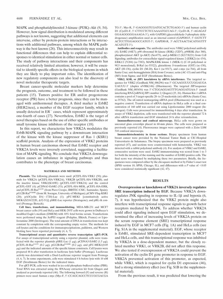
VRK2A downregulates the transactivation of transcriptioninduced by ErbB2, H-Ras(G12V), B-Raf(V600E), and activeMEK. The above observations suggest that expression ofVRK2A protein might have an effect on growth factor re-sponse signaling mediated by the Ras-Raf-MEK-Erk pathway,among which ErbB2 is the most relevant for breast and othercarcinomas. ErbB2 is active by itself and has no known ligand(18, 52). Initially, a dose-response assay to ErbB2 was per-
formed in HEK293T cells with the expected increase in SRE-dependent transcription (see Fig. S3A in the supplementalmaterial). A 1.5-�g amount of ErbB2 was selected to deter-mine the effect of the VRK2A protein on the transcriptionalresponse to ErbB2 in MCF7 cells. As the VRK2A protein levelincreased, there was a significant reduction, in a dose-depen-dent manner, of the transcriptional response induced by ErbB2in MCF7 cells (Fig. 2A) and HEK293T cells (see Fig. S3B inthe supplemental material). These data indicated that ErbB2was able to activate SRE-dependent gene transcription andthat its response was inhibited by VRK2A.
The signal transduction pathway activating SRE in responseto EGF, or ErbB2, is mediated by the Ras oncogene (40) andthe B-Raf Ser-Thr kinase (24), both frequently mutated inmany types of human cancer (34, 38), and thus cells becomeindependent of EGF/ErbB signaling (8). Therefore, we testedwhether the VRK2A protein was acting downstream of theRas/Raf proteins, since they are in intracellular membranes(16). First, a dose-response assay for H-Ras(G12V) was per-formed (see Fig. S3C in the supplemental material), and a doseof 200 ng was selected to determine the effect of VRK2A inHEK293T (see Fig. S3C and D in the supplemental material)and MCF7 (Fig. 2B) cells. VRK2A inhibited H-Ras(G12V)activation of SRE-dependent transcription in a dose-depen-dent manner (Fig. 2B). Since the Ras oncogene was thestronger inducer of transcription, it was used to determine ifthe effect of VRK2A was due either to its kinase activity orto a protein interaction. Overexpression of kinase-deadVRK2A(K169E) (4) was equally effective in inhibiting theSRE-dependent transcription induced by H-Ras(G12V)(Fig. 2B, right), suggesting that the observed effect was aconsequence of a VRK2A protein interaction with a com-ponent of the MAPK pathway.
A similar approach was followed using the B-Raf(V600E)mutant and a constitutively active MEK1(S218/222E, �32-51)(plasmid pFC-MEK1), two downstream components of Ras inthe MAPK signaling pathway. Activation of transcription byB-Raf(V600E) (Fig. 2C) or MEK1(S218/222E, �32-51) (Fig.2D) was also blocked by VRK2A in a dose-dependent manner.Therefore, it can be concluded that VRK2A acts downstreamof Ras, B-Raf, and MEK1 in the ErbB signal transductionpathway.
VRK2A blocks the phosphorylation of ERK in response toErbB2 and H-Ras(G12V).. The signal initiated in the EGFreceptor (ErbB1) is partly transmitted by phosphorylation ofERK, which activates SRE-dependent transcription. To gaininsight into how VRK2A can regulate ERK signaling, we de-termined whether overexpression of ErbB2 or H-Ras(G12V)affected the phosphorylation of ERK, the last kinase in theMAPK complex. Both ErbB2 and H-Ras(G12V) induced in-creased levels of p-MEK and p-ERK in MCF7 cells (Fig. 3Aand B). However, when VRK2A was overexpressed, there wasa reduction in the level of p-ERK, but not of p-MEK, inresponse to either ErbB2 or H-Ras(G12V), suggesting it mightbe acting between MEK and ERK (Fig. 3A and B). In theseexperiments, AKT activation by phosphorylation was not af-fected by VRK2A overexpression, since there were no changesin its phosphorylation level (Fig. 3A and B). These data indi-cated that VRK2A acts downstream of MEK and does notaffect PI3K-AKT signaling, one of the most relevant pathways
FIG. 1. VRK2A protein modulates EGF activation of SRE-depen-dent transcription. (A) Effect of VRK2A overexpression. MCF7 cellswere transfected with 1 �g of the SRE-luciferase reporter, as well asplasmids, for overexpression of VRK2A or VRK1. The immunoblotshows the correct expression of the corresponding VRK proteins de-tected with an antibody to the HA epitope. The error bars indicatestandard deviations. ***, P � 0.001; *, P � 0.05. (B) (Top) Knock-down of the VRK2 intracellular protein level with siVRK2-06 inMDA-MB-231 breast cancer cells and HeLa cells. MDA-MB-231 cellsexpress only VRK2A, and HeLa cells express both VRK2 isoforms.Mean values with standard deviations for three experiments are shown.*, P � 0.05. (Bottom) Effect of VRK2 knockdown on the activation ofSRE-dependent transcription induced by EGF in MDA-MB-231 andHeLa cells. MDA-MB-231 cells have autocrine stimulation by EGF.MCF7 cells express both isoforms of VRK2, which are not affected bythe specific siVRK2-06, while its RNA is effectively reduced (see Fig.S2B in the supplemental material). In the HeLa cell line, siVRK2-06was very effective at reducing VRK2A protein. The reduction of VRK2by siVRK2-06 was approximately 63% (see Fig. S2A in the supple-mental material).
VOL. 30, 2010 VRK2 REGULATES MAPK SIGNALING 4689
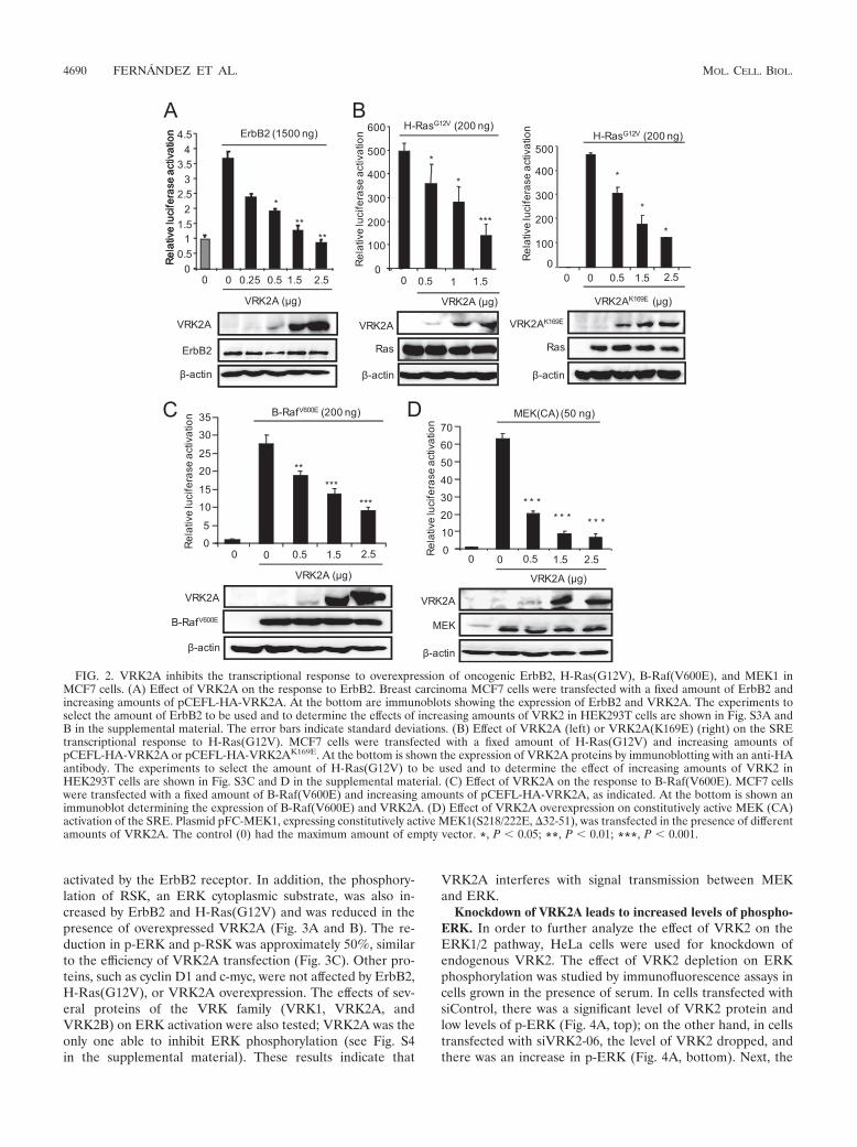
activated by the ErbB2 receptor. In addition, the phosphory-lation of RSK, an ERK cytoplasmic substrate, was also in-creased by ErbB2 and H-Ras(G12V) and was reduced in thepresence of overexpressed VRK2A (Fig. 3A and B). The re-duction in p-ERK and p-RSK was approximately 50%, similarto the efficiency of VRK2A transfection (Fig. 3C). Other pro-teins, such as cyclin D1 and c-myc, were not affected by ErbB2,H-Ras(G12V), or VRK2A overexpression. The effects of sev-eral proteins of the VRK family (VRK1, VRK2A, andVRK2B) on ERK activation were also tested; VRK2A was theonly one able to inhibit ERK phosphorylation (see Fig. S4in the supplemental material). These results indicate that
VRK2A interferes with signal transmission between MEKand ERK.
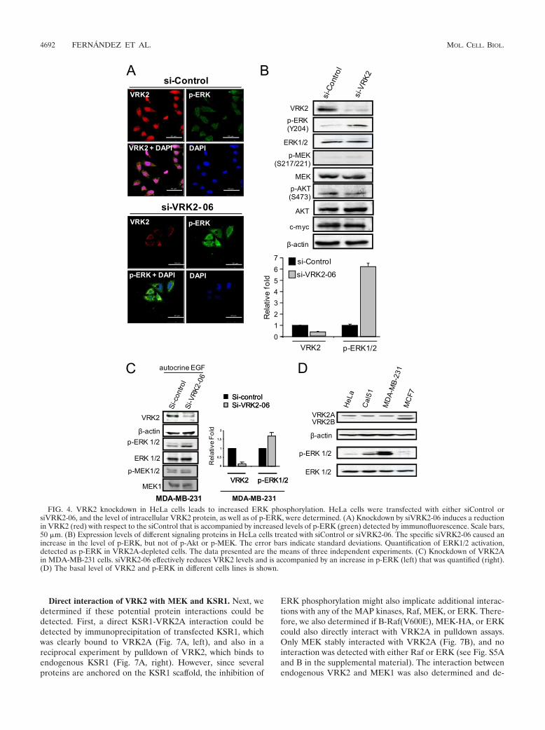
Knockdown of VRK2A leads to increased levels of phospho-ERK. In order to further analyze the effect of VRK2 on theERK1/2 pathway, HeLa cells were used for knockdown ofendogenous VRK2. The effect of VRK2 depletion on ERKphosphorylation was studied by immunofluorescence assays incells grown in the presence of serum. In cells transfected withsiControl, there was a significant level of VRK2 protein andlow levels of p-ERK (Fig. 4A, top); on the other hand, in cellstransfected with siVRK2-06, the level of VRK2 dropped, andthere was an increase in p-ERK (Fig. 4A, bottom). Next, the
FIG. 2. VRK2A inhibits the transcriptional response to overexpression of oncogenic ErbB2, H-Ras(G12V), B-Raf(V600E), and MEK1 inMCF7 cells. (A) Effect of VRK2A on the response to ErbB2. Breast carcinoma MCF7 cells were transfected with a fixed amount of ErbB2 andincreasing amounts of pCEFL-HA-VRK2A. At the bottom are immunoblots showing the expression of ErbB2 and VRK2A. The experiments toselect the amount of ErbB2 to be used and to determine the effects of increasing amounts of VRK2 in HEK293T cells are shown in Fig. S3A andB in the supplemental material. The error bars indicate standard deviations. (B) Effect of VRK2A (left) or VRK2A(K169E) (right) on the SREtranscriptional response to H-Ras(G12V). MCF7 cells were transfected with a fixed amount of H-Ras(G12V) and increasing amounts ofpCEFL-HA-VRK2A or pCEFL-HA-VRK2AK169E. At the bottom is shown the expression of VRK2A proteins by immunoblotting with an anti-HAantibody. The experiments to select the amount of H-Ras(G12V) to be used and to determine the effect of increasing amounts of VRK2 inHEK293T cells are shown in Fig. S3C and D in the supplemental material. (C) Effect of VRK2A on the response to B-Raf(V600E). MCF7 cellswere transfected with a fixed amount of B-Raf(V600E) and increasing amounts of pCEFL-HA-VRK2A, as indicated. At the bottom is shown animmunoblot determining the expression of B-Raf(V600E) and VRK2A. (D) Effect of VRK2A overexpression on constitutively active MEK (CA)activation of the SRE. Plasmid pFC-MEK1, expressing constitutively active MEK1(S218/222E, �32-51), was transfected in the presence of differentamounts of VRK2A. The control (0) had the maximum amount of empty vector. *, P � 0.05; **, P � 0.01; ***, P � 0.001.
4690 FERNANDEZ ET AL. MOL. CELL. BIOL.

levels of different proteins were determined by Western blot-ting under these two conditions. VRK2 knockdown resulted inaccumulation of p-ERK but had no effect on either p-MEK orp-AKT (Fig. 4B). The changes in VRK2A and p-ERK werequantified, and the reduction in VRK2 was accompanied by a6-fold increase in p-ERK (Fig. 4B). These results are consis-tent with the effect on transcription levels observed in thesecells (Fig. 1B) and with VRK2A overexpression results (Fig. 3).The effect of siVRK2 on p-ERK was also determined in MDA-MB-231 cells, in which the reduction in VRK2A was accom-panied by increased p-ERK without affecting MEK phosphor-ylation; in this cell line, the increase in p-ERK was smaller dueto its high basal level of p-ERK (Fig. 4C). The levels of VRK2and p-ERK in different cell lines are shown in Fig. 4D; inMDA-MB-231 cells, there was a very high basal level of p-ERK, consistent with its autocrine secretion of EGF and itsRas and B-Raf mutations (19).
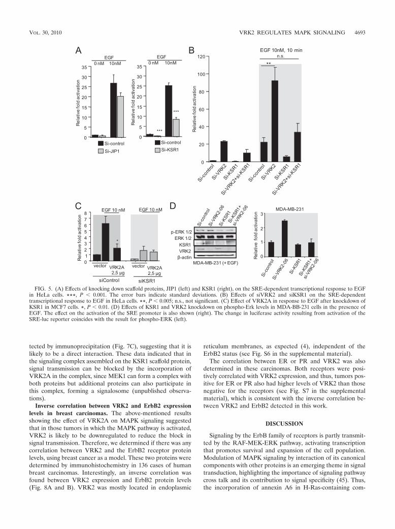
Effect of knockdown of JIP1 and KSR1 on the transcrip-tional response to EGF. Transmission by MAPK protein com-plexes requires their assembly on scaffold proteins (13). Two ofthem, KSR1 and JIP1, are likely to be implicated in the SREtranscriptional response to EGF. Therefore, it was tested ifknockdown of each of them affected the activation of transcrip-tion induced by EGF. Both siKSR1 (10) and siJIP1 (7) werevery effective in knocking down their respective protein levels.The SRE transcriptional response to EGF was very sensitive tothe knockdown of KSR1, resulting in its loss, but was onlyslightly affected by the knockdown of JIP1 (Fig. 5A). HeLacells can respond very well to EGF; thus, it was also deter-mined if knockdown of VRK2, KSR1, or both affected thetranscriptional response to EGF. VRK2 knockdown by itselfsignificantly increased the transcriptional response to EGF, aresponse that is lost in combination with siKSR1 (Fig. 5B). Theknockdown of KSR1 showed that this protein is necessary for
the EGF response, and the VRK2A inhibitory effect was de-tected only in the presence of KSR1 (Fig. 5C). These resultssuggested that the EGF response is at least partially mediatedby the KSR1 scaffold protein. Also, in MDA-MB231 cells inthe presence of EGF, knockdown of VRK2 resulted in anincrease in phospho-Erk levels, which was also lost by knock-down of KSR1 in combination with VRK2 knockdown (Fig.5D). These data point to contributions by both proteins tosignal transmission and rule out the possibility that the effect ofVRK2 is mediated by an alternative mechanism independentof KSR1.
Colocalization of KSR1 with calnexin and VRK2. Mechanis-tically, the effect of VRK2 on the ERK pathway may be aconsequence of a direct interaction between VRK2 and KSR1,or one of the kinases in the complex, as has been described forJNK-JIP1 regulation mediated by VRK2A (6, 7). Initially, thepotential colocalization of endogenous KSR1 and calnexin inthree cell lines was determined, and in all of them, the KSR1pool was larger than the calnexin pool (Fig. 6A), which iswhere VRK2A was localized (4); the fraction of KSR1 colo-calizing with calnexin varied depending on the cell line used(Fig. 6A). In MCF7 cells, almost all KSR1 colocalized withcalnexin, and thus, a reduction in VRK2 can explain the largeincrease in p-ERK observed in this cell line. In the case ofMDA-MB-231 cells, the overlapping signal represents a smallfraction. In these cells, there is much more KSR1, and most ofit is dispersed in the cytosol; this distribution explains whyeliminating VRK2 had a smaller effect on the level of p-ERK(Fig. 4C), which already had a very high basal level (Fig. 4D).The colocalization of KSR1 with calnexin indicates that KSR1,even if it is a subpopulation, might also colocalize with VRK2in the endoplasmic reticulum. This VRK2-KSR1 colocalizationwas confirmed by immunofluorescence (Fig. 6B).
FIG. 3. VRK2A inhibits ErbB2 and H-Ras(G12V) activation of ERK phosphorylation without affecting MEK or AKT phosphorylation. MCF7cells were transfected with empty vector (control) or with plasmids expressing ErbB2 (A) or H-Ras(G12V) (B), and the effect of VRK2Aoverexpression on the activation at different levels of several pathways, such as the MAPK and AKT routes, that respond to EGF was determined.Proteins were detected in immunoblots with the corresponding antibodies (see Materials and Methods). (C) Changes in relative p-ERK and p-RSKlevels induced by ErbB2 (A) and H-Ras(G12V) (B).
VOL. 30, 2010 VRK2 REGULATES MAPK SIGNALING 4691
Direct interaction of VRK2 with MEK and KSR1. Next, wedetermined if these potential protein interactions could bedetected. First, a direct KSR1-VRK2A interaction could bedetected by immunoprecipitation of transfected KSR1, whichwas clearly bound to VRK2A (Fig. 7A, left), and also in areciprocal experiment by pulldown of VRK2, which binds toendogenous KSR1 (Fig. 7A, right). However, since severalproteins are anchored on the KSR1 scaffold, the inhibition of
ERK phosphorylation might also implicate additional interac-tions with any of the MAP kinases, Raf, MEK, or ERK. There-fore, we also determined if B-Raf(V600E), MEK-HA, or ERKcould also directly interact with VRK2A in pulldown assays.Only MEK stably interacted with VRK2A (Fig. 7B), and nointeraction was detected with either Raf or ERK (see Fig. S5Aand B in the supplemental material). The interaction betweenendogenous VRK2 and MEK1 was also determined and de-
FIG. 4. VRK2 knockdown in HeLa cells leads to increased ERK phosphorylation. HeLa cells were transfected with either siControl orsiVRK2-06, and the level of intracellular VRK2 protein, as well as of p-ERK, were determined. (A) Knockdown by siVRK2-06 induces a reductionin VRK2 (red) with respect to the siControl that is accompanied by increased levels of p-ERK (green) detected by immunofluorescence. Scale bars,50 �m. (B) Expression levels of different signaling proteins in HeLa cells treated with siControl or siVRK2-06. The specific siVRK2-06 caused anincrease in the level of p-ERK, but not of p-Akt or p-MEK. The error bars indicate standard deviations. Quantification of ERK1/2 activation,detected as p-ERK in VRK2A-depleted cells. The data presented are the means of three independent experiments. (C) Knockdown of VRK2Ain MDA-MB-231 cells. siVRK2-06 effectively reduces VRK2 levels and is accompanied by an increase in p-ERK (left) that was quantified (right).(D) The basal level of VRK2 and p-ERK in different cells lines is shown.
4692 FERNANDEZ ET AL. MOL. CELL. BIOL.
tected by immunoprecipitation (Fig. 7C), suggesting that it islikely to be a direct interaction. These data indicated that inthe signaling complex assembled on the KSR1 scaffold protein,signal transmission can be blocked by the incorporation ofVRK2A in the complex, since MEK1 can form a complex withboth proteins but additional proteins can also participate inthis complex, forming a signalosome (unpublished observa-tions).
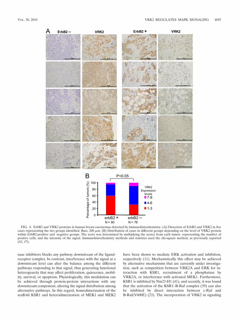
Inverse correlation between VRK2 and ErbB2 expressionlevels in breast carcinomas. The above-mentioned resultsshowing the effect of VRK2A on MAPK signaling suggestedthat in those tumors in which the MAPK pathway is activated,VRK2 is likely to be downregulated to reduce the block insignal transmission. Therefore, we determined if there was anycorrelation between VRK2 and the ErbB2 receptor proteinlevels, using breast cancer as a model. These two proteins weredetermined by immunohistochemistry in 136 cases of humanbreast carcinomas. Interestingly, an inverse correlation wasfound between VRK2 expression and ErbB2 protein levels(Fig. 8A and B). VRK2 was mostly located in endoplasmic
reticulum membranes, as expected (4), independent of theErbB2 status (see Fig. S6 in the supplemental material).
The correlation between ER or PR and VRK2 was alsodetermined in these carcinomas. Both receptors were posi-tively correlated with VRK2 expression, and thus, tumors pos-itive for ER or PR also had higher levels of VRK2 than thosenegative for the receptors (see Fig. S7 in the supplementalmaterial), which is consistent with the inverse correlation be-tween VRK2 and ErbB2 detected in this work.
DISCUSSION
Signaling by the ErbB family of receptors is partly transmit-ted by the RAF-MEK-ERK pathway, activating transcriptionthat promotes survival and expansion of the cell population.Modulation of MAPK signaling by interaction of its canonicalcomponents with other proteins is an emerging theme in signaltransduction, highlighting the importance of signaling pathwaycross talk and its contribution to signal specificity (45). Thus,the incorporation of annexin A6 in H-Ras-containing com-
FIG. 5. (A) Effects of knocking down scaffold proteins, JIP1 (left) and KSR1 (right), on the SRE-dependent transcriptional response to EGFin HeLa cells. ***, P � 0.001. The error bars indicate standard deviations. (B) Effects of siVRK2 and siKSR1 on the SRE-dependenttranscriptional response to EGF in HeLa cells. **, P � 0.005; n.s., not significant. (C) Effect of VRK2A in response to EGF after knockdown ofKSR1 in MCF7 cells. *, P � 0.01. (D) Effects of KSR1 and VRK2 knockdown on phospho-Erk levels in MDA-BB-231 cells in the presence ofEGF. The effect on the activation of the SRE promoter is also shown (right). The change in luciferase activity resulting from activation of theSRE-luc reporter coincides with the result for phospho-ERK (left).
VOL. 30, 2010 VRK2 REGULATES MAPK SIGNALING 4693

plexes inhibits EGFR and Ras signaling in breast cancer cells(12). Also, thyroid receptor �1 blocks the response to EGF,IGF1, and transforming growth factor � (TGF-�) functioningas a suppressor of tumor invasion (32). The VRK2A protein,
by interacting with the JIP1 scaffold protein, blocks JNK acti-vation in response to interleukin 1� or hypoxia (6, 7).
Blocking of EGF or its receptor (ErbB) at the membranelevel by monoclonal antibodies or small-molecule tyrosine ki-
FIG. 6. (A) Colocalization of KSR1 with calnexin, an endoplasmic reticulum marker, in three cell lines. Bars, 10 �m. (B) Colocalization ofVRK2 and KSR1 in HeLa cells by confocal microscopy. Bars, 10 �m.
FIG. 7. (A) Interaction between VRK2A and the KSR1 scaffold. 293T cells were transfected with plasmid pCMV-Flag-KSR1 or pCMV-Flag(control) and pCEFL-GST-VRK2A or pCEFL-GST, the cell extract was immunoprecipitated (IP) with an anti-Flag antibody, and the proteins presentin the immunoprecipitate were detected with antibodies for the epitopes. For the pulldown (PD) assay, cells were transfected with pCEFL-GST-VRK2Aor pCEFL-GST, and endogenous KSR1 was detected with a specific antibody. (B) Interaction of VRK2A with MEK. HEK293T cells were transfectedwith plasmid pCEFL-GST VRK2A or pCEFL-GST as a control, in combination with plasmid pCEFL-HA-MEK. The lysates were used for pulldown,and proteins (right) were detected by immunoblotting. (C) Direct interaction between endogenous MEK1 and VRK2 proteins in MCF7 cells.Endogenous MEK1 was immunoprecipitated with 9G3 monoclonal antibody (Ab), and the endogenous VRK2 protein present in the immunoprecipitatewas detected by Western blotting.
4694 FERNANDEZ ET AL. MOL. CELL. BIOL.
nase inhibitors blocks any pathway downstream of the ligand-receptor complex. In contrast, interference with the signal at adownstream level can alter the balance among the differentpathways responding to that signal, thus generating functionalheterogeneity that may affect proliferation, quiescence, mobil-ity, survival, or apoptosis. Physiologically, this modulation canbe achieved through protein-protein interactions with anydownstream component, altering the signal distribution amongalternative pathways. In this regard, homodimerization of thescaffold KSR1 and heterodimerization of MEK1 and MEK2
have been shown to mediate ERK activation and inhibition,respectively (11). Mechanistically this effect may be achievedby alternative mechanisms that are currently under investiga-tion, such as competition between VRK2A and ERK for in-teraction with KSR1, recruitment of a phosphatase byVRK2A, or interference with activated MEK1. Furthermore,KSR1 is inhibited by Nm23-H1 (41), and recently, it was foundthat the activation of the KSR1–B-Raf complex (39) can alsobe inhibited by direct interaction between c-Raf andB-Raf(V600E) (23). The incorporation of VRK2 in signaling
FIG. 8. ErbB2 and VRK2 proteins in human breast carcinomas detected by immunohistochemistry. (A) Detection of ErbB2 and VRK2 in fivecases representing the two groups identified. Bars, 200 �m. (B) Distribution of cases in different groups depending on the level of VRK2 proteinwithin ErbB2-positive and -negative groups. The score was determined by multiplying the scores from each tumor, representing the number ofpositive cells, and the intensity of the signal. Immunohistochemistry methods and statistics used the chi-square method, as previously reported(42, 47).
VOL. 30, 2010 VRK2 REGULATES MAPK SIGNALING 4695
complexes assembled on KSR1, and its regulation, is a processthat will need to be further characterized, since KSR1 alsointeracts with members of the 14-3-3 protein family, whichmodulates signal transduction (20), and these alternative com-plexes can modify the balance among different signals andbiological effects. The subcellular location of KSR1 in thecytoplasm is partly dispersed and partly colocalized with en-dogenous calnexin, an endoplasmic reticulum marker, but theproportions vary depending on the cell line (Fig. 6A). KSR1 ismostly cytoplasmic and has no membrane-spanning sequenceor hydrophobic region (35, 51). Thus, its association with theendoplasmic reticulum must occur through interactions withproteins anchored to the endoplasmic reticulum, as is the casewith VRK2A (4), or with common interacting proteins thatfunction as a bridge between them.
In ErbB2� breast tumors, we have detected a significantreduction in VRK2 levels, but not lack of expression, whichfunctionally may alter the balance among EGF- or ErbB2-dependent pathways. VRK2 downregulation resulted in in-creased levels of p-ERK-, p-RSK-, and SRE-dependent tran-scriptional response, while overexpression of VRK2 had theopposite effect. This is a consequence of blocking the MAPKsignaling pathway at the level of its last component; thus, notall EGF response routes are likely to be affected, as is the casewith the AKT-mediated response. Hence, the low level ofVRK2 in ErbB2� tumors suggests that the proliferative signalis stronger, and thus, the likelihood of a more aggressive tumoris increased. The identification of VRK2A as a downstreammodulator of ErbB2 can have wider implications than thoserelated to breast carcinoma. The alteration of MAPK signalingby mutation of some of its components is common to manycarcinomas. Thus, a specific B-Raf(V600E) mutation is com-monly detected in melanomas (38), and Ras mutations aredetected in many types of carcinomas (8). In these cancers, theprediction is that the level of VRK2A will be downregulated intumors with any of these mutations compared with those thatdo not have alterations in MAPK signaling. This is consistentwith the loss of heterozygosity detected in the VRK2 locus inthe lung (3) and adrenocortical carcinomas (25).
In this work, we have shown that VRK2A, a cytoplasmickinase, is able to reduce the signal from ErbB2 or EGF. Thus,in those tumors, like breast carcinomas, that have amplifiedErbB2, its signal could be potentiated if there was a reductionin the levels of VRK2A, which functions as an inhibitor. Theeffect of removing an inhibitor of the MAPK route is consistentwith the relative importance of this signaling pathway in car-cinomas. Thus, in colon carcinomas treated with cetuximab, ifthere is an activating Ras mutation, the effect of the antibodyis reduced, pointing to the importance of the route (2, 22). Theuse of targeted therapies directed at kinases (40) will requirethe characterization of the mutational status of signaling mol-ecules downstream in the pathway, such as Ras or B-Raf, aswell as that of modulators of the signal, such as VRK2, par-ticularly in the subgroup with high ErbB2 and VRK2 levels.However, at this time, it is not known if this VRK2 downregu-lation will identify a subpopulation of ErbB2� tumors or iftherapeutic decisions will be affected by this change. Betterselection of patients based on signaling criteria might contrib-ute to achieving better clinical responses to current inhibitors,either antibodies or small molecules. Thus, identification of
new regulators opens up an area for potential intervention inthose tumors that, because of downstream mutations, such asin Ras or Raf, can make patients less responsive to currenttherapies directed at the EGF/ErbB2 signaling pathway.
ACKNOWLEDGMENTS
This work was funded by grants from the Ministerio de Educacion yCiencia (SAF2007-60242, SAF2010-14935, and CSD2007-0017), Juntade Castilla y Leon (Consejería de Educacion, CSI14A08; Consejeríade Sanidad, GR-15), Fundacion Sandra Ibarra to P.A.L. and grantsBFU2007-66100 and SAF2010-20203 from the Ministerio de Educa-cion y Ciencia to J.L. I.F.F. has a JAE-CSIC predoctoral fellowship.
REFERENCES
1. Albanese, C., J. Johnson, G. Watanabe, N. Eklund, D. Vu, A. Arnold, andR. G. Pestell. 1995. Transforming p21ras mutants and c-Ets-2 activate thecyclin D1 promoter through distinguishable regions. J. Biol. Chem. 270:23589–23597.
2. Amado, R. G., M. Wolf, M. Peeters, E. Van Cutsem, S. Siena, D. J. Freeman,T. Juan, R. Sikorski, S. Suggs, R. Radinsky, S. D. Patterson, and D. D.Chang. 2008. Wild-type KRAS is required for panitumumab efficacy inpatients with metastatic colorectal cancer. J. Clin. Oncol. 26:1626–1634.
3. Benachenhou, N., S. Guiral, I. Gorska-Flipot, D. Labuda, and D. Sinnett.1998. High resolution deletion mapping reveals frequent allelic losses at theDNA mismatch repair loci hMLH1 and hMSH3 in non-small cell lungcancer. Int. J. Cancer 77:173–180.
4. Blanco, S., L. Klimcakova, F. M. Vega, and P. A. Lazo. 2006. The subcellularlocalization of vaccinia-related kinase-2 (VRK2) isoforms determines theirdifferent effect on p53 stability in tumour cell lines. FEBS J. 273:2487–2504.
5. Blanco, S., and P. A. Lazo. 2009. Vaccinia-related kinase-2. UCSD-NatureMolecule Page doi:10.1038/mp.a000905.01.
6. Blanco, S., C. Santos, and P. A. Lazo. 2007. Vaccinia-related kinase 2modulates the stress response to hypoxia mediated by TAK1. Mol. Cell. Biol.27:7273–7283.
7. Blanco, S., M. Sanz-Garcia, C. R. Santos, and P. A. Lazo. 2008. Modulationof interleukin-1 transcriptional response by the interaction between VRK2and the JIP1 scaffold protein. PLoS One 3:e1660.
8. Bos, J. L. 1989. ras oncogenes in human cancer: a review. Cancer Res.49:4682–4689.
9. Burgess, A. W. 2008. EGFR family: structure physiology signalling and ther-apeutic targets. Growth Factors 26:263–274.
10. Casar, B., I. Arozarena, V. Sanz-Moreno, A. Pinto, L. Agudo-Ibanez, R.Marais, R. E. Lewis, M. T. Berciano, and P. Crespo. 2009. Ras subcellularlocalization defines extracellular signal-regulated kinase 1 and 2 substratespecificity through distinct utilization of scaffold proteins. Mol. Cell. Biol.29:1338–1353.
11. Chen, C., R. E. Lewis, and M. A. White. 2008. IMP modulates KSR1-dependent multivalent complex formation to specify ERK1/2 pathway acti-vation and response thresholds. J. Biol. Chem. 283:12789–12796.
12. de Muga, S. V., P. Timpson, L. Cubells, R. Evans, T. E. Hayes, C. Rentero,A. Hegemann, M. Reverter, J. Leschner, A. Pol, F. Tebar, R. J. Daly, C.Enrich, and T. Grewal. 2009. Annexin A6 inhibits Ras signalling in breastcancer cells. Oncogene 28:363–377.
13. Dhanasekaran, D. N., K. Kashef, C. M. Lee, H. Xu, and E. P. Reddy. 2007.Scaffold proteins of MAP-kinase modules. Oncogene 26:3185–3202.
14. Dhillon, A. S., S. Hagan, O. Rath, and W. Kolch. 2007. MAP kinase signal-ling pathways in cancer. Oncogene 26:3279–3290.
15. Di Cosimo, S., and J. Baselga. 2008. Targeted therapies in breast cancer:where are we now? Eur. J. Cancer 44:2781–2790.
16. Fehrenbacher, N., D. Bar-Sagi, and M. Philips. 2009. Ras/MAPK signalingfrom endomembranes. Mol. Oncol. 3:297–307.
17. Gorjanacz, M., E. P. Klerkx, V. Galy, R. Santarella, C. Lopez-Iglesias, P.Askjaer, and I. W. Mattaj. 2008. Caenorhabditis elegans BAF-1 and itskinase VRK-1 participate directly in post-mitotic nuclear envelope assembly.EMBO J. 26:132–143.
18. Hynes, N. E., and H. A. Lane. 2005. ERBB receptors and cancer: the com-plexity of targeted inhibitors. Nat. Rev. Cancer. 5:341–354.
19. Ikediobi, O. N., H. Davies, G. Bignell, S. Edkins, C. Stevens, S. O’Meara, T.Santarius, T. Avis, S. Barthorpe, L. Brackenbury, G. Buck, A. Butler, J.Clements, J. Cole, E. Dicks, S. Forbes, K. Gray, K. Halliday, R. Harrison, K.Hills, J. Hinton, C. Hunter, A. Jenkinson, D. Jones, V. Kosmidou, R. Lugg,A. Menzies, T. Mironenko, A. Parker, J. Perry, K. Raine, D. Richardson, R.Shepherd, A. Small, R. Smith, H. Solomon, P. Stephens, J. Teague, C. Tofts,J. Varian, T. Webb, S. West, S. Widaa, A. Yates, W. Reinhold, J. N. Wein-stein, M. R. Stratton, P. A. Futreal, and R. Wooster. 2006. Mutation analysisof 24 known cancer genes in the NCI-60 cell line set. Mol. Cancer Ther.5:2606–2612.
20. Jagemann, L. R., L. G. Perez-Rivas, E. J. Ruiz, J. A. Ranea, F. Sanchez-
4696 FERNANDEZ ET AL. MOL. CELL. BIOL.
Jimenez, A. R. Nebreda, E. Alba, and J. Lozano. 2008. The functionalinteraction of 14-3-3 proteins with the ERK1/2 scaffold KSR1 occurs in anisoform-specific manner. J. Biol. Chem. 283:17450–17462.
21. Kang, T. H., D. Y. Park, W. Kim, and K. T. Kim. 2008. VRK1 phosphorylatesCREB and mediates CCND1 expression. J. Cell Sci. 121:3035–3041.
22. Karapetis, C. S., S. Khambata-Ford, D. J. Jonker, C. J. O’Callaghan, D. Tu,N. C. Tebbutt, R. J. Simes, H. Chalchal, J. D. Shapiro, S. Robitaille, T. J.Price, L. Shepherd, H. J. Au, C. Langer, M. J. Moore, and J. R. Zalcberg.2008. K-ras mutations and benefit from cetuximab in advanced colorectalcancer. N. Engl. J. Med. 359:1757–1765.
23. Karreth, F. A., G. M. DeNicola, S. P. Winter, and D. A. Tuveson. 2009. C-Rafinhibits MAPK activation and transformation by B-Raf(V600E). Mol. Cell36:477–486.
24. Kizaka-Kondoh, S., K. Sato, K. Tamura, H. Nojima, and H. Okayama. 1992.Raf-1 protein kinase is an integral component of the oncogenic signal cas-cade shared by epidermal growth factor and platelet-derived growth factor.Mol. Cell. Biol. 12:5078–5086.
25. Kjellman, M., L. Roshani, B. T. Teh, O. P. Kallioniemi, A. Hoog, S. Gray,L. O. Farnebo, M. Holst, M. Backdahl, and C. Larsson. 1999. Genotyping ofadrenocortical tumors: very frequent deletions of the MEN1 locus in 11q13and of a 1-centimorgan region in 2p16. J. Clin. Endocrinol. Metab. 84:730–735.
26. Klerkx, E. P., P. A. Lazo, and P. Askjaer. 2009. Emerging biological functionsof the Vaccinia-Related Kinase (VRK) family. Histol. Histopathol. 24:749–759.
27. Klijn, J. G., M. P. Look, H. Portengen, J. Alexieva-Figusch, W. L. van Putten,and J. A. Foekens. 1994. The prognostic value of epidermal growth factorreceptor (EGF-R) in primary breast cancer: results of a 10 year follow-upstudy. Breast Cancer Res. Treat. 29:73–83.
28. Kolch, W. 2005. Coordinating ERK/MAPK signalling through scaffolds andinhibitors. Nat. Rev. Mol. Cell Biol. 6:827–837.
29. Lopez-Borges, S., and P. A. Lazo. 2000. The human vaccinia-related kinase1 (VRK1) phosphorylates threonine-18 within the mdm-2 binding site of thep53 tumour suppressor protein. Oncogene 19:3656–3664.
30. Martin, K. J., D. R. Patrick, M. J. Bissell, and M. V. Fournier. 2008.Prognostic breast cancer signature identified from 3D culture model accu-rately predicts clinical outcome across independent datasets. PLoS One3:e2994.
31. Martinez-Carpio, P. A., C. Mur, P. Rosel, and M. A. Navarro. 1999. Con-stitutive and regulated secretion of epidermal growth factor and transform-ing growth factor-beta1 in MDA-MB-231 breast cancer cell line in 11-daycultures. Cell Signal. 11:753–757.
32. Martinez-Iglesias, O., S. Garcia-Silva, S. P. Tenbaum, J. Regadera, F.Larcher, J. M. Paramio, B. Vennstrom, and A. Aranda. 2009. Thyroid hor-mone receptor beta1 acts as a potent suppressor of tumor invasiveness andmetastasis. Cancer Res. 69:501–509.
33. McKay, M. M., and D. K. Morrison. 2007. Integrating signals from RTKs toERK/MAPK. Oncogene 26:3113–3121.
34. Michaloglou, C., L. C. Vredeveld, M. S. Soengas, C. Denoyelle, T. Kuilman,C. M. van der Horst, D. M. Majoor, J. W. Shay, W. J. Mooi, and D. S. Peeper.2005. BRAFE600-associated senescence-like cell cycle arrest of humannaevi. Nature 436:720–724.
35. Muller, J., S. Ory, T. Copeland, H. Piwnica-Worms, and D. K. Morrison.2001. C-TAK1 regulates Ras signaling by phosphorylating the MAPK scaf-fold, KSR1. Mol. Cell 8:983–993.
36. Nichols, R. J., and P. Traktman. 2004. Characterization of three paralogousmembers of the mammalian vaccinia related kinase family. J. Biol. Chem.279:7934–7946.
37. Nichols, R. J., M. S. Wiebe, and P. Traktman. 2006. The vaccinia-relatedkinases phosphorylate the N� terminus of BAF, regulating its interactionwith DNA and its retention in the nucleus. Mol. Biol. Cell 17:2451–2464.
38. Patton, E. E., H. R. Widlund, J. L. Kutok, K. R. Kopani, J. F. Amatruda,R. D. Murphey, S. Berghmans, E. A. Mayhall, D. Traver, C. D. Fletcher, J. C.Aster, S. R. Granter, A. T. Look, C. Lee, D. E. Fisher, and L. I. Zon. 2005.BRAF mutations are sufficient to promote nevi formation and cooperatewith p53 in the genesis of melanoma. Curr. Biol. 15:249–254.
39. Rajakulendran, T., M. Sahmi, M. Lefrancois, F. Sicheri, and M. Therrien.2009. A dimerization-dependent mechanism drives RAF catalytic activation.Nature 461:542–545.
40. Roberts, P. J., and C. J. Der. 2007. Targeting the Raf-MEK-ERK mitogen-activated protein kinase cascade for the treatment of cancer. Oncogene26:3291–3310.
41. Salerno, M., D. Palmieri, A. Bouadis, D. Halverson, and P. S. Steeg. 2005.Nm23-H1 metastasis suppressor expression level influences the bindingproperties, stability, and function of the kinase suppressor of Ras1 (KSR1)Erk scaffold in breast carcinoma cells. Mol. Cell. Biol. 25:1379–1388.
42. Santos, C. R., M. Rodriguez-Pinilla, F. M. Vega, J. L. Rodriguez-Peralto, S.Blanco, A. Sevilla, A. Valbuena, T. Hernandez, A. J. van Wijnen, F. Li, E. deAlava, M. Sanchez-Cespedes, and P. A. Lazo. 2006. VRK1 signaling pathwayin the context of the proliferation phenotype in head and neck squamous cellcarcinoma. Mol. Cancer Res. 4:177–185.
43. Sevilla, A., C. R. Santos, R. Barcia, F. M. Vega, and P. A. Lazo. 2004. c-Junphosphorylation by the human vaccinia-related kinase 1 (VRK1) and itscooperation with the N-terminal kinase of c-Jun (JNK). Oncogene 23:8950–8958.
44. Sevilla, A., C. R. Santos, F. M. Vega, and P. A. Lazo. 2004. Human vaccinia-related kinase 1 (VRK1) activates the ATF2 transcriptional activity by novelphosphorylation on Thr-73 and Ser-62 and cooperates with JNK. J. Biol.Chem. 279:27458–27465.
45. Shin, S. Y., O. Rath, S. M. Choo, F. Fee, B. McFerran, W. Kolch, and K. H.Cho. 2009. Positive- and negative-feedback regulations coordinate the dy-namic behavior of the Ras-Raf-MEK-ERK signal transduction pathway.J. Cell Sci. 122:425–435.
46. Valbuena, A., I. Lopez-Sanchez, and P. A. Lazo. 2008. Human VRK1 is anearly response gene and its loss causes a block in cell cycle progression. PLoSOne 3:e1642.
47. Valbuena, A., A. Suarez-Gauthier, F. Lopez-Rios, A. Lopez-Encuentra, S.Blanco, P. L. Fernandez, M. Sanchez-Cespedes, and P. A. Lazo. 2007. Al-teration of the VRK1-p53 autoregulatory loop in human lung carcinomas.Lung Cancer 58:303–309.
48. Valbuena, A., F. M. Vega, S. Blanco, and P. A. Lazo. 2006. p53 downregulatesits activating vaccinia-related kinase 1, forming a new autoregulatory loop.Mol. Cell. Biol. 26:4782–4793.
49. Vega, F. M., P. Gonzalo, M. L. Gaspar, and P. A. Lazo. 2003. Expression ofthe VRK (vaccinia-related kinase) gene family of p53 regulators in murinehematopoietic development. FEBS Lett. 544:176–180.
50. Vega, F. M., A. Sevilla, and P. A. Lazo. 2004. p53 stabilization and accumu-lation induced by human vaccinia-related kinase 1. Mol. Cell. Biol. 24:10366–10380.
51. Xing, H., K. Kornfeld, and A. J. Muslin. 1997. The protein kinase KSRinteracts with 14-3-3 protein and Raf. Curr. Biol. 7:294–300.
52. Yarden, Y., and M. X. Sliwkowski. 2001. Untangling the ErbB signallingnetwork. Nat. Rev. Mol. Cell Biol. 2:127–137.
53. Yuste, L., J. C. Montero, A. Esparis-Ogando, and A. Pandiella. 2005. Acti-vation of ErbB2 by overexpression or by transmembrane neuregulin resultsin differential signaling and sensitivity to herceptin. Cancer Res. 65:6801–6810.
54. Zandi, R., A. B. Larsen, P. Andersen, M. T. Stockhausen, and H. S. Poulsen.2007. Mechanisms for oncogenic activation of the epidermal growth factorreceptor. Cell. Signal. 19:2013–2023.
VOL. 30, 2010 VRK2 REGULATES MAPK SIGNALING 4697

Figure S1
Figure S1A. Effect of VRK2 overexpression on transcriptional response mediated by SRE inHeLa cells stimulated with EGF. HeLa cells were transfected with plasmid pCEFL-HA-VRK2A, pCEFL-HA-VRK1 or pCEFL-HA-VRK2B. Forty-eight hours after transfection 10nM EGF was added to the culture for 10 minutes. Luciferase was determined using a dual-luciferase reporter system. Mean of three independent experiments. P<0.05. At the bottom isshown a western blot to detect the transfected proteins and the loading control. The threeVRK proteins were detected with an anti-HA antibody, the common tag epitope in the threeVRK proteins.
Vector 0 1 1.5 2
VRK1 VRK2BVRK2A
α-HA (VRK)
β-actin
0
1
2
3
4
EGF 10nM (10 min)
***
Rel
ativ
e lu
cife
rase
act
ivat
ion
1.5 (µg)1.5
A
Figure S1B. Effect of VRK proteins overexpression on the transcriptional response to EGF ofcyclin D1 promoter in MCF7 cells. MCF7 cells were transfected with 1 μg of plasmidpA·cyclin D1 (-1720)-Luc construct, Cells were serum starved for 12 hours before stimulationwith 10 nM EGF for 6 hours. Ct: control with empty vector plasmid. ** (P<0.001). *** (P<0.0005).
B
012345678
Rel
ativ
e lu
cife
rase
act
ivat
ion
EGF 10 nM
VRK2A VRK1Ct Ct
******
**

Figure S2
Figure S2A. Quantification of VRK2A protein level in HeLa cells that were transfected with siVRK2-06 or siControl (Fig. 2). Data are presented as the mean of three independent experiments.
Rel
ativ
e pr
otei
n le
vel
0
1
siVRK2-06siControl
HeLa cellsA
Figure S2B.Quantification by semiquantitative RT-PCR of the VRK2 mRNA in MCF7 cellstransfected with si-VRK2-06 or si-control (left) and effect on VRK2 protein level (middle)showing it has no effect on protein despite the effective knockdown of its RNA. Therefore thislack of VRK2 protein reduction predicts that in this cell line on EGF stimulation of transcriptionshould not be affected as shown using the pSRE-Luc reporter plasmid (right).
siControl
siVRK2
00,20,40,6
0,81
1,2
VRK2
1
1
Rel
ativ
e R
NA
leve
l
B
VRK2AVRK2B
β-actin
si-C
ont
si-V
RK2
-06
MCF7
0
20
40
60
80
100
120
si-control
EGF
Rel
ativ
e lu
cife
rase
act
ivat
ion
si-VRK2-06

Figure S3
C
Rel
ativ
e fo
ld a
ctiv
atio
n
H-RasG12V (ng)
0
10
20
30
40
50
60
5 10 20 50 100 3000
Figure S3. A. Dose–dependent activation of transcription induced by ErbB2in HEK293T cells. B. Inhibition of ErbB2 (500 ng) activation oftranscription by VRK2 in a dose-dependent manner. C. Dose–dependentactivation of transcription induced by K-RasG12V. D. Inhibition of H-RasG12V
(200 ng) activation of transcription by VRK2 in a dose-dependent manner.These experiments were performed in HEK293T cells. Luciferase activitywas measured with an SRE-Luc reporter plasmid. In control points (0) themaximaun amount of empty vector was used, and the specific points it wasused to reach the same amount of DNA in transfections.
B
0 0 50 250 500 25000
2
4
6
8
10
12
VRK2A (ng)
VRK2A
ErbB2
β-actin
500 ng ErbB20 ng
Rel
ativ
e fo
ld a
ctiv
atio
n
D
VRK2A
β-actin
Rel
ativ
e fo
ld a
ctiv
atio
n
0 0 250 500 10002500VRK2A ( ng)
0
10
20
30
40
50
60 H-RasG12V (200 ng)
RasRas
β-actin
A
02468
1012141618
100 200 500 1000ErbB2 (ng)
Rel
ativ
e fo
ld a
ctiv
atio
n
ErbB2β-actin
0

Figure S4
ErbB2
α-HA( VRKs)
α -p-ERK1/2
ERK 1/2
p-AKT
AKT
ErbB2
p-MEK 1/2
MEK 1/2
Figure S4. VRK2A overexpression leads to reduced, ERK1/2 phosphorylation inresponse to ErbB2 overexpression, without affecting AKT or MEK1/2phosphorylation, in HEK293T cell line. VRK1 and VRKK2B overexpression does notaffect ERK1/2 phosphorylation. HEK293T cells were transfected with thecorreponding plasmids and forty-eight hours later the lysates were used forimmunoblot analysis with specific antibodies indicated in the Methods section.

Figure S5
Figure S5. Lack of interaction between VRK2A and B-RafV600E (left) or ERK-HA(right).A 293T cells were transfected with plasmid pCEFL-GST-VRK2A or pCEFL-GST as control in combination with plasmids pG12B-RafV600E or empty vector. Afterforty-eight hours cell lysates (left) were used for pull-down (PD), and the proteins in it(right) detected by western blot with the corresponding antibodies. B. 293T cells weretransfected with plasmids pCEFL-GST-VRK2A or pCEFL-GST as control incombination with pCEFL-HA-ERK1 or empty vector.
A
B-RafV600E
GST PDGST
lysate pulldown
B
HA-(ERK)
GST PD
lysate pulldown
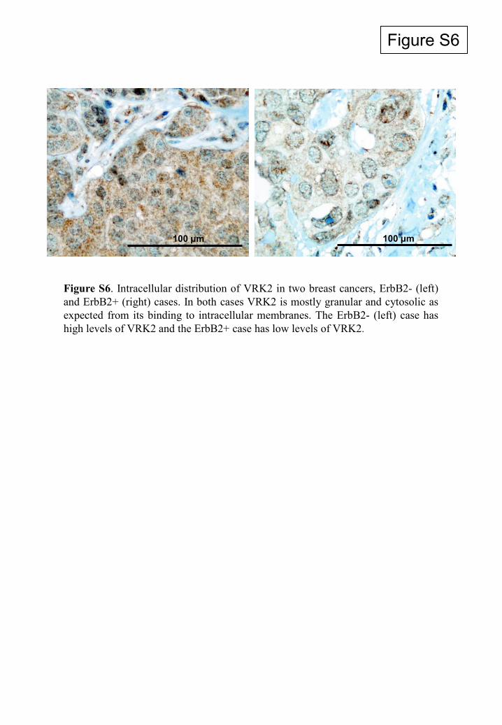
Figure S6
Figure S6. Intracellular distribution of VRK2 in two breast cancers, ErbB2- (left)and ErbB2+ (right) cases. In both cases VRK2 is mostly granular and cytosolic asexpected from its binding to intracellular membranes. The ErbB2- (left) case hashigh levels of VRK2 and the ErbB2+ case has low levels of VRK2.
100 μm 100 μm
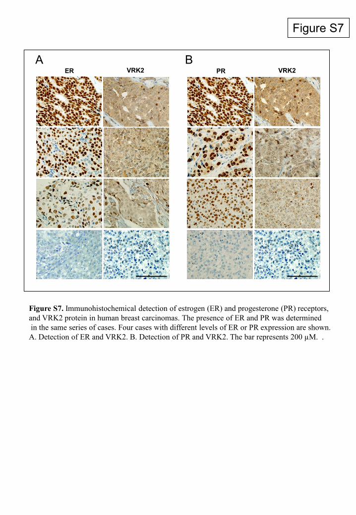
Figure S7
ER VRK2 PR VRK2 A B
Figure S7. Immunohistochemical detection of estrogen (ER) and progesterone (PR) receptors,and VRK2 protein in human breast carcinomas. The presence of ER and PR was determinedin the same series of cases. Four cases with different levels of ER or PR expression are shown.A. Detection of ER and VRK2. B. Detection of PR and VRK2. The bar represents 200 µM. .
Copyright © 2022 FDOKUMEN