Combination of TLR2 and TLR3 agonists derepress infectious ...
Please cite this article in press as: Thaikoottathil, J., Chu, H.W., MAPK/AP-1 activation mediates TLR2 agonist-induced SPLUNC1 expression in
human lung epithelial cells. Mol. Immunol. (2011), doi:10.1016/j.molimm.2011.08.005
ARTICLE IN PRESSG Model
MIMM-3785; No. of Pages 8
Molecular Immunology xxx (2011) xxx– xxx
Contents lists available at ScienceDirect
Molecular Immunology
jo u rn al hom epa ge: www.elsev ier .com/ locate /mol imm
MAPK/AP-1 activation mediates TLR2 agonist-induced SPLUNC1 expression in
human lung epithelial cells
Jyoti Thaikoottathil a, Hong Wei Chua,b,∗
a Department of Medicine, National Jewish Health, University of Colorado Denver, Denver, CO, USAb Department of Immunology, National Jewish Health, University of Colorado Denver, Denver, CO, USA
a r t i c l e i n f o
Article history:
Received 21 June 2011
Received in revised form 2 August 2011
Accepted 6 August 2011
Available online xxx
Keywords:
Lung epithelium
SPLUNC1
TLR2
MAPK
AP-1
Gene regulation
a b s t r a c t
Background: Short Palate Lung and Nasal epithelium Clone 1 (SPLUNC1) is a newly described host defense
protein, primarily expressed in large airway epithelial cells. Reduced SPLUNC1 has been reported in aller-
gic and cigarette smoke-exposed airways. We found that Mycoplasma pneumoniae increases SPLUNC1
in airway epithelium in part via activating TLR2-NF-�B pathway. However, the contribution of addi-
tional signaling pathways to TLR2-mediated SPLUNC1 expression remains unclear. In the present study,
we investigated if TLR2-induced mitogen-activated protein kinase (MAPK)/activator protein-1 (AP-1)
signaling regulates SPLUNC1 expression in human lung epithelial cells.
Methods: Human lung epithelial NCI-H292 cells were stimulated with a TLR2 agonist Palmitoyl (3)-Cys-
Ser-Lys (4)-OH (Pam3CSK4). MAPK/AP-1 activation and its role in SPLUNC1 regulation were investigated
by Western blot, c-Jun activation assay, chromatin immunoprecipitation (ChIP) and real-time PCR.
SPLUNC1 promoter activity was assessed by a luciferase reporter assay.
Results: Pam3CSK4 increased SPLUNC1 expression in NCI-H292 cells in a dose- and time-dependent man-
ner, and enhanced SPLUNC1 promoter activity. Pam3CSK4-treated cells demonstrated activated MAPK
and c-Jun compared to untreated cells. ChIP assay indicated increased c-Jun binding to the SPLUNC1 pro-
moter following Pam3CSK4 stimulation. Inhibition of ERK1/2 significantly reduced Pam3CSK4-mediated
c-Jun activation and SPLUNC1 expression.
Conclusions: Our results for the first time demonstrate that TLR2-mediated MAPK/AP-1 activation up-
regulates lung epithelial SPLUNC1 expression at the transcriptional level. Understanding SPLUNC1 gene
regulation should provide more specific therapeutic targets to restore deficient SPLUNC1 production in
diseased airways.
© 2011 Elsevier Ltd. All rights reserved.
1. Introduction
Human lungs are constantly challenged with harmful environ-
mental agents including infectious and non-infectious stimuli that
initiate innate and adaptive immune responses in the airways. Air-
way epithelial cells provide an active mucosal barrier to protect
lungs against various microbial and non-infectious agents in part
through Toll-like receptors (TLRs) (Lafferty et al., 2010; Opitz et al.,
Abbreviations: ACD, actinomycin D; AP-1, activator protein-1; ChIP, chromatin
immunoprecipitation; ERK1/2, extracellular signal response kinase 1/2; JNK1/2,
c-Jun N-terminal kinase 1/2; MAPK, mitogen-activated protein kinase; MEK1/2,
mitogen-activated protein kinase kinase 1/2; Pam3CSK4, Palmitoyl (3)-Cys-Ser-Lys
(4)-OH; SPLUNC1, Short Palate, Lung and Nasal epithelium Clone 1; TLR2, Toll-like
receptor 2.∗ Corresponding author at: Department of Medicine, National Jewish Health,
Room A639, 1400 Jackson Street, Denver, CO 80206, USA. Tel.: +1 303 398 1689;
fax: +1 303 270 2319.
E-mail address: [email protected] (H.W. Chu).
2010; Randhawa and Hawn, 2008). Short Palate, Lung and Nasal
epithelium Clone 1 (SPLUNC1), a newly described host defense
protein, is abundant in airway lining fluid of healthy individu-
als, and is primarily expressed and secreted form large airway
epithelial cells (Bingle and Bingle, 2000). SPLUNC1 is significantly
reduced in allergic and cigarette smoke-exposed airways, which
may render the hosts more susceptible to bacterial infections (Chu
et al., 2007; Steiling et al., 2009). Our group as well as others
have reported antimicrobial activity of SPLUNC1 against several
respiratory pathogens including Mycoplasma pneumoniae and Pseu-
domonas aeruginosa (Chu et al., 2007; Gally et al., 2011; Lukinskiene
et al., 2011; Zhou et al., 2008).
Although SPLUNC1 is an abundant protein in the airways, its
gene regulation under basal as well as diseased conditions such as
infection is poorly understood. We have shown that TLR2 activation
by M. pneumoniae and a TLR2 agonist Palmitoyl (3)-Cys-Ser-Lys (4)-
OH (Pam3CSK4) enhanced SPLUNC1 expression in airway epithelial
cells in part via activating NF-�B signaling pathway (Chu et al.,
2010). This leads to the question of what other transcription
0161-5890/$ – see front matter © 2011 Elsevier Ltd. All rights reserved.doi:10.1016/j.molimm.2011.08.005
Please cite this article in press as: Thaikoottathil, J., Chu, H.W., MAPK/AP-1 activation mediates TLR2 agonist-induced SPLUNC1 expression in
human lung epithelial cells. Mol. Immunol. (2011), doi:10.1016/j.molimm.2011.08.005
ARTICLE IN PRESSG Model
MIMM-3785; No. of Pages 8
2 J. Thaikoottathil, H.W. Chu / Molecular Immunology xxx (2011) xxx– xxx
factors also regulate SPLUNC1 at the transcriptional and/or post-
transcriptional levels.
Mammalian cells express multiple mitogen-activated protein
kinases (MAPK) to mediate the effects of extracellular signals on
a wide variety of biological processes including cell growth, pro-
liferation, differentiation and apoptosis (Garrington and Johnson,
1999). One of the downstream events following MAPK activation
is activation and nuclear translocation of transcription factor c-Jun,
a member of the activator protein-1 (AP-1) family. Activation of
TLRs including TLR2 has been shown to activate MAPK/AP-1 signal-
ing in airway epithelial cells, which may promote the production
of inflammatory mediators and host defense proteins such as �-
defensin-2 (Scharf et al., 2010; Schmeck et al., 2006). However,
the role of TLR2-mediated MAPK/AP-1 activation in SPLUNC1 reg-
ulation remains to be determined. The aim of the present study
is to investigate if TLR2-induced MAPK/AP-1 signaling regulates
SPLUNC1 expression in lung epithelial cells. Understanding the
functional role of key transcription factors/signaling pathways in
SPLUNC1 gene regulation can provide new targets for add-on ther-
apy aimed at improving mucosal immunity in diseased airways.
2. Materials and methods
2.1. Lung epithelial cell culture
NCI-H292 cells, a human pulmonary mucoepidermoid carci-
noma cell line (ATCC, Manassas, VA) were cultured in RPMI-1640
medium supplemented with 10% FBS and penicillin-streptomycin
at 37 ◦C, 5% CO2. We selected NCI-H292 cells to study TLR2-
mediated SPLUNC1 gene regulation because in our previous
experiments they have been shown to express SPLUNC1 in
response to TLR2 stimulation in a similar fashion to well-
differentiated human primary airway epithelial cells (Chu et al.,
2010). Since growth-arrested (100% confluence) NCI-H292 cells
mimic most of the features of well-differentiated human primary
airway epithelial cells including SPLUNC1 gene modulation, they
were utilized in all the experiments except transient transfec-
tion study. NCI-H292 cells were cultured overnight in reduced
serum (i.e., 1% FBS) containing RPMI-1640 medium. Next day, cells
were stimulated with different doses (1–1000 ng/ml) of TLR2 ago-
nist Palmitoyl(3)-Cys-Ser-Lys(4)-OH (Pam3CSK4) (InvivoGen, San
Diego, CA) in reduced serum medium for indicated time points to
measure various parameters.
2.2. Transient transfection of SPLUNC1 promoter construct and
luciferase reporter assay
SPLUNC1 promoter reporter construct containing 5′ UTR region
(−943 bp/+47 bp) of SPLUNC1 gene and a firefly luciferase reporter
gene (pGL4-SPLUNC1) and renilla luciferase construct (pRL-
TK) were purchased from SwitchGear Genomics (Menlo Park,
CA). NCI-H292 cells were seeded into a 12-well plate (2 × 10⁵
cells/well). Next day, cells reached 80–85% confluence, and were co-
transfected with pGL4-SPLUNC1 and pRL-TK (5:1 ratio) by utilizing
the DNApolyjet (SignaGen Laboratories, Rockville, MD) transfec-
tion reagent as per the manufacturers’ protocol. Next day cells
were stimulated with or without Pam3CSK4 (100 ng/ml) for up
to 48 h after overnight culture in reduced serum medium. Cells
were then lysed in 1× passive lysis buffer. Firefly luciferase (F-
luc) and renilla luciferase (R-luc) activity was determined using a
dual luciferase reporter assay kit and a Glomax luminescent plate
reader (Promega, Madison, WI). In order to normalize the trans-
fection efficiency among different samples, ratio of F-luc and R-luc
activity was utilized to determine the change in SPLUNC1 promoter
activity.
2.3. mRNA stability
NCI-H292 cells were stimulated with Pam3CSK4 (100 ng/ml) for
16 h to induce SPLUNC1 mRNA expression. Cells were then divided
into two groups where one group received 0.1% DMSO as a vehicle
control, and other group received 5 �g/ml actinomycin D (ACD) to
inhibit de novo mRNA synthesis. Effect of Pam3CSK4 on SPLUNC1
mRNA stability was determined by comparing SPLUNC1 mRNA lev-
els in the presence or absence of ACD for up to 12 h. Results were
expressed as percent of change in SPLUNC1 mRNA levels with ACD
over their respective controls without ACD. To determine if ACD
was cytotoxic, lactate dehydrogenase (LDH) release was measured
by using a cytotoxicity detection kit (Roche Applied Science, Indi-
anapolis, IN).
2.4. c-Jun activation assay
NCI-H292 cells were stimulated with Pam3CSK4 (100 ng/ml) for
up to 2 h. Cells were then harvested, and nuclear and cytosolic
fractions were separated using a NXTRACT 1KT nuclear protein
extraction kit (Sigma–Aldrich Corp, St. Louis, MO). Total protein
was quantified using a BCA kit (Pierce, Rockford, IL) and 15 �g
nuclear proteins were utilized to determine c-Jun activation by
using an ELISA-based TransAM c-Jun activation assay kit (Active
Motif, Carlsbad, CA). This assay detects N-terminal Ser 73 phospho-
rylated c-Jun binding to oligonucleotide containing AP-1 consensus
sequence [5′-TGA(C/G)TCA-3′]. Results of c-Jun activation assay
were expressed as absorbance read at 450 nm with a reference
wavelength at 650 nm.
2.5. Chromatin immunoprecipitation (ChIP) assay
NCI-H292 cells were stimulated with or without Pam3CSK4 in
reduced serum medium for up to 2 h. Cells were harvested, and
DNA and chromatin were cross-linked using 1.6% formaldehyde
for 10 min at room temperature. DNA–protein cross-linking was
quenched by adding 0.125 M glycine. Soluble chromatin (fragment
size range, 0.3–1.5 kb) was prepared by mechanical shearing using
a Bioruptor sonicator in ChIP lysis buffer containing 1% SDS, 10 mM
EDTA and 50 mM Tris (pH 8.0) with freshly added protease inhibitor
cocktail and phosphate inhibitors. Chromatin was pre-cleared with
protein A/G agarose beads for 1 h at 4 ◦C. Equal amount of chromatin
(50 �g/IP) was utilized to perform immunoprecipitation with an
anti-c-Jun antibody (Santa Cruz, sc-74543) and a corresponding
mouse control IgG (CT IgG) using protein A/G agarose beads. Ten
percent of immunoprecipitated chromatin (5 �g) was collected as
an input control for each sample. The immunoprecipitated chro-
matin was sequentially washed with low salt buffer, high salt
buffer, LiCl2 buffer and then 50 mM Tris–EDTA buffer. DNA–protein
complex was eluted with 1% SDS elution buffer with 0.1 M NaHCO3
and reverse cross-linked, followed by protein digestion with pro-
teinase K. DNA was extracted using the phenol-chloroform method
and dissolved in molecular grade water. Immunoprecipitated DNA
along with input DNA was subjected to PCR using specific primer
sets amplifying DNA sequences flanking putative AP-1/c-Jun tran-
scription factor binding motifs present on 5′ UTR region of SPLUNC1
gene. PCR products were visualized on the agarose gel, and the
amplicon band densities were quantified using the NIH Image J
software. Chromatin enrichment following Pam3CSK4 treatment
was determined using the PCR amplicon band density ratio of c-Jun
antibody over CT IgG.
2.6. Real-time quantitative PCR
Cells were harvested in Trizol (Invitrogen, Carlsbad, CA) to
extract total RNA, followed by DNase treatment to remove
Please cite this article in press as: Thaikoottathil, J., Chu, H.W., MAPK/AP-1 activation mediates TLR2 agonist-induced SPLUNC1 expression in
human lung epithelial cells. Mol. Immunol. (2011), doi:10.1016/j.molimm.2011.08.005
ARTICLE IN PRESSG Model
MIMM-3785; No. of Pages 8
J. Thaikoottathil, H.W. Chu / Molecular Immunology xxx (2011) xxx– xxx 3
any genomic DNA contamination. RNA (1 �g/sample) was con-
verted to cDNA by reverse transcription (RT) using Applied
biosystems RT reagents. Primers and probe [forward primer,
5′-GGGCCTGTTGGGCATTCT-3′; reverse primer, 5′-CCTCCTCCAG-
GCTTCAGGAT-3′; probe, 5′-AAACCTTCCGCTCCTGGA-3′] for human
SPLUNC1 gene (gene bank accession # NM 016583) were designed
using Primer Express Software (Applied biosystems, Carlsbad, CA).
SPLUNC1 mRNA levels were determined by real-time quantitative
PCR using 30 ng of cDNA in a BioRad CFX96 real-time PCR machine.
Housekeeping gene glyceraldehydes 3-phosphate dehydrogenase
(GAPDH) mRNA levels were examined using 20× primers and probe
mix from Applied biosystems. SPLUNC1 mRNA levels were normal-
ized with GAPDH mRNA. Comparative threshold cycle (Ct) method
was applied to determine the fold change of SPLUNC1 mRNA levels
following Pam3CSK4 treatment relative to untreated samples.
2.7. Western blot analysis
Cells were lysed in RIPA lysis buffer containing protease and
phosphate inhibitors. Total protein was quantified using the BCA
kit (Pierce, Rockford, IL) and equal amount of proteins were
resolved by SDS-PAGE and transferred to a polyvinylidene diflu-
oride membrane (BioRad Laboratories, Hercules, CA). Membranes
were blocked in 2.5% dry fat milk prepared in PBS with 0.05%
Tween 20 for 1 h, and then probed with primary antibodies against
SPLUNC1 (R&D Systems), ERK (Millipore), JNK1/2 (Cell signaling),
pERK1/2, pJNK1/2, p-p38MAPK, p38MAPK, GAPDH, �-actin (San-
tacruz), phospo c-Jun Ser63 (cell signaling) and total c-Jun (Abcam)
at 1:1000 dilutions. Immuno-reactivity of primary antibody was
detected using corresponding secondary HRP-conjugated antibody
at 1:10,000 dilution and ECL substrate from Pierce.
2.8. Statistical analyses
Data presented as means ± SEM. Statistical significance
(p ≤ 0.05) was determined among various treatment groups
by applying unpaired Student’s t-test and ANOVA, followed by
Tukey’s multiple comparison tests appropriately.
3. Results
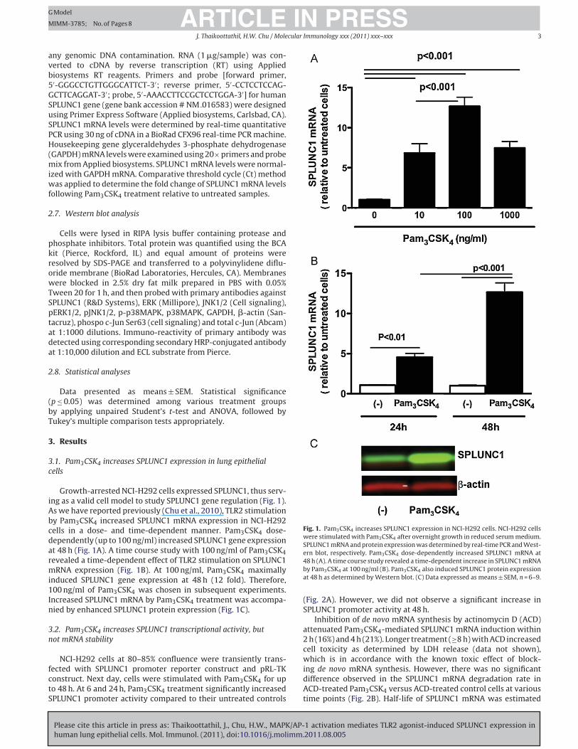
3.1. Pam3CSK4 increases SPLUNC1 expression in lung epithelial
cells
Growth-arrested NCI-H292 cells expressed SPLUNC1, thus serv-
ing as a valid cell model to study SPLUNC1 gene regulation (Fig. 1).
As we have reported previously (Chu et al., 2010), TLR2 stimulation
by Pam3CSK4 increased SPLUNC1 mRNA expression in NCI-H292
cells in a dose- and time-dependent manner. Pam3CSK4 dose-
dependently (up to 100 ng/ml) increased SPLUNC1 gene expression
at 48 h (Fig. 1A). A time course study with 100 ng/ml of Pam3CSK4
revealed a time-dependent effect of TLR2 stimulation on SPLUNC1
mRNA expression (Fig. 1B). At 100 ng/ml, Pam3CSK4 maximally
induced SPLUNC1 gene expression at 48 h (12 fold). Therefore,
100 ng/ml of Pam3CSK4 was chosen in subsequent experiments.
Increased SPLUNC1 mRNA by Pam3CSK4 treatment was accompa-
nied by enhanced SPLUNC1 protein expression (Fig. 1C).
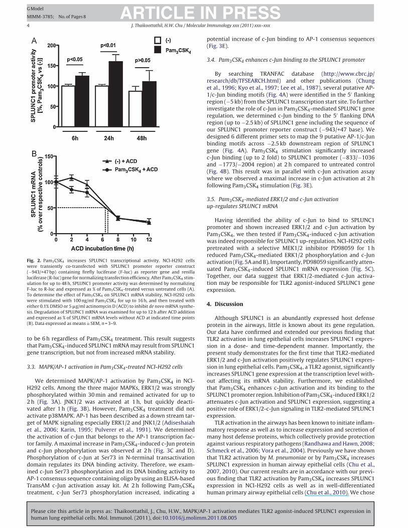
3.2. Pam3CSK4 increases SPLUNC1 transcriptional activity, but
not mRNA stability
NCI-H292 cells at 80–85% confluence were transiently trans-
fected with SPLUNC1 promoter reporter construct and pRL-TK
construct. Next day, cells were stimulated with Pam3CSK4 for up
to 48 h. At 6 and 24 h, Pam3CSK4 treatment significantly increased
SPLUNC1 promoter activity compared to their untreated controls
Fig. 1. Pam3CSK4 increases SPLUNC1 expression in NCI-H292 cells. NCI-H292 cells
were stimulated with Pam3CSK4 after overnight growth in reduced serum medium.
SPLUNC1 mRNA and protein expression was determined by real-time PCR and West-
ern blot, respectively. Pam3CSK4 dose-dependently increased SPLUNC1 mRNA at
48 h (A). A time course study revealed a time-dependent increase in SPLUNC1 mRNA
by Pam3CSK4 at 100 ng/ml (B). Pam3CSK4 also induced SPLUNC1 protein expression
at 48 h as determined by Western blot. (C) Data expressed as means ± SEM, n = 6–9.
(Fig. 2A). However, we did not observe a significant increase in
SPLUNC1 promoter activity at 48 h.
Inhibition of de novo mRNA synthesis by actinomycin D (ACD)
attenuated Pam3CSK4-mediated SPLUNC1 mRNA induction within
2 h (16%) and 4 h (21%). Longer treatment (≥8 h) with ACD increased
cell toxicity as determined by LDH release (data not shown),
which is in accordance with the known toxic effect of block-
ing de novo mRNA synthesis. However, there was no significant
difference observed in the SPLUNC1 mRNA degradation rate in
ACD-treated Pam3CSK4 versus ACD-treated control cells at various
time points (Fig. 2B). Half-life of SPLUNC1 mRNA was estimated
Please cite this article in press as: Thaikoottathil, J., Chu, H.W., MAPK/AP-1 activation mediates TLR2 agonist-induced SPLUNC1 expression in
human lung epithelial cells. Mol. Immunol. (2011), doi:10.1016/j.molimm.2011.08.005
ARTICLE IN PRESSG Model
MIMM-3785; No. of Pages 8
4 J. Thaikoottathil, H.W. Chu / Molecular Immunology xxx (2011) xxx– xxx
Fig. 2. Pam3CSK4 increases SPLUNC1 transcriptional activity. NCI-H292 cells
were transiently co-transfected with SPLUNC1 promoter reporter construct
(−943/+47 bp) containing firefly luciferase (F-luc) as reporter gene and renilla
luciferase (R-luc) gene for normalizing transfection efficiency. After Pam3CSK4 stim-
ulation for up to 48 h, SPLUNC1 promoter activity was determined by normalizing
F-luc to R-luc and expressed as % of Pam3CSK4-treated versus untreated cells (A).
To determine the effect of Pam3CSK4 on SPLUNC1 mRNA stability, NCI-H292 cells
were stimulated with 100 ng/ml Pam3CSK4 for up to 16 h, and then treated with
either 0.1% DMSO or 5 �g/ml actinomycin D (ACD) to inhibit de novo mRNA synthe-
sis. Degradation of SPLUNC1 mRNA was examined for up to 12 h after ACD addition
and expressed as % of SPLUNC1 mRNA levels without ACD at indicated time points
(B). Data expressed as means ± SEM, n = 3–9.
to be 6 h regardless of Pam3CSK4 treatment. This result suggests
that Pam3CSK4-induced SPLUNC1 mRNA may result from SPLUNC1
gene transcription, but not from increased mRNA stability.
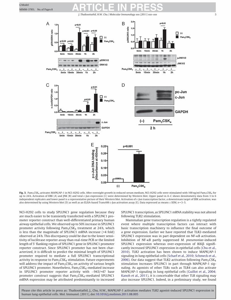
3.3. MAPK/AP-1 activation in Pam3CSK4-treated NCI-H292 cells
We determined MAPK/AP-1 activation by Pam3CSK4 in NCI-
H292 cells. Among the three major MAPKs, ERK1/2 was strongly
phosphorylated within 30 min and remained activated for up to
2 h (Fig. 3A). JNK1/2 was activated at 1 h, but quickly deacti-
vated after 1 h (Fig. 3B). However, Pam3CSK4 treatment did not
activate p38MAPK. AP-1 has been described as a down stream tar-
get of MAPK signaling especially ERK1/2 and JNK1/2 (Adiseshaiah
et al., 2006; Karin, 1995; Pulverer et al., 1991). We determined
the activation of c-Jun that belongs to the AP-1 transcription fac-
tor family. A maximal increase in Pam3CSK4-induced c-Jun protein
and c-Jun phosphorylation was observed at 2 h (Fig. 3C and D).
Phosphorylation of c-Jun at Ser73 in N-terminal transactivation
domain regulates its DNA binding activity. Therefore, we exam-
ined c-Jun Ser73 phosphorylation and its DNA binding activity to
AP-1 consensus sequence containing oligo by using an ELISA-based
TransAM c-Jun activation assay kit. At 2 h following Pam3CSK4
treatment, c-Jun Ser73 phosphorylation increased, indicating a
potential increase of c-Jun binding to AP-1 consensus sequences
(Fig. 3E).
3.4. Pam3CSK4 enhances c-Jun binding to the SPLUNC1 promoter
By searching TRANFAC database (http://www.cbrc.jp/
research/db/TFSEARCH.html) and other publications (Chung
et al., 1996; Kyo et al., 1997; Lee et al., 1987), several putative AP-
1/c-Jun binding motifs (Fig. 4A) were identified in the 5′ flanking
region (−5 kb) from the SPLUNC1 transcription start site. To further
investigate the role of c-Jun in Pam3CSK4-mediated SPLUNC1 gene
regulation, we determined c-Jun binding to the 5′ flanking DNA
region (up to −2.5 kb) of SPLUNC1 gene including the sequence of
our SPLUNC1 promoter reporter construct (−943/+47 base). We
designed 6 different primer sets to map the 9 putative AP-1/c-Jun
binding motifs across −2.5 kb downstream region of SPLUNC1
gene (Fig. 4A). Pam3CSK4 stimulation significantly increased
c-Jun binding (up to 2 fold) to SPLUNC1 promoter (−833/−1036
and −1773/−2004 region) at 2 h compared to untreated control
(Fig. 4B). This result was in parallel with c-Jun activation assay
where we observed a maximal increase in c-Jun activation at 2 h
following Pam3CSK4 stimulation (Fig. 3E).
3.5. Pam3CSK4-mediated ERK1/2 and c-Jun activation
up-regulates SPLUNC1 mRNA
Having identified the ability of c-Jun to bind to SPLUNC1
promoter and shown increased ERK1/2 and c-Jun activation by
Pam3CSK4, we then tested if Pam3CSK4-induced c-Jun activation
was indeed responsible for SPLUNC1 up-regulation. NCI-H292 cells
pretreated with a selective MEK1/2 inhibitor PD98059 for 1 h
reduced Pam3CSK4-mediated ERK1/2 phosphorylation and c-Jun
activation (Fig. 5A and B). Importantly, PD98059 significantly atten-
uated Pam3CSK4-induced SPLUNC1 mRNA expression (Fig. 5C).
Together, our data suggest that ERK1/2-mediated c-Jun activa-
tion may be responsible for TLR2 agonist-induced SPLUNC1 gene
expression.
4. Discussion
Although SPLUNC1 is an abundantly expressed host defense
protein in the airways, little is known about its gene regulation.
Our data have confirmed and extended our previous finding that
TLR2 activation in lung epithelial cells increases SPLUNC1 expres-
sion in a dose- and time-dependent manner. Importantly, the
present study demonstrates for the first time that TLR2-mediated
ERK1/2 and c-Jun activation positively regulates SPLUNC1 expres-
sion in lung epithelial cells. Pam3CSK4, a TLR2 agonist, significantly
increases SPLUNC1 gene expression at the transcription level with-
out affecting its mRNA stability. Furthermore, we established
that Pam3CSK4 enhances c-Jun activation and its binding to the
SPLUNC1 promoter region. Inhibition of Pam3CSK4-induced ERK1/2
attenuates c-Jun activation and SPLUNC1 expression, suggesting a
positive role of ERK1/2-c-Jun signaling in TLR2-mediated SPLUNC1
expression.
TLR activation in the airways has been known to initiate inflam-
matory response as well as to increase expression and secretion of
many host defense proteins, which collectively provide protection
against various respiratory pathogens (Randhawa and Hawn, 2008;
Schmeck et al., 2006; Vora et al., 2004). Previously we have shown
that TLR2 activation by M. pneumoniae or by Pam3CSK4 increases
SPLUNC1 expression in human airway epithelial cells (Chu et al.,
2007, 2010). Our current results are in accordance with our previ-
ous finding that TLR2 activation by Pam3CSK4 increases SPLUNC1
expression in NCI-H292 cells as well as in well-differentiated
human primary airway epithelial cells (Chu et al., 2010). We chose
Please cite this article in press as: Thaikoottathil, J., Chu, H.W., MAPK/AP-1 activation mediates TLR2 agonist-induced SPLUNC1 expression in
human lung epithelial cells. Mol. Immunol. (2011), doi:10.1016/j.molimm.2011.08.005
ARTICLE IN PRESSG Model
MIMM-3785; No. of Pages 8
J. Thaikoottathil, H.W. Chu / Molecular Immunology xxx (2011) xxx– xxx 5
Fig. 3. Pam3CSK4 activates MAPK/AP-1 in NCI-H292 cells. After overnight growth in reduced serum medium, NCI-H292 cells were stimulated with 100 ng/ml Pam3CSK4 for
up to 24 h. Activation of ERK (A) and JNK (B) and total c-Jun expression (C) were determined by Western blot. Upper panel in A–C shows densitometry data from 3 to 6
independent replicates and lower panel is a representative picture of their Western blot. Activation of c-Jun transcription factor, a downstream target of ERK activation, was
also determined by using Western blot (D) as well as an ELISA-based TransAM c-Jun activation assay (E). Data expressed as means ± SEM, n = 3–5.
NCI-H292 cells to study SPLUNC1 gene regulation because they
are much easier to be transiently transfected with a SPLUNC1 pro-
moter reporter construct than well-differentiated primary human
airway epithelial cells. We observed up to 50% increase in SPLUNC1
promoter activity following Pam3CSK4 treatment at 24 h, which
is less than the magnitude of SPLUNC1 mRNA increase (>4 fold)
observed at 24 h. This discrepancy could be due to the lower sensi-
tivity of luciferase reporter assay than real-time PCR or the limited
length of 5′ flanking region of SPLUNC1 gene in SPLUNC1 promoter
reporter construct. Since SPLUNC1 promoter has not been char-
acterized, it is difficult to predict the minimal length of SPLUNC1
promoter required to mediate a full SPLUNC1 transcriptional
activity in response to Pam3CSK4 stimulation. Future experiments
will address the impact of Pam3CSK4 on activity of various length
of SPLUNC1 promoter. Nonetheless, Pam3CSK4-mediated increase
in SPLUNC1 promoter reporter activity with −943/+47 base
promoter construct suggests that Pam3CSK4-mediated SPLUNC1
mRNA expression may be attributed predominantly to increased
SPLUNC1 transcription, as SPLUNC1 mRNA stability was not altered
following TLR2 stimulation.
Mammalian gene transcription regulation is a tightly regulated
event where multiple transcription factors can interact with
basic transcription machinery to influence the final outcome of
a gene expression. Earlier we have reported that TLR2-mediated
SPLUNC1 expression was in part dependent on NF-�B activation.
Inhibition of NF-�B partly suppressed M. pneumoniae-induced
SPLUNC1 expression whereas over-expression of IKK� signifi-
cantly increased SPLUNC1 expression in epithelial cells (Chu et al.,
2010). TLR2 activation has been shown to induce MAPK/AP-1
signaling in lung epithelial cells (Scharf et al., 2010; Schmeck et al.,
2006). Our data suggest that TLR2 activation following Pam3CSK4
stimulation increases SPLUNC1 in part through MAPK/AP-1 sig-
naling. As agonists of other TLRs such as TLR4 can also activate
MAPK/AP-1 signaling in lung epithelial cells (Guillot et al., 2004;
Kanoh et al., 2011), it is conceivable that other TLR signaling may
also increase SPLUNC1. Indeed, in a preliminary study, we found

Please cite this article in press as: Thaikoottathil, J., Chu, H.W., MAPK/AP-1 activation mediates TLR2 agonist-induced SPLUNC1 expression in
human lung epithelial cells. Mol. Immunol. (2011), doi:10.1016/j.molimm.2011.08.005
ARTICLE IN PRESSG Model
MIMM-3785; No. of Pages 8
6 J. Thaikoottathil, H.W. Chu / Molecular Immunology xxx (2011) xxx– xxx
Fig. 4. Pam3CSK4 enhances c-Jun binding to the SPLUNC1 promoter. Several puta-
tive AP-1/c-Jun binding motifs were identified in the −2.5 kb flanking region of
SPLUNC1 gene including SPLUNC1 promoter sequence. Six different primer sets
were designed to map 9 putative AP-1/c-Jun binding motifs across −2.5 kb flank-
ing region of SPLUNC1 gene (A). After overnight growth in reduced serum medium,
NCI-H292 cells were stimulated with Pam3CSK4 (100 ng/ml) for up to 2 h. Binding
of c-Jun to SPLUNC1 promoter region (−833/−1036 and −1733/−2004 bases) con-
taining putative Ap-1/c-Jun DNA binding motifs was determined by ChIP assay (B).
Upper panel shows a representative picture of agarose gels; lower panel shows den-
sitometric analysis of PCR products visualized on agarose gel electrophoresis. Data
expressed as means ± SEM, n = 3.
that TLR4 agonist LPS increases SPLUNC1 mRNA expression (up to
3 fold) in NCI-H292 cells in a dose-dependent manner. However,
at the same dose (100 ng/ml), LPS versus Pam3CSK4 resulted in
less (6-fold) SPLUNC1 mRNA induction. Why TLR2, as compared to
TLR4 activation, more robustly up-regulates SPLUNC1 expression
in epithelial cells deserves further studies.
In the present study we found that TLR2 activation by Pam3CSK4
induces a sustained ERK1/2 phosphorylation along with a transient
increase in JNK1/2 phosphorylation in NCI-H292 cells. Tradition-
ally ERK phosphorylation has been implicated in a wide variety of
cellular responses including cell growth, proliferation and differ-
entiation whereas JNK1/2 was more involved in inducing apoptosis
or stress activated signaling (Ballif and Blenis, 2001; Gauthier et al.,
2001; Li et al., 2004). Different MAPKs can regulate distinct cellu-
lar activities even though they share common substrate including
transcription factor AP-1 (Adiseshaiah et al., 2006; Karin, 1995;
Pulverer et al., 1991). AP-1 is a family of transcription factors that
has been known to modulate various gene expressions via form-
ing a homo- or hetero-dimer and also via interacting with other
transcription factors (Bakiri et al., 2002; Hess et al., 2004). Tran-
scription factor c-Jun is a component of AP-1 transcription family,
which can be activated by ERK1/2 and JNK1/2 (Karin, 1995; Leppa
et al., 1998). Although c-Jun has been suggested as a preferred
substrate for JNK1/2, there are studies reporting N-terminal trans-
activation domain phosphorylation of c-Jun at Ser63 and Ser73
by ERK1/2 (Leppa et al., 1998; Pulverer et al., 1991). ERK activa-
tion has been shown to up-regulate de novo expression of c-Jun
in rat phaeochromocytome PC12 cells (Leppa et al., 1998). Such
dual input of increased c-Jun expression and phosphorylation of
Fig. 5. Pam3CSK4-mediated ERK1/2 and c-Jun activation up-regulates SPLUNC1
expression. NCI-H292 cells were pre-treated with DMSO (0.1%) or MEK1/2 inhibitor
PD98059 (10 �M). An hour later, cells were stimulated with Pam3CSK4 for up to 24 h.
ERK1/2 inhibition by PD98059 resulted in decreased ERK1/2 and c-Jun activation as
determined by Western blot (A) and ELISA-based TransAM c-Jun activation assay
(B). Effect of PD98059 on Pam3CSK4-induced SPLUNC1 mRNA was determined by
real-time quantitative PCR (C). Data expressed as means ± SEM, n = 3–5.
c-Jun by ERK1/2 and JNK1/2 have been shown to enhance cell
differentiation in PC12 cells (Leppa et al., 1998). Our results also
indicate a transient increase in c-Jun phosphorylation as well as
increase in total c-Jun levels following Pam3CSK4 treatment at
2 h (Fig. 3C and E). As our c-Jun Western blotting for total c-Jun
revealed multiple bands, we speculate that Pam3CSK4 may induce
c-Jun phosphorylation at other sites in addition to Ser63/Ser73.
Based on the presence of multiple bands in c-Jun Western blot,
similar speculations were reported by Satomi and colleagues in
perillyl alcohol-treated human breast cancer cell line T47D-C4-2W
(Satomi et al., 1999). Increased c-Jun phosphorylation by MAPK
has been reported to increase c-Jun protein stability and de novo
expression (Leppa et al., 1998; Musti et al., 1997). We observed
in our previous studies that basal SPLUNC1 expression in airway
Please cite this article in press as: Thaikoottathil, J., Chu, H.W., MAPK/AP-1 activation mediates TLR2 agonist-induced SPLUNC1 expression in
human lung epithelial cells. Mol. Immunol. (2011), doi:10.1016/j.molimm.2011.08.005
ARTICLE IN PRESSG Model
MIMM-3785; No. of Pages 8
J. Thaikoottathil, H.W. Chu / Molecular Immunology xxx (2011) xxx– xxx 7
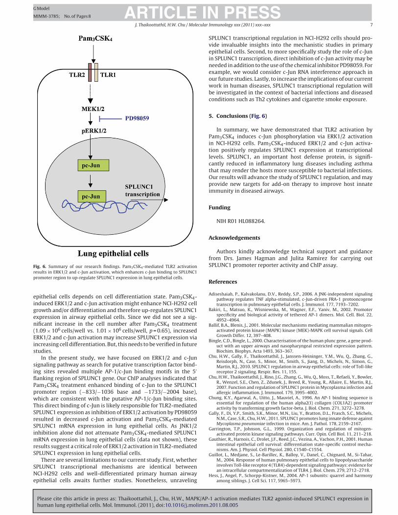
Fig. 6. Summary of our research findings. Pam3CSK4-mediated TLR2 activation
results in ERK1/2 and c-Jun activation, which enhances c-Jun binding to SPLUNC1
promoter region to up-regulate SPLUNC1 expression in lung epithelial cells.
epithelial cells depends on cell differentiation state. Pam3CSK4-
induced ERK1/2 and c-Jun activation might enhance NCI-H292 cell
growth and/or differentiation and therefore up-regulates SPLUNC1
expression in airway epithelial cells. Since we did not see a sig-
nificant increase in the cell number after Pam3CSK4 treatment
(1.09 × 10⁶ cells/well vs. 1.01 × 10⁶ cells/well, p = 0.65), increased
ERK1/2 and c-Jun activation may increase SPLUNC1 expression via
increasing cell differentiation. But, this needs to be verified in future
studies.
In the present study, we have focused on ERK1/2 and c-Jun
signaling pathway as search for putative transcription factor bind-
ing sites revealed multiple AP-1/c-Jun binding motifs in the 5′
flanking region of SPLUNC1 gene. Our ChIP analyses indicated that
Pam3CSK4 treatment enhanced binding of c-Jun to the SPLUNC1
promoter region (−833/−1036 base and −1733/−2004 base),
which are consistent with the putative AP-1/c-Jun binding sites.
This direct binding of c-Jun is likely responsible for TLR2-mediated
SPLUNC1 expression as inhibition of ERK1/2 activation by PD98059
resulted in decreased c-Jun activation and Pam3CSK4-mediated
SPLUNC1 mRNA expression in lung epithelial cells. As JNK1/2
inhibition alone did not attenuate Pam3CSK4-mediated SPLUNC1
mRNA expression in lung epithelial cells (data not shown), these
results suggest a critical role of ERK1/2 activation in TLR2-mediated
SPLUNC1 expression in lung epithelial cells.
There are several limitations to our current study. First, whether
SPLUNC1 transcriptional mechanisms are identical between
NCI-H292 cells and well-differentiated primary human airway
epithelial cells awaits further studies. Nonetheless, unraveling
SPLUNC1 transcriptional regulation in NCI-H292 cells should pro-
vide invaluable insights into the mechanistic studies in primary
epithelial cells. Second, to more specifically study the role of c-Jun
in SPLUNC1 transcription, direct inhibition of c-Jun activity may be
needed in addition to the use of the chemical inhibitor PD98059. For
example, we would consider c-Jun RNA interference approach in
our future studies. Lastly, to increase the implications of our current
work in human diseases, SPLUNC1 transcriptional regulation will
be investigated in the context of bacterial infections and diseased
conditions such as Th2 cytokines and cigarette smoke exposure.
5. Conclusions (Fig. 6)
In summary, we have demonstrated that TLR2 activation by
Pam3CSK4 induces c-Jun phosphorylation via ERK1/2 activation
in NCI-H292 cells. Pam3CSK4-induced ERK1/2 and c-Jun activa-
tion positively regulates SPLUNC1 expression at transcriptional
levels. SPLUNC1, an important host defense protein, is signifi-
cantly reduced in inflammatory lung diseases including asthma
that may render the hosts more susceptible to bacterial infections.
Our results will advance the study of SPLUNC1 regulation, and may
provide new targets for add-on therapy to improve host innate
immunity in diseased airways.
Funding
NIH R01 HL088264.
Acknowledgements
Authors kindly acknowledge technical support and guidance
from Drs. James Hagman and Julita Ramirez for carrying out
SPLUNC1 promoter reporter activity and ChIP assay.
References
Adiseshaiah, P., Kalvakolanu, D.V., Reddy, S.P., 2006. A JNK-independent signalingpathway regulates TNF alpha-stimulated, c-Jun-driven FRA-1 protooncogenetranscription in pulmonary epithelial cells. J. Immunol. 177, 7193–7202.
Bakiri, L., Matsuo, K., Wisniewska, M., Wagner, E.F., Yaniv, M., 2002. Promoterspecificity and biological activity of tethered AP-1 dimers. Mol. Cell. Biol. 22,4952–4964.
Ballif, B.A., Blenis, J., 2001. Molecular mechanisms mediating mammalian mitogen-activated protein kinase (MAPK) kinase (MEK)-MAPK cell survival signals. CellGrowth Differ. 12, 397–408.
Bingle, C.D., Bingle, L., 2000. Characterisation of the human plunc gene, a gene prod-uct with an upper airways and nasopharyngeal restricted expression pattern.Biochim. Biophys. Acta 1493, 363–367.
Chu, H.W., Gally, F., Thaikoottathil, J., Janssen-Heininger, Y.M., Wu, Q., Zhang, G.,Reisdorph, N., Case, S., Minor, M., Smith, S., Jiang, D., Michels, N., Simon, G.,Martin, R.J., 2010. SPLUNC1 regulation in airway epithelial cells: role of Toll-likereceptor 2 signaling. Respir. Res. 11, 155.
Chu, H.W., Thaikoottathil, J., Rino, J.G., Zhang, G., Wu, Q., Moss, T., Refaeli, Y., Bowler,R., Wenzel, S.E., Chen, Z., Zdunek, J., Breed, R., Young, R., Allaire, E., Martin, R.J.,2007. Function and regulation of SPLUNC1 protein in Mycoplasma infection andallergic inflammation. J. Immunol. 179, 3995–4002.
Chung, K.Y., Agarwal, A., Uitto, J., Mauviel, A., 1996. An AP-1 binding sequence isessential for regulation of the human alpha2(I) collagen (COL1A2) promoteractivity by transforming growth factor-beta. J. Biol. Chem. 271, 3272–3278.
Gally, F., Di, Y.P., Smith, S.K., Minor, M.N., Liu, Y., Bratton, D.L., Frasch, S.C., Michels,N.M., Case, S.R., Chu, H.W., 2011. SPLUNC1 promotes lung innate defense againstMycoplasma pneumoniae infection in mice. Am. J. Pathol. 178, 2159–2167.
Garrington, T.P., Johnson, G.L., 1999. Organization and regulation of mitogen-activated protein kinase signaling pathways. Curr. Opin. Cell Biol. 11, 211–218.
Gauthier, R., Harnois, C., Drolet, J.F., Reed, J.C., Vezina, A., Vachon, P.H., 2001. Humanintestinal epithelial cell survival: differentiation state-specific control mecha-nisms. Am. J. Physiol. Cell Physiol. 280, C1540–C1554.
Guillot, L., Medjane, S., Le-Barillec, K., Balloy, V., Danel, C., Chignard, M., Si-Tahar,M., 2004. Response of human pulmonary epithelial cells to lipopolysaccharideinvolves Toll-like receptor 4 (TLR4)-dependent signaling pathways: evidence foran intracellular compartmentalization of TLR4. J. Biol. Chem. 279, 2712–2718.
Hess, J., Angel, P., Schorpp-Kistner, M., 2004. AP-1 subunits: quarrel and harmonyamong siblings. J. Cell Sci. 117, 5965–5973.

Please cite this article in press as: Thaikoottathil, J., Chu, H.W., MAPK/AP-1 activation mediates TLR2 agonist-induced SPLUNC1 expression in
human lung epithelial cells. Mol. Immunol. (2011), doi:10.1016/j.molimm.2011.08.005
ARTICLE IN PRESSG Model
MIMM-3785; No. of Pages 8
8 J. Thaikoottathil, H.W. Chu / Molecular Immunology xxx (2011) xxx– xxx
Kanoh, S., Tanabe, T., Rubin, B.K., 2011. Dapsone inhibits IL-8 secretion from humanbronchial epithelial cells stimulated with LPS and resolves airway inflammationin the ferret. Chest, in press.
Karin, M., 1995. The regulation of AP-1 activity by mitogen-activated protein kinases.J. Biol. Chem. 270, 16483–16486.
Kyo, S., Klumpp, D.J., Inoue, M., Kanaya, T., Laimins, L.A., 1997. Expression of AP1 dur-ing cellular differentiation determines human papillomavirus E6/E7 expressionin stratified epithelial cells. J. Gen. Virol. 78 (Pt 2), 401–411.
Lafferty, E.I., Qureshi, S.T., Schnare, M., 2010. The role of toll-like receptors in acuteand chronic lung inflammation. J. Inflamm. (Lond.) 7, 57.
Lee, W., Mitchell, P., Tjian, R., 1987. Purified transcription factor AP-1 interacts withTPA-inducible enhancer elements. Cell 49, 741–752.
Leppa, S., Saffrich, R., Ansorge, W., Bohmann, D., 1998. Differential regulation of c-Junby ERK and JNK during PC12 cell differentiation. EMBO J. 17, 4404–4413.
Li, S., Gerrard Jr., E.R., Balkovetz, D.F., 2004. Evidence for ERK1/2 phosphorylationcontrolling contact inhibition of proliferation in Madin-Darby canine kidneyepithelial cells. Am. J. Physiol. Cell Physiol. 287, C432–C439.
Lukinskiene, L., Liu, Y., Reynolds, S.D., Steele, C., Stripp, B.R., Leikauf, G.D., Kolls, J.K.,Di, Y.P., 2011. Antimicrobial activity of PLUNC protects against Pseudomonasaeruginosa infection. J. Immunol. 187, 382–390.
Musti, A.M., Treier, M., Bohmann, D., 1997. Reduced ubiquitin-dependent degrada-tion of c-Jun after phosphorylation by MAP kinases. Science 275, 400–402.
Opitz, B., van Laak, V., Eitel, J., Suttorp, N., 2010. Innate immune recognition in infec-tious and noninfectious diseases of the lung. Am. J. Respir. Crit. Care Med. 181,1294–1309.
Pulverer, B.J., Kyriakis, J.M., Avruch, J., Nikolakaki, E., Woodgett, J.R., 1991. Phospho-rylation of c-jun mediated by MAP kinases. Nature 353, 670–674.
Randhawa, A.K., Hawn, T.R., 2008. Toll-like receptors: their roles in bacterial recog-nition and respiratory infections. Expert Rev. Anti Infect. Ther. 6, 479–495.
Satomi, Y., Miyamoto, S., Gould, M.N., 1999. Induction of AP-1 activity by perillylalcohol in breast cancer cells. Carcinogenesis 20, 1957–1961.
Scharf, S., Hippenstiel, S., Flieger, A., Suttorp, N., N’Guessan, P.D., 2010. Inductionof human beta-defensin-2 in pulmonary epithelial cells by Legionella pneu-mophila: involvement of TLR2 and TLR5, p38 MAPK, JNK, NF-kappaB, and AP-1.Am. J. Physiol. Lung Cell Mol. Physiol. 298, L687–L695.
Schmeck, B., Moog, K., Zahlten, J., van Laak, V., N’Guessan, P.D., Opitz, B., Rosseau,S., Suttorp, N., Hippenstiel, S., 2006. Streptococcus pneumoniae induced c-Jun-N-terminal kinase- and AP-1-dependent IL-8 release by lung epithelial BEAS-2Bcells. Respir. Res. 7, 98.
Steiling, K., Kadar, A.Y., Bergerat, A., Flanigon, J., Sridhar, S., Shah, V., Ahmad, Q.R.,Brody, J.S., Lenburg, M.E., Steffen, M., Spira, A., 2009. Comparison of proteomicand transcriptomic profiles in the bronchial airway epithelium of current andnever smokers. PLoS One 4, e5043.
Vora, P., Youdim, A., Thomas, L.S., Fukata, M., Tesfay, S.Y., Lukasek, K., Michelsen, K.S.,Wada, A., Hirayama, T., Arditi, M., Abreu, M.T., 2004. Beta-defensin-2 expres-sion is regulated by TLR signaling in intestinal epithelial cells. J. Immunol. 173,5398–5405.
Zhou, H.D., Li, X.L., Li, G.Y., Zhou, M., Liu, H.Y., Yang, Y.X., Deng, T., Ma, J., Sheng, S.R.,2008. Effect of SPLUNC1 protein on the Pseudomonas aeruginosa and Epstein-Barrvirus. Mol. Cell. Biochem. 309, 191–197.
Copyright © 2022 FDOKUMEN