The Effect of a TLR4 Agonist/Cationic Liposome Adjuvant on ...
19
pharmaceutics Article The Effect of a TLR4 Agonist/Cationic Liposome Adjuvant on Varicella-Zoster Virus Glycoprotein E Vaccine Efficacy: Antigen Presentation, Uptake, and Delivery to Lymph Nodes Seo Ri Wui 1 , Ara Ko 1 , Ji In Ryu 1 , Eojin Sim 1 , Soo Jeong Lim 1 , Shin Ae Park 2 , Kwang Sung Kim 1,2 , Ha Kim 3,4 , Hyewon Youn 3,4 and Na Gyong Lee 1, * Citation: Wui, S.R.; Ko, A.; Ryu, J.I.; Sim, E.; Lim, S.J.; Park, S.A.; Kim, K.S.; Kim, H.; Youn, H.; Lee, N.G. The Effect of a TLR4 Agonist/Cationic Liposome Adjuvant on Varicella-Zoster Virus Glycoprotein E Vaccine Efficacy: Antigen Presentation, Uptake, and Delivery to Lymph Nodes. Pharmaceutics 2021, 13, 390. https://doi.org/10.3390/ pharmaceutics13030390 Academic Editor: Nirmal Marasini Received: 2 February 2021 Accepted: 12 March 2021 Published: 15 March 2021 Publisher’s Note: MDPI stays neutral with regard to jurisdictional claims in published maps and institutional affil- iations. Copyright: © 2021 by the authors. Licensee MDPI, Basel, Switzerland. This article is an open access article distributed under the terms and conditions of the Creative Commons Attribution (CC BY) license (https:// creativecommons.org/licenses/by/ 4.0/). 1 Department of Integrated Bioscience and Biotechnology, Sejong University, Seoul 05006, Korea; [email protected] (S.R.W.); [email protected] (A.K.); [email protected] (J.I.R.); [email protected] (E.S.); [email protected] (S.J.L.); [email protected] (K.S.K.) 2 R & D Center, EyeGene, Goyang 10551, Korea; [email protected] 3 Cancer Research Institute, College of Medicine, Seoul National University, Seoul 03080, Korea; [email protected] (H.K.); [email protected] (H.Y.) 4 Department of Nuclear Medicine, Cancer Imaging Center, Seoul National University Hospital, Seoul 03080, Korea * Correspondence: [email protected]; Tel.: +82-2-3408-3765; Fax: +82-2-3408-3334 Abstract: Adjuvant CIA09, composed of 1,2-dioleoyl-3-trimethylammonium-propane (DOTAP)- based cationic liposomes and the toll-like receptor 4 agonist de-O-acylated lipooligosaccharide (dLOS), has been shown to enhance antibody and cellular immune responses to varicella-zoster virus (VZV) glycoprotein E (gE), recombinant tuberculosis vaccine antigen, and inactivated Japanese encephalitis vaccine. In this study, we investigated its modes of action using VZV gE as a model antigen. Liposomes adsorbed gE and cooperatively with dLOS promoted endocytosis-mediated cellular uptake of gE by mouse dendritic cells in vitro. CIA09 increased the stability and cellular uptake of the antigen at the muscle site of injection, and induced immune cell recruitment and cytokine and chemokine production, which led to efficient antigen delivery to draining lymph nodes. Mouse bone marrow-derived dendritic cells, pulsed with CIA09-adjuvanted gE, efficiently presented gE to antigen-specific T cells, inducing Th1-type biased immunity, as shown by high IFN-γ production. The data indicate that liposomes and dLOS cooperate in the adjuvant activity of CIA09 by promoting antigen uptake and delivery to lymph nodes as well as antigen presentation to T cells. Keywords: antigen delivery to lymph nodes; cationic liposomes; cellular antigen uptake; CIA09; TLR4 agonist de-O-acylated lipooligosaccharides; vaccine adjuvant; VZV gE antigen 1. Introduction Adjuvants are an important component of many vaccines, especially recombinant subunit vaccines. Vehicle type adjuvants, which act as antigen carriers, include alum, oil-in- water emulsions, and liposomes [1–3]. They serve as an antigen depot, increase the stability of the antigen, enhance uptake by antigen-presenting cells (APCs), and facilitate delivery to lymph nodes [2–5]. Immunomodulatory adjuvants directly activate immune cells, induce cytokines and chemokines, and recruit immune cells [2,5]. They are typically ligands for cellular pattern recognition receptors (PRRs), including toll-like receptors (TLRs), C-type lectin receptors (CLRs), nucleotide-binding oligomerization domain (NOD)-like receptors (NLRs), retinoic acid-inducible gene I (RIG-I)-like receptors (RLRs), and cytosolic dsDNA sensors (CDSs) [6–11]. These ligands mimic pathogen or damage-associated molecular patterns (PAMPs/DAMPs) to recognize PRRs and induce the rapid activation of innate immune responses, leading to adaptive immunity [6–11]. Recently designed adjuvant formulations, containing two or more adjuvants with different, cooperative modes of action, provide high adjuvant activity, optimizing the efficacy of vaccines [12,13]. Pharmaceutics 2021, 13, 390. https://doi.org/10.3390/pharmaceutics13030390 https://www.mdpi.com/journal/pharmaceutics
-
Upload
khangminh22 -
Category
Documents
-
view
0 -
download
0
Transcript of The Effect of a TLR4 Agonist/Cationic Liposome Adjuvant on ...

pharmaceutics
Article
The Effect of a TLR4 Agonist/Cationic Liposome Adjuvant onVaricella-Zoster Virus Glycoprotein E Vaccine Efficacy: AntigenPresentation, Uptake, and Delivery to Lymph Nodes
Seo Ri Wui 1, Ara Ko 1 , Ji In Ryu 1, Eojin Sim 1, Soo Jeong Lim 1, Shin Ae Park 2, Kwang Sung Kim 1,2, Ha Kim 3,4,Hyewon Youn 3,4 and Na Gyong Lee 1,*
�����������������
Citation: Wui, S.R.; Ko, A.; Ryu, J.I.;
Sim, E.; Lim, S.J.; Park, S.A.; Kim, K.S.;
Kim, H.; Youn, H.; Lee, N.G. The
Effect of a TLR4 Agonist/Cationic
Liposome Adjuvant on
Varicella-Zoster Virus Glycoprotein E
Vaccine Efficacy: Antigen
Presentation, Uptake, and Delivery to
Lymph Nodes. Pharmaceutics 2021, 13,
390. https://doi.org/10.3390/
pharmaceutics13030390
Academic Editor: Nirmal Marasini
Received: 2 February 2021
Accepted: 12 March 2021
Published: 15 March 2021
Publisher’s Note: MDPI stays neutral
with regard to jurisdictional claims in
published maps and institutional affil-
iations.
Copyright: © 2021 by the authors.
Licensee MDPI, Basel, Switzerland.
This article is an open access article
distributed under the terms and
conditions of the Creative Commons
Attribution (CC BY) license (https://
creativecommons.org/licenses/by/
4.0/).
1 Department of Integrated Bioscience and Biotechnology, Sejong University, Seoul 05006, Korea;[email protected] (S.R.W.); [email protected] (A.K.); [email protected] (J.I.R.);[email protected] (E.S.); [email protected] (S.J.L.); [email protected] (K.S.K.)
2 R & D Center, EyeGene, Goyang 10551, Korea; [email protected] Cancer Research Institute, College of Medicine, Seoul National University, Seoul 03080, Korea;
[email protected] (H.K.); [email protected] (H.Y.)4 Department of Nuclear Medicine, Cancer Imaging Center, Seoul National University Hospital,
Seoul 03080, Korea* Correspondence: [email protected]; Tel.: +82-2-3408-3765; Fax: +82-2-3408-3334
Abstract: Adjuvant CIA09, composed of 1,2-dioleoyl-3-trimethylammonium-propane (DOTAP)-based cationic liposomes and the toll-like receptor 4 agonist de-O-acylated lipooligosaccharide(dLOS), has been shown to enhance antibody and cellular immune responses to varicella-zostervirus (VZV) glycoprotein E (gE), recombinant tuberculosis vaccine antigen, and inactivated Japaneseencephalitis vaccine. In this study, we investigated its modes of action using VZV gE as a modelantigen. Liposomes adsorbed gE and cooperatively with dLOS promoted endocytosis-mediatedcellular uptake of gE by mouse dendritic cells in vitro. CIA09 increased the stability and cellularuptake of the antigen at the muscle site of injection, and induced immune cell recruitment andcytokine and chemokine production, which led to efficient antigen delivery to draining lymphnodes. Mouse bone marrow-derived dendritic cells, pulsed with CIA09-adjuvanted gE, efficientlypresented gE to antigen-specific T cells, inducing Th1-type biased immunity, as shown by high IFN-γproduction. The data indicate that liposomes and dLOS cooperate in the adjuvant activity of CIA09by promoting antigen uptake and delivery to lymph nodes as well as antigen presentation to T cells.
Keywords: antigen delivery to lymph nodes; cationic liposomes; cellular antigen uptake; CIA09;TLR4 agonist de-O-acylated lipooligosaccharides; vaccine adjuvant; VZV gE antigen
1. Introduction
Adjuvants are an important component of many vaccines, especially recombinantsubunit vaccines. Vehicle type adjuvants, which act as antigen carriers, include alum, oil-in-water emulsions, and liposomes [1–3]. They serve as an antigen depot, increase the stabilityof the antigen, enhance uptake by antigen-presenting cells (APCs), and facilitate delivery tolymph nodes [2–5]. Immunomodulatory adjuvants directly activate immune cells, inducecytokines and chemokines, and recruit immune cells [2,5]. They are typically ligands forcellular pattern recognition receptors (PRRs), including toll-like receptors (TLRs), C-typelectin receptors (CLRs), nucleotide-binding oligomerization domain (NOD)-like receptors(NLRs), retinoic acid-inducible gene I (RIG-I)-like receptors (RLRs), and cytosolic dsDNAsensors (CDSs) [6–11]. These ligands mimic pathogen or damage-associated molecularpatterns (PAMPs/DAMPs) to recognize PRRs and induce the rapid activation of innateimmune responses, leading to adaptive immunity [6–11]. Recently designed adjuvantformulations, containing two or more adjuvants with different, cooperative modes ofaction, provide high adjuvant activity, optimizing the efficacy of vaccines [12,13].
Pharmaceutics 2021, 13, 390. https://doi.org/10.3390/pharmaceutics13030390 https://www.mdpi.com/journal/pharmaceutics

Pharmaceutics 2021, 13, 390 2 of 19
Liposomes, which are biocompatible, biodegradable, non-toxic, and have long beenused as drug delivery systems, also function as adjuvants for human vaccines [14–18]. Lipo-somal adjuvants serve as persistent depots of vaccine antigens at the site of injection (SOI),facilitate antigen delivery to lymph nodes, and increase antigen uptake and processingby APCs [14–18]. Cationic liposomes, which bind electrostatically to negatively chargedprotein antigens and cell surface membranes, are particularly effective adjuvants [19–21].Unlike neutral or anionic liposomes, cationic liposomes directly activate immune cells,such as dendritic cells (DCs) and macrophages [22–25]. A cationic liposome adjuvant,based on 1,2-dioleoyl-3-trimethylammonium-propane (DOTAP), enhances humoral andcell-mediated immune responses to antigens produced in vitro or in vivo in mice [26–28];it also induces activation of the extracellular-signal-regulated kinase (ERK) pathway andthe synthesis of chemokines in mouse bone marrow-derived DCs (BMDCs) [29]. However,cationic liposomes require other immune stimulants for optimal adjuvant activity [24,30];for example, the liposome-based adjuvant CAF01 includes the cationic lipid dimethyldioc-tadecylammonium (DDA) and synthetic trehalose-6,6-dibehenate (TDB) [31,32]. CAF01induces strong humoral and cellular immune responses and is a potential adjuvant for a tu-berculosis vaccine, having elicited protective immunity against a Mycobacterium tuberculosischallenge in mouse and human clinical trials [33–35].
De-O-acylated lipooligosaccharide (dLOS) is a TLR4 agonist derived from Escherichia colilipopolysaccharide (LPS) with an additional short carbohydrate moiety [36,37]. It inducesthe secretion of cytokines from murine peritoneal macrophages similarly to MPL but withmore potent activation of human monocytes and DCs [37]. dLOS in combination withaluminum hydroxide (designated CIA06) has adjuvant activity in several viral and bacterialvaccines [38–42] and performed well in a phase I clinical study of a CIA06-adjuvantedhuman papillomavirus (HPV) virus-like particle (VLP) vaccine (unpublished data). Wedeveloped cationic liposome-based adjuvant CIA09, composed of DOTAP-based cationicliposomes and dLOS, which enhances antibody and cell-mediated immune responses torecombinant tuberculosis antigens, inactivated Japanese encephalitis vaccine (JEV), andrecombinant varicella-zoster virus (VZV) glycoprotein E (gE) antigen [42–44]. CIA09 isparticularly effective in eliciting a Th1-type biased response, as determined by interferon-γ(IFN-γ) cytokine production, compared with CIA06 [42–44]. Using VZV gE as the modelantigen, we investigated the mechanism of action of CIA09 and demonstrate here thatliposomes and dLOS cooperatively promote (i) the immunogenicity of VZV gE antigenby increasing the antigen stability, (ii) antigen uptake at the site of injection (SOI), (iii) therecruitment of immune cells, (iv) antigen delivery to the lymph nodes, and v) antigenpresentation by APCs to T cells.
2. Materials and Methods2.1. Experimental Animals
BALB/c and C57BL/6 mice used for experiments were purchased from SLC (Hama-matsu, Japan) or Orient Bio (Orient Bio, Gyeonggi-do, Korea). Mice were housed in atemperature- and humidity-controlled chamber with a 12-h light/dark cycle and providedwith free access to food and water. Mice were anesthetized with an intraperitoneal injec-tion of a ketamine/xylazine mixture before being used for experiments or sacrificed fortissue samples.
2.2. Materials
The Madin-Darby canine kidney (MDCK) cell line and the J774A.1 mouse mono-cyte/macrophage cell line were obtained from ATCC (Manassas, VA, USA), while theDC2.4 mouse immature dendritic cell line was kindly provided by Prof. I. Rhee of SejongUniversity, Republic of Korea. Two phospholipids, DOTAP and 1,2-dimyristoyl-sn-glycero-3-phosphocholine (DMPC), were purchased from Avanti Polar Lipids (Alabaster, AL, USA)and/or NOF (Tokyo, Japan). NBD-labeled DOTAP was obtained from Avanti Polar Lipids,whereas 1,1′-dioctadecyl-3,3,3′,3′-tetramethylindotricarbocyanine iodide (DiR) and DAPI

Pharmaceutics 2021, 13, 390 3 of 19
were from Invitrogen (Carlsbad, CA, USA) and Lonza (Basel, Switzerland), respectively.Cytochalasin D (actin polymerization inhibitor), chlorpromazine (clathrin-mediated endo-cytosis inhibitor), and genistein (caveolae-mediated endocytosis inhibitor) were purchasedfrom Sigma-Aldrich (St. Louis, MO, USA). Cell culture media and antibiotics were obtainedfrom Welgene (Gyeongsangbuk-do, Korea), whereas fetal bovine serum (FBS) was fromGibco/Invitrogen (Grand Island, NY, USA). The IFN-γ cytokine ELISA kit and the multi-plex assay kit using Luminex® were from R&D Systems (Minneapolis, MN, USA), whileinterleukin-5 (IL-5) and monocyte chemoattractant protein-1 (MCP-1) ELISA kits werefrom BD Bioscience (San Jose, CA, USA). Anti-mouse CD16/CD32 Ab, anti–CD11b-FITC,anti-CD11c-FITC, anti-Ly6C-PE, anti-F4/80-PE, anti-Ly6G-PE, anti-MHCII-PE, anti-CD3ε-PE, anti-CD11b-PE-Cy7, IC fixation buffer, and permeabilization buffer were purchasedfrom eBioscience (San Diego, CA, USA) or BioLegend (San Diego, CA, USA).
2.3. Preparation of dLOS and Recombinant VZV gE Antigen
The TLR4 agonist dLOS was isolated from an E. coli strain that expresses LPS lackingO-polysaccharides and prepared as previously described [36,37], visualized on a silver-stained SDS-polyacrylamide gel, and quantified using the 2-keto-3-deoxyoctonate as-say [45].
Recombinant VZV gE antigen was expressed in Chinese hamster ovary (CHO) cellsand purified as previously described by Wui et al. [44]. Protein was quantified with theBradford assay kit, while the endotoxin contents in gE preparations were determined usingthe Endosafe-PTS test system (Charles River Laboratories, Wilmington, MA, USA) andconfirmed to be lower than 1 EU/µg protein. To prepare fluorescently labeled gE antigen,recombinant VZV gE was conjugated with fluorescent dyes using an Alexa Fluor (AF) 488or 647 protein labeling kit (Invitrogen) or HiLyteTM Fluor 647 amine (AnaSpec, Fremont,CA, USA) according to the manufacturer’s instructions.
2.4. Preparation and Characterization of Cationic Liposomes2.4.1. Liposome Preparation
Liposomes were prepared with cationic lipid DOTAP and neutral lipid DMPC by a drycake method as previously described by Wui et al. [44]. Briefly, DOTAP and DMPC wereeach dissolved in tertiary-butyl alcohol and mixed at a molar ratio of 1:1. The lipid mixturewas lyophilized overnight using a freeze-dryer (Lyoph-Pride; Ilshin BioBase, Gyeonggi-do,Korea). The dry cakes were hydrated with a 10% sucrose solution to a final concentrationof 2 mg lipid/mL and homogenized using a microfluidizer (Avestin, Ottawa, ON, Canada).Liposome preparations were aliquoted in sealed glass vials, freeze-dried, and stored at−20 ◦C. For experiments, freeze-dried liposomes were reconstituted with deionized waterto the original volume. Fluorescently labeled liposomes were made by adding 1% DiR- orNBD-labeled DMPC to the phospholipid mixtures (w/w) before lyophilization. CIA09 wasprepared by combining liposomes with dLOS at various ratios.
2.4.2. Measurement of Particle Size and Zeta Potential of Liposomes
The particle size and polydispersity index (PDI) of liposomes were determined bydynamic light scattering (DLS) using a Zetasizer Nano ZSP (Malvern Instruments, Worces-tershire, UK). Liposome powder was rehydrated with deionized water to its originalvolume and further diluted tenfold with a 10% sucrose solution. Samples were analyzedat a detection angle of 173◦ and a recording temperature of 25 ◦C. The zeta potential ofliposomes was measured by laser doppler microelectrophoresis on the Zetasizer Nano ZSP.Malvern Zetasizer DTS software (version 7.12) was used for data acquisition and analysis.All measurements were carried out in triplicate.
2.4.3. Morphology
The morphology of liposomes was determined by cryogenic transmission electronmicroscopy (Cryo-TEM). Lyophilized liposome powder was rehydrated with deionized

Pharmaceutics 2021, 13, 390 4 of 19
water, and 3 µL of the liposome preparation was placed on a lacey carbon film 200-meshcopper grid and vitrified semi-automatically using a Vitrobot™ (FEI, Hillsboro, OR, USA).Vitrified samples were maintained at liquid nitrogen temperatures during sample transferand imaged with a Tecnai G2 Spirit Twin electron microscope (FEI) at 120 keV.
2.4.4. Cytotoxicity Assay
The cytotoxicity of liposomes was determined by the MTT assay in MDCK and DC2.4cells. Cells were seeded in DMEM or RPMI1640 supplemented with 10% FBS at a densityof 2 × 10⁵ cells/mL in a 96-well plate and cultured overnight. Cells were treated withliposomes (25, 50, 100, or 200 µg/mL) in the presence or absence of dLOS (0.5, 1, 2, or4 µg/mL) for 24 h. The next day, CCK-8 solution (Dojindo, Tokyo, Japan) was added tothe culture, and cells were incubated for 2 h. Optical density at 450 nm was measuredusing a microplate reader (Infinit M200; Tecan, Mannedorf, Switzerland). All assays wereperformed in triplicate. Cell viability relative to that of control cells was calculated as:cell viability (%) = ((OD450 of test cells − OD450 of blank well)/(OD450 of media controlcells − OD450 of blank well)) × 100.
2.5. Measurement of gE Antigen Adsorption on Liposomes
Recombinant VZV gE antigen (10 µg) was mixed with liposomes (200 µg) in thepresence or absence of dLOS (4 µg) in a total volume of 200 µL. The mixture was ultracen-trifuged at 100,000× g at 4 ◦C for 1 h. The supernatant was removed, and the pellet wasdissolved in the original volume of phosphate-buffered saline (PBS, pH 7.2). gE proteinin the supernatant and pellet was resolved on SDS-PAGE gels and silver-stained. Theintensity of the protein bands on gels was determined by image processing of the bandsusing Image J program (NIH, Bethesda, MD, USA).
2.6. Determination of Cellular Uptake of gE Antigen2.6.1. Imaging of Cellular Antigen-Uptake by Confocal Microscopy
DC2.4 cells were seeded at a density of 2 × 10⁵ cells/mL in 4-well cell culture slidesand cultured overnight. Cells were incubated with fluorescently labeled gE (5 µg/mL),alone or in combination with dLOS (3 µg/mL), NBD-labeled liposomes (100 µg/mL), orboth (CIA09) for 4 h. Cells were washed, fixed, and stained with DAPI. The slides werewashed, mounted with antifade mounting medium (Invitrogen), and observed under aconfocal microscope (Carl Zeiss, Oberkochen, Germany).
For live-cell image analysis, DC2.4 cells were seeded at a density of 1 × 10⁴ cells/wellin a Scar™ Block confocal dish (SPL, Gyeonggi-do, Korea) and cultured overnight. Cellswere treated with fluorescently labeled gE (5 µg/mL), alone or in combination with dLOS(2 µg/mL), DiR-labeled liposomes (100 µg/mL), or both, and then immediately observedin the live cell chamber system under a laser scanning confocal microscope (Carl Zeiss).Live-cell images were acquired every 15 s over a 30-min period and data analysis wasperformed using ZEN software Blue lite edition (Carl Zeiss).
2.6.2. Measurement of Cellular Antigen Uptake by Flow Cytometry
DC2.4 cells were seeded at a density of 5 × 10⁵ cells/mL in a 24-well plate andcultured overnight. Cells were incubated for 1 h at 4 ◦C or 37 ◦C with fresh medium,followed by treatment with fluorescently labeled gE (5 µg/mL), alone or in combinationwith dLOS (1 or 3 µg/mL), liposomes (50 or 100 µg/mL), or both for 4 h. Endocytosisinhibitors chlorpromazine (50 µM), cytochalasin D (5 µM), or genistein (200 µM) wereadded to the cells 1 h before sample treatment. Cells were washed three times with PBS andfluorescently labeled gE-associated cells were analyzed by flow cytometry (FACSCanto II;Becton Dickinson, CA, USA). Data acquisition and analysis were performed using FlowJosoftware (FlowJo, Ashland, OR, USA).

Pharmaceutics 2021, 13, 390 5 of 19
2.7. Measurement of Cytokines and Chemokines Secreted from Mouse Immune Cells
J774A.1 cells were seeded at a density of 2 × 10⁵ cells/mL in a 96-well plate, culturedovernight, and treated with liposomes (25, 50, or 100 µg/mL), dLOS (0.5, 1, or 2 µg/mL),or both for 24 h. IL-12 and MCP-1 secreted into the culture medium were measured usingcytokine sandwich ELISA kits.
2.8. Measurement of Cytokine and Chemokine Secretion at the SOI
Six-week-old female BALB/c mice (SLC) were given an intramuscular injection withdLOS, liposomes, or both in a total volume of 100 µL. Control mice were administeredPBS. Muscle tissue was collected from the SOI at various times post-injection and homog-enized in organ lysis buffer (R&D systems) containing a protease inhibitor cocktail andphenylmethylsulfonyl fluoride (1 mM) using a homogenizer (IKA, Staufen, Germany).Homogenates were centrifugated at 12,000 rpm for 10 min at 4 ◦C, and the supernatantwas assessed for total protein concentration using a BCA protein assay kit (Pierce, MA,USA). Cytokine and chemokine amounts in muscle lysates were determined by a multiplexassay using Luminex® according to the manufacturer’s instructions (R&D Systems). Thepanel of cytokines and chemokines included: IFN-γ, IL-10, IL-12p70, IL-17A, IL-1α, IL-1β,IL-2, IL-5, IL-6, IP-10, MCP-1, MIP-1α, MIP-1β, MIP-2, RANTES, TNF-α, and MIG.
2.9. Immune Cell Phenotyping of Mouse Muscle Samples
Six-week-old female BALB/c mice (SLC) were given an intramuscular injection withfluorescently labeled gE, alone or in combination with dLOS, liposomes, or both. Controlmice were administered PBS. Muscle samples taken from the SOI were chopped intosmall pieces using scissors and digested with type II collagenase (1 mg/mL) in Hank’sbalanced salt solution for 1 h at 37 ◦C. Cell suspensions were filtered through a 40-µm cellstrainer and centrifuged. Cells were resuspended and treated with anti-mouse CD16/CD32Ab for 10 min to block the FcR, followed by staining with combinations of anti-mouseantibodies: anti-CD11b-FITC, anti-CD11c-FITC, anti-Ly6C-PE, anti-F4/80-PE, anti-Ly6G-PE, anti-MHCII-PE, anti-CD3ε-PE, and/or anti-CD11b-PE-Cy7. Immune cell and antigen-positive cell populations of the muscle tissue samples were determined by flow cytometry(FACSCanto II), and data acquisition and analysis were performed using FlowJo software.Flow cytometry gating strategies are presented in Supplementary Figure S1.
2.10. In Vivo Imaging Analysis2.10.1. Fluorescence Imaging Analysis
Five-week-old female BALB/c mice (Orient Bio) in groups of six mice were admin-istered fluorescently labeled gE (5 µg), alone or in combination with dLOS (2 µg), lipo-somes (100 µg), or both in a total volume of 100 µL via intramuscular injection. At 0, 1,and 24 h post-injection, two mice from each group were anesthetized and monitored forthe distribution of the fluorescent dye using an in vivo imaging system (IVIS Lumina II;PerkinElmer Health Sciences, Waltham, MA, USA) with excitation/emission wavelengthsof 620 nm/670 nm. The acquisition time was 10 sec per view, and the total radiance onthe surface of mice was expressed as photons per second per microwatt per centimetersquared ((p/s)/(µW/cm2)).
2.10.2. SPECT/CT Imaging Analysis
For single-photon emission-computed tomography (SPECT)/CT imaging analysis, gEantigen was radiolabeled with 125I using pre-coated iodination tubes (Pierce, Rockford, IL,USA) according to the manufacturer’s instructions. Five-week-old female C57BL/6 mice(Orient Bio) were given an intramuscular injection with 125I-labeled gE alone (20 µg) orin combination with dLOS (2 µg), liposomes (100 µg), or both in a total volume of 100 µL.SPECT was performed with a four-headed multiplexing multi-pinhole NanoSPECT/CTPlus (Mediso, Budapest, Hungary) using a mouse aperture. An acquisition time of 20 sper view was chosen for SPECT, resulting in acquisition times ranging from 15–20 min per

Pharmaceutics 2021, 13, 390 6 of 19
animal. The 7-min CT imaging was performed immediately following the whole-bodySPECT imaging at 48-µm resolution. SPECT scans were acquired at 1, 24, 48, and 120 hpost-injection. Reconstruction of images was performed using HiSPECT software (Bioscan,Washington DC, USA) without attenuation correction. Reconstructed data from SPECTand CT were co-registered using InVivoScope (Bioscan) for analysis and interpretation.
2.11. Measurement of In Vivo Cellular Antigen Uptake and Delivery to Lymph Nodes
Six-week-old female BALB/c mice (SLC) were given an intramuscular injection withfluorescently labeled gE (10 µg), alone or in combination with dLOS (3 µg), liposomes(100 µg), or both. Control mice were administered PBS. Muscle samples and draininglymph nodes were collected at 24 h post-injection. Single cells were prepared from tissuesamples and analyzed for antigen-positive cells by flow cytometry.
2.12. Antigen-Presenting Cell Assay
To prepare antigen-presenting cells, mouse BMDCs were prepared from 5-week-old fe-male C57BL/6 mice (SLC) as previously described by Han et al. [34] and seeded at a densityof 1 × 10⁴ cells/well in a 96-well cell culture plate. Cells were pulsed with VZV gE antigen(2.5 µg/mL) alone or in combination with dLOS (0.5 µg/mL), liposomes (50 µg/mL), orboth for 24 h and washed three times with fresh medium. For gE-specific responder cells,CD3+ T cells were purified from the spleens of VZV gE-immunized C57BL/6 mice usingthe magnetic antibody cell sorting (MACS) CD3 MicroBead Kit according to the manufac-turer’s instructions (Miltenyi Biotec, Germany). Purified CD3+ T cells (1 × 10⁵ cells/well)were added to the pulsed presenter cells at a ratio of 10:1 (T cells:BMDCs) and culturedin a 37 ◦C CO2 incubator. The cell culture medium was collected at 3, 7, and 10 dayspost-treatment and assayed for IFN-γ and IL-5 by sandwich ELISA.
2.13. Statistical Analysis
SPSS 18.0 software (IBM, Armonk, NY, USA) was used for statistical analysis. Dif-ferences among the experimental groups were analyzed using one-way ANOVA withTukey’s multiple comparison test. A two-tailed Student’s t-test was used to comparetwo experimental groups. P value of less than 0.05 was considered statistically significant.
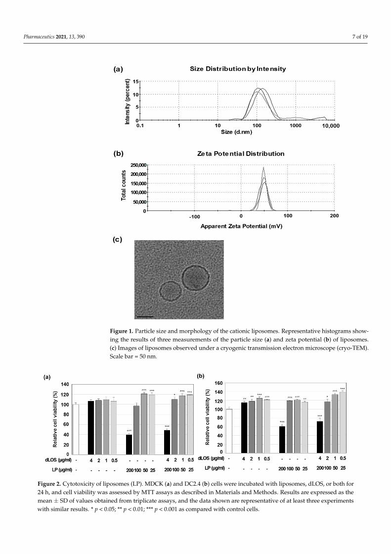
3. Results3.1. Characterization of Liposomes
Liposomes prepared using cationic lipid DOTAP and neutral lipid DMPC (1:1, w/w),had a Z-average diameter of 80–120 nm with a polydispersity index (PDI) of 0.2–0.3 and azeta potential of + 55–70 mV (Figure 1a,b). The liposomes, observed by cryo-TEM, weremostly small, unilamellar vesicles with a diameter of 50–80 nm (Figure 1c). The addition ofdLOS to liposomes did not change the Z-average diameter but tended to reduce the zetapotential of the liposomes by about 10 mV. Since dLOS is negatively charged due to itsphosphate moieties, it should interact with positively charged liposomes, thus explainingthe decrease in their zeta potential.
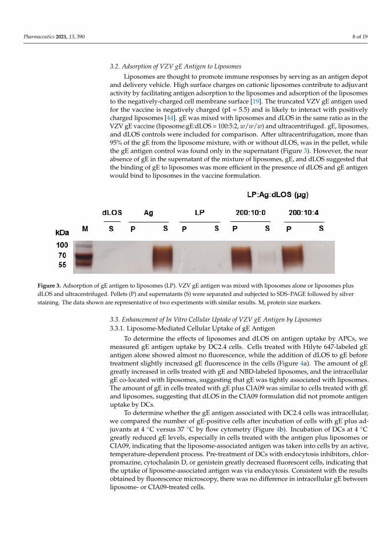
Liposomes were not cytotoxic up to a concentration of 100 µg/mL in either MDCKor DC2.4 cells, but at 200 µg/mL, they killed 40% and 60% of MDCK and DC2.4 cells,respectively (Figure 2). dLOS did not exhibit any cytotoxicity at concentrations up to4 µg/mL, and its addition to liposomes decreased their cytotoxicity at 200 µg/mL, possiblyby lowering the surface charges of the liposomes by their interaction with dLOS. Based onthese data, a liposome concentration of 100 µg/mL was used in experiments.
Pharmaceutics 2021, 13, 390 7 of 19
Figure 1. Particle size and morphology of the cationic liposomes. Representative histograms show-ing the results of three measurements of the particle size (a) and zeta potential (b) of liposomes.(c) Images of liposomes observed under a cryogenic transmission electron microscope (cryo-TEM).Scale bar = 50 nm.
Figure 2. Cytotoxicity of liposomes (LP). MDCK (a) and DC2.4 (b) cells were incubated with liposomes, dLOS, or both for24 h, and cell viability was assessed by MTT assays as described in Materials and Methods. Results are expressed as themean ± SD of values obtained from triplicate assays, and the data shown are representative of at least three experimentswith similar results. * p < 0.05; ** p < 0.01; *** p < 0.001 as compared with control cells.
Pharmaceutics 2021, 13, 390 8 of 19
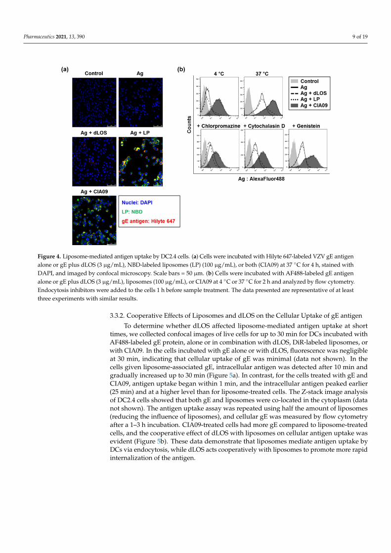
3.2. Adsorption of VZV gE Antigen to Liposomes
Liposomes are thought to promote immune responses by serving as an antigen depotand delivery vehicle. High surface charges on cationic liposomes contribute to adjuvantactivity by facilitating antigen adsorption to the liposomes and adsorption of the liposomesto the negatively-charged cell membrane surface [19]. The truncated VZV gE antigen usedfor the vaccine is negatively charged (pI = 5.5) and is likely to interact with positivelycharged liposomes [44]. gE was mixed with liposomes and dLOS in the same ratio as in theVZV gE vaccine (liposome:gE:dLOS = 100:5:2, w/w/w) and ultracentrifuged. gE, liposomes,and dLOS controls were included for comparison. After ultracentrifugation, more than95% of the gE from the liposome mixture, with or without dLOS, was in the pellet, whilethe gE antigen control was found only in the supernatant (Figure 3). However, the nearabsence of gE in the supernatant of the mixture of liposomes, gE, and dLOS suggested thatthe binding of gE to liposomes was more efficient in the presence of dLOS and gE antigenwould bind to liposomes in the vaccine formulation.
Figure 3. Adsorption of gE antigen to liposomes (LP). VZV gE antigen was mixed with liposomes alone or liposomes plusdLOS and ultracentrifuged. Pellets (P) and supernatants (S) were separated and subjected to SDS–PAGE followed by silverstaining. The data shown are representative of two experiments with similar results. M, protein size markers.
3.3. Enhancement of In Vitro Cellular Uptake of VZV gE Antigen by Liposomes3.3.1. Liposome-Mediated Cellular Uptake of gE Antigen
To determine the effects of liposomes and dLOS on antigen uptake by APCs, wemeasured gE antigen uptake by DC2.4 cells. Cells treated with Hilyte 647-labeled gEantigen alone showed almost no fluorescence, while the addition of dLOS to gE beforetreatment slightly increased gE fluorescence in the cells (Figure 4a). The amount of gEgreatly increased in cells treated with gE and NBD-labeled liposomes, and the intracellulargE co-located with liposomes, suggesting that gE was tightly associated with liposomes.The amount of gE in cells treated with gE plus CIA09 was similar to cells treated with gEand liposomes, suggesting that dLOS in the CIA09 formulation did not promote antigenuptake by DCs.
To determine whether the gE antigen associated with DC2.4 cells was intracellular,we compared the number of gE-positive cells after incubation of cells with gE plus ad-juvants at 4 ◦C versus 37 ◦C by flow cytometry (Figure 4b). Incubation of DCs at 4 ◦Cgreatly reduced gE levels, especially in cells treated with the antigen plus liposomes orCIA09, indicating that the liposome-associated antigen was taken into cells by an active,temperature-dependent process. Pre-treatment of DCs with endocytosis inhibitors, chlor-promazine, cytochalasin D, or genistein greatly decreased fluorescent cells, indicating thatthe uptake of liposome-associated antigen was via endocytosis. Consistent with the resultsobtained by fluorescence microscopy, there was no difference in intracellular gE betweenliposome- or CIA09-treated cells.
Pharmaceutics 2021, 13, 390 9 of 19
Figure 4. Liposome-mediated antigen uptake by DC2.4 cells. (a) Cells were incubated with Hilyte 647-labeled VZV gE antigenalone or gE plus dLOS (3 µg/mL), NBD-labeled liposomes (LP) (100 µg/mL), or both (CIA09) at 37 ◦C for 4 h, stained withDAPI, and imaged by confocal microscopy. Scale bars = 50 µm. (b) Cells were incubated with AF488-labeled gE antigenalone or gE plus dLOS (3 µg/mL), liposomes (100 µg/mL), or CIA09 at 4 ◦C or 37 ◦C for 2 h and analyzed by flow cytometry.Endocytosis inhibitors were added to the cells 1 h before sample treatment. The data presented are representative of at leastthree experiments with similar results.
3.3.2. Cooperative Effects of Liposomes and dLOS on the Cellular Uptake of gE antigen
To determine whether dLOS affected liposome-mediated antigen uptake at shorttimes, we collected confocal images of live cells for up to 30 min for DCs incubated withAF488-labeled gE protein, alone or in combination with dLOS, DiR-labeled liposomes, orwith CIA09. In the cells incubated with gE alone or with dLOS, fluorescence was negligibleat 30 min, indicating that cellular uptake of gE was minimal (data not shown). In thecells given liposome-associated gE, intracellular antigen was detected after 10 min andgradually increased up to 30 min (Figure 5a). In contrast, for the cells treated with gE andCIA09, antigen uptake began within 1 min, and the intracellular antigen peaked earlier(25 min) and at a higher level than for liposome-treated cells. The Z-stack image analysisof DC2.4 cells showed that both gE and liposomes were co-located in the cytoplasm (datanot shown). The antigen uptake assay was repeated using half the amount of liposomes(reducing the influence of liposomes), and cellular gE was measured by flow cytometryafter a 1–3 h incubation. CIA09-treated cells had more gE compared to liposome-treatedcells, and the cooperative effect of dLOS with liposomes on cellular antigen uptake wasevident (Figure 5b). These data demonstrate that liposomes mediate antigen uptake byDCs via endocytosis, while dLOS acts cooperatively with liposomes to promote more rapidinternalization of the antigen.

Pharmaceutics 2021, 13, 390 10 of 19
Figure 5. Cooperative effects of liposomes (LP) and dLOS on the cellular uptake of gE antigen.(a) DC2.4 cells were treated with AF488-labeled gE antigen (green) in combination with DiR-labeledliposomes (red) or CIA09 and cultured at 37 ◦C. Cumulative cellular fluorescence was acquiredover 30 min using a laser scanning confocal microscope. The data presented are representativeof two experiments with similar results. Scale bar = 20 µm. (b) DC2.4 cells were treated withAF647-labeled gE antigen, alone or in combination with dLOS (1 µg/mL), liposomes (50 µg/mL),or both, and cultured at 37 ◦C for 1, 2, or 3 h followed by flow cytometry. The data presented arerepresentative of three independent experiments with similar results.
3.4. Immune Cell Activation and Recruitment by CIA09
We found previously that dLOS stimulates immune activity in human monocytes aswell as in mouse monocyte/macrophage J774A.1 cells and BMDCs [34]. Here, we measuredcytokine and chemokine secretion in J774A.1 cells treated with various concentrations ofliposomes without dLOS or with dLOS in the same proportion as in the VZV gE vaccineformulation. IL-12 in the culture medium from dLOS-treated cells was about 1 ng/mL,independent of dLOS concentration (Figure 6a). These results were similar to those from

Pharmaceutics 2021, 13, 390 11 of 19
our previous studies showing that IL-12 induced by dLOS reached a peak at as low as0.1 µg/mL of dLOS in these cells (unpublished data). Liposomes alone did not inducecytokine secretion. Neither IL-12 (Figure 6a) nor TNF-α (data not shown) was detected inthe liposome-treated cells. Liposomes with dLOS (i.e., CIA09) induced IL-12 secretion ininverse proportion to the adjuvant concentration, suggesting that while liposomes withlow concentrations of dLOS enhanced IL-12 release from the cells, at higher concentrationsliposomes inhibited the dLOS activity. Although liposomes barely induced cytokines,they stimulated the release of chemokine MCP-1, and that release was enhanced by dLOS(Figure 6b). Liposomes did not induce cytokine secretion or costimulatory surface moleculeexpression in mouse BMDCs (data not shown).
Figure 6. In vitro immune-stimulating activity of liposomes (LP). J774A.1 cells were cultured with liposomes, dLOS, orboth for 24 h. IL-12 (a) and MCP-1 (b) released into the culture medium were measured by sandwich ELISA. Results areexpressed as mean ± SD of triplicate cultures. Data presented are representative of at least two experiments with similarresults. * p < 0.05; ** p < 0.01; *** p < 0.001 as compared with control cells.
Since CIA09 induced cytokine and chemokine release in vitro, we determined itsability to recruit and stimulate immune cells in vivo. Cytokine and chemokine secretionwas assayed in mouse muscle tissues 4 h and 24 h after intramuscular injection with dLOS,liposomes, or CIA09 (Figure 7a). dLOS induced strong cytokine and chemokine secretionat 4 h post-injection, which decreased by 50% at 24 h. Liposomes weakly induced cytokineand chemokine secretion and MCP-1was the most abundant species, consistent with thein vitro data (Figure 6). CIA09 was more effective in stimulating the secretion of cytokineand chemokines than dLOS, and high levels of secretion were maintained at 24 h in theCIA09-treated mice but not in the dLOS-treated mice. While there were differences in thecytokine and chemokine profiles of the two groups, both groups had high levels of MCP-1and IL-6 at 4 h. The predominant species in CIA09-treated mice at 4 h was IL-1α, whichdecreased to basal level at 24 h.
The high levels of chemokines secreted at the SOI in mice treated with dLOS or CIA09would be expected to recruit immune cells to the site; however, mice treated with dLOSshowed low levels of immune cell recruitment (Figure 7b). In contrast, liposomes increasedthe frequency of immune cells at 4 h, which doubled at 24 h, while CIA09-treated mice hadthe largest immune cell population among the groups at 4 h and 24 h. These data suggestthat dLOS and liposomes cooperatively activate and recruit immune cells to the SOI.

Pharmaceutics 2021, 13, 390 12 of 19
Figure 7. Cytokine and chemokine induction and immune cell recruitment at SOI. Groups of mice (n = 3) were given anintramuscular injection with dLOS (3 µg), liposomes (LP) (100 µg), or both (CIA09), and muscle samples were collected at4 h and 24 h post-injection. (a) Muscle tissue homogenates were prepared and analyzed for cytokines and chemokines bymultiplex assays using Luminex®. (b) Single cells were prepared, stained with antibodies specific for immune cell markers,and analyzed by flow cytometry. Data are expressed as the mean of three muscle samples for each group.
3.5. Increase in Antigen Uptake at the Site of Injection by CIA09
To determine whether CIA09 enhances antigen uptake, mice were injected intramus-cularly with AF647-labeled gE antigen, alone or in combination with dLOS, liposomes, orCIA09. Muscle samples were collected at 1, 24, 48, and 168 h post-injection and analyzedby flow cytometry for antigen-positive immune cells and total immune cells (Figure 8).Injection of gE antigen did not change the immune cell frequency in muscle cells, whilegE antigen with dLOS resulted in only a minimal, temporary increase in immune cells.However, administration of gE antigen with liposomes or CIA09 dramatically increasedthe immune cell population at the SOI at 24 h post-injection, consistent with the resultsshown in Figure 7b. While immune cell recruitment reached a peak at 48 h in mice injectedwith gE plus liposomes and then decreased rapidly to basal level at 168 h, mice adminis-tered gE plus CIA09 maintained a high immune cell population up to 168 h post-injection.The antigen-positive immune cell profiles in the muscle samples were similar to the totalimmune cell population, except that antigen-positive immune cells were greater in micetreated with liposomes compared to CIA09 (Figure 8b). We conclude that liposomes recruitimmune cells and mediate their uptake of gE, while dLOS contributes to immune cellretention at the SOI.
Figure 8. Antigen uptake at the SOI. Groups of mice (n = 3) were given an intramuscular injection with AF647-labeled gE(5 µg), alone or in combination with dLOS (2 µg), liposomes (LP) (100 µg), or CIA09, and muscle samples were collected at1, 24, 48, and 168 h post-injection. Single cells were prepared, stained with antibodies specific for immune cell markers,and analyzed by flow cytometry. (a) Immune cell populations at SOI; (b) the frequency of antigen-positive immune cells.The results are expressed as the mean of values obtained from three muscle samples for each group. The data shown arerepresentative of three independent experiments with similar results.

Pharmaceutics 2021, 13, 390 13 of 19
3.6. Increased In Vivo Stability of Liposome-Associated gE Antigen
Since vehicle-type adjuvants such as alum and liposomes form an antigen depot at theSOI, we determined whether liposomes serve as an antigen reservoir at the SOI, increasingantigen stability. The distribution of AF647-conjugated gE after intramuscular injection,alone or in combination with dLOS, liposomes, or CIA09, was monitored at 0, 1, or 24 hpost-injection using an in vivo imaging system. A very high level of fluorescent gE wasdetected immediately after injection at the SOI and at 1 h post-injection (Figure 9a), butdecreased by more than 90% after 24 h, indicating that the antigen was quickly degraded.The addition of dLOS to gE yielded results similar to gE alone. However, when gE antigenwas mixed with liposomes or CIA09, the fluorescence signal from the gE antigen wasreduced about five-fold due to the fluorescence quenching effect of liposomes. Despite thisquenching, fluorescence for the group given gE antigen mixed with liposomes or CIA09 at24 h post-injection was 3–5 times greater than for the group given gE alone (see the insertsof Figure 9a) and was almost the same as the initial level, indicating that the gE antigen atthe SOI was retained over time. Postmortem examination of the leg muscles of mice givenliposomes or CIA09 revealed a strong blue color of the fluorescently labeled gE antigen,while no trace of color was left in the muscles of the other two groups (Figure 9b).
Figure 9. In vivo imaging of CIA09-adjuvanted gE antigen in mice. (a) Groups of mice (n = 6) were injected with AF647-labeled gE (5 µg), alone or in combination with dLOS (2 µg), liposomes (LP) (100 µg), or CIA09. Two mice from eachgroup were monitored at 0, 1, and 24 h post-injection for the distribution of the fluorescence dye using in vivo imaging.Fluorescence detected at a lower scale are shown in photo inserts. The data shown are representative of two independentexperiments with similar results. (b) Mouse legs taken at 24 h were opened to monitor fluorescence. An image of one mousefrom each group is shown. (c) Mice were injected with 125I-labeled gE (20 µg), alone or in combination with dLOS (1 µg),liposomes (100 µg), or CIA09, and their SPECT/CT images were obtained at 1, 24, 48, and 120 h post-injection. Arrowsmark injection sites.
The in vivo imaging analysis was repeated using 125I-labeled gE antigen. The imagesof the mice injected with 125I-labeled gE alone revealed high gE at the SOI one hour afterinjection but none at 24 h (Figure 9c). 125I released from the labeled gE was detected in thethyroid and bladder of these animals. Mice administered 125I-gE mixed with liposomesor CIA09 retained large amounts of gE at the SOI at 24 h after injection, with a detectablesignal at 48 h that disappeared by 120 h. Interestingly, however, the antigen spread fromthe SOI in the dLOS-treated mice, probably due to the increased immune cell mobility

Pharmaceutics 2021, 13, 390 14 of 19
activated by dLOS. Overall, these findings indicate that liposomes, with or without dLOS,serve as an antigen reservoir and increase the stability of antigen at the SOI.
3.7. Efficient Delivery to Lymph Nodes of CIA09-Adjuvanted gE Antigen
To determine a possible role of adjuvants in the delivery of antigens to lymph nodes,mice were injected with AF647-labeled gE, alone or with dLOS, liposomes, or CIA09, andthe antigen-positive cells at the SOI and in the draining lymph nodes were measured byflow cytometry (Supplementary Figure S2). While the addition of dLOS did not affectthe number of gE-positive cells at the SOI compared to antigen alone (Figure 10a), thegE-positive cells increased 1.7 times in mice given gE plus liposomes, although the differ-ence between the two groups was not statistically significant (p = 0.48). The addition ofCIA09 to gE increased the number of antigen-positive cells at the SOI 1.3-fold compared tothe liposome-treated group (p = 0.60). There were few antigen-positive cells in draininglymph nodes in mice given gE alone, gE with dLOS, or with liposomes. However, miceinjected with CIA09-adjuvanted antigen were significantly higher in antigen-positive cellsin lymph nodes compared to those given antigen alone or antigen plus dLOS (p < 0.05).Furthermore, they showed a 5.2-fold increase in antigen-positive cells when compared tothose given antigen with liposomes, although the difference between the two groups wasnot statistically significant (p = 0.73). These data suggested that dLOS is the primary adju-vant component involved in efficient antigen delivery to lymph nodes, even though bothliposomes and dLOS contribute to increased antigen uptake by APCs at SOI. Phenotypinganalysis of antigen-bearing immune cells at SOI and lymph nodes revealed that DCs arethe primary cells that deliver the antigens to lymph nodes, although macrophages andmonocytes are the cells that occupy the most antigens at SOI (Supplementary Figure S3).
Figure 10. Increased antigen delivery to lymph nodes in the mice administered CIA09-adjuvanted gE antigen. Groups ofmice (n = 3) were given an intramuscular injection with AF647-labeled gE antigen (10 µg), alone or in combination withdLOS (3 µg), liposomes (LP) (100 µg), or both. Cells were collected from the muscles at SOI (a) and draining lymph nodes(b) at 24 h post-injection and analyzed for antigen-positive cells by flow cytometry. Results are expressed as the mean ± SDof values obtained from 3 mice for each group. * p < 0.05 as compared with the group given antigen alone or antigenplus dLOS.
3.8. Efficient Antigen Presentation to T Cells by DCs Pulsed with CIA09-Associated gE
To determine whether CIA09 enhances antigen presentation to T cells, presenterBMDCs isolated from naïve C57BL/6 mice were pulsed with gE alone or in combinationwith dLOS, liposomes, or CIA09, and co-cultured with syngeneic CD3+ T responder cellspurified from VZV gE-immunized mice. The culture medium was harvested after 3, 7,and 10 days and assayed for IFN-γ and IL-5 (Figure 11). The amount of IFN-γ secretedby T cells that had been co-cultured with gE-pulsed DCs was very low and similar to thecontrol DCs. When presenter cells were pulsed with gE plus either dLOS or liposomes,

Pharmaceutics 2021, 13, 390 15 of 19
there was significantly more IFN-γ secreted by T cells at day 3 (p < 0.01), which decreasedthereafter. The addition of CIA09 doubled the amount of IFN-γ secreted by T cells at day3 compared to either dLOS or liposomes (p < 0.01), and it remained high until day 10(p < 0.001), indicating efficient antigen presentation by CIA09-pulsed DCs. In contrast toIFN-γ, there was no significant difference in IL-5 between DCs pulsed with gE alone or incombination with liposomes or dLOS. However, DCs pulsed with gE plus CIA09 elicitedIL-5 secretion from T cells at day 7, which was significantly greater than the other groups(p < 0.05). These data indicate that CIA09-adjuvanted gE is taken up by DCs, which areactivated and efficiently present the antigen to T cells.
Figure 11. Efficient antigen presentation to T cells by DCs pulsed with CIA09-adjuvanted gE antigen. (a) T cells isolatedfrom gE-immunized mice were co-cultured for 24 h with BMDCs that had been pulsed with gE, alone or in combinationwith dLOS, liposomes (LP), or CIA09. The culture medium was harvested at 3, 7, and 10 days post-treatment and assayedfor IFN-γ (b) and IL-5 (c) by sandwich ELISA. Results are expressed as the mean ± SD of values obtained from triplicatecultures. The data presented are representative of two independent experiments with similar results. * p < 0.05; ** p < 0.01;*** p < 0.001 as compared with antigen-treated cells.
4. Discussion
We have previously developed a VZV gE subunit vaccine for the prevention of herpeszoster that elicited both humoral and cellular immune responses in a mouse model [44].The vaccine contains an adjuvant formulation CIA09, composed of DOTAP-based cationicliposomes and the TLR4 agonist dLOS, which enhances antibody and cell-mediated im-mune responses to tuberculosis protein antigens and inactivated JEV vaccine [42,43]. Inthis study, we investigated the mechanism of action of CIA09 as an adjuvant for the modelVZV gE antigen. The two components of CIA09, cationic liposomes and dLOS, cooperate toinduce adaptive immunity to vaccine antigens, including antigen uptake by DCs, antigendelivery to lymph nodes, recruitment and activation of innate immune cell to the SOI, andantigen-presentation to T cells.
The protective immunity induced by vaccination depends on the interaction of APCswith T cells, which links innate and acquired immunity, and activation of DCs is essentialfor antigen processing and presentation to T cells [46,47]. Tissue-resident DCs weaklyexpress surface costimulatory molecules and MHC class II molecules but readily captureantigens from the environment [46,47]. Once activated, DCs lose the ability to take up

Pharmaceutics 2021, 13, 390 16 of 19
antigens but express high levels of surface molecules and migrate to draining lymphnodes where they present antigens to T cells [46,47]. Cationic liposomes, such as DDA-based liposomes, can adsorb antigens, enhancing antigen uptake and presentation byAPCs [19,23]. In this study, cationic liposomes mediated gE antigen uptake by DCs bothin vitro and in vivo, and DCs pulsed with gE plus liposomes efficiently presented gE toantigen-specific T cells leading to IFN-γ secretion. However, the cationic liposomes didnot activate surface molecule or cytokine expression in DCs (data not shown). dLOS, aTLR4 agonist, directly activates DCs leading to high levels of CD40, CD80, and CD86;expression of MHC class II costimulatory molecules; and secretion of TNF-α, IL-6, andIL-12 [37]. While dLOS- and MPL-activated DCs induce IFN-γ, IL-5, and IL-17 secretionfrom T cells in an allogeneic T cell response assay, dLOS is more effective than MPL [37].In our current study, dLOS promoted antigen uptake by DCs, especially in the presenceof liposomes (Figure 5). Liposomes may increase the targeting and uptake of antigens byDCs, while dLOS contributes to the activation of DCs, since CIA09-treated DC cells wereactivated faster than liposome-treated cells (Figure 5). Generally, CIA09 was more effectivethan each component alone, suggesting that dLOS and liposomes work cooperatively topresent antigen to T cells. The migration of antigen-containing DCs to draining lymphnodes occurred more efficiently in the mice that received antigen plus CIA09 than thoseadministered antigen plus liposomes, supporting the role of dLOS in the activation of DCs.However, the number of mice used in the experiment was too small to see a statisticallysignificant difference between the groups, and the experiment should be repeated usingmore mice per group in order to draw a definite conclusion.
CIA06, like CIA09, is a dLOS-based adjuvant formulation with aluminum hydroxideinstead of liposomes as the antigen carrier [38,39]. CIA06 enhances the serum antibodyand CMI responses to vaccines against anthrax, influenza, Pseudomonas, and HPV [38–41],inducing protective antibodies as demonstrated by neutralization assays and challenges inmodel animals [38–41]. An HPV vaccine with CIA06 demonstrated the safety and efficacyof CIA06 in humans in a phase I clinical study (unpublished data). Alum is the mostcommonly used adjuvant in human vaccines with excellent safety and efficacy [48]. It is astrong activator of Th2-type humoral response but tends to inhibit cellular immunity [48].We found that the alum in CIA06 in HPV VLP and anthrax vaccines suppressed the Th1-type immune response to the antigens induced by dLOS in proportion to the alum dose,consistent with previous reports [38,39]. Unlike alum, liposomes are known to stimulateTh-1 type immunity when used as vaccine adjuvants. Both DOTAP-based and DDA-basedcationic liposomes enhance the CMI response to vaccine antigens [42–44]. While liposomesalone stimulated the Th1-type response in the antigen-presentation assay, dLOS additionenhanced the effect (Figure 11). CIA06 and CIA09 elicit similar antibody responses toJEV, but CIA09 is superior in its ability to promote CMI responses [42]. The results of thisstudy indicate that the optimal activity of an adjuvant requires an appropriate combinationof components acting via different mechanisms; however, CIA09 would be particularlyappropriate for vaccines that require a strong CMI response for efficacy.
Understanding the mechanisms of action of adjuvants is crucial for vaccine design,with the potential to allow an assessment of vaccine safety and efficacy at the developmentstage and at the regulatory level [49,50]. Hauguel and Hackett suggested that vaccine adju-vants can be designed so that the efficacy and toxicity of adjuvants are separated, whichwould allow adjuvants with optimal efficacy without unintended side effects [51]. Manyimmunostimulatory adjuvants, especially TLR agonists, act via innate immune receptorsand trigger cellular signaling pathways, leading to the production of cytokines such asTNF-α, IL-1β, and IL-6 that may cause inflammation. dLOS, an immunostimulant in CIA09,directly activates innate immune cells, including DCs and monocytes, thereby inducingcytokines that are essential for its adjuvant efficacy [37]. Administration of dLOS viaintramuscular injection resulted in high serum levels of cytokines, with particularly highamounts of pro-inflammatory IL-6 and several chemokines (manuscript in preparation).The addition of cationic liposomes attenuated the activity of dLOS, and the serum cytokine

Pharmaceutics 2021, 13, 390 17 of 19
and chemokine levels declined 10-fold compared to dLOS alone, potentially preventinga non-specific inflammatory response (manuscript in preparation). While a high concen-tration of cationic liposomes, which serve as the antigen carrier in CIA09, was cytotoxicto MDCK and DC cell lines (Figure 2), the addition of dLOS reduced the cytotoxicity ofliposomes, probably as a result of the decreased surface charge of liposomes interactingwith dLOS. We found that a lower ratio of cationic lipids to neutral lipids in liposomesresulted in a lower surface charge of the particles, with decreased stimulation of immuneactivity and cytotoxicity (data not shown). Overall, our results indicate that a formulationcontaining two adjuvants from different classes can maximize adjuvant activity throughtheir cooperative effects, while also reducing the potential toxicity of each component.
Supplementary Materials: The following are available online at https://www.mdpi.com/1999-4923/13/3/390/s1, Figure S1: Flow cytometry gating strategy for immune cell phenotyping andantigen-positive cells. Figure S2: Flow cytometric analysis of VZV gE-positive cells in the muscletissues at SOI and draining lymph nodes. Live cells were gated (FSC/SSC) and analyzed for antigen-containing cells by gating AF647-positive cells. Figure S3: Antigen delivery to lymph nodes in themice administered VZV gE antigen combined with liposomes or CIA09.
Author Contributions: Conceptualization, S.R.W. and N.G.L.; methodology, S.R.W., S.J.L., S.A.P.,K.S.K., H.Y., and N.G.L.; investigation, S.R.W., A.K., J.I.R., S.A.P., K.S.K., E.S., and H.K.; writing—original draft preparation, S.R.W., A.K., and N.G.L.; writing—review & editing, A.K. and N.G.L.;visualization, S.R.W., H.K., and A.K.; supervision, H.Y. and N.G.L.; project administration and fund-ing acquisition, N.G.L. All authors have read and agreed to the published version of the manuscript.
Funding: This research was funded by Korean Health Technology R&D Project, Ministry of Health &Welfare, Republic of Korea (grant number HI14C2664/grant number HI13C0826) and by the Ministryof Food and Drug Safety of Republic of Korea (grant number 20172MFDS284).
Institutional Review Board Statement: The study was conducted according to the guidelines ofthe Declaration of Helsinki and approved by the Institutional Animal Care and Use Committees ofSejong University (approval no.: SJ-20150301) and Seoul National University (approval no.: 15-0277).
Informed Consent Statement: Not applicable.
Data Availability Statement: Not applicable.
Acknowledgments: The authors are grateful to I.M. Rhee of Sejong University for providing mouseimmature dendritic cell line DC2.4 cells. Cryo-TEM of liposomes was conducted at the Korea Instituteof Science and Technology (Seoul, Korea).
Conflicts of Interest: S.A.P., and K.S.K. are currently employees of EyeGene. N.G.L. is a scientificadvisor for EyeGene. S.A.P., K.S.K., and N.G.L. are listed as inventors on EyeGene-owned patentsrelated to the adjuvant formulation CIA09. The other authors have no conflicts of interest. Thesponsors had no role in the design, execution, interpretation, or writing of the study.
References1. Guy, B. The perfect mix: Recent progress in adjuvant research. Nat. Rev. Microbiol. 2007, 5, 505–517. [CrossRef] [PubMed]2. Reed, S.G.; Orr, M.T.; Fox, C.B. Key roles of adjuvants in modern vaccines. Nat. Med. 2013, 19, 1597–1608. [CrossRef] [PubMed]3. Di Pasquale, A.; Preiss, S.; Da Silva, F.T.; Garcon, N. Vaccine adjuvants: From 1920 to 2015 and beyond. Vaccines 2015, 3, 320–343.
[CrossRef]4. Smith, D.M.; Simon, J.K.; Baker, J.R. Applications of nanotechnology for immunology. Nat. Rev. Immunol. 2013, 13, 592–605.
[CrossRef] [PubMed]5. Shi, S.; Zhu, H.; Xia, X.; Liang, Z.; Ma, X.; Sun, B. Vaccine adjuvants: Understanding the structure and mechanism of adjuvanticity.
Vaccine 2019, 37, 3167–3178. [CrossRef] [PubMed]6. Kawai, T.; Akira, S. The roles of TLRs, RLRs and NLRs in pathogen recognition. Int. Immunol. 2009, 21, 317–337. [CrossRef]7. De Gregorio, E.; D’Oro, U.; Wack, A. Immunology of TLR-independent vaccine adjuvants. Curr. Opin. Immunol. 2009, 21, 339–345.
[CrossRef] [PubMed]8. Toussi, D.N.; Massari, P. Immune adjuvant effect of molecularly-defined toll-like receptor ligands. Vaccines 2014, 2, 323–353.
[CrossRef]9. Bonam, S.R.; Partidos, C.D.; Halmuthur, S.K.M.; Muller, S. An overview of novel adjuvants designed for improving vaccine
efficacy. Trends Pharmacol. Sci. 2017, 38, 771–793. [CrossRef]

Pharmaceutics 2021, 13, 390 18 of 19
10. Bonam, S.R.; Bhunia, D.; Muller, S.; Nerella, S.G.; Alvala, M.; Mahabalarao, S.K.H. Novel trisaccharide based phospholipids asimmunomodulators. Int. Immunopharmacol. 2019, 74, e105684. [CrossRef] [PubMed]
11. Schijns, V.; Fernandez-Tejada, A.; Barjaktarovic, Z.; Bouzalas, I.; Brimnes, J.; Chernysh, S.; Gizurarson, S.; Gursel, I.; Jakopin, Z.;Lawrenz, M.; et al. Modulation of immune responses using adjuvants to facilitate therapeutic vaccination. Immunol. Rev. 2020,296, 169–190. [CrossRef] [PubMed]
12. O’Hagan, D.T.; Valiante, N.M. Recent advances in the discovery and delivery of vaccine adjuvants. Nat. Rev. Drug Discov. 2003, 2,727–735. [CrossRef] [PubMed]
13. Mount, A.; Koernig, S.; Silva, A.; Drane, D.; Maraskovsky, E.; Morelli, A.B. Combination of adjuvants: The future of vaccinedesign. Expert Rev. Vaccines 2013, 12, 733–746. [CrossRef] [PubMed]
14. Schmidt, S.T.; Foged, C.; Korsholm, K.S.; Rades, T.; Christensen, D. Liposome-based adjuvants for subunit vaccines: Formulationstrategies for subunit antigens and immunostimulators. Pharmaceutics 2016, 8, 7. [CrossRef]
15. Alving, C.R.; Beck, Z.; Matyas, G.R.; Rao, M. Liposomal adjuvants for human vaccines. Expert Opin. Drug Deliv. 2016, 13, 807–816.[CrossRef]
16. Didierlaurent, A.M.; Laupeze, B.; Di Pasquale, A.; Hergli, N.; Collignon, C.; Garcon, N. Adjuvant system AS01: Helping toovercome the challenges of modern vaccines. Expert Rev. Vaccines 2017, 16, 55–63. [CrossRef] [PubMed]
17. Nisini, R.; Poerio, N.; Mariotti, S.; De Santis, F.; Fraziano, M. The multirole of liposomes in therapy and prevention of infectiousdiseases. Front. Immunol. 2018, 9, 155. [CrossRef] [PubMed]
18. Wang, N.; Chen, M.N.; Wang, T. Liposomes used as a vaccine adjuvant-delivery system: From basics to clinical immunization. J.Control Release 2019, 303, 130–150. [CrossRef] [PubMed]
19. Henriksen-Lacey, M.; Christensen, D.; Bramwell, V.W.; Lindenstrom, T.; Agger, E.M.; Andersen, P.; Perrie, Y. Liposomal cationiccharge and antigen adsorption are important properties for the efficient deposition of antigen at the injection site and ability ofthe vaccine to induce a CMI response. J. Control Release 2010, 145, 102–108. [CrossRef] [PubMed]
20. Ma, Y.; Zhuang, Y.; Xie, X.; Wang, C.; Wang, F.; Zhou, D.; Zeng, J.; Cai, L. The role of surface charge density in cationicliposome-promoted dendritic cell maturation and vaccine-induced immune responses. Nanoscale 2011, 3, 2307–2314. [CrossRef][PubMed]
21. Perrie, Y.; Kastner, E.; Kaur, R.; Wilkinson, A.; Ingham, A.J. A case-study investigating the physicochemical characteristics thatdictate the function of a liposomal adjuvant. Hum. Vaccin. Immunother. 2013, 9, 1374–1381. [CrossRef]
22. Vangasseri, D.P.; Cui, Z.R.; Chen, W.H.; Hokey, D.A.; Falo, L.D.; Huang, L. Immunostimulation of dendritic cells by cationicliposomes. Mol. Membr. Biol. 2006, 23, 385–395. [CrossRef] [PubMed]
23. Korsholm, K.S.; Agger, E.M.; Foged, C.; Christensen, D.; Dietrich, J.; Andersen, C.S.; Geisler, C.; Andersen, P. The adjuvantmechanism of cationic dimethyldioctadecylammonium liposomes. Immunology 2007, 121, 216–226. [CrossRef]
24. Christensen, D.; Korsholm, K.S.; Andersen, P.; Agger, E.M. Cationic liposomes as vaccine adjuvants. Expert Rev. Vaccines 2011, 10,513–521. [CrossRef] [PubMed]
25. Wang, C.; Liu, P.; Zhuang, Y.; Li, P.; Jiang, B.L.; Pan, H.; Liu, L.L.; Cai, L.T.; Ma, Y.F. Lymphatic-targeted cationic liposomes: Arobust vaccine adjuvant for promoting long-term immunological memory. Vaccine 2014, 32, 5475–5483. [CrossRef] [PubMed]
26. Perrie, Y.; Frederik, P.M.; Gregoriadis, G. Liposome-mediated DNA vaccination: The effect of vesicle composition. Vaccine 2001,19, 3301–3310. [CrossRef]
27. Tada, R.; Suzuki, H.; Takahashi, S.; Negishi, Y.; Kiyono, H.; Kunisawa, J.; Aramaki, Y. Nasal vaccination with pneumococcalsurface protein A in combination with cationic liposomes consisting of DOTAP and DC-chol confers antigen-mediated protectiveimmunity against Streptococcus pneumoniae infections in mice. Int. Immunopharmacol. 2018, 61, 385–393. [CrossRef] [PubMed]
28. Mansury, D.; Ghazvini, K.; Jamehdar, S.A.; Badiee, A.; Tafaghodi, M.; Nikpooe, A.R.; Amini, Y.; Jaafari, M.R. Increasing cellularimmune response in liposomal formulations of DOTAP encapsulated by fusion protein Hspx, PPE44, and Esxv, as a potentialtuberculosis vaccine candidate. Rep. Biochem. Mol. Biol. 2019, 7, 156–166. [PubMed]
29. Yan, W.; Chen, W.; Huang, L. Mechanism of adjuvant activity of cationic liposome: Phosphorylation of a MAP kinase, ERK andinduction of chemokines. Mol. Immunol. 2007, 44, 3672–3681. [CrossRef]
30. Christensen, D.; Agger, E.M.; Andreasen, L.V.; Kirby, D.; Andersen, P.; Perrie, Y. Liposome-based cationic adjuvant formulations(CAF): Past, present, and future. J. Liposome Res. 2009, 19, 2–11. [CrossRef]
31. Agger, E.M.; Rosenkrands, I.; Hansen, J.; Brahimi, K.; Vandahl, B.S.; Aagaard, C.; Werninghaus, K.; Kirschning, C.; Lang, R.;Christensen, D.; et al. Cationic liposomes formulated with synthetic mycobacterial cordfactor (CAF01): A versatile adjuvant forvaccines with different immunological requirements. PLoS ONE 2008, 3, e3116. [CrossRef] [PubMed]
32. Henriksen-Lacey, M.; Bramwell, V.W.; Christensen, D.; Agger, E.M.; Andersen, P.; Perrie, Y. Liposomes based on dimethyldioc-tadecylammonium promote a depot effect and enhance immunogenicity of soluble antigen. J. Control Release 2010, 142, 180–186.[CrossRef] [PubMed]
33. Davidsen, J.; Rosenkrands, I.; Christensen, D.; Vangala, A.; Kirby, D.; Perrie, Y.; Agger, E.M.; Andersen, P. Characterizationof cationic liposomes based on dimethyldioctadecylammonium and synthetic cord factor from M. tuberculosis (trehalose 6,6′-dibehenate)—A novel adjuvant inducing both strong CMI and antibody responses. Biochim. Biophys. Acta 2005, 1718, 22–31.[CrossRef] [PubMed]

Pharmaceutics 2021, 13, 390 19 of 19
34. Aagaard, C.; Hoang, T.; Dietrich, J.; Cardona, P.J.; Izzo, A.; Dolganov, G.; Schoolnik, G.K.; Cassidy, J.P.; Billeskov, R.; Andersen,P. A multistage tuberculosis vaccine that confers efficient protection before and after exposure. Nat. Med. 2011, 17, 189–U224.[CrossRef] [PubMed]
35. Van Dissel, J.T.; Joosten, S.A.; Hoff, S.T.; Soonawala, D.; Prins, C.; Hokey, D.A.; O’Dee, D.M.; Graves, A.; Thierry-Carstensen, B.;Andreasen, L.V.; et al. A novel liposomal adjuvant system, CAF01, promotes long-lived Mycobacterium tuberculosis-specific T-cellresponses in human. Vaccine 2014, 32, 7098–7107. [CrossRef] [PubMed]
36. Cho, Y.J.; Ahn, B.Y.; Lee, N.G.; Lee, D.H.; Kim, D.S. A combination of E. coli DNA fragments and modified lipopolysaccharides asa cancer immunotherapy. Vaccine 2006, 24, 5862–5871. [CrossRef]
37. Han, J.E.; Wui, S.R.; Kim, K.S.; Cho, Y.J.; Cho, W.J.; Lee, N.G. Characterization of the structure and immunostimulatory activity ofa vaccine adjuvant, de-O-acylated lipooligosaccharide. PLoS ONE 2014, 9, e85838. [CrossRef]
38. Wui, S.R.; Kim, H.K.; Han, J.E.; Kim, J.M.; Kim, Y.H.; Chun, J.H.; Cho, Y.J.; Lee, N.G. A combination of the TLR4 agonist CIA05 andalum promotes the immune responses to Bacillus anthracis protective antigen in mice. Int. Immunopharmacol. 2011, 11, 1195–1204.[CrossRef]
39. Han, J.E.; Wui, S.R.; Park, S.A.; Lee, N.G.; Kim, K.S.; Cho, Y.J.; Kim, H.J.; Kim, H.J. Comparison of the immune responses tothe CIA06-adjuvanted human papillomavirus L1 VLP vaccine with those against the licensed HPV vaccine Cervarix™ in mice.Vaccine 2012, 30, 4127–4134. [CrossRef]
40. Ryu, J.I.; Park, S.A.; Wui, S.R.; Ko, A.; Han, J.E.; Choi, J.A.; Song, M.K.; Kim, K.S.; Cho, Y.J.; Lee, N.G. A de-O-acylatedlipooligosaccharide-based adjuvant system promotes antibody and Th1-type immune responses to H1N1 pandemic influenzavaccine in mice. Biomed. Res. Int. 2016, 2016. [CrossRef]
41. Ryu, J.I.; Wui, S.R.; Ko, A.; Do, H.T.T.; Lee, Y.J.; Kim, H.J.; Rhee, I.; Park, S.A.; Kim, K.S.; Cho, Y.J.; et al. Increased immunogenicityand protective efficacy of a P. aeruginosa vaccine in mice using an alum and de-O-acylated lipooligosaccharide adjuvant system. J.Microbiol. Biotechnol. 2017, 27, 1539–1548. [CrossRef] [PubMed]
42. Ko, A.; Wui, S.R.; Ryu, J.I.; Do, H.T.T.; Lee, Y.J.; Lim, S.J.; Rhee, I.; Jung, D.I.; Park, J.A.; Choi, J.A.; et al. Comparison of theadjuvanticity of two adjuvant formulations containing de-O-acylated lipooligosaccharide on Japanese encephalitis vaccine inmice. Arch. Pharm. Res. 2018, 41, 219–228. [CrossRef]
43. Ko, A.; Wui, S.R.; Ryu, J.I.; Lee, Y.J.; Hien, D.T.T.; Rhee, I.; Shin, S.J.; Park, S.A.; Kim, K.S.; Cho, Y.J.; et al. Potentiation ofTh1-type immune responses to Mycobacterium tuberculosis antigens in mice by cationic liposomes combined with de-O-acylatedlipooligosaccharide. J. Microbiol. Biotechnol. 2018, 28, 136–144. [CrossRef]
44. Wui, S.R.; Kim, K.S.; Ryu, J.I.; Ko, A.; Do, H.T.T.; Lee, Y.J.; Kim, H.J.; Lim, S.J.; Park, S.A.; Cho, Y.J.; et al. Efficient induction ofcell-mediated immunity to varicella-zoster virus glycoprotein E co-lyophilized with a cationic liposome-based adjuvant in mice.Vaccine 2019, 37, 2131–2141. [CrossRef]
45. Lee, C.H.; Tsai, C.M. Quantification of bacterial lipopolysaccharides by the purpald assay: Measuring formaldehyde generatedfrom 2-keto-3-deoxyoctonate and heptose at the inner core by periodate oxidation. Anal. Biochem. 1999, 267, 161–168. [CrossRef]
46. Guermonprez, P.; Valladeau, J.; Zitvogel, L.; Thery, C.; Amigorena, S. Antigen presentation and T cell stimulation by dendriticcells. Annu. Rev. Immunol. 2002, 20, 621–667. [CrossRef] [PubMed]
47. Banchereau, J.; Steinman, R.M. Dendritic cells and the control of immunity. Nature 1998, 392, 245–252. [CrossRef]48. Lindblad, E.B. Aluminium compounds for use in vaccines. Immunol. Cell Biol. 2004, 82, 497–505. [CrossRef] [PubMed]49. Mastelic, B.; Ahmed, S.; Egan, W.M.; Del Giudice, G.; Golding, H.; Gust, I.; Neels, P.; Reed, S.G.; Sheets, R.L.; Siegrist, C.A.; et al.
Mode of action of adjuvants: Implications for vaccine safety and design. Biologicals 2010, 38, 594–601. [CrossRef]50. Batista-Duharte, A.; Lindblad, E.B.; Oviedo-Orta, E. Progress in understanding adjuvant immunotoxicity mechanisms. Toxicol.
Lett. 2011, 203, 97–105. [CrossRef] [PubMed]51. Hauguel, T.M.; Hackett, C.J. Rationally-designed vaccine adjuvants: Separating efficacy from toxicity. Front. Biosci. 2008, 13,
2806–2813. [CrossRef] [PubMed]