Glucocorticosteroids enhance replication of respiratory viruses: effect of adjuvant interferon
11
Glucocorticosteroids enhance replication of respiratory viruses: effect of adjuvant interferon Belinda J. Thomas 1,2,3 , Rebecca A. Porritt 2,3 , Paul J. Hertzog 2,3 , Philip G. Bardin 1,2,3 * & Michelle D. Tate 2,3 * 1 Monash Lung and Sleep, Monash Medical Centre, Clayton, Victoria, 3168, Australia, 2 Centre for Innate Immunity and Infectious Diseases, MIMR-PHI Institute of Medical Research, Clayton, Victoria, 3168, Australia, 3 Monash University, Clayton, Victoria, 3168, Australia. Glucocorticosteroids (GCS) are used on a daily basis to reduce airway inflammation in asthma and chronic obstructive pulmonary disease (COPD). This treatment is usually escalated during acute disease exacerbations, events often associated with virus infections. We examined the impact of GCS on anti-viral defences and virus replication and assessed supplementary interferon (IFN) treatment. Here, we report that treatment of primary human airway cells in vitro with GCS prior to rhinovirus (RV) or influenza A virus (IAV) infection significantly reduces the expression of innate anti-viral genes and increases viral replication. Mice given intranasal treatment with GCS prior to IAV infection developed more severe disease associated with amplified virus replication and elevated inflammation in the airways. Adjuvant IFN treatment markedly reduced GCS-amplified infections in human airway cells and in mouse lung. This study demonstrates that GCS cause an extrinsic compromise in anti-viral defences, enhancing respiratory virus infections and provides a rationale for adjuvant IFN treatment. I nfluenza A virus (IAV) and rhinovirus (RV) cause human respiratory infections leading to acute exacerbations of chronic respiratory diseases such as asthma and chronic obstructive pulmonary disease (COPD). These infections place patients at increased risk of severe lung disease, often requiring hospitalization 1,2 . For example, during the 2009 IAV H1N1 pandemic, pneumonia occurred chiefly in persons who had exacerbations of pre- existing asthma or COPD 3,4 . It is as yet unclear why this patient group is susceptible to viruses and go on to develop life-threatening lower airway infections. Intrinsic disease-related or extrinsic treatment-associated fac- tors may contribute. GCS have potent anti-inflammatory actions that are clinically effective and therapy has become entrenched as daily inhaled treatment for patients with asthma and COPD. Despite known beneficial clinical effects, GCS have documented risks 5,6 and recent evidence indicates that this treatment requires caution. Inhaled GCS may be associated with an increased incidence of pneumonia in individuals with asthma and COPD exacerbations 7,8 and GCS inhalers may offer little protection against virus-induced exacerbations of asthma 9 and COPD 10 . It reflects a complex pathology initiated when respiratory virus infection occurs in the context of pre-existing airway inflam- mation and a therapy (GCS) having both anti-inflammatory and immune-suppressive characteristics. Studies examining the impact of GCS therapy on respiratory virus infection in healthy individuals suggest that GCS may enhance virus replication. Treatment with intranasal GCS during naturally occurring RV infections was associated with prolonged shedding of virus in nasal washes 11 , whilst oral GCS therapy prior to experimental RV infections in healthy individuals increased virus replication in nasal washes 12 . This evidence, albeit circumstantial, implies that GCS therapy, when combined with intercurrent respiratory virus infection, may lead to more severe infections and detrimental outcomes. Innate anti-viral immunity is a key defence mechanism against viruses but the impact of GCS on these responses in the airways is also unclear. Although in vitro studies have illustrated the ability of GCS to suppress RV-induced pro-inflammatory cytokine production by lung epithelial cells 13,14 and fibroblasts 15 , the influence of GCS on type I and III interferon (IFN) production and subsequent induction of IFN-stimulated genes (ISGs) is not well characterized. In the only pertinent study, GCS were shown to inhibit the induction of the ISG MxA in RV-stimulated human peripheral blood mononuclear cells from healthy individuals 16 . In this proof-of-concept study we demonstrate that GCS treatment dampens anti-viral responses and enhances respiratory virus replication. Furthermore, intranasal treatment of mice with GCS enhances IAV replication, OPEN SUBJECT AREAS: INNATE IMMUNITY VIRAL HOST RESPONSE VIRAL INFECTION Received 13 August 2014 Accepted 5 November 2014 Published 24 November 2014 Correspondence and requests for materials should be addressed to M.D.T. (michelle.tate@ monash.edu) or B.J.T. (belinda.thomas@ monash.edu) * These authors contributed equally to this work. SCIENTIFIC REPORTS | 4 : 7176 | DOI: 10.1038/srep07176 1
Transcript of Glucocorticosteroids enhance replication of respiratory viruses: effect of adjuvant interferon
Glucocorticosteroids enhancereplication of respiratory viruses: effect ofadjuvant interferonBelinda J. Thomas1,2,3, Rebecca A. Porritt2,3, Paul J. Hertzog2,3, Philip G. Bardin1,2,3* & Michelle D. Tate2,3*
1Monash Lung and Sleep, Monash Medical Centre, Clayton, Victoria, 3168, Australia, 2Centre for Innate Immunity and InfectiousDiseases, MIMR-PHI Institute of Medical Research, Clayton, Victoria, 3168, Australia, 3Monash University, Clayton, Victoria, 3168,Australia.
Glucocorticosteroids (GCS) are used on a daily basis to reduce airway inflammation in asthma and chronicobstructive pulmonary disease (COPD). This treatment is usually escalated during acute diseaseexacerbations, events often associated with virus infections. We examined the impact of GCS on anti-viraldefences and virus replication and assessed supplementary interferon (IFN) treatment. Here, we report thattreatment of primary human airway cells in vitro with GCS prior to rhinovirus (RV) or influenza A virus(IAV) infection significantly reduces the expression of innate anti-viral genes and increases viral replication.Mice given intranasal treatment with GCS prior to IAV infection developed more severe disease associatedwith amplified virus replication and elevated inflammation in the airways. Adjuvant IFN treatmentmarkedly reduced GCS-amplified infections in human airway cells and in mouse lung. This studydemonstrates that GCS cause an extrinsic compromise in anti-viral defences, enhancing respiratory virusinfections and provides a rationale for adjuvant IFN treatment.
Influenza A virus (IAV) and rhinovirus (RV) cause human respiratory infections leading to acute exacerbationsof chronic respiratory diseases such as asthma and chronic obstructive pulmonary disease (COPD). Theseinfections place patients at increased risk of severe lung disease, often requiring hospitalization1,2. For example,
during the 2009 IAV H1N1 pandemic, pneumonia occurred chiefly in persons who had exacerbations of pre-existing asthma or COPD3,4. It is as yet unclear why this patient group is susceptible to viruses and go on todevelop life-threatening lower airway infections. Intrinsic disease-related or extrinsic treatment-associated fac-tors may contribute.
GCS have potent anti-inflammatory actions that are clinically effective and therapy has become entrenched asdaily inhaled treatment for patients with asthma and COPD. Despite known beneficial clinical effects, GCS havedocumented risks5,6 and recent evidence indicates that this treatment requires caution. Inhaled GCS may beassociated with an increased incidence of pneumonia in individuals with asthma and COPD exacerbations7,8 andGCS inhalers may offer little protection against virus-induced exacerbations of asthma9 and COPD10. It reflects acomplex pathology initiated when respiratory virus infection occurs in the context of pre-existing airway inflam-mation and a therapy (GCS) having both anti-inflammatory and immune-suppressive characteristics.
Studies examining the impact of GCS therapy on respiratory virus infection in healthy individuals suggest thatGCS may enhance virus replication. Treatment with intranasal GCS during naturally occurring RV infections wasassociated with prolonged shedding of virus in nasal washes11, whilst oral GCS therapy prior to experimental RVinfections in healthy individuals increased virus replication in nasal washes12. This evidence, albeit circumstantial,implies that GCS therapy, when combined with intercurrent respiratory virus infection, may lead to more severeinfections and detrimental outcomes.
Innate anti-viral immunity is a key defence mechanism against viruses but the impact of GCS on theseresponses in the airways is also unclear. Although in vitro studies have illustrated the ability of GCS to suppressRV-induced pro-inflammatory cytokine production by lung epithelial cells13,14 and fibroblasts15, the influence ofGCS on type I and III interferon (IFN) production and subsequent induction of IFN-stimulated genes (ISGs) isnot well characterized. In the only pertinent study, GCS were shown to inhibit the induction of the ISG MxA inRV-stimulated human peripheral blood mononuclear cells from healthy individuals16.
In this proof-of-concept study we demonstrate that GCS treatment dampens anti-viral responses and enhancesrespiratory virus replication. Furthermore, intranasal treatment of mice with GCS enhances IAV replication,
OPEN
SUBJECT AREAS:INNATE IMMUNITY
VIRAL HOST RESPONSE
VIRAL INFECTION
Received13 August 2014
Accepted5 November 2014
Published24 November 2014
Correspondence andrequests for materials
should be addressed toM.D.T. ([email protected]) or B.J.T.
* These authorscontributed equally to
this work.
SCIENTIFIC REPORTS | 4 : 7176 | DOI: 10.1038/srep07176 1

reduces anti-viral responses and increases mortality. Addition ofIFNs lessens the impact of GCS on virus replication. Together, thesedata demonstrate that GCS may have deleterious consequences dur-ing respiratory virus infections by blunting IFN-mediated anti-viralresponses, effects that may be offset by adjuvant IFN therapy.
ResultsGCS inhibit anti-viral and cytokine responses in primary humanairway cells. Innate anti-viral responses induced by type I and IIIIFNs play an important role in host defense following virus infection.To examine the effects of GCS on anti-viral and cytokine responses,primary human airway cells isolated from healthy subjects weretreated with fluticasone propionate (GCS) for 24 hrs, with aconcentration as reported and reflective of levels achieved byinhalation in humans (10 nM14,17). Cell monolayers were thenwashed and incubated with RV (RV16) or IAV (A/SolomonIslands/3/2006, H1N1) and anti-viral and cytokine responses wereexamined at several time points following infection.
We first examined type I and III IFN proteins in cell supernatantsfrom virus-infected primary human airway cells by ELISA.Consistent with previous reports15,18,19, the levels of IFNa and IFNlproteins in cell supernatants from human primary bronchial epithe-lial cells (PBEC), primary airway fibroblasts (PAF) and primary air-way macrophages (PAM) were below the level of detection, at 24 and48 hrs following RV or IAV infection (data not shown). We thereforeexamined the mRNA expression of IFN genes by RT-PCR. GCStreatment of PBEC, PAF or PAM prior to IAV or RV infection didnot significantly alter the expression of IFNa, IFNb or IFNl1 genes,24 hrs post-infection (Fig. 1A–C).
Binding of type I and III IFNs to their unique receptors results inthe induction of a large number of ISGs with anti-viral properties20.We next assessed if GCS treatment of human airway cells prior tovirus infection alters the induction of anti-viral genes using RT-PCR.Preliminary experiments were performed to determine the time atwhich significant up-regulation of ISG mRNA was detected follow-ing RV and IAV infection. mRNA expression of the ISGs IFIT1,ISG15, MxA, 2959OAS, viperin and RIG-I was significantly reduced24 hrs following IAV infection and 48 hrs following RV infection, inGCS pre-treated PBEC (Fig. 1D) and PAF (Fig. 1E). In addition,mRNA levels of MxA, viperin and RIG-I were significantly lowerin GCS pre-treated PAM 24 hrs following IAV infection, whileISG15, viperin and RIG-I gene expression was significantly decreased24 hrs post-RV infection in PAM pre-treated with GCS (Fig. 1F).
We next aimed to confirm the previously reported inhibitoryeffects of GCS on virus-induced pro-inflammatory cytokine produc-tion in vitro13,15,18. Initial experiments were performed to determinethe optimal time points to examine cytokine production by each celltype, following RV and IAV infection. GCS pre-treatment of PBECsignificantly reduced IL-6, IL-8 and IP-10 production, 24 and 48 hrsfollowing IAV and RV infection, respectively (Fig. 2A). Significantproduction of MCP-1 was not observed following RV or IAV infec-tion (data not shown). GCS pre-treatment of PAF prior to IAVinfection decreased IL-6, IL-8 and IP-10 production at 48 hrs post-infection but had no effect on MCP-1 production (Fig. 2B).Production of IP-10 was not altered by GCS pre-treatment of PAFprior to RV infection and levels of IL-6, IL-8 and MCP-1 were notincreased by RV infection alone at 48 hrs post-infection (Fig. 2B).There was no significant production of RANTES by PAF followingIAV or RV infection (data not shown). Finally, GCS pre-treatment ofPAM reduced levels of IL-6, IL-8 and TNFa at 24 hrs following IAVinfection, as well as IL-8 and IP-10 following RV infection (Fig. 2C).Significant production of IL-1b, MCP-1 or RANTES by PAM wasnot observed in response to RV or IAV infection (data not shown).Taken together these in vitro studies demonstrate that GCS inhibitanti-viral responses and confirm the reported anti-inflammatoryactions of GCS in vitro.
GCS treatment enhances viral replication in primary humanairway cells. IAV and RV infection of airway epithelial cells andfibroblasts leads to virus replication21,22. In contrast, residentalveolar macrophages are susceptible to IAV and RV infection butvirus replication is limited23,24. We investigated if the GCS-mediatedimpairment of anti-viral responses in primary human airway cells weobserved (Fig. 1) was associated with changes in virus infection and/or replication.
Treatment of PBEC and PAF with GCS for 24 hrs prior to IAVinfection significantly increased the levels of infectious virus in cellsupernatants 24, 48 and 72 hrs following infection (Fig. 3A, leftpanels). In agreement with previous reports18,25, GCS pre-treat-ment of PBEC did not alter RV replication at 24 or 48 hrs fol-lowing infection but levels of infectious virus in supernatants fromPBEC were significantly increased by GCS at 72 hrs. In addition,we did not observe any significant differences in viral titers 24 hrsfollowing RV infection of PAF (Fig. 3A, right panel), as describedby Val Ly et al15. We did, however, detect elevated viral replicationin GCS pre-treated PAF at 48 and 72 hrs following RV infection.In addition, the GCS-mediated elevation of infectious RV in cellsupernatants correlated with significant increases in viral RNAisolated from PBEC and PAF (Supplementary Fig. S1). No sub-stantial virus amplification was detected between 2 and 24 hrsfollowing IAV or RV infection of PAM (data not shown).Immunofluorescent staining for viral proteins indicated thatGCS pre-treatment of PBEC, PAF or PAM did not significantlyalter the proportion of cells infected at 8 hrs following RV or IAVinfection (Supplementary Table S1). This finding indicates thatthe observed effects on viral titers were due to enhanced virusreplication overall rather than being the result of greater numbersof cells primarily infected.
We have demonstrated that pre-treatment of PBEC and PAF withfluticasone propionate (GCS) increases virus replication (Fig. 3A).We next examined the effects of additional GCS routinely used forthe management of respiratory diseases, such as dexamethasone(Dex) and budesonide (Bud), on virus replication. Treatment ofPBEC with 10 nM Dex or 10 nM Bud for 24 hrs prior to IAV infec-tion resulted in increased levels of infectious virus in cell superna-tants at 24, 48 and 72 hrs post-infection (Supplementary Fig. S2).
Type I and III IFNs counteract the effect of GCS on virusreplication in primary human airway cells. We next examinedthe ability of human type I and III IFNs to reduce the impact ofGCS on viral replication in primary human airway cells. It hasbeen reported that fibroblasts do not respond to IFNl26. Wetherefore performed initial studies to evaluate whether stimula-tion of PBEC, PAF or PAM with human IFNa2, IFNb or IFNl1
(250 IU/ml) increased mRNA expression of ISGs. Addition ofIFNa2 or IFNb to PBEC, PAF and PAM resulted in significantinduction of ISG15, IFIT1 and viperin at 3 hrs following stimula-tion. However, as expected only PBEC and PAM responded toIFNl1 (data not shown).
We next pre-treated PBEC and PAF with GCS for 24 hrs, infectedwith IAV or RV for 1 hr and then stimulated with human IFN ormedia alone. Addition of IFNa2, IFNb or IFNl1 to GCS pre-treatedPBEC reduced the levels of infectious RV and IAV in cell super-natants compared to control cells not treated with IFN (Fig. 3B).Furthermore, addition of IFNa2 or IFNb to GCS-treated PAF 1 hrfollowing infection significantly lessened RV and IAV titers.
Treatment of mice with GCS prior to IAV infection results in theinduction of severe disease associated with amplified viralreplication. Having established in vitro that GCS treatment ofprimary human airway cells dampens the innate anti-viral respon-se (Fig. 1) and enhances viral replication (Fig. 3), we treated primarymouse airway epithelial cells (mAEC) cultured from C57BL/6 micewith GCS in vitro for 24 hrs prior to IAV infection (HKx31, H3N2).
www.nature.com/scientificreports
SCIENTIFIC REPORTS | 4 : 7176 | DOI: 10.1038/srep07176 2

Figure 1 | Treatment of primary human airway cells with GCS prior to respiratory virus infection reduces innate anti-viral responses. Monolayers of
PBEC, PAF or PAM were treated with GCS for 24 hrs, washed and then infected with IAV (left panels) or RV (right panels). RNA was extracted from cell
monolayers at 24 or 48 hrs post-infection and RT-PCR performed. mRNA expression of (A–C) IFNa, IFNb and IFNl1 genes and (D–F) ISGs IFIT1,
ISG15, MxA, 2959OAS, viperin and RIG-I was measured. Data is relative to the expression of 18S and represents the mean of a minimum of 3 replicate
samples 6 SD. Data is representative of cultures from a minimum of 3 independent donors. Expression significantly reduced in virus-infected samples
treated with GCS, * p , 0.05, ** p , 0.01, *** p , 0.001, one-way ANOVA and Tukey’s post-test.
www.nature.com/scientificreports
SCIENTIFIC REPORTS | 4 : 7176 | DOI: 10.1038/srep07176 3

Consistent with our experiments in human PBEC, GCS pre-treatment of primary mAEC significantly increased IAV replica-tion at 24 and 48 hrs following infection (Fig. 4A).
We next used a well-characterized mouse model of IAV infection(HKx31) to examine the effects of GCS treatment in vivo. Mice weretreated with GCS (20 mg) or PBS alone via the intranasal route 48 and
Figure 2 | GCS pre-treatment of primary human airway cells reduces the production of pro-inflammatory cytokines following virus infection.Monolayers of (A) PBEC, (B) PAF or (C) PAM were treated with GCS for 24 hrs, washed and then infected with RV or IAV. Concentrations of cytokines
in cell culture supernatants were determined by CBA at 24 or 48 hrs post-infection. The detection limit of each mediator is indicated as a dotted line. Data
represents the mean of a minimum of 3 replicate samples 6 SD and is representative of cultures from a minimum of 3 independent donors. Levels
significantly reduced in virus-infected samples treated with GCS, * p , 0.05, ** p , 0.01, *** p , 0.001, one-way ANOVA and Tukey’s post-test.
www.nature.com/scientificreports
SCIENTIFIC REPORTS | 4 : 7176 | DOI: 10.1038/srep07176 4

24 hrs prior to infection with 102 PFU of IAV. Mice received addi-tional doses of GCS 24 hrs following infection and every 48 hrsthereafter. This GCS dose schedule is comparable to the doses ofinhaled GCS given to humans and has been used in other studies27,28.Animals were monitored daily for changes in body weight and sur-vival over an 8-day period. GCS-treated mice infected with IAV lostsignificant weight, displayed signs of disease (inactivity and ruffledfur) and all mice were euthanized 5 days following infection (Fig. 4B).In contrast, IAV-infected mice treated with PBS did not lose weightor display visible signs of disease over the 5-day period, nor diduninfected PBS or GCS-treated control animals (Fig. 4B).
We next examined the viral loads in the upper (nasal tissues) andlower (lung) respiratory tract of IAV-infected mice treated with PBSor GCS. Viral loads were significantly elevated in the lung and nasaltissues of GCS-treated mice compared to PBS-treated controls ondays 1, 3 and 5 following IAV infection (Fig. 4C).
GCS treatment of mice results in heightened inflammatory re-sponses in the airways following IAV infection. The cellularinfiltrate and pro-inflammatory cytokines produced in the airwayscould be critical factors contributing to the enhanced disease severityobserved in GCS-treated mice infected with IAV. The cellular
Figure 3 | GCS pre-treatment of primary human airway cells enhances viral replication; an effect blunted by IFN. PBEC or PAF were treated with GCS
for 24 hrs, washed and then infected with IAV (left panels) or RV (right panels). (A) Levels of infectious virus in cell supernatants were determined by
plaque assay (IAV) or titration (RV). Data represent the mean 6 SD from triplicate samples and are representative of a minimum of cultures from 3
independent donors. Infectious virus in cell supernatants significantly increased with GCS treatment, * p , 0.05, *** p , 0.001, Student’s t-test.
(B) At 1 hr following IAV or RV infection, monolayers were stimulated with 250 IU/ml of human IFNa2, IFNb or IFNl1 and levels of infectious virus in
cell supernatants were determined at the time points indicated. The detection limit of the assay is indicated as a dotted line. Data represent the mean 6 SD
from triplicate samples and are representative of a minimum of cultures from 3 independent donors. Infectious virus in cell supernatants significantly
reduced by addition of IFN in both GCS treated and mock-treated infected cells, * p , 0.05, ** p , 0.01, *** p , 0.001, one-way ANOVA and Tukey’s
post-test.
www.nature.com/scientificreports
SCIENTIFIC REPORTS | 4 : 7176 | DOI: 10.1038/srep07176 5

inflammatory response to IAV infection was assessed in the airwaysvia flow cytometry analysis of bronchoalveolar lavage (BAL) fluid.Mice were treated with GCS or PBS 48 and 24 hrs prior to infectionwith IAV as described previously. At days 3 and 5 following IAVinfection, no differences were noted in the total numbers of CD451
leukocytes in the BAL fluid between GCS and PBS-treated mice(Fig. 5A). Increased numbers of neutrophils were detected in theairways of GCS-treated mice at day 3 post-infection (Fig. 5A).Airway macrophages (AM), dendritic cells (DCs) and inflamma-tory macrophages (IM) numbers in the BAL fluid were decreasedby GCS treatment at day 3 but not at day 5 post-infection (Fig. 5A).
We next examined levels of cytokines in BAL fluids from IAV-infected mice. Significantly elevated levels of IL-6 and MCP-1 weredetected in the BAL fluid from mice treated with GCS compared toPBS controls, at day 3 and 5 post-infection (Fig. 5B). Levels of TNFaand KC were increased in GCS-treated mice at day 3 but not at day 5post-infection. No differences were seen in the levels of RANTES orIFNc at either time point (data not shown).
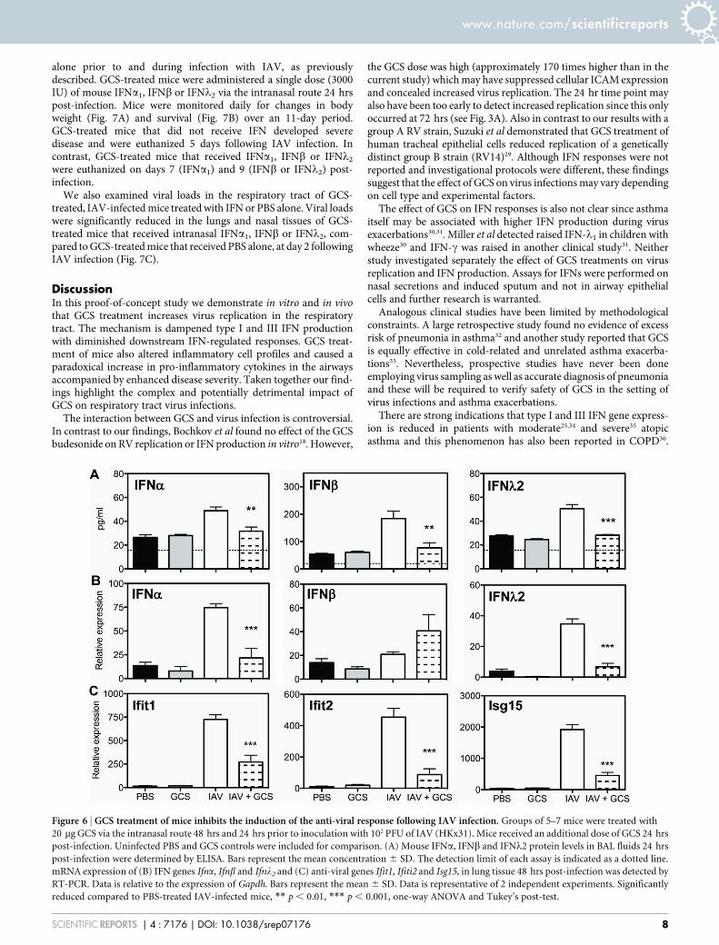
Treatment of mice with GCS prior to IAV infection inhibits theanti-viral response. Having established that GCS treatment in miceprior to IAV infection results in more severe disease, enhanced viralreplication and an increased inflammatory response, we sought toexamine if treatment of mice with GCS alters the anti-viral responseelicited following IAV infection. Mice were treated with GCS andinfected with IAV, as previously described. At day 1 following IAV-infection, GCS-treated mice had significantly reduced levels of IFNa,IFNb and IFNl2 proteins in BAL fluid (Fig. 6A). Furthermore, at day2 post-infection, mRNA expression of Ifna and Ifnl2 as well as theISGs Ifit1, Ifit2 and Isg15, were significantly reduced in lung tissuefrom mice treated with GCS (Fig. 6B–C).
A single intranasal treatment of IFN delays the onset of severedisease in GCS-treated animals. As we had established a deficiencyin the anti-viral response in mice treated with GCS, we next evaluatedthe ability of intranasal administration of type I and III IFNs to lessendisease severity in these mice. Mice were treated with GCS or PBS
Figure 4 | Mice treated with GCS prior to IAV infection develop severe disease. (A) Monolayers of primary mouse airway epithelial cells (mAEC) were
treated with GCS for 24 hrs, washed and infected with IAV (HKx31, MOI 1). Levels of infectious virus in cell supernatants were determined by plaque
assay at 24 and 48 hrs post-infection. Bars represent the mean 6 SD from triplicate samples. Viral titer significantly increased with GCS treatment,
*** p , 0.001, Student’s t-test. (B) Groups of 5 C57BL/6 mice were treated with 20 mg GCS via the intranasal route 48 hrs and 24 hrs prior to inoculation
with 102 PFU of IAV (HKx31). Mice received additional doses of GCS 24 hr post-infection and every 48 hrs thereafter. Uninfected mice treated with PBS
or GCS were included for comparison. Mice were observed daily for weight loss (left panel). Mice which had lost $15% of their original body weight were
euthanized. Data represents the mean % weight change 6 SEM. Survival plots are shown (right panel). (C) Titers of infectious virus in lung (left panel)
and nasal tissue (right panel) homogenates were determined by plaque assay at days 1, 3 and 5 post-infection. Bars represent the mean viral titer from a
group of 5 mice 6 SD. Virus titers from GCS-treated mice were significantly higher than those from PBS-treated mice, *** p , 0.001, one-way ANOVA
and Tukey’s post-test. Data is representative of a minimum of 2 independent experiments.
www.nature.com/scientificreports
SCIENTIFIC REPORTS | 4 : 7176 | DOI: 10.1038/srep07176 6
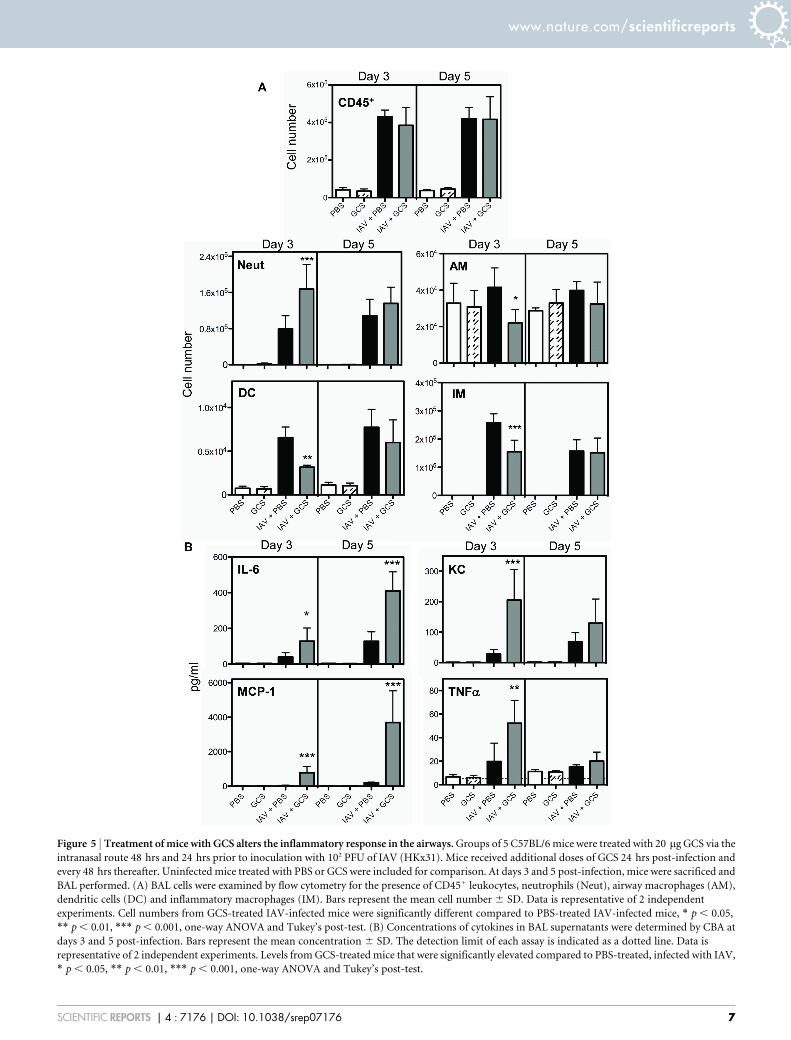
Figure 5 | Treatment of mice with GCS alters the inflammatory response in the airways. Groups of 5 C57BL/6 mice were treated with 20 mg GCS via the
intranasal route 48 hrs and 24 hrs prior to inoculation with 102 PFU of IAV (HKx31). Mice received additional doses of GCS 24 hrs post-infection and
every 48 hrs thereafter. Uninfected mice treated with PBS or GCS were included for comparison. At days 3 and 5 post-infection, mice were sacrificed and
BAL performed. (A) BAL cells were examined by flow cytometry for the presence of CD451 leukocytes, neutrophils (Neut), airway macrophages (AM),
dendritic cells (DC) and inflammatory macrophages (IM). Bars represent the mean cell number 6 SD. Data is representative of 2 independent
experiments. Cell numbers from GCS-treated IAV-infected mice were significantly different compared to PBS-treated IAV-infected mice, * p , 0.05,
** p , 0.01, *** p , 0.001, one-way ANOVA and Tukey’s post-test. (B) Concentrations of cytokines in BAL supernatants were determined by CBA at
days 3 and 5 post-infection. Bars represent the mean concentration 6 SD. The detection limit of each assay is indicated as a dotted line. Data is
representative of 2 independent experiments. Levels from GCS-treated mice that were significantly elevated compared to PBS-treated, infected with IAV,
* p , 0.05, ** p , 0.01, *** p , 0.001, one-way ANOVA and Tukey’s post-test.
www.nature.com/scientificreports
SCIENTIFIC REPORTS | 4 : 7176 | DOI: 10.1038/srep07176 7
alone prior to and during infection with IAV, as previouslydescribed. GCS-treated mice were administered a single dose (3000IU) of mouse IFNa1, IFNb or IFNl2 via the intranasal route 24 hrspost-infection. Mice were monitored daily for changes in bodyweight (Fig. 7A) and survival (Fig. 7B) over an 11-day period.GCS-treated mice that did not receive IFN developed severedisease and were euthanized 5 days following IAV infection. Incontrast, GCS-treated mice that received IFNa1, IFNb or IFNl2
were euthanized on days 7 (IFNa1) and 9 (IFNb or IFNl2) post-infection.
We also examined viral loads in the respiratory tract of GCS-treated, IAV-infected mice treated with IFN or PBS alone. Viral loadswere significantly reduced in the lungs and nasal tissues of GCS-treated mice that received intranasal IFNa1, IFNb or IFNl2, com-pared to GCS-treated mice that received PBS alone, at day 2 followingIAV infection (Fig. 7C).
DiscussionIn this proof-of-concept study we demonstrate in vitro and in vivothat GCS treatment increases virus replication in the respiratorytract. The mechanism is dampened type I and III IFN productionwith diminished downstream IFN-regulated responses. GCS treat-ment of mice also altered inflammatory cell profiles and caused aparadoxical increase in pro-inflammatory cytokines in the airwaysaccompanied by enhanced disease severity. Taken together our find-ings highlight the complex and potentially detrimental impact ofGCS on respiratory tract virus infections.
The interaction between GCS and virus infection is controversial.In contrast to our findings, Bochkov et al found no effect of the GCSbudesonide on RV replication or IFN production in vitro18. However,
the GCS dose was high (approximately 170 times higher than in thecurrent study) which may have suppressed cellular ICAM expressionand concealed increased virus replication. The 24 hr time point mayalso have been too early to detect increased replication since this onlyoccurred at 72 hrs (see Fig. 3A). Also in contrast to our results with agroup A RV strain, Suzuki et al demonstrated that GCS treatment ofhuman tracheal epithelial cells reduced replication of a geneticallydistinct group B strain (RV14)29. Although IFN responses were notreported and investigational protocols were different, these findingssuggest that the effect of GCS on virus infections may vary dependingon cell type and experimental factors.
The effect of GCS on IFN responses is also not clear since asthmaitself may be associated with higher IFN production during virusexacerbations30,31. Miller et al detected raised IFN-l1 in children withwheeze30 and IFN-c was raised in another clinical study31. Neitherstudy investigated separately the effect of GCS treatments on virusreplication and IFN production. Assays for IFNs were performed onnasal secretions and induced sputum and not in airway epithelialcells and further research is warranted.
Analogous clinical studies have been limited by methodologicalconstraints. A large retrospective study found no evidence of excessrisk of pneumonia in asthma32 and another study reported that GCSis equally effective in cold-related and unrelated asthma exacerba-tions33. Nevertheless, prospective studies have never been doneemploying virus sampling as well as accurate diagnosis of pneumoniaand these will be required to verify safety of GCS in the setting ofvirus infections and asthma exacerbations.
There are strong indications that type I and III IFN gene express-ion is reduced in patients with moderate25,34 and severe35 atopicasthma and this phenomenon has also been reported in COPD36.
Figure 6 | GCS treatment of mice inhibits the induction of the anti-viral response following IAV infection. Groups of 5–7 mice were treated with
20 mg GCS via the intranasal route 48 hrs and 24 hrs prior to inoculation with 102 PFU of IAV (HKx31). Mice received an additional dose of GCS 24 hrs
post-infection. Uninfected PBS and GCS controls were included for comparison. (A) Mouse IFNa, IFNb and IFNl2 protein levels in BAL fluids 24 hrs
post-infection were determined by ELISA. Bars represent the mean concentration 6 SD. The detection limit of each assay is indicated as a dotted line.
mRNA expression of (B) IFN genes Ifna, Ifnb and Ifnl2 and (C) anti-viral genes Ifit1, Ifiti2 and Isg15, in lung tissue 48 hrs post-infection was detected by
RT-PCR. Data is relative to the expression of Gapdh. Bars represent the mean 6 SD. Data is representative of 2 independent experiments. Significantly
reduced compared to PBS-treated IAV-infected mice, ** p , 0.01, *** p , 0.001, one-way ANOVA and Tukey’s post-test.
www.nature.com/scientificreports
SCIENTIFIC REPORTS | 4 : 7176 | DOI: 10.1038/srep07176 8

Impairment is associated with increased RV replication25,34.Importantly, individuals with mild/well-controlled asthma37,38 andmild asthma39 displayed no differences in IFN levels suggesting thatreduced IFN production may be the consequence of airway inflam-mation and connected to disease severity. A key consideration is thatmoderate and severe asthmatics and patients with COPD are invari-ably treated with GCS that may cause immune suppression, more soat higher doses. Intrinsic disease-related immune suppression hasthus made it problematic to investigate whether extrinsic factors(GCS) contribute to a blunted immune response. This is furthercomplicated by anti-inflammatory benefits of GCS that may obscuretheir detrimental actions. With this perspective we have excluded theeffect of underlying lung disease and primarily examined GCS activ-ities in healthy human lung cells in vitro and an in vivo mouse model.Additionally, we have examined the effect of a single dose of GCStreatment of human cells in vitro, which may underestimate theeffects of repetitive use of GCS by patients with asthma or COPD.
GCS in clinically relevant doses increased virus replication but wedid not observe significant reductions in the expression of IFNa,IFNb and IFNl genes in PBEC, PAF or PAM treated with GCS priorto RV or IAV infection (Fig. 1). This was not unexpected since IFNresponses are transient, peaking at variable times, therefore changesmay be difficult to detect in vitro. However, our in vivo studiesunambiguously demonstrated that mRNA expression and proteinlevels of IFNa, IFNb and IFNl were significantly reduced in thelungs of GCS-treated mice following IAV infection (Fig. 6). As analternative approach we also measured ISG expression since they arereliable surrogate indicators of IFN-signaling20. As shown, GCSreduced the expression of ISGs such as IFIT1 and ISG15 (Fig. 1),whilst increasing virus replication (Fig. 3, Supplementary Fig. S2), inboth primary human airway cells and mouse lung (Fig. 4C). In the
mouse model GCS-mediated enhanced infection was associated withmore severe illness and in human disease it is possible that amplifiedvirus infections may predispose to more frequent bacterial secondaryinfections and pneumonia40,41. Higher levels of virus infection mayalso oppose some of the beneficial anti-inflammatory actions of GCSas suggested by findings noted in our mouse model.
GCS treatment of human PBEC, PAF and PAM inhibited produc-tion of pro-inflammatory cytokines such as IL-8 and IL-6, followingvirus infection (Fig. 2) but this contrasted with responses in ourmouse model. In mice, GCS treatment was associated with anincrease in neutrophils and a reduction in dendritic cell (DC), airwaymacrophage (AM) and inflammatory macrophage (IM) numbers, 3days following infection (Fig. 5A). In addition, GCS-treated micedisplayed elevated levels of the cytokines IL-6, MCP-1 and KC inthe airways (Fig. 5B). The usual anti-inflammatory properties of GCSmay therefore be counterbalanced in vivo by enhancement of virusreplication and augmented inflammatory responses. This obser-vation is in accord with BAL and biopsy studies that have shownthat GCS do not prevent airway inflammation caused by RV infec-tion of both healthy and asthmatic volunteers42,43.
GCS therapies have been implicated in the development of pneu-monia in asthma and COPD in some7,8 but not all studies32. In addi-tion, GCS may have limited ability to prevent virus exacerbations9,10.Given the anti-inflammatory clinical benefits of GCS in these dis-eases, the development of improved preventive therapies or strategiesto counter virus infections will be a significant advance. IFNa andIFNb have been used therapeutically to treat several conditions suchas viral hepatitis and multiple sclerosis and a recent study of inhaledIFNb in asthma demonstrated safety and efficacy in patients withdisease exacerbations44. In the mouse model, intranasal delivery ofIFNa, IFNb or IFNl2 into the lung of GCS-treated mice 24 hours
Figure 7 | Intranasal administration of recombinant IFN following IAV infection improves the severe disease observed in GCS-treated mice. Groups of
5 C57BL/6 mice were treated with 20 mg of GCS via the intranasal route 48 hrs and 24 hrs prior to inoculation with 102 PFU of IAV (HKx31). Mice
received additional doses of GCS at 24 hrs post-infection and every 48 hrs thereafter. At day 1 post-infection, GCS-treated mice were administered 3000
IU of mouse IFNa1, IFNb or IFNl2 via the intranasal route. (A) Mice were weighed daily and data represents the mean % weight change 6 SEM. Animals
that had lost $15% of their original body weight were euthanized. (B) Survival curves are shown. (C) Titers of infectious virus in lung homogenates were
determined by plaque assay at day 2 post-infection (24 hrs following IFN treatment). Bars represent the mean viral titer from a group of 5 mice 6 SD.
Virus titers from GCS-treated mice treated with IFN were significantly lower than GCS-treated mice not treated with IFN, *** p , 0.001, one-way
ANOVA and Tukey’s post-test.
www.nature.com/scientificreports
SCIENTIFIC REPORTS | 4 : 7176 | DOI: 10.1038/srep07176 9

following infection delayed the onset of disease by several days(Fig. 7A/B) and reduced viral loads (Fig. 7C). This approach is feas-ible since the type I IFN receptor complex (IFNAR1/2) is ubiqui-tously expressed on all cell types, while the type III receptor(IFNlR1) is selectively expressed on epithelial cells, such as thosethat line the airways (reviewed by45). Recent studies have suggestedthat type III IFNs could provide a therapeutic alternative over type IIFNs, as they appear to induce fewer side effects, possibly due to theircell-specific actions46. Inhaled IFNl may therefore be a potentialtherapeutic adjuvant for improved treatment of virus infections inpatients undergoing GCS therapy and further studies are needed.
In summary, our findings indicate that GCS suppress host innateimmune responses, reducing cardinal early protective mechanismsand causing more severe infections. We also demonstrate the poten-tial of IFNs to offset the impact of GCS on innate immune responses.Overall, although GCS therapy can be beneficial, our studies providea rationale for further studies to examine GCS activities and thepotential of IFN as an adjuvant therapy for individuals who rely onGCS treatments.
MethodsCulture of primary human airway cells. Primary human airway cells were obtainedfrom normal subjects whom were non-smokers or had not smoked for .15 years andnone had a diagnosis of asthma or COPD (normal FEV1 measurements). Studies wereapproved by the Monash Health and Monash Medical Centre Human ResearchEthics Committee, consent was obtained from all subjects and studies were conductedin accordance with the approved guidelines. Primary bronchial epithelial cells(PBEC) were obtained from bronchial brushings and cultured under submergedconditions on collagen-coated flasks (MP Biomedicals, USA) in supplementedbronchial epithelial growth medium (BEGM; Lonza, Australia). Primary airwayfibroblasts (PAF) were obtained from bronchial tissue obtained from resection orbiopsy as described previously21. All bronchial biopsies and brushings were obtainedfrom the same anatomical regions (bronchial generations 4–7) and were used within 5passages. Primary airway macrophages (PAM) were obtained via bronchoalveolarlavage (BAL) during routine bronchoscopy and cultured overnight beforeexperimentation. PAF and PAM were cultured in minimum essential medium(MEM, Gibco, USA) supplemented with 10% FCS, 2 mM L-glutamine, 100 U/mlpenicillin and 100 mg/ml streptomycin.
Virus infection and GCS treatment of primary human airway cells. Human RVserotype 16 and the seasonal IAV strain A/Solomon Islands/3/2006 (H1N1) werepropagated, purified and titrated as previously described21,23. Preliminaryexperiments were performed to determine the optimal virus dose for each cell type.Human airway cells were treated with 10 nM fluticasone propionate (GCS, SigmaAldrich, USA) or diluent alone (mock) for 24 hrs in 24-well plates. Cell counts wereperformed to ensure GCS did not significantly alter cell numbers 24 hrs followingtreatment. In some experiments, cells were treated for 24 hrs with 10 nMdexamethasone (Dex; Sigma Aldrich, USA) or 10 nM budesonide (Bud; AstraZeneca,Australia).
Primary human airway cells (approximately 80% confluency) were washed andinfected with RV or IAV in serum free MEM or BEGM without hydrocortisone for1 hr at a multiplicity of infection (MOI) of 1 (PBEC), 3 (PAF) or 5 (PAM). Cellmonolayers were then washed and incubated in BEGM without hydrocortisone(PBEC) or MEM supplemented with 2% FCS (PAF and PAM). Samples infected withIAV were cultured in the presence of 4 mg/ml trypsin to permit multiple rounds ofreplication.
In some experiments, primary human cells were treated with GCS for 24 hrs,infected and then incubated in media containing 250 IU/ml human IFNa2a
(Roferon-A, Roche, USA), IFNb1a (Rebif 44, Merck, Germany) or IFNl1 (PBLInterferon Source, USA) for 24, 48 or 72 hrs.
Levels of infectious virus in primary cell culture supernatants were determined at24, 48 and 72 hrs following infection by titration on Ohio Hela cells (RV) or bystandard plaque assay on MDCK cells (IAV). To examine virus infection, cells werefixed at 8 hrs post-infection with 80% acetone and then stained with anti-nuclearprotein (IAV) or anti-3C protease (RV) antibodies (a gift from James Gern, Madison,WI), as previously described23.
Gene expression in human airway cells. Total RNA was isolated from frozenprimary human cell lysates 24 and 48 hrs following RV or IAV infection using TRIReagent (Molecular Research Centre, Cincinnati, USA) and reverse transcriptionperformed using Multiscribe Reverse Transcriptase (Invitrogen, USA). RT-PCRreactions were performed using FastStart SYBR Green Master Mix (Roche, USA) on aCorbett Rotor-Gene 3000 machine21. Gene expression was calculated relative to 18S.See Supplementary Table S2 in the supplemental material for target genes and primersequences. Viral RV RNA was quantified as previously described21.
Detection of human cytokines. Levels of cytokines in primary human cellsupernatants were determined at 24 and 48 hrs following virus infection byCytometric Bead Array (CBA) Human Inflammation and Chemokine Kits (BDBiosciences, USA). Levels of human IFNa in cell supernatants were determined bysandwich ELISA using mouse monoclonal (clone MMHA-11) and rabbit polyclonalantibodies (PBL Interferon Source, USA). Human IFNl2 was quantified by ELISA(R&D Systems, USA).
Preparation and infection of primary mouse airway epithelial cells (mAEC).Mouse AEC cultures were prepared from female C57BL/6 mice as previouslydescribed22, with minor modifications. Briefly, lungs from mice were digested in1.5 mg/ml Pronase (Roche, USA), 0.1 mg/ml DNase I (Sigma-Aldrich, USA) for 1 hrat 37uC in 5% CO2. Single cell suspensions were incubated with purified rat anti-mouse CD45 antibody (BD Biosciences, USA) and epithelial cells negatively enrichedwith BioMag goat anti-rat Ig-coupled magnetic beads (Qiagen, USA). Cells werecultured on collagen-coated (MP Biomedicals, USA) plates in DMEM/F-12 media(Gibco, USA) supplemented with 10% FCS, 2 mM L-glutamine, 100 U/ml penicillin,100 mg/ml streptomycin and 5 mg/ml insulin (Sigma-Aldrich). To confirm cellpurity, monolayers were detached with 3 mM EDTA and cells stained with mouseanti-EpCAM antibodies (BioLegend, USA) and examined by flow cytometry. Cellmonolayers were negative for the presence of fibroblasts (anti-mouse vimentin (SantaCruz, USA)). Monolayers were infected with the HKx31 strain of IAV (H3N2, MOI 1)in serum free DMEM/F-12 for 1 hr and then washed and incubated in DMEM/F-12supplemented with 2% FCS and 4 mg/ml trypsin for 24 or 48 hrs.
Influenza virus infection and treatment of mice. Mice (6–8 week old female C57BL/6) were anesthetized and infected with 102 PFU of HKx31 (H3N2) via the intranasal(i.n.) route in 50 ml PBS. Groups of 5 mice were treated i.n. with 20 mg fluticasonepropionate diluted in 50 ml PBS or PBS alone for 48 and 24 hrs prior to infection andadditional doses 24 hrs post-infection and every 48 hrs thereafter. In someexperiments, mice were treated i.n. with 3000 IU of mouse IFNa1, IFNb or IFNl2
(PBL Interferon Source, USA) in 50 ml of PBS, 24 hrs post-infection.Mice were weighed daily and assessed for visual signs of clinical disease, including
inactivity, ruffled fur, labored breathing and huddling behavior. Animals that had lost$15% of their original body weight were euthanized. Research was approved by theMonash University Animal Ethics Committee and studies were conducted inaccordance with the approved guidelines. Levels of infectious virus in lung and nasaltissue homogenates were determined by standard plaque assay on MDCK cells.
Recovery and characterization of leukocytes in the airways of mice. BAL cells wereobtained and analysed by flow cytometry as previously described23. For flowcytometric analysis, BAL cells were incubated with supernatants from hybridoma2.4G2 to block Fc receptors, followed by staining with monoclonal antibodies toCD45, Ly6C, Ly6G, CD11c and I-Ab (BD Biosciences, USA). Leucocytes, neutrophils,airway macrophages (AM) and dendritic cells (DC) were identified as describedpreviously23. Inflammatory macrophages (IM) were identified as Ly6G2 Ly6C1.Living cells (propidium iodine negative) were analysed using a BD FACS Canto IIflow cytometer (BD Biosciences, USA).
Detection of cytokines in mouse BAL fluids. Levels of cytokines in BAL fluid of micewere determined using Cytometric Bead Array (CBA) Mouse Inflammation or FlexKits (BD Biosciences, USA). Levels of mouse IFNa were determined by sandwichELISA using mouse monoclonal (clone F18, Thermo Scientific, USA) and rabbitpolyclonal antibodies (PBL Interferon Source, USA). Levels of mouse IFNb weredetermined by sandwich ELISA using mouse monoclonal (clone 7F-D3, Abcam, UK)and rabbit polyclonal antibodies (PBL Interferon Source, USA). Mouse IFNl2 wasquantified by ELISA (R&D Systems, USA).
Analysis of gene expression in mouse lung tissue. Lungs were excised from mice andimmediately frozen in liquid nitrogen. Total RNA was extracted using TRIsure(Bioline, UK). RNA (1 mg) was DNase-treated (Promega, USA) and reverse-transcribed to cDNA using M-MLV reverse transcriptase (Promega, USA), accordingto the manufacturer’s instructions. RT-PCR reactions were performed using SYBRGreen (Invitrogen, USA) on an Applied Biosystems 7900HT Fast Real-Time PCRMachine. Gene expression was calculated relative to the housekeeping gene Gapdh.See Supplementary Table S2 for target genes and primer sequences.
Statistical analyses. For comparison of two sets of values, Student’s t-test (two-tailed,two-sample equal variance) was used. When comparing $ three sets of values, datawere analyzed by one-way ANOVA and Tukey’s multiple comparison test.
1. Dulek, D. E. & Peebles, R. S., Jr. Viruses and asthma. Biochim. Biophys. Acta. 1810,1080–1090 (2011).
2. Singanayagam, A., Joshi, P. V., Mallia, P. & Johnston, S. L. Viruses exacerbatingchronic pulmonary disease: the role of immune modulation. BMC Med. 10, 27(2012).
3. Bergman, H., Livornese, L. L., Jr., Sambhara, S., Santoro, J. & Dessain, S. K.Patients Hospitalized with pH1N1 Influenza in an Academic CommunityMedical Center. Open Respir Med J. 5, 19–23 (2011).
4. Zarogoulidis, P. et al. Long-term respiratory follow-up of H1N1 infection. Virol J.8, 319 (2011).
www.nature.com/scientificreports
SCIENTIFIC REPORTS | 4 : 7176 | DOI: 10.1038/srep07176 10
5. Nannini, L. J., Poole, P., Milan, S. J., Holmes, R. & Normansell, R. Combinedcorticosteroid and long-acting beta2-agonist in one inhaler versus placebo forchronic obstructive pulmonary disease. Cochrane Database Syst Rev. 11,CD003794 (2013).
6. White, P., Thornton, H., Pinnock, H., Georgopoulou, S. & Booth, H. P.Overtreatment of COPD with Inhaled Corticosteroids - Implications for Safetyand Costs: Cross-Sectional Observational Study. PLoS One. 8, e75221 (2013).
7. McKeever, T., Harrison, T. W., Hubbard, R. & Shaw, D. Inhaled corticosteroidsand the risk of pneumonia in people with asthma: a case-control study. Chest. 144,1788–1794 (2013).
8. Suissa, S., Patenaude, V., Lapi, F. & Ernst, P. Inhaled corticosteroids in COPD andthe risk of serious pneumonia. Thorax. 68, 1029–1036 (2013).
9. Ducharme, F. M. et al. Preemptive use of high-dose fluticasone for virus-inducedwheezing in young children. N. Engl. J. Med. 360, 339–353 (2009).
10. Price, D., Yawn, B., Brusselle, G. & Rossi, A. Risk-to-benefit ratio of inhaledcorticosteroids in patients with COPD. Prim Care Respir J. 22, 92–100 (2013).
11. Puhakka, T. et al. The common cold: effects of intranasal fluticasone propionatetreatment. J. Allergy Clin. Immunol. 101, 726–731 (1998).
12. Gustafson, L. M., Proud, D., Hendley, J. O., Hayden, F. G. & Gwaltney, J. M., Jr.Oral prednisone therapy in experimental rhinovirus infections. J. Allergy Clin.Immunol. 97, 1009–1014 (1996).
13. Skevaki, C. L. et al. Budesonide and formoterol inhibit inflammatory mediatorproduction by bronchial epithelial cells infected with rhinovirus. Clin. Exp.Allergy. 39, 1700–1710 (2009).
14. Edwards, M. R., Haas, J., Panettieri, R. A., Jr., Johnson, M. & Johnston, S. L.Corticosteroids and beta2 agonists differentially regulate rhinovirus-inducedinterleukin-6 via distinct Cis-acting elements. J. Biol. Chem. 282, 15366–15375(2007).
15. Van Ly, D. et al. Effects of beta(2) Agonists, Corticosteroids, and Novel Therapieson Rhinovirus-Induced Cytokine Release and Rhinovirus Replication in PrimaryAirway Fibroblasts. J Allergy (Cairo). 2011, 457169 (2011).
16. Davies, J. M. et al. Budesonide and formoterol reduce early innate anti-viralimmune responses in vitro. PLoS One. 6, e27898 (2011).
17. Edwards, M. R., Johnson, M. W. & Johnston, S. L. Combination therapy:Synergistic suppression of virus-induced chemokines in airway epithelial cells.Am. J. Respir. Cell Mol. Biol. 34, 616–624 (2006).
18. Bochkov, Y. A. et al. Budesonide and formoterol effects on rhinovirus replicationand epithelial cell cytokine responses. Resp Res. 14, 98 (2013).
19. Wang, J. et al. Differentiated human alveolar type II cells secrete antiviral IL-29(IFN-lambda 1) in response to influenza A infection. J. Immunol. 182, 1296–1304(2009).
20. Schoggins, J. W. & Rice, C. M. Interferon-stimulated genes and their antiviraleffector functions. Curr Opin Virol. 1, 519–525 (2011).
21. Thomas, B. J. et al. Transforming growth factor-beta enhances rhinovirusinfection by diminishing early innate responses. Am. J. Respir. Cell Mol. Biol. 41,339–347 (2009).
22. Tate, M. D., Schilter, H. C., Brooks, A. G. & Reading, P. C. Responses of mouseairway epithelial cells and alveolar macrophages to virulent and avirulent strainsof influenza A virus. Viral Immunol. 24, 77–88 (2011).
23. Tate, M. D., Pickett, D. L., van Rooijen, N., Brooks, A. G. & Reading, P. C. Criticalrole of airway macrophages in modulating disease severity during influenza virusinfection of mice. J. Virol. 84, 7569–7580 (2010).
24. Gern, J. E. et al. Rhinovirus enters but does not replicate inside monocytes andairway macrophages. J. Immunol. 156, 621–627 (1996).
25. Wark, P. A. et al. Asthmatic bronchial epithelial cells have a deficient innateimmune response to infection with rhinovirus. J. Exp. Med. 201, 937–947 (2005).
26. Witte, K. et al. Despite IFN-lambda receptor expression, blood immune cells, butnot keratinocytes or melanocytes, have an impaired response to type IIIinterferons: implications for therapeutic applications of these cytokines. GenesImmun. 10, 702–714 (2009).
27. Singam, R. et al. Combined fluticasone propionate and salmeterol reduces RSVinfection more effectively than either of them alone in allergen-sensitized mice.Virol J. 3, 32 (2006).
28. Chu, H. W., Campbell, J. A., Rino, J. G., Harbeck, R. J. & Martin, R. J. Inhaledfluticasone propionate reduces concentration of Mycoplasma pneumoniae,inflammation, and bronchial hyperresponsiveness in lungs of mice. J. Infect. Dis.189, 1119–1127 (2004).
29. Suzuki, T. et al. Effects of dexamethasone on rhinovirus infection in culturedhuman tracheal epithelial cells. Am J Physiol Lung Cell Mol Physiol. 278, L560–571(2000).
30. Miller, E. K. et al. A mechanistic role for type III IFN-lambda1 in asthmaexacerbations mediated by human rhinoviruses. Am. J. Respir. Crit. Care Med.185, 508–516 (2012).
31. Schwantes, E. A. et al. Interferon gene expression in sputum cells correlates withthe Asthma Index Score during virus-induced exacerbations. Clin. Exp. Allergy.44, 813–821 (2014).
32. O’Byrne, P. M. et al. Risks of pneumonia in patients with asthma taking inhaledcorticosteroids. Am. J. Respir. Crit. Care Med. 183, 589–595 (2011).
33. Reddel, H. K. et al. Effect of different asthma treatments on risk of cold-relatedexacerbations. Eur. Respir. J. 38, 584–593 (2011).
34. Contoli, M. et al. Role of deficient type III interferon-lambda production inasthma exacerbations. Nat. Med. 12, 1023–1026 (2006).
35. Edwards, M. R. et al. Impaired innate interferon induction in severe therapyresistant atopic asthmatic children. Mucosal Immunol. 6, 797–806 (2013).
36. Mallia, P. et al. Experimental rhinovirus infection as a human model of chronicobstructive pulmonary disease exacerbation. Am. J. Respir. Crit. Care Med. 183,734–742 (2011).
37. Sykes, A. et al. TLR3, TLR4 and TLRs7-9 Induced Interferons Are Not Impaired inAirway and Blood Cells in Well Controlled Asthma. PLoS One. 8, e65921 (2013).
38. Sykes, A. et al. Rhinovirus-induced interferon production is not deficient in wellcontrolled asthma. Thorax. 69, 240–246 (2014).
39. Bochkov, Y. A. et al. Rhinovirus-induced modulation of gene expression inbronchial epithelial cells from subjects with asthma. Mucosal Immunol. 3, 69–80(2010).
40. McCullers, J. A. The co-pathogenesis of influenza viruses with bacteria in the lung.Nat Rev Microbiol. 12, 252–262 (2014).
41. Molyneaux, P. L. et al. Outgrowth of the bacterial airway microbiome afterrhinovirus exacerbation of chronic obstructive pulmonary disease. Am. J. Respir.Crit. Care Med. 188, 1224–1231 (2013).
42. Grunberg, K. et al. Rhinovirus-induced airway inflammation in asthma: effect oftreatment with inhaled corticosteroids before and during experimental infection.Am. J. Respir. Crit. Care Med. 164, 1816–1822 (2001).
43. de Kluijver, J. et al. Interleukin-1beta and interleukin-1ra levels in nasal lavagesduring experimental rhinovirus infection in asthmatic and non-asthmaticsubjects. Clin. Exp. Allergy. 33, 1415–1418 (2003).
44. Djukanovic, R. et al. The Effect of Inhaled IFN-beta on Worsening of AsthmaSymptoms Caused by Viral Infections. A Randomized Trial. Am. J. Respir. Crit.Care Med. 190, 145–154 (2014).
45. Levy, D. E., Marie, I. J. & Durbin, J. E. Induction and function of type I and IIIinterferon in response to viral infection. Curr Opin Virol. 1, 476–486 (2011).
46. Donnelly, R. P., Dickensheets, H. & O’Brien, T. R. Interferon-lambda and therapyfor chronic hepatitis C virus infection. Trends Immunol. 32, 443–450 (2011).
AcknowledgmentsThis work was supported by the Victorian State Government Operational InfrastructureScheme. M.D. Tate and P.J. Hertzog were funded by National Health and Medical ResearchCouncil (NHMRC) of Australia Fellowships. B.J. Thomas was funded by Monash Lung &Sleep and Monash Lung & Sleep Institute.
Author contributionsB.J.T. and M.D.T. designed, performed and analysed experiments. R.A.P. performedexperiments. B.J.T., P.G.B., M.D.T. and P.J.H. conceived and contributed to theexperimental design and wrote the paper.
Additional informationSupplementary information accompanies this paper at http://www.nature.com/scientificreports
Competing financial interests: The authors declare no competing financial interests.
How to cite this article: Thomas, B.J., Porritt, R.A., Hertzog, P.J., Bardin, P.G. & Tate, M.D.Glucocorticosteroids enhance replication of respiratory viruses: effect of adjuvantinterferon. Sci. Rep. 4, 7176; DOI:10.1038/srep07176 (2014).
This work is licensed under a Creative Commons Attribution-NonCommercial-NoDerivs 4.0 International License. The images or other third party material inthis article are included in the article’s Creative Commons license, unless indicatedotherwise in the credit line; if the material is not included under the CreativeCommons license, users will need to obtain permission from the license holderin order to reproduce the material. To view a copy of this license, visit http://creativecommons.org/licenses/by-nc-nd/4.0/
www.nature.com/scientificreports
SCIENTIFIC REPORTS | 4 : 7176 | DOI: 10.1038/srep07176 11