
Alveolar Macrophages Are the Primary Interferon-α Producer in Pulmonary Infection with RNA Viruses
13
Immunity Article Alveolar Macrophages Are the Primary Interferon- a Producer in Pulmonary Infection with RNA Viruses Yutaro Kumagai, 1,3 Osamu Takeuchi, 1,3 Hiroki Kato, 1 Himanshu Kumar, 1 Kosuke Matsui, 1,3 Eiichi Morii, 2 Katsuyuki Aozasa, 2 Taro Kawai, 1,3 and Shizuo Akira 1,3, * 1 Department of Host Defense, Research Institute for Microbial Diseases, Osaka University 2 Department of Pathology, Graduate School of Medicine, Osaka University 3 ERATO, Japan Science and Technology Agency 3-1 Yamada-oka, Suita, Osaka 565-0871, Japan *Correspondence: [email protected] DOI 10.1016/j.immuni.2007.07.013 SUMMARY Type I interferons (IFNs) are critical for antiviral responses. Here we generated a knockin mouse in which green fluorescence protein (GFP) was expressed under the control of the Ifna6 pro- moter. Virus-induced expression of GFP reca- pitulated various IFN-a subtypes. Systemic in- fection of the mice with Newcastle disease virus (NDV) increased GFP + plasmacytoid den- dritic cells (pDCs) via the Toll-like receptor sys- tem, and GFP + conventional dendritic cells (cDCs) and macrophages via the RIG-I-like heli- case system. By contrast, lung infection with NDV led to IFN-a production in alveolar macro- phages (AMs) and cDCs, but not in pDCs. Spe- cific depletion of AMs caused a marked defect in the initial viral elimination in the lung. pDCs produced IFN-a in the absence of AM-mediated viral recognition, suggesting that pDCs function when the first defense line is broken. Thus, AMs act as a type I IFN producer that is important for the initial responses to viral infection in the lung. INTRODUCTION The innate immune system senses viral invasion and evokes quick responses by producing various cytokines. Among them, type I interferons (IFNs) are pleiotropic cyto- kines essential for antiviral immune responses. They are comprised of multiple IFN-as and single IFN-b, and other members such as IFN-u,-3, and -k (Honda et al., 2006). Humans and mice have more than 13 IFN-a family mem- bers. Type I IFNs induce apoptosis of virus-infected cells and cellular resistance to viral infection, and also activate natural killer (NK) and T cells (Stetson and Medzhitov, 2006). Thus, type I IFNs have an important role not only in the innate antiviral responses, but also in the activation of the adaptive immune system. Two innate immune receptor families, Toll-like recep- tors (TLRs) and RIG-I-like helicases (RLHs), have been shown to recognize viral components and induce type I IFNs (Akira et al., 2006). The TLR system senses vari- ous viral components, including double-stranded RNA (dsRNA) single-stranded RNA (ssRNA), and unmethylated DNA with CpG motifs via TLR3, TLR7, and TLR9, respec- tively. TLR3 triggers signaling cascades via an adaptor protein, the Toll-IL-1 receptor (TIR) domain containing adaptor-inducing IFN-b (TRIF), which activates two IkB kinase (IKK)-related kinases, TANK-binding kinase 1 (TBK1) and inducible IKK (IKK-i). These kinases are known to directly phosphorylate transcription factors of IFN reg- ulatory factor 3 (IRF-3) and IRF-7 (Kawai and Akira, 2006). These transcription factors then form a dimer, translocate to the nucleus, and activate the transcription of type I IFNs and IFN-inducible genes. On the other hand, TLR7 and TLR9 activate IRF-7 via an adaptor, MyD88 (Honda et al., 2004; Kawai et al., 2004), IL-1R-associated kinase 1 (IRAK1) (Uematsu et al., 2005), and IKK-a (Hoshino et al., 2006), but not TBK1 or IKK-i. The RLH family is comprised of the retinoic acid-induc- ible gene I (RIG-I), the melanoma differentiation-associ- ated gene 5 (MDA5), and Lgp2 (Akira et al., 2006). RIG-I and MDA5, but not Lgp2, contain caspase-recruit do- mains (CARDs) in addition to a RNA helicase domain. RIG-I is responsible for detection of various RNA viruses (Kato et al., 2005; Yoneyama et al., 2004), in vitro tran- scribed dsRNA (Kato et al., 2006), and 5 0 -triphosphate RNA (Hornung et al., 2006; Pichlmair et al., 2006), whereas MDA5 recognizes picornaviruses and polyinosinic polycy- tidylic acid [poly (I:C)] (Kato et al., 2006). RIG-I and MDA5 activate TBK1 and IKK-i via a CARD domain containing IFN-b promoter stimulator-1 (IPS-1), an adaptor also known as MAVS, CARDIF, or VISA (Kawai et al., 2005; Kumar et al., 2006; Meylan et al., 2005; Seth et al., 2005; Sun et al., 2006; Xu et al., 2005). Although various cells are reported to have the potential to produce type I IFNs when exposed to viruses in vitro, 240 Immunity 27, 240–252, August 2007 ª2007 Elsevier Inc.
-
Upload
independent -
Category
Documents
-
view
0 -
download
0
Transcript of Alveolar Macrophages Are the Primary Interferon-α Producer in Pulmonary Infection with RNA Viruses
Immunity
Article
Alveolar Macrophages Are thePrimary Interferon-a Producerin Pulmonary Infection with RNA VirusesYutaro Kumagai,1,3 Osamu Takeuchi,1,3 Hiroki Kato,1 Himanshu Kumar,1 Kosuke Matsui,1,3 Eiichi Morii,2
Katsuyuki Aozasa,2 Taro Kawai,1,3 and Shizuo Akira1,3,*1Department of Host Defense, Research Institute for Microbial Diseases, Osaka University2Department of Pathology, Graduate School of Medicine, Osaka University3ERATO, Japan Science and Technology Agency
3-1 Yamada-oka, Suita, Osaka 565-0871, Japan
*Correspondence: [email protected]
DOI 10.1016/j.immuni.2007.07.013
SUMMARY
Type I interferons (IFNs) are critical for antiviralresponses. Here we generated a knockin mousein which green fluorescence protein (GFP) wasexpressed under the control of the Ifna6 pro-moter. Virus-induced expression of GFP reca-pitulated various IFN-a subtypes. Systemic in-fection of the mice with Newcastle diseasevirus (NDV) increased GFP+ plasmacytoid den-dritic cells (pDCs) via the Toll-like receptor sys-tem, and GFP+ conventional dendritic cells(cDCs) and macrophages via the RIG-I-like heli-case system. By contrast, lung infection withNDV led to IFN-a production in alveolar macro-phages (AMs) and cDCs, but not in pDCs. Spe-cific depletion of AMs caused a marked defectin the initial viral elimination in the lung. pDCsproduced IFN-a in the absence of AM-mediatedviral recognition, suggesting that pDCs functionwhen the first defense line is broken. Thus, AMsact as a type I IFN producer that is importantfor the initial responses to viral infection inthe lung.
INTRODUCTION
The innate immune system senses viral invasion and
evokes quick responses by producing various cytokines.
Among them, type I interferons (IFNs) are pleiotropic cyto-
kines essential for antiviral immune responses. They are
comprised of multiple IFN-as and single IFN-b, and other
members such as IFN-u, -3, and -k (Honda et al., 2006).
Humans and mice have more than 13 IFN-a family mem-
bers. Type I IFNs induce apoptosis of virus-infected cells
and cellular resistance to viral infection, and also activate
natural killer (NK) and T cells (Stetson and Medzhitov,
2006). Thus, type I IFNs have an important role not only
240 Immunity 27, 240–252, August 2007 ª2007 Elsevier Inc.
in the innate antiviral responses, but also in the activation
of the adaptive immune system.
Two innate immune receptor families, Toll-like recep-
tors (TLRs) and RIG-I-like helicases (RLHs), have been
shown to recognize viral components and induce type
I IFNs (Akira et al., 2006). The TLR system senses vari-
ous viral components, including double-stranded RNA
(dsRNA) single-stranded RNA (ssRNA), and unmethylated
DNA with CpG motifs via TLR3, TLR7, and TLR9, respec-
tively. TLR3 triggers signaling cascades via an adaptor
protein, the Toll-IL-1 receptor (TIR) domain containing
adaptor-inducing IFN-b (TRIF), which activates two IkB
kinase (IKK)-related kinases, TANK-binding kinase 1
(TBK1) and inducible IKK (IKK-i). These kinases are known
to directly phosphorylate transcription factors of IFN reg-
ulatory factor 3 (IRF-3) and IRF-7 (Kawai and Akira, 2006).
These transcription factors then form a dimer, translocate
to the nucleus, and activate the transcription of type I IFNs
and IFN-inducible genes. On the other hand, TLR7 and
TLR9 activate IRF-7 via an adaptor, MyD88 (Honda
et al., 2004; Kawai et al., 2004), IL-1R-associated kinase
1 (IRAK1) (Uematsu et al., 2005), and IKK-a (Hoshino
et al., 2006), but not TBK1 or IKK-i.
The RLH family is comprised of the retinoic acid-induc-
ible gene I (RIG-I), the melanoma differentiation-associ-
ated gene 5 (MDA5), and Lgp2 (Akira et al., 2006). RIG-I
and MDA5, but not Lgp2, contain caspase-recruit do-
mains (CARDs) in addition to a RNA helicase domain.
RIG-I is responsible for detection of various RNA viruses
(Kato et al., 2005; Yoneyama et al., 2004), in vitro tran-
scribed dsRNA (Kato et al., 2006), and 50-triphosphate
RNA (Hornung et al., 2006; Pichlmair et al., 2006), whereas
MDA5 recognizes picornaviruses and polyinosinic polycy-
tidylic acid [poly (I:C)] (Kato et al., 2006). RIG-I and MDA5
activate TBK1 and IKK-i via a CARD domain containing
IFN-b promoter stimulator-1 (IPS-1), an adaptor also
known as MAVS, CARDIF, or VISA (Kawai et al., 2005;
Kumar et al., 2006; Meylan et al., 2005; Seth et al., 2005;
Sun et al., 2006; Xu et al., 2005).
Although various cells are reported to have the potential
to produce type I IFNs when exposed to viruses in vitro,
Immunity
Identification of IFN-a-Producing Cells In Vivo
stimulation of human peripheral blood mononuclear cells
with viruses has revealed that plasmacytoid dendritic cells
(pDCs), a rare subset of dendritic cells (DCs), are the major
producer of type I IFNs (Cella et al., 1999; Siegal et al.,
1999). Subsequently, a mouse counterpart of human
pDC was identified based on the surface expression of
CD11c and B220 (Asselin-Paturel et al., 2001; Bjorck,
2001; Nakano et al., 2001). It has been shown by ex vivo
experiments that the TLR system is responsible for the se-
cretion of type I IFNs in pDCs (Kato et al., 2005). pDCs pro-
duced IFN-a irrespective of the presence of type I IFN
receptor (Barchet et al., 2002; Prakash et al., 2005). In
contrast, conventional DCs (cDCs) and fibroblasts pro-
duce type I IFNs in response to viral infection in vitro in
a RLH-dependent fashion (Diebold et al., 2003; Kato
et al., 2005). These cell types produce IFN-a via type I
IFN receptor-mediated positive feedback. IFN-producing
killer dendritic cells (IKDCs) are also reported to produce
type I IFNs (Chan et al., 2006; Taieb et al., 2006), although
this has been questioned in a recent report (Vremec et al.,
2007). However, the contribution of each cell type to the
production of type I IFNs in vivo remains to be determined.
Additionally, the in vivo role of the two abovementioned
viral detector systems in the production of type I IFNs is
unknown.
Currently used methods have not succeeded in moni-
toring the expression of type I IFNs in vivo. For instance,
enzyme-linked immunosorbent assay (ELISA) is not suit-
able for detecting type I IFNs at a single-cell level. Al-
though intracellular staining of IFN-a is sensitive enough
to detect IFN-a expression in ex vivo experiments, it is
difficult to monitor IFN-a-producing cells in vivo. There-
fore, we generated a reporter mouse strain in which the
coding sequence of the Ifna6 gene was replaced by the
GFP coding sequence. Comparison between GFP ex-
pression and intracellular IFN-a staining, and quantitative
real-time PCR (Q-PCR) analysis for the expression of mul-
tiple IFN-a genes in GFP+ cells, revealed that this reporter
recapitulated the expression of various IFN-a genes.
pDCs were the sole producer of IFN-a in response to
TLR7 and TLR9 ligand inoculation, whereas cDCs were
the main producer in response to poly (I:C), the TLR3-
MDA5 ligand. In response to systemic RNA virus infec-
tion, not only pDCs, but also cDCs, macrophages, and
monocytes, produced IFN-a. The TLR system was re-
sponsible for the production of IFN-a in pDCs, whereas
cDCs and macrophages utilized the RLH system. When
the same virus was introduced intranasally, the IFN-a-
producing cells shifted from pDCs to alveolar macro-
phages (AMs) and cDCs. Depletion of AMs increased
the virus yield after NDV infection, further supporting
the role of this cell type in the control of viral infection.
Production of IFNs in AMs and cDCs depended on the
RLH system, whereas IPS-1 deficiency led to IFN-a
production in pDCs. Our data clearly demonstrates that
distinct IFN-producing cells (IPCs) are activated in a tis-
sue-specific manner. This serves to emphasize the im-
portance of local antiviral cells in the natural course of
the response to infection.
RESULTS
Generation of Ifna6gfp Knockin Locus and ItsValidation In VitroTo investigate which cell population or populations pro-
duce IFN-a in vivo, we generated a mouse strain in which
the coding sequence of the Ifna6 gene was replaced by
the GFP coding sequence (Figures 1A and 1B). Among
multiple IFN-as, we chose IFN-a6 because IFN-a6 ex-
pression is strongly induced in response to viral infection
in various cell types in vitro, as detected by Q-PCR and
DNA microarray analysis (Matsui et al., 2006). Additionally,
IFN-a6 was reported to be regulated solely by IRF-7, as is
the case for various IFN-as, except for IFN-a4; IFN-b and
IFN-a4 are reported to be regulated by IRF-3 in addition to
IRF-7 (Honda et al., 2005). In the targeted allele, the GFP
coding sequence and loxP-flanked neomycin-resistance
cassette replaced the complete sequence encoding
IFN-a6. The neomycin-resistance cassette was excised
in targeted embryonic stem (ES) cells in vitro by transfect-
ing a plasmid containing the Cre recombinase coding
gene. The resulting allele Ifna6gfp produced GFP instead
of IFN-a6. We used mice heterozygous for Ifna6gfp to
circumvent the interference in IFN-a6 production.
We first examined whether the Ifna6gfp allele recapitu-
lated IFN-a expression in vitro. Flt3L-induced bone mar-
row DCs (Flt3L-BMDCs) prepared from Ifna6gfp/+ mice
were infected with Newcastle disease virus (NDV) for 6
hr, and intracellular IFN-a staining was performed (Fig-
ure 1C). Flow cytometry analysis revealed that the number
of GFP-positive CD11c+B220+ pDCs markedly increased
in infected cells, and most of the intracellular IFN-a-posi-
tive cells were also positive for GFP. Next, GFP-positive
(GFP+) and GFP-negative (GFP�) pDCs were FACS-sorted
after NDV infection. The expression of type I IFN genes was
then examined by Q-PCR (Figure 1D). Expression of the
Ifna6, Ifna2, Ifna4, and Ifna5 genes, as well as the Ifnb1
gene, in GFP+ cells was markedly augmented, being about
ten times higher than that in GFP� cells. By contrast, the
expression of the Cxcl10 gene was almost equally upregu-
lated in both GFP+ and GFP� cells. This result indicates
that the expression of GFP reflects the induction of IFN-
as and IFN-b, but not the general transcriptional upregula-
tion invoked by NDV infection. Production of IFN-a was not
altered between Ifna6+/+ and Ifna6gfp/+ cells. This indicated
that the heterozygous Ifna6gfp allele does not affect IFN-a
production overall (Figure 1E). Taken as a whole, these re-
sults showed that the expression of GFP in Ifna6gfp/+ cells
is an appropriate reporter for IFN-a production in vitro.
IPCs in Response to TLR Ligand Stimulation In VivoWe next examined the responses of Ifna6gfp mice to TLR
and RLH ligands in vivo. Systemic administration of syn-
thetic nucleotide analogs, such as D-type CpG-oligo-
deoxynucleotide (CpG-ODN) (a ligand for TLR9), R-848
(TLR7), and poly (I:C) (TLR3-MDA5), are known to induce
IFN-a production in vivo (Asselin-Paturel et al., 2005;
Hemmi et al., 2002; Kato et al., 2006). CpG-ODN was
administered in Ifna6gfp/+ mice and GFP expression was
Immunity 27, 240–252, August 2007 ª2007 Elsevier Inc. 241
Immunity
Identification of IFN-a-Producing Cells In Vivo
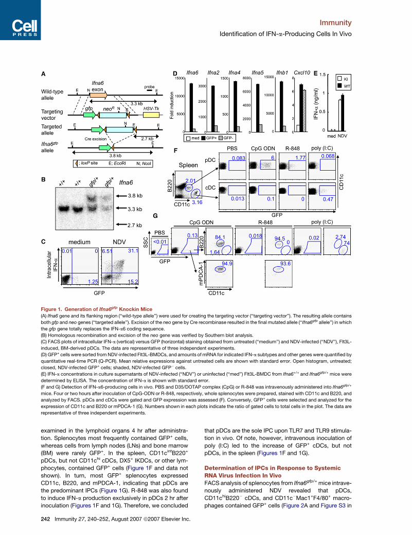
Figure 1. Generation of Ifna6gfp Knockin Mice
(A) Ifna6 gene and its flanking region (‘‘wild-type allele’’) were used for creating the targeting vector (‘‘targeting vector’’). The resulting allele contains
both gfp and neo genes (‘‘targeted allele’’). Excision of the neo gene by Cre recombinase resulted in the final mutated allele (‘‘Ifna6gfp allele’’) in which
the gfp gene totally replaces the IFN-a6 coding sequence.
(B) Homologous recombination and excision of the neo gene was verified by Southern blot analysis.
(C) FACS plots of intracellular IFN-a (vertical) versus GFP (horizontal) staining obtained from untreated (‘‘medium’’) and NDV-infected (‘‘NDV’’), Flt3L-
induced, BM-derived pDCs. The data are representative of three independent experiments.
(D) GFP+ cells were sorted from NDV-infected Flt3L-BMDCs, and amounts of mRNA for indicated IFN-a subtypes and other genes were quantified by
quantitative real-time PCR (Q-PCR). Mean relative expressions against untreated cells are shown with standard error. Open histogram, untreated;
closed, NDV-infected GFP+ cells; shaded, NDV-infected GFP� cells.
(E) IFN-a concentrations in culture supernatants of NDV-infected (‘‘NDV’’) or uninfected (‘‘med’’) Flt3L-BMDC from Ifna6+/+ and Ifna6gfp/+ mice were
determined by ELISA. The concentration of IFN-a is shown with standard error.
(F and G) Detection of IFN-a6-producing cells in vivo. PBS and D35/DOTAP complex (CpG) or R-848 was intravenously administered into Ifna6gfp/+
mice. Four or two hours after inoculation of CpG-ODN or R-848, respectively, whole splenocytes were prepared, stained with CD11c and B220, and
analyzed by FACS. pDCs and cDCs were gated and GFP expression was assessed (F). Conversely, GFP+ cells were selected and analyzed for the
expression of CD11c and B220 or mPDCA-1 (G). Numbers shown in each plots indicate the ratio of gated cells to total cells in the plot. The data are
representative of three independent experiments.
examined in the lymphoid organs 4 hr after administra-
tion. Splenocytes most frequently contained GFP+ cells,
whereas cells from lymph nodes (LNs) and bone marrow
(BM) were rarely GFP+. In the spleen, CD11cintB220+
pDCs, but not CD11chi cDCs, DX5+ IKDCs, or other lym-
phocytes, contained GFP+ cells (Figure 1F and data not
shown). In turn, most GFP+ splenocytes expressed
CD11c, B220, and mPDCA-1, indicating that pDCs are
the predominant IPCs (Figure 1G). R-848 was also found
to induce IFN-a production exclusively in pDCs 2 hr after
inoculation (Figures 1F and 1G). Therefore, we concluded
242 Immunity 27, 240–252, August 2007 ª2007 Elsevier Inc.
that pDCs are the sole IPC upon TLR7 and TLR9 stimula-
tion in vivo. Of note, however, intravenous inoculation of
poly (I:C) led to the increase of GFP+ cDCs, but not
pDCs, in the spleen (Figures 1F and 1G).
Determination of IPCs in Response to SystemicRNA Virus Infection In VivoFACS analysis of splenocytes from Ifna6gfp/+ mice intrave-
nously administered NDV revealed that pDCs,
CD11chiB220� cDCs, and CD11c�Mac1+F4/80+ macro-
phages contained GFP+ cells (Figure 2A and Figure S3 in
Immunity
Identification of IFN-a-Producing Cells In Vivo
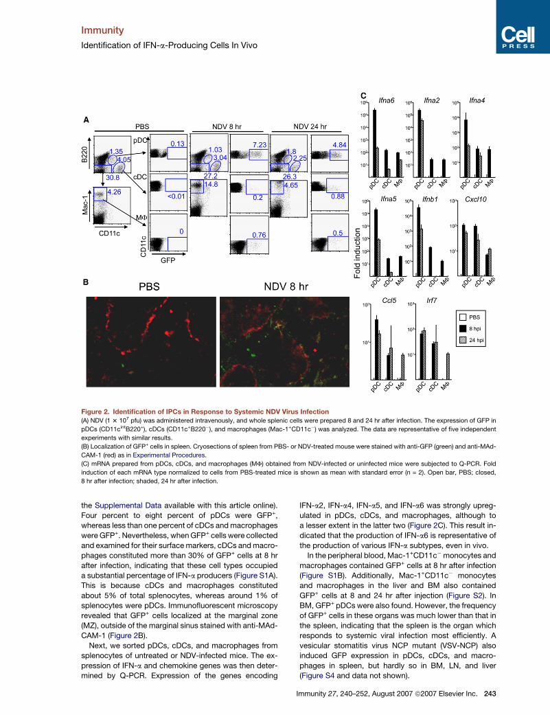
Figure 2. Identification of IPCs in Response to Systemic NDV Virus Infection
(A) NDV (1 3 107 pfu) was administered intravenously, and whole splenic cells were prepared 8 and 24 hr after infection. The expression of GFP in
pDCs (CD11cintB220+), cDCs (CD11c+B220�), and macrophages (Mac-1+CD11c�) was analyzed. The data are representative of five independent
experiments with similar results.
(B) Localization of GFP+ cells in spleen. Cryosections of spleen from PBS- or NDV-treated mouse were stained with anti-GFP (green) and anti-MAd-
CAM-1 (red) as in Experimental Procedures.
(C) mRNA prepared from pDCs, cDCs, and macrophages (MF) obtained from NDV-infected or uninfected mice were subjected to Q-PCR. Fold
induction of each mRNA type normalized to cells from PBS-treated mice is shown as mean with standard error (n = 2). Open bar, PBS; closed,
8 hr after infection; shaded, 24 hr after infection.
the Supplemental Data available with this article online).
Four percent to eight percent of pDCs were GFP+,
whereas less than one percent of cDCs and macrophages
were GFP+. Nevertheless, when GFP+ cells were collected
and examined for their surface markers, cDCs and macro-
phages constituted more than 30% of GFP+ cells at 8 hr
after infection, indicating that these cell types occupied
a substantial percentage of IFN-a producers (Figure S1A).
This is because cDCs and macrophages constituted
about 5% of total splenocytes, whereas around 1% of
splenocytes were pDCs. Immunofluorescent microscopy
revealed that GFP+ cells localized at the marginal zone
(MZ), outside of the marginal sinus stained with anti-MAd-
CAM-1 (Figure 2B).
Next, we sorted pDCs, cDCs, and macrophages from
splenocytes of untreated or NDV-infected mice. The ex-
pression of IFN-a and chemokine genes was then deter-
mined by Q-PCR. Expression of the genes encoding
IFN-a2, IFN-a4, IFN-a5, and IFN-a6 was strongly upreg-
ulated in pDCs, cDCs, and macrophages, although to
a lesser extent in the latter two (Figure 2C). This result in-
dicated that the production of IFN-a6 is representative of
the production of various IFN-a subtypes, even in vivo.
In the peripheral blood, Mac-1+CD11c�monocytes and
macrophages contained GFP+ cells at 8 hr after infection
(Figure S1B). Additionally, Mac-1+CD11c� monocytes
and macrophages in the liver and BM also contained
GFP+ cells at 8 and 24 hr after injection (Figure S2). In
BM, GFP+ pDCs were also found. However, the frequency
of GFP+ cells in these organs was much lower than that in
the spleen, indicating that the spleen is the organ which
responds to systemic viral infection most efficiently. A
vesicular stomatitis virus NCP mutant (VSV-NCP) also
induced GFP expression in pDCs, cDCs, and macro-
phages in spleen, but hardly so in BM, LN, and liver
(Figure S4 and data not shown).
Immunity 27, 240–252, August 2007 ª2007 Elsevier Inc. 243
Immunity
Identification of IFN-a-Producing Cells In Vivo
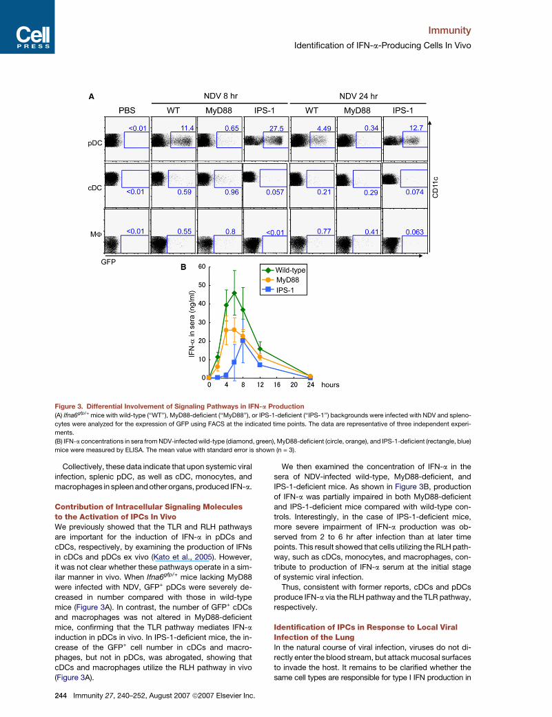
Figure 3. Differential Involvement of Signaling Pathways in IFN-a Production
(A) Ifna6gfp/+ mice with wild-type (‘‘WT’’), MyD88-deficient (‘‘MyD88’’), or IPS-1-deficient (‘‘IPS-1’’) backgrounds were infected with NDV and spleno-
cytes were analyzed for the expression of GFP using FACS at the indicated time points. The data are representative of three independent experi-
ments.
(B) IFN-a concentrations in sera from NDV-infected wild-type (diamond, green), MyD88-deficient (circle, orange), and IPS-1-deficient (rectangle, blue)
mice were measured by ELISA. The mean value with standard error is shown (n = 3).
Collectively, these data indicate that upon systemic viral
infection, splenic pDC, as well as cDC, monocytes, and
macrophages in spleen and other organs, produced IFN-a.
Contribution of Intracellular Signaling Moleculesto the Activation of IPCs In VivoWe previously showed that the TLR and RLH pathways
are important for the induction of IFN-a in pDCs and
cDCs, respectively, by examining the production of IFNs
in cDCs and pDCs ex vivo (Kato et al., 2005). However,
it was not clear whether these pathways operate in a sim-
ilar manner in vivo. When Ifna6gfp/+ mice lacking MyD88
were infected with NDV, GFP+ pDCs were severely de-
creased in number compared with those in wild-type
mice (Figure 3A). In contrast, the number of GFP+ cDCs
and macrophages was not altered in MyD88-deficient
mice, confirming that the TLR pathway mediates IFN-a
induction in pDCs in vivo. In IPS-1-deficient mice, the in-
crease of the GFP+ cell number in cDCs and macro-
phages, but not in pDCs, was abrogated, showing that
cDCs and macrophages utilize the RLH pathway in vivo
(Figure 3A).
244 Immunity 27, 240–252, August 2007 ª2007 Elsevier Inc.
We then examined the concentration of IFN-a in the
sera of NDV-infected wild-type, MyD88-deficient, and
IPS-1-deficient mice. As shown in Figure 3B, production
of IFN-a was partially impaired in both MyD88-deficient
and IPS-1-deficient mice compared with wild-type con-
trols. Interestingly, in the case of IPS-1-deficient mice,
more severe impairment of IFN-a production was ob-
served from 2 to 6 hr after infection than at later time
points. This result showed that cells utilizing the RLH path-
way, such as cDCs, monocytes, and macrophages, con-
tribute to production of IFN-a serum at the initial stage
of systemic viral infection.
Thus, consistent with former reports, cDCs and pDCs
produce IFN-a via the RLH pathway and the TLR pathway,
respectively.
Identification of IPCs in Response to Local ViralInfection of the LungIn the natural course of viral infection, viruses do not di-
rectly enter the blood stream, but attack mucosal surfaces
to invade the host. It remains to be clarified whether the
same cell types are responsible for type I IFN production in
Immunity
Identification of IFN-a-Producing Cells In Vivo
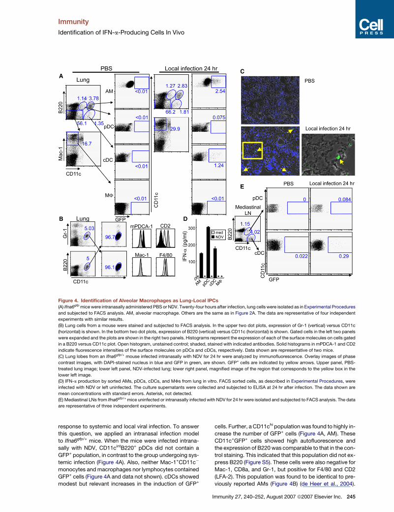
Figure 4. Identification of Alveolar Macrophages as Lung-Local IPCs
(A) Ifna6gfp mice were intranasally administered PBS or NDV. Twenty-four hours after infection, lung cells were isolated as in Experimental Procedures
and subjected to FACS analysis. AM, alveolar macrophage. Others are the same as in Figure 2A. The data are representative of four independent
experiments with similar results.
(B) Lung cells from a mouse were stained and subjected to FACS analysis. In the upper two dot plots, expression of Gr-1 (vertical) versus CD11c
(horizontal) is shown. In the bottom two dot plots, expression of B220 (vertical) versus CD11c (horizontal) is shown. Gated cells in the left two panels
were expanded and the plots are shown in the right two panels. Histograms represent the expression of each of the surface molecules on cells gated
in a B220 versus CD11c plot. Open histogram, unstained control; shaded, stained with indicated antibodies. Solid histograms in mPDCA-1 and CD2
indicate fluorescence intensities of the surface molecules on pDCs and cDCs, respectively. Data shown are representative of two mice.
(C) Lung lobes from an Ifna6gfp/+ mouse infected intranasally with NDV for 24 hr were analyzed by immunofluorescence. Overlay images of phase
contrast images, with DAPI-stained nucleus in blue and GFP in green, are shown. GFP+ cells are indicated by yellow arrows. Upper panel, PBS-
treated lung image; lower left panel, NDV-infected lung; lower right panel, magnified image of the region that corresponds to the yellow box in the
lower left image.
(D) IFN-a production by sorted AMs, pDCs, cDCs, and MFs from lung in vitro. FACS sorted cells, as described in Experimental Procedures, were
infected with NDV or left uninfected. The culture supernatants were collected and subjected to ELISA at 24 hr after infection. The data shown are
mean concentrations with standard errors. Asterisk, not detected.
(E) Mediastinal LNs from Ifna6gfp/+ mice uninfected or intranasally infected with NDV for 24 hr were isolated and subjected to FACS analysis. The data
are representative of three independent experiments.
response to systemic and local viral infection. To answer
this question, we applied an intranasal infection model
to Ifna6gfp/+ mice. When the mice were infected intrana-
sally with NDV, CD11cintB220+ pDCs did not contain a
GFP+ population, in contrast to the group undergoing sys-
temic infection (Figure 4A). Also, neither Mac-1+CD11c�
monocytes and macrophages nor lymphocytes contained
GFP+ cells (Figure 4A and data not shown). cDCs showed
modest but relevant increases in the induction of GFP+
cells. Further, a CD11chi population was found to highly in-
crease the number of GFP+ cells (Figure 4A, AM). These
CD11c+GFP+ cells showed high autofluorescence and
the expression of B220 was comparable to that in the con-
trol staining. This indicated that this population did not ex-
press B220 (Figure S5). These cells were also negative for
Mac-1, CD8a, and Gr-1, but positive for F4/80 and CD2
(LFA-2). This population was found to be identical to pre-
viously reported AMs (Figure 4B) (de Heer et al., 2004).
Immunity 27, 240–252, August 2007 ª2007 Elsevier Inc. 245
Immunity
Identification of IFN-a-Producing Cells In Vivo
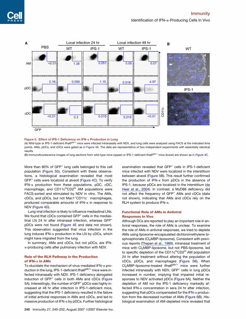
Figure 5. Effect of IPS-1 Deficiency on IFN-a Production in Lung(A) Wild-type or IPS-1-deficient Ifna6gfp/+ mice were infected intranasally with NDV, and lung cells were analyzed using FACS at the indicated time
points. AMs, pDCs, and cDCs were gated as in Figure 4A. The data are representative of two independent experiments with essentially identical
results.
(B) Immunofluorescence images of lung sections from wild-type mice (upper) or IPS-1-deficient Ifna6gfp/+ mice (lower) are shown as in Figure 4C.
More than 80% of GFP+ lung cells belonged to this cell
population (Figure S5). Consistent with these observa-
tions, a histological examination revealed that most
GFP+ cells were localized at alveoli (Figure 4C). To verify
IFN-a production from these populations, pDC, cDC,
macrophage, and CD11chiCD2hi AM populations were
FACS-sorted and stimulated by NDV in vitro. The AMs,
cDCs, and pDCs, but not Mac1+CD11c� macrophages,
produced comparable amounts of IFN-a in response to
NDV (Figure 4D).
Lung viral infection is likely to influence mediastinal LNs.
We found that cDCs contained GFP+ cells in the medias-
tinal LN 24 hr after intranasal infection, whereas GFP+
pDCs were not found (Figure 4E and data not shown).
This observation suggested that virus infection in the
lung induces IFN-a production in the LN by cDCs, which
might have migrated from the lung.
In summary, AMs and cDCs, but not pDCs, are IFN-
a-producing cells after pulmonary infection with NDV.
Role of the RLH Pathway in the Productionof IFN-a in AMsTo elucidate the mechanism of virus-mediated IFN-a pro-
duction in the lung, IPS-1-deficient Ifna6gfp/+ mice were in-
fected intranasally with NDV. IPS-1 deficiency abrogated
induction of GFP+ cells in both AMs and cDCs (Figure
5A). Interestingly, the number of GFP+ pDCs was highly in-
creased at 48 hr after infection in IPS-1-deficient mice,
suggesting that the IPS-1 deficiency resulted in the failure
of initial antiviral responses in AMs and cDCs, and led to
massive production of IFN-a by pDCs. Further histological
246 Immunity 27, 240–252, August 2007 ª2007 Elsevier Inc.
examination revealed that GFP+ cells in IPS-1-deficient
mice infected with NDV were localized in the interstitium
between alveoli (Figure 5B). This result further confirmed
the production of IFN-a from pDCs in the absence of
IPS-1, because pDCs are localized in the interstitium (de
Heer et al., 2004). In contrast, a MyD88 deficiency did
not affect the frequency of GFP+ AMs and cDCs (data
not shown), indicating that AMs and cDCs rely on the
RLH system to produce IFN-a.
Functional Role of AMs in AntiviralResponses In VivoAlthough DCs are reported to play an important role in an-
tiviral responses, the role of AMs is unclear. To examine
the role of AMs in antiviral responses, we tried to deplete
AMs using liposome-encapsulated dichloromethylene bi-
sphosphonate (Cl2MBP-liposome). Consistent with previ-
ous reports (Thepen et al., 1989), intranasal treatment of
mice with Cl2MBP-liposome, but not PBS-liposome, led
to specific depletion of the CD11chiCD2hi AM population
24 hr after treatment without altering the population of
cDCs, pDCs, and macrophages (Figure S6). When
Cl2MBP-liposome-treated Ifna6gfp/+ mice were further
infected intranasally with NDV, GFP+ cells in lung pDCs
increased in number, implying that impaired initial re-
sponses to NDV activated pDCs (Figure 6A). Neither the
depletion of AM nor the IPS-1 deficiency markedly af-
fected IFN-a concentration in sera 24 hr after infection,
suggesting that pDCs compensated for the IFN-a produc-
tion from the decreased number of AMs (Figure 6B). His-
tological examination of AM-depleted mice revealed that
Immunity
Identification of IFN-a-Producing Cells In Vivo
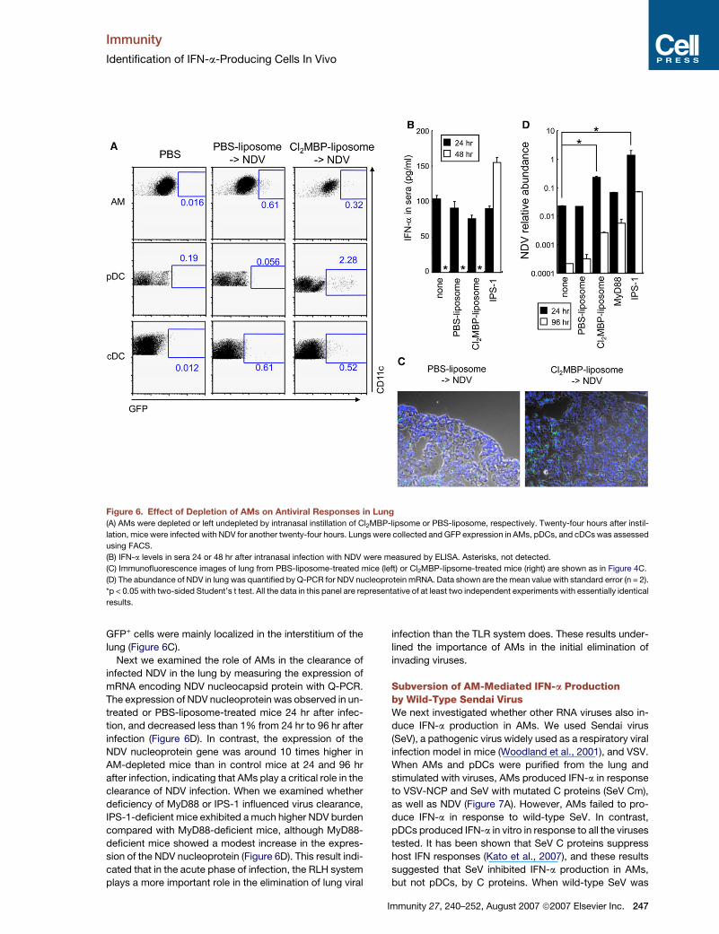
Figure 6. Effect of Depletion of AMs on Antiviral Responses in Lung
(A) AMs were depleted or left undepleted by intranasal instillation of Cl2MBP-lipsome or PBS-liposome, respectively. Twenty-four hours after instil-
lation, mice were infected with NDV for another twenty-four hours. Lungs were collected and GFP expression in AMs, pDCs, and cDCs was assessed
using FACS.
(B) IFN-a levels in sera 24 or 48 hr after intranasal infection with NDV were measured by ELISA. Asterisks, not detected.
(C) Immunofluorescence images of lung from PBS-liposome-treated mice (left) or Cl2MBP-lipsome-treated mice (right) are shown as in Figure 4C.
(D) The abundance of NDV in lung was quantified by Q-PCR for NDV nucleoprotein mRNA. Data shown are the mean value with standard error (n = 2).
*p < 0.05 with two-sided Student’s t test. All the data in this panel are representative of at least two independent experiments with essentially identical
results.
GFP+ cells were mainly localized in the interstitium of the
lung (Figure 6C).
Next we examined the role of AMs in the clearance of
infected NDV in the lung by measuring the expression of
mRNA encoding NDV nucleocapsid protein with Q-PCR.
The expression of NDV nucleoprotein was observed in un-
treated or PBS-liposome-treated mice 24 hr after infec-
tion, and decreased less than 1% from 24 hr to 96 hr after
infection (Figure 6D). In contrast, the expression of the
NDV nucleoprotein gene was around 10 times higher in
AM-depleted mice than in control mice at 24 and 96 hr
after infection, indicating that AMs play a critical role in the
clearance of NDV infection. When we examined whether
deficiency of MyD88 or IPS-1 influenced virus clearance,
IPS-1-deficient mice exhibited a much higher NDV burden
compared with MyD88-deficient mice, although MyD88-
deficient mice showed a modest increase in the expres-
sion of the NDV nucleoprotein (Figure 6D). This result indi-
cated that in the acute phase of infection, the RLH system
plays a more important role in the elimination of lung viral
infection than the TLR system does. These results under-
lined the importance of AMs in the initial elimination of
invading viruses.
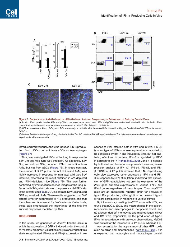
Subversion of AM-Mediated IFN-a Productionby Wild-Type Sendai VirusWe next investigated whether other RNA viruses also in-
duce IFN-a production in AMs. We used Sendai virus
(SeV), a pathogenic virus widely used as a respiratory viral
infection model in mice (Woodland et al., 2001), and VSV.
When AMs and pDCs were purified from the lung and
stimulated with viruses, AMs produced IFN-a in response
to VSV-NCP and SeV with mutated C proteins (SeV Cm),
as well as NDV (Figure 7A). However, AMs failed to pro-
duce IFN-a in response to wild-type SeV. In contrast,
pDCs produced IFN-a in vitro in response to all the viruses
tested. It has been shown that SeV C proteins suppress
host IFN responses (Kato et al., 2007), and these results
suggested that SeV inhibited IFN-a production in AMs,
but not pDCs, by C proteins. When wild-type SeV was
Immunity 27, 240–252, August 2007 ª2007 Elsevier Inc. 247
Immunity
Identification of IFN-a-Producing Cells In Vivo
Figure 7. Subversion of AM-Mediated or cDC-Mediated Antiviral Responses, or Subversion of Both, by Sendai Virus(A) In vitro IFN-a production by AMs and pDCs in response to various viruses. AMs and pDCs were sorted and infected in vitro for 24 hr. IFN-a
concentrations in the culture supernatants were measured with ELISA. Asterisk, not detected.
(B) GFP expressions in AMs, pDCs, and cDCs were analyzed at 24 hr after intranasal infection with wild-type Sendai virus (SeV WT) or its mutant,
SeV Cm.
(C) Immunofluorescence images of lung infected with SeV Cm (left panel) or SeV WT (right) are shown. The data are representative of two independent
experiments with same results.
introduced intravenously, the virus induced IFN-a produc-
tion from pDCs, but not from cDCs or macrophages
(Figure S7).
Thus, we investigated IPCs in the lung in response to
SeV Cm and wild-type SeV infection. As expected, SeV
Cm, as well as NDV, induced IFN-a production from
AMs, but not from pDCs (Figure 7B). In sharp contrast,
the number of GFP+ pDCs, but not cDCs and AMs, was
highly increased in response to intranasal wild-type SeV
infection, resembling the result obtained in AM-depleted
and IPS-1-deficient mice (Figure 7B). This was further
confirmed by immunofluorescence images of the lung in-
fected with SeV, which showed the presence of GFP+ cells
in the interstitium (Figure 7C). In contrast, SeV Cm induced
GFP expression in AMs. These results suggested that SeV
targets AMs for suppressing IFN-a production, and that
the subversion is essential for SeV virulence. Collectively,
these data emphasized the importance of the first-line
antiviral responses mediated by AMs.
DISCUSSION
In this study, we generated an Ifna6gfp knockin allele in
which the expression of GFP is induced under the control
of the Ifna6 promoter. Validation analysis showed that this
allele recapitulated IFN-as and IFN-b expression in re-
248 Immunity 27, 240–252, August 2007 ª2007 Elsevier Inc.
sponse to viral infection both in vitro and in vivo. IFN-a6
is a subtype of IFN-as whose expression is reported to
be controlled by IRF-7 and induced by viral, but not bac-
terial, infections. In contrast, IFN-b is regulated by IRF-3
in addition to IRF-7 (Honda et al., 2005), and it is induced
by both viral and bacterial components. However, an ex-
pression analysis of IFN-a2, IFN-a4, IFN-a5, and IFN-
b mRNA in GFP+ pDCs revealed that IFN-a6-producing
cells also expressed other subtypes of IFN-a and IFN-
b in response to NDV stimulation, indicating that expres-
sion of GFP recapitulates not only the expression of the
Ifna6 gene but also expressions of various IFN-a and
IFN-b genes regardless of the subtypes. Thus, Ifna6gfp/+
mice are an appropriate reporter strain for analysis of
type I IFN production, although it is not proven if type I
IFNs are coregulated in response to various stimuli.
By intravenously treating Ifna6gfp/+ mice with NDV, we
found that pDCs, cDCs, and macrophages in the spleen,
monocytes and macrophages in peripheral blood, and
(to a lesser degree) monocytes and macrophages in liver
and BM were responsible for the production of type I
IFNs. In accordance with previous observations, MyD88
was critical for the increase in GFP+ pDCs, whereas IPS-
1 was essential for the appearance of other GFP+ cells
such as cDCs and macrophages (Kato et al., 2005). It is
unexpected that monocytes and macrophages consist
Immunity
Identification of IFN-a-Producing Cells In Vivo
of a large portion of IPCs in vivo. Because the RLH system
is essential for IFN-a production in this cell type, mono-
cytes and macrophages seem to be actually infected. Im-
paired production of serum IFN-a in IPS-1-deficient mice
also implies the importance of non-pDCs in virus-induced
IFN-a production. Our data indicated that cell types other
than pDCs also make a large contribution to type I IFN
production during systemic viral infections.
The IPS-1-dependent pathway contributes substan-
tially to increases in serum IFN-a at an early time point af-
ter infection with NDV, although the MyD88-dependent
pathways are also critical at later time points. Accordingly,
about 30% of GFP+ splenocytes were macrophages and
cDCs at 8 hr after NDV infection, but the ratio dropped
to below 10% at 24 hr. In the spleen of NDV-infected
Ifna6gfp mice, GFP+ cells were localized at the MZ. This
is consistent with previous reports that used an IFN-a an-
tibody to show that IPCs are localized at the MZ (Asselin-
Paturel et al., 2005; Dalod et al., 2002). Upon systemic
NDV infection, GFP+ cells were almost exclusively found
in the spleen, though quite a few GFP+ pDCs were ob-
served in other lymphatic organs, such as BM and LNs.
VSV and SeV also induced GFP expression mostly in the
spleen, indicating that the spleen is the most important or-
gan for the production of type I IFNs in response to blood-
borne viruses. Because the MZ is important for sequester-
ing blood-borne pathogens, and mice with abnormal MZ
organization failed to mount proper IFN-a production in re-
sponse to viral infection (Louten et al., 2006), IPCs acti-
vated by systemic NDV infection are probably localized
in the splenic MZ. pDCs are mostly localized in the T cell
area and in the red pulp, but rarely localize in the MZ al-
though they migrate to the MZ to form clusters in response
to CpG-DNA stimulation (Asselin-Paturel et al., 2005). In
contrast, cDCs originally localize in the MZ. Thus, it is pos-
sible that invading NDV encounters macrophages and
cDCs at the MZ first, and then pDCs that have migrated
to the MZ. Although further detailed analysis of localization
and time-course changes needs to be addressed, this
may explain the temporal differences in the contributions
of IPS-1- and MyD88-dependent pathways in the produc-
tion of IFN-a.
In contrast to systemic virus infection, intranasal treat-
ment of Ifna6gfp mice with NDV failed to increase the num-
ber of GFP+ cells in pDCs. By characterizing GFP+ cells
upon intranasal infection, we identified AMs and cDCs,
particularly AMs, as the major IFN-a producers. AMs are
reported to sample and respond to microorganisms that
have entered the alveolar space. They phagocytose in-
vading bacteria and evoke inflammation by producing
proinflammatory cytokines (Peters-Golden, 2004). They
are also reported to phagocytose virus-infected apoptotic
cells (Hashimoto et al., 2007). However, our results sug-
gested that direct infection of AMs with viruses is required
for the production of IFN-a. The role of AMs in the clear-
ance of viral infection was also demonstrated by the con-
sequences of the depletion of AMs. Thus, AMs function as
sentinels, recognizing infectious viruses and alerting sur-
rounding cells. AMs do not seem to migrate to mediastinal
LNs even after viral infection, suggesting that type I IFNs
produced by AMs activate surrounding cells in an auto-
crine or paracrine manner to prepare for any virus infec-
tion. Taken together, our data highlighted the importance
of the initial production of IFN-a and the elimination of
invading viruses by AMs.
cDCs, but not pDCs, produced IFN-a in response to lo-
cal infection in mediastinal LNs. Because GFP+ cDCs
were also found in lung tissue, it is possible that cDCs
activated in the lung tissue are migrating to mediastinal
LNs. Given the function of type I IFNs in the activation of
acquired immune responses (Stetson and Medzhitov,
2006), the major role of cDCs may be to activate T cells
in LNs rather than function as local IPCs.
Earlier reports indicated that pDCs produce type I IFNs
to help elicit the acquired immune response (Yoneyama
et al., 2005), although we failed to detect GFP+ pDCs
in response to intranasal NDV infection in Ifna6gfp mice.
However, massive amounts of GFP+ pDCs were de-
tected either when AMs were depleted or in the absence
of IPS-1. These results suggested that pDCs start to
function when AM- and cDC-mediated host defense
mechanisms are impaired, and that pDCs may function as
the backup system to achieve robust antiviral IFN-a
production.
Wild-type SeV is reported to repress host antiviral
responses mainly by inhibiting type I IFN signaling via C
proteins; this activity is essential for the pathogenicity of
the virus (Kato et al., 2007). In vitro experiments showed
that wild-type SeV did not induce IFN-a production in
AMs, whereas SeV Cm highly induced IFN-a. Upon local
lung infection with wild-type SeV, AMs and cDCs did not
appear to contain GFP+ cells, indicating that AMs and
cDCs failed to produce IFN-a. This result indicates that
SeV has a sophisticated mechanism or mechanisms to
subvert host antiviral response by AMs and cDCs. Resem-
bling the response in the absence of AMs, the failure in an-
tiviral response by AMs and cDCs led to massive produc-
tion of IFN-a from pDCs. Intranasal local infection of SeV
Cm, however, induced IFN-a production from AMs, but
not from pDCs. Thus, if SeV fails to subvert the host anti-
viral response due to its deficiency of C proteins, AMs will
exert an effective antiviral response against SeV to control
viral dissemination.
From these observations, we propose that three differ-
ent IPCs in the lung, i.e. AMs, cDCs, and pDCs, are acti-
vated sequentially, but not simultaneously, for mounting
antiviral immune responses. AMs act as the first-line sen-
sor of invading viruses, and produce IFN-a at the site of
infection. cDCs also initially produce IFN-a against viral
infection, and have an additional role in producing IFNs
in regional LNs. In contrast, pDCs are not activated until
the initial defense line is broken by the viruses. It is of
note that pDCs utilize the TLR system for type I IFN pro-
duction, which is different from AM and cDC usage of
the RLH system. Given that several virulent RNA viruses,
such as SeV and influenza virus, suppress the RLH-medi-
ated signaling pathway, type I IFN signaling pathway, or
both (Kato et al., 2007; Pichlmair et al., 2006), it is tempting
Immunity 27, 240–252, August 2007 ª2007 Elsevier Inc. 249
Immunity
Identification of IFN-a-Producing Cells In Vivo
to speculate that hosts have evolved two different type I
IFN production systems to make it more difficult for
viruses to escape the antiviral response.
This study showed that the Ifna6gfp/+ mice are a quite
useful tool for identifying IPCs in response to viral infec-
tion. Although we used respiratory virus infection as
a model of local viral infection, as yet uncharacterized
IPCs can be activated in other organs and mucosal tis-
sues, such as intestine. Thus, this mouse model will be
useful in identifing IPCs in different tissues in relation to
various viral infections. Recent expansion in IFN biology
has also revealed that type I IFNs are not only involved
in antiviral responses, but also in responses to bacteria
and autoimmune diseases (Banchereau and Pascual,
2006). For instance, overproduction of type I IFNs is re-
ported in some autoimmune diseases, such as systemic
lupus erythematosus, although the identity or identities
of the type I IFN-producing cells are not clear in mouse
models of the autoimmune disease. Therefore, we believe
that this mouse model will benefit research in the IFN field
by providing a vehicle for in vivo insights.
EXPERIMENTAL PROCEDURES
Generation of Ifna6gfp Knockin Mice
The Ifna6 gene and its flanking region were isolated from genomic DNA
extracts of ES cells (clone GSI-I) by PCR. The targeting vector was
constructed by replacing a 1.0 kb fragment encoding the entire Ifna6
open reading frame with a neomycin-resistance gene cassette (neo)
and a fragment encoding EGFP (gfp) from pEGFP-1 (Clontech). A her-
pes simplex virus thymidine kinase driven by PGK promoter (HSV-Tk)
was inserted into the genomic fragment for negative selection. After
the targeting vector was transfected into ES cells, G418 and gancyclo-
vir doubly resistant colonies were selected and screened by PCR and
Southern blotting. A plasmid that contains a gene encoding Cre re-
combinase was transfected into the clone and G418-sensitive colonies
were selected. Clones in which excision of the neo gene took place
were screened by Southern blotting. These clones were microinjected
into C57BL/6 female mice, and heterozygous Ifna6gfp/+ F1 progenies
were backcrossed five times to C57BL/6 before analysis.
Mice
Mice deficient in Myd88 and Ips-1 have been described previously
(Adachi et al., 1998; Kumar et al., 2006). All mice were bred and main-
tained in a specific pathogen-free facility of the Research Institute for
Microbial Diseases, Osaka University, in accordance with the specifi-
cations of the Association for Assessment and Accreditation of Labo-
ratory Animal Care. Mouse protocols were approved by Osaka Univer-
sity Animal Care and Use Committee.
Reagents and Viruses
All fluorochrome-conjugated and biotin-conjugated antibodies were
purchased from BD PharMingen unless otherwise indicated. Flt3L
was purchased from Peprotec. When administered intravenously,
5 mg of CpG-ODN D35 (Uematsu et al., 2005) was complexed to
30 ml of DOTAP transfection reagent (Boeringer-Manheim) at the final
concentration of 25 mg/ml, and the conjugate was administered.
R-848 has also been described (Hemmi et al., 2002), and 100 nmol
of R-848 was administered intravenously. Poly (I:C) was purchased
from Amersham, and 100 mg of poly (I:C) was intravenously adminis-
tered. NDV and VSV-NCP were a kind gift from Dr. T. Abe and Dr.
Y. Matsuura (Research Institute for Microbial Diseases, Osaka Univer-
sity). NDV suspended in chick allantoic fluid was administered both in-
travenously and intranasally at a titer of 1 3 107 plaque forming unit
250 Immunity 27, 240–252, August 2007 ª2007 Elsevier Inc.
(pfu) after anesthesia. Allantoic fluid does not induce IFN-a either
in vitro or in vivo by itself (data not shown). VSV-NCP was administered
intravenously at a titer of 1 3 106 pfu. Wild-type SeV and SeV Cm were
kindly provided by Dr. A. Kato (National Institute for Infectious Dis-
eases, Japan) and administered both intravenously and intranasally
at a titer of 1 3 107 pfu. The IFN-a ELISA kit was obtained from PBL.
Isolation of Cells from Tissues
Isolation of lung cells was performed as essentially described with
some modifications (de Heer et al., 2004). Briefly, mice were sacrificed
and perfused with PBS containing 10 mM EDTA from the right ventri-
cle. Lung lobes were isolated and collagenase buffer (150 units/ml of
collagenase [purchased from Wako Chemicals], 10 mg/ml of DNaseI
[from Sigma], and 5% of FCS in RPMI1640 medium) was injected
into the lobes using a 27G needle. The lobes were then shredded
into small pieces and incubated at 37�C for 45 min. During the last
5 min, EDTA was added at 10 mM. Any remaining small pieces were
dispersed by passage in and out through a 20G needle, and the sus-
pension was passed through nylon mesh to remove debris. A single-
cell suspension was prepared after RBC lysis. Cells from LNs (both
inguinal and mediastinal) and liver were isolated essentially in the
same manner as lung cells were.
Flow-Cytometric Analysis and Sorting
Intracellular staining of IFN-a was performed as described (Kato et al.,
2005). For sorting pDCs, cDCs, and macrophages from spleen, CD19+
cells and Thy1.2+ cells in splenocytes were depleted by a magnetic
bead sorting system using magnetic bead-conjugated anti-CD19
and anti-Thy1.2 antibodies (Miltenyi Biotech). The cells of the depleted
fraction were stained with PerCP-conjugated anti-B220, APC-conju-
gated anti-CD11c, and FITC-conjugated anti-Mac-1 antibodies. Gates
were set as in Figure 2A. For sorting AMs, a single-cell suspension
of lung cells was stained with APC-conjugated anti-CD11c, FITC-
conjugated anti-CD2, and PE-conjugated anti-Mac-1 antibodies.
CD11chiCD2hiMac-1� cells were sorted as AMs. Stained cells were
sorted using a FACSArea (BD Bioscience). Sorted cells had more
than 95% purity.
Immunofluorescence Staining
Isolated tissues were embedded into OCT compound (Sakura Fine-
technical) and subjected to rapid cooling in n-hexane cooled in liquid
nitrogen and stored at �80�C until further analysis. Five-micrometer
thick cryosections were fixed in acetone at�30�C for 20 min, air-dried,
and rehydrated in PBS and PBS containing 0.05% Tween 20 (PBST)
for 1 min each before staining. Sections were incubated in 2% BSA
in PBST for 30 min at room temperature. If required, streptavidin-biotin
blocking was also performed using a streptavidin-biotin blocking kit
(Vector laboratories). After blocking, sections were stained with anti-
GFP rabbit polyclonal antibody (SantaCruz) and anti-MAdCAM-1 rat
monoclonal antibody (clone MECA75). Alexa 488-conjugated anti-rab-
bit IgG (Invitrogen) and biotinylated anti-rat IgG (Jackson Immunore-
search) were then applied as secondary antibodies. Finally, sections
were stained with Alexa 594-conjugated streptavidin (Invitrogen).
The resulting stained specimens were mounted by VectaShield
Mounting Medium Hard (Vector Laboratories) and observed under
an Olympus IX81 microscope (Olympus). Obtained images were pro-
cessed using Metamorph software.
RNA Isolation; Q-PCR
Total RNA was isolated using TRIzol (Invitrogen) according to the man-
ufacturer’s instructions. One microgram of RNA was reverse tran-
scribed using ReverTraAce (Toyobo) following the manufacturer’s pro-
tocol. For sorting cells, total RNA was first purified using an RNA
isolation and purification Mini Kit (QIAGEN), and 10 ng of obtained pu-
rified RNA was reverse transcribed using an Ovation Biotin System
(Nugen) according to manufacturer’s instructions. The resulting
cDNA was subjected to Q-PCR as described (Matsui et al., 2006).
Probes were purchased from Applied Biosystems. The probe for
Immunity
Identification of IFN-a-Producing Cells In Vivo
a gene encoding NDV nucleocapsid protein has been described
(Matsui et al., 2006).
Depletion of AMs with Cl2MBP-Liposome
The preparation of liposomes was performed as described (Thepen
et al., 1989) with some modifications. Briefly, cholesterol (CL, pur-
chased from Sigma) and L-a-phosphatidylcholine (from yolk egg,
Type XVI-E, Sigma) were dissolved in chloroform at concentrations
of 8 and 100 mg/ml, respectively. One milliliter of CL solution was
mixed with eighty-six microliters of PC solution in a two-milliliter
tube. The chloroform in the mixture was evaporated in a centrifuge
evaporator. Either 1 ml of PBS or Cl2MBP (from Sigma) in PBS at a con-
centration of 0.25 g/ml was added into the tube and mixed well by mild
vortexing for 30 min. The mixture was incubated at room temperature
for 2 hr. It was then sonicated for 3 min in a waterbath sonicator and
incubated for an additional 2 hr. The resulting liposomes were washed
by PBS and resuspended in 200 ml of PBS. To deplete AMs in vivo, 50
ml of Cl2MBP-liposome was administered intranasally. Twenty-four
hours later, mice were subjected to further analysis.
Supplemental Data
The Supplemental Data, which consists of seven additional figures,
can be found online at http://www.immunity.com/cgi/content/full/27/
2/240/DC1/.
ACKNOWLEDGMENTS
We thank all colleagues in our laboratory; A. Kato, T. Abe, Y. Matsuura,
T. Shioda, and E. Nakayama for providing viruses; K. Nakamura for cell
sorting; P.Y. Lee for critical reading of the manuscript; M. Hashimoto
for secretarial assistance; and Y. Fujiwara, M. Shiokawa, and N. Kita-
gaki for technical assistance.
This work was supported in part by grants from the Ministry of
Education, Culture, Sports, Science, and Technology in Japan;
the 21st Century Center of Excellence Program of Japan; and NIH
(AI070167). The authors declare that they have no competing financial
interests.
Received: March 16, 2007
Revised: May 22, 2007
Accepted: July 3, 2007
Published online: August 23, 2007
REFERENCES
Adachi, O., Kawai, T., Takeda, K., Matsumoto, M., Tsutsui, H., Saka-
gami, M., Nakanishi, K., and Akira, S. (1998). Targeted disruption of
the MyD88 gene results in loss of IL-1- and IL-18-mediated function.
Immunity 9, 143–150.
Akira, S., Uematsu, S., and Takeuchi, O. (2006). Pathogen recognition
and innate immunity. Cell 124, 783–801.
Asselin-Paturel, C., Boonstra, A., Dalod, M., Durand, I., Yessaad, N.,
Dezutter-Dambuyant, C., Vicari, A., O’Garra, A., Biron, C., Briere, F.,
and Trinchieri, G. (2001). Mouse type I IFN-producing cells are imma-
ture APCs with plasmacytoid morphology. Nat. Immunol. 2, 1144–
1150.
Asselin-Paturel, C., Brizard, G., Chemin, K., Boonstra, A., O’Garra, A.,
Vicari, A., and Trinchieri, G. (2005). Type I interferon dependence of
plasmacytoid dendritic cell activation and migration. J. Exp. Med.
201, 1157–1167.
Banchereau, J., and Pascual, V. (2006). Type I interferon in systemic
lupus erythematosus and other autoimmune diseases. Immunity 25,
383–392.
Barchet, W., Cella, M., Odermatt, B., Asselin-Paturel, C., Colonna, M.,
and Kalinke, U. (2002). Virus-induced interferon alpha production by
a dendritic cell subset in the absence of feedback signaling in vivo.
J. Exp. Med. 195, 507–516.
Bjorck, P. (2001). Isolation and characterization of plasmacytoid den-
dritic cells from Flt3 ligand and granulocyte-macrophage colony-stim-
ulating factor-treated mice. Blood 98, 3520–3526.
Cella, M., Jarrossay, D., Facchetti, F., Alebardi, O., Nakajima, H.,
Lanzavecchia, A., and Colonna, M. (1999). Plasmacytoid monocytes
migrate to inflamed lymph nodes and produce large amounts of type
I interferon. Nat. Med. 5, 919–923.
Chan, C.W., Crafton, E., Fan, H.N., Flook, J., Yoshimura, K., Skarica,
M., Brockstedt, D., Dubensky, T.W., Stins, M.F., Lanier, L.L., et al.
(2006). Interferon-producing killer dendritic cells provide a link be-
tween innate and adaptive immunity. Nat. Med. 12, 207–213.
Dalod, M., Salazar-Mather, T.P., Malmgaard, L., Lewis, C., Asselin-
Paturel, C., Briere, F., Trinchieri, G., and Biron, C.A. (2002). Interferon
alpha/beta and interleukin 12 responses to viral infections: pathways
regulating dendritic cell cytokine expression in vivo. J. Exp. Med.
195, 517–528.
de Heer, H.J., Hammad, H., Soullie, T., Hijdra, D., Vos, N., Willart, M.A.,
Hoogsteden, H.C., and Lambrecht, B.N. (2004). Essential role of lung
plasmacytoid dendritic cells in preventing asthmatic reactions to
harmless inhaled antigen. J. Exp. Med. 200, 89–98.
Diebold, S.S., Montoya, M., Unger, H., Alexopoulou, L., Roy, P., Has-
well, L.E., Al-Shamkhani, A., Flavell, R., Borrow, P., and Reis e Sousa,
C. (2003). Viral infection switches non-plasmacytoid dendritic cells into
high interferon producers. Nature 424, 324–328.
Hashimoto, Y., Moki, T., Takizawa, T., Shiratsuchi, A., and Nakanishi,
Y. (2007). Evidence for phagocytosis of influenza virus-infected, apo-
ptotic cells by neutrophils and macrophages in mice. J. Immunol.
178, 2448–2457.
Hemmi, H., Kaisho, T., Takeuchi, O., Sato, S., Sanjo, H., Hoshino, K.,
Horiuchi, T., Tomizawa, H., Takeda, K., and Akira, S. (2002). Small
anti-viral compounds activate immune cells via the TLR7 MyD88-
dependent signaling pathway. Nat. Immunol. 3, 196–200.
Honda, K., Yanai, H., Mizutani, T., Negishi, H., Shimada, N., Suzuki, N.,
Ohba, Y., Takaoka, A., Yeh, W.C., and Taniguchi, T. (2004). Role of
a transductional-transcriptional processor complex involving MyD88
and IRF-7 in Toll-like receptor signaling. Proc. Natl. Acad. Sci. USA
101, 15416–15421.
Honda, K., Yanai, H., Negishi, H., Asagiri, M., Sato, M., Mizutani, T.,
Shimada, N., Ohba, Y., Takaoka, A., Yoshida, N., and Taniguchi, T.
(2005). IRF-7 is the master regulator of type-I interferon-dependent
immune responses. Nature 434, 772–777.
Honda, K., Takaoka, A., and Taniguchi, T. (2006). Type I interferon [cor-
rected] gene induction by the interferon regulatory factor family of tran-
scription factors. Immunity 25, 349–360.
Hornung, V., Ellegast, J., Kim, S., Brzozka, K., Jung, A., Kato, H.,
Poeck, H., Akira, S., Conzelmann, K.K., Schlee, M., et al. (2006). 50-
Triphosphate RNA is the ligand for RIG-I. Science 314, 994–997.
Hoshino, K., Sugiyama, T., Matsumoto, M., Tanaka, T., Saito, M.,
Hemmi, H., Ohara, O., Akira, S., and Kaisho, T. (2006). IkappaB ki-
nase-alpha is critical for interferon-alpha production induced by Toll-
like receptors 7 and 9. Nature 440, 949–953.
Kato, A., Kiyotani, K., Kubota, T., Yoshida, T., Tashiro, M., and Nagai,
Y. (2007). Importance of Anti-Interferon Capacity of the Sendai Virus C
Protein for Pathogenicity in Mice. J Virol. 81, 3264–3271.
Kato, H., Sato, S., Yoneyama, M., Yamamoto, M., Uematsu, S., Mat-
sui, K., Tsujimura, T., Takeda, K., Fujita, T., Takeuchi, O., and Akira,
S. (2005). Cell type-specific involvement of RIG-I in antiviral response.
Immunity 23, 19–28.
Kato, H., Takeuchi, O., Sato, S., Yoneyama, M., Yamamoto, M., Mat-
sui, K., Uematsu, S., Jung, A., Kawai, T., Ishii, K.J., et al. (2006). Differ-
ential roles of MDA5 and RIG-I helicases in the recognition of RNA
viruses. Nature 441, 101–105.
Kawai, T., and Akira, S. (2006). Innate immune recognition of viral in-
fection. Nat. Immunol. 7, 131–137.
Immunity 27, 240–252, August 2007 ª2007 Elsevier Inc. 251
Immunity
Identification of IFN-a-Producing Cells In Vivo
Kawai, T., Sato, S., Ishii, K.J., Coban, C., Hemmi, H., Yamamoto, M.,
Terai, K., Matsuda, M., Inoue, J., Uematsu, S., et al. (2004). Inter-
feron-alpha induction through Toll-like receptors involves a direct in-
teraction of IRF7 with MyD88 and TRAF6. Nat. Immunol. 5, 1061–1068.
Kawai, T., Takahashi, K., Sato, S., Coban, C., Kumar, H., Kato, H., Ishii,
K.J., Takeuchi, O., and Akira, S. (2005). IPS-1, an adaptor triggering
RIG-I- and Mda5-mediated type I interferon induction. Nat. Immunol.
6, 981–988.
Kumar, H., Kawai, T., Kato, H., Sato, S., Takahashi, K., Coban, C.,
Yamamoto, M., Uematsu, S., Ishii, K.J., Takeuchi, O., and Akira, S.
(2006). Essential role of IPS-1 in innate immune responses against
RNA viruses. J. Exp. Med. 203, 1795–1803.
Louten, J., van Rooijen, N., and Biron, C.A. (2006). Type 1 IFN defi-
ciency in the absence of normal splenic architecture during lympho-
cytic choriomeningitis virus infection. J. Immunol. 177, 3266–3272.
Matsui, K., Kumagai, Y., Kato, H., Sato, S., Kawagoe, T., Uematsu, S.,
Takeuchi, O., and Akira, S. (2006). Cutting edge: Role of TANK-binding
kinase 1 and inducible IkappaB kinase in IFN responses against vi-
ruses in innate immune cells. J. Immunol. 177, 5785–5789.
Meylan, E., Curran, J., Hofmann, K., Moradpour, D., Binder, M., Bar-
tenschlager, R., and Tschopp, J. (2005). Cardif is an adaptor protein
in the RIG-I antiviral pathway and is targeted by hepatitis C virus. Na-
ture 437, 1167–1172.
Nakano, H., Yanagita, M., and Gunn, M.D. (2001). CD11c(+)B220(+)Gr-
1(+) cells in mouse lymph nodes and spleen display characteristics of
plasmacytoid dendritic cells. J. Exp. Med. 194, 1171–1178.
Peters-Golden, M. (2004). The alveolar macrophage: the forgotten cell
in asthma. Am. J. Respir. Cell Mol. Biol. 31, 3–7.
Pichlmair, A., Schulz, O., Tan, C.P., Naslund, T.I., Liljestrom, P., We-
ber, F., and Reis e Sousa, C. (2006). RIG-I-mediated antiviral re-
sponses to single-stranded RNA bearing 50-phosphates. Science
314, 997–1001.
Prakash, A., Smith, E., Lee, C.K., and Levy, D.E. (2005). Tissue-specific
positive feedback requirements for production of type I interferon fol-
lowing virus infection. J. Biol. Chem. 280, 18651–18657.
Seth, R.B., Sun, L., Ea, C.K., and Chen, Z.J. (2005). Identification and
characterization of MAVS, a mitochondrial antiviral signaling protein
that activates NF-kappaB and IRF 3. Cell 122, 669–682.
252 Immunity 27, 240–252, August 2007 ª2007 Elsevier Inc.
Siegal, F.P., Kadowaki, N., Shodell, M., Fitzgerald-Bocarsly, P.A.,
Shah, K., Ho, S., Antonenko, S., and Liu, Y.J. (1999). The nature of
the principal type 1 interferon-producing cells in human blood. Science
284, 1835–1837.
Stetson, D.B., and Medzhitov, R. (2006). Type I interferons in host
defense. Immunity 25, 373–381.
Sun, Q., Sun, L., Liu, H.H., Chen, X., Seth, R.B., Forman, J., and Chen,
Z.J. (2006). The specific and essential role of MAVS in antiviral innate
immune responses. Immunity 24, 633–642.
Taieb, J., Chaput, N., Menard, C., Apetoh, L., Ullrich, E., Bonmort, M.,
Pequignot, M., Casares, N., Terme, M., Flament, C., et al. (2006). A
novel dendritic cell subset involved in tumor immunosurveillance.
Nat. Med. 12, 214–219.
Thepen, T., Van Rooijen, N., and Kraal, G. (1989). Alveolar macrophage
elimination in vivo is associated with an increase in pulmonary immune
response in mice. J. Exp. Med. 170, 499–509.
Uematsu, S., Sato, S., Yamamoto, M., Hirotani, T., Kato, H., Takeshita,
F., Matsuda, M., Coban, C., Ishii, K.J., Kawai, T., et al. (2005). Interleu-
kin-1 receptor-associated kinase-1 plays an essential role for Toll-like
receptor (TLR)7- and TLR9-mediated interferon-{alpha} induction. J.
Exp. Med. 201, 915–923.
Vremec, D., O’Keeffe, M., Hochrein, H., Fuchsberger, M., Caminschi,
I., Lahoud, M., and Shortman, K. (2007). Production of interferons by
dendritic cells, plasmacytoid cells, natural killer cells, and interferon-
producing killer dendritic cells. Blood 109, 1165–1173.
Woodland, D.L., Hogan, R.J., and Zhong, W. (2001). Cellular immunity
and memory to respiratory virus infections. Immunol. Res. 24, 53–67.
Xu, L.G., Wang, Y.Y., Han, K.J., Li, L.Y., Zhai, Z., and Shu, H.B. (2005).
VISA is an adapter protein required for virus-triggered IFN-beta signal-
ing. Mol. Cell 19, 727–740.
Yoneyama, M., Kikuchi, M., Natsukawa, T., Shinobu, N., Imaizumi, T.,
Miyagishi, M., Taira, K., Akira, S., and Fujita, T. (2004). The RNA heli-
case RIG-I has an essential function in double-stranded RNA-induced
innate antiviral responses. Nat. Immunol. 5, 730–737.
Yoneyama, H., Matsuno, K., Toda, E., Nishiwaki, T., Matsuo, N.,
Nakano, A., Narumi, S., Lu, B., Gerard, C., Ishikawa, S., and Matsush-
ima, K. (2005). Plasmacytoid DCs help lymph node DCs to induce anti-
HSV CTLs. J. Exp. Med. 202, 425–435.