Monomorphic Epithelial Proliferations Characterization and ...
Upload
khangminh22Category
view
3download
0
Page 1/23
Response of Human Alveolar Epithelial (hAELVi)Cells to Heavy Metals in Airborne ParticulateHui-Fang Chang
Academia SinicaJi-Yen Cheng ( [email protected] )
Academia Sinica
Research Article
Keywords: human alveolar epithelial cells (hAELVi), heavy metals, cytotoxicity, TEER, ROS, cytokine
Posted Date: November 1st, 2021
DOI: https://doi.org/10.21203/rs.3.rs-1023251/v1
License: This work is licensed under a Creative Commons Attribution 4.0 International License. Read Full License

Page 2/23
AbstractIn vitro human alveolar models provide a platform to investigate the cellular behavior and activity ofhuman lung cells for toxicity assays. However, there has been relatively rare research on the new humanalveolar epithelial cell line (hAELVi) for the toxicity induced by heavy metal ions. In this study, hAELVi cellswere exposed to various concentrations of Cd, As, and Zn for 24h. The morphological changes of hAELVicells were observed after exposure to heavy metals. A high concentration of Cd, As, and Zn led to celldeath. However, the cytotoxicity was not reected by the transepithelial electrical resistance (TEER) valueafter exposure to 250 μM of As and Zn. In addition, Cd at a concentration of 200 μM induced thesignicant production of TNF-α whereas As and Zn did not induce observable secretion of the cytokine.On the contrary, a high concentration of Cd, As, and Zn inhibited the production of IL-6. In addition, thereis not a direct relationship between the metal ion cytotoxicity and the ROS production in the hAELVi cells.Accordingly, this study contributes to elucidating the cytotoxicity, TEER variation, ROS production, andcytokine secretion of hAELVi cells to acute exposure to heavy metal in vitro. The discrepancy between thecytotoxicity and the TEER result suggests that hAELVi cell cultured on permeable membrane exhibit verydifferent behavior, which is worth pursuing.
IntroductionAmbient particulate matters (PM) have critical impacts on air quality deterioration. 1. PM is an aggregateof a particle and a large number of chemicals and metal materials, and the compositions of PM candiffer by time and place 2. Several studies have indicated that elevated concentrations of inhalableparticles were associated with increased respiratory diseases 3–5. Epidemiological studies have shownthat increases in the ambient nickel (Ni) and vanadium (V) concentrations are signicantly associatedwith an increased probability of wheezing in young children 6. Increasing concentrations of ambient zinc(Zn) and iron (Fe) have been associated with decrements in lung function indices (FEV1/FVC) in chronicobstructive pulmonary disease (COPD) subjects 7. Increases in ambient Zn have been associated withincreases in asthma emergency department visits 8.
To date, ambient PM pollution is still one of the most concerning environmental issues in Taiwan. In areport by Chen et al., the authors investigated the concentrations and metal compositions of PM2.5 andPM2.5–10 in Yunlin County, Taiwan, during air quality deterioration. The total mean concentrations ofmetals (such as Cd, Sb, Zn, Pb, Se, As, Mo, Cu, Cr, and Ni) with anthropogenic sources in ne and coarseparticles during the episode period were ~1.5 times higher than those during the non-episode period. Themean value of ne-size As (6.67 ng/m3) obtained from the episode period exceeds the air qualitystandards in the European Union (6 ng/m3) 9. Previous studies have shown that toxic metals (Cd and As)are associated with respiratory diseases. The fumes or gaseous forms of cadmium (Cd) may lead toacute chemical pneumonitis and pulmonary edema or acute tracheobronchitis 10. Cd inhibits thesynthesis and expression of inammatory cytokines like IL-1β, IL-4, IL-6, TNF-α, IFN-γ, and ICAM-1leadingto the prevalence of asthma 11. Chronic exposure to arsenic (As) has been associated with impaired lung
Page 3/23
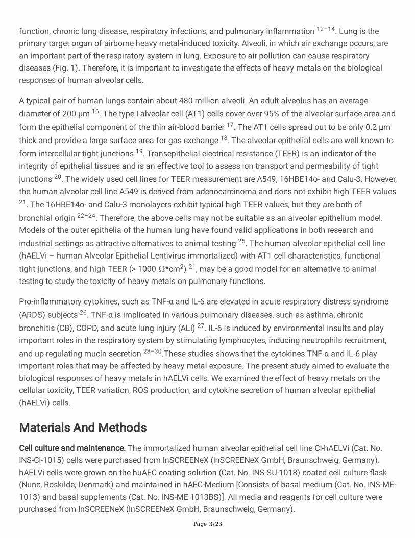
function, chronic lung disease, respiratory infections, and pulmonary inammation 12–14. Lung is theprimary target organ of airborne heavy metal-induced toxicity. Alveoli, in which air exchange occurs, arean important part of the respiratory system in lung. Exposure to air pollution can cause respiratorydiseases (Fig. 1). Therefore, it is important to investigate the effects of heavy metals on the biologicalresponses of human alveolar cells.
A typical pair of human lungs contain about 480 million alveoli. An adult alveolus has an averagediameter of 200 µm 16. The type I alveolar cell (AT1) cells cover over 95% of the alveolar surface area andform the epithelial component of the thin air-blood barrier 17. The AT1 cells spread out to be only 0.2 µmthick and provide a large surface area for gas exchange 18. The alveolar epithelial cells are well known toform intercellular tight junctions 19. Transepithelial electrical resistance (TEER) is an indicator of theintegrity of epithelial tissues and is an effective tool to assess ion transport and permeability of tightjunctions 20. The widely used cell lines for TEER measurement are A549, 16HBE14o- and Calu-3. However,the human alveolar cell line A549 is derived from adenocarcinoma and does not exhibit high TEER values21. The 16HBE14o- and Calu-3 monolayers exhibit typical high TEER values, but they are both ofbronchial origin 22–24. Therefore, the above cells may not be suitable as an alveolar epithelium model.Models of the outer epithelia of the human lung have found valid applications in both research andindustrial settings as attractive alternatives to animal testing 25. The human alveolar epithelial cell line(hAELVi – human Alveolar Epithelial Lentivirus immortalized) with AT1 cell characteristics, functionaltight junctions, and high TEER (> 1000 Ω*cm2) 21, may be a good model for an alternative to animaltesting to study the toxicity of heavy metals on pulmonary functions.
Pro-inammatory cytokines, such as TNF-α and IL-6 are elevated in acute respiratory distress syndrome(ARDS) subjects 26. TNF-α is implicated in various pulmonary diseases, such as asthma, chronicbronchitis (CB), COPD, and acute lung injury (ALI) 27. IL-6 is induced by environmental insults and playimportant roles in the respiratory system by stimulating lymphocytes, inducing neutrophils recruitment,and up-regulating mucin secretion 28–30.These studies shows that the cytokines TNF-α and IL-6 playimportant roles that may be affected by heavy metal exposure. The present study aimed to evaluate thebiological responses of heavy metals in hAELVi cells. We examined the effect of heavy metals on thecellular toxicity, TEER variation, ROS production, and cytokine secretion of human alveolar epithelial(hAELVi) cells.
Materials And MethodsCell culture and maintenance. The immortalized human alveolar epithelial cell line CI-hAELVi (Cat. No.INS-CI-1015) cells were purchased from InSCREENeX (InSCREENeX GmbH, Braunschweig, Germany).hAELVi cells were grown on the huAEC coating solution (Cat. No. INS-SU-1018) coated cell culture ask(Nunc, Roskilde, Denmark) and maintained in hAEC-Medium [Consists of basal medium (Cat. No. INS-ME-1013) and basal supplements (Cat. No. INS-ME 1013BS)]. All media and reagents for cell culture werepurchased from InSCREENeX (InSCREENeX GmbH, Braunschweig, Germany).

Page 4/23
The cells were placed in an incubator lled with 5% CO2 atmosphere and maintained at 37°C. The cellswere subcultured every 3–4 days and the cells used in the present study were within 10 to 15 passages.The TEER of the monolayer formed by the hAELVi cells was measured and the tight-junction formedamong the cells was observed by ZO-1 staining (61-7300, Thermo Fisher Scientic). The cultured cellswere routinely tested for mycoplasma using a commercial PCR kit (e-Myco plus, iNtRON Biotech, Korea).All the cells used in the present study were free of mycoplasma contamination.
In vitro cytotoxicity assay. The cell cytotoxicity was measured using the Live-Dead cell staining kit (ALX-850-249, Enzo). The kit utilizes Live-dyeTM, a cell-permeable green uorescent dye (Ex (max): 488 nm; Em(max): 518 nm), to stain live cells. Dead cells can be easily stained by propidium iodide (PI), a reduorescent dye (Ex (max): 488 nm; Em (max): 615 nm).
The hAELVi cells (2 × 10⁴ cells/well) were seeded in 96-well huAEC coating solution-coated black plates(Corning 3603, Costar) and incubated for 24 h in a complete medium at 37°C in a humidied atmosphereof 5% CO2. After 24 h incubation, the cadmium chloride (CdCl2, 20899, Fluka, referred to as Cd below),sodium (meta) arsenite (AsNaO2, S7400, Sigma-Aldrich, referred to as As below), and zinc sulfateheptahydrate (ZnSO4 7H2O, 204986, Sigma-Aldrich, referred to as Zn below) was added to the 96-wellblack plate at different concentrations (20 µM, 40 µM, 80 µM, 100 µM, 200 µM, and 250 µM) and the cellswere further cultured for 24 h at 37°C. The cells in the control group (CTL) were grown in the absence ofheavy metals. After exposure to the heavy metals, the medium was harvested and stored at −20°C forcytokines analysis. 100 µL of Live-Dead cell staining reagent was added into each well and incubated for15 min at 37°C. After the incubation, the uorescence was measured using a uorescence microplatereader (SynergyTM 2, BioTek). Results are expressed as the percentage of viable cells compared tountreated cells (0 µM).
Transepithelial electrical resistance (TEER) measurements and crystal violet assay. The TEER values weremeasured using the ECIS TEER24 (Applied BioPhysics, New York, USA). The hAELVi cells (1x10⁵
cells/transwell) were seeded on huAEC coating solution-coated transwell inserts with the permeablemembrane pore size of 0.4 µm and a growth area of 0.33 cm2 (Corning 3470). Wells in a TEER24 plate(Applied BioPhysics, T24W) were lled with 1 mL of culture medium. After that, the inserts were put intothe wells and incubated at 37°C in a humidied atmosphere of 5% CO2. After 3 days, the medium in boththe upper and lower chamber was replaced with fresh medium.
Each well exhibit TEER values of typically 2000 Ω across the permeable support after 6 days of culture.Then, continuous measurement of TEER value after the exposure with 250 µM of the heavy metals (Cd,As, and Zn) was conducted. To determine the integrity of the cell monolayer, the cellular image wasobserved after the cells were stained with 1% crystal violet solution (V5265, Sigma-Aldrich). After 24h ofthe heavy metal exposure, the medium was aspirated and replaced by 50 µL of crystal violet stain andthen incubated at room temperature for 20 minutes. Excessive dye was then washed off with

Page 5/23
demineralized water and the cells were observed under an inverted phase-contrast microscope (CKX41,Olympus).
Cellular reactive oxygen species (ROS) assay. The intracellular ROS production levels were determinedusing an oxidation sensitive uorescent dye, 2´,7´-dichlorouorescin diacetate (DCFDA, also known asH2DCFDA and as DCFH-DA) (ab113851, Abcam). The uorogenic dye measures hydroxyl, peroxyl andother ROS activity within the cell 31.
The hAELVi cells (2.5 × 10⁴ cells/well) were seeded in 96-well huAEC coating solution-coated black plates(Corning 3603, Costar) and incubated for 24 h in a complete medium at 37°C in a humidied atmosphereof 5% CO2. After that, the cells were washed gently with phosphate-buffered saline (PBS) and incubatedat 37°C with 25 µM DCFDA for 45 min in the dark. Then, the cells were washed gently with PBS andtreated with the heavy metals at different concentrations (20 µM, 40 µM, 80 µM, 100 µM, 200 µM, and250 µM) and further incubated for 24 h. A common inducer of ROS production tert-butyl hydroperoxide(TBHP), was used as a positive control. The uorescence of oxidized DCF was measured immediatelyafter the incubation at an excitation/emission wavelength of 488/535 nm using a uorescencemicroplate reader (SynergyTM 2, BioTek).
Quantitation of cytokines in the culture supernatants. The levels of TNF-α (ab181421, Abcam) and IL-6(ab178013, Abcam) in the culture medium were measured by ELISA according to the manufacturer’sinstructions. After the exposure to the heavy metals, the medium was harvested and centrifuged at 2000× g for 10 min to remove oating cells. The cell culture supernatants were stored at −20°C until analysis.The absorbance of each well was measured at 450 nm using a microplate reader (SPECTROstarNano,BMG LABTECH). The data from each exposure condition was from three independent experiments. Eachexperiment had three technical replicates. The measurement of each independent experiment wasrepeated at least twice to ensure reproducibility and allow for statistical evaluation.
Statistical analysisCell viability parameters were analysed as follows:
(1) F(518)sam is the uorescence at 518 nm in the experimental group, which is labelled with the greenuorescent dye and PI.(2) F(518)min is the uorescence at 518 nm in the control group, where all cells are alive and labelled withPI.(3) F(518)max is the uorescence at 518 nm in the control group, where all cells are alive and labelled withthe green uorescent dye.For cell viability analysis, the percentage of live cells was calculated from the uorescence readings bythe equation:
Page 6/23
For each assay, all conditions were tested in triplicate. All data were expressed as mean ± SEM. Statisticalsignicance was determined using Student’s t-test. p < 0.05 represents statistical signicance. Theasterisk (*) denotes p < 0.05, double asterisks (**) denote p < 0.01 and triple asterisks (***) denote p <0.001.
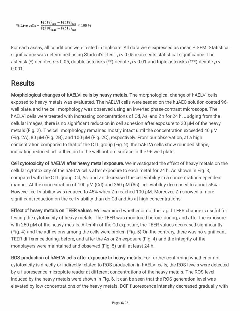
ResultsMorphological changes of hAELVi cells by heavy metals. The morphological change of hAELVi cellsexposed to heavy metals was evaluated. The hAELVi cells were seeded on the huAEC solution-coated 96-well plate, and the cell morphology was observed using an inverted phase-contrast microscope. ThehAELVi cells were treated with increasing concentrations of Cd, As, and Zn for 24 h. Judging from thecellular images, there is no signicant reduction in cell adhesion after exposure to 20 µM of the heavymetals (Fig. 2). The cell morphology remained mostly intact until the concentration exceeded 40 µM(Fig. 2A), 80 µM (Fig. 2B), and 100 µM (Fig. 2C), respectively. From our observation, at a highconcentration compared to that of the CTL group (Fig. 2), the hAELVi cells show rounded shape,indicating reduced cell adhesion to the well bottom surface in the 96 well plate.
Cell cytotoxicity of hAELVi after heavy metal exposure. We investigated the effect of heavy metals on thecellular cytotoxicity of the hAELVi cells after exposure to each metal for 24 h. As shown in Fig. 3,compared with the CTL group, Cd, As, and Zn decreased the cell viability in a concentration-dependentmanner. At the concentration of 100 µM (Cd) and 250 µM (As), cell viability decreased to about 55%.However, cell viability was reduced to 45% when Zn reached 100 µM. Moreover, Zn showed a moresignicant reduction on the cell viability than do Cd and As at high concentrations.
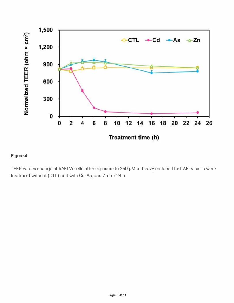
Effect of heavy metals on TEER values. We examined whether or not the rapid TEER change is useful fortesting the cytotoxicity of heavy metals. The TEER was monitored before, during, and after the exposurewith 250 µM of the heavy metals. After 4h of the Cd exposure, the TEER values decreased signicantly(Fig. 4) and the adhesions among the cells were broken (Fig. 5) On the contrary, there was no signicantTEER difference during, before, and after the As or Zn exposure (Fig. 4) and the integrity of themonolayers were maintained and observed (Fig. 5) until at least 24 h.
ROS production of hAELVi cells after exposure to heavy metals. For further conrming whether or notcytotoxicity is directly or indirectly related to ROS production in hAELVi cells, the ROS levels were detectedby a uorescence microplate reader at different concentrations of the heavy metals. The ROS levelinduced by the heavy metals were shown in Fig. 6. It can be seen that the ROS generation level waselevated by low concentrations of the heavy metals. DCF uorescence intensity decreased gradually with

Page 7/23
increasing concentrations of the heavy metals; the cell cytotoxicity was negatively correlated with theROS generation.
Effect of heavy metal exposure on cytokine secretion from hAELVi cells. The expression levels ofcytokines in hAELVi cells exposed to heavy metals can provide information on the possible role of heavymetals on respiratory illnesses due to inammation. TNF-α and IL-6 released or suppressed in hAELVicells after the heavy metal exposure was further evaluated. The Cd-induced secretion of TNF-α in hAELVicells were shown in Fig. 7. The absence of basal levels of TNF-α measured in the controls suggests thatthis pro-inammatory cytokine is not secreted or present at concentrations lower than 2 pg/ml (thedetection limit for the assay employed). Also, the expression levels of TNF-α were lower than thedetection limit after the As and Zn exposure, and therefore the data is not shown.
The secretion of IL-6 was reduced in the hAELVi cells after the heavy metal exposure. The mostsignicant effect was observed at the higher concentration of 200 µM (Cd) and 250 µM (As or Zn)(Fig. 8). The levels of IL-6 at the ion concentrations of 200 µM and 250 µM were signicantly lower thanthat of the CTL. Zn decreased the release of IL-6 in a concentration-dependent manner. Cd, As, and Znsuppressed the IL-6 cytokine releases at 200~250 µM, at which concentration these heavy metals alsoshowed inhibition of cell viability in the cell viability assay.
DiscussionIn vitro human alveolar models provide a platform to investigate the cellular behavior and activity ofhuman lung cells 32. The purpose of this study is to investigate the effect of heavy metals on the cellularcytotoxicity, TEER variation, ROS production, and cytokine secretion of human alveolar epithelial (hAELVi)cells in vitro. The responses may be correlated to the observed pulmonary physiological responses raisedby the heavy metals. The human lung carcinoma cells and human bronchial epithelial cells exposed toheavy metals have been reported 2,33. In the report by Honda et al., the authors have shown that thebiological reaction of human bronchial epithelial BEAS-2B cells to metals in the atmosphere can lead toairway damage and the exacerbation of respiratory diseases 2. In the report by Mu et al., the authorsdetermined the biological responses of sodium arsenite on the human alveolar cell line A549 bymonitoring the arsenite-induced toxicity 33. However, the A549 and BEAS-2B are not derived from thenormal lung; therefore, these cells may not be suitable as alveolar epithelium models. The hAELVi cellsare derived from human alveolar epithelial cells and with functional tight junctions 21. The hAELVi cellsmay be suitable for an alternative to animal testing to study the toxicity of heavy metals on pulmonaryfunctions.
In this study, we examined the effect of Cd, As, and Zn on hAELVi cells. The morphological change ofhAELVi cells exposed to Cd, As, and Zn was examined at concentrations of 40 µM, 80 µM, and 100 µM,respectively (Fig. 2). Signicant cytotoxicity of these heavy metals on the cells was observed at aconcentration exceeding 100 µM (Fig. 3). In a report by Takano et al., Cd concentrations causing a 50%loss in cell viability was 220 µM to 250 µM in alveolar type II cells 34. In our study, we also observed that

Page 8/23
250 µM Cd induced a similar level of cell viability reduction in hAELVi cells. In a report by Mu et al., thecell viability of A549 cells was decreased to about 70% at the highest concentration of NaAsO2 employed
(80 µM) 33. Our results also indicate that the hAELVi cell viability was similarly reduced to 70% at 80 µMof As.
Several studies have demonstrated that zinc plays a critical role in diverse cellular functions such asgrowth and development, maintenance and priming of the immune system, and tissue repair 35. However,zinc is also an important environmental pollutant that becomes dangerous after excessive intake orexposure 36. In a report by Sharif et al., cell growth and viability were decreased by 100 µM of ZnSO4 in
the WIL2-NS human lymphoblastoid cell line 37. In a report by Li et al., the authors have found that ZnSO4
(50 to 150 µM) reduced the cell viability of immortalized murine ovarian granular KK-1 cells 38. As shownin Fig. 3, we observed that the cell viability of hAELVi cells was decreased to about 45% by Zn at aconcentration of 100 µM. In our present comparative study of the acute cytotoxicity of heavy metals tohAELVi cells, Zn at concentrations higher than 100 uM was more toxic than the other heavy metals.These results indicated that the cell viability of WIL2-NS, KK-1, and hAELVi cells was all decreased afterthe treatment with Zn. In summary, all the tested heavy metals show signicant toxicity to the hAELVicells. Zinc ions seemed to be the most toxic metal ions in concentrations higher than 100 µM.
TEER has been widely used to check the level of toxicity of drugs on the epithelial cells 39,40. However,previous studies have reported that cell viability is a more sensitive indicator of cytotoxicity than TEER39,41,42. In a report by Calabro et al., the cell viability decreases before the integrity of the monolayer arecompromised. In a report by Konsoula et al., the authors demonstrated that the MTT assay is a moresensitive indicator of chemical exposure than the TEER measurements. In a report by Mukherjee et al., theauthors have suggested that TEER mainly represents the tight junctions and does not reect cell viability.In this study, we demonstrated that the cell viability was not reected by the TEER value after exposed to250 µM of As and Zn in hAELVi cells (Fig. 3 and Fig. 4). For the Cd exposure, the TEER value decreased tonear zero while the cell viability sustained at 50 %. On the contrary, for the exposure to the other two metalions, the TEER values remained virtually unaffected while drastic cell viability deterioration down to ~50% was indeed observed. The electrical resistance of the monolayers was maintained even while thecellular viability had been severely reduced. In summary, the measurement of TEER seems not a reliablemethod to reect the toxicity of heavy metals on the hAELVi monolayers. The discrepant result betweenthe two assays may rise from the substrate on which the cells adhere.
The integrity shown in Fig. 5, especially for As and Zn, seems to be in contradiction to the deteriorated cellviability shown in Fig. 2. However, the viability test was done with the cells grown on solid substrate whilethe cells in the TEER test was grown on porous membrane, which is supposed to be more relevant to theniche for the alveolar cells in vivo. The fact that the completeness of the Zn- or As- treated monolayer(Fig. 5) resembles that in the CTL group explains the observed resilient TEER values (Fig. 4). The resultsuggests that the hAELVi cells cultured on the membrane survived the damages caused by the metals.

Page 9/23
The escape from the damage is in consistence with the observation that the level of the cytokine TNF-αwas not elevated by As and Zn, as discussed below.
Our results showed that the heavy metals had a signicant cytotoxic effect on the hAELVi cells (Fig. 3).However, what mechanisms are involved in the metal-induced cell damage needs to be questioned. In thisstudy, we observed that the ROS level was elevated at low concentrations of 20 µM heavy metals (Fig. 6).However, there is no signicant effect on cell viability at this concentration. A signicant effect on cellviability was observed at higher concentrations of the heavy metals (100~250 µM) (Fig. 3). Someresearch efforts suggested that the effect between cytotoxicity and ROS production might not be simpleas direct correlation 43. Previous studies have also reported that the behaviour of some cell lines to ROS-mediated oxidative stress is strongly dependent on the scavenging enzymes, like SOD, Cat, GPx 44–47.hAELVi cells are alveolar cells, and in principle, they are expected to have a robust antioxidant systemsince these cells are exposed to gas exchanges in vivo. In addition, cell death is one of the manyscenarios that could be initiated via activating mitochondrial or alternative pathway apoptosis, orinammatory phenomenon could exist 48,49. In a report by Shen et al., the authors suggested thatincreases in intracellular ROS levels were not the sole cause of cytotoxicity. ROS may be a result ofcytotoxicity rather than a causal factor 50. In summary, the previous reports indicated the biphasic effectof ROS-induced growth, ROS-induced apoptosis, or necrosis. However, our results indicate that thecytotoxicity was negatively correlated with ROS generation in the hAELVi cells. Therefore, we suggest thatthere is not a direct relationship between cytotoxicity and ROS production on the hAELVi cells, at least byusing the analysis method employed in this study.
Tumor necrosis factor (TNF), a vital cytokine in physiology and pathology processes, involves in celldifferentiation, apoptotic and necrotic cell death, inammation, and tumorigenesis 51. Highconcentrations of TNF can be deleterious and promotes inammation and organ injury 52. Previous reporthas shown that human peripheral blood mononuclear cells exposed to CdSO4 (1, 5, and 10 µM) or other
metal ions produces an increase in the levels of TNF-α 53. In our study, the TNF-α showed a signicantdose-dependent increase in hAELVi cells after exposure to a high concentration of Cd (Fig. 7). In addition,the cell viability decreased after the treatment of the high concentration of Cd (Fig. 3). Previous researchhas reported that Taraxacum ocinale induces cytotoxicity through tumor necrosis factor-α (TNF-α)secretion in Hep G2 cells 54. Our nding also suggested that exposure to Cd induced the secretion of TNF-α by hAELVi cells and the signicant increase in cell death and the accompanying drastic TEER decrease.In our study, As did not induce TNF-α production. Similar study on lung cells is rare. As a comparison, astudy has shown that 2-8 µM of As increased the expression of TNF-α in human uroepithelial cells, whilethe expression is decreased under 10 µM As 55. Apparently, the hAELVi cells respond to As very differentlyfrom the uroepithelial cells. Concerning the fact that Zn did not induce TNF-a in the hAELVi cells in thisstudy, there has been relatively rare research on the Zn’s effect on the production of TNF-α and thereforeis not compared in more depth. Further studies on the toxicity of heavy metal on hAELVi cells areobviously in need.

Page 10/23
IL-6 is pleiotropic and acts as both an anti-inammatory cytokine 56 and a pro-inammatory cytokine 29,which are mediated by classic signaling and by trans-signaling, respectively 57. IL-6 has a wide range ofbiological activities, including immune responses, acute-phase response, and inammation 58. In ourstudy, As and Zn at low concentrations (20 µM) increased the levels of IL6. At higher concentrations (250µM), an opposite effect was seen (Fig. 8), while TNF-α secretion remain un-detectable. A highconcentration of Cd signicantly increased the secretion of TNF-α but decreased the secretion of IL6 fromthe hAELVi cells (Fig. 7 and Fig. 8), which is very different from the effect by the other two metal ions.
Previous studies have reported that TNF-α and IL-6 production is mutually counter-regulated 59–62. In areport by Gonzalez et al., the negative relationship between TNF-α and IL-6 in patients with acute stageliver abscess has been observed 59. In a report by Yimin et al., IL-6 has a suppressive effect on TNF-αproduction and down-regulates granulomatous inammation reaction 60. In a report by Diao et al., IL-6plays an important role in the negative feedback of the immune response via suppression of TNF-αproduction in Streptococcus pyogenes-infected mice 61. In the report by Yimin et al., it has been shownthat TNF-α and IL-6 production exhibit negatively regulatory effects by each other 62. These resultsindicated that the negatively regulated mechanism between IL-6 and TNF-α secretion. In our study, wealso observed that the concentration of 250 µM Cd signicantly increased the secretion of TNF-α butdecreased the secretion of IL6. However, for As and Zn, the decrease of IL-6 was not accompanied by theincrease of TNF-α. Previous research efforts suggested that the cytokines assayed were differentiallyaffected by heavy metal exposure 53. Our results also suggested that the cytokines of hAELVi cells weredifferentially affected by heavy metal exposure. Therefore, although signicant cell death was indeedinduced by Zn, there was no corresponding TNF-α secretion. It may be inuenced by other cytokinesreleased.
In conclusion, we conrmed that Cd, As, and Zn induced variable extents of cellular toxicity on hAELVicells in a dose-dependent manner when the cells were cultured on a solid substrate. However, thecytotoxicity was not reected by the TEER value after exposure to As and Zn. The discrepancy betweenthe viability assay and the TEER assay may rise from the substrate effect because the TEER wasmeasured with cells cultured on permeable porous membrane, a niche that resembles in vivo better thanon solid substrate. Further studies observing the cytokine expression from cells cultured on permeablemembrane is worth of pursuing. In addition, we found that there is no direct relationship between thecytotoxicity and the ROS production in the hAELVi cells. The exposure to Cd, As and Zn result in differingcytokine secretion of the cells. This study contributes to elucidating the cytotoxicity, TEER values, ROSproduction, and cytokine secretion of hAELVi cells to acute exposure to heavy metal in vitro.
DeclarationsAcknowledgements
Page 11/23
The authors acknowledge nancial support from Ministry of Science and Technology, Taiwan (No. MOST110-2113-M-001-021).
Author contributions statement
HFC designed the study, carried out the experiments, data analysis, and drafted the manuscript. JYCparticipated in the design of the study, interpreted the data, and helped to draft the manuscript.
Conict of interest
The authors declare that they have no conicts of interest.
References1 Huang, W., Long, E., Wang, J., Huang, R. & Ma, L. Characterizing spatial distribution and temporalvariation of PM10 and PM2.5 mass concentrations in an urban area of Southwest China. AtmosphericPollution Research6, 842-848, doi:10.5094/APR.2015.093 (2015).
2 Honda, A. et al. Effects of air pollution-related heavy metals on the viability and inammatoryresponses of human airway epithelial cells. International journal of toxicology34, 195-203,doi:10.1177/1091581815575757 (2015).
3 Feng, S., Gao, D., Liao, F., Zhou, F. & Wang, X. The health effects of ambient PM2.5 and potentialmechanisms. Ecotoxicol Environ Saf128, 67-74, doi:10.1016/j.ecoenv.2016.01.030 (2016).
4 Costa, A. F., Hoek, G., Brunekreef, B. & Ponce de Leon, A. C. Air Pollution and Deaths among ElderlyResidents of Sao Paulo, Brazil: An Analysis of Mortality Displacement. Environ Health Perspect125, 349-354, doi:10.1289/EHP98 (2017).
5 Baxter, L. K., Crooks, J. L. & Sacks, J. D. Inuence of exposure differences on city-to-city heterogeneity inPM2.5-mortality associations in US cities. Environ Health16, 1, doi:10.1186/s12940-016-0208-y (2017).
6 Patel, M. M. et al. Ambient metals, elemental carbon, and wheeze and cough in New York City childrenthrough 24 months of age. Am J Respir Crit Care Med180, 1107-1113, doi:10.1164/rccm.200901-0122OC(2009).
7 Lagorio, S. et al. Air pollution and lung function among susceptible adult subjects: a panel study.Environ Health5, 11, doi:10.1186/1476-069X-5-11 (2006).
8 Hirshon, J. M. et al. Elevated ambient air zinc increases pediatric asthma morbidity. Environ HealthPerspect116, 826-831, doi:10.1289/ehp.10759 (2008).
9 Chen, Y. C. et al. Characteristics of Concentrations and Metal Compositions for PM2.5 and PM2.5–10 inYunlin County, Taiwan during Air Quality Deterioration. Aerosol and Air Quality Research15, 2571-2583,

Page 12/23
doi:10.4209/aaqr.2015.04.0261 (2015).
10 Nemery, B. Metal toxicity and the respiratory tract. Eur Respir J3, 202-219 (1990).
11 Chen, Y. et al. Summer-winter differences of PM2.5 toxicity to human alveolar epithelial cells (A549)and the roles of transition metals. Ecotoxicol Environ Saf165, 505-509, doi:10.1016/j.ecoenv.2018.09.034(2018).
12 Ramsey, K. in Handbook of Arsenic Toxicology (ed S. J. S. Flora) 335-347 (Academic Press, 2015).
13 Parvez, F. et al. Arsenic exposure and impaired lung function. Findings from a large population-basedprospective cohort study. Am J Respir Crit Care Med188, 813-819, doi:10.1164/rccm.201212-2282OC(2013).
14 De, B. K., Majumdar, D., Sen, S., Guru, S. & Kundu, S. Pulmonary involvement in chronic arsenicpoisoning from drinking contaminated ground-water. J Assoc Physicians India52, 395-400 (2004).
15 Stedman, T. L. Stedman's medical dictionary. 27th ed. edn, 1035 (Lippincott Williams & Wilkins, 2000).
16 Ochs, M. et al. The number of alveoli in the human lung. Am J Respir Crit Care Med169, 120-124,doi:10.1164/rccm.200308-1107OC (2004).
17 Wang, Y. et al. Pulmonary alveolar type I cell population consists of two distinct subtypes that differ incell fate. Proc Natl Acad Sci U S A115, 2407-2412, doi:10.1073/pnas.1719474115 (2018).
18 Kemp, S. J. et al. Immortalization of human alveolar epithelial cells to investigate nanoparticle uptake.Am J Respir Cell Mol Biol39, 591-597, doi:10.1165/rcmb.2007-0334OC (2008).
19 Dekali, S. et al. Assessment of an in vitro model of pulmonary barrier to study the translocation ofnanoparticles. Toxicol Rep1, 157-171, doi:10.1016/j.toxrep.2014.03.003 (2014).
20 Poenar, D. P., Yang, G., Wan, W. K. & Feng, S. Low-Cost Method and Biochip for Measuring the Trans-Epithelial Electrical Resistance (TEER) of Esophageal Epithelium. Materials (Basel)13, 2354,doi:10.3390/ma13102354 (2020).
21 Kuehn, A. et al. Human alveolar epithelial cells expressing tight junctions to model the air-blood barrier.ALTEX33, 251-260, doi:10.14573/altex.1511131 (2016).
22 Ehrhardt, C. et al. 16HBE14o- human bronchial epithelial cell layers express P-glycoprotein, lungresistance-related protein, and caveolin-1. Pharm Res20, 545-551, doi:10.1023/a:1023230328687 (2003).
23 Forbes, B., Shah, A., Martin, G. P. & Lansley, A. B. The human bronchial epithelial cell line 16HBE14o- asa model system of the airways for studying drug transport. Int J Pharm257, 161-167, doi:10.1016/s0378-5173(03)00129-7 (2003).

Page 13/23
24 Grainger, C. I., Greenwell, L. L., Lockley, D. J., Martin, G. P. & Forbes, B. Culture of Calu-3 cells at the airinterface provides a representative model of the airway epithelial barrier. Pharm Res23, 1482-1490,doi:10.1007/s11095-006-0255-0 (2006).
25 Gordon, S. et al. Non-animal models of epithelial barriers (skin, intestine and lung) in research,industrial applications and regulatory toxicology. ALTEX32, 327-378, doi:10.14573/altex.1510051 (2015).
26 Han, S. & Mallampalli, R. K. The acute respiratory distress syndrome: from mechanism to translation.J Immunol194, 855-860, doi:10.4049/jimmunol.1402513 (2015).
27 Mukhopadhyay, S., Hoidal, J. R. & Mukherjee, T. K. Role of TNFalpha in pulmonary pathophysiology.Respir Res7, 125, doi:10.1186/1465-9921-7-125 (2006).
28 Chen, Y. et al. Stimulation of airway mucin gene expression by interleukin (IL)-17 through IL-6paracrine/autocrine loop. J Biol Chem278, 17036-17043, doi:10.1074/jbc.M210429200 (2003).
29 Thacker, E. L. Lung inammatory responses. Veterinary research37, 469-486,doi:10.1051/vetres:2006011 (2006).
30 Bautista, M. V. et al. IL-8 regulates mucin gene expression at the posttranscriptional level in lungepithelial cells. J Immunol183, 2159-2166, doi:10.4049/jimmunol.0803022 (2009).
31 Figueroa, D., Asaduzzaman, M. & Young, F. Real time monitoring and quantication of reactive oxygenspecies in breast cancer cell line MCF-7 by 2′,7′–dichlorouorescin diacetate (DCFDA) assay. Journal ofPharmacological and Toxicological Methods94, 26-33, doi:10.1016/j.vascn.2018.03.007 (2018).
32 Evans, K. V. & Lee, J. H. Alveolar wars: The rise of in vitro models to understand human lung alveolarmaintenance, regeneration, and disease. Stem Cells Transl Med9, 867-881, doi:10.1002/sctm.19-0433(2020).
33 Mu, Y., Wang, R. & Wang, H. Programmed Cell Death of Cultured A549 Lung Epithelial Cells Induced bySodium Arsenite Exposure. Journal of Medicinal Chemistry & Toxicology2, 85-89, doi:10.15436/2575-808X.17.1471 (2017).
34 Takano, Y., Taguchi, T., Suzuki, I., Balis, J. U. & Yuri, K. Cytotoxicity of heavy metals on primary culturedalveolar type II cells. Environ Res89, 138-145, doi:10.1006/enrs.2002.4354 (2002).
35 Truong-Tran, A. Q., Carter, J., Run, R. & Zalewski, P. D. New insights into the role of zinc in therespiratory epithelium. Immunol Cell Biol79, 170-177, doi:10.1046/j.1440-1711.2001.00986.x (2001).
36 Marcinčáková, D. et al. Impact of Zinc Sulfate Exposition on Viability, Proliferation and Cell CycleDistribution of Epithelial Kidney Cells. Polish Journal of Environmental Studies28, 3279-3286,doi:10.15244/pjoes/94045 (2019).
Page 14/23
37 Sharif, R., Thomas, P., Zalewski, P., Graham, R. D. & Fenech, M. The effect of zinc sulphate and zinccarnosine on genome stability and cytotoxicity in the WIL2-NS human lymphoblastoid cell line. MutatRes720, 22-33, doi:10.1016/j.mrgentox.2010.12.004 (2011).
38 Li, Y. et al. Zinc inhibits the reproductive toxicity of Zearalenone in immortalized murine ovariangranular KK-1 cells. Sci Rep5, 14277, doi:10.1038/srep14277 (2015).
39 Mukherjee, T., Squillantea, E., Gillespieb, M. & Shao, J. Transepithelial electrical resistance is not areliable measurement of the Caco-2 monolayer integrity in Transwell. Drug Deliv11, 11-18,doi:10.1080/10717540490280345 (2004).
40 Narai, A., Arai, S. & Shimizu, M. Rapid decrease in transepithelial electrical resistance of humanintestinal Caco-2 cell monolayers by cytotoxic membrane perturbents. Toxicol In Vitro11, 347-354,doi:10.1016/s0887-2333(97)00026-x (1997).
41 Calabro, A. R., Konsoula, R. & Barile, F. A. Evaluation of in vitro cytotoxicity and paracellularpermeability of intact monolayers with mouse embryonic stem cells. Toxicol In Vitro22, 1273-1284,doi:10.1016/j.tiv.2008.02.023 (2008).
42 Konsoula, R. & Barile, F. A. Correlation of in vitro cytotoxicity with paracellular permeability in Caco-2cells. Toxicol In Vitro19, 675-684, doi:10.1016/j.tiv.2005.03.006 (2005).
43 Zhu, C., Hu, W., Wu, H. & Hu, X. No evident dose-response relationship between cellular ROS level andits cytotoxicity--a paradoxical issue in ROS-based cancer therapy. Sci Rep4, 5029, doi:10.1038/srep05029(2014).
44 Ighodaro, O. M. & Akinloye, O. A. First line defence antioxidants-superoxide dismutase (SOD), catalase(CAT) and glutathione peroxidase (GPX): Their fundamental role in the entire antioxidant defence grid.Alexandria Journal of Medicine54, 287-293, doi:10.1016/j.ajme.2017.09.001 (2019).
45 He, L. et al. Antioxidants Maintain Cellular Redox Homeostasis by Elimination of Reactive OxygenSpecies. Cell Physiol Biochem44, 532-553, doi:10.1159/000485089 (2017).
46 Birben, E., Sahiner, U. M., Sackesen, C., Erzurum, S. & Kalayci, O. Oxidative stress and antioxidantdefense. World Allergy Organ J5, 9-19, doi:10.1097/WOX.0b013e3182439613 (2012).
47 Day, R. M. & Suzuki, Y. J. Cell proliferation, reactive oxygen and cellular glutathione. Dose Response3,425-442, doi:10.2203/dose-response.003.03.010 (2006).
48 Green, D. R. & Llambi, F. Cell Death Signaling. Cold Spring Harb Perspect Biol7, a006080,doi:10.1101/cshperspect.a006080 (2015).
49 Belizario, J., Vieira-Cordeiro, L. & Enns, S. Necroptotic Cell Death Signaling and Execution Pathway:Lessons from Knockout Mice. Mediators Inamm2015, 128076, doi:10.1155/2015/128076 (2015).
Page 15/23
50 Shen, C. et al. Relating cytotoxicity, zinc ions, and reactive oxygen in ZnO nanoparticle-exposed humanimmune cells. Toxicol Sci136, 120-130, doi:10.1093/toxsci/kft187 (2013).
51 Locksley, R. M., Killeen, N. & Lenardo, M. J. The TNF and TNF receptor superfamilies: integratingmammalian biology. Cell104, 487-501, doi:10.1016/s0092-8674(01)00237-9 (2001).
52 Steeland, S., Libert, C. & Vandenbroucke, R. E. A New Venue of TNF Targeting. Int J Mol Sci19, 1442,doi:10.3390/ijms19051442 (2018).
53 Maria, B., Koizumi, S. & Jonai, H. Cytokine Production by Human Peripheral Blood Mononuclear cellsafter Exposure to Heavy Metals. JOURNAL OF HEALTH SCIENCE46, 358-362, doi:10.1248/jhs.46.358(2000).
54 Koo, H. N. et al. Taraxacum ocinale induces cytotoxicity through TNF-alpha and IL-1alpha secretionin Hep G2 cells. Life Sci74, 1149-1157, doi:10.1016/j.lfs.2003.07.030 (2004).
55 Liu, S. et al. Arsenic induced overexpression of inammatory cytokines based on the human urothelialcell model in vitro and urinary secretion of individuals chronically exposed to arsenic. Chem ResToxicol27, 1934-1942, doi:10.1021/tx5002783 (2014).
56 Riemschneider, S., Herzberg, M. & Lehmann, J. Subtoxic Doses of Cadmium Modulate InammatoryProperties of Murine RAW 264.7 Macrophages. Biomed Res Int2015, 295303, doi:10.1155/2015/295303(2015).
57 Scheller, J., Chalaris, A., Schmidt-Arras, D. & Rose-John, S. The pro- and anti-inammatory properties ofthe cytokine interleukin-6. Biochim Biophys Acta1813, 878-888, doi:10.1016/j.bbamcr.2011.01.034(2011).
58 Kayaalti, Z. et al. Effects of the interleukin-6 (IL-6) polymorphism on toxic metal and trace elementlevels in placental tissues. Sci Total Environ409, 4929-4933, doi:10.1016/j.scitotenv.2011.08.036 (2011).
59 Gonzalez-Amaro, R. et al. Plasma levels and in vitro production of tumor necrosis factor-alpha andinterleukin-6 in patients with amebic liver abscess. Rev Invest Clin46, 209-213 (1994).
60 Yimin, Kohanawa, M. & Minagawa, T. Up-regulation of granulomatous inammation in interleukin-6knockout mice infected with Rhodococcus aurantiacus. Immunology110, 501-506, doi:10.1111/j.1365-2567.2003.01762.x (2003).
61 Diao, H. & Kohanawa, M. Endogenous interleukin-6 plays a crucial protective role in streptococcal toxicshock syndrome via suppression of tumor necrosis factor alpha production. Infect Immun73, 3745-3748,doi:10.1128/IAI.73.6.3745-3748.2005 (2005).
62 Yimin & Kohanawa, M. A regulatory effect of the balance between TNF-alpha and IL-6 in thegranulomatous and inammatory response to Rhodococcus aurantiacus infection in mice. J

Page 16/23
Immunol177, 642-650, doi:10.4049/jimmunol.177.1.642 (2006).
Figures
Figure 1
The respiratory system can suffer effects after exposure to air pollution from factories (modied fromStedman's Medical Dictionary 15).

Page 17/23
Figure 2
Micrographs of hAELVi cells exposed to heavy metals for 24 h. Cell morphology changes relative toconcentrations (20, 40, 80, 100, 200 and 250 μM) of (A) absence of heavy metals (CTL) (B) Cd, (C) As,and (D) Zn respectively. All micrographs are at the same magnication; Scale bars, 50 μm.

Page 18/23
Figure 3
The cell viability of hAELVi cells after exposure to various doses of air pollution-related heavy metals. ThehAELVi cells were incubated in the presence of Cd, As, and Zn for 24 h.
Page 19/23
Figure 4
TEER values change of hAELVi cells after exposure to 250 μM of heavy metals. The hAELVi cells weretreatment without (CTL) and with Cd, As, and Zn for 24 h.

Page 20/23
Figure 5
The integrity of hAELVi cells monolayers after exposure to 250 μM of heavy metals. The hAELVi cellswere treatment without (CTL) and with Cd, As, and Zn for 24 h. The cells were stained with crystal violet.All micrographs are at the same magnication; Scale bars, 200 μm.

Page 21/23
Figure 6
Production of ROS in hAELVi cells. The hAELVi cells were incubated in the presence of Cd, As, and Zn for24 h. 50 μM Tert-Butyl Hydrogen Peroxide (TBHP) was used as a positive control.

Page 22/23
Figure 7
Secretion of TNF-α by hAELVi cells treated with CdCl2. The hAELVi cells were incubated in the presence ofCd for 24 h and TNF-α concentrations measured in the supernatants.

Page 23/23
Figure 8
Secretion of IL-6 by hAELVi cells treated with heavy metals after 24h. The hAELVi cells were incubated inthe presence of Cd, As, and Zn for 24 h and IL-6 concentrations measured in the supernatants.
Copyright © 2022 FDOKUMEN