
morphology of bacteria, viruses and protozoa
78
МІНІСТЕРСТВО ОХОРОНИ ЗДОРОВ'Я УКРАЇНИ Харківський національний медичний університет MORPHOLOGY OF BACTERIA, VIRUSES AND PROTOZOA Learning guide for the 2 nd and 3 rd year English media students of the Faculty of Medicine and the Faculty of Dentistry (Microbiology, virology and immunology) МОРФОЛОГІЯ БАКТЕРІЙ, ВІРУСІВ І НАЙПРОСТІШИХ Методичні вказівки з дисципліни «Мікробіологія, вірусологія та імунологія» для студентів II і III курсів медичного та стоматологічного факультетів з англійською мовою викладання ЗАТВЕРДЖЕНО вченою радою ХНМУ. Протокол № 4 від 27.04.2017. Харків ХНМУ 2017
-
Upload
khangminh22 -
Category
Documents
-
view
0 -
download
0
Transcript of morphology of bacteria, viruses and protozoa

1
МІНІСТЕРСТВО ОХОРОНИ ЗДОРОВ'Я УКРАЇНИ
Харківський національний медичний університет
MORPHOLOGY
OF BACTERIA, VIRUSES
AND PROTOZOA Learning guide for the 2
nd and 3
rd year English media
students of the Faculty of Medicine and the Faculty
of Dentistry (Microbiology, virology and immunology)
МОРФОЛОГІЯ БАКТЕРІЙ,
ВІРУСІВ І НАЙПРОСТІШИХ Методичні вказівки з дисципліни
«Мікробіологія, вірусологія та імунологія»
для студентів II і III курсів медичного
та стоматологічного факультетів
з англійською мовою викладання
ЗАТВЕРДЖЕНО
вченою радою ХНМУ.
Протокол № 4 від 27.04.2017.
Харків ХНМУ 2017
2
Morphology of bacteria, viruses and protozoa: Learning guide for the 2nd
and
3rd
year English media students of the Faculty of Medicine and the Faculty of
Dentistry (Microbiology, virology and immunology) / comp. N. I. Kovalenko. –
Kharkiv : Kharkiv National Medical University, 2017. – 76 p.
Compiler N. I. Kovalenko
Learning guide is related to the program of Ministry of Health of Ukraine
and is recommended to students of medical and dentistry faculties of high
medical schools of III-IV level accreditation.
Learning guide includes sections of taxonomy, morphology and
ultrastructure of bacteria, viruses and pathogenic protozoa. The most modern
information on methods of microscopic examination is represented.
Морфологія бактерій, вірусів і найпростіших : метод. вказівки
з дисципліни «Мікробіологія, вірусологія та імунологія» для студентів II
і III курсів мед. та стомат. фак-тів з англ. мовою викладання / упоряд.
Н. І. Коваленко. – Харків : ХНМУ, 2017. – 76 с.
Упорядник Н. І. Коваленко

3
MORPHOLOGY OF COCCI. MICROSCOPIC EXAMINATION. SIMPLE TECHNIQUES OF STAINING
Actuality of the theme.
Infections may be caused by bacteria, viruses, fungi, or parasites. Infection
may be endogenous or exogenous. In endogenous infections, the
microorganism (usually a bacterium) is a component of the patient's indigenous
flora. Endogenous infections can occur when the microorganism is aspirated
from the upper to the lower respiratory tract or when it penetrates the skin or
mucosal barrier as a result of trauma or surgery. In contrast, in exogenous
infections, the microorganism is acquired from the environment (e.g., from soil
or water) or from another person or an animal. Although it is important to
establish the cause of an infection, the differential diagnosis is based on a
careful history, physical examination, and appropriate radiographic and
laboratory studies, including the selection of appropriate specimens for
microbiologic examination. Results of the history, physical examination, and
radiographic and laboratory studies allow the physician to request tests for the
microorganisms most likely to be the cause of the infection.
Some infectious diseases are distinctive enough to be identified clinically.
Most pathogens, however, can cause a wide spectrum of clinical syndromes in
humans. Conversely, a single clinical syndrome may result from infection with
any one of many pathogens. Influenza virus infection, for example, causes a
wide variety of respiratory syndromes that cannot be distinguished clinically
from those caused by streptococci, or more than 100 other viruses.
Most often, therefore, it is necessary to use microbiologic laboratory
methods to identify a specific etiologic agent. Diagnostic medical microbiology
is the discipline that identifies etiologic agents of disease. The job of the
clinical microbiology laboratory is to test specimens from patients for
microorganisms that are, or may be, a cause of the illness and to provide
information (when appropriate) about the in vitro activity of antimicrobial
drugs against the microorganisms identified.
Direct examination of specimens frequently provides the most rapid
indication of microbial infection. A variety of microscopic, immunologic, and
hybridization techniques have been developed for rapid diagnosis. The
sensitivity of a technique usually depends on the number of microorganisms in
the specimen. Its specificity depends on how morphologically unique a specific
microorganism appears microscopically.
Goal: Studying of morphology of cocci by microscopic examination.
Concrete goals:
1. Study of the rules of work in a bacteriological laboratory.
2. Study of arrangement of cocci in microslide.
4
3. Study of simple method of staining of microslide.
4. Study of structure of immersion microscope.
Students should be able to:
1. Perform simple method of staining of microslide.
2. Using a microscope, identify different bacterial shapes and arrangements.
Equipment: museum cultures of bacteria, bacteriological loops, gas
burners, basic days, immersion microscopes, immersion oil, tables, atlas.
Rules of work in bacteriological laboratories. 1. The personnel working
at laboratories is supplied with medical coats and kerchiefs or caps. Special
clothes protect the worker and also prevent contamination of the material to be
studied with foreign microflora.
2. Eating and smoking in the laboratory are strictly forbidden.
3. Unnecessary walking about the laboratory, sharp movements, and
irrelevant conversations should be discouraged. It is also necessary to avoid
rubbing one's eyes or nose, scratching one's head, biting nails, pencils, etc.
4. In the process of examination the working place should be kept clean and
tidy. Bacteriological loops are rendered harmless by burning them in the
burner's flame; used spatulas, glass slides, pipettes, and other instruments are
placed into jars with disinfectant solution.
5. Upon the completion of work the nutrient media with inoculated cultures
are placed into an incubator; museum cultures, into safe-refrigerators; devices
and apparatuses are set up in places specially intended for them. Wipe tables
with disinfectant solution and thoroughly wash the hands.
6. If the material to be analyzed or the culture of microorganisms is
accidentally spilt onto the hands, table, coat, or shoes, they should be
immediately treated with disinfectant.
Students must observe the principles of hygiene. They must disinfect and
wash their hands always after contaminating them with a biological material
and before leaving the hall. For disinfecting hands, 0.5 % chloramine is used
for 2 minutes. Then the hands are to be rinsed with warm water and washed
with soap.
Morphology of cocci
Bacteria (Gk bakterion small staff) are, for the most part, unicellular organisms
lacking chlorophyll. Their biological properties and predominant reproduction
relates them to prokaryotes. Bacteria divide by binary fission, a process by which
one bacterium splits into two. The size of bacteria is measured in micrometres
(mcm). Most pathogenic bacteria measure 0.2 to 10 mcm.
Morphologically, bacteria possess three main forms. They are either
spherical (cocci), rod-shaped (bacteria, bacilli, and clostridia) or spiral-shaped
(vibrios, spirilla, and spirochetes).

5
Cocci (Gk. chokes berry). These forms of bacteria are spherical, ellipsoidal,
bean-shaped, and lancelet. Cocci are approximately 0.5 micrometer (µm) in
diameter and may be seen, based on their planes of division and tendency to
remain attached after replication, in one of the following arrangements (Fig. 1):
Fig. 1. Arrangements of cocci
1. Micrococci. The cells are arranged singly or irregularly They are sapro-
phytes, and live in water and in air (Micrococcus agilis, M. roseus, M. luteus, etc).
2. Diplococci (Gk. diplos double) divide in one plane and remain attached
in pairs. These include Neisseria meningitidis or meningoccus (Fig. 2), causative
agent of epidemic cerebrospinal meningitis, N.gonorrhoeae or gonococcus,
causative agent of gonorrhoea and blennorrhoea, and Streptococcus
pneumoniae, causative agent of respiratory infections, meningitis etc. (Fig. 3).
Fig. 2. Neisseria meningitidis Fig. 3. Streptococcus pneumoniae
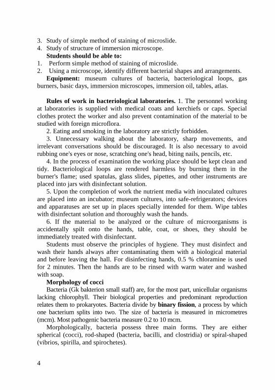
3. Streptococci (Gk. streptos curved, kokkos berry) divide in one plane and are
arranged in chains of different length. Some streptococci are pathogenic for humans
and are responsible for various diseases (Streptococcus pyogenes) (Fig. 4).
4. Tetracocci (Gk. tetra four) divide in two planes at right angles to one another
and form groups of 4 cells (Fig. 5). They very rarely produce diseases in humans.
6
Fig. 4. Streptococcus pyogenes Fig. 5. Tetracoccus arrangement
5. Sarcinae (L sarcio to tie) divide in three planes at right angles to one
another and resemble packets of 8, 16 or more cells (Fig. 6). They are
frequently found in the air. Virulent species have not been encountered.
6. Staphylococci (Gk. staphyle cluster of grapes) divide in several planes
resulting in irregular bunches of cells, sometimes resembling clusters of grapes
(Fig. 7). Some species of Staphylococci cause diseases in man and animals
(Staphylococcus aureus, S. epidermidis, S. saprophyticus).
Fig. 6. Sarcina arrangement Fig. 7. Staphylococcus aureus
As you observe these different cocci, keep in mind that the procedures used in
slide preparation may cause some arrangements to break apart or clump
together. The correct form, however, should predominate. Also remember that each
coccus in an arrangement represents a complete, single, one-celled organism.
Principal microbiological procedures. A complex of bacterioscopic,
bacteriological, serological, allergological, and biological techniques is used in
the microbiological diagnosis of bacterial infections. Depending on the nature of
the given infectious disease, one of these methods is used as the main one, while
the others are supplementary. Such biological substances as blood, faeces, urine,
cerebrospinal fluid, bile, etc. serve as the material for microbiological diagnosis.
Microscopic examination. Light microscopy. A light microscope is fitted with
dry and immersion objectives. A dry objective with a relatively large focal distance and
weak magnification power is ordinarily utilized for studying large biological and
7
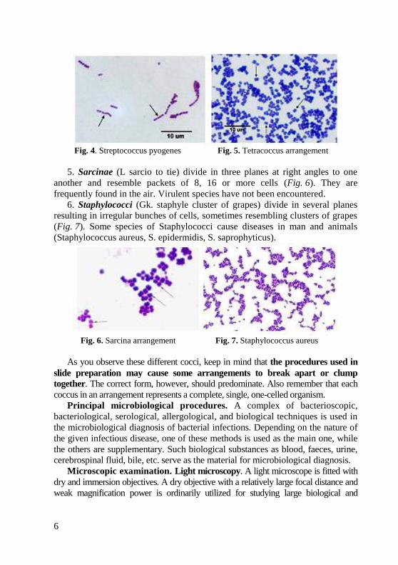
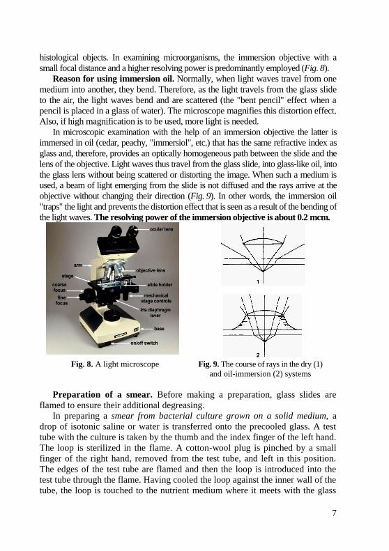
histological objects. In examining microorganisms, the immersion objective with a
small focal distance and a higher resolving power is predominantly employed (Fig. 8).
Reason for using immersion oil. Normally, when light waves travel from one
medium into another, they bend. Therefore, as the light travels from the glass slide
to the air, the light waves bend and are scattered (the "bent pencil" effect when a
pencil is placed in a glass of water). The microscope magnifies this distortion effect.
Also, if high magnification is to be used, more light is needed.
In microscopic examination with the help of an immersion objective the latter is
immersed in oil (cedar, peachy, "immersiol", etc.) that has the same refractive index as
glass and, therefore, provides an optically homogeneous path between the slide and the
lens of the objective. Light waves thus travel from the glass slide, into glass-like oil, into
the glass lens without being scattered or distorting the image. When such a medium is
used, a beam of light emerging from the slide is not diffused and the rays arrive at the
objective without changing their direction (Fig. 9). In other words, the immersion oil
"traps" the light and prevents the distortion effect that is seen as a result of the bending of
the light waves. The resolving power of the immersion objective is about 0.2 mcm.
Fig. 8. A light microscope Fig. 9. The course of rays in the dry (1)
and oil-immersion (2) systems
Preparation of a smear. Before making a preparation, glass slides are
flamed to ensure their additional degreasing.
In preparing a smear from bacterial culture grown on a solid medium, a
drop of isotonic saline or water is transferred onto the precooled glass. A test
tube with the culture is taken by the thumb and the index finger of the left hand.
The loop is sterilized in the flame. A cotton-wool plug is pinched by a small
finger of the right hand, removed from the test tube, and left in this position.
The edges of the test tube are flamed and then the loop is introduced into the
test tube through the flame. Having cooled the loop against the inner wall of the
tube, the loop is touched to the nutrient medium where it meets with the glass
8
wall (if the loop is not sufficiently cooled, it induces cracking and melts the
medium). Then the loop is touched to the culture of the microorganisms on the
surface of the medium. Then the loop is withdrawn, the edges of the test tube
are quickly flamed, the tube is closed with a stopper passed through the flame,
and then replaced into the test tube rack. All the above described procedures are
made above the flame. The culture sample is placed with the loop into a drop of
water on the glass slide and spread uniformly with circular movements on an
area of 1-1.5 cm in diameter then the loop is flamed.
Drying and fixation of the smear. The dried smears are flamed to kill and fix
the bacteria on the glass slide, preventing thereby their washing off during staining.
The dead microorganisms are more receptive to dyes and present no danger for the
personnel working with them. The glass slide is grasped with a forceps or with the
thumb and index finger of the right hand, the smear being in the upside position,
and passed three times through the hottest part of the burner's flame.
Staining of a smear. Staining of bacteria is a complex physicochemical
process. Interaction of the dye with the cell substances results in the formation
of salts ensuring stability of staining. Relationship between various types of
microorganisms and dyes is called a tinctorial property. The following dyes are employed most extensively: (1) red (basic fuchsine,
acid fuchsine, safranine, neutral red, Congo red); (2) blue (methylene blue,
toluidine blue, trypan blue, etc.); (3) violet (gentian, methyl or crystal); and
(4) yellow-brown (vesuvin, chrysoidine).
Stains are generally salts in which one of the ions is colored. (A salt is a
compound composed of a positively charged ion and a negatively charged ion.) For
example, the dye methylene blue is actually the salt methylene blue chloride, which
will dissociate in water into a positively charged methylene blue ion which is blue in
color and a negatively charged chloride ion which is colorless.
Dyes or stains may be divided into two groups: basic and acidic. If the color portion
of the dye resides in the positive ion, as in the above case, it is called a basic dye
(examples: methylene blue, crystal violet, safranin). If the color portion is in the
negatively charged ion, it is called an acidic dye (examples: nigrosin, congo red).
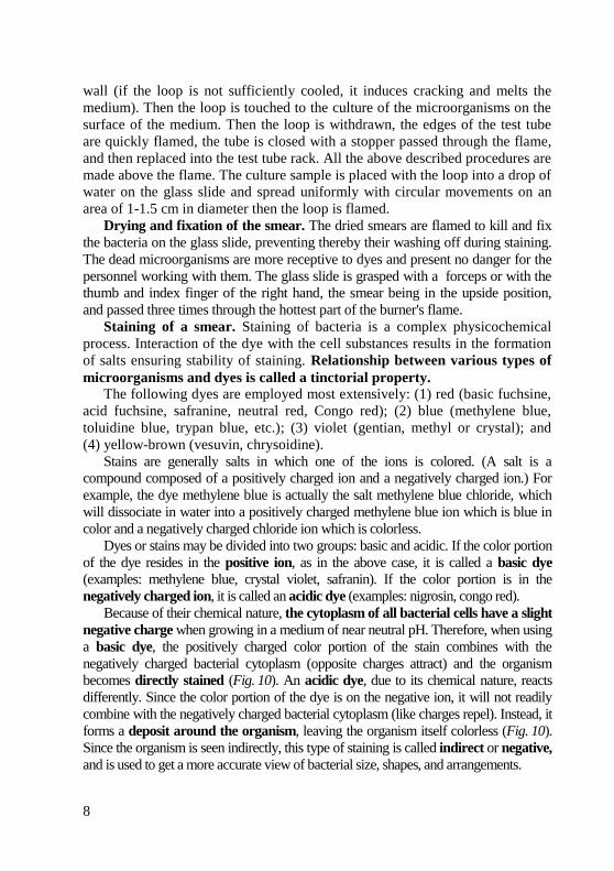
Because of their chemical nature, the cytoplasm of all bacterial cells have a slight
negative charge when growing in a medium of near neutral pH. Therefore, when using
a basic dye, the positively charged color portion of the stain combines with the
negatively charged bacterial cytoplasm (opposite charges attract) and the organism
becomes directly stained (Fig. 10). An acidic dye, due to its chemical nature, reacts
differently. Since the color portion of the dye is on the negative ion, it will not readily
combine with the negatively charged bacterial cytoplasm (like charges repel). Instead, it
forms a deposit around the organism, leaving the organism itself colorless (Fig. 10).
Since the organism is seen indirectly, this type of staining is called indirect or negative,
and is used to get a more accurate view of bacterial size, shapes, and arrangements.
9
Fig. 10. Direct staining and indirect staining
Simple techniques of staining make use of only one dye and demonstrate the
form of bacteria. The fixed preparation is placed, the smear upward, on the support.
A dye solution is pipetted onto the entire surface of the smear. With fuchsine the
staining lasts 1-2 min, with alkaline solution of Loeffler's methylene blue or water-
alcoholic solution of methylene blue, 3-5 min. Following the staining procedure the
dye is dispensed, the preparation is washed with water, dried between sheets of
filter paper, and then examined under the oil-immersion objective.
Introduction to staining. Bacterial morphology (form and structure) may
be examined in two ways: by observing living unstained organisms (wet
mount), or by observing killed stained organisms.
Since bacteria are almost colorless and therefore show little contrast with
the broth in which they are suspended, they are difficult to observe when
unstained. Staining microorganisms enables one to: see greater contrast
between the organism and the background, differentiate various morphological
types (by shape, arrangement, gram reaction, etc.), observe certain structures
(flagella, capsules, endospores, etc.).
Before staining bacteria, you must first understand how to "fix" the
organisms to the glass slide. If the preparation is not fixed, the organisms will
be washed off the slide during staining. A simple method is that of air drying
and heat fixing. The organisms are heat fixed by passing an air-dried smear of
the organisms through the flame of a gas burner. The heat coagulates the
organisms' proteins causing the bacteria to stick to the slide.
The procedure for heat fixation is as follows:
1. If the culture is taken from an agar medium:
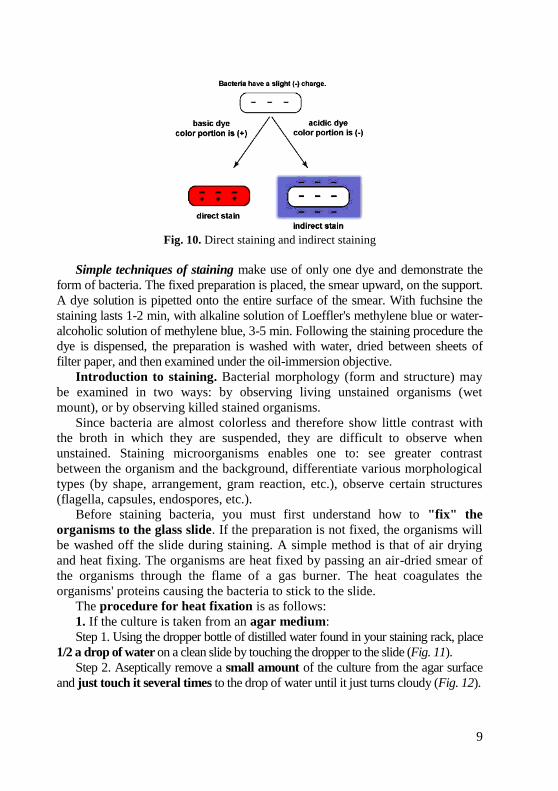
Step 1. Using the dropper bottle of distilled water found in your staining rack, place
1/2 a drop of water on a clean slide by touching the dropper to the slide (Fig. 11).
Step 2. Aseptically remove a small amount of the culture from the agar surface
and just touch it several times to the drop of water until it just turns cloudy (Fig. 12).

10
Fig. 11. Preparing the slide for Fig. 12. Preparing the slide for
staining: Step 1. staining: Step 2.
Step 3. Burn the remaining bacteria off of the loop. (If too much culture
is added to the water, you will not see stained individual bacteria.)
Step 4. Using the loop spread the suspension over the entire slide to form
a thin film (Fig. 13).
Step 5. Allow this thin suspension to completely air dry (Fig. 14).
Fig. 13. Preparing the slide for Fig. 14. Preparing the slide for
staining: Step 4. staining: Step 5.
Step 6. Pass the slide (film-side up) through the flame of the bunsen burner 3 or 4
times to heat-fix (Fig. 15). Caution: Too much heat might distort the organism and, in
the case of the gram stain, may cause gram-positive organisms to stain gram-
negatively. The slide should feel very warm but not too hot to hold.
Fig. 15. Preparing the slide for staining: Step 6.
2. If the organism is taken from a broth culture:
a. Aseptically place 2 or 3 loops of the culture on a clean slide. Do not use water.
b. Using the loop spread the suspension over the entire slide to form a thin film.
c. Allow this thin suspension to completely air dry.
d. Pass the slide (film-side up) through the flame of the gas burner 3 or
4 times to heat-fix.
11
The procedure for staining is as follows:
Step 1. Place the slide on a staining tray and cover the entire film with
carbol fuchsin. Stain for one minute.
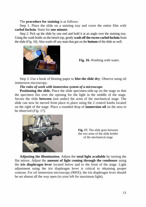
Step 2. Pick up the slide by one end and hold it at an angle over the staining tray.
Using the wash bottle on the bench top, gently wash off the excess carbol fuchsin from
the slide (Fig. 16). Also wash off any stain that got on the bottom of the slide as well.
Fig. 16. Washing with water.
Step 3. Use a book of blotting paper to blot the slide dry. Observe using oil
immersion microscopy.
The rules of work with immersion system of a microscope.
Positioning the slide. Place the slide specimen-side-up on the stage so that
the specimen lies over the opening for the light in the middle of the stage.
Secure the slide between (not under) the arms of the mechanical stage. The
slide can now be moved from place to place using the 2 control knobs located
on the right of the stage. Place a rounded drop of immersion oil on the area to
be observed (Fig. 17).
Fig. 17. The slide goes between
the two arms of the slide holder
of the mechanical stage
Adjusting the illumination. Adjust the total light available by turning the
flat mirror. Adjust the amount of light coming through the condenser using
the iris diaphragm lever located below and to the front of the stage. Light
adjustment using the iris diaphragm lever is critical to obtaining proper
contrast. For oil immersion microscopy (900X), the iris diaphragm lever should
be set almost all the way open (to your left for maximum light).
12
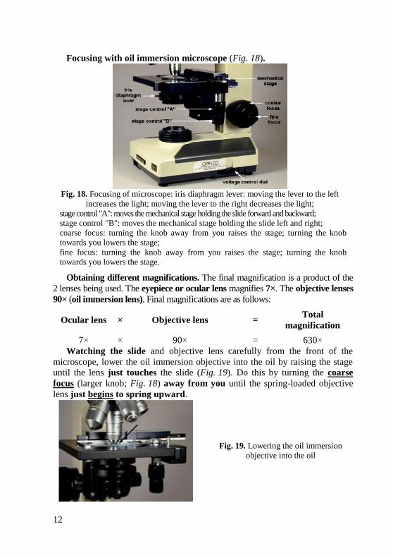
Focusing with oil immersion microscope (Fig. 18).
Fig. 18. Focusing of microscope: iris diaphragm lever: moving the lever to the left
increases the light; moving the lever to the right decreases the light;
stage control "A": moves the mechanical stage holding the slide forward and backward;
stage control "B": moves the mechanical stage holding the slide left and right;
coarse focus: turning the knob away from you raises the stage; turning the knob
towards you lowers the stage;
fine focus: turning the knob away from you raises the stage; turning the knob
towards you lowers the stage.
Obtaining different magnifications. The final magnification is a product of the
2 lenses being used. The eyepiece or ocular lens magnifies 7×. The objective lenses
90× (oil immersion lens). Final magnifications are as follows:
Ocular lens × Objective lens = Total
magnification
7× × 90× = 630×
Watching the slide and objective lens carefully from the front of the
microscope, lower the oil immersion objective into the oil by raising the stage
until the lens just touches the slide (Fig. 19). Do this by turning the coarse
focus (larger knob; Fig. 18) away from you until the spring-loaded objective
lens just begins to spring upward.
Fig. 19. Lowering the oil immersion
objective into the oil
13
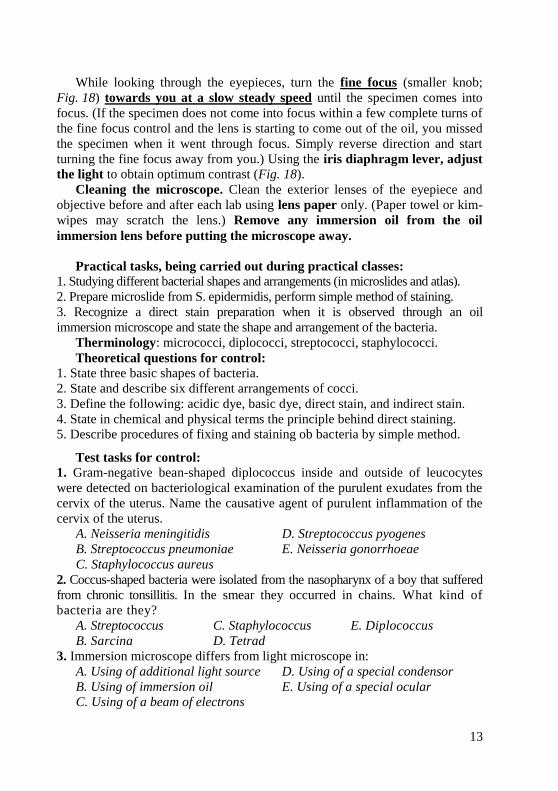
While looking through the eyepieces, turn the fine focus (smaller knob;
Fig. 18) towards you at a slow steady speed until the specimen comes into
focus. (If the specimen does not come into focus within a few complete turns of
the fine focus control and the lens is starting to come out of the oil, you missed
the specimen when it went through focus. Simply reverse direction and start
turning the fine focus away from you.) Using the iris diaphragm lever, adjust
the light to obtain optimum contrast (Fig. 18).
Cleaning the microscope. Clean the exterior lenses of the eyepiece and
objective before and after each lab using lens paper only. (Paper towel or kim-
wipes may scratch the lens.) Remove any immersion oil from the oil
immersion lens before putting the microscope away.
Practical tasks, being carried out during practical classes:
1. Studying different bacterial shapes and arrangements (in microslides and atlas).
2. Prepare microslide from S. epidermidis, perform simple method of staining.
3. Recognize a direct stain preparation when it is observed through an oil
immersion microscope and state the shape and arrangement of the bacteria.
Therminology: micrococci, diplococci, streptococci, staphylococci.
Theoretical questions for control:
1. State three basic shapes of bacteria.
2. State and describe six different arrangements of cocci.
3. Define the following: acidic dye, basic dye, direct stain, and indirect stain.
4. State in chemical and physical terms the principle behind direct staining.
5. Describe procedures of fixing and staining ob bacteria by simple method.
Test tasks for control:
1. Gram-negative bean-shaped diplococcus inside and outside of leucocytes
were detected on bacteriological examination of the purulent exudates from the
cervix of the uterus. Name the causative agent of purulent inflammation of the
cervix of the uterus.
A. Neisseria meningitidis D. Streptococcus pyogenes
B. Streptococcus pneumoniae E. Neisseria gonorrhoeae
C. Staphylococcus aureus
2. Coccus-shaped bacteria were isolated from the nasopharynx of a boy that suffered
from chronic tonsillitis. In the smear they occurred in chains. What kind of
bacteria are they?
A. Streptococcus C. Staphylococcus E. Diplococcus
B. Sarcina D. Tetrad
3. Immersion microscope differs from light microscope in:
A. Using of additional light source D. Using of a special condensor
B. Using of immersion oil E. Using of a special ocular
C. Using of a beam of electrons

14
MORPHOLOGY OF ROD-SHAPED BACTERIA. STRUCTURE OF BACTERIAL CELL. GRAM’S METHOD
Goal: Studying of morphology of rod-shaped bacteria by microscopic
examination.
Concrete goals:
1. Study of arrangement of rod-shaped bacteria in microslides.
2. Study of principle and mechanism of Gram’s method of staining of microslide.
3. Study of structure of bacterial cell.
Students should be able to:
1. Perform Gram’s method of staining of microslide.
2. Using a microscope, identify different bacterial shapes and arrangements.
Equipment: museum cultures of bacteria, bacteriological loops, gas
burners, gentian violet, fuchsine, alcohol, water, immersion microscopes,
immersion oil, tables, atlas.
Rods. Rod-shaped or cylindrical forms are subdivided into bacteria, bacilli,
and clostridia. Bacteria include those microorganisms which, as a rule, do not
produce spores (Escherichia coli, and Salmonella typhi - organism responsible
for enteric fever, paratyphoids, Shigella dysentery, Corynebacterium diphtheria,
Mycobacterium tuberculosis, etc.) Bacilli and clostridia include organisms the
majority of which produce spores (Bacillus anthracis that causes anthrax,
Clostridium tetani causes tetanus, causative agents of anaerobic infections).
Rod-shaped bacteria exhibit differences in form. Some are short
(Escherichia coli), others are long (Bacillus anthracis), the majority has blunted
ends, and others have tapered ends (fusobacteria). A single bacillus is typically
0.5–1.0 µm wide and from 1–4 µm long. Small bacilli or bacilli that have just
divided by binary fission may at first glance be confused for cocci so they must
be observed carefully.
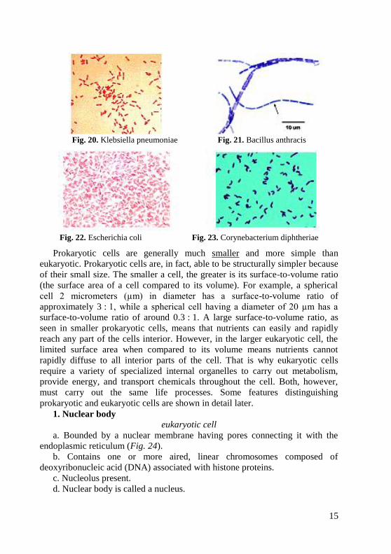
According to their arrangement, cylindrical forms can be subdivided into
three groups (1) diplobacilli occurring in pairs (Klebsiella pneumoniae)
(Fig. 20); (2) streptobacilli occurring in chains of different length (causative
agents of anthrax) (Fig. 21), (3) bacilli which are not arranged in a regular
pattern (these comprise the majority of the rod-shaped forms – Escherichia coli
(Fig. 22), Salmonella, Shigella). Some rod-shaped bacteria have pin-head
thickenings at the ends arranged like Chinees or Latin letters (Corynebacterium
diphtheriae – causative agents of diphtheria) (Fig. 23); others form lateral
branchings (Mycobacterium tuberculosis and leprosy).
Cellular organization: prokaryotic and eukaryotic cells. The cell is the basic unit
of life. Based on the organization of their cellular structures, all living cells can be
divided into two groups: prokaryotic and eukaryotic. Animals, plants, fungi, protozoans,
and algae all possess eukaryotic cell types. Only bacteria have prokaryotic cell types.
15
Fig. 20. Klebsiella pneumoniae Fig. 21. Bacillus anthracis
Fig. 22. Escherichia coli Fig. 23. Corynebacterium diphtheriae
Prokaryotic cells are generally much smaller and more simple than
eukaryotic. Prokaryotic cells are, in fact, able to be structurally simpler because
of their small size. The smaller a cell, the greater is its surface-to-volume ratio
(the surface area of a cell compared to its volume). For example, a spherical
cell 2 micrometers (µm) in diameter has a surface-to-volume ratio of
approximately 3 : 1, while a spherical cell having a diameter of 20 µm has a
surface-to-volume ratio of around 0.3 : 1. A large surface-to-volume ratio, as
seen in smaller prokaryotic cells, means that nutrients can easily and rapidly
reach any part of the cells interior. However, in the larger eukaryotic cell, the
limited surface area when compared to its volume means nutrients cannot
rapidly diffuse to all interior parts of the cell. That is why eukaryotic cells
require a variety of specialized internal organelles to carry out metabolism,
provide energy, and transport chemicals throughout the cell. Both, however,
must carry out the same life processes. Some features distinguishing
prokaryotic and eukaryotic cells are shown in detail later.
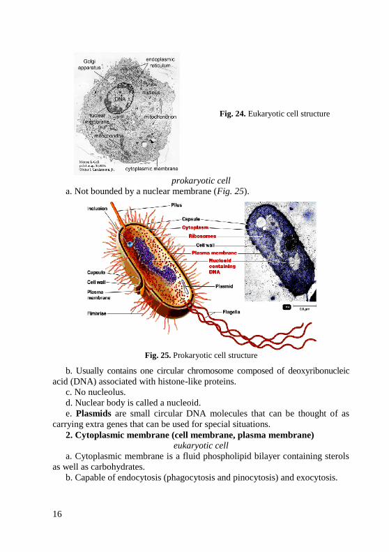
1. Nuclear body
eukaryotic cell
a. Bounded by a nuclear membrane having pores connecting it with the
endoplasmic reticulum (Fig. 24).
b. Contains one or more aired, linear chromosomes composed of
deoxyribonucleic acid (DNA) associated with histone proteins.
c. Nucleolus present.
d. Nuclear body is called a nucleus.
16
Fig. 24. Eukaryotic cell structure
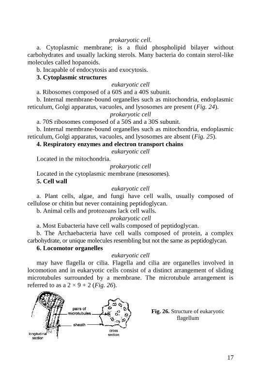
prokaryotic cell
a. Not bounded by a nuclear membrane (Fig. 25).
Fig. 25. Prokaryotic cell structure
b. Usually contains one circular chromosome composed of deoxyribonucleic
acid (DNA) associated with histone-like proteins.
c. No nucleolus.
d. Nuclear body is called a nucleoid.
e. Plasmids are small circular DNA molecules that can be thought of as
carrying extra genes that can be used for special situations.
2. Cytoplasmic membrane (cell membrane, plasma membrane)
eukaryotic cell
a. Cytoplasmic membrane is a fluid phospholipid bilayer containing sterols
as well as carbohydrates.
b. Capable of endocytosis (phagocytosis and pinocytosis) and exocytosis.
17
prokaryotic cell.
a. Cytoplasmic membrane; is a fluid phospholipid bilayer without
carbohydrates and usually lacking sterols. Many bacteria do contain sterol-like
molecules called hopanoids.
b. Incapable of endocytosis and exocytosis.
3. Cytoplasmic structures eukaryotic cell
a. Ribosomes composed of a 60S and a 40S subunit.
b. Internal membrane-bound organelles such as mitochondria, endoplasmic
reticulum, Golgi apparatus, vacuoles, and lysosomes are present (Fig. 24).
prokaryotic cell
a. 70S ribosomes composed of a 50S and a 30S subunit.
b. Internal membrane-bound organelles such as mitochondria, endoplasmic
reticulum, Golgi apparatus, vacuoles, and lysosomes are absent (Fig. 25).
4. Respiratory enzymes and electron transport chains eukaryotic cell
Located in the mitochondria.
prokaryotic cell
Located in the cytoplasmic membrane (mesosomes).
5. Cell wall eukaryotic cell
a. Plant cells, algae, and fungi have cell walls, usually composed of
cellulose or chitin but never containing peptidoglycan.
b. Animal cells and protozoans lack cell walls.
prokaryotic cell
a. Most Eubacteria have cell walls composed of peptidoglycan.
b. The Archaebacteria have cell walls composed of protein, a complex
carbohydrate, or unique molecules resembling but not the same as peptidoglycan.
6. Locomotor organelles eukaryotic cell
may have flagella or cilia. Flagella and cilia are organelles involved in
locomotion and in eukaryotic cells consist of a distinct arrangement of sliding
microtubules surrounded by a membrane. The microtubule arrangement is
referred to as a 2 × 9 + 2 (Fig. 26).
Fig. 26. Structure of eukaryotic
flagellum

18
prokaryotic cell
Some have flagella, each composed of a single, rotating fibril and not
surrounded by a membrane (Fig. 27). No cilia.
Bacteria often store reserve materials
in the form of insoluble cytoplasmic
granules, which are deposited as
osmotically inert, neutral polymers.
Many bacteria accumulate reserves of
inorganic phosphate as granules of
polymerized metaphosphate, called
volutin. Volutin granules are also called
metachromatic granules because they
stain red with a blue dye. They are
characteristic features of corynebacteria.
The bacterial cell wall is a unique structure which surrounds the cell
membrane. Structurally, the wall is necessary for:
Maintaining the cell's characteristic shape – the rigid wall compensates
for the flexibility of the phospholipid membrane and keeps the cell from
assuming a spherical shape.
Countering the effects of osmotic pressure – the strength of the wall is
responsible for keeping the cell from bursting when the intracellular osmolarity
is much greater than the extracellular osmolarity.
Gram’s method of staining. An important taxonomic characteristic of
bacteria is their response to Gram's stain. The Gram’s stain is the most widely
used staining procedure in bacteriology. It is called a differential stain since it
differentiates between Gram-positive and Gram-negative bacteria. Bacteria,
which stain purple with the Gram staining procedure are termed Gram-
positive; those which stain pink are said to be Gram-negative. The terms
positive and negative have nothing to do with electrical charge, but simply
designate two distinct morphological groups of bacteria.
The Gram-staining property appears to be a fundamental one, since the
Gram reaction is correlated with many other morphologic properties in
phylogenetically related forms.
Gram-positive and Gram-negative bacteria stain differently because of
fundamental differences in the structure of their cell walls. The bacterial cell wall
serves to give the organism its size and shape as well as to prevent osmotic lysis. The
material in the bacterial cell wall which confers rigidity is peptidoglycan.
The Gram-staining procedure begins with the application of a basic dye,
crystal (gentian) violet. A solution of iodine is then applied; all bacteria will be
stained blue at this point in the procedure. The cells are then treated with
alcohol. Gram-positive cells retain the crystal violet-iodine complex, remaining
Fig. 27. Insertion structure of bacterial
flagellum

19
blue; Gram-negative cells are completely decolorized by alcohol. As a last step,
a counter stain such as the red dye fuchsine is applied so that the decolorized
Gram-negative cells will take on a contrasting pink color; the Gram-positive
cells now appear purple.
The cell walls of all bacteria are not identical. In fact, cell wall composition
is one of the most important factors in bacterial species analysis and
differentiation. There are two major types of walls: Gram-positive and Gram-
negative. The cell wall of Gram-positive bacteria consists of many polymer
layers of peptidoglycan connected by amino acid bridges. The peptidoglycan
polymer is composed of an alternating sequence of N-acetylglucosamine and
N-acetyl-muraminic acid. Each peptidoglycan layer is connected, or
crosslinked, to the other by a bridge made of amino acids and amino acid
derivatives. Also, 90 % of the Gram-positive cell wall is comprised of
peptidoglycan. In electron micrographs, the Gram-positive cell wall appears as
a broad, dense wall 20–80 nm thick and consisting of numerous interconnecting
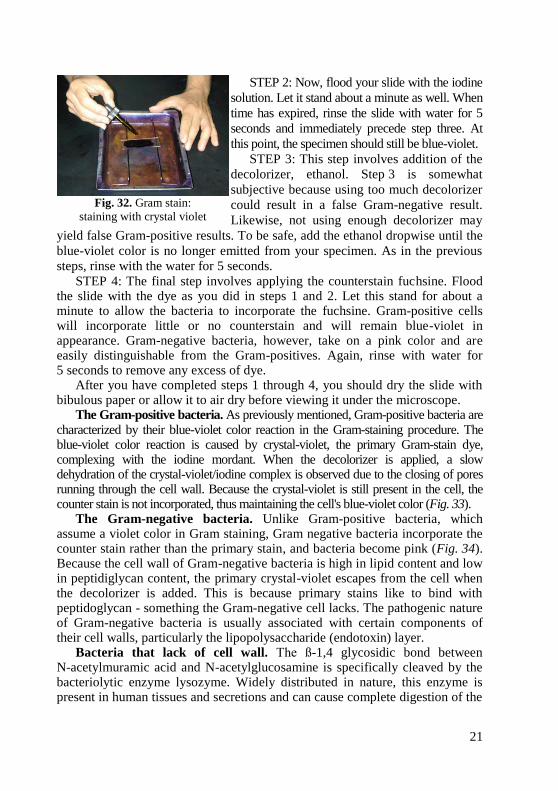
layers of peptidoglycan (Figs. 28). Interwoven in the cell wall of Gram-positive
are teichoic acids. Teichoic acids, which extend through and beyond the rest of
the cell wall, are composed of polymers of glycerol, phosphates, and the sugar
alcohol ribitol. Some have a lipid attached (lipoteichoic acid) (Fig. 30). The
outer surface of the peptidoglycan is studded with proteins that differ with the
strain and species of the bacterium.
Fig. 28. Electron micrograph
of a Gram-positive cell wall
Common Gram-positive bacteria of medical importance include
Streptococcus pyogenes, Streptococcus pneumoniae, Staphylococcus aureus,
Enterococcus faecalis, Bacillus anthracis, and Clostridium species.
The Gram-negative cell wall, on the other hand, contains only 2–3 layers
of peptidoglycan (Fig. 29). The cell wall of Gram-negative bacteria is much
thinner, being comprised of only 20 % peptidoglycan. Gram-negative bacteria
also have two unique regions which surround the outer plasma membrane: the
periplasmic space and the lipopolysaccharide layer (Fig. 31). The periplasmic
space separates the outer plasma membrane from the peptidoglycan layer. It
contains binding proteins and proteins which destroy potentially dangerous
foreign matter present in this space. The lipopolysaccharide (LPS) layer is
located adjacent to the exterior peptidoglycan layer. It is a phospholipid bilayer
construction similar to that in the cell membrane and is attached to the
peptidoglycan by lipoproteins. The lipid portion of the LPS contains a toxic
20
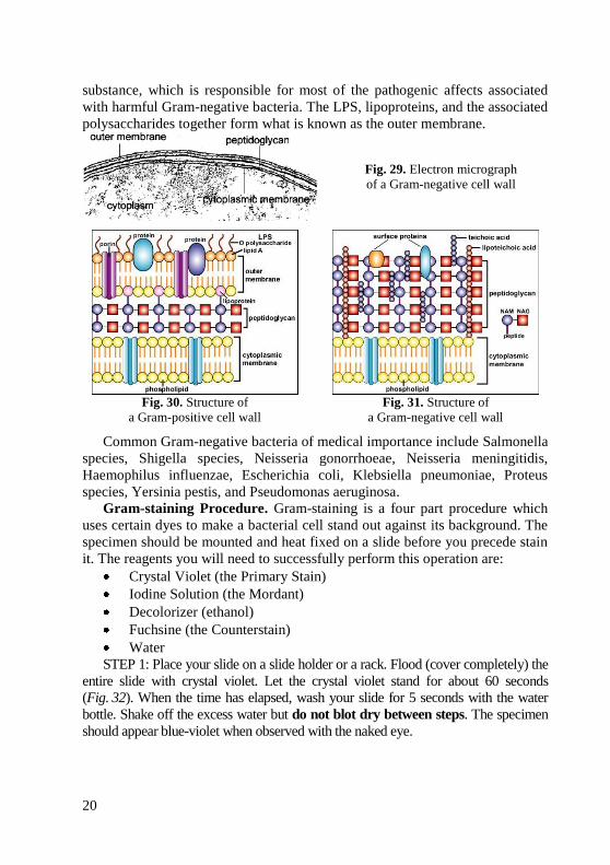
substance, which is responsible for most of the pathogenic affects associated
with harmful Gram-negative bacteria. The LPS, lipoproteins, and the associated
polysaccharides together form what is known as the outer membrane.
Fig. 29. Electron micrograph
of a Gram-negative cell wall
Fig. 30. Structure of
a Gram-positive cell wall Fig. 31. Structure of
a Gram-negative cell wall
Common Gram-negative bacteria of medical importance include Salmonella
species, Shigella species, Neisseria gonorrhoeae, Neisseria meningitidis,
Haemophilus influenzae, Escherichia coli, Klebsiella pneumoniae, Proteus
species, Yersinia pestis, and Pseudomonas aeruginosa.
Gram-staining Procedure. Gram-staining is a four part procedure which
uses certain dyes to make a bacterial cell stand out against its background. The
specimen should be mounted and heat fixed on a slide before you precede stain
it. The reagents you will need to successfully perform this operation are:
Crystal Violet (the Primary Stain)
Iodine Solution (the Mordant)
Decolorizer (ethanol)
Fuchsine (the Counterstain)
Water
STEP 1: Place your slide on a slide holder or a rack. Flood (cover completely) the
entire slide with crystal violet. Let the crystal violet stand for about 60 seconds
(Fig. 32). When the time has elapsed, wash your slide for 5 seconds with the water
bottle. Shake off the excess water but do not blot dry between steps. The specimen
should appear blue-violet when observed with the naked eye.
21
STEP 2: Now, flood your slide with the iodine
solution. Let it stand about a minute as well. When
time has expired, rinse the slide with water for 5
seconds and immediately precede step three. At
this point, the specimen should still be blue-violet.
STEP 3: This step involves addition of the
decolorizer, ethanol. Step 3 is somewhat
subjective because using too much decolorizer
could result in a false Gram-negative result.
Likewise, not using enough decolorizer may
yield false Gram-positive results. To be safe, add the ethanol dropwise until the
blue-violet color is no longer emitted from your specimen. As in the previous
steps, rinse with the water for 5 seconds.
STEP 4: The final step involves applying the counterstain fuchsine. Flood the slide with the dye as you did in steps 1 and 2. Let this stand for about a minute to allow the bacteria to incorporate the fuchsine. Gram-positive cells will incorporate little or no counterstain and will remain blue-violet in appearance. Gram-negative bacteria, however, take on a pink color and are easily distinguishable from the Gram-positives. Again, rinse with water for 5 seconds to remove any excess of dye.
After you have completed steps 1 through 4, you should dry the slide with bibulous paper or allow it to air dry before viewing it under the microscope.
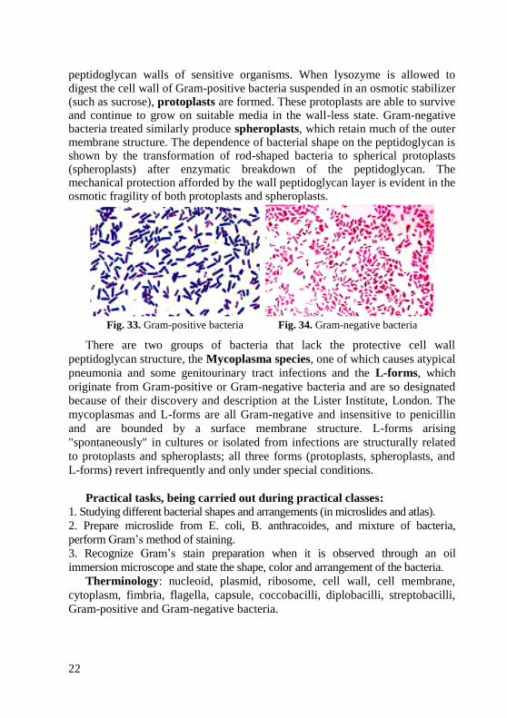
The Gram-positive bacteria. As previously mentioned, Gram-positive bacteria are characterized by their blue-violet color reaction in the Gram-staining procedure. The blue-violet color reaction is caused by crystal-violet, the primary Gram-stain dye, complexing with the iodine mordant. When the decolorizer is applied, a slow dehydration of the crystal-violet/iodine complex is observed due to the closing of pores running through the cell wall. Because the crystal-violet is still present in the cell, the counter stain is not incorporated, thus maintaining the cell's blue-violet color (Fig. 33).
The Gram-negative bacteria. Unlike Gram-positive bacteria, which assume a violet color in Gram staining, Gram negative bacteria incorporate the counter stain rather than the primary stain, and bacteria become pink (Fig. 34). Because the cell wall of Gram-negative bacteria is high in lipid content and low in peptidiglycan content, the primary crystal-violet escapes from the cell when the decolorizer is added. This is because primary stains like to bind with peptidoglycan - something the Gram-negative cell lacks. The pathogenic nature of Gram-negative bacteria is usually associated with certain components of their cell walls, particularly the lipopolysaccharide (endotoxin) layer.
Bacteria that lack of cell wall. The ß-1,4 glycosidic bond between N-acetylmuramic acid and N-acetylglucosamine is specifically cleaved by the bacteriolytic enzyme lysozyme. Widely distributed in nature, this enzyme is present in human tissues and secretions and can cause complete digestion of the
Fig. 32. Gram stain:
staining with crystal violet
22
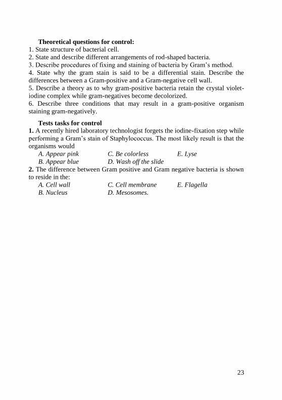
peptidoglycan walls of sensitive organisms. When lysozyme is allowed to digest the cell wall of Gram-positive bacteria suspended in an osmotic stabilizer (such as sucrose), protoplasts are formed. These protoplasts are able to survive and continue to grow on suitable media in the wall-less state. Gram-negative bacteria treated similarly produce spheroplasts, which retain much of the outer membrane structure. The dependence of bacterial shape on the peptidoglycan is shown by the transformation of rod-shaped bacteria to spherical protoplasts (spheroplasts) after enzymatic breakdown of the peptidoglycan. The mechanical protection afforded by the wall peptidoglycan layer is evident in the osmotic fragility of both protoplasts and spheroplasts.
Fig. 33. Gram-positive bacteria Fig. 34. Gram-negative bacteria
There are two groups of bacteria that lack the protective cell wall
peptidoglycan structure, the Mycoplasma species, one of which causes atypical
pneumonia and some genitourinary tract infections and the L-forms, which
originate from Gram-positive or Gram-negative bacteria and are so designated
because of their discovery and description at the Lister Institute, London. The
mycoplasmas and L-forms are all Gram-negative and insensitive to penicillin
and are bounded by a surface membrane structure. L-forms arising
"spontaneously" in cultures or isolated from infections are structurally related
to protoplasts and spheroplasts; all three forms (protoplasts, spheroplasts, and
L-forms) revert infrequently and only under special conditions.
Practical tasks, being carried out during practical classes:
1. Studying different bacterial shapes and arrangements (in microslides and atlas).
2. Prepare microslide from E. coli, B. anthracoides, and mixture of bacteria,
perform Gram’s method of staining.
3. Recognize Gram’s stain preparation when it is observed through an oil
immersion microscope and state the shape, color and arrangement of the bacteria.
Therminology: nucleoid, plasmid, ribosome, cell wall, cell membrane,
cytoplasm, fimbria, flagella, capsule, coccobacilli, diplobacilli, streptobacilli,
Gram-positive and Gram-negative bacteria.
23
Theoretical questions for control:
1. State structure of bacterial cell.
2. State and describe different arrangements of rod-shaped bacteria.
3. Describe procedures of fixing and staining of bacteria by Gram’s method.
4. State why the gram stain is said to be a differential stain. Describe the
differences between a Gram-positive and a Gram-negative cell wall.
5. Describe a theory as to why gram-positive bacteria retain the crystal violet-
iodine complex while gram-negatives become decolorized.
6. Describe three conditions that may result in a gram-positive organism
staining gram-negatively.
Tests tasks for control
1. A recently hired laboratory technologist forgets the iodine-fixation step while
performing a Gram’s stain of Staphylococcus. The most likely result is that the
organisms would
A. Appear pink C. Be colorless E. Lyse
B. Appear blue D. Wash off the slide
2. The difference between Gram positive and Gram negative bacteria is shown
to reside in the:
A. Cell wall C. Cell membrane E. Flagella
B. Nucleus D. Mesosomes.
24
MORPHOLOGY OF SPIRAL BACTERIA. METHODS OF EXAMINATION OF FLAGELLA
Goal: Studying of morphology of spiral bacteria by microscopic examination.
Concrete goals:
1. Study of arrangement of spiral bacteria in microslides.
2. Study of method of detection of motility of bacteria.
3. Study of structure of bacterial flagella.
Students should be able to:
1. Perform Gram’s method of staining of microslide.
2. Using a microscope, identify different bacterial shapes and arrangements.
Equipment: museum cultures of bacteria,
bacteriological loops, gas burners, gentian
violet, fuchsine, alcohol, water, immersion
microscopes, immersion oil, tables, atlas.
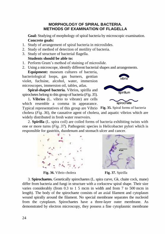
Spiral-shaped bacteria. Vibrios, spirilla and
spirochetes belong to this group of bacteria (Fig. 35).
1. Vibrios (L. vibrio to vibrate) are cells
which resemble a comma in appearance.
Typical representatives of this group are Vibrio
cholera (Fig. 36), the causative agent of cholera, and aquatic vibrios which are
widely distributed in fresh water reservoirs.
2. Spirilla (L. spira coil) are coiled forms of bacteria exhibiting twists with
one or more turns (Fig. 37). Pathogenic species is Helicobacter pylori which is
responsible for gastritis, duodenum and stomach ulcer and cancer.
Fig. 36. Vibrio cholera Fig. 37. Spirilla
3. Spirochaetes. Genetically spirochaetes (L. spira curve, Gk. chaite cock, mane)
differ from bacteria and fungi in structure with a corkscrew spiral shape. Their size
varies considerably (from 0.3 to 1 5 mcm in width and from 7 to 500 mcm in
length). The body of the spirochaete consists of an axial filament and cytoplasm
wound spirally around the filament. No special membrane separates the nucleoid
from the cytoplasm. Spirochaetes have a three-layer outer membrane. As
demonstrated by electron microscopy, they possess a fine cytoplasmic membrane
Fig. 35. Spiral forms of bacteria

25
enclosing the cytoplasm. The spirochaetes do not possess the cell wall characteristic
of bacteria, but electron microscopy has revealed that they have a thin cell wall
(periplast) which encloses the cytoplasm.
In spite of the absence of flagella, spirochaetes are actively motile due to the
distinct flexibility of their bodies. Spirochaetes have a rotating motion which is
performed axially, a translational motion forwards and backwards, an
undulating motion along the whole body of the microorganism, and a bending
motion when the body bends at a certain angle.
Classification of Spirochaetes. The order Spirochaetales, family
Spirochaetaceae includes two pathogenic genera (Borrelia, Treponema), and
one belong to family Leptospiraceae (Leptospira) (Fig. 38).
The organisms of genus Borrelia
differ from spirochaetes in that their cells
have large, obtuse-angled, irregular spirals,
the number of which varies from 3 to 10
(Fig. 38). Pathogenic for man are the
causative agents of relapsing fever trans-
mitted by lice (Borrelia hispanica), and by
ticks (Borrelia persica, etc.), causative
agent of Lime disease (B. burgdorferi).
These stain blue-violet with the
Romanowsky–Giemsa stain (Fig. 39).
The genus Treponema (Gk. trepein turn, nema thread) exhibits thin,
flexible cells with 6–14 twists (Fig. 38). The microorganisms do not appear to
have a visible axial filament or an axial crest when viewed under the
microscope. The ends of treponemas are either tapered or rounded, some
species have thin elongated threads on the poles. The organisms stain pale-pink
with the Romanowsky–Giemsa stain (Fig. 40). Burry stain with Indian ink
(negative staining) is more suitable (Fig. 41). A typical representative is the
causative agent of syphilis Treponema pallidum.
Fig. 39. Borrelia in blood.
Romanowsky–Giemsa stain Fig. 40. Treponema pallidum.
Romanowsky–Giemsa stain
Fig. 38. Morphology of spirochaetes
26
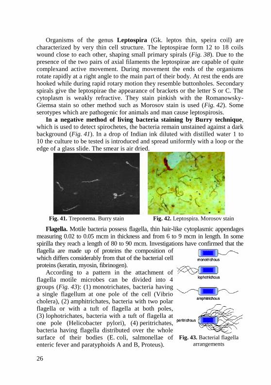
Organisms of the genus Leptospira (Gk. leptos thin, speira coil) are characterized by very thin cell structure. The leptospirae form 12 to 18 coils wound close to each other, shaping small primary spirals (Fig. 38). Due to the presence of the two pairs of axial filaments the leptospirae are capable of quite complexand active movement. During movement the ends of the organisms rotate rapidly at a right angle to the main part of their body. At rest the ends are hooked while during rapid rotary motion they resemble buttonholes. Secondary spirals give the leptospirae the appearance of brackets or the letter S or C. The cytoplasm is weakly refractive. They stain pinkish with the Romanowsky-Giemsa stain so other method such as Morosov stain is used (Fig. 42). Some serotypes which are pathogenic for animals and man cause leptospirosis.
In a negative method of living bacteria staining by Burry technique, which is used to detect spirochetes, the bacteria remain unstained against a dark background (Fig. 41). In a drop of Indian ink diluted with distilled water 1 to 10 the culture to be tested is introduced and spread uniformly with a loop or the edge of a glass slide. The smear is air dried.
Fig. 41. Treponema. Burry stain Fig. 42. Leptospira. Morosov stain
Flagella. Motile bacteria possess flagella, thin hair-like cytoplasmic appendages measuring 0.02 to 0.05 mcm in thickness and from 6 to 9 mcm in length. In some spirilla they reach a length of 80 to 90 mcm. Investigations have confirmed that the flagella are made up of proteins the composition of which differs considerably from that of the bacterial cell proteins (keratin, myosin, fibrinogen).
According to a pattern in the attachment of flagella motile microbes can be divided into 4 groups (Fig. 43): (1) monotrichates, bacteria having a single flagellum at one pole of the cell (Vibrio cholera), (2) amphitrichates, bacteria with two polar flagella or with a tuft of flagella at both poles, (3) lophotrichates, bacteria with a tuft of flagella at one pole (Helicobacter pylori), (4) peritrichates, bacteria having flagella distributed over the whole surface of their bodies (E. coli, salmonellae of enteric fever and paratyphoids A and B, Proteus).
Fig. 43. Bacterial flagella
arrangements
27
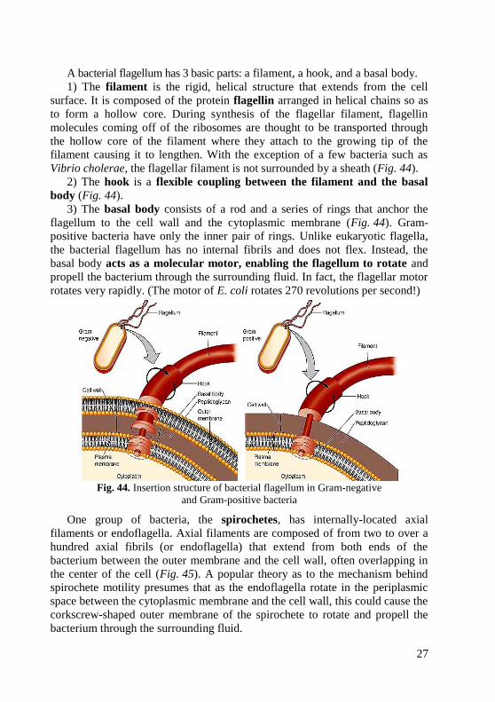
A bacterial flagellum has 3 basic parts: a filament, a hook, and a basal body.
1) The filament is the rigid, helical structure that extends from the cell
surface. It is composed of the protein flagellin arranged in helical chains so as
to form a hollow core. During synthesis of the flagellar filament, flagellin
molecules coming off of the ribosomes are thought to be transported through
the hollow core of the filament where they attach to the growing tip of the
filament causing it to lengthen. With the exception of a few bacteria such as
Vibrio cholerae, the flagellar filament is not surrounded by a sheath (Fig. 44).
2) The hook is a flexible coupling between the filament and the basal
body (Fig. 44).
3) The basal body consists of a rod and a series of rings that anchor the
flagellum to the cell wall and the cytoplasmic membrane (Fig. 44). Gram-
positive bacteria have only the inner pair of rings. Unlike eukaryotic flagella,
the bacterial flagellum has no internal fibrils and does not flex. Instead, the
basal body acts as a molecular motor, enabling the flagellum to rotate and
propell the bacterium through the surrounding fluid. In fact, the flagellar motor
rotates very rapidly. (The motor of E. coli rotates 270 revolutions per second!)
Fig. 44. Insertion structure of bacterial flagellum in Gram-negative
and Gram-positive bacteria
One group of bacteria, the spirochetes, has internally-located axial
filaments or endoflagella. Axial filaments are composed of from two to over a
hundred axial fibrils (or endoflagella) that extend from both ends of the
bacterium between the outer membrane and the cell wall, often overlapping in
the center of the cell (Fig. 45). A popular theory as to the mechanism behind
spirochete motility presumes that as the endoflagella rotate in the periplasmic
space between the cytoplasmic membrane and the cell wall, this could cause the
corkscrew-shaped outer membrane of the spirochete to rotate and propell the
bacterium through the surrounding fluid.

28
Fig. 45. Spirochete axial filaments
Function: Flagella are the organelles of locomotion for most of the bacteria
that are capable of motility. Two proteins in the flagellar motor, called MotA
and MotB, form a proton channel through the cytoplasmic membrane and
rotation of the flagellum is driven by a proton gradient. This driving proton
motive force occurs as protons accumulating in the space between the
cytoplasmic membrane and the cell wall as a result of the electron transport
system travel through the channel back into the bacterium's cytoplasm.
The bacterial flagellum can rotate both
counterclockwise and clockwise (Fig. 46).
This is controlled by a protein switch in
the molecular motor of the basal body.
Clockwise rotation results in a tumbling
motion and changes the direction of
bacterial movement. On the other hand,
counterclockwise rotation leads to long,
straight or curved runs without a change
in direction. During a run, that lasts
about one second, the bacterium moves
10–20 times its length before it stops. In
the case of a tumble, the movement lasts
only about one-tenth of a second and no
real forward progress is made.
Around half of all known bacteria are
motile. Motility serves to keep bacteria in
an optimum environment via taxis.
Taxis is a motile response to an environmental stimulus. Bacteria can
respond to chemicals (chemotaxis), light (phototaxis), osmotic pressure
(osmotaxis), oxygen (aerotaxis), and temperature (thermotaxis).
Chemotaxis is a response to a chemical gradient of attractant or repellent molecules
in the bacterium's environment. In an environment that lacks such a gradient, the
bacterium moves randomly. It travels in a straight line, or runs, for a few seconds, then
stops, tumbles, and runs in a different direction. However, when the bacterium is
Fig. 46. Flagella and bacterial motility.
Notice that direction of flagellar rotation
determines which of these movements
occurs. Arrows indicate direction
of movement
29
exposed to a chemical gradient of, for example, an attractant, it tumbles less frequently
(has longer runs) as it moves up the gradient, but tumbles at the normal rate if it travels
down the gradient. In this way, the net movement is towards a more optimum
environment. Chemotaxis is regulated by chemoreceptors located in the cytoplasmic
membrane or periplasm of the bacterium bind chemical attractants or repellents. An
increasing concentration of attractant or decreasing concentration of repellent (both
conditions beneficial) causes less tumbling and longer runs; a decreasing concentration
of attractant or increasing concentration of repellent (both conditions harmful) causes
normal tumbling and a greater chance of reorienting in a "better" direction. As a result,
the organism's net movement is toward the optimum environment.
Motility and chemotaxis probably help some intestinal pathogens to move
through the mucous layer so they can attach to the epithelial cells of the
mucous membranes. They also enable spirochetes to move through viscous
environments and penetrate cell membranes.
The flagella can be observed by the following three methods: 1) direct
observation by means of special-purpose microscopes (phase-contrast and dark-
field), and by electron microscopy, 2) motility media, 3) flagella staining by
special methods involving treatment with mordants, adsorption of various
substances and dyes on their surfaces.
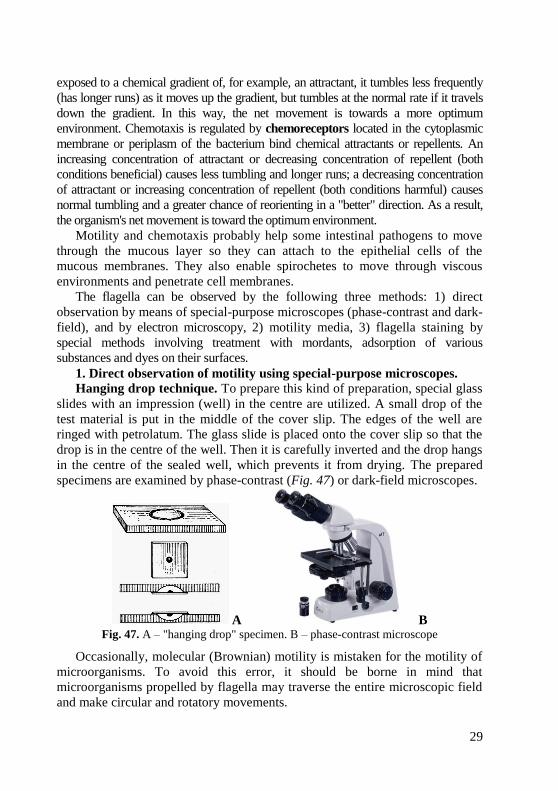
1. Direct observation of motility using special-purpose microscopes.
Hanging drop technique. To prepare this kind of preparation, special glass
slides with an impression (well) in the centre are utilized. A small drop of the
test material is put in the middle of the cover slip. The edges of the well are
ringed with petrolatum. The glass slide is placed onto the cover slip so that the
drop is in the centre of the well. Then it is carefully inverted and the drop hangs
in the centre of the sealed well, which prevents it from drying. The prepared
specimens are examined by phase-contrast (Fig. 47) or dark-field microscopes.
A B Fig. 47. A – "hanging drop" specimen. B – phase-contrast microscope
Occasionally, molecular (Brownian) motility is mistaken for the motility of
microorganisms. To avoid this error, it should be borne in mind that
microorganisms propelled by flagella may traverse the entire microscopic field
and make circular and rotatory movements.
30
Phase-contrast microscope uses special phase-contrast objectives and a
condenser assembly to control illumination and give an optical effect of direct
staining. The special optics convert slight variations in specimen thickness into
corresponding visible variation in brightness. Thus, the bacterium and its
structures appear darker than the background. Phase-contrast microscope is
based on the fact that the optical length of the light travelling in any substance
depends on its refractive index. Light waves transversing through optically
denser sites of the object lag in their phase behind the light waves which do not
have to pass through these sites. The intensity of light in this case remains
unaltered but the phase of fluctuation, detected by neither eye nor photoplate, is
changed. To increase resolution of the image, the objective is fitted with a
special semi-transparent phase plate to create difference in the wave length
between the rays of the background and the object. If this difference reaches
one-fourth of the wave length, a visually tangible effect occurs when a dark
object is clearly seen against a light background (positive contrast) or vice
versa (negative contrast) depending on the structure of the phase plate.
Phase-contrast microscopy does not enhance the resolving power of the
optical system but helps to elucidate new details of the structure of living
microorganisms and to study different stages of their development, the effect
on them of chemical agents, antibiotics, and other factors.
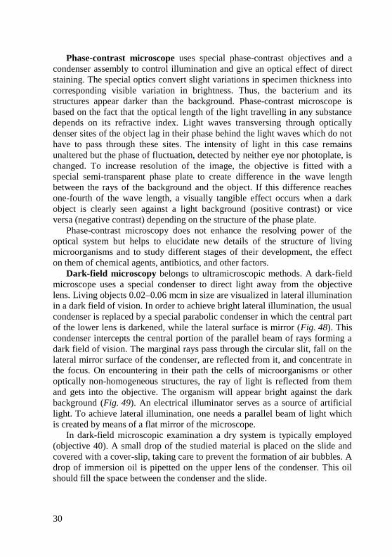
Dark-field microscopy belongs to ultramicroscopic methods. A dark-field
microscope uses a special condenser to direct light away from the objective
lens. Living objects 0.02–0.06 mcm in size are visualized in lateral illumination
in a dark field of vision. In order to achieve bright lateral illumination, the usual
condenser is replaced by a special parabolic condenser in which the central part
of the lower lens is darkened, while the lateral surface is mirror (Fig. 48). This
condenser intercepts the central portion of the parallel beam of rays forming a
dark field of vision. The marginal rays pass through the circular slit, fall on the
lateral mirror surface of the condenser, are reflected from it, and concentrate in
the focus. On encountering in their path the cells of microorganisms or other
optically non-homogeneous structures, the ray of light is reflected from them
and gets into the objective. The organism will appear bright against the dark
background (Fig. 49). An electrical illuminator serves as a source of artificial
light. To achieve lateral illumination, one needs a parallel beam of light which
is created by means of a flat mirror of the microscope.
In dark-field microscopic examination a dry system is typically employed
(objective 40). A small drop of the studied material is placed on the slide and
covered with a cover-slip, taking care to prevent the formation of air bubbles. A
drop of immersion oil is pipetted on the upper lens of the condenser. This oil
should fill the space between the condenser and the slide.
31
Fig. 48. Diagram of a dark-field microscope
shows the path of light. The dark-field ring
in the condenser blocks the direct passage
of light through the specimen and into the
objective lens. Only light that is reflected
off a specimen will enter the objective lens
and be seen
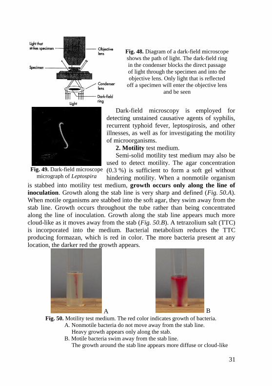
Dark-field microscopy is employed for
detecting unstained causative agents of syphilis,
recurrent typhoid fever, leptospirosis, and other
illnesses, as well as for investigating the motility
of microorganisms.
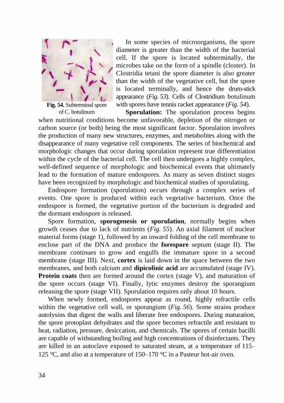
2. Motility test medium.
Semi-solid motility test medium may also be
used to detect motility. The agar concentration
(0.3 %) is sufficient to form a soft gel without
hindering motility. When a nonmotile organism
is stabbed into motility test medium, growth occurs only along the line of
inoculation. Growth along the stab line is very sharp and defined (Fig. 50.A).
When motile organisms are stabbed into the soft agar, they swim away from the
stab line. Growth occurs throughout the tube rather than being concentrated
along the line of inoculation. Growth along the stab line appears much more
cloud-like as it moves away from the stab (Fig. 50.B). A tetrazolium salt (TTC)
is incorporated into the medium. Bacterial metabolism reduces the TTC
producing formazan, which is red in color. The more bacteria present at any
location, the darker red the growth appears.
A B
Fig. 50. Motility test medium. The red color indicates growth of bacteria.
A. Nonmotile bacteria do not move away from the stab line.
Heavy growth appears only along the stab.
B. Motile bacteria swim away from the stab line.
The growth around the stab line appears more diffuse or cloud-like
Fig. 49. Dark-field microscope
micrograph of Leptospira

32
3. Flagella staining (Loeffler stain) can
be used indirectly to denote bacterial motility.
Since flagella are very thin (20–28 nm in
diameter), they are below the resolution
limits of a normal light microscope and
cannot be seen unless one first treats
them with special dyes and mordants
which build up as layers of precipitate
along the length of the flagella, making
them microscopically visible. This is a
delicate staining procedure.
Practical tasks, being carried out during practical classes:
1. Studying different bacterial shapes and arrangements (in microslides and atlas).
2. Prepare microslide from V. cholerae, perform Gram’s method of staining.
3. Recognize a direct stain preparation when it is observed through an oil
immersion microscope and state the shape and arrangement of the bacteria.
4. Observe the phase-contrast microscopy demonstration of motile Vibrio.
5. Observe the flagella stain demonstrations of a Vibrio species, Proteus vulgaris.
Therminology: spirochetes, spirilla, vibrio, flagella, dark-field microscope,
phase-contrast microscope, hanging drop specimen.
Theoretical questions for control:
1. State three basic shapes of spiral bacteria.
2. Define the following: Giemsa stain, Burry stain, Morosov stain, mordants.
3. State in the principle of dark-field microscope and phase-contrast microscope.
4. Describe procedures of hanging drop preparation.
5. Interpret the results of motility test medium.
6. Recognize monotrichous, lophotrichous, amphitrichous, and peritrichous
flagellar arrangements.
Test tasks for control:
1. Bacterioscopic examination of chancre material revealed some mobile, long,
convoluted microorganisms with 8-12 regular coils. These features are typical for:
A. Vibrios C. Leptospira E. Treponema
B. Borrellia D. Campylobacter
2. Negative staining is especially useful in demonstration:
A. Vibrios C. Neisseria E. Staphylococcus
B. Spirochetes D. Clostridia
Fig. 51. Vibrio cholerae. Loeffler stain

33
SPORES. METHODS OF SPORE STATINING
Goal: Studying of morphology of spore-forming bacteria by microscopic
examination.
Concrete goals:
1. Study of structure and arrangement of bacterial spores in microslides.
2. Study of methods of bacterial spores detection.
Students should be able to:
1. Perform Ozeshko method of staining.
2. Using a microscope, identify bacterial shapes and spore arrangements.
Equipment: museum cultures of bacteria, bacteriological loops, gas
burners, carbol fuchsin, 5 % H2SO4, methylene blue, water, immersion
microscopes, immersion oil, tables, atlas.
Endospores. Endospores are highly heat-resistant, dehydrated resting cells
formed intracellularly in members of the genera Bacillus and Clostridium.
Endospores are small spherical or oval bodies formed within the cell. These
include the causative agents of anthrax (Bacillus anthracis), tetanus
(Clostridium tetani), anaerobic infections-gas gangrene (C.perfringens,
C.septicum, C.hystolyticum, C.novyi), botulism (C.botulinum),
pseudomembranous colitis (C.difficile) and also saprophytic species living in
the soil, water and bodies of animals. Spore formation only rarely occurs in
cocci (Sarcina lutea, Sarcina ureae) and in spiral forms (Desulfovibrio
desulfuricans). Sporulation occurs in the environment (in soil and on nutrient
media), and is not observed in human or animal tissues.
In bacteria spores are located (1) centrally, in the centre of the cell
(causative agent of anthrax) (Fig. 52); (2) terminally, at the ends of the rod
(causative agent of tetanus) (Fig. 53); (3) subterminally, towards the ends
(causative agents of botulism, anaerobic infections, etc.) (Fig. 54).
Fig. 52. Central spore of B. anthracis.
Note the endospores within the
streptobacillus
Fig. 53. Terminal spore of C.tetani
34
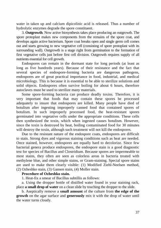
In some species of microorganisms, the spore
diameter is greater than the width of the bacterial
cell. If the spore is located subterminally, the
microbes take on the form of a spindle (closter). In
Clostridia tetani the spore diameter is also greater
than the width of the vegetative cell, but the spore
is located terminally, and hence the drum-stick
appearance (Fig. 53). Cells of Clostridium botulinum
with spores have tennis racket appearance (Fig. 54).
Sporulation: The sporulation process begins
when nutritional conditions become unfavorable, depletion of the nitrogen or
carbon source (or both) being the most significant factor. Sporulation involves
the production of many new structures, enzymes, and metabolites along with the
disappearance of many vegetative cell components. The series of biochemical and
morphologic changes that occur during sporulation represent true differentiation
within the cycle of the bacterial cell. The cell then undergoes a highly complex,
well-defined sequence of morphologic and biochemical events that ultimately
lead to the formation of mature endospores. As many as seven distinct stages
have been recognized by morphologic and biochemical studies of sporulating.
Endospore formation (sporulation) occurs through a complex series of
events. One spore is produced within each vegetative bacterium. Once the
endospore is formed, the vegetative portion of the bacterium is degraded and
the dormant endospore is released.
Spore formation, sporogenesis or sporulation, normally begins when
growth ceases due to lack of nutrients (Fig. 55). An axial filament of nuclear
material forms (stage 1), followed by an inward folding of the cell membrane to
enclose part of the DNA and produce the forespore septum (stage II). The
membrane continues to grow and engulfs the immature spore in a second
membrane (stage III). Next, cortex is laid down in the space between the two
membranes, and both calcium and dipicolinic acid are accumulated (stage IV).
Protein coats then are formed around the cortex (stage V), and maturation of
the spore occurs (stage VI). Finally, lytic enzymes destroy the sporangium
releasing the spore (stage VII). Sporulation requires only about 10 hours.
When newly formed, endospores appear as round, highly refractile cells
within the vegetative cell wall, or sporangium (Fig. 56). Some strains produce
autolysins that digest the walls and liberate free endospores. During maturation,
the spore protoplast dehydrates and the spore becomes refractile and resistant to
heat, radiation, pressure, desiccation, and chemicals. The spores of certain bacilli
are capable of withstanding boiling and high concentrations of disinfectants. They
are killed in an autoclave exposed to saturated steam, at a temperature of 115–
125 C, and also at a temperature of 150–170 C in a Pasteur hot-air oven.
Fig. 54. Subterminal spore
of C. botulinum

35
Fig. 55. Stages of sporulation
The impermeability of the spore coat is thought to be responsible for the
endospore's resistance to chemicals. The heat resistance of endospores is due to
a variety of factors: a) calcium-dipicolinate, abundant within the endospore,
may stabilize and protect the endospore's DNA. As much as 15 % of the spore’s
dry weight consists of dipicolinic acid complexes with calcium ions;
b) specialized DNA-binding proteins saturate the endospore's DNA and protect
it from heat, drying, chemicals, and radiation; c) the cortex may osmotically
remove water from the interior of the endospore and the dehydration that
results is thought to be very important in the endospore's resistance to heat and
radiation; d) DNA repair enzymes contained within the endospore are able to
repair damaged DNA during germination.
Since one vegetative cell gives rise
to one endospore, sporulation is obviously
not a means of cell reproduction. Spores
make survival of bacteria possible
under unfavourable condition.
Structure and properties of
endospores.
1. Core. The spore protoplast, or
core contains a complete nucleoid
(chromosome), ribosomes, and energy
generating components that are
enclosed within a modified cytoplasmic membrane. A number of vegetative
cell enzymes are increased in amount (eg, alanine racemase), and a number of
unique enzymes are formed (eg, dipicolinic acid synthetase). The energy for
germination is stored as 3-phosphoglycerate rather than as ATP. The heat
resistance of spores is due in pan to their dehydrated state and in part to the
presence of large amounts (5–15 % of the spore dry weight) of calcium
dipicolinate, which is formed from an intermediate of the lysine biosynthetic
Fig. 56. Endospore within a vegetative cell

36
pathway. In some way not yet understood, these properties result in the
stabilization of the spore enzymes, most of which exhibit normal heat lability
when isolated in soluble form.
2. Spore wall. The innermost layer surrounding the inner spore membrane
is called the spore wall. It contains normal peptidoglycan and becomes the cell
wall of the germinating vegetative cell.
3. Cortex is the thickest layer of the spore envelope. It contains an unusual
type of peptidoglycan, with many fewer cross-links than are found in cell wall
peptidoglycan. Cortex peptidoglycan is extremely sensitive to lysozyme, and its
autolysis plays a key role in spore germination.
4. Coat is composed of a keratin-like protein containing many
intramolecular disulfide bonds. The impermeability of this layer confers on
spores their relative resistance to antibacterial chemical agents.
5. Exosporium is a lipoprotein membrane containing some carbohydrate.
Germination is a transformation of spore into vegetative cell.
The germination process occurs in 3 stages (Fig. 57):
1. Activation-Even when placed in an environment that favors germination
(eg, a nutritionally rich medium), bacterial spores will not germinate unless first
activated by one or another agent that damages the spore coat. Among the
agents that can overcome spore dormancy are heat (60 to 70 °C for a few
minutes), abrasion, acidity, and compounds containing free sulfhydryl groups.
2. Initiation-Once activated, a spore will initiate germination if the
environmental conditions are favorable. Initiation is characterized by spore
swelling, rupture or absorption of the spore coat, loss of resistance to heat and
stress, release of spore component, increase in metabolic activity. Species of
bacteria after getting activated recognize effectors as signalling a rich medium.
Many normal metabolic nutrients (e.g. amino acids and sugars) can trigger
germination after activation. Binding of the effector substance to spore coat
activates an autolysin, which destroys peptidoglycan of the cortex. After this
Fig. 57. Germination
of spore
37
water in taken up and calcium dipicolinic acid is released. Thus a number of
hydrolytic enzymes degrade the spore constituent.
3. Outgrowth. Now active biosynthesis takes place producing an outgrowth. The
spore protoplast makes new components from the remains of the spore coat, and
develops again active bacterium. Spore coat breaks open and single germ cell comes
out and starts growing to new vegetative cell (consisting of spore protoplast with its
surrounding wall). Outgrowth is a stage right from germination to the formation of
first vegetative cells just before first cell division. Outgrowth requires supply of all
nutrients essential for cell growth.
Endospores can remain in the dormant state for long periods (at least as
long as five hundreds years). Because of their resistance and the fact that
several species of endospore-forming bacteria are dangerous pathogens,
endospores are of great practical importance in food, industrial, and medical
microbiology. This is because it is essential to be able to sterilize solutions and
solid objects. Endospores often survive boiling for about 6 hours, therefore
autoclaves must be used to sterilize many materials.
Some spore-forming bacteria can produce deadly toxins. Therefore, it is
very important that foods that may contain these spores be processed
adequately to insure that endospores are killed. Many people have died of
botulism after ingesting improperly canned food that contained spores of
botulism. In such improperly processed food, the heat-resistant spores
germinated into vegetative cells under the appropriate conditions. These cells
then synthesized the toxin, which when ingested causes botulism. However,
since the toxin is destroyed by heat, boiling contaminated food for 30 minutes
will destroy the toxin, although such treatment will not kill the endospores.
Due to the resistant nature of the endospore coats, endospores are difficult
to stain. Strong dyes and vigorous staining conditions such as heat are needed.
Once stained, however, endospores are equally hard to decolorize. Since few
bacterial genera produce endospores, the endospore stain is a good diagnostic
test for species of Bacillus and Clostridium. Because spores are impermeable to
most stains, they often are seen as colorless areas in bacteria treated with
methylene blue, and other simple stains, or Gram-staining. Special spore stains
are used to make them clearly visible: (1) Modified Ziehl-Neelsen method,
(2) Ozheshko stain, (3) Gansen stain, (4) Moller stain.
Procedure of Ozheshko stain. 1. Heat-fix a smear of Bacillus subtillis as follows:
a. Using the dropper bottle of distilled water found in your staining rack,
place a small drop of water on a clean slide by touching the dropper to the slide.
b. Aseptically remove a small amount of the culture from the edge of the
growth on the agar surface and generously mix it with the drop of water until
the water turns cloudy.
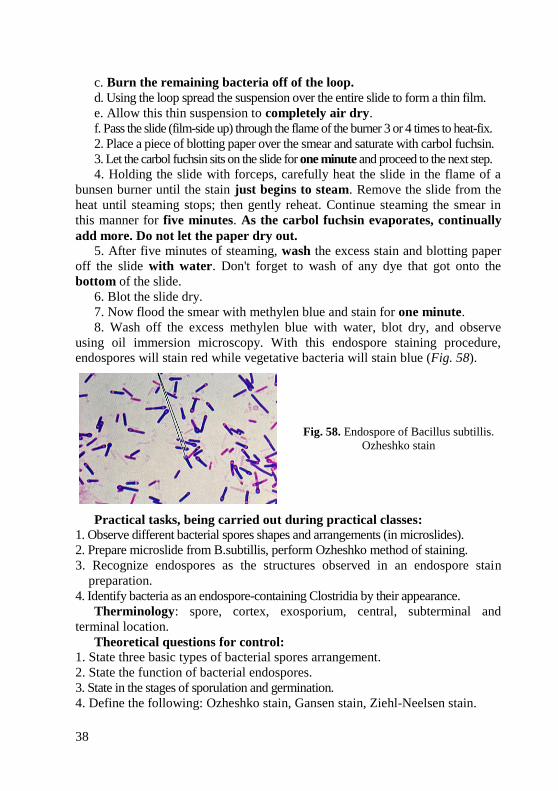
38
c. Burn the remaining bacteria off of the loop.
d. Using the loop spread the suspension over the entire slide to form a thin film.
e. Allow this thin suspension to completely air dry.
f. Pass the slide (film-side up) through the flame of the burner 3 or 4 times to heat-fix.
2. Place a piece of blotting paper over the smear and saturate with carbol fuchsin.
3. Let the carbol fuchsin sits on the slide for one minute and proceed to the next step.
4. Holding the slide with forceps, carefully heat the slide in the flame of a
bunsen burner until the stain just begins to steam. Remove the slide from the
heat until steaming stops; then gently reheat. Continue steaming the smear in
this manner for five minutes. As the carbol fuchsin evaporates, continually
add more. Do not let the paper dry out. 5. After five minutes of steaming, wash the excess stain and blotting paper
off the slide with water. Don't forget to wash of any dye that got onto the
bottom of the slide.
6. Blot the slide dry.
7. Now flood the smear with methylen blue and stain for one minute.
8. Wash off the excess methylen blue with water, blot dry, and observe
using oil immersion microscopy. With this endospore staining procedure,
endospores will stain red while vegetative bacteria will stain blue (Fig. 58).
Fig. 58. Endospore of Bacillus subtillis.
Ozheshko stain
Practical tasks, being carried out during practical classes:
1. Observe different bacterial spores shapes and arrangements (in microslides).
2. Prepare microslide from B.subtillis, perform Ozheshko method of staining.
3. Recognize endospores as the structures observed in an endospore stain
preparation.
4. Identify bacteria as an endospore-containing Clostridia by their appearance.
Therminology: spore, cortex, exosporium, central, subterminal and
terminal location.
Theoretical questions for control:
1. State three basic types of bacterial spores arrangement.
2. State the function of bacterial endospores.
3. State in the stages of sporulation and germination.
4. Define the following: Ozheshko stain, Gansen stain, Ziehl-Neelsen stain.
39
Test tasks for control:
1. Microscopic examination of a microbal culture revealed fusiform spore-forming
microorganisms that get violet-blue Gram’s stain. What microorganisms were
revealed?
A. Actinomycete C. Diplococci E. Spirochaete
B. Streptococci D. Clostridia
2. Quite often, the soil may contain a number of pathogenic microorganisms. The
causative agents of the following disease may stay viable in the soil for a long time:
A. Сholera C. Diphtheria E. Dysentery
B. Anthrax D. Viral hepatitis
3. In the surgical ward, the dressing material was undergoing sterilization in an
autoclave. Through an oversight of a nurse the mode of sterilization was
changed and the temperature in the autoclave reached only 100 °C instead of
the due 120 °C. What microorganisms can stay viable under these conditions?
A. Yeast fungi C. Staphylococci E. Mycobacteria
B. Bacilli D. Salmonella
4. There are areas where humans or animals are exposed to the constant risk of
contracting certain types of bacteria. What feature of these bacteria is
responsible for their long viability in the soil?
A. No cell wall C. Plasmids E. Capsule formation
B. Spore formation D. Thick cell wall
5. A smear of streptobacillus preparation stained by Ozheshko method has been
studied microscopically with oil immersion. What structural feature of the
bacteria has been studied?
A. Spores C. Inclusions E. Flagella
B. Capsule D. Cell wall
40
CAPSULES OF BACTERIA. METHODS OF DETECTION OF CAPSULE
Goal: Studying of morphology of capsule-producing bacteria by microscopic examination.
Concrete goals: 1. Study of arrangement of capsule-producing bacteria in microslides. 2. Study of method of detection of bacterial capsule. 3. Study of structure of bacterial capsule.
Students should be able to: 1. Perform Burri-Gins method of staining of microslide. 2. Using a microscope, identify different bacterial shapes and arrangements.
Capsules and Loose Slime Some bacteria form capsules, which constitute the outermost layer of the
bacterial cell and surround it with a relatively thick layer of viscous gel. Capsules may be up to 10 µm thick. Some organisms lack a well-defined capsule but have loose, amorphous slime layers external to the cell wall or cell envelope. The hemolytic Streptococcus mutans, the primary organism found in dental plaque is able to synthesis a large extracellular mucoid glucans from sucrose. Not all bacterial species produce capsules; however, the capsules of encapsulated pathogens are often important determinants of virulence. Encapsulated species are found among both Gram-positive and Gram-negative bacteria. In both groups, most capsules are composed of highmolecular-weight viscous polysaccharides that are retained as a thick gel outside the cell wall or envelope (Table 1). The capsule of Bacillus anthracis (the causal agent of anthrax) is unusual in that it is composed of a g-glutamyl polypeptide. Table presents the various capsular substances formed by a selection of Gram-positive and Gram-negative bacteria. A plasma membrane stage is involved in the biosynthesis and assembly of the capsular substances, which are extruded or secreted through the outer wall or envelope structures. Mutational loss of enzymes involved in the biosynthesis of the capsular polysaccharides can result in loss of virulence of bacteria.
The capsule is not essential for viability. Viability is not affected when capsular polysaccharides are removed enzymatically from the cell surface. The exact functions of capsules are not fully understood, but they do confer resistance to phagocytosis and hence provide the bacterial cell with protection against host defenses to invasion. In soil and water, capsules help prevent bacteria from being engulfed by protozoans. Capsules also help many bacteria to adhere to surfaces and thus resist flushing.
Capsules are usually demonstrated by the negative staining procedure with Indian ink (Burri-Gins stain). A smear is prepared from pure culture of bacteria from nutrient medium, after staining capsule is colorless, bacteria cell – pink, background – black (Fig. 59). To detect capsule in a smear-imprint from fresh tissue simple method of staining with methylene blue is used (Fig. 60).

41
Table 1. Nature of capsular substances formed by various bacteria
Species Capsular substance
S. pneumoniae Polysaccharides
K. pneumoniae Polysaccharides
N. meningitidis Polysaccharides
N. gonorrhoeae Polysaccharides
S. aureus Polysaccharides
B. anthracis Polypeptide
S. pyogenes Hyaluronic acid
Procedure of Burri-Gins stain
1. Aseptically remove a small amount of the culture from the edge of the
growth on the agar surface and generously mix it with the drop of Indian ink
on a slide.
2. Using your loop spread it out over the entire slide to form a thin film.
3. Let it completely air dry. Do not heat fix. Capsules stick well to glass,
and heat may destroy the capsule.
4. Stain with fuchsin for one minute.
5. Wash off the excess stain with water.
6. Blot dry and observe using oil immersion microscopy. The organisms on
the slide will be pink while the capsule will remain colorless (Fig. 59).
Fig. 59. Capsule of
K. pneumoniae by Burri-Gins stain Fig. 60. Capsule of K. pneumonia
with methylene blue stain
Practical tasks, being carried out during practical classes:
1. Observe the demonstration of capsule stain of Streptococcus pneumoniae,
an encapsulated bacterium often with a diplococcus arrangement.
2. Observe the demonstration capsule stain of Klebsiella pneumoniae.
3. Prepare microslide from K.pneumoniae, perform Burri-Gins method of
staining.
Therminology: glycocalix, Burri-Gins stain, negative staining, Indian ink.
42
Theoretical questions for control:
1. State the chemical nature and major functions of bacterial capsules.
2. Recognize capsules as the structures observed when microscopically
viewing a capsule stain preparation.
3. Define the following: Burri-Gins stain, methylene blue stain.
Test tasks for control:
1. During examination of the sputum of a patient suffering from pneumonia a
lot of Gram-negative rod-shaped capsule-producing bacteria were detected.
What microorganisms are revealed in the sputum?
A. E. coli C. S. aureus E. N. meningitides
B. N. gonorrhoeae D. K. pneumoniae
2. A smear of streptobacillus preparation stained by Burry-Gins method has
been studied microscopically with oil immersion. What color of capsule was
observed?
A. Red C. Blue E. Yellow
B. Colorless D. Pink
3. A patient has a suspected pneumonia. In his sputum there were revealed
capsulated Gram-positive diplococci, prolonged with the slightly pointed
opposite ends. What microorganisms are revealed in the sputum?
A. S. pneumoniae C. S. aureus E. N. meningitides
B. N. gonorrhoeae D. K. pneumoniae
4. A patient was admitted to an infectious department with complaints of
headache, fatigue, cough and high temperature. In the smears from the patient's
sputum Gram-positive capsular elongated diplococci with lanceolate shape
were revealed. What pathogen was found in the specimen?
A. S. pneumoniae C. S. aureus E. N. meningitides
B. N. gonorrhoeae D. K. pneumoniae
5. For the microscopic examination of sputum of patients with suspected
pulmonary disease Burry-Gins method was used. What kind of paint was used
in the first stage?
A. Carbol fuchsin C. Gentian violet E. Fuchsin
B. Methylene blue D. Indian ink
6. Gram-positive cocci of lanceolate form arranged in pairs and short chains
with the capsule were isolated from patient nasopharynx. Carrier of
Streptococcus pneumonia was suspected. What method of staining should be
used to identify capsule?
A. Gram C. Ozheshko E. Giemsa
B. Burri-Gins D. Gansen
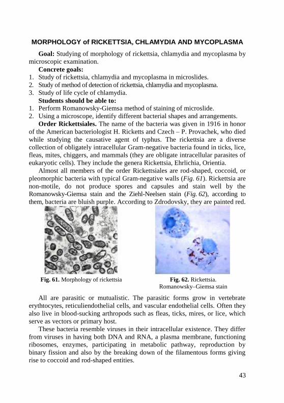
43
MORPHOLOGY of RICKETTSIA, CHLAMYDIA AND MYCOPLASMA
Goal: Studying of morphology of rickettsia, chlamydia and mycoplasma by
microscopic examination.
Concrete goals:
1. Study of rickettsia, chlamydia and mycoplasma in microslides.
2. Study of method of detection of rickettsia, chlamydia and mycoplasma.
3. Study of life cycle of chlamydia.
Students should be able to:
1. Perform Romanowsky-Giemsa method of staining of microslide.
2. Using a microscope, identify different bacterial shapes and arrangements.
Order Rickettsiales. The name of the bacteria was given in 1916 in honor
of the American bacteriologist H. Ricketts and Czech – P. Provachek, who died
while studying the causative agent of typhus. The rickettsia are a diverse
collection of obligately intracellular Gram-negative bacteria found in ticks, lice,
fleas, mites, chiggers, and mammals (they are obligate intracellular parasites of
eukaryotic cells). They include the genera Rickettsia, Ehrlichia, Orientia.
Almost all members of the order Rickettsiales are rod-shaped, coccoid, or
pleomorphic bacteria with typical Gram-negative walls (Fig. 61). Rickettsia are
non-motile, do not produce spores and capsules and stain well by the
Romanowsky-Giemsa stain and the Ziehl-Neelsen stain (Fig. 62), according to
them, bacteria are bluish purple. According to Zdrodovsky, they are painted red.
Fig. 61. Morphology of rickettsia Fig. 62. Rickettsia.
Romanowsky–Giemsa stain
All are parasitic or mutualistic. The parasitic forms grow in vertebrate
erythtocytes, reticuliendothelial cells, and vascular endothelial cells. Often they
also live in blood-sucking arthropods such as fleas, ticks, mires, or lice, which
serve as vectors or primary host.
These bacteria resemble viruses in their intracellular existence. They differ
from viruses in having both DNA and RNA, a plasma membrane, functioning
ribosomes, enzymes, participating in metabolic pathway, reproduction by
binary fission and also by the breaking down of the filamentous forms giving
rise to coccoid and rod-shaped entities.
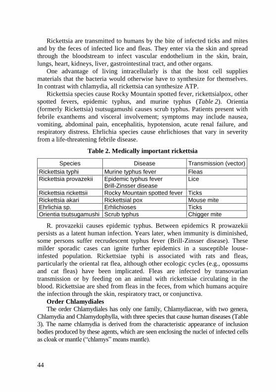
44
Rickettsia are transmitted to humans by the bite of infected ticks and mites
and by the feces of infected lice and fleas. They enter via the skin and spread
through the bloodstream to infect vascular endothelium in the skin, brain,
lungs, heart, kidneys, liver, gastrointestinal tract, and other organs.
One advantage of living intracellularly is that the host cell supplies
materials that the bacteria would otherwise have to synthesize for themselves.
In contrast with chlamydia, all rickettsia can synthesize ATP.
Rickettsia species cause Rocky Mountain spotted fever, rickettsialpox, other
spotted fevers, epidemic typhus, and murine typhus (Table 2). Orientia
(formerly Rickettsia) tsutsugamushi causes scrub typhus. Patients present with
febrile exanthems and visceral involvement; symptoms may include nausea,
vomiting, abdominal pain, encephalitis, hypotension, acute renal failure, and
respiratory distress. Ehrlichia species cause ehrlichioses that vary in severity
from a life-threatening febrile disease.
Table 2. Medically important rickettsia
Species Disease Transmission (vector)
Rickettsia typhi Murine typhus fever Fleas
Rickettsia provazekii Epidemic typhus fever Brill-Zinsser disease
Lice
Rickettsia rickettsii Rocky Mountain spotted fever Ticks
Rickettsia akari Rickettsial pox Mouse mite
Ehrlichia sp. Erhlichioses Ticks
Orientia tsutsugamushi Scrub typhus Chigger mite
R. provazekii causes epidemic typhus. Between epidemics R prowazekii
persists as a latent human infection. Years later, when immunity is diminished,
some persons suffer recrudescent typhus fever (Brill-Zinsser disease). These
milder sporadic cases can ignite further epidemics in a susceptible louse-
infested population. Rickettsiae typhi is associated with rats and fleas,
particularly the oriental rat flea, although other ecologic cycles (e.g., opossums
and cat fleas) have been implicated. Fleas are infected by transovarian
transmission or by feeding on an animal with rickettsiae circulating in the
blood. Rickettsiae are shed from fleas in the feces, from which humans acquire
the infection through the skin, respiratory tract, or conjunctiva.
Order Chlamydiales
The order Chlamydiales has only one family, Chlamydiaceae, with two genera,
Chlamydia and Chlamydophylla, with three species that cause human diseases (Table
3). The name chlamydia is derived from the characteristic appearance of inclusion
bodies produced by these agents, which are seen enclosing the nuclei of infected cells
as cloak or mantle (“chlamys” means mantle).
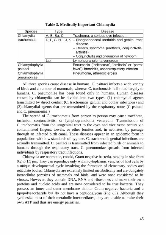
45
Table 3. Medically Important Chlamydia
Species Type Disease
Chlamydia trachomatis
A, B, Ba, C Trachoma, a serious eye infection.
D, F, G, H, I, J, K – Nongonococcal urethritis and genital tract disease.
– Reiter’s syndrome (urethritis, conjunctivitis, arthritis).
– Conjunctivitis and pneumonia of newborn
L1-3 Lymphogranuloma venereum
Chlamydophylla psittaci
Pneumonia (“psittacosis”, “ornitosis” or “parrot fever”), bronchitis, upper respiratory infection
Chlamydophylla pneumoniae
Pneumonia, atherosclerosis
All three species cause disease in humans. C. psittaci infects a wide variety
of birds and a number of mammals, whereas C. trachomatis is limited largely to
humans. C. pneumoniae has been found only in humans. Human diseases
caused by chlamydia can be divided into two types: (1) chlamydial agents
transmitted by direct contact (C. trachomatis genital and ocular infections) and
(2) chlamydial agents that are transmitted by the respiratory route (C psittaci
and C. pneumoniae.)
The spread of C. trachomatis from person to person may cause trachoma,
inclusion conjunctivitis, or lymphogranuloma venereum. Transmission of
C. trachomatis from the urogenital tract to the eyes and vice versa occurs via
contaminated fingers, towels, or other fomites and, in neonates, by passage
through an infected birth canal. These diseases appear in an epidemic form in
populations with low standards of hygiene. C. trachomatis genital infections are
sexually transmitted. C. psittaci is transmitted from infected birds or animals to
humans through the respiratory tract. C. pneumoniae spreads from infected
individuals by respiratory tract infections.
Chlamydia are nonmotile, cocoid, Gram-negative bacteria, ranging in size from
0.2 to 1.5 μm. They can reproduce only within cytoplasmic vesicles of host cells by
a unique developmental cycle involving the formation of elementary bodies and
reticulate bodies. Chlamydia are extremely limited metabolically and are obligately
intracellular parasites of mammals and birds, and were once considered to be
viruses. However, they contain DNA, RNA and ribosomes and make their own
proteins and nucleic acids and are now considered to be true bacteria. They
possess an inner and outer membrane similar Gram-negative bacteria and a
lipopolysaccharide but do not have a peptidoglycan (Fig. 63). Although they
synthesize most of their metabolic intermediates, they are unable to make their
own ATP and thus are energy parasites.
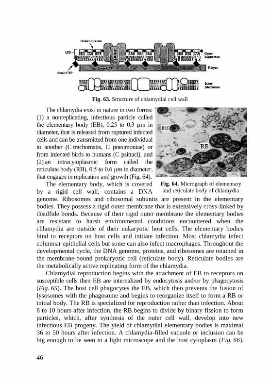
46
Fig. 63. Structure of chlamydial cell wall
The chlamydia exist in nature in two forms:
(1) a nonreplicating, infectious particle called
the elementary body (EB), 0.25 to 0.3 µm in
diameter, that is released from ruptured infected
cells and can be transmitted from one individual
to another (C trachomatis, C pneumoniae) or
from infected birds to humans (C psittaci), and
(2) an intracytoplasmic form called the
reticulate body (RB), 0.5 to 0.6 µm in diameter,
that engages in replication and growth (Fig. 64).
The elementary body, which is covered
by a rigid cell wall, contains a DNA
genome. Ribosomes and ribosomal subunits are present in the elementary
bodies. They possess a rigid outer membrane that is extensively cross-linked by
disulfide bonds. Because of their rigid outer membrane the elementary bodies
are resistant to harsh environmental conditions encountered when the
chlamydia are outside of their eukaryotic host cells. The elementary bodies
bind to receptors on host cells and initiate infection. Most chlamydia infect
columnar epithelial cells but some can also infect macrophages. Throughout the
developmental cycle, the DNA genome, proteins, and ribosomes are retained in
the membrane-bound prokaryotic cell (reticulate body). Reticulate bodies are
the metabolically active replicating form of the chlamydia.
Chlamydial reproduction begins with the attachment of EB to receptors on
susceptible cells then EB are internalized by endocytosis and/or by phagocytosis
(Fig. 65). The host cell phagocytes the EB, which then prevents the fusion of
lysosomes with the phagosome and begins to reorganize itself to form a RB or
initial body. The RB is specialized for reproduction rather than infection. About
8 to 10 hours after infection, the RB begins to divide by binary fission to form
particles, which, after synthesis of the outer cell wall, develop into new
infectious EB progeny. The yield of chlamydial elementary bodies is maximal
36 to 50 hours after infection. A chlamydia-filled vacuole or inclusion can be
big enough to be seen in a light microscope and the host cytoplasm (Fig. 66).
Fig. 64. Micrograph of elementary
and reticulate body of chlamydia

47
The resulting inclusions may contain 100–500 progeny EB reproduction
continues until the host cell dies.
Fig. 65. Developmental cycle of the chlamydia
Fig. 66. Inclusion of chlamydia in host cell
Mycoplasma and Ureaplasma
The family Mycoplasmataceae contains two genera that infect humans:
Mycoplasma and Ureaplasma. Although there are many species of mycoplasmas,
only four are recognized as human pathogens: Mycoplasma pneumoniae,
Mycoplasma hominis, Mycoplasma genitalium, and Ureaplasma urealyticum
(Table 4). All mycoplasmas cultivated and identified thus far are parasites of
humans, animals, plants, or arthropods. The primary habitats of human and
animal mycoplasmas are the mucous surfaces of the respiratory and urogenital
tracts and the joints in some animals. Although some mycoplasmas belong to
the normal flora, many species are pathogens, causing various diseases that
tend to run a chronic course.
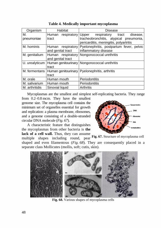
48
Table 4. Medically important mycoplasma
Organism Habitat Disease
M. pneumoniae
Human respiratory tract
Upper respiratory tract disease, tracheobronchitis, atypical pneumonia, pericarditis, meningitis, polyartritis
M. hominis Human respiratory and genital tract
Pyelonephritis, postpartum fever, pelvic inflammatory disease
M. genitalium Human respiratory and genital tract
Nongonococcal urethritis
U. urealyticum Human genitourinary tract
Nongonococcal urethritis
M. fermentans Human genitourinary tract
Pyelonephritis, arthritis
M. orale Human mouth Periodontitis
M. salivarium Human mouth Periodontitis
M. arthritidis Sinovial liquid Arthritis
Mycoplasmas are the smallest and simplest self-replicating bacteria. They range
from 0.2–0.8 mcm. They have the smallest
genome size. The mycoplasma cell contains the
minimum set of organelles essential for growth
and replication: a plasma membrane, ribosomes,
and a genome consisting of a double-stranded
circular DNA molecule (Fig. 67).
A characteristic feature that distinguishes
the mycoplasmas from other bacteria is the
lack of a cell wall. Thus, they can assume
multiple shapes including round, pear
shaped and even filamentous (Fig. 68). They are consequently placed in a
separate class Mollicutes (mollis, soft; cutis, skin).
Fig. 68. Various shapes of mycoplasma cells
Fig. 67. Structure of mycoplasma cell

49
In most mycoplasma cultures, elongated or filamentous forms (up to 100 µm
long and about 0.4 µm thick) also occur. The filaments tend to produce truly
branched mycelioid structures, hence the name mycoplasma (myces, a fungus;
plasma, a form). Mycoplasmas reproduce by binary fission, but cytoplasmic
division frequently may lag behind genome replication, resulting in formation
of multinuclear filaments.
Practical tasks, being carried out during practical classes:
1. Observe the demonstration of chlamydia, rickettsia and mycoplasma.
Therminology: elementary body, reticulate body.
Theoretical questions for control:
1. State the stages of life cycle of clamydia.
2. Recognize mycoplasma and rickettsia observed when microscopically
viewing Romanowsky-Giemsa stain.
3. Recognize inclusion of chlamydia inclusion observed when microscopically
viewing Romanowsky-Giemsa stain.
4. Define the following: Romanowsky-Giemsa stain, Ziehl-Neelsen stain.
Test task for control:
1. While microscopy of the scraping from urethra cytoplasmic inclusions were
identified in epithelial cells in the form of characteristic “cap” over the nucleus
of the cell. Which of the following disease can be assumed?
A. Chlamydiosis C. Gonorrhea E. Syphilis
B. Candidiasis D. Mycoplasmosis

50
STRUCTURE AND CHEMICAL COMPOSITION OF VIRUSES
Goal: Studying morphology of viruses and virus-induced damage to host cell.
Concrete goals: 1. Study of structure of virion. 2. Study of virus-induced damage to host cell and detection by microscopic
examination. 3. Study of type of reproduction of viruses. 4. Study of structure of bacteriophages.
Students should be able to: 1. Recognize virus-induced damage to host cell by microscopic examination.
Viruses are the smallest infectious agents (20–300 nm in diameter), containing one kind of nucleic acid (RNA or DNA) as their genome, usually as single molecule. The nucleic acid is encased in a protein shell (capsid), some viruses have additional outer envelope (supercapsid) (Fig. 69). The entire extracellular infectious unit is termed a virion.
Viruses replicate only in living cells. They are obligatory intracel-
lular parasite. The viral nucleic acid contains information necessary for programming the infected host cell to synthesize a number of virus-specific macromolecules required for the production of virus progeny. During the replicative cycle, numerous copies of viral nucleic acid and coat proteins are produced. The coat protein assemble together to form the capsid, which encases and stabilizes the viral nucleic acid against the extracellular environment (nucleases) and facilitates the attachment and perhaps penetration of the virus upon contact with new susceptible cells.
Some useful definitions in virology Capsid: The symmetric protein shell that encloses the nucleic acid genome. Capsomeres: Morphologic units seen in the electron microscope on the
surface of virus particles. Capsomeres represent clusters of polypeptides, which when completely assembled form the capsid.
Nucleocapsid: The capsid together with the enclosed nucleic acid. In many viruses, the capsid is the only outer coat and the virion is termed a
naked virus. Outer envelope (supercapsid) consists of lipid bilyer and surface spikes of
glycoproteins. Viruses that have an additional outer envelope are called
enveloped viruses.
Fig. 69. Structure of viral particle
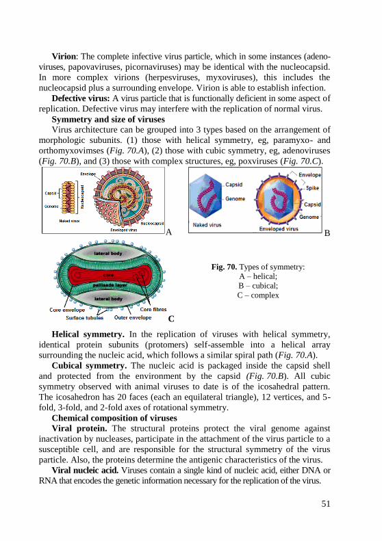
51
Virion: The complete infective virus particle, which in some instances (adeno-
viruses, papovaviruses, picornaviruses) may be identical with the nucleocapsid.
In more complex virions (herpesviruses, myxoviruses), this includes the
nucleocapsid plus a surrounding envelope. Virion is able to establish infection.
Defective virus: A virus particle that is functionally deficient in some aspect of
replication. Defective virus may interfere with the replication of normal virus.
Symmetry and size of viruses
Virus architecture can be grouped into 3 types based on the arrangement of
morphologic subunits. (1) those with helical symmetry, eg, paramyxo- and
orthomyxovimses (Fig. 70.A), (2) those with cubic symmetry, eg, adenoviruses
(Fig. 70.B), and (3) those with complex structures, eg, poxviruses (Fig. 70.C).
A B
C
Fig. 70. Types of symmetry:
A – helical;
B – cubical;
C – complex
Helical symmetry. In the replication of viruses with helical symmetry,
identical protein subunits (protomers) self-assemble into a helical array
surrounding the nucleic acid, which follows a similar spiral path (Fig. 70.A).
Cubical symmetry. The nucleic acid is packaged inside the capsid shell
and protected from the environment by the capsid (Fig. 70.B). All cubic
symmetry observed with animal viruses to date is of the icosahedral pattern.
The icosahedron has 20 faces (each an equilateral triangle), 12 vertices, and 5-
fold, 3-fold, and 2-fold axes of rotational symmetry.
Chemical composition of viruses
Viral protein. The structural proteins protect the viral genome against
inactivation by nucleases, participate in the attachment of the virus particle to a
susceptible cell, and are responsible for the structural symmetry of the virus
particle. Also, the proteins determine the antigenic characteristics of the virus.
Viral nucleic acid. Viruses contain a single kind of nucleic acid, either DNA or
RNA that encodes the genetic information necessary for the replication of the virus.

52
RNA of viruses can be:
(-) genome and must be converted to RNA or DNA before translation;
(+) genome is ready to be translated into proteins.
Viruses can have many genome arrangements (Fig. 71).
Fig. 71. Possible viral genome arrangements
Viral lipids. A number of different viruses contain lipids as part of their structure.
Viral carbohydrates. Virus envelopes contain glycoproteins.
Classification of viruses (Fig. 72)
Fig. 72. Classification of viruses
The following properties, listed in the order of preference or importance,
have been used as a basis for the classification of viruses.
(1) Nucleic acid type: RNA or DNA; single-stranded or double-stranded;
strategy of replication.

53
(2) Size and morphology, including type of symmetry, number of
capsomeres, and presence of envelope.
(3) Presence of specific enzymes, particularly RNA and DNA
polymerases concerned with genome (HIV), and neuraminidase
necessary for release of certain virus particles (influenza) from the
cells in which they were formed.
(4) Susceptibility to physical and chemical agents, especially ether.
(5) Immunologic properties.
(6) Natural methods of transmission.
(7) Host, tissue, and cell tropisms.
Pathology; inclusion body formation.
Measuring the size of viruses
Direct observation in the electron microscope is used for determining the
sizes of viruses and their components.
As compared with the light microscope, the electron microscope uses
electrons rather than light waves and electromagnetic lenses rather than glass
lenses. The electron beam obtained has a much shorter wavelength than that of
light, so that objects much smaller than the wavelength of visible or ultraviolet
light can be visualized. Viruses can be visualized in preparations from tissue
extracts and in ultrathin sections of infected cells. Electron microscopy is the
most widely used method for estimating particle size.
Quantification of virus Cytopathic effect – virus-induced damage to host cell (Fig. 73):
1. Changes in size and shape.
2. Cytoplasmic inclusion body.
3. Nuclear inclusion body.
4. Cells fuse to form multinucleated cells.
5. Cell lysis.
6. Alter DNA.
7. Transform cells into cancerous cell.
Inclusion body formation. In the course of virus multiplication within
cells, virus-specific structures called inclusion bodies may be produced. They
become far larger than the individual virus particle and often have an affinity
for acid dyes (eg, eosin). They may be situated in the nucleus (herpesvirus), in
the cytoplasm (rabies virus, influenza virus, pox virus), or in both (measles
virus). In many viral infections, the inclusion bodies are the site of development
of the virions (the virus factories).
The presence of inclusion bodies may be of considerable diagnostic aid. The
intracytoplasmic inclusion in nerve cells, the Babesh-Negri body (Fig. 73), is
pathognomonic for rabies. Guaenieri body is inclusion of smallpox virus in
cytoplasm of epithelial cells.

54
Cytoplasmic inclusion body
Nuclear inclusion body
Cells fuse to form multinucleated cells
Changes in size and shape
(rounding of cells)
Fig. 73. Cytopathic effect of viruses
Viruses of bacteria (bacteriophages)
Bacteria are host to a special group of viruses called bacteriophage, or
«phage», which pass through fine-porous filters and
develop at the expense of reproducing bacteria.
Morphology. A typical phage particle consists of a
“head” and a “tail” (Fig. 74). The head represents a
tightly packed core of nucleic acid surrounded by a
protein coat, or capsid. The protein capsid of the head
is made up of identical subunits, packed to form a
prismatic structure, usually hexagonal in cross section.
The smallest known phage has a head diameter of 25
nm; others range from 55 × 40 nm up to 100 × 70 nm.
The phage tail varies tremendously in its complexity from one phage to another. The most complex tail is found in phage T2 and in a number of other coli and typhoid phages. In these phages, the tail consists of at least 3 parts: a hollow core, a contractile sheath, and a terminal base-plate, hexagonal in shape, to which may be attached prongs, tail fibers, or both.
A number of other tail morphologies have been reported. In some of these, sheaths are visible but the contracted state has not been observed; and in one case no sheath can be seen. The phages also vary with respect to the terminal structure of the tail: some have base-plates, some have «knobs», and some appear to lack specific terminal structures (Fig. 75).
The phage tail is the adsorption organ for those phages that possess them. Some phages lack tails altogether; in the RNA phages, for example, the capsid is a simple icosahedron.
Fig. 74. Structure
of bacteriophage
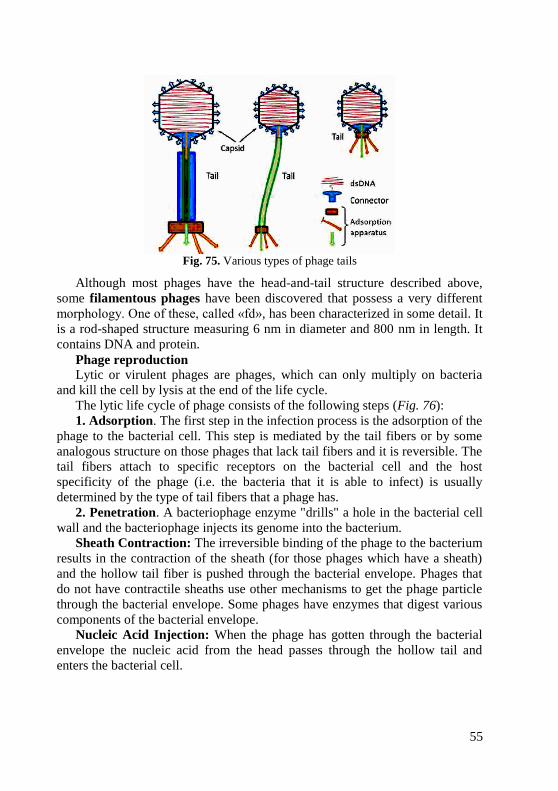
55
Fig. 75. Various types of phage tails
Although most phages have the head-and-tail structure described above,
some filamentous phages have been discovered that possess a very different
morphology. One of these, called «fd», has been characterized in some detail. It
is a rod-shaped structure measuring 6 nm in diameter and 800 nm in length. It
contains DNA and protein.
Phage reproduction
Lytic or virulent phages are phages, which can only multiply on bacteria
and kill the cell by lysis at the end of the life cycle.
The lytic life cycle of phage consists of the following steps (Fig. 76):
1. Adsorption. The first step in the infection process is the adsorption of the
phage to the bacterial cell. This step is mediated by the tail fibers or by some
analogous structure on those phages that lack tail fibers and it is reversible. The
tail fibers attach to specific receptors on the bacterial cell and the host
specificity of the phage (i.e. the bacteria that it is able to infect) is usually
determined by the type of tail fibers that a phage has.
2. Penetration. A bacteriophage enzyme "drills" a hole in the bacterial cell
wall and the bacteriophage injects its genome into the bacterium.
Sheath Contraction: The irreversible binding of the phage to the bacterium
results in the contraction of the sheath (for those phages which have a sheath)
and the hollow tail fiber is pushed through the bacterial envelope. Phages that
do not have contractile sheaths use other mechanisms to get the phage particle
through the bacterial envelope. Some phages have enzymes that digest various
components of the bacterial envelope.
Nucleic Acid Injection: When the phage has gotten through the bacterial
envelope the nucleic acid from the head passes through the hollow tail and
enters the bacterial cell.
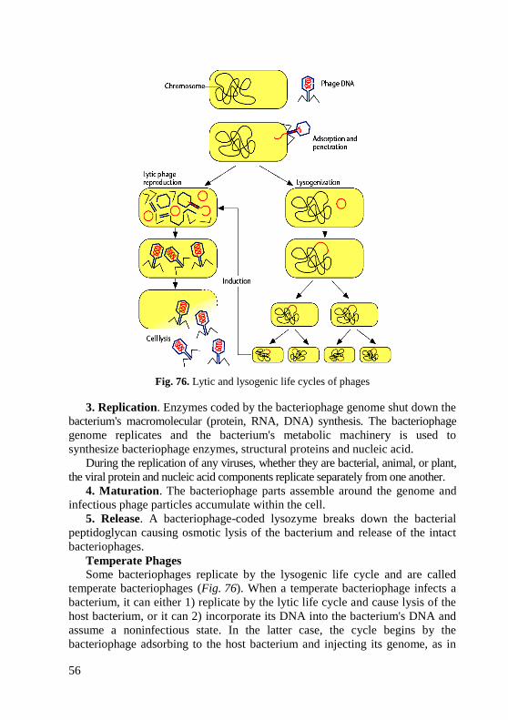
56
Fig. 76. Lytic and lysogenic life cycles of phages
3. Replication. Enzymes coded by the bacteriophage genome shut down the
bacterium's macromolecular (protein, RNA, DNA) synthesis. The bacteriophage
genome replicates and the bacterium's metabolic machinery is used to
synthesize bacteriophage enzymes, structural proteins and nucleic acid.
During the replication of any viruses, whether they are bacterial, animal, or plant,
the viral protein and nucleic acid components replicate separately from one another.
4. Maturation. The bacteriophage parts assemble around the genome and
infectious phage particles accumulate within the cell.
5. Release. A bacteriophage-coded lysozyme breaks down the bacterial
peptidoglycan causing osmotic lysis of the bacterium and release of the intact
bacteriophages.
Temperate Phages
Some bacteriophages replicate by the lysogenic life cycle and are called
temperate bacteriophages (Fig. 76). When a temperate bacteriophage infects a
bacterium, it can either 1) replicate by the lytic life cycle and cause lysis of the
host bacterium, or it can 2) incorporate its DNA into the bacterium's DNA and
assume a noninfectious state. In the latter case, the cycle begins by the
bacteriophage adsorbing to the host bacterium and injecting its genome, as in
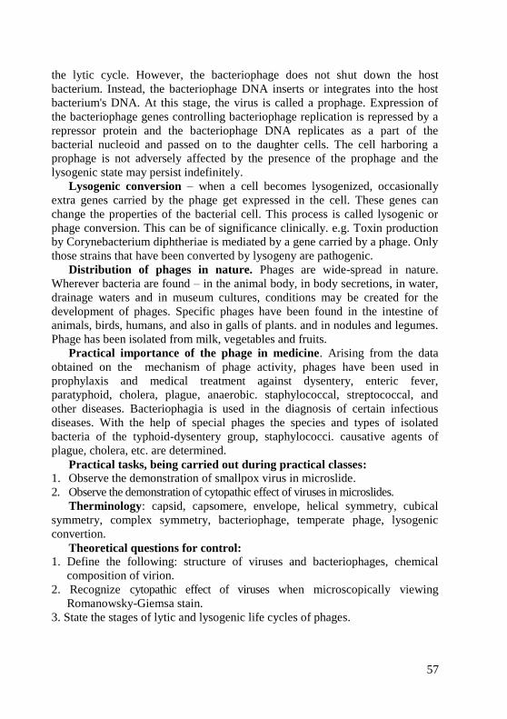
57
the lytic cycle. However, the bacteriophage does not shut down the host
bacterium. Instead, the bacteriophage DNA inserts or integrates into the host
bacterium's DNA. At this stage, the virus is called a prophage. Expression of
the bacteriophage genes controlling bacteriophage replication is repressed by a
repressor protein and the bacteriophage DNA replicates as a part of the
bacterial nucleoid and passed on to the daughter cells. The cell harboring a
prophage is not adversely affected by the presence of the prophage and the
lysogenic state may persist indefinitely.
Lysogenic conversion – when a cell becomes lysogenized, occasionally
extra genes carried by the phage get expressed in the cell. These genes can
change the properties of the bacterial cell. This process is called lysogenic or
phage conversion. This can be of significance clinically. e.g. Toxin production
by Corynebacterium diphtheriae is mediated by a gene carried by a phage. Only
those strains that have been converted by lysogeny are pathogenic.
Distribution of phages in nature. Phages are wide-spread in nature.
Wherever bacteria are found – in the animal body, in body secretions, in water,
drainage waters and in museum cultures, conditions may be created for the
development of phages. Specific phages have been found in the intestine of
animals, birds, humans, and also in galls of plants. and in nodules and legumes.
Phage has been isolated from milk, vegetables and fruits.
Practical importance of the phage in medicine. Arising from the data
obtained on the mechanism of phage activity, phages have been used in
prophylaxis and medical treatment against dysentery, enteric fever,
paratyphoid, cholera, plague, anaerobic. staphylococcal, streptococcal, and
other diseases. Bacteriophagia is used in the diagnosis of certain infectious
diseases. With the help of special phages the species and types of isolated
bacteria of the typhoid-dysentery group, staphylococci. causative agents of
plague, cholera, etc. are determined.
Practical tasks, being carried out during practical classes:
1. Observe the demonstration of smallpox virus in microslide.
2. Observe the demonstration of cytopathic effect of viruses in microslides.
Therminology: capsid, capsomere, envelope, helical symmetry, cubical
symmetry, complex symmetry, bacteriophage, temperate phage, lysogenic
convertion.
Theoretical questions for control:
1. Define the following: structure of viruses and bacteriophages, chemical
composition of virion.
2. Recognize cytopathic effect of viruses when microscopically viewing
Romanowsky-Giemsa stain.
3. State the stages of lytic and lysogenic life cycles of phages.
58
Test task for control:
1. Which of the following is found in all infectious virions, regardless of the
type of the virus?
A. Nucleocapsid C. DNA genome E. Envelope
B. Matrix protein D. One enzyme
2. Every virion contains a genome surrounded by capsid, which consists of:
A. Lipid C. Lipoprotein E. Protein
B. Polysacharide D. Nucleic acid
3. The following statements regarding viruses are correct, except:
A. Have icosahedral symmetry D. Have helical symmetry
B. They are unicellular E. They possess DNA or RNA
C. They are acellular
4. Viruses possess all, except:
A. Сontain DNA or RNA D. Have genome
B. Divide by binary fission E. Lack enzymes
C. Have symmetrical structure for protein synthesis
5. Morphology of viruses is studied by which of the following methods:
A. Immersion microscopy D. Electrophoresis
B. Electron microscopy E. Phase-contrast microscopy
C. Dark-field microscopy
59
MORPHOLOGY OF PATHOGENIC PROTOZOA
Goal: Studying of morphology of pathogenic protozoa by microscopic
examination.
Concrete goals:
1. Study of classification of pathogenic protozoa.
2. Study of morphology of pathogenic protozoa in microslides.
3. Study of method of staining of pathogenic protozoa.
4. Study of structure of protozoan cell.
Students should be able to:
1. Using a microscope, identify pathogenic protozoa.
Equipment: immersion microscopes, immersion oil, tables, atlas.
Structure of protozoan cell
Protozoa are microscopic unicellular eukaryotes that have a relatively
complex internal structure and carry out complex metabolic activities. Some
protozoa have structures for propulsion or other types of movement. Most
parasitic protozoa in humans are less than 50 µm in size. The smallest (mainly
intracellular forms) are 1 to 10 µm long, but Balantidium coli may measure 150
µm. As in all eukaryotes, the nucleus is enclosed in a membrane. In protozoa
other than ciliates, the nucleus is vesicular, with scattered chromatin giving a
diffuse appearance to the nucleus. The ciliates have both a micronucleus and
macronucleus, which appear quite homogeneous in composition.
The organelles of protozoa have functions similar to the organs of higher
animals. The plasma membrane enclosing the cytoplasm also covers the
projecting locomotory structures such as pseudopodia, cilia, and flagella. The
outer surface layer of some protozoa, termed a pellicle, is sufficiently rigid to
maintain a distinctive shape, as in the trypanosomes and Giardia. However,
these organisms can readily twist and bend when moving through their
environment. In most protozoa the cytoplasm is differentiated into ectoplasm
(the outer, transparent layer) and endoplasm (the inner layer containing
organelles); the structure of the cytoplasm is most easily seen in species with
projecting pseudopodia, such as the amebas. Some protozoa have a cytosome or
cell "mouth" for ingesting fluids or solid particles. Contractile vacuoles for
osmoregulation occur in some, such as Balantidium. Many protozoa have
subpellicular microtubules; in the Apicomplexa, which have no external
organelles for locomotion, these provide a means for slow movement. The
trichomonads and trypanosomes have a distinctive undulating membrane
between the body wall and a flagellum. Many other structures occur in parasitic
protozoa, including the Golgi apparatus, mitochondria, lysosomes, food
vacuoles, conoids in the Apicomplexa, and other specialized structures.
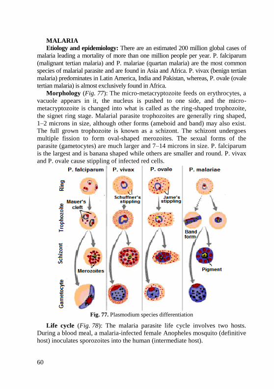
60
MALARIA
Etiology and epidemiology: There are an estimated 200 million global cases of
malaria leading a mortality of more than one million people per year. P. falciparum
(malignant tertian malaria) and P. malariae (quartan malaria) are the most common
species of malarial parasite and are found in Asia and Africa. P. vivax (benign tertian
malaria) predominates in Latin America, India and Pakistan, whereas, P. ovale (ovale
tertian malaria) is almost exclusively found in Africa.
Morphology (Fig. 77): The micro-metacryptozoite feeds on erythrocytes, a
vacuole appears in it, the nucleus is pushed to one side, and the micro-
metacryptozoite is changed into what is called as the ring-shaped trophozoite,
the signet ring stage. Malarial parasite trophozoites are generally ring shaped,
1–2 microns in size, although other forms (ameboid and band) may also exist.
The full grown trophozoite is known as a schizont. The schizont undergoes
multiple fission to form oval-shaped merozoites. The sexual forms of the
parasite (gametocytes) are much larger and 7–14 microns in size. P. falciparum
is the largest and is banana shaped while others are smaller and round. P. vivax
and P. ovale cause stippling of infected red cells.
Fig. 77. Plasmodium species differentiation
Life cycle (Fig. 78): The malaria parasite life cycle involves two hosts.
During a blood meal, a malaria-infected female Anopheles mosquito (definitive
host) inoculates sporozoites into the human (intermediate host).
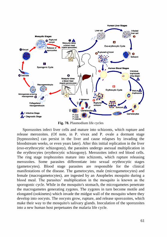
61
Fig. 78. Plasmodium life cycles
Sporozoites infect liver cells and mature into schizonts, which rupture and
release merozoites. (Of note, in P. vivax and P. ovale a dormant stage
[hypnozoites] can persist in the liver and cause relapses by invading the
bloodstream weeks, or even years later). After this initial replication in the liver
(exo-erythrocytic schizogony), the parasites undergo asexual multiplication in
the erythrocytes (erythrocytic schizogony). Merozoites infect red blood cells.
The ring stage trophozoites mature into schizonts, which rupture releasing
merozoites. Some parasites differentiate into sexual erythrocytic stages
(gametocytes). Blood stage parasites are responsible for the clinical
manifestations of the disease. The gametocytes, male (microgametocytes) and
female (macrogametocytes), are ingested by an Anopheles mosquito during a
blood meal. The parasites’ multiplication in the mosquito is known as the
sporogonic cycle. While in the mosquito's stomach, the microgametes penetrate
the macrogametes generating zygotes. The zygotes in turn become motile and
elongated (ookinetes) which invade the midgut wall of the mosquito where they
develop into oocysts. The oocysts grow, rupture, and release sporozoites, which
make their way to the mosquito's salivary glands. Inoculation of the sporozoites
into a new human host perpetuates the malaria life cycle.
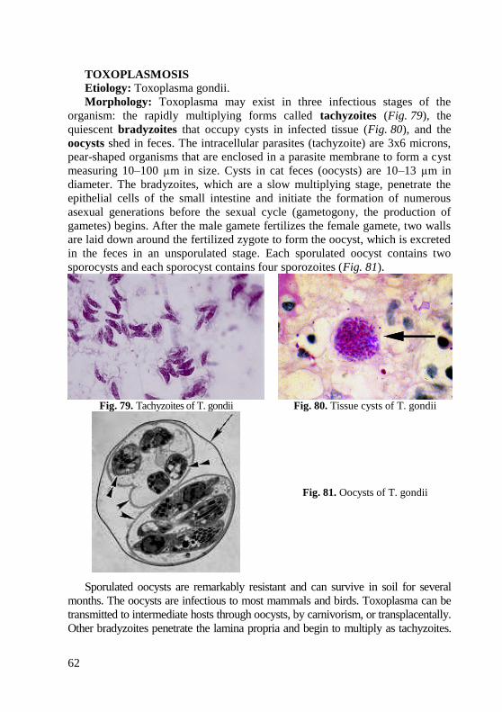
62
TOXOPLASMOSIS
Etiology: Toxoplasma gondii.
Morphology: Toxoplasma may exist in three infectious stages of the
organism: the rapidly multiplying forms called tachyzoites (Fig. 79), the
quiescent bradyzoites that occupy cysts in infected tissue (Fig. 80), and the
oocysts shed in feces. The intracellular parasites (tachyzoite) are 3x6 microns,
pear-shaped organisms that are enclosed in a parasite membrane to form a cyst
measuring 10–100 µm in size. Cysts in cat feces (oocysts) are 10–13 µm in
diameter. The bradyzoites, which are a slow multiplying stage, penetrate the
epithelial cells of the small intestine and initiate the formation of numerous
asexual generations before the sexual cycle (gametogony, the production of
gametes) begins. After the male gamete fertilizes the female gamete, two walls
are laid down around the fertilized zygote to form the oocyst, which is excreted
in the feces in an unsporulated stage. Each sporulated oocyst contains two
sporocysts and each sporocyst contains four sporozoites (Fig. 81).
Fig. 79. Tachyzoites of T. gondii Fig. 80. Tissue cysts of T. gondii
Fig. 81. Oocysts of T. gondii
Sporulated oocysts are remarkably resistant and can survive in soil for several
months. The oocysts are infectious to most mammals and birds. Toxoplasma can be
transmitted to intermediate hosts through oocysts, by carnivorism, or transplacentally.
Other bradyzoites penetrate the lamina propria and begin to multiply as tachyzoites.
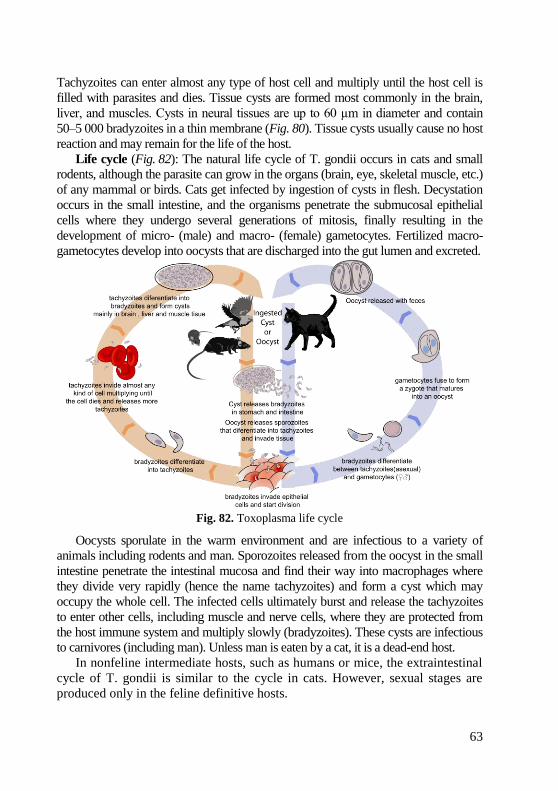
63
Tachyzoites can enter almost any type of host cell and multiply until the host cell is
filled with parasites and dies. Tissue cysts are formed most commonly in the brain,
liver, and muscles. Cysts in neural tissues are up to 60 µm in diameter and contain
50–5 000 bradyzoites in a thin membrane (Fig. 80). Tissue cysts usually cause no host
reaction and may remain for the life of the host.
Life cycle (Fig. 82): The natural life cycle of T. gondii occurs in cats and small
rodents, although the parasite can grow in the organs (brain, eye, skeletal muscle, etc.)
of any mammal or birds. Cats get infected by ingestion of cysts in flesh. Decystation
occurs in the small intestine, and the organisms penetrate the submucosal epithelial
cells where they undergo several generations of mitosis, finally resulting in the
development of micro- (male) and macro- (female) gametocytes. Fertilized macro-
gametocytes develop into oocysts that are discharged into the gut lumen and excreted.
Fig. 82. Toxoplasma life cycle
Oocysts sporulate in the warm environment and are infectious to a variety of
animals including rodents and man. Sporozoites released from the oocyst in the small
intestine penetrate the intestinal mucosa and find their way into macrophages where
they divide very rapidly (hence the name tachyzoites) and form a cyst which may
occupy the whole cell. The infected cells ultimately burst and release the tachyzoites
to enter other cells, including muscle and nerve cells, where they are protected from
the host immune system and multiply slowly (bradyzoites). These cysts are infectious
to carnivores (including man). Unless man is eaten by a cat, it is a dead-end host.
In nonfeline intermediate hosts, such as humans or mice, the extraintestinal
cycle of T. gondii is similar to the cycle in cats. However, sexual stages are
produced only in the feline definitive hosts.
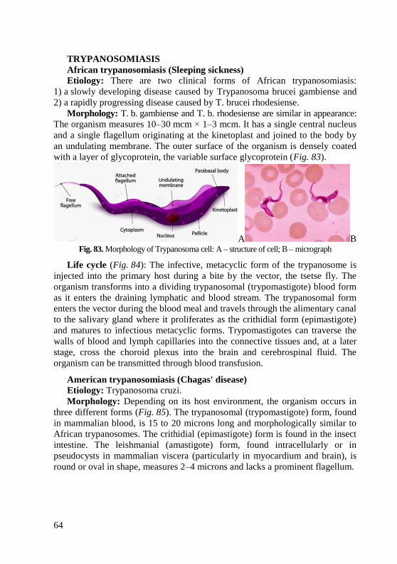
64
TRYPANOSOMIASIS
African trypanosomiasis (Sleeping sickness)
Etiology: There are two clinical forms of African trypanosomiasis:
1) a slowly developing disease caused by Trypanosoma brucei gambiense and
2) a rapidly progressing disease caused by T. brucei rhodesiense.
Morphology: T. b. gambiense and T. b. rhodesiense are similar in appearance:
The organism measures 10–30 mcm × 1–3 mcm. It has a single central nucleus
and a single flagellum originating at the kinetoplast and joined to the body by
an undulating membrane. The outer surface of the organism is densely coated
with a layer of glycoprotein, the variable surface glycoprotein (Fig. 83).
A B Fig. 83. Morphology of Trypanosoma cell: A – structure of cell; B – micrograph
Life cycle (Fig. 84): The infective, metacyclic form of the trypanosome is
injected into the primary host during a bite by the vector, the tsetse fly. The
organism transforms into a dividing trypanosomal (trypomastigote) blood form
as it enters the draining lymphatic and blood stream. The trypanosomal form
enters the vector during the blood meal and travels through the alimentary canal
to the salivary gland where it proliferates as the crithidial form (epimastigote)
and matures to infectious metacyclic forms. Trypomastigotes can traverse the
walls of blood and lymph capillaries into the connective tissues and, at a later
stage, cross the choroid plexus into the brain and cerebrospinal fluid. The
organism can be transmitted through blood transfusion.
American trypanosomiasis (Chagas' disease)
Etiology: Trypanosoma cruzi.
Morphology: Depending on its host environment, the organism occurs in
three different forms (Fig. 85). The trypanosomal (trypomastigote) form, found
in mammalian blood, is 15 to 20 microns long and morphologically similar to
African trypanosomes. The crithidial (epimastigote) form is found in the insect
intestine. The leishmanial (amastigote) form, found intracellularly or in
pseudocysts in mammalian viscera (particularly in myocardium and brain), is
round or oval in shape, measures 2–4 microns and lacks a prominent flagellum.
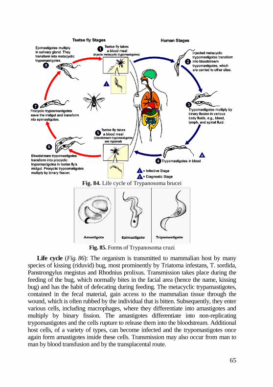
65
Fig. 84. Life cycle of Trypanosoma brucei
Fig. 85. Forms of Trypanosoma cruzi
Life cycle (Fig. 86): The organism is transmitted to mammalian host by many species of kissing (riduvid) bug, most prominently by Triatoma infestans, T. sordida, Panstrongylus megistus and Rhodnius prolixus. Transmission takes place during the feeding of the bug, which normally bites in the facial area (hence the name, kissing bug) and has the habit of defecating during feeding. The metacyclic trypamastigotes, contained in the fecal material, gain access to the mammalian tissue through the wound, which is often rubbed by the individual that is bitten. Subsequently, they enter various cells, including macrophages, where they differentiate into amastigotes and multiply by binary fission. The amastigotes differentiate into non-replicating trypomastigotes and the cells rupture to release them into the bloodstream. Additional host cells, of a variety of types, can become infected and the trypomastigotes once again form amastigotes inside these cells. Transmission may also occur from man to man by blood transfusion and by the transplacental route.
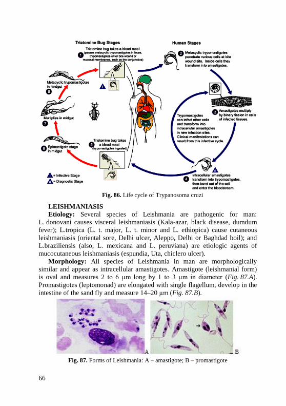
66
Fig. 86. Life cycle of Trypanosoma cruzi
LEISHMANIASIS
Etiology: Several species of Leishmania are pathogenic for man:
L. donovani causes visceral leishmaniasis (Kala-azar, black disease, dumdum
fever); L.tropica (L. t. major, L. t. minor and L. ethiopica) cause cutaneous
leishmaniasis (oriental sore, Delhi ulcer, Aleppo, Delhi or Baghdad boil); and
L.braziliensis (also, L. mexicana and L. peruviana) are etiologic agents of
mucocutaneous leishmaniasis (espundia, Uta, chiclero ulcer).
Morphology: All species of Leishmania in man are morphologically
similar and appear as intracellular amastigotes. Amastigote (leishmanial form)
is oval and measures 2 to 6 µm long by 1 to 3 µm in diameter (Fig. 87.A).
Promastigotes (leptomonad) are elongated with single flagellum, develop in the
intestine of the sand fly and measure 14–20 µm (Fig. 87.B).
Fig. 87. Forms of Leishmania: A – amastigote; B – promastigote
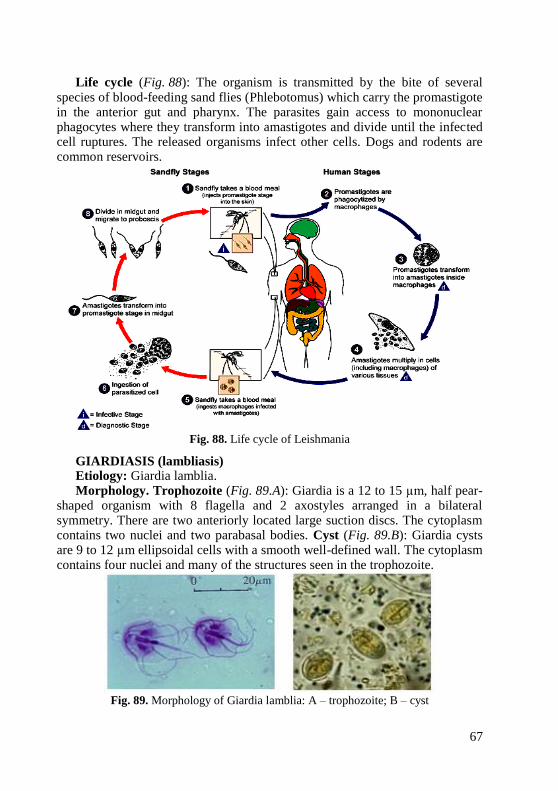
67
Life cycle (Fig. 88): The organism is transmitted by the bite of several species of blood-feeding sand flies (Phlebotomus) which carry the promastigote in the anterior gut and pharynx. The parasites gain access to mononuclear phagocytes where they transform into amastigotes and divide until the infected cell ruptures. The released organisms infect other cells. Dogs and rodents are common reservoirs.
Fig. 88. Life cycle of Leishmania
GIARDIASIS (lambliasis) Etiology: Giardia lamblia. Morphology. Trophozoite (Fig. 89.A): Giardia is a 12 to 15 µm, half pear-
shaped organism with 8 flagella and 2 axostyles arranged in a bilateral symmetry. There are two anteriorly located large suction discs. The cytoplasm contains two nuclei and two parabasal bodies. Cyst (Fig. 89.B): Giardia cysts are 9 to 12 µm ellipsoidal cells with a smooth well-defined wall. The cytoplasm contains four nuclei and many of the structures seen in the trophozoite.
Fig. 89. Morphology of Giardia lamblia: A – trophozoite; B – cyst
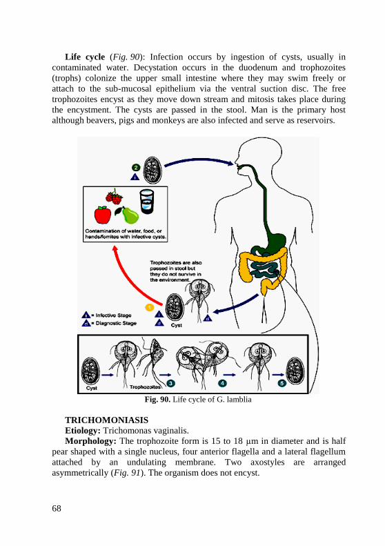
68
Life cycle (Fig. 90): Infection occurs by ingestion of cysts, usually in
contaminated water. Decystation occurs in the duodenum and trophozoites
(trophs) colonize the upper small intestine where they may swim freely or
attach to the sub-mucosal epithelium via the ventral suction disc. The free
trophozoites encyst as they move down stream and mitosis takes place during
the encystment. The cysts are passed in the stool. Man is the primary host
although beavers, pigs and monkeys are also infected and serve as reservoirs.
TRICHOMONIASIS
Etiology: Trichomonas vaginalis.
Morphology: The trophozoite form is 15 to 18 µm in diameter and is half
pear shaped with a single nucleus, four anterior flagella and a lateral flagellum
attached by an undulating membrane. Two axostyles are arranged
asymmetrically (Fig. 91). The organism does not encyst.
Fig. 90. Life cycle of G. lamblia
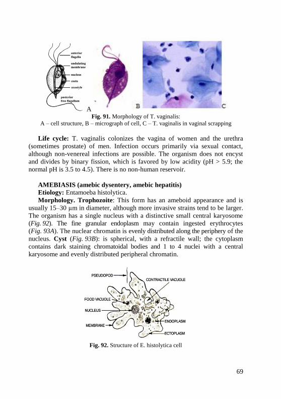
69
A Fig. 91. Morphology of T. vaginalis:
A – cell structure, B – micrograph of cell, C – T. vaginalis in vaginal scrapping
Life cycle: T. vaginalis colonizes the vagina of women and the urethra
(sometimes prostate) of men. Infection occurs primarily via sexual contact,
although non-venereal infections are possible. The organism does not encyst
and divides by binary fission, which is favored by low acidity (pH > 5.9; the
normal pH is 3.5 to 4.5). There is no non-human reservoir.
AMEBIASIS (amebic dysentery, amebic hepatitis)
Etiology: Entamoeba histolytica.
Morphology. Trophozoite: This form has an ameboid appearance and is
usually 15–30 µm in diameter, although more invasive strains tend to be larger.
The organism has a single nucleus with a distinctive small central karyosome
(Fig. 92). The fine granular endoplasm may contain ingested erythrocytes
(Fig. 93A). The nuclear chromatin is evenly distributed along the periphery of the
nucleus. Cyst (Fig. 93B): is spherical, with a refractile wall; the cytoplasm
contains dark staining chromatoidal bodies and 1 to 4 nuclei with a central
karyosome and evenly distributed peripheral chromatin.
Fig. 92. Structure of E. histolytica cell
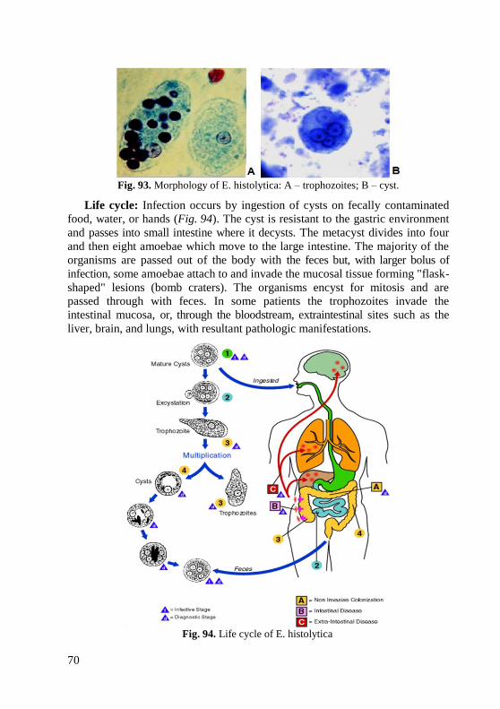
70
Fig. 93. Morphology of E. histolytica: A – trophozoites; B – cyst.
Life cycle: Infection occurs by ingestion of cysts on fecally contaminated
food, water, or hands (Fig. 94). The cyst is resistant to the gastric environment
and passes into small intestine where it decysts. The metacyst divides into four
and then eight amoebae which move to the large intestine. The majority of the
organisms are passed out of the body with the feces but, with larger bolus of
infection, some amoebae attach to and invade the mucosal tissue forming "flask-
shaped" lesions (bomb craters). The organisms encyst for mitosis and are
passed through with feces. In some patients the trophozoites invade the
intestinal mucosa, or, through the bloodstream, extraintestinal sites such as the
liver, brain, and lungs, with resultant pathologic manifestations.
Fig. 94. Life cycle of E. histolytica
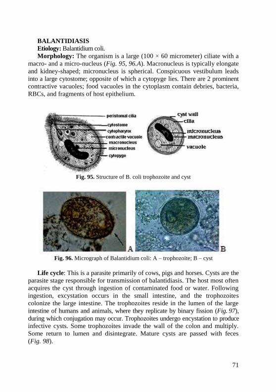
71
BALANTIDIASIS
Etiology: Balantidium coli.
Morphology: The organism is a large (100 × 60 micrometer) ciliate with a
macro- and a micro-nucleus (Fig. 95, 96.A). Macronucleus is typically elongate
and kidney-shaped; micronucleus is spherical. Conspicuous vestibulum leads
into a large cytostome; opposite of which a cytopyge lies. There are 2 prominent
contractive vacuoles; food vacuoles in the cytoplasm contain debries, bacteria,
RBCs, and fragments of host epithelium.
Fig. 95. Structure of B. coli trophozoite and cyst
Fig. 96. Micrograph of Balantidium coli: A – trophozoite; B – cyst
Life cycle: This is a parasite primarily of cows, pigs and horses. Cysts are the
parasite stage responsible for transmission of balantidiasis. The host most often
acquires the cyst through ingestion of contaminated food or water. Following
ingestion, excystation occurs in the small intestine, and the trophozoites
colonize the large intestine. The trophozoites reside in the lumen of the large
intestine of humans and animals, where they replicate by binary fission (Fig. 97),
during which conjugation may occur. Trophozoites undergo encystation to produce
infective cysts. Some trophozoites invade the wall of the colon and multiply.
Some return to lumen and disintegrate. Mature cysts are passed with feces
(Fig. 98).
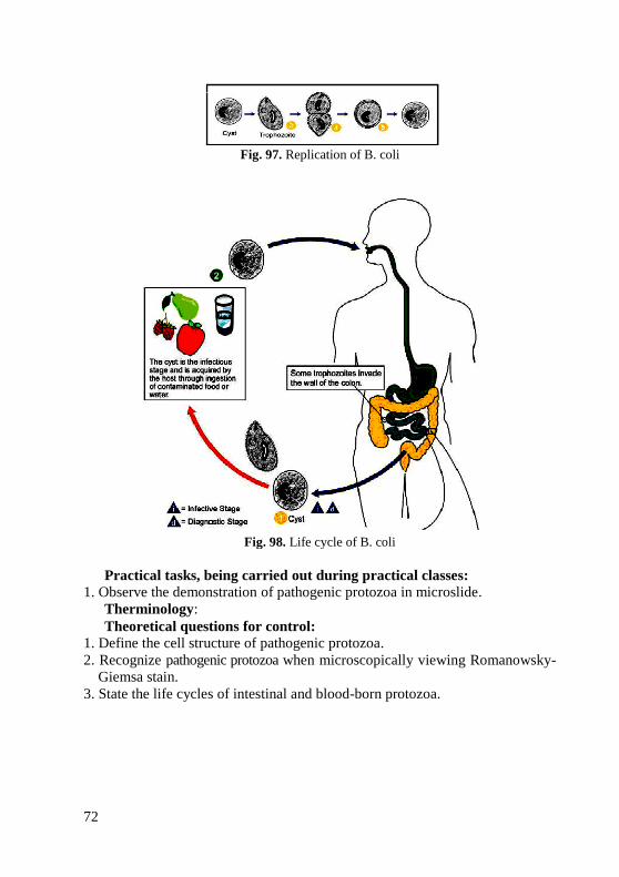
72
Fig. 97. Replication of B. coli
Fig. 98. Life cycle of B. coli
Practical tasks, being carried out during practical classes:
1. Observe the demonstration of pathogenic protozoa in microslide.
Therminology:
Theoretical questions for control:
1. Define the cell structure of pathogenic protozoa.
2. Recognize pathogenic protozoa when microscopically viewing Romanowsky-
Giemsa stain.
3. State the life cycles of intestinal and blood-born protozoa.
73
Test task for control:
1. Microscopy of dental plaque revealed unicellular organisms. Their cytoplasm
had two distinct layers, barely visible core, wide pseudopodia. The patient is
most likely to have:
A. Entamoeba gingivalis D. Lamblia
B. Entamoeba histolytica E. Entamoeba coli
C. Trichomonas tenax
2. A 13 year old child complains about poor appetite, pain in the right subcostal
area. Microscopical examination of duodenal contents revealed big pyriform
cells with two nuclei. What microorganism was revealed?
A. Lamblia C. Trichomonas E. Amoeba
B. Trypanosoma D. Toxoplasma
3. While examining a blood smear taken form a patient and stained by Romanovsky’s
method a doctor revealed some protozoa and diagnosed the patient with Chagas
disease. What protozoan is the causative agent of this disease?
A. Leishmania donovani D. Toxoplasma gondii
B. Trypanosoma cruzi E. Leishmania tropica
C. Trypanosoma brucei
4. A smear from frothy and purulent vaginal discharges of a 42 y.o. woman was
stained by Romanovsky-Giemsa method. Its analysis revealed some microorganisms
of flagellates class. What microorganism were the most probably revealed?
A. Leishmania donovani D. Trypanosoma brucei
B. Trypanosoma cruzi E. Lamblia intestinalis
C. Trihomonas vaginalis
5. Finding intraerythrocytic trophozoite, schizont, and gametocytes stages in
stained thin blood smears during febrile periods is the usual means for
diagnosis of which of the following?
A. African trypanosomiasis D. Toxoplasmosis
B. Malaria E. Visceral leishmaniasis
C. American trypanosomiasis
74
REFERENCES
1. Microbiology. Handbook to laboratory classes in microbiology /
A. Ya. Tsyganenko, I. L. Dikiy, V. L. Tkachenko et al. – Kharkiv : Osnova,
2005. – 210 p.
2. Tsyganenko A. Ya. Practical exercises of morphology of bacteria. Short
textbook for students of high medical schools / A. Ya. Tsyganenko,
V. N. Vasilchenko, N. I. Kovalenko. – Kharkiv : KSMU, 2015. – 284 p.
3. Murray P. R. Manual of Clinical Microbiology / P. R. Murray,
K. S. Rosenthal, M. A. Pfaller. – 8th
ed. – Washington : ASM Press, 2015. –
848 p.
4. Ananthanarayan P. Textbook of Microbiology. / P. Ananthanarayan. –
9th
ed. – Orient Blackswan, 2013. – 657 p.
5. Manual of Clinical Microbiology / Karen C. Carroll et al. – 10th
edition. – Vol. 2. – Washington, DC : ASM Press, 2011. – 2630 p.
6. Summaries of Infectious Diseases. Red Book® Online. – 2006.
https://redbook.solutions.aap.org/
7. CDC Alphabetical Index of Parasitic Diseases. Access:
https://www.cdc.gov/parasites/az/index.html#a
ADDITIONAL REFERENCES
1. Gupte S. The Short Textbook of Medical Microbiology. / S. Gupte. –
9th
ed. – New Delhi: Jaypee Brothers Medical Publishers Ltd., 2006. – 509 p.
2. Kaiser G. E. Microbiology Lab Manuel / G. E. Kaiser. – Raven Press,
New York, 2004.
3. Milton R. J. Salton, Kwang-Shin Kim. Structure. In Medical
Microbiology. / S. Baron. – 4th
ed. – Galveston (TX): University of Texas
Medical Branch at Galveston; 1996. Access:
https://www.ncbi.nlm.nih.gov/books/NBK8477/
4. Prescoti L.M. Microbiology / L. M. Prescoti, J. E. Harley, D. A. Klein. –
3th ed. – Wm. C. Prown Publishers, 1994.
5. Virella G., M.D. Microbiology and Infectious Diseases / G. Virella. –
3th ed. – Mass Publishing Co. – Egypt, 1997. – 575 p.
75
CONTENTS
Morphology of cocci. Microscopic examination.
Simple techniques of staining . . . . . . . . . . . . . . . . . . . . . . . . . . . . . . . . .
3
Morphology of rod-shaped bacteria. Structure of bacterial cell.
Gram’s method . . . . . . . . . . . . . . . . . . . . . . . . . . . . . . . . . . . . . . . . . . . .
14
Morphology of spiral bacteria. Methods of examination of flagella . . . 24
Spores. Methods of spore statining . . . . . . . . . . . . . . . . . . . . . . . . . . . . . 33
Capsules of bacteria. Methods of detection of capsule . . . . . . . . . . . . . . 40
Morphology of rickettsia, chlamydia and mycoplasma . . . . . . . . . . . . . 43
Structure and chemical composition of viruses . . . . . . . . . . . . . . . . . . . 50
Morphology of pathogenic protozoa . . . . . . . . . . . . . . . . . . . . . . . . . . . . 59
References . . . . . . . . . . . . . . . . . . . . . . . . . . . . . . . . . . . . . . . . . . . . . . . . 74
Additional references . . . . . . . . . . . . . . . . . . . . . . . . . . . . . . . . . . . . . . . 74
76
Навчальне видання
МОРФОЛОГІЯ БАКТЕРІЙ,
ВІРУСІВ І НАЙПРОСТІШИХ
Методичні вказівки
для студентів II і III курсів
медичного та стоматологічного факультетів
з англійською мовою викладання
Упорядник Коваленко Наталія Іллівна
Відповідальний за випуск Н. І. Коваленко
Комп'ютерний набір Н. І. Коваленко
Комп'ютерна верстка О. Ю. Лавриненко
Формат А5. Ум. друк. арк. 4,8. Зам. № 17-33410.
______________________________________________________________
Редакційно-видавничий відділ
ХНМУ, пр. Науки, 4, м. Харків, 61022
Свідоцтво про внесення суб’єкта видавничої справи до Державного реєстру видавництв,
виготівників і розповсюджувачів видавничої продукції серії ДК № 3242 від 18.07.2008 р.
77
MORPHOLOGY OF BACTERIA,
VIRUSES AND PROTOZOA Learning guide for the 2
nd and 3
rd year English media
students of the Faculty of Medicine and the Faculty of
Dentistry (Microbiology, virology and immunology)
МОРФОЛОГІЯ БАКТЕРІЙ,
ВІРУСІВ І НАЙПРОСТІШИХ Методичні вказівки з дисципліни
«Мікробіологія, вірусологія та імунологія»
для студентів II і III курсів
медичного та стоматологічного факультетів
з англійською мовою викладання

78


