Gene Repertoire of Amoeba-Associated Giant Viruses
14
Fax +41 61 306 12 34 E-Mail [email protected] www.karger.com Intervirology 2010;53:330–343 DOI: 10.1159/000312918 Gene Repertoire of Amoeba-Associated Giant Viruses Philippe Colson a, b Didier Raoult a, b a URMITE UMR CNRS 6236 IRD 198, Facultés de Médecine et de Pharmacie, Université de la Méditerranée, et b Pôle des Maladies Infectieuses et Tropicales Clinique et Biologique, Fédération de Bactériologie-Hygiène-Virologie, Centre Hospitalo-Universitaire Timone, Marseille, France plicating sympatric bacteria and viruses with an intra-amoe- bal lifestyle. In addition, lineage-specific gene expansion may have played a major role in the genome shaping. Alto- gether, the data so far accumulated on amoeba-associated giant viruses are a powerful incentive to isolate and study additional strains to gain better understanding of their pangenome. Copyright © 2010 S. Karger AG, Basel Introduction Viruses are ubiquitous components of the biosphere, outnumbering cells by at least 10-fold, and they have ex- tremely variable genomes in terms of composition, orga- nization, and size [1, 2]. Nevertheless, they have not been incorporated into the universal tree of life [3, 4], are not considered living beings according to the International Committee on Taxonomy of Viruses [5], and are still de- fined on the basis of negative criteria and as specialized and simple organisms [6, 7]. The recent discoveries of giant viruses associated with amoebae have strongly challenged this paradigm [8–11]. Acanthamoeba polyphaga mimivirus (APMV) is the first member of Mimiviridae, a new family of nucleo-cytoplas- mic large DNA viruses (NCLDVs) that form a monophy- letic class on the basis of a small subset of about 41 con- Key Words Acanthamoeba polyphaga mimivirus Core genes Gene duplication Giant virus Lateral gene transfer Marseillevirus Mimiviridae Nucleo-cytoplasmic large DNA viruses Virophage Abstract Acanthamoeba polyphaga mimivirus, Marseillevirus, and Sputnik, a virophage, are intra-amoebal viruses that have been isolated from water collected in cooling towers. They have provided fascinating data and have raised exciting questions about viruses definition and evolution. Mimivirus and Marseillevirus have been classified in the nucleo-cyto- plasmic large DNA viruses (NCLDVs) class. Their genomes are the largest and fifth largest viral genomes sequenced so far. The gene repertoire of these amoeba-associated viruses can be divided into four groups: the core genome, genes acquired by lateral gene transfer, duplicated genes, and ORFans. Open reading frames (ORFs) that have homologs in the NCLDVs core gene set represent 2.9 and 6.1% of the Mimivirus and Marseillevirus gene contents, respectively. A substantial proportion of the Mimivirus, Marseillevirus and Sputnik ORFs exhibit sequence similarities to homologs found in bacteria, archaea, eukaryotes or viruses. The large amount of chimeric genes in these viral genomes might have resulted from acquisitions by lateral gene transfers, im- Published online: June 15, 2010 Didier Raoult, MD, PhD Unité des Rickettsies, URMITE UMR CNRS 6236 IRD 198 Faculté de Médecine, Université de la Méditerranée, 27 Boulevard Jean Moulin FR–13385 Marseille Cedex 05 (France) Tel. +33 491 324 375, Fax +33 491 387 772, E-Mail didier.raoult @ gmail.com © 2010 S. Karger AG, Basel 0300–5526/10/0535–0330$26.00/0 Accessible online at: www.karger.com/int
-
Upload
khangminh22 -
Category
Documents
-
view
2 -
download
0
Transcript of Gene Repertoire of Amoeba-Associated Giant Viruses

Fax +41 61 306 12 34E-Mail [email protected]
Intervirology 2010;53:330–343 DOI: 10.1159/000312918
Gene Repertoire of Amoeba-Associated Giant Viruses
Philippe Colson a, b Didier Raoult a, b
a URMITE UMR CNRS 6236 IRD 198, Facultés de Médecine et de Pharmacie, Université de la Méditerranée,et b Pôle des Maladies Infectieuses et Tropicales Clinique et Biologique, Fédération deBactériologie-Hygiène-Virologie, Centre Hospitalo-Universitaire Timone, Marseille , France
plicating sympatric bacteria and viruses with an intra-amoe-bal lifestyle. In addition, lineage-specific gene expansion may have played a major role in the genome shaping. Alto-gether, the data so far accumulated on amoeba-associated giant viruses are a powerful incentive to isolate and study additional strains to gain better understanding of their pangenome. Copyright © 2010 S. Karger AG, Basel
Introduction
Viruses are ubiquitous components of the biosphere, outnumbering cells by at least 10-fold, and they have ex-tremely variable genomes in terms of composition, orga-nization, and size [1, 2] . Nevertheless, they have not been incorporated into the universal tree of life [3, 4] , are not considered living beings according to the International Committee on Taxonomy of Viruses [5] , and are still de-fined on the basis of negative criteria and as specialized and simple organisms [6, 7] .
The recent discoveries of giant viruses associated with amoebae have strongly challenged this paradigm [8–11] . Acanthamoeba polyphaga mimivirus (APMV) is the first member of Mimiviridae, a new family of nucleo-cytoplas-mic large DNA viruses (NCLDVs) that form a monophy-letic class on the basis of a small subset of about 41 con-
Key Words Acanthamoeba polyphaga mimivirus � Core genes � Geneduplication � Giant virus � Lateral gene transfer � Marseillevirus � Mimiviridae � Nucleo-cytoplasmic large DNA viruses � Virophage
Abstract Acanthamoeba polyphaga mimivirus , Marseillevirus, and Sputnik, a virophage, are intra-amoebal viruses that have been isolated from water collected in cooling towers. They have provided fascinating data and have raised exciting questions about viruses definition and evolution. Mimivirus and Marseillevirus have been classified in the nucleo-cyto-plasmic large DNA viruses (NCLDVs) class. Their genomes are the largest and fifth largest viral genomes sequenced sofar. The gene repertoire of these amoeba-associated viruses can be divided into four groups: the core genome, genesacquired by lateral gene transfer, duplicated genes, andORFans. Open reading frames (ORFs) that have homologsin the NCLDVs core gene set represent 2.9 and 6.1% of the Mimivirus and Marseillevirus gene contents, respectively. A substantial proportion of the Mimivirus, Marseillevirus and Sputnik ORFs exhibit sequence similarities to homologs found in bacteria, archaea, eukaryotes or viruses. The large amount of chimeric genes in these viral genomes might have resulted from acquisitions by lateral gene transfers, im-
Published online: June 15, 2010
Didier Raoult, MD, PhD Unité des Rickettsies, URMITE UMR CNRS 6236 IRD 198 Faculté de Médecine, Université de la Méditerranée, 27 Boulevard Jean Moulin FR–13385 Marseille Cedex 05 (France) Tel. +33 491 324 375, Fax +33 491 387 772, E-Mail didier.raoult @ gmail.com
© 2010 S. Karger AG, Basel0300–5526/10/0535–0330$26.00/0
Accessible online at:www.karger.com/int

Gene Repertoire of Amoeba Viruses Intervirology 2010;53:330–343 331
served genes [12] . It is currently the largest known virus (0.8 � m), being larger than some bacteria, and has a1.18-Mb genome that encodes more than 100 proteins [9] . Moreover, some of these proteins are components of the translation machinery never before found in viruses. A second virus, Sputnik, is a parasite of a new strain of APMV [8] . It is 50 nm in size and cannot be classified in a known viral family. Due to its functional analogy with bacteriophages, it has been named a virophage. Another giant virus that has been classified into a new viral fam-ily of NCLDVs, the Marseillevirus, was isolated using amoebae from a cooling tower [11] . It is the fifth largest viral genome sequenced so far. Here we review data about the content, origin, and significance of the gene reper-toire of amoeba-associated giant viruses.
Mimivirus, Sputnik and Marseillevirus Genomes
The Mimivirus genome (GenBank accession number NC_006450.1) is a double-stranded, linear DNA mole-cule of 1,181,404 bp, as confirmed by restriction digests and pulse-field gel electrophoresis [9] . It is the largest among viral genomes and is, in fact, larger than that of several bacterial and archaeal parasites [13] . On the basis of the presence of two inverted repeats of approximately 900 nucleotides located near its tips, it has been hypoth-esized that Mimivirus may adopt a circular, Q-shaped conformation, with a long tail and a short tail formed by pairing between these repeats [9, 14] . In contrast, the APMV genome lacks the large, terminal, inverted repeats found in the genome of phycodnaviruses, other NCLDVs [15, 16] . The nucleotide composition of the APMV ge-nome is 72% A+T, with a significant strand asymmetry corresponding to a slope reversal of the cumulative A+C and gene excesses around position 400,000 [9] . This sug-gests, based on observations in bacteria [17] , that the ori-gin of replication may be located near this latter position. The number of genes transcribed from either strand is similar despite their asymmetry, with 450 and 461 ‘R’ [(+) strand] and ‘L’ [(–) strand] open reading frames (ORFs), respectively, and genes tend to be transcribed away from the putative origin of replication [9] . Raoult et al. have identified in the Mimivirus genome 1,262 putative ORFs of lengths 6 100 amino acid residues, of which 911 are predicted to be protein-coding genes [9] . The DNA mol-ecule has a theoretical coding density of 90.5% and a mean intergenic space of 157 nt, thus exhibiting a genome compaction similar to that of other DNA viruses [9, 14] . A total of 298 ORFs (24% of the predicted genes) could be
associated with functional attributes. This is a low pro-portion when compared to that found in the genomes of small bacteria and archaea (70%) [18] , raising questions about the origin and significance of these genes with un-known functions. Among the predicted APMV ORFs, 194 were found to match significantly with 108 clusters of orthologous groups (COGs) [9] , which correspond to groups of at least three proteins from distant genomes that are more similar to each other than they are to any other proteins from the same genome [19] . Notably, this is more than twice the number of COGs represented in the genome of Paramecium bursaria Chlorella virus NY2A (PBCV-1) [20] . The 108 COGs have been linked with 17 functional categories. Interestingly, codon usage in APMV is practically the opposite of that exhibited by its amoebal host, Acanthamoeba castellanii . Thus, re-garding its overall amino acid composition, the predicted APMV proteome shows a strong positive bias for residues encoded by codons rich in A+T. For instance, isoleucine, asparagine, and tyrosine are twice as frequent in Mimi-virus as in amoebae or human proteins, with mimiviral frequencies of 9.9, 8.9 and 5.4%, respectively [9] .
In 2008, La Scola et al. reported the isolation of a new strain of APMV, called Mamavirus, by inoculating amoe-bae with water from a cooling tower located in France [8] . The Mamavirus genome is about 1,200 kbp long, and 99% of the predicted Mamavirus genes are orthologous to Mimivirus ORFs, with amino acid identity levels ranging from 75 to 100%. More remarkable than the discovery of Mamavirus in amoebal cells was the concurrent observa-tion of unknown, icosahedral, small viral particles (50 nm in size) in the amoebal cytoplasm of infected cells and inside the Mamaviruses factories [8] . Unexpectedly, this new virus, called Sputnik, only multiplies within A. cas-tellanii if these cells are co-infected with Mimivirus or Mamavirus. Sputnik and Mamavirus co-infections re-vealed that the two viruses are produced in the same viral factory but with different locations and kinetics, Sputnik having the shorter multiplication cycle. Furthermore, co-infection of Sputnik and Mamavirus was associated with a three-fold decrease in amoeba lysis at day one post-in-fection and with a 70% decrease in the yield of infective Mamavirus. Concomitantly, a significant increase in the production of abnormal Mamavirus virions was noted. For instance, a six-fold increase in the thickness of the capsid was observed in some virions. Altogether, these findings indicate that Sputnik is a Mamavirus parasite that substantially affects the reproduction of its host vi-rus. In light of its functional analogy to bacteriophages, it was classified as a virophage. The Sputnik genome is a

Colson /Raoult Intervirology 2010;53:330–343332
circular, double-stranded DNA with a size of 18,343 bp (GenBank accession number NC_011132.1) [8] . Its orga-nization is typical of viral genomes, showing little overlap but tight arrangement of the ORFs, and it displays a high A+T content, similar to that observed in the APMV ge-nome (73%). Twenty-one predicted protein-coding genes were identified, which range in size from 88 to 779 amino acids. The classification of Sputnik as a virophage is sup-ported by the identification of hairpin structures recent-ly found to coincide with polyadenylation signals specif-ically used by the Mimivirus-encoded transcription ma-chinery and detected at the 3 � -end of nearly all its mature transcripts [21] . A total of 16 Mimivirus-like putative hairpin structures have been identified in the Sputnik ge-nome, located in intergenic regions in all but two cases (88%) despite the fact that these regions only represent 20% of its genome [14] . This finding, together with the rarity of the AAAATTGA motif that is thought to be a gene promoter region specific to and highly conserved within the APMV genome [22] , suggests the late expres-sion of Sputnik genes in APMV factories [14] .
Recently, a new giant virus termed Marseillevirus has been isolated on amoeba from a cooling tower [11] . Its genome is a circular, double-stranded DNA molecule of 368,453 bp. Thus, it is the fifth largest viral genome se-quenced so far behind those of Mimivirus, Mamavirus, Emiliania huxleyi virus 86 (EhV86), and Paramecium bursaria Chlorella virus NY2A [20, 23] ; of note, a size of about 500,000 bp was reported, on the basis of its mo-lecular weight and sequencing, for the unpublished ge-nome of Bacillus megaterium bacteriophage G [24, 25] . The Marseillevirus genome has a G+C content of 44.7%. A total of 457 ORFs were predicted to encode proteins that range from 50 to 1,537 amino acids, the coding se-quence representing 89.3% of the genome. The ORFs are equally distributed on both strands, 233 and 224 being on the negative and positive strand, respectively. For 188 ORFs, significant matches with sequence databases were obtained and/or conserved domains were identified. Phylogenetic analysis has shown that Marseillevirus rep-resents a new viral family of NCLDVs.
Gene Content of Amoeba-Associated Viruses
The gene repertoire of amoeba-associated viruses can be divided into four groups: the core genome, genes ac-quired by lateral gene transfer (LGT), paralogs originat-ing from duplication events, and ORFans ( fig. 1 ).
The Core Genome No common gene is shared by all viruses [26] , in con-
trast to the situation in bacteria. Nevertheless, monophy-ly of five large classes of viruses and selfish genetic ele-ments have been described based upon comparativegenomics [12, 26, 27] . Mimivirus, Mamavirus, and Mar-seillevirus are classified in one of them, the so-called NCLDV class. Iyer et al. first performed a comparative genomic study of NCLDVs of eukaryotes that revealed the monophyletic origin of four viral families: Poxviri-dae, Asfarviridae, Iridoviridae and Phycodnaviridae [27] . Thereafter, they updated their analysis by including the Mimivirus genome as well as several additional genomes of iridoviruses, phycodnaviruses and poxviruses [12] . From the most to the least evolutionarily conserved, NCLDVs core genes have been classified as class I when found in all clades, class II when they are missing insome species despite being present in all clades, class III when absent from one clade, and class IV when absent from more than one clade. In the original analysis, the NCLDVs core gene sets was found to include 9 genes shared by all families of NCLDVs and 22 additional genes shared by at least three of the four families [27] . Thereaf-ter, it was hypothesized, based on a parsimonious recon-struction of its gene complement, that the ancestral NCLDV was a complex virus with at least 41 genes [12] . These putative genes encode proteins implicated in the replication machinery: up to four RNA polymerase sub-units, at least three transcription factors, capping and polyadenylation enzymes, the DNA packaging appara-tus, and structural components of an icosahedral capsid and the viral membrane. Additionally, a set of 11 con-served genes was identified for all NCLDVs, which in-cludes genes encoding the jelly-roll capsid protein and the superfamily 3 helicase, two proteins that are among the most widely dispersed among viruses [12, 25–27] . In a very recent paper, Yutin et al. identified 47 core genes on the basis of the construction of COGs for a still extended number of NCLDV genomes, which included the Mar-seillevirus genome [28] ; only five NCLDV COGs includ-ed proteins from all 45 analyzed viruses.
Homologs for 9/9 (100%) class I genes, 6/8 (75%) class II genes, 11/14 (79%) class III genes, and 16/30 (53%) class IV genes could be identified within the APMV genome [9] . In comparison with other NCLDVs, the COG pattern of APMV shows significant over-representation in the functional categories of translation, post-translational modification, and amino acid transport and metabolism. The APMV genome lacks two class II genes that are rel-evant to the biosynthesis of 3 � -deoxythymidine 5 � -tri-

Gene Repertoire of Amoeba Viruses Intervirology 2010;53:330–343 333
phosphate and a class III core gene encoding an ATP-dependent DNA ligase [9] . Nevertheless, it contains ho-mologs for the class IV core genes thymidylate synthase and thymidine kinase. Additionally, the missing ATP-dependent DNA ligase might have been replaced by a nic-otinamide adenine dinucleotide-dependent ATP ligase also found in iridoviruses [27] . Among the additional nu-cleotide synthesis enzymes encoded by the APMV ge-nome is the first nucleoside diphosphate kinase identified in a double-stranded DNA virus [9] . Moreover, it has been pointed out that with the exception of RNA poly-merase subunit 10, the APMV genome harbors the same
transcription-related core genes as the Poxviridae and As-farviridae genomes. This supports the hypothesis that the transcription of at least some APMV genes is cytoplasmic [9] . Among viral transcripts detected within Mimivirus particles, three correspond to NCLDV core genes of class I or II, encoding a putative capsid protein, a DNA poly-merase, and a TFII-like transcription factor [9] .
In the Marseillevirus genome, 28 of the 457 ORFs are bona fide NCLDV core genes, out of the 41 previously defined classes I–III genes [11] . Six ORFs are universal NCLDV proteins, and 17 are shared with Mimivirus/Mamavirus but are absent in other NCLDVs. Based on
0
5
10
15
20
25
30
Core genome Lateral gene transfers
ORFans
Mimivirus genome: 1,181,404 base pairs [9]
1,262 putativeORFs
911 predictedprotein-coding
genes
194 ORFsmatching
significantly with108 COGs
289 (32%) ORFswith functional
attributes
Class I: 9
Class II: 6
Class III: 11
Class IV: 16
Eukaryotes/Archaea/Bacteria Eukaryotes/Archaea Eukaryotes/Bacteria
Archaea/Bacteria Eukaryotes Archaea
Bacteria
42 NCLDV core genes [9]4.6% of ORFs
39% of ORFs [9] Between 26.3 and 35.0% of the genes have atleast one paralog in the genome [109]
Duplicated genes
126 out of 198 Mimivirusproteins ascribed toCOGs have clearhomologs in the threedomains of life [9, 28]
Number of homologs inthe three domains of life formimiviral ORFs
Functional classes ofgenes
Source of homologs in the three domains of life for mimiviral ORFs
E F G I J K L M O Q R S T U
Fig. 1. Summarized representation of the gene content of the Mimivirus genome. Abbreviations for the COG functional class-es: E = amino acid transport and metabolism; F = nucleotide transport and metabolism; G = carbohydrate transport and me-tabolism; I = lipid transport and metabolism; J = translation; K = transcription; L = replication, recombination and repair; M = cell
wall/membrane biogenesis; O = post-translational modification, protein turnover, chaperones; Q = secondary metabolite biosyn-thesis, transport and catabolism; R = general function prediction only; S = function unknown; T = signal transduction mecha-nisms; U = intracellular trafficking and secretion [9] .

Colson /Raoult Intervirology 2010;53:330–343334
phylogenetic analysis, 51 Marseillevirus ORFs might be of NCLDV origin. As in other viruses within the NCLDV class, including APMV, the proportion of Marseillevirus ORFs that belong to the NCLDV core gene set is very small (6.1%). Moreover, as observed for the Mimivirus genome, the core genome of Marseillevirus encodes pro-teins expressed and packaged in the virion particle.
Genes Acquired through LGT Viruses are known to evolve and recombine at much
higher rates than cells [29, 30] . Moreover, they have been labeled ‘gene robbers’ since most viral genes involved in energy and carbon metabolism, transcription, transla-tion and replication with cellular homologs were likely acquired by viruses through LGT, and the flux of genes from cells to viruses is thought to outweigh the flux of genes in the opposite direction [31] . Regarding APMV, it has been questioned whether it is the ‘king among these gene robbers’ [32] . The 42 ORFS identified in the APMV genome that have homologs in the NCLDV core gene set only represent 4.6% of its gene content [9] . This means that the majority of the APMV genome is lineage-specif-ic. Recent findings indicate that lateral gene transfer and gene duplication have strongly influenced the composi-tion of the majority of the APMV genome. However, the question of its origin remains poorly resolved, as most Mimivirus ORFs are classified as ORFans.
LGT, or horizontal gene transfer, refers to the exchange of genes between unrelated organisms [33] . It is consid-ered a major factor of evolution in microbiology [34, 35] . For instance, LGT has had a major impact on the evolu-tion of the genomes of bacteriophages. Their patchwork structure, made up of DNA of various and sometimes re-mote origins, indicates that they likely represent a sub-stantial portion of the mobile DNA, and lysogeny is re-garded as a mechanism of bacterial genomic evolution [36] . Due to the high frequency of LGT between bacterio-phage families, phylogenetic boundaries may be blurred in some cases, which hampers their classification on the basis of gene content [37] . Aside from LGT between bac-teriophages, LGT between bacteriophages and their host is considered a major driver of bacterial evolution, and gene subversion has provided selective advantages to phages or hosts as well [37–40] . LGT has also been studied in herpesviruses . These large enveloped DNA viruses have some of the largest viral genomes, approximately 200 kb comprising over 100 genes, and they are widely distributed in humans and other vertebrates [41] . Among the proteins encoded by herpesviruses, many are thought to have been acquired from their host to mimic or block
normal cellular functions [42–44] . Recently, it was found that most herpesvirus proteins were clustered with those of mammals, which might reflect the co-evolution of her-pesviruses with their mammalian hosts over long periods of time [45] .
Almost all lineages of NCLDVs contain genes with bacterial and eukaryotic homologs [12] , and the study of NCLDV non-core genes has suggested massive LGT, im-plicating various sources [46] ( fig. 2 ). Noteworthy, it has been shown that genes that have homologs in host ge-nomes tend to be clustered at the tips of the viral genome, which were identified as highly recombinogenic regions in poxviruses [47] . The main evolutionary features of NCLDV genomes have recently been summarized [48] . Only APMV and Chlorella phycodnaviruses acquired more than 2% of their genes from bacteria, the highest proportion being in APMV (9.6%), which is twice as high as that of some phycodnaviruses. Conversely, the APMV genome has, along with Aedes taeniorhynchus iridescent virus, which belongs to the Iridoviridae family, the lowest proportion of genes acquired from their hosts. This pro-portion (0.8%) is approximately one-third to one-tenth of that observed in Poxviridae. Duplicated genes constitute greater than 25% of the gene content of only four virus genomes, including the Poxviridae (Fowlpoxvirus and Ansacta moorei Entomopoxvirus) and Iridoviridae ( A. taeniorhynchus iridescent virus) families, while APMV harbors the highest proportion (43%). These data empha-size the outstanding and extreme features of the APMV genome among the NCLDVs, regarding lateral gene transfer and lineage-specific gene expansion.
Using methods that do not require inference of phylo-genetic trees, it was found that 30 Mimivirus ORFs (8.3%) out of 363 that exhibit recognizable homologs in other organisms likely originated from recent LGT [49] , where-as about 40 ORFs (4.4%) among the entire set of APMV ORFs were found to have eukaryotic or bacterial se-quences as best BLAST matches [13, 50, 51] . Instead of BLAST analysis, Moreira and Brochier-Armanet specifi-cally studied a set of 198 APMV proteins previously as-cribed to COG families [9, 31, 52] ( fig. 2 ). Among 126 ORFs for which clear homologs were retrieved, the most abundant group was ORFs present only in eukaryotes and bacteria (n = 47 ORFs, 37%), followed by ORFs pres-ent in all three domains (n = 29, 23%), and lastly ORFs present in eukaryotes only (n = 21, 17%). Homologous ORFs found only in bacteria (n = 12), in bacteria and ar-chaea (n = 9), and in archaea and eukaryotes (n = 8) each made up less than 10% of those 126 AMPV ORFs. In ad-dition, phylogenetic analysis inferred a eukaryotic origin
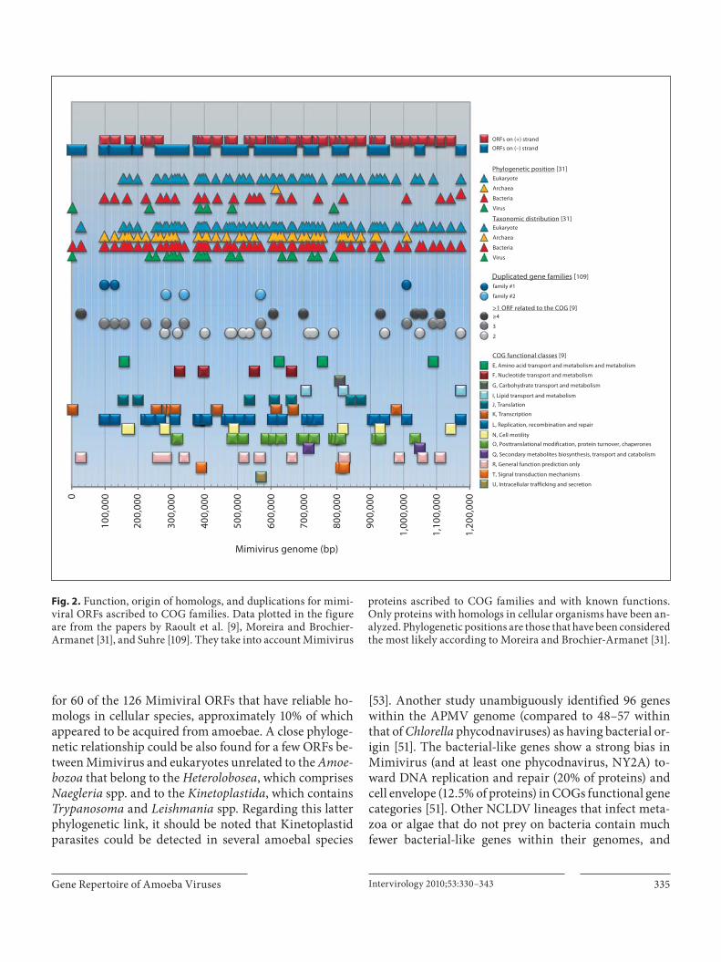
Gene Repertoire of Amoeba Viruses Intervirology 2010;53:330–343 335
for 60 of the 126 Mimiviral ORFs that have reliable ho-mologs in cellular species, approximately 10% of which appeared to be acquired from amoebae. A close phyloge-netic relationship could be also found for a few ORFs be-tween Mimivirus and eukaryotes unrelated to the Amoe-bozoa that belong to the Heterolobosea, which comprises Naegleria spp. and to the Kinetoplastida, which contains Trypanosoma and Leishmania spp. Regarding this latter phylogenetic link, it should be noted that Kinetoplastid parasites could be detected in several amoebal species
[53] . Another study unambiguously identified 96 genes within the APMV genome (compared to 48–57 within that of Chlorella phycodnaviruses) as having bacterial or-igin [51] . The bacterial-like genes show a strong bias in Mimivirus (and at least one phycodnavirus, NY2A) to-ward DNA replication and repair (20% of proteins) and cell envelope (12.5% of proteins) in COGs functional gene categories [51] . Other NCLDV lineages that infect meta-zoa or algae that do not prey on bacteria contain much fewer bacterial-like genes within their genomes, and
family #1
family #2
Eukaryote
Archaea
Bacteria
Virus
Eukaryote
Archaea
Bacteria
Virus
≥4
3
2
Mimivirus genome (bp)
100,
0000
200,
000
300,
000
400,
000
500,
000
600,
000
700,
000
800,
000
900,
000
1,00
0,00
0
1,10
0,00
0
1,20
0,00
0
ORFs on (+) strand
ORFs on (–) strand
Phylogenetic position [31]
Taxonomic distribution [31]
Duplicated gene families [109]
>1 ORF related to the COG [9]
COG functional classes [9]E, Amino acid transport and metabolism and metabolism
F, Nucleotide transport and metabolism
G, Carbohydrate transport and metabolism
I, Lipid transport and metabolism
J, Translation
K, Transcription
L, Replication, recombination and repair
N, Cell motility
O, Posttranslational modification, protein turnover, chaperones
Q, Secondary metabolites biosynthesis, transport and catabolism
R, General function prediction only
T, Signal transduction mechanisms
U, Intracellular trafficking and secretion
Fig. 2. Function, origin of homologs, and duplications for mimi-viral ORFs ascribed to COG families. Data plotted in the figure are from the papers by Raoult et al. [9] , Moreira and Brochier-Armanet [31] , and Suhre [109] . They take into account Mimivirus
proteins ascribed to COG families and with known functions. Only proteins with homologs in cellular organisms have been an-alyzed. Phylogenetic positions are those that have been considered the most likely according to Moreira and Brochier-Armanet [31] .

Colson /Raoult Intervirology 2010;53:330–343336
these genes are rather scattered throughout the genome [51] . These findings suggest that eukaryotic hosts using bacteria as food may provide a hotspot for the promiscu-ous exchange of DNA between replicating viruses and bacteria, thus providing a biological niche with access to bacterial genes ( fig. 3 ).
Interestingly, Filee et al. examined the distribution and location of bacterial-like genes in giant viruses, in-cluding APMV [51] . They identified three consecutive co-inherited reading frames, encoding a sugar transami-nase, a glycosyltransferase, and a protein of unknown function in the APMV genome, that are syntenic with three ORFs in the genome of Clostridium acetobutylicum , suggesting the inheritance of these bacterial-like genes as a short contiguous block. Additional findings also sug-gested co-inheritance of bacterial-like genes by NY2A,
a member of the Phycodnaviridae family. Another impor-tant finding of their work was that bacterial-likegenes tend to be clustered toward the extremities of the NCLDVs’ genomes, possibly in islands. In the APMV ge-nome, these regions were found positioned within the first and last 250 kb of the genome. In contrast, NCLDV core genes and eukaryotic-like genes are positioned to-ward the middle of the genome. It is noteworthy that nu-merous mobile genetic elements have been detected in the APMV genome, as these were previously thought to be specific to prokaryotes [51] . They include insertion se-quences, considered major agents of LGT in prokaryotes [54] , two homing endonucleases, and an intein. The in-sertion sequences contain two ORFs, one encoding a transposase and the other a protein of unknown function [55] . These two elements had previously only been identi-
Prom
iscu
ity
betw
een
the
DN
A o
f rep
licat
ing
viru
ses
and
bact
eria
Rate
of l
ater
al g
ene
tran
sfer
sG
enom
e si
ze o
f int
race
llula
r org
anis
ms
BACTERIA VIRUSES
Mimivirus, Marseillevirus
Sputnik
Major evolutionary driving forces:•Lateral gene transfers•Gene duplications
?
G
G
GG
G
G
G
G
METAZOA, ALGA,do not feed on bacteria
GIANT VIRUSES WITH COMPLEX CHIMERIC GENOMES GIANT VIRUSES WITH COMPLEX CHIMERIC GENOMES
Other NCLDVsincluding poxviruses,iridoviruses,asfarviruses, andphycodnavirusesother than Chlorellaphycodnaviruses
AMOEBAfeed on bacteria
N
N
N
-
+
G
Other EUKARYOTES
G
NCLDVs other thanMimiviridae and Marseillevirus
G
GG
Fig. 3. Intra-amoebal lifestyle as a source of complex chimeric gene contents. Col-ored boxes containing a G indicate genes from various origins (bacteria, viruses, eukaryotes).

Gene Repertoire of Amoeba Viruses Intervirology 2010;53:330–343 337
fied in the genome of one other eukaryotic virus, a brown alga virus ( Ectocarpus silicosus virus 1). Those detected in the APMV genome appear to be full-length copies and belong to the IS607 family. These insertion sequences co-localize with bacterial-like genes, being embedded in some cases within stretches of contiguous bacterial-like genes (or with ORFans), further supporting their putative co-inheritance with bacterial DNA. Homing endonucle-ases are characterized by an HNH motif and are usually encoded by genes within mobile, self-splicing introns that promote the movement of the DNA sequences that en-code them [56] . The two Mimivirus HNH endonuclease genes flank a unique gene typically detected in prophag-es of bacteria, supporting the hypothesis of acquisition by LGT from a bacteriophage [51] . Finally, an intein has been detected in the APMV genome [57] . Inteins, first discov-ered in 1990 [58, 59] , are segments of proteins that cata-lyze self-splicing at the protein level. They have been found in bacteria, archaea and unicellular eukaryotes, and it has been proposed that they may be transferred between species by viruses. They have been detected in many prophages and bacteriophages, but APMV is one of the few eukaryote-infecting viruses, with iridoviruses, and Chlorella and Ostreococcus phycodnaviruses, that has been found to harbor an intein [51, 60, 61] . The Mimi-virus intein is most likely to be functional and is closest to extremophilic archaeal-type inteins. Its association with a mesophilic eukaryotic-like DNA polymerase PolB gene in the APMV genome is consistent with the hypoth-esis that DNA viruses may have represented a central res-ervoir of inteins during the evolutionary course [57] .
Of the 21 ORFs identified in the Sputnik genome, the 8 non-ORFan proteins have either viral, plasmidic, bacte-rial or eukaryotic homologs and/or homologs within the environmental Global Ocean Survey (GOS) sequencedatabase [8, 62] . Three ORFs are most closely related to Mimivirus and Mamavirus ORFs [8] . Among the most noteworthy features, ORF 13 contains two domains in-volved in viral DNA replication. Its N-terminal domain has homologs with high similarity among proteins from the GOS database and may correspond to an archaeo-eukaryotic primase, whereas its C-terminal domain is a highly conserved superfamily 3 helicase of NCLDVs. ORF 3 has limited similarity with a packaging ATPase conserved in all NCLDVs and in many bacteriophages [62, 63] . ORF 17 encodes a protein with homologs in the GOS dataset and that belongs to the family of bacterial insertion sequence transposase DNA-binding subunits, and ORF 10 is closely related to integrases of the tyrosine recombinase family from archaeal viruses and provirus-
es. The presence of these two latter ORFs within the Sput-nik genome suggests that a provirophage stage may exist. This would reinforce the hypothesis of a putative role for Sputnik as a vehicle of genes. Indeed, globally, Sputnik’s genomic content appears to be of chimeric origin and to be evolutionarily related to at least three distinct sources: Mimivirus/Mamavirus, an archaeal virus or a plasmid, and a putative novel family of viruses [8] . The relation-ship between Sputnik and Mimi-/Mamavirus genomes may be explained by gene exchange within viral factories during co-infection, as a result of recombination events. Efficient plasmid replication has been reported in poxvi-rus and in Asfarviridae [64, 65] , supporting the hypoth-esis that Mimi- or Mamavirus factories may be able to replicate foreign DNA, namely that of Sputnik.
Regarding Marseillevirus, 59, 57, 70 and 2 of its pre-dicted proteins exhibit the highest sequence similarityto viral, bacterial, eukaryotic, and archaeal homologs,respectively [11] . In 8 cases, the homologue that showsthe closest similarity to the Marseillevirus ORF is from Acanthamoeba . On the basis of phylogenetic analyses,the Marseillevirus genome contains 49 genes of proba-ble bacterial or bacteriophage origin and 85 of apparent eukaryotic origin. For 22 of these latter ORFs, a phylo-genetic relationship was found with Mimivirus and Acanthamoeba ORFs. Three genes appear to originate in other Amoebozoa. Among the 188 predicted Marseillevi-rus ORFs, 20 encode bacterial-like membrane-occupa-tion and recognition-nexus repeat domains, typically im-plicated in interactions between membranes or between membranes and cytoskeleton [66] , and 10 encode bacte-riophage HNH endonucleases and restriction-like endo-nucleases, which typically reside in mobile selfish genetic elements and might have been acquired by LGT. Addi-tionally, 3 ORFs are homologous to genes encoding his-tone-like proteins previously found only in the genomes of two polydnaviruses ( Heliothis zea virus 1, a insect nudivirus, and Cotesia plutellae bracovirus) [67, 68] . These histone proteins have been detected in the viral particle and may therefore play a role in viral DNA pack-aging [11] . Interestingly, the inferred origin for the pre-dicted Marseillevirus genes tends to be non-randomly connected with their function. Thus, mixed eukaryotic and bacterial origins were inferred for genes encoding metabolic enzymes and proteins implicated in protein and lipid modification or degradation, whereas genes im-plicated in signal transduction are primarily of eukary-otic origin, and many of the genes for defense and repair functions, in particular nucleases, seem to be of bacterial or bacteriophages origin. Altogether, the findings report-

Colson /Raoult Intervirology 2010;53:330–343338
ed by Boyer et al. led them to hypothesize that numerous Marseillevirus ORFs were acquired from many different sources through LGT and that the mosaicism exhibited by the Marseillevirus genome could have resulted from gene exchange with sympatric amoeba-infecting bacteria and viruses that have an intra-amoebal lifestyle, as well as with their amoebal host [11] . LGT may be common, as suggested by several cases where related genes seemed to be acquired from independent sources by Marseillevirus and Mimivirus [11] .
The substantial flow of genes due to LGT that has been detected in amoeba-associated viruses prompts the de-sire to understand the mechanisms of bacterial-like gene acquisition by APMV. It is assumed that free-living amoe-bae harbor a spectrum of pathogenic and non-pathogen-ic bacteria [69] . Although the current knowledge of the replication cycle of APMV and other NCLDVs is very limited [70] , it is conceivable that amoebae may promote gene exchange between bacterial and viral DNA during their replication in a same cell compartment [8, 69, 71] . One possible mechanism for LGT between bacterial and APMV genomes may be suggested by the replication mode of poxviruses, other NCLDVs [72] . Indeed, repli-cating poxviruses have been found to display very high levels of homologous recombination that requires little sequence homology [73] and appears to occur predomi-nantly at the ends of the viral genome [74, 75] . Interest-ingly, similar recombination frequencies have been noted in phycodnaviruses [76] . Another mechanism may be similar to that observed during recombination in Herpes-viridae, which are also large DNA viruses [77] . This pro-cess involves a DNA-binding protein and an exonuclease, and replication is unnecessary. Alternatively, it has been proposed that recombination may be promoted by an en-zyme similar to the topoisomerase IB of the vaccinia vi-rus [51, 78] . In support of this, a DNA topoisomerase IB encoded by the APMV genome has been produced, puri-fied and characterized [79] . Interestingly, an evolutionary and functional relationship was demonstrated among the APMV, poxvirus, and bacterial topo isomerase IB pro-teins, and dissemination of an ancestral bacterial/viral enzyme by LGT was hypothesized.
The survival and life cycle of giant viruses in amoebae may be the cornerstone of their gene repertoire ( fig. 3 ). Free-living amoebae of the genus Acanthamoeba are ubiquitous in the environment and can be isolated from air, soil and water of various sources [80, 81] . Amoebae are wild phagocytes that ingest any particle larger than 0.5 � m [82] . They harbor a variety of intracellular bacte-ria (either transiently or for long periods), including Chla-
mydia , Bacteroidetes , Actinobacteria , Firmicutes and Pro-teobacteria [69] . Several mimiviral ORFs have homologs in bacterial species that are common inhabitants of amoe-bae [31] . For instance, two ORFs corresponding to a Zn-dependant alcohol dehydrogenase and an outer-mem-brane lipoprotein seem to have homologs in Legionella pneumophila and Campylobacter spp., respectively. In the genome of Marseillevirus, the most recently described amoeba-infecting giant virus, 41% of ORFs exhibit the highest sequence similarities to homologs found in vi-ruses, bacteria, archaea and eukaryotes, and it has been suggested that the large proportion of chimeric genes ob-served in Marseillevirus might have resulted from LGT with sympatric intra-amoebal bacteria and viruses [11] . A recent work has underscored the observation that amoebal endosymbionts appear to have larger genomes than their relatives [83] , in contrast to other intracellular bacteria whose lifestyle is associated with genome reduc-tion [84, 85] . Thus, Moliner et al. reported that the Legio-nella drancourtii genome is larger than those sequenced from L. pneumophila strains (4.2 Mb vs. 3.5 Mb on aver-age) [83, 86, 87] . Along the same lines, the genome of Rickettsia bellii is the largest among Rickettsia species [71] , and the genomes of Candidatus ‘Protochlamydia amoebophila’ and of Parachlamydia acanthamoebae are approximately twice as large as those of pathogenic Chla-mydia species [88, 89] . Among viruses, APMV and Mar-seillevirus have the largest and fifth-largest genomes, re-spectively [9, 11] . Therefore, in all those previous exam-ples, the intra-amoebal lifestyle has been positively associated with genome size. This may be explained by possible foraging of genes by sympatric microorganisms due to the promiscuity between their replicating ge-nomes. In contrast, the lifestyle of obligate intracellular bacteria in other eukaryotic cells limits such genetic pro-miscuity and the concomitant capacity to acquire foreign genes ( fig. 3 ). The work by Moliner et al. further provides a very good illustration of how the intra-amoebal lifestyle might enable the exchange of genes across the boundaries of phyla, kingdoms, and even domains, and that some of these genes might have ancient origins [83] . Thus, a sterol delta-7 reductase was detected among orthologous pro-teins of Candidatus ‘Protochlamydia amoebophila’, L. drancourtii, and Coxiella burnetii , three amoeba-resis-tant bacteria, but not in other related species. Further-more, this protein is closely related to sterol delta-7 reduc-tases of Viridiplantae, which led to the hypothesis that its gene may have been acquired by amoeba-resistant bacte-ria following a transfer from Viridiplantae. Other studies have pointed out the putative role of amoebae in gene ex-

Gene Repertoire of Amoeba Viruses Intervirology 2010;53:330–343 339
changes between sympatric intra-cellular pathogens. For instance, ATP/ADP translocases were found to be a com-mon feature of obligate intra-cellular amoebal symbionts related to Chlamydiae and Rickettsiae [90] . Additionally, it has been suggested that amoeba-like ancestral protozoa might have served as a genetic ‘melting pot’ where the ancestors of Rickettsia and other bacteria exchanged genes that may have permitted their adaptation to an in-tracellular lifestyle in eukaryotic cells [71] . In contrast, species living in restricted environments and lacking mechanisms of gene exchange may have evolved with considerably less variation. An example is the obligate in-tracellular endosymbiont of aphids, Buchnera aphidicola , in which no chromosome rearrangements or gene acqui-sitions have occurred in the past 50–70 million years [91] . Furthermore, a massive comparative analysis of 317 ge-nomes of bacteria with different lifestyles recently dem-onstrated a convergent evolution for obligate intracellular bacteria, which included massive gene loss as a driving force in the adaptation of parasites to eukaryote cells [92] .
The remarkably extensive and diverse gene content of amoeba-associated giant viruses may provide them sev-eral benefits. L. pneumophila has acquired by LGT eu-karyotic genes involved in a variety of cell functions, in-cluding two serine/threonine protein kinases [93] . Such enzymes have been showed to inhibit phagosome-lyso-some fusion in several pathogens, promoting their intra-cellular survival and the disruption of host defenses by interfering with eukaryotic signal transduction [94] . In this view, the presence in the Mimivirus genome of three serine/threonine protein kinases acquired from its amoe-bal hosts and a RAS GTPase suggest that APMV may regulate the host cell cycle for its own benefit [31] . Fur-thermore, in addition to the fact that Acanthamoeba spe-cies can, themselves, infect a large spectrum of mammals, including humans [95] , their role as ‘Trojan horses’ has been discussed [81, 96] . Thus, most bacteria that multiply in amoebae are human pathogens [97] . The large gene content of giant viruses that multiply in amoebae may al-low them to survive and be pathogenic outside their host after this latter has played its role of environmental res-ervoir and gene-transfer vehicle [69] . Ghigo et al. demon-strated that APMV could enter macrophages by internal-ization, a process that closely resembles that mediated by amoeba [50, 98] . This suggests that amoebae may pro-mote the adaptation of some microorganisms to macro-phages. For instance, L. pneumophila, C. burnetii and Parachlamydiaceae are resistant to amoebae and have been shown to be pathogens for macrophages [98, 99] . The finding that the intracellular survival strategies of
Cryptococcus neoformans in amoebae and macrophages are very similar also suggests that selective pressures placed by amoebae on microorganisms may be a key fac-tor in the maintenance of their virulence characteristics for animal hosts [100] . These data further suggest a patho-genic role for APMV and are in agreement with findings that support a causative role for it in pneumonia [101–105] . The detection in Mimivirus and Marseillevirus of LGTs involving different eukaryotic sources and the rela-tive paucity of genes of Acanthamoeba origin further sug-gest that these viruses may have a larger spectrum of hosts than was historically thought and that they may have changed hosts in the course of their evolution [9, 11, 48] . In agreement with this hypothesis, very recent find-ings suggest that Mimivirus relatives may be very ubiqui-tous in the biosphere, and microalgae and modern spong-es might be hosts to yet unidentified members of the Mimiviridae [106, 107] . Finally, previous findings have underscored that amoebae are unique host cells where bacteria and viruses are in proximity. Besides exchanging genes, these microorganisms might compete to survive and multiply. Amoebae might therefore represent a bat-tlefield where sympatric microorganisms struggle for life with their gene armories. In this setting, possessing a ple-thoric and/or chimeric gene repertoire might be very beneficial to intra-amoebal giant viruses because, as pro-posed in the Red Queen hypothesis, no species can ever win in such an environment as the amoebal arena [108] .
Duplicated Genes Besides LGT, it has been suggested that gene and ge-
nome duplication played a major role in shaping the APMV genome [109] . Such events were defined several decades ago and are associated with the emergence of new gene material with different fates [110] . Duplicated genes can evolve towards nonfunctionality, with degen-eration into a pseudogene and possible elimination, or towards sub- or neo-functionalization. The divergence of duplicated genes may be very important in evolution, so much so that the homology between many genes with common ancestry can no longer be detected [111] . Suhre determined that about one-third of APMV genes have one or more paralogs in the genome [109] . Depending on the choice of the e-value cutoff, between 26.3 and 35.0% of APMV genes were found to have at least one paralog. The number of paralogs was estimated to be as high as 66, and the orientation and location of gene duplication events appear to be non-random. Indeed, duplicated genes are inserted about twice as frequently in the paral-lel orientation as in the antiparallel orientation, with re-

Colson /Raoult Intervirology 2010;53:330–343340
spect to the coding direction of the matching gene (20.2 vs. 11.7%), and the proportion of duplications that are lo-calized to the same half of the genome is 79% for duplica-tions in cis versus 39% for duplications in trans . Interest-ingly, among the major families of paralogs, several showed homology, although often remote or only puta-tive, with genes encoding proteins that are likely to play a role in virus–host interactions by interfering with im-portant host processes such as transcription, protein deg-radation, or cellular regulatory processes. One such fam-ily includes proteins with a Pfam F-box domain, which is a receptor that serves as a link between a target protein and a ubiquitin-conjugating enzyme. The largest paralo-gous family corresponds to ankyrin repeat-containing proteins that might have a structural role. Finally, aside from gene duplication, Suhre described data that suggest the occurrence of segmental duplication implicating a large part of the 5 � -end and 3 � -end of the genome. The large number of gene duplications in the APMV genome demonstrated in the study of Suhre lends support to the theory that duplication events contributed substantially to its large size, without a substantial requirement of LGT for the acquisition of genes. As a matter of fact, the gene duplication rate of APMV is within the range of frequen-cies observed in the three domains of life. The hypothesis that duplicated genes interfere with host processes may be compatible with the accretion and fixation of the large gene content of the APMV genome because the latter would allow APMV to mimic large microbial prey for amoebae. Supporting this hypothesis, a log-linear corre-lation could be identified for APMV and other large DNA viruses between the number of paralogous genes and the number of predicted genes within the genome [109] .
ORFans in Mimivirus, Marseillevirus and the Sputnik Virophage ORFans refer to genes without detectable homologs in
sequence databases [112] . A challenging feature of the ge-nomes of APMV, Marseillevirus and the Sputnik viro-phage are that a substantial part of their predicted coding genes do not exhibit clear homology to proteins of known function [8, 9, 11] . Furthermore, 39% of APMV ORFs and 13 of Sputnik’s 21 predicted genes do not clearly match any sequence found in databases [8, 9] , whereas sequence similarity and conserved domain searches against NCBI databases identified significant matches and/or con-served domains for only 188 of the 457 Marseillevirus predicted ORFs. The substantial proportion of ORFans in the genomes of amoeba-associated viruses under-scores the fact that our sequence databases, although rap-
idly growing, may still cover only a small fraction of the pangenome of our biosphere. As ORFans of amoeba-as-sociated giant viruses are the topic of another review in this journal issue, they are not described here.
Conclusion
In a recent correspondence letter on the complexity of the virus world, Koonin et al. stated that ‘the virus world is a dynamic network of relationships in which genes have diverse, variously intertwined histories’ [113] . This para-digm seems to apply perfectly to the giant viruses isolated from amoebae. The genomes of Mimivirus and Marseil-levirus contain core genes of NCLDVs but also genes of various origins in the three domains of life. Thus, they are made up of a backbone of genes upon which is grafted a complex, chimeric, dispensable genome acquired large-ly through LGT or gene duplication events. Moreover, the genomes of Mimivirus, Marseillevirus and Sputnik con-tain many ORFans. Gene accretion, leading to unexpect-edly large repertoires of genes of various and sometimes remote origins, probably occurs as a consequence of an intra-amoebal lifestyle, which enables the sharing of genes between sympatric bacteria, viruses and eukary-otes. This provides amoeba-associated giant viruses with gene armories that may help them out-compete other mi-croorganisms present in their hosts. Finally, the remark-ably extensive and diverse gene content of Mimivirus and Marseillevirus may enable them to venture outside the amoebae. In support of this, Mimivirus has been shown to be internalized by macrophages, leading to productive replication [98] , and several studies implicate this virus as a possible causative agent of pneumonia [101–105] . Altogether, the findings reviewed herein are a powerful incentive to find and study other amoeba-associated gi-ant viruses, so that we may gain a better understanding of their pangenome.

Gene Repertoire of Amoeba Viruses Intervirology 2010;53:330–343 341
References
1 Fuhrman JA: Marine viruses and their bio-geochemical and ecological effects. Nature 1999; 399: 541–548.
2 Suttle CA: Viruses in the sea. Nature 2005; 437: 356–361.
3 Woese C: The universal ancestor. Proc Natl Acad Sci USA 1998; 95: 6854–6859.
4 Woese CR, Kandler O, Wheelis ML: Towards a natural system of organisms: proposal for the domains Archaea, Bacteria, and Eucarya. Proc Natl Acad Sci USA 1990; 87: 4576–4579.
5 van Regenmortel MHV: 7th report of the In-ternational Committee on taxonomy of Vi-ruses. San Diego, Academic Press, 2000, pp 3–16.
6 Lwoff A: The concept of virus. J Gen Micro-biol 1957; 17: 239–253.
7 Raoult D, La Scola B, Birtles R: The discovery and characterization of Mimivirus, the larg-est known virus and putative pneumonia agent. Clin Infect Dis 2007; 45: 95–102.
8 La Scola B, Desnues C, Pagnier I, Robert C, Barrassi L, Fournous G, Merchat M, Suzan-Monti M, Forterre P, Koonin E, Raoult D: The virophage as a unique parasite of the gi-ant mimivirus. Nature 2008; 455: 100–104.
9 Raoult D, Audic S, Robert C, Abergel C,Renesto P, Ogata H, La Scola B, Suzan M, Claverie JM: The 1.2-megabase genome se-quence of Mimivirus. Science 2004; 306: 1344–1350.
10 La Scola B, Audic S, Robert C, Jungang L, de Lamballerie X, Drancourt M, Birtles R, Cla-verie JM, Raoult D: A giant virus in amoe-bae. Science 2003; 299: 2033.
11 Boyer L, Yutin N, Pagnier I, Barassi L, Four-nous G, Espinosa L, Robert C, Azza S, Sun S, Rossmann MG, Suzan-Monti M, La Scola B, Koonin EV, Raoult D: Giant Marseillevirus highlights the role of amoebae as a melting pot in emergence of chimeric microorgan-isms. Proc Natl Acad Sci USA 2009; 106: 21848–21853.
12 Iyer LM, Balaji S, Koonin EV, Aravind L: Evolutionary genomics of nucleo-cytoplas-mic large DNA viruses. Virus Res 2006; 117: 156–184.
13 Koonin EV: Virology: Gulliver among the Lilliputians. Curr Biol 2005; 15:R167–R169.
14 Claverie JM, Abergel C: Mimivirus and its virophage. Annu Rev Genet 2009; 43: 49–66.
15 Yamada T, Onimatsu H, Van Etten JL: Chlo-rella viruses. Adv Virus Res 2006; 66: 293–336.
16 Delaroque N, Boland W, Muller DG, Knip-pers R: Comparisons of two large phaeoviral genomes and evolutionary implications. J Mol Evol 2003; 57: 613–622.
17 Freeman MJ, Plasterer NT, Smith FT, Mohr CS: Patterns of genome organization in bac-teria. Science 1998; 279: 1827.
18 Galperin MY, Koonin EV: ‘Conserved hypo-thetical’ proteins: prioritization of targets for experimental study. Nucleic Acids Res 2004; 32: 5452–5463.
19 Koonin EV: Orthologs, paralogs, and evolu-tionary genomics. Annu Rev Genet 2005; 39: 309–338.
20 Fitzgerald LA, Graves MV, Li X, Feldblyum T, Nierman WC, Van Etten JL: Sequence and annotation of the 369-kb NY-2A and the 345-kb AR158 viruses that infect Chlorella NC64A. Virology 2007; 358: 472–484.
21 Byrne D, Grzela R, Lartigue A, Audic S, Chenivesse S, Encinas S, Claverie JM, Aber-gel C: The polyadenylation site of Mimivirus transcripts obeys a stringent ‘hairpin rule’. Genome Res 2009; 19: 1233–1242.
22 Suhre K, Audic S, Claverie JM: Mimivirus gene promoters exhibit an unprecedented conservation among all eukaryotes. Proc Natl Acad Sci USA 2005; 102: 14689–14693.
23 Wilson WH, Schroeder DC, Allen MJ, Hol-den MT, Parkhill J, Barrell BG, Churcher C, Hamlin N, Mungall K, Norbertczak H, Quail MA, Price C, Rabbinowitsch E, Walker D, Craigon M, Roy D, Ghazal P: Complete ge-nome sequence and lytic phase transcription profile of a Coccolithovirus. Science 2005; 309: 1090–1092.
24 Hutson MS, Duke T, Viovy JL: Two-dimen-sional motion of DNA bands during 120° pulsed-field gel electrophoresis. I. Effect of molecular weight. Biopolymers 1995; 35: 297–306.
25 Claverie JM, Ogata H, Audic S, Abergel C, Suhre K, Fournier PE: Mimivirus and the emerging concept of ‘giant’ virus. Virus Res 2006; 117: 133–144.
26 Koonin EV, Senkevich TG, Dolja VV: The ancient Virus World and evolution of cells. Biol Direct 2006; 1: 29.
27 Iyer LM, Aravind L, Koonin EV: Common origin of four diverse families of large eu-karyotic DNA viruses. J Virol 2001; 75: 11720–11734.
28 Yutin N, Wolf YI, Raoult D, Koonin EV: Eu-karyotic large nucleo-cytoplasmic DNA vi-ruses: clusters of orthologous genes and re-construction of viral genome evolution. Virol J 2009; 6: 223.
29 Awadalla P: The evolutionary genomics of pathogen recombination. Nat Rev Genet 2003; 4: 50–60.
30 Moreira D, Lopez-Garcia P: Ten reasons to exclude viruses from the tree of life. Nat Rev Microbiol 2009; 7: 306–311.
31 Moreira D, Brochier-Armanet C: Giant vi-ruses, giant chimeras: the multiple evolu-tionary histories of Mimivirus genes. BMC Evol Biol 2008; 8: 12.
32 Moreira D, Lopez-Garcia P: Comment on ‘The 1.2-megabase genome sequence of Mimivirus’. Science 2005; 308: 1114.
33 Andersson JO: Lateral gene transfer in eu-karyotes. Cell Mol Life Sci 2005; 62: 1182–1197.
34 Lawrence JG, Ochman H: Amelioration of bacterial genomes: rates of change and ex-change. J Mol Evol 1997; 44: 383–397.
35 Jain R, Rivera MC, Moore JE, Lake JA: Hori-zontal gene transfer accelerates genome in-novation and evolution. Mol Biol Evol 2003; 20: 1598–1602.
36 Canchaya C, Fournous G, Chibani-Chen-noufi S, Dillmann ML, Brussow H: Phage as agents of lateral gene transfer. Curr OpinMicrobiol 2003; 6: 417–424.
37 Filee J, Bapteste E, Susko E, Krisch HM: A selective barrier to horizontal gene transfer in the T4-type bacteriophages that has pre-served a core genome with the viral replica-tion and structural genes. Mol Biol Evol 2006; 23: 1688–1696.
38 Filee J, Tetart F, Suttle CA, Krisch HM: Ma-rine T4-type bacteriophages, a ubiquitous component of the dark matter of the bio-sphere. Proc Natl Acad Sci USA 2005; 102: 12471–12476.
39 Lindell D, Sullivan MB, Johnson ZI, Tolonen AC, Rohwer F, Chisholm SW: Transfer of photosynthesis genes to and from Prochlo-rococcus viruses. Proc Natl Acad Sci USA 2004; 101: 11013–11018.
40 Millard A, Clokie MR, Shub DA, Mann NH: Genetic organization of the psbAD regionin phages infecting marine Synechococcus strains. Proc Natl Acad Sci USA 2004; 101: 11007–11012.
41 McGeoch DJ, Rixon FJ, Davison AJ: Topics in herpesvirus genomics and evolution. Vi-rus Res 2006; 117: 90–104.
42 Alcami A, Koszinowski UH: Viral mecha-nisms of immune evasion. Trends Microbiol 2000; 8: 410–418.
43 McFadden G, Murphy PM: Host-related im-munomodulators encoded by poxviruses and herpesviruses. Curr Opin Microbiol 2000; 3: 371–378.
44 Moore PS, Boshoff C, Weiss RA, Chang Y: Molecular mimicry of human cytokine and cytokine response pathway genes by KSHV. Science 1996; 274: 1739–1744.
45 Fu M, Deng R, Wang J, Wang X: Detection and analysis of horizontal gene transfer in herpesvirus. Virus Res 2008; 131: 65–76.
46 Filee J, Pouget N, Chandler M: Phylogenetic evidence for extensive lateral acquisition of cellular genes by nucleocytoplasmic large DNA viruses. BMC Evol Biol 2008; 8: 320.
47 Esposito JJ, Sammons SA, Frace AM, Os-borne JD, Olsen-Rasmussen M, Zhang M, Govil D, Damon IK, Kline R, Laker M, Li Y, Smith GL, Meyer H, Leduc JW, Wohlhueter RM: Genome sequence diversity and clues to the evolution of variola (smallpox) virus. Sci-ence 2006; 313: 807–812.
48 Filee J: Lateral gene transfer, lineage-specific gene expansion and the evolution of Nucleo Cytoplasmic Large DNA viruses. J Invertebr Pathol 2009; 101: 169–171.
49 Ogata H, Abergel C, Raoult D, Claverie JM: Response to Comment on ‘The 1.2-Mega-base genome sequence of Mimivirus’. Sci-ence 2005; 308: 1114b.

Colson /Raoult Intervirology 2010;53:330–343342
50 Suzan-Monti M, La Scola B, Raoult D: Ge-nomic and evolutionary aspects of Mimivi-rus. Virus Res 2006; 117: 145–155.
51 Filee J, Siguier P, Chandler M: I am what I eat and I eat what I am: acquisition of bacterial genes by giant viruses. Trends Genet 2007; 23: 10–15.
52 Tatusov RL, Fedorova ND, Jackson JD, Ja-cobs AR, Kiryutin B, Koonin EV, Krylov DM, Mazumder R, Mekhedov SL, Nikolska-ya AN, Rao BS, Smirnov S, Sverdlov AV, Va-sudevan S, Wolf YI, Yin JJ, Natale DA: The COG database: an updated version includes eukaryotes. BMC Bioinformatics 2003; 4: 41.
53 Dyková I, Fiala I, Lom J, Lukeš J: Perkinsiella amoebae -like endosymbionts of Neopar-amoeba spp., relatives of the kinetoplastid Ichthyobodo . Eur J Protistol 2003; 39: 37–52.
54 Frost LS, Leplae R, Summers AO, Toussaint A: Mobile genetic elements: the agents of open source evolution. Nat Rev Microbiol 2005; 3: 722–732.
55 Ton-Hoang B, Guynet C, Ronning DR, Coin-tin-Marty B, Dyda F, Chandler M: Transpo-sition of ISHp608, member of an unusual family of bacterial insertion sequences. EMBO J 2005; 24: 3325–3338.
56 Guhan N, Muniyappa K: Structural and functional characteristics of homing endo-nucleases. Crit Rev Biochem Mol Biol 2003; 38: 199–248.
57 Ogata H, Raoult D, Claverie JM: A new ex-ample of viral intein in Mimivirus. Virol J 2005; 2: 8.
58 Hirata R, Ohsumk Y, Nakano A, Kawasaki H, Suzuki K, Anraku Y: Molecular structure of a gene, VMA1, encoding the catalytic sub-unit of H(+)-translocating adenosine tri-phosphatase from vacuolar membranes of Saccharomyces cerevisiae . J Biol Chem 1990; 265: 6726–6733.
59 Kane PM, Yamashiro CT, Wolczyk DF, Neff N, Goebl M, Stevens TH: Protein splicing converts the yeast TFP1 gene product to the 69-kD subunit of the vacuolar H(+)-adeno-sine triphosphatase. Science 1990; 250: 651–657.
60 Pietrokovski S: Identification of a virus in-tein and a possible variation in the protein-splicing reaction. Curr Biol 1998; 8:R634–R635.
61 Weynberg KD, Allen MJ, Ashelford K, Scan-lan DJ, Wilson WH: From small hosts come big viruses: the complete genome of a second Ostreococcus tauri virus, OtV-1. Environ Microbiol 2009; 11: 2821–2839.
62 Williamson SJ, Rusch DB, Yooseph S, Hal-pern AL, Heidelberg KB, Glass JI, Andrews-Pfannkoch C, Fadrosh D, Miller CS, Sutton G, Frazier M, Venter JC: The Sorcerer II Global Ocean Sampling Expedition: meta-genomic characterization of viruses within aquatic microbial samples. PLoS One 2008; 3:e1456.
63 Iyer LM, Makarova KS, Koonin EV, Aravind L: Comparative genomics of the FtsK-HerA superfamily of pumping ATPases: implica-tions for the origins of chromosome segrega-tion, cell division and viral capsid packaging. Nucleic Acids Res 2004; 32: 5260–5279.
64 De Silva FS, Moss B: Origin-independent plasmid replication occurs in vaccinia virus cytoplasmic factories and requires all five known poxvirus replication factors. Virol J 2005; 2: 23.
65 Rojo G, Garcia-Beato R, Vinuela E, Salas ML, Salas J: Replication of African swine fever vi-rus DNA in infected cells. Virology 1999; 257: 524–536.
66 Gubbels MJ, Vaishnava S, Boot N, Du-bremetz JF, Striepen B: A MORN-repeat pro-tein is a dynamic component of the Toxoplas-ma gondii cell division apparatus. J Cell Sci 2006; 119: 2236–2245.
67 Cheng CH, Liu SM, Chow TY, Hsiao YY, Wang DP, Huang JJ, Chen HH: Analysis of the complete genome sequence of the Hz-1 virus suggests that it is related to members of the Baculoviridae. J Virol 2002; 76: 9024–9034.
68 Ibrahim AMA, Choi JY, Je YH, Kim Y: Struc-ture and expression profile of two putative Cotesia plutellae bracovirus genes (CpBV-H4 and CpBV-E94{alpha}) in parasitized Plutel-la xylostella . J Asia Pacific Entomol 2005; 8: 359–366.
69 Horn M, Wagner M: Bacterial endosymbi-onts of free-living amoebae. J Eukaryot Mi-crobiol 2004; 51: 509–514.
70 Suzan-Monti M, La Scola B, Barrassi L, Espi-nosa L, Raoult D: Ultrastructural character-ization of the giant volcano-like virus facto-ry of Acanthamoeba polyphaga Mimivirus. PLoS One 2007; 2:e328.
71 Ogata H, La Scola B, Audic S, Renesto P, Blanc G, Robert C, Fournier PE, Claverie JM, Raoult D: Genome sequence of Rickettsia bellii illuminates the role of amoebae in gene exchanges between intracellular pathogens. PLoS Genet 2006; 2:e76.
72 Yao XD, Evans DH: Effects of DNA structure and homology length on vaccinia virus re-combination. J Virol 2001; 75: 6923–6932.
73 Willer DO, Yao XD, Mann MJ, Evans DH: In vitro concatemer formation catalyzed by vaccinia virus DNA polymerase. Virology 2000; 278: 562–569.
74 Ball LA: High-frequency homologous re-combination in vaccinia virus DNA. J Virol 1987; 61: 1788–1795.
75 Evans DH, Stuart D, McFadden G: High lev-els of genetic recombination among cotrans-fected plasmid DNAs in poxvirus-infected mammalian cells. J Virol 1988; 62: 367–375.
76 Tessman I: Genetic recombination of the DNA plant virus PBCV1 in a Chlorella -like alga. Virology 1985; 145: 319–322.
77 Reuven NB, Willcox S, Griffith JD, Weller SK: Catalysis of strand exchange by the HSV-1 UL12 and ICP8 proteins: potent ICP8 re-combinase activity is revealed upon resec-tion of dsDNA substrate by nuclease. J Mol Biol 2004; 342: 57–71.
78 Cheng C, Shuman S: Recombinogenic flap ligation pathway for intrinsic repair of topo-isomerase IB-induced double-strand breaks. Mol Cell Biol 2000; 20: 8059–8068.
79 Benarroch D, Claverie JM, Raoult D, Shu-man S: Characterization of mimivirus DNA topoisomerase IB suggests horizontal gene transfer between eukaryal viruses and bacte-ria. J Virol 2006; 80: 314–321.
80 Rodriguez-Zaragoza S: Ecology of free-liv-ing amoebae. Crit Rev Microbiol 1994; 20: 225–241.
81 Mattana A, Serra C, Mariotti E, Delogu G, Fiori PL, Cappuccinelli P: Acanthamoeba castellanii promotion of in vitro survival and transmission of coxsackie b3 viruses. Eu-karyot Cell 2006; 5: 665–671.
82 Audic S, Robert C, Campagna B, Parinello H, Claverie JM, Raoult D, Drancourt M: Ge-nome analysis of Minibacterium massilien-sis highlights the convergent evolution of water-living bacteria. PLoS Genet 2007; 3:e138.
83 Moliner C, Raoult D, Fournier PE: Evidence that the intra-amoebal Legionella drancour-tii acquired a sterol reductase gene from eu-karyotes. BMC Res Notes 2009; 2: 51.
84 Boussau B, Karlberg EO, Frank AC, Legault BA, Andersson SG: Computational infer-ence of scenarios for alpha-proteobacterial genome evolution. Proc Natl Acad Sci USA 2004; 101: 9722–9727.
85 Sakharkar KR, Dhar PK, Chow VT: Genome reduction in prokaryotic obligatory intracel-lular parasites of humans: a comparative analysis. Int J Syst Evol Microbiol 2004; 54: 1937–1941.
86 Chien M, Morozova I, Shi S, Sheng H, Chen J, Gomez SM, Asamani G, Hill K, Nuara J, Feder M, Rineer J, Greenberg JJ, Steshenko V, Park SH, Zhao B, Teplitskaya E, Edwards JR, Pampou S, Georghiou A, Chou IC, Ian-nuccilli W, Ulz ME, Kim DH, Geringer-Sa-meth A, Goldsberry C, Morozov P, Fischer SG, Segal G, Qu X, Rzhetsky A, Zhang P, Cayanis E, De Jong PJ, Ju J, Kalachikov S, Shuman HA, Russo JJ: The genomic se-quence of the accidental pathogen Legionella pneumophila . Science 2004; 305: 1966–1968.
87 Cazalet C, Rusniok C, Bruggemann H,Zidane N, Magnier A, Ma L, Tichit M, Jar-raud S, Bouchier C, Vandenesch F, KunstF, Etienne J, Glaser P, Buchrieser C: Evidence in the Legionella pneumophila genome for exploitation of host cell functions and high genome plasticity. Nat Genet 2004; 36: 1165–1173.

Gene Repertoire of Amoeba Viruses Intervirology 2010;53:330–343 343
88 Horn M, Collingro A, Schmitz-Esser S, Beier CL, Purkhold U, Fartmann B, Brandt P, Nyakatura GJ, Droege M, Frishman D, Rattei T, Mewes HW, Wagner M: Illuminating the evolutionary history of chlamydiae. Science 2004; 304: 728–730.
89 Greub G, Kebbi-Beghdadi C, Bertelli C, Col-lyn F, Riederer BM, Yersin C, Croxatto A, Raoult D: High throughput sequencing and proteomics to identify immunogenic pro-teins of a new pathogen: the dirty genome approach. PLoS One 2009; 4:e8423.
90 Schmitz-Esser S, Linka N, Collingro A, Beier CL, Neuhaus HE, Wagner M, Horn M: ATP/ADP translocases: a common feature of ob-ligate intracellular amoebal symbionts relat-ed to Chlamydiae and Rickettsiae. J Bacteriol 2004; 186: 683–691.
91 Tamas I, Klasson L, Canback B, Naslund AK, Eriksson AS, Wernegreen JJ, Sandstrom JP, Moran NA, Andersson SG: 50 million years of genomic stasis in endosymbiotic bacteria. Science 2002; 296: 2376–2379.
92 Merhej V, Royer-Carenzi M, Pontarotti P, Raoult D: Massive comparative genomic analysis reveals convergent evolution of spe-cialized bacteria. Biol Direct 2009; 4: 13.
93 Bruggemann H, Cazalet C, Buchrieser C: Adaptation of Legionella pneumophila to the host environment: role of protein secretion, effectors and eukaryotic-like proteins. Curr Opin Microbiol 2006; 9: 86–94.
94 Walburger A, Koul A, Ferrari G, Nguyen L, Prescianotto-Baschong C, Huygen K, Klebl B, Thompson C, Bacher G, Pieters J: Protein kinase G from pathogenic mycobacteria pro-motes survival within macrophages. Science 2004; 304: 1800–1804.
95 Meersseman W, Lagrou K, Sciot R, de JJ, Haberler C, Walochnik J, Peetermans WE, van WE: Rapidly fatal Acanthamoeba en-cephalitis and treatment of cryoglobuline-mia. Emerg Infect Dis 2007; 13: 469–471.
96 Abu KY, Gao LY, Stone BJ, Venkataraman C, Harb OS: Invasion of protozoa by Legio-nella pneumophila and its role in bacterial ecology and pathogenesis. Appl Environ Microbiol 1998; 64: 3127–3133.
97 Barker J, Brown MR: Trojan horses of the microbial world: protozoa and the survival of bacterial pathogens in the environment. Microbiology 1994; 140: 1253–1259.
98 Ghigo E, Kartenbeck J, Lien P, Pelkmans L, Capo C, Mege JL, Raoult D: Ameobal pathogen Mimivirus infects macrophages through phagocytosis. PLoS Pathog 2008; 4:e1000087.
99 Greub G, Raoult D: Microorganisms resis-tant to free-living amoebae. Clin Microbiol Rev 2004; 17: 413–433.
100 Steenbergen JN, Shuman HA, Casadevall A: Cryptococcus neoformans interactions with amoebae suggest an explanation for its virulence and intracellular pathogenic strategy in macrophages. Proc Natl Acad Sci USA 2001; 98: 15245–15250.
101 La Scola B, Marrie TJ, Auffray JP, Raoult D: Mimivirus in pneumonia patients. Emerg Infect Dis 2005; 11: 449–452.
102 Berger P, Papazian L, Drancourt M, La Scola B, Auffray JP, Raoult D: Ameba-asso-ciated microorganisms and diagnosis of nosocomial pneumonia. Emerg Infect Dis 2006; 12: 248–255.
103 Raoult D, Renesto P, Brouqui P: Laboratory infection of a technician by mimivirus. Ann Intern Med 2006; 144: 702–703.
104 Khan M, La Scola B, Lepidi H, Raoult D: Pneumonia in mice inoculated experimen-tally with Acanthamoeba polyphaga mimi-virus. Microb Pathog 2007; 42: 56–61.
105 Vincent A, La Scola B, Forel JM, Pauly V, Raoult D, Papazian L: Clinical significance of a positive serology for mimivirus in pa-tients presenting a suspicion of ventilator-associated pneumonia. Crit Care Med 2009; 37: 111–118.
106 Monier A, Larsen JB, Sandaa RA, Bratbak G, Claverie JM, Ogata H: Marine mimivi-rus relatives are probably large algal virus-es. Virol J 2008; 5: 12.
107 Claverie JM, Grzela R, Lartigue A, Berna-dac A, Nitsche S, Vacelet J, Ogata H, Aber-gel C: Mimivirus and Mimiviridae: giant viruses with an increasing number of po-tential hosts, including corals and sponges. J Invertebr Pathol 2009; 101: 172–180.
108 Van Valen L: A new evolutionary law. Evo-lutionary Theory 1973; 1: 1–30.
109 Suhre K: Gene and genome duplication in Acanthamoeba polyphaga Mimivirus. J Vi-rol 2005; 79: 14095–14101.
110 Zhang J: Evolution by gene duplication: an update. Trends Ecol Evol 2003; 18: 292–298.
111 Hurles M: Gene duplication: the genomic trade in spare parts. PLoS Biol 2004; 2:E206.
112 Fischer D, Eisenberg D: Finding families for genomic ORFans. Bioinformatics 1999; 15: 759–762.
113 Koonin EV, Wolf YI, Nagasaki K, Dolja VV: The complexity of the virus world. Nat Rev Microbiol 2009, E-pub ahead of print.