A «Repertoire for Repertoire» Hypothesis: Repertoires of Type Three Effectors are Candidate...
21
A «Repertoire for Repertoire» Hypothesis: Repertoires of Type Three Effectors are Candidate Determinants of Host Specificity in Xanthomonas Ahmed Hajri 1 , Chrystelle Brin 1 , Gilles Hunault 2 , Fre ´de ´ ric Lardeux 2 , Christophe Lemaire 3 , Charles Manceau 1 , Tristan Boureau 3. *, Ste ´ phane Poussier 4. * 1 De ´ partement Sante ´ des Plantes et Environnement, Institut National de la Recherche Agronomique (INRA), Beaucouze ´ , France, 2 De ´partement d’Informatique, Universite ´ d’Angers, Angers, France, 3 De ´ partement de Biologie, Universite ´ d’Angers, Angers, Beaucouze ´, France, 4 De ´partement de Sciences Biologiques, Agrocampus Ouest centre d’Angers, Institut National d’Horticulture et de Paysage (INHP), Beaucouze ´, France Abstract Background: The genetic basis of host specificity for animal and plant pathogenic bacteria remains poorly understood. For plant pathogenic bacteria, host range is restricted to one or a few host plant species reflecting a tight adaptation to specific hosts. Methodology/Principal Findings: Two hypotheses can be formulated to explain host specificity: either it can be explained by the phylogenetic position of the strains, or by the association of virulence genes enabling a pathological convergence of phylogenically distant strains. In this latter hypothesis, host specificity would result from the interaction between repertoires of bacterial virulence genes and repertoires of genes involved in host defences. To challenge these two hypotheses, we selected 132 Xanthomonas axonopodis strains representative of 18 different pathovars which display different host range. First, the phylogenetic position of each strain was determined by sequencing the housekeeping gene rpoD. This study showed that many pathovars of Xanthomonas axonopodis are polyphyletic. Second, we investigated the distribution of 35 type III effector genes (T3Es) in these strains by both PCR and hybridization methods. Indeed, for pathogenic bacteria T3Es were shown to trigger and to subvert host defences. Our study revealed that T3E repertoires comprise core and variable gene suites that likely have distinct roles in pathogenicity and different evolutionary histories. Our results showed a correspondence between composition of T3E repertoires and pathovars of Xanthomonas axonopodis. For polyphyletic pathovars, this suggests that T3E genes might explain a pathological convergence of phylogenetically distant strains. We also identified several DNA rearrangements within T3E genes, some of which correlate with host specificity of strains. Conclusions/Significance: These data provide insight into the potential role played by T3E genes for pathogenic bacteria and support a ‘‘repertoire for repertoire’’ hypothesis that may explain host specificity. Our work provides resources for functional and evolutionary studies aiming at understanding host specificity of pathogenic bacteria, functional redundancy between T3Es and the driving forces shaping T3E repertoires. Citation: Hajri A, Brin C, Hunault G, Lardeux F, Lemaire C, et al. (2009) A «Repertoire for Repertoire» Hypothesis: Repertoires of Type Three Effectors are Candidate Determinants of Host Specificity in Xanthomonas. PLoS ONE 4(8): e6632. doi:10.1371/journal.pone.0006632 Editor: Ching-Hong Yang, University of Wisconsin-Milwaukee, United States of America Received April 20, 2009; Accepted July 9, 2009; Published August 14, 2009 Copyright: ß 2009 Hajri et al. This is an open-access article distributed under the terms of the Creative Commons Attribution License, which permits unrestricted use, distribution, and reproduction in any medium, provided the original author and source are credited. Funding: This study is part of programs funded by the Region Pays de la Loire and by the Plant Health and Environment Department of INRA. AH is supported by a grant from the Tunisian Government, from the Conseil General du Maine-et-Loire and from INRA. The funders had no role in study design, data collection and analysis, decision to publish, or preparation of the manuscript. Competing Interests: The authors have declared that no competing interests exist. * E-mail: [email protected] (TB); [email protected] (SP) . These authors contributed equally to this work. Introduction Deciphering the mechanisms used by bacterial pathogens to evolve and adapt to new hosts is a major issue for both medical and plant sciences. Despite the tremendous achievements of 30 years of intensive research and the mass of information provided by the sequenced genomes in understanding the interactions between bacterial pathogens and their hosts, the molecular factors underlying host specificity of pathogenic bacteria still remain to be identified. Such fundamental knowledge tackles both the co- evolution between the host and the pathogen, as well as the factors underlying the emergence of new pathogens. For example, recent studies investigated the similarities between human and avian pathogenic strains of Escherichia coli, to gain insight into potential zoonotic risks of avian pathogenic strains [1–5]. Indeed, strains of E. coli belonging to the same phylogenetic groups may display pathogenicity either on poultry or on humans, and the question whether avian strains may serve as potential reservoir of antibiotic resistance or virulence genes is of crucial importance [2]. For plant pathogenic bacteria, host specificity of strains is usually very high and well characterized. In many species of plant pathogenic bacteria numerous pathovars are defined. A pathovar PLoS ONE | www.plosone.org 1 August 2009 | Volume 4 | Issue 8 | e6632
Transcript of A «Repertoire for Repertoire» Hypothesis: Repertoires of Type Three Effectors are Candidate...

A «Repertoire for Repertoire» Hypothesis: Repertoires ofType Three Effectors are Candidate Determinants of HostSpecificity in XanthomonasAhmed Hajri1, Chrystelle Brin1, Gilles Hunault2, Frederic Lardeux2, Christophe Lemaire3, Charles
Manceau1, Tristan Boureau3.*, Stephane Poussier4.*
1 Departement Sante des Plantes et Environnement, Institut National de la Recherche Agronomique (INRA), Beaucouze, France, 2 Departement d’Informatique, Universite
d’Angers, Angers, France, 3 Departement de Biologie, Universite d’Angers, Angers, Beaucouze, France, 4 Departement de Sciences Biologiques, Agrocampus Ouest centre
d’Angers, Institut National d’Horticulture et de Paysage (INHP), Beaucouze, France
Abstract
Background: The genetic basis of host specificity for animal and plant pathogenic bacteria remains poorly understood. Forplant pathogenic bacteria, host range is restricted to one or a few host plant species reflecting a tight adaptation to specifichosts.
Methodology/Principal Findings: Two hypotheses can be formulated to explain host specificity: either it can be explainedby the phylogenetic position of the strains, or by the association of virulence genes enabling a pathological convergence ofphylogenically distant strains. In this latter hypothesis, host specificity would result from the interaction between repertoiresof bacterial virulence genes and repertoires of genes involved in host defences. To challenge these two hypotheses, weselected 132 Xanthomonas axonopodis strains representative of 18 different pathovars which display different host range.First, the phylogenetic position of each strain was determined by sequencing the housekeeping gene rpoD. This studyshowed that many pathovars of Xanthomonas axonopodis are polyphyletic. Second, we investigated the distribution of 35type III effector genes (T3Es) in these strains by both PCR and hybridization methods. Indeed, for pathogenic bacteria T3Eswere shown to trigger and to subvert host defences. Our study revealed that T3E repertoires comprise core and variablegene suites that likely have distinct roles in pathogenicity and different evolutionary histories. Our results showed acorrespondence between composition of T3E repertoires and pathovars of Xanthomonas axonopodis. For polyphyleticpathovars, this suggests that T3E genes might explain a pathological convergence of phylogenetically distant strains. Wealso identified several DNA rearrangements within T3E genes, some of which correlate with host specificity of strains.
Conclusions/Significance: These data provide insight into the potential role played by T3E genes for pathogenic bacteriaand support a ‘‘repertoire for repertoire’’ hypothesis that may explain host specificity. Our work provides resources forfunctional and evolutionary studies aiming at understanding host specificity of pathogenic bacteria, functional redundancybetween T3Es and the driving forces shaping T3E repertoires.
Citation: Hajri A, Brin C, Hunault G, Lardeux F, Lemaire C, et al. (2009) A «Repertoire for Repertoire» Hypothesis: Repertoires of Type Three Effectors are CandidateDeterminants of Host Specificity in Xanthomonas. PLoS ONE 4(8): e6632. doi:10.1371/journal.pone.0006632
Editor: Ching-Hong Yang, University of Wisconsin-Milwaukee, United States of America
Received April 20, 2009; Accepted July 9, 2009; Published August 14, 2009
Copyright: � 2009 Hajri et al. This is an open-access article distributed under the terms of the Creative Commons Attribution License, which permits unrestricteduse, distribution, and reproduction in any medium, provided the original author and source are credited.
Funding: This study is part of programs funded by the Region Pays de la Loire and by the Plant Health and Environment Department of INRA. AH is supported bya grant from the Tunisian Government, from the Conseil General du Maine-et-Loire and from INRA. The funders had no role in study design, data collection andanalysis, decision to publish, or preparation of the manuscript.
Competing Interests: The authors have declared that no competing interests exist.
* E-mail: [email protected] (TB); [email protected] (SP)
. These authors contributed equally to this work.
Introduction
Deciphering the mechanisms used by bacterial pathogens to
evolve and adapt to new hosts is a major issue for both medical
and plant sciences. Despite the tremendous achievements of 30
years of intensive research and the mass of information provided
by the sequenced genomes in understanding the interactions
between bacterial pathogens and their hosts, the molecular factors
underlying host specificity of pathogenic bacteria still remain to be
identified. Such fundamental knowledge tackles both the co-
evolution between the host and the pathogen, as well as the factors
underlying the emergence of new pathogens. For example, recent
studies investigated the similarities between human and avian
pathogenic strains of Escherichia coli, to gain insight into potential
zoonotic risks of avian pathogenic strains [1–5]. Indeed, strains of
E. coli belonging to the same phylogenetic groups may display
pathogenicity either on poultry or on humans, and the question
whether avian strains may serve as potential reservoir of antibiotic
resistance or virulence genes is of crucial importance [2].
For plant pathogenic bacteria, host specificity of strains is
usually very high and well characterized. In many species of plant
pathogenic bacteria numerous pathovars are defined. A pathovar
PLoS ONE | www.plosone.org 1 August 2009 | Volume 4 | Issue 8 | e6632

is a subspecific division that groups all bacterial strains that cause
the same symptoms on the same plant host range [6]. Within a
pathovar, a second level of specificity is defined: races were defined
based on the observation that some strains, although fully
pathogenic on most host cultivars, may reveal avirulent on a
certain cultivar. Such specificity between bacterial races and
cultivars follows Flor’s « gene for gene » theory [7], and has been
largely exploited in crop breeding. In the last 20 years, examples
accumulated of bacterial plant pathogens bypassing monogenic
resistances introduced in crops. By contrast, host jumps only
seldom occur, suggesting that host specificity barriers are more
difficult to bypass. Therefore, understanding the molecular
mechanisms underlying host specificity may lead to engineering
more durable resistances for crop protection.
In the last ten years, genomes of many animal and plant
pathogenic bacteria were completely sequenced. Comparative
genomics studies demonstrated that repertoires of virulence
associated genes can be highly variable among the sequenced
strains, thus suggesting a role in host specificity [8–14]. However,
despite the increasing number of microbial genomes available,
genome comparison of several model strains may not fully
represent the extreme variability of host specializations that can
be found within one bacterial genus. For example, the Xanthomonas
genus is constituted of 27 species causing diseases on more than
400 different host plants, among which many economically
important crops [15]. At present, genomes of only 12 model
strains belonging to diverse species and pathovars of Xanthomonas
are sequenced or on the way to be sequenced (http://www.
genomesonline.org/). As long as at least one strain representative
of each pathovar is not sequenced, comparative genomics,
although highly informative, is not fully suited for the identification
of the molecular determinants of host specificity.
A pioneer study by Sarkar and colleagues [16] determined the
distribution of a large scope of virulence associated genes in a
collection of 91 strains of the plant pathogenic bacterium
Pseudomonas syringae, isolated from diverse host plants. They
postulated that looking at the distribution of these virulence-
associated genes may provide clues on their possible role in host
specificity: genes that are highly conserved among all strains,
irrespective to the host of isolation, probably do not play a major
role in host specificity. On the contrary, virulence-associated genes
heterogeneously distributed among strains are good candidates to
explain host specificity.
Among pathogenicity determinants shown to display heteroge-
neous distribution between strains are Type III Effectors (T3Es)
[12,16]. T3Es are bacterial proteins that are directly injected
inside the cytoplasm of the host cell by a bacterial molecular
apparatus called Type III Secretion System (T3SS). This system is
conserved among most of the gram negative plant and animal
pathogenic bacteria. Using the T3SS, each bacterial strain can
inject up to 30 T3Es in the eukaryotic host cell simultaneously
[17,18]. In Xanthomonas, a mutation in the T3SS impairs the ability
to inject T3Es in the host plant, and as a consequence abolishes
pathogenicity and multiplication in planta [19]. More precisely,
many studies demonstrated that T3Es alter the physiology of the
host cell in a way that is beneficial for the pathogen [11,20].
In 2006, Jones and Dangl [21] proposed a mechanistic model
for the interaction between gram negative bacteria and plants in
which the combined action of T3Es leads to suppression of host
defence reactions that are induced after recognition of Pathogen
Associated Molecular Patterns (PAMPs), thus resulting in an
effector-triggered susceptibility. In some cases, individual T3E (or
its action inside the cell) may specifically be recognized by the
plant. This recognition induces a hypersensitive response (HR),
resulting in an effector induced resistance. Such specific recogni-
tion of a T3E in the host cell by the product of a plant resistance
gene constitutes the molecular basis of the ‘‘gene for gene’’ theory
ruling race/cultivar specificity. Thus, T3Es may enlarge the host
range of a given bacterium by suppressing host defences, or
narrow the host range when one of them is specifically recognized
by the plant.
Experimental data suggest that T3Es may also be involved in
host specificity. Indeed, several studies used suppression subtrac-
tive hybridization (SSH) approaches to perform genomic compar-
ison of non-sequenced strains, that are very closely related
phylogenetically but differing in the hosts they attack. Among
the genes isolated in these SSHs were single T3Es, suggesting that
they can play a role in host specificity in plant pathogenic bacteria
[22–24] as well as in animal pathogenic bacteria [2].
Regarding the different T3Es injected in the host cell, they
probably act collectively. Supporting this idea, mutations achieved
in one single T3E rarely affect the virulence phenotype of strains.
This suggests that among all the T3Es injected in the host cell,
some have redundant functions virulence [25,26]. T3Es seem to
act synergistically or antagonistically on different pathways of the
host cell, to create a physiological status of the host that would be
optimal for the development of the pathogen. Examples
illustrating such idea can be found in plant and animal pathogenic
bacteria. Among the conserved T3Es of Pseudomonas syringae pv.
tomato DC3000, HopPtoM induces an increase of the number and
the size of the lesions, whereas HopPtoN induces a decrease of the
number and the size of the lesions [27,28]. In E. amylovora, similar
antagonism can be found also between HrpN and HrpW. Indeed,
HrpN induces ion fluxes that induce cell death in Arabidopsis,
whereas HrpW induce ion fluxes that prevent cell death induced
by HrpN. When both purified proteins HrpW and HrpN are
added in the culture medium, the quantity of cell death depends
on the relative concentration of the proteins [29]. In Salmonella
typhimurium, the T3E AvrA stabilizes cell permeability and tight
junctions in epithelial cells, whereas the T3Es SopB, SopE and
SopE2 were shown to destabilize tight junctions [30].
Moreover, it was shown that in maize, resistance genes involved
in race/cultivar specificity may also play a role in the recognition
of the non-host bacterium Xanthomonas oryzae [31]. This shows that
the same type of genes may be involved at both levels, race/
cultivar resistance and non-host resistance. Host specificity is most
probably not governed by a unique gene. If so, emergence of new
plant diseases would be as frequent as cases of bacterial pathogens
escaping monogenic resistances. May we generalize the model
described by Jones and Dangl [21], the outcome of the interaction
would depend greatly on the confrontation of repertoires of T3Es
and ‘‘guard genes’’ of the plant. Race/cultivar specificity would
then be explained in a ‘‘Gene for Gene’’ manner [7] whereas host
specificity would be explained in a ‘‘Repertoire for Repertoire’’
manner.
In the present study, we have chosen, as a model for investi-
gating the molecular determinants of host specialization, the
species Xanthomonas axonopodis [32]. Indeed, within this bacterial
species, numerous pathovars displaying various plant host ranges
have been defined [32–35]. Furthermore, it is worth noting that
recent phylogenetic studies performed on strains belonging to X.
axonopodis demonstrated that some pathovars did not form
monophyletic groups [36,37]. Some strains being phylogeneti-
cally very close may belong to different pathovars, whereas some
strains belonging to the same pathovar may be distant
phylogenetically. For example, the pathovar phaseoli, that groups
all the strains pathogenic on bean, comprises four distinct genetic
lineages [24]. Strains belonging to the genetic lineages 2 and 3 of
Xanthomonas Host Specificity
PLoS ONE | www.plosone.org 2 August 2009 | Volume 4 | Issue 8 | e6632

X. axonopodis pv. phaseoli are phylogenetically closer to strains
pathogenic on citrus or cotton, than strains belonging to the
genetic lineage 1 of the pathovar phaseoli [24]. This suggests that
in X. axonopodis, host specialization results from phylogeny-
independent factors.
Interestingly, Xanthomonas species comprise pathovars that have
different tissue specificities. For example, the species X. oryzae and
X. campestris include pathovars that invade their hosts through the
vascular system ( = vascular pathogens) and pathovars that
colonize the intercellular spaces of the parenchyma tissue
( = non-vascular pathogens) [38,39]. Like X. oryzae and X.
campestris, X. axonopodis also comprises vascular and non-vascular
pathovars [40–56]. Tissue specificity is also reported to be targeted
by T3Es [57].
In this study, we assayed for the presence of 35 T3Es in a set of
132 strains of the species X. axonopodis, representative of 18
different pathovars. We then tested for associations between
pathovars and the repertoire of T3Es of the tested strains, to
provide data challenging a ‘‘repertoire for repertoire’’ hypothesis
that may explain host and tissue specificity.
Results
Presence or absence of 35 T3Es in strains used in this work
was achieved by using specific primers for PCR amplification,
as well as by dot blot hybridization. Both methods were used
simultaneously to obtain complementary results: hybridi-
zation tells whether strains contain orthologs of the probed
T3E gene, whereas PCR approach provides clues on whether
genetic rearrangements may have occurred in the target
sequence.
Phylogenetic position of strains used in this work was obtained
by sequencing the housekeeping gene rpoD. Comparison of
dendrograms obtained using the rpoD phylogenetic data and the
dendrogram obtained based on presence or absence of T3Es
documents the involvement of T3E repertoires in host specialisa-
tion.
The X. axonopodis species comprises monophyletic andpolyphyletic pathovars
In order to test whether the phylogeny could explain the
distribution of the strains of our collection (Table 1) among the 18
pathovars of X. axonopodis, we sequenced rpoD, one of the
housekeeping genes commonly used in multilocus sequence
analysis and typing (MLSA and MLST) studies [37,58–61].
Phylogenetic trees based in rpoD sequences were constructed using
the method of Maximum Likelihood. Akaike information criterion
used in Modeltest [62] selected the GTR+I+G model with the
following parameters: f(A) = 0.1368, f(C) = 0.3256, f(G) = 0.3110;
rate matrix R(A–C) = 1.9312, R(A–G) = 12.5584, R(A–
T) = 0.4209, R(C–G) = 1.2602, R(C–T) = 2.6835, R(G–T) = 1;
proportion of invariable sites (I) = 0.5065 and Gamma distribution
shape parameter (G) = 0.7388. The Maximum Likelihood tree,
rooted with the orthologous rpoD sequences from X. campestris pv.
campestris strain CFBP5241, is presented in Figure 1. A very similar
tree was also obtained with sequences of four housekeeping genes
originating from about forty strains belonging to diverse pathovars
of X. axonopodis [36,37]. The tree constructed based on rpoD
sequences matched well with the 6 rep-PCR clusters (from 9.1 to
9.6) defined within the X. axonopodis species (Figure 1) [34,35].
Moreover, high bootstrap values indicated that this clustering was
well supported and that the tree was robust (Figure 1). It is worth
observing that, depending on the X. axonopodis pathovars, strains
were grouped together or were distributed into phylogenetically
unrelated groups (Figure 1). From this phylogenetic analysis,
pathovars anacardii, axonopodis, begoniae, citri, mangiferaeindicae, mal-
vacearum, manihotis, ricini, vesicatoria and vignicola can be considered as
monophyletic whereas pathovars alfalfae, allii, aurantifolii, citrumelo,
dieffenbachiae, glycines, phaseoli and vasculorum can be considered as
polyphyletic.
The rpoD-based tree was sufficient to highlight the existence of
different genetic lineages within a pathovar. As an example, the
four genetic lineages of the pathovar phaseoli that have been
determined by Amplified Fragment Length Polymorphism (AFLP)
[24, our unpublished data], were supported by the rpoD sequence
analysis (Figure 1). Our rpoD-based tree confirmed that strains
belonging to the genetic lineage 1 of the pathovar phaseoli are
phylogenetically distant from strains belonging to the three other
genetic lineages. X. axonopodis pv. phaseoli strain CFBP6987
diverged from all other strains belonging to this pathovar, which
also supports our previous AFLP analyses [our unpublished data].
Concerning strains of the pathovar dieffenbachiae, it is interesting to
notice that rpoD sequences analysis gather strains into three
phylogenetic groups according to their host of isolation (Anthurium
sp., Dieffenbachia sp. and Philodendron sp.) (Figure 1). Another striking
observation was that some strains belonging to different pathovars
(aurantifolii, citri and glycines in a first case and anacardii, aurantifolii
and citrumelo in a second case) exhibited identical rpoD sequences
(Figure 1).
T3E repertoires of X. axonopodis strains combineubiquitous and variable effectors
Before investigating the T3E gene distribution among our
collection of strains, we checked that all strains of our collection
have a T3SS of the Hrp2 family that is usually present in
xanthomonads [8,9,63–66]. This analysis, performed by using
specific PCR primers [55], revealed that all strains of our
collection carry this T3SS (data not shown). Then, the
distribution study of the 35 selected T3E genes among the X.
axonopodis strains was studied and the results are presented in
Figure 2 and in Table S1. When we did not detect the presence of
a T3E gene in a given strain, we considered that this gene is
absent or too divergent to be detected. Indeed, our approach
cannot completely rule out the fact that some T3E genes may
have been subjected to diversifying selection which resulted in a
sufficient divergence sequence to avoid detection through dot-
blot hybridization.
Our results clearly revealed that T3E repertoires contained
two categories of genes. Some genes showed a broad distribution
among strains whereas the remaining ones displayed a variable
distribution. The first class comprised T3E genes (xopF1, avrBs2,
xopN, pthA1, xopX, xopQ, avrXacE3 and xopE2) that were present in
at least 87% of the strains tested. These genes will be referred to
as ubiquitous T3Es. We could then consider ubiquitous T3Es as
the core suite of T3E genes for strains of X. axonopodis. The other
T3Es, which distribution were not as broad as the ubiquitous
genes, constituted a second class of genes: for instance xopP was
detected in 67% of the strains, but 10 other genes were present in
less than 10% of the X. axonopodis strains tested. Two of them,
avrXccA1 and XCC2565, were not detected in any of the strains,
although they were found in X. campestris pv. campestris strain
CFBP5241. This second class of genes will be referred to as
variable T3Es. We could then consider variable T3Es as the
variable suite of T3E genes for X. axonopodis strains. Interestingly,
all ubiquitous T3Es have a G+C content (,65%) similar to the
average value of total DNA for X. axonopodis strains [8,32,64]
whereas the majority of variable T3E genes have a G+C content
considerably lower (until 42.1%) (Figure 2). Another point of
Xanthomonas Host Specificity
PLoS ONE | www.plosone.org 3 August 2009 | Volume 4 | Issue 8 | e6632

Table 1. Bacterial strains used in this study.
Species/Pathovars Strains Other collections Host of isolation Geographic originYear ofisolation
X. axonopodis pv.
alfalfae CFBP 3835 ICMP 3376 Medicago sativa Australia 1972
alfalfae CFBP 3836 ICMP 5718, LMG 497, NCPPB 2062 Medicago sativa Sudan NA
alfalfae CFBP 3837 ICMP 9115 Medicago sativa United States 1965
alfalfae CFBP 7120 NCPPB 1821 Medicago sativa Japan 1962
alfalfae CFBP 7121 NCPPB 480 Medicago sativa India NA
allii CFBP 6107 MAFF 311173 Allium fistulosum Japan 1998
allii CFBP 6369 Allium cepa Reunion Island 1996
allii CFBP 6358 Allium sativum Reunion Island 1994
allii CFBP 6359 LMG 580, ICPB XC177 Allium cepa United States 1980
allii CFBP 6362 IBSBF 594, ICMP 9278 Allium cepa Brazil 1986
allii CFBP 6364 Allium sativum Cuba 1986
allii CFBP 6367 Allium cepa Barbados NA
allii CFBP 6376 Allium cepa Mauritius 1997
allii CFBP 6383 Allium cepa United States NA
allii CFBP 6385 Allium cepa South Africa NA
anacardii CFBP 2914 ICMP 4087 Mangifera indica Brazil NA
anacardii CFBP 2913 ICMP 4088 Mangifera indica Brazil NA
anacardii CFBP 7240 JY542 Anacardium occidentale Brazil 2001
anacardii CFBP 7241 LA099 Anacardium occidentale Brazil 2004
anacardii CFBP7242 LA100 Anacardium occidentale Brazil 2004
anacardii CFBP 7243 LA102 Anacardium occidentale Brazil 2004
aurantifolii CFBP 3528 Citrus limon Argentina 1988
aurantifolii CFBP 3529 Citrus limon Uruguay 1983
aurantifolii CFBP 3530 Citrus limon Uruguay 1984
aurantifolii CFBP 2901 Citrus limon Argentina NA
aurantifolii CFBP 2866 NCPPB 3233 Citrus aurantiifolia Brazil 1982
axonopodis CFBP 4924 LMG 539, ICMP 698, NCPPB 2375 Axonopus scoparius Colombia 1949
axonopodis CFBP 5141 LMG 538, ICMP 50, NCPPB 457 Axonopus scoparius Colombia 1949
begoniae CFBP 2524 ICMP 194, LMG 7303, NCPPB 1926 Begonia sp. New Zealand 1962
begoniae CFBP 5677 Begonia pendula France 1991
begoniae CFBP 1421 Begonia sp. France NA
begoniae CFBP 5676 Begonia rugosa Antilles 1988
begoniae CFBP 5678 Begonia eliator Germany 1994
citri CFBP 2525 NCPPB 409, ICMP 24, LMG 682 Citrus limon New Zealand 1956
citri CFBP 3369 LMG 9322, ATCC 49118. Citrus aurantifolia United States 1989
citri CFBP 1209 NCPPB 1472 Citrus grandis Hong Kong 1963
citri CFBP 1814 Citrus sp. Reunion Island 1978
citri CFBP 5280 Citrus hystrix Thailand 1998
citri CFBP 5284 Citrus sp. Malaysia 1999
citri CFBP 2900 Citrus sp. Japan NA
citri 306 NA NA NA
citrumelo CFBP 3371 LMG 9325, ICPB 10483 NA NA 1989
citrumelo CFBP 3541 Citrus aurantiifolia Mexico NA
citrumelo CFBP 3841 LMG 9160. Poncirus trifoliata x citrus sinensis United States NA
citrumelo CFBP 3842 LMG 9167. Poncirus trifoliata x citrus paradisi United States NA
citrumelo CFBP 3843 LMG 9172. Citrus paradisi United States NA
citrumelo CFBP 3114 Citrumelo cv. Swingle United States 1984
Xanthomonas Host Specificity
PLoS ONE | www.plosone.org 4 August 2009 | Volume 4 | Issue 8 | e6632

i-
Species/Pathovars Strains Other collections Host of isolation Geographic originYear ofisolation
dieffenbachiae CFBP 3133 NCPPB 1833, ICMP 5727, LMG 695 Anthurium sp. Brazil 1965
dieffenbachiae CFBP 3132 NCPPB 985, LMG 7399 Diffenbachia sp. United States 1950
dieffenbachiae CFBP 5688 Anthurium andreanum Venezuela NA
dieffenbachiae CFBP 5693 Philodendron scandens United States NA
dieffenbachiae CFBP 5691 Anthurium sp. Mauritius NA
glycines CFBP 1519 NCPPB 1717. Glycine hispida Zimbabwe 1962
glycines CFBP 2526 NCPPB 554, ICMP 5732, LMG 712. Glycine hispida Sudan 1956
glycines CFBP 7119 NCPPB 3658 Glycine max Brazil 1981
glycines CFBP 7118 NCPPB 1716 Glycine javanica Zambia 1963
glycines CFBP 1559 Glycine hispida France 1974
malvacearum CFBP 2035 Gossypium hirsutum Argentina 1981
malvacearum CFBP 2530 NCPPB 633, ICMP 5739, LMG 761. Gossypium hirsutum Sudan 1958
malvacearum CFBP 5700 Gossypium hirsutum Senegal 1990
malvacearum CFBP 5701 Gossypium hirsutum Madagascar 1990
malvacearum CFBP 5726 Gossypium barbadense Sudan 1991
mangiferaeindicae CFBP 1716 ICMP 5740, LMG 941, NCPPB 490 Mangifera indica India 1957
mangiferaeindicae CFBP 2933 Mangifera indica Reunion Island 1981
mangiferaeindicae CFBP 7236 JN576 Mangifera indica Japan 1993
mangiferaeindicae CFBP 2915 NCPPB 2438 Mangifera indica South Africa 1971
mangiferaeindicae CFBP 2935 Mangifera indica Australia 1978
mangiferaeindicae CFBP 2939 Schinus terebenthifolius Reunion Island 1987
mangiferaeindicae CFBP 2940 Schinus terebenthifolius Reunion Island 1987
mangiferaeindicae CFBP 7238 JP742 Schinus terebenthifolius Reunion Island 1994
mangiferaeindicae CFBP 7239 JP757 Schinus terebenthifolius Reunion Island 1994
mangiferaeindicae CFBP 7237 JP740 Schinus terebenthifolius Reunion Island 1994
manihotis CFBP 1860 Manihot esculenta Nigeria 1978
manihotis CFBP 2603 NCPPB 2443. Manihot esculenta Colombia 1972
manihotis CFBP 1851 CIAT111 Manihot esculenta United States NA
manihotis CFBP 2624 Manihot esculenta Reunion Island 1986
manihotis CFBP 6544 Manihot esculenta Brazil 1992
manihotis CFBP 1865 Manihot esculenta Congo 1977
phaseoli var fuscans CFBP 1815 Phaseolus sp. Greece 1978
phaseoli var fuscans CFBP 4834 Phaseolus vulgaris France 1998
phaseoli var fuscans CFBP 6165 LMG 826, ICMP 239, NCPPB 381. Phaseolus vulgaris Canada 1957
phaseoli var fuscans CFBP 6167 LMG 7511, ICMP 242. Phaseolus sp. United States 1964
phaseoli var fuscans CFBP 6969 Phaseolus vulgaris Tanzania 2001
phaseoli var fuscans CFBP 6166 LMG 837, NCPPB 1654. Phaseolus vulgaris South Africa 1963
phaseoli var fuscans CFBP 6965 Phaseolus vulgaris NA NA
phaseoli var fuscans CFBP 6975 Phaseolus sp. France 1994
phaseoli var fuscans CFBP 6976 Phaseolus sp. Switzerland 1994
phaseoli var fuscans CFBP 6979 Phaseolus vulgaris Tanzania 2001
phaseoli var fuscans CFBP 1845 Phaseolus sp. Greece 1978
phaseoli var fuscans CFBP 6960 Phaseolus vulgaris Reunion Island 2000
phaseoli var fuscans CFBP 6970 Phaseolus sp. United States 1990
phaseoli var fuscans CFBP 6971 Phaseolus sp. Tanzania 1992
phaseoli GL1 CFBP 2534 ATCC 9563, NCPPB 3035, ICMP 5834 Phaseolus vulgaris United States NA
phaseoli GL1 CFBP 6164 LMG 8014, NCPPB 1811. Phaseolus vulgaris Romania 1966
phaseoli GL1 CFBP 6987 Phaseolus vulgaris Tanzania NA
phaseoli GL1 CFBP 6984 Phaseolus vulgaris Reunion Island 2000
Table 1. Cont.
Xanthomonas Host Specificity
PLoS ONE | www.plosone.org 5 August 2009 | Volume 4 | Issue 8 | e6632

nterest is the association of the selected T3Es with mobile
elements when looking at the sequenced genomes of Xanthomonas
strains [8,64]. Indeed, the majority of the variable T3E genes
appeared to be associated with IS elements, contrary to what is
observed for ubiquitous T3E genes (Figure 2). Moreover,
ubiquitous T3E genes are flanked by orthologous genes in X.
axonopodis genomes, in contrast to what is seen for the majority of
variable T3E genes [8,64].
Species/Pathovars Strains Other collections Host of isolation Geographic originYear ofisolation
phaseoli GL1 CFBP 6982 Phaseolus vulgaris Reunion Island 2000
phaseoli GL1 CFBP 412 Phaseolus vulgaris United States NA
phaseoli GL1 CFBP 6983 Phaseolus vulgaris Reunion Island 2000
phaseoli GL1 CFBP 6985 Phaseolus vulgaris Reunion Island 2000
phaseoli GL2 CFBP 6989 Phaseolus vulgaris Reunion Island 2000
phaseoli GL2 CFBP 6990 Phaseolus vulgaris Reunion Island 2000
phaseoli GL2 CFBP 6991 Phaseolus vulgaris Reunion Island 2000
phaseoli GL2 CFBP 6988 Phaseolus vulgaris Reunion Island 2000
phaseoli GL3 CFBP 6992 Phaseolus vulgaris Reunion Island 2000
phaseoli GL3 CFBP 6994 Phaseolus vulgaris Tanzania 1990
phaseoli GL3 CFBP 6996 Phaseolus vulgaris Reunion Island 2000
phaseoli GL3 CFBP 6993 Phaseolus vulgaris Reunion Island 2000
ricini CFBP 5863 IBSBF 313. Ricinus communis Brazil 1981
ricini CFBP 5864 IBSBF 1191. Ricinus communis Brazil 1995
ricini CFBP 5865 IBSBF 1192 Ricinus communis Brazil 1995
ricini CFBP 6541 Ricinus communis Brazil 1981
ricini CFBP 6542 Ricinus communis Brazil 1985
vasculorum CFBP 1215 Saccharum officinarum Kenya NA
vasculorum CFBP 5694 Zea mays Reunion Island NA
vasculorum CFBP 5695 Tripsacum laxum Reunion Island NA
vasculorum CFBP 5696 Thysanolena maxima Reunion Island NA
vasculorum CFBP 5698 Saccharum officinarum Trinidad NA
vasculorum CFBP 5823 LMG 901, ICMP 5757, NCPPB 796. Saccharum officinarum Mauritius 1979
vasculorum CFBP 1289 Saccharum officinarum Reunion Island 1970
vesicatoria CFBP 1604 Capsicum annuum Guadeloupe. NA
vesicatoria CFBP 5594 Lycopersicon esculentum Guadeloupe 1993
vesicatoria CFBP 5618 Xcv. 85-10 Capsicum annuum United States NA
vesicatoria 75-3 Lycopersicon esculentum NA NA
vesicatoria CFBP 2484 Lycopersicon esculentum Guadeloupe 1980
vesicatoria CFBP 6864 Capsicum frutescens United States 1947
vesicatoria CFBP 6817 NA Thailand 1997
vignicola CFBP 7110 LMG 831, NCPPB 638 Vigna unguiculata Zimbabwe NA
vignicola CFBP 7111 NCPPB 1633 Vigna sinensis United States 1942
vignicola CFBP 7112 LMG 8752, NCPPB 1838 Vigna unguiculata United States 1942
vignicola CFBP 7113 LMG 840, NCPPB 2061 Vigna unguiculata Sudan 1966
vignicola CFBP 7115 NCPPB 3187 Vigna sinensis Brazil 1978
X. campestris pv.campestris
CFBP 5241 LMG 568, ICMP 13, NCPPB 528 Brassica oleracea United Kingdom 1957
X. campestris pv.campestris
CFBP 6650 LMG 8004, NCPPB 1145 Brassica oleracea United Kingdom 1958
X. oryzae pv. oryzae CFBP 7088 KACC10331 Oryza sativa Korea
X. oryzae pv. oryzicola CFBP 7109 BLS256 Oryza sativa Philippines 1984
X. albilineans CFBP 7063 Saccharum spp. Guadeloupe. 2003
Escherichia coli DH5a
doi:10.1371/journal.pone.0006632.t001
Table 1. Cont.
Xanthomonas Host Specificity
PLoS ONE | www.plosone.org 6 August 2009 | Volume 4 | Issue 8 | e6632

Figure 1. Phylogeny of 18 pathovars of Xanthomonas axonopodis based on rpoD gene sequences. The phylogenetic tree was constructedusing the method of maximum likelihood. Confidence on nodes was tested with 1000 bootstrap replicates. Bootstrap values under 50 are notreported. Bar, 0.01 substitution per site. The tree is rooted with the rpoD gene sequence of strain CFBP 5241 of Xanthomonas campestris pv.campestris. The tree constructed based on rpoD sequences is congruent with previous grouping based on Rep-PCR profiles by Rademaker [34].Polyphyletic pathovars are reported in red, whereas monophyletic are reported in black. *GL1, GL2, GL3, and var. fuscans correspond to the 4 geneticlineages previously described in the pv. phaseoli [24]. The dendrogram also displays the evolutionnary history of the T3E xac3090 as inferred from theparsimony method implemented in Mesquite [89]. Occurence of xac3090 is indicated on the tree by gray branches. For example, xac3090 is present inall the strains of pv. glycines. The parsimony analysis indicates that this T3E appeared several independent times in strains of X. axonopodis pv.glycines, as well as in other parts of the tree.doi:10.1371/journal.pone.0006632.g001
Xanthomonas Host Specificity
PLoS ONE | www.plosone.org 7 August 2009 | Volume 4 | Issue 8 | e6632

Figure 2. Distribution of T3E genes among strains belonging to 18 pathovars of Xanthomonas axonopodis. In this figure, the presence orthe absence of an ortholog of each selected T3E gene was determined by dot-blot hybridizations. Black squares represent presence of thecorresponding gene, whereas white squares represent absence of sequence similar to the probe used. In the latter case, gene may be absent or itssequence is too divergent to be detected. The GC% of each T3E gene is indicated on the basis of sequences of the orthologs found in the databases.*AMGE indicates whether the considered gene was reported to be Associated to Mobile Genetic Elements in Xanthomonas strains whose genomewas sequenced (Y:Yes; N: No) [8,64].doi:10.1371/journal.pone.0006632.g002
Xanthomonas Host Specificity
PLoS ONE | www.plosone.org 8 August 2009 | Volume 4 | Issue 8 | e6632

Different pathovars have different T3E repertoires. Somediversity in T3E repertoires may be observed within somepathovars
T3E repertoires were highly variable between X. axonopodis
strains, both in terms of T3E present and of size of the repertoires
(Figure 2). Our results showed that repertoires of T3Es were
different between strains belonging to different pathovars.
When looking at the size of T3E repertoires, we observed a
large variability. Strains of the pathovar vasculorum harboured the
smallest T3E repertoire (6 or 7 of the 35 selected T3E genes
depending on the strains) whereas strains of pathovar vesicatoria
exhibited the largest T3E repertoire (from 22 to 26 of the 35
selected T3E genes depending on the strains). Regarding strains of
the 16 remaining pathovars, their T3E repertoires were composed
of 10 to 20 of the 35 selected T3E genes.
Within most pathovars, repertoires of T3Es were conserved.
Indeed, we observed identical or almost identical (only one T3E
gene in one strain differs) T3E repertoires from strains of the
monophyletic pathovars anacardii, axonopodis, malvacearum, manihotis,
mangiferaeindicae, ricini and vignicola (Figure 2). We also observed
almost identical T3E repertoires (only one T3E gene in one or two
strains differs) from strains belonging to the polyphyletic pathovars
dieffenbachiae, glycines and vasculorum. For example, strain CFBP3132
of the pathovar dieffenbachiae carries one more gene (avrXccA2) than
other strains of this pathovar (Figure 2).
When a significant variation in T3E repertoires occurred
between strains of the same pathovar, the observed variation can
be linked to the reported genetic diversity within this pathovar. For
example, the four genetic lineages, that were defined in the
pathovar phaseoli [24], possess T3E repertoires that are similar but
not identical (Figure 2). Some variations in T3E repertoires within
genetic lineages of the pathovar phaseoli were noticed as well, but
differences are smaller among strains belonging to the same
genetic lineage than among strains belonging to different genetic
lineage. In other cases, the variation observed within a pathovar
can be linked to the host of isolation. For example, among strains
of the pathovar anacardii, T3E repertoires are almost identical, but
strains isolated from Mangifera indica (CFBP2913 and CFBP2914)
carry one more T3E gene (xopX) than strains isolated from
Anacardium occidentale (CFBP7240, CFBP7241, CFBP7242, 7243)
(Figure 2). A similar observation can be made for pathovar glycines
strains isolated from Glycine hispida (CFBP1519, CFBP1559 and
CFBP2526) which carry one more T3E gene (xopQ) than strains
isolated from Glycine javanica or Glycine max (CFBP7118 and
CFBP7119) (Figure 2).
Distance between studied repertoires highlights acorrespondence between T3E repertoires and pathovarsof X. axonopodis
To test the hypothesis of a correspondence between repertoires
of T3Es and pathovars, we constructed a dendrogram from a
matrice summarizing presence/absence of the T3Es for each of
the 132 selected strains. Strikingly, this dendrogram grouped the
tested strains according to their pathovar, and these groupings
were supported by high bootstrap values (Figure 3). However, one
exception was observed since strains of the pathovar aurantifolii
were distributed in two distinct groups. Such result was
particularly interesting when looking at the case of pathovars that
appeared polyphyletic based on our rpoD sequence analysis (see
above). For example, strains of the pathovars citrumelo, dieffenbachiae,
glycines, and vasculorum, which have been split into phylogenetic
distinct groups (Figure 1), clustered together based on T3E
repertoires (Figure 3). Regarding the pathovar phaseoli, the four
genetic lineages appeared tightly related when looking at the T3E
repertoires whereas one genetic lineage was phylogenetically
distant from the three other ones (Figures 1 and 3). The same
observation could be made for the pathovar phaseoli strain
CFBP6987, that was evolutionary divergent based on rpoD
sequencing (Figure 1) but indistinguishable from other strains of
the genetic lineage 1 of the pathovar phaseoli based on T3E
repertoires (Figure 3).
Therefore, our results revealed some correlations between
repertoires of T3Es and pathovars. This suggests that T3E
repertoires might promote the pathogenicity of strains that are
phylogenetically distinct on the same host plants. Thus, T3E
repertoires represent candidate determinants of the pathological
adaptation of the X. axonopodis strains on their hosts.
Furthermore, some T3Es may allow to discrimate between
pathovars. Indeed, among the T3Es we tested, some appear
specific of certain pathovars. For example, xopD and xopO were
specific of the pathovar vesicatoria. Some T3E genes also allowed
discrimination between different genetic lineages of polyphyletic
pathovars. For example, avrRxo1 allowed discrimination between
genetic lineage 1 of the pathovar phaseoli and the other genetic
lineages. Genetic lineages 2 and fuscans may be discriminated from
the two other genetic lineages of the pathovar phaseoli by the
presence of avrXacE1 and avrXacE2.
When one considered the tissue specificity of the X. axonopodis
strains, our results presented in Figures 2 and 3 showed no clear
delineation between vascular and non-vascular pathogens. We did
not detect T3E genes that allow distinction between vascular and
non-vascular pathovars. No apparent correlation was observed
between T3E repertoires and tissue specificity of the tested strains
in contrast to our results between T3E repertoires and host
specificity. Nevertheless, in Figure 3 we noted that certain vascular
pathogens, such as pathovars vasculorum and manihotis, or certain
non-vascular pathogens, such as pathovars citrumelo and alfalfae,
appeared closely related in the dendrogram but these groupings
were not supported by high bootstrap values.
Association between T3E genes within repertoiresThe notion that a T3E repertoire enables a pathological
convergence on a particular host implies that it is the coordinated
action of several T3Es rather than the action of one unique T3E
that matters. Thus, we wanted to estimate potential associations
between T3Es, which may provide clues on potential functional
synergies or redundancies between pairs of T3Es.
Therefore, we calculated for each pair of T3E gene a frequency
of association, and we considered only cases where both T3E
genes in a pair are present in the tested strains (Figure 4). When we
found high frequencies of association between T3E genes in X.
axonopodis, we revealed that either these genes are genetically-
linked or -unlinked based on the genome sequence of the X.
axonopodis pv. vesicatoria strain CFBP5618 ( = strain 85-10) [64]. The
first case is illustrated by avrBs1 and avrBs1.1 that are genetically-
linked and showed 100% of association (Figure 4). Interestingly,
the observed genetic linkage of xopN and xopF2 in X. axonopodis pv.
vesicatoria strain CFBP5618 [64] does not seem to be conserved in
all X. axonopodis strains since both genes, when present, did not
exhibit 100% of association but 45% of association (Figure 4). The
second case is illustrated for instance by avrBsT and xccB (46% of
association) or by avrRxv and xopJ (43% of association) (Figure 4)
that are genetically-unlinked. This point is particularly important
when one consider the functional families of T3Es and then
functional redundancy between T3Es. For instance, avrBsT, xccB,
avrRxv and xopJ belong to the same functional family, namely the
YopJ/AvrRxv family of cysteine proteases (Table S2) [67,68].
Xanthomonas Host Specificity
PLoS ONE | www.plosone.org 9 August 2009 | Volume 4 | Issue 8 | e6632

Figure 3. Dendrogram constructed based on results of presence/absence of T3Es in 18 pathovars of Xanthomonas axonopodis. Thetree was constructed with the Neighbor Joining method using Jaccard distances and rooted with the strain CFBP 5241 of Xanthomonas campestris pv.campestris. Confidence on nodes was tested with 1000 bootstrap replicates. Bootstrap values under 50 are not reported. Polyphyletic pathovars arereported in red, whereas monophyletic are reported in black. *GL1, GL2, GL3, and fuscans refer to the 4 genetic lineages previously described in X.axonopodis pv. phaseoli [24].doi:10.1371/journal.pone.0006632.g003
Xanthomonas Host Specificity
PLoS ONE | www.plosone.org 10 August 2009 | Volume 4 | Issue 8 | e6632

Interestingly, we also found high frequencies of association
between genetically-unlinked T3Es belonging to the HopX/
AvrPphE family (Table S2) [8,68]. For example, we found that
avrXacE1 was highly associated with xopE1 (87%), xopE2 (60%),
avrXacE3 (60%) and avrXacE2 (59%) (Figure 4). Regarding
genetically-unlinked hpaF and xac3090 which encode T3Es
belonging to the PopC family (Table S2) [8], it appeared that
both genes showed 39% of association (Figure 4). In contrast,
genetically-unlinked xopX and ecf that encode T3Es belonging to
the HopAE1 family (Table S2) [68] exhibited a very low frequency
of association (only 6%) in X. axonopodis strains (Figure 4).
Few DNA rearrangements are identified within T3E genesWe tried to detect the presence of the 35 selected T3E genes in
our large X. axonopodis strains collection by both PCR and dot-blot
hybridization methods. Interestingly, we observed that some PCR
products were clearly different in size from the expected PCR
products. Indeed, 22 PCR products were larger and one was
smaller as compared with PCR products of the reference strains,
suggesting insertion or deletion of DNA sequences within some
T3E genes. To further characterize the DNA rearrangements
within these T3E genes, we sequenced the PCR fragments
generated from these genes. The sequence analyses revealed three
types of DNA rearrangements: deletion, tandem duplication and
insertions of IS element (Table 2). We identified one in-frame
deletion of 384 bp in the xopF2 gene of X. axonopodis pv. aurantifolii
strain CFBP2866, the only strain of this pathovar carrying this
gene (Table 2 and Figure 2). One perfect tandem duplication of
90 bp in size was identified in xopD of the X. axonopodis pv. vesicatoria
strain CFBP6817 (Table 2).
Most of the DNA rearrangements (21/23) corresponds to
insertions of IS elements. We found that seven T3E genes (avrXv3,
avrXacE2, avrRxo1, ecf, xopC, xopO, xopN) from strains belonging to 6
pathovars of X. axonopodis were disrupted by 6 different IS elements
(IS1595, ISXca2, ISXac2, IS1389, IS1404 and IS1479). Interest-
ingly, except IS1479 and IS1595, these IS elements are closely
related since they are classified within the single IS3 family - IS407
group (http://www-IS.biotoul.fr/is.html). The determination of
the usual 4 bp DRs generated by insertions of the IS elements
belonging to the IS3 family-IS407 group revealed no consensus
sequence thus reflecting no insertion site specificity (Table 2). The
determination of the location of IS element insertions (Table 2)
showed that a T3E gene can be disrupted at the same position by
the same IS element in all strains of the same pathovar (for
example avrXv3 disrupted by IS1595 at position 513 in all
pathovar alfalfae strains) or at different positions by different IS
elements in different strains of the same pathovar (for instance
avrRxo1 disrupted by ISXca2 and IS1389 at positions 770 and 411
respectively in strains CFBP6369 and CFBP6107 of the pathovar
allii). We also observed that a T3E gene can be disrupted by
different IS elements at different positions in strains belonging to
different pathovars. This is the case of xopC that is disrupted by
IS1404 and IS1479 in strains of pathovars citrumelo and
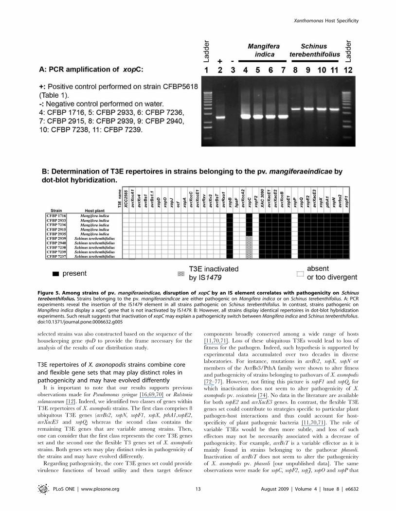
mangiferaeindicae respectively. In this latter example, it is interesting
to note that xopC, carried by pathovar mangiferaeindicae strains, is
altered in strains isolated from Schinus terebenthifolius but not in those
isolated from Mangifera indica. Since strains from both hosts of
isolation exhibited identical T3E repertoires (Figures 2 and 5), this
result might suggest that the alteration of this T3E gene might
have a role in host adaptation for pathovar mangiferaeindicae strains.
To gain insight on sequence variation among orthologs of
variable T3E, a subset of 120 sequences of variable T3Es was
obtained. Genetic diversity thus observed was extremely reduced,
and sequences obtained were almost identical to that of sequences of
the functional orthologs found in the databases (data not shown).
Only the sequence of avrXccB in strain CFBP1845 of the pathovar
phaseoli displayed a premature stop codon (data not shown).
Discussion
In this paper, we investigated the distribution of 35 T3Es among
132 strains belonging to 18 pathovars of the species X. axonopodis
[32]. To our knowledge, this strain collection is the largest used in
any other distribution study of virulence-associated genes in plant
pathogenic bacteria. To provide the largest diversity, strains were
chosen to represent the broad host range, wide geographic
distribution, and genetic diversity of the species X. axonopodis
[32,34,35]. In the course of this study, the phylogeny of the 132
Figure 4. Frequency of association between T3Es in Xanthomonas axonopodis. Numbers represent the frequency of cases when both T3Es inthe pair are present in the same strains.doi:10.1371/journal.pone.0006632.g004
Xanthomonas Host Specificity
PLoS ONE | www.plosone.org 11 August 2009 | Volume 4 | Issue 8 | e6632

Ta
ble
2.
DN
Are
arra
ng
em
en
tsfo
un
din
T3
Eg
en
es
fro
mX
an
tho
mo
na
sa
xon
op
od
isst
rain
s.
T3
Eg
en
es
Pa
tho
va
rsS
tra
ins
DN
Are
arr
an
ge
me
nts
Ty
pe
aS
ize
(bp
)P
osi
tio
nb
ISd
ire
ctre
pe
at
seq
ue
nce
ge
ne
rate
db
yIS
inse
rtio
na
nd
du
pli
cate
do
rd
ele
ted
seq
ue
nce
inT
3E
ge
ne
sp
lus
fla
nk
ing
seq
ue
nce
c
avr
Xv3
alf
alf
ae
CFB
P3
83
5,
CFB
P3
83
6,
CFB
P3
83
7,
CFB
P7
12
0,
CFB
P7
12
1IS
1595
(IS1
595
fam
ily,
IS15
95g
rou
p)
10
72
51
3A
AG
AT
TC
A
xop
Ove
sica
tori
aC
FBP
68
17
ISX
ca2
(IS3
fam
ily,
IS40
7g
rou
p)
12
00
33
CC
AT
ecf
alf
alf
ae
CFB
P3
83
6IS
Xca
2(I
S3fa
mily
,IS
407
gro
up
)1
20
01
42
3C
GT
T
avr
Rxo
1a
llii
CFB
P6
36
9,
CFB
P6
35
8IS
Xca
2(I
S3fa
mily
,IS
407
gro
up
)1
20
07
70
GA
CG
avr
Rxo
1a
llii
CFB
P6
10
7IS
1389
(IS3
fam
ily,
IS40
7g
rou
p)
12
07
41
1G
TC
C
avr
Xa
cE2
alf
alf
ae
CFB
P3
83
6,
CFB
P3
83
7IS
Xca
2(I
S3fa
mily
,IS
407
gro
up
)1
20
05
58
AG
GG
xop
Cci
tru
mel
oC
FBP
31
14
,C
FBP
33
71
,C
FBP
38
43
IS14
04(I
S3fa
mily
,IS
407
gro
up
)1
20
32
03
4G
CG
A
xop
Cm
an
gif
era
e-in
dic
ae
CFB
P2
93
9,
CFB
P2
94
0,
JP7
40
,JP
74
2,
JP7
57
IS14
79(I
S5fa
mily
,IS
5g
rou
p)
11
54
10
91
CT
AG
xop
Na
ura
nti
folii
CFB
P2
86
6IS
Xa
c2(I
S3fa
mily
,IS
407
gro
up
)1
19
5N
D
xop
F2a
ura
nti
folii
CFB
P2
86
6D
ele
tio
n3
84
10
30
–1
41
3g
ccg
t[A
TC
GT
CC
AG
CA
CG
GA
CT
GA
CG
CT
GC
GC
GA
-T
TC
CT
GC
CC
GA
CG
GT
CA
TA
GG
CC
TG
TG
CC
GT
GA
G-
CT
CG
GC
AC
GG
GT
CG
GC
GT
AC
CG
TT
TT
CT
CC
GG
GA
C-
GC
AC
GA
AG
CG
CT
TG
CT
TC
CC
TC
AG
GG
GT
GA
TT
TC
C-
AA
TA
GG
AC
CG
GC
GG
CG
GG
TC
CG
GC
AC
CA
GG
AT
CT
-T
GG
GA
TC
GA
TC
GC
CT
GG
AA
AC
GC
GG
CA
GA
TT
GG
CG
-A
CG
CG
CG
CG
CG
CC
TG
TC
CA
TC
GA
TG
GA
AT
CA
AC
A-
GC
GT
GT
CG
CA
GG
CA
TG
CG
AC
CC
CA
TT
CC
CG
AG
AT
-C
AC
CC
CA
GC
CA
GG
GT
CG
CT
GT
AC
TC
AA
TG
GC
TC
GG
-A
AG
CG
GG
CG
GG
CG
GT
TG
CT
CT
GG
TA
GG
CA
GT
GG
C-
TG
CA
GA
AA
TG
CC
GA
GC
GG
CG
TC
CG
CA
AC
GC
GC
C-
AA
CG
AA
AA
AC
GA
]tct
ga
xop
Dve
sica
tori
aC
FBP
68
17
Tan
de
md
up
licat
ion
90
52
7–
61
6tc
cccA
TA
CT
CC
GG
CG
GG
TT
CT
TC
CT
AT
TC
GT
CC
C-
TG
TT
CC
CG
CC
CA
CC
CC
TT
CT
GG
CG
GT
TG
GC
-C
GC
AG
AA
CG
CA
TC
AG
GT
GA
GT
GG
CA
TC
CC
G-
AT
AC
TC
CG
GC
GG
GT
TC
TT
CC
TA
TT
CG
TC
C-
CT
GT
TC
CC
GC
CC
AC
CC
CT
TC
TG
GC
GG
TT
GG
-
CC
GC
AG
AA
CG
CA
TC
AG
GT
GA
GT
GG
CA
TC
CC
Gatact
aIS
nam
es,
fam
ilie
san
dg
rou
ps
are
giv
en
acco
rdin
gto
ISFi
nd
er
dat
abas
e(h
ttp
://w
ww
.is.b
ioto
ul.f
r/is
.htm
l).
bN
ucl
eo
tid
en
um
be
rsar
eg
ive
nco
nsi
de
rin
gth
en
ucl
eo
tid
es
Ao
fth
est
art
cod
on
of
T3
Eg
en
es
as1
.cT
arg
ets
for
du
plic
atio
nar
ein
bo
ldca
pit
als,
resu
ltin
gta
nd
em
du
plic
atio
nar
ein
bo
ldan
din
ital
ic,a
nd
de
lete
dre
gio
ns
are
inca
pit
als
insq
uar
eb
rack
ets
.Nu
cle
oti
de
sfl
anki
ng
targ
et
reg
ion
s,ta
nd
em
du
plic
atio
ns
and
de
lete
dre
gio
ns
are
inlo
we
rca
se.
do
i:10
.13
71
/jo
urn
al.p
on
e.0
00
66
32
.t0
02
Xanthomonas Host Specificity
PLoS ONE | www.plosone.org 12 August 2009 | Volume 4 | Issue 8 | e6632
selected strains was also constructed based on the sequence of the
housekeeping gene rpoD to provide the frame necessary for the
analysis of the results of our distribution study.
T3E repertoires of X. axonopodis strains combine coreand flexible gene sets that may play distinct roles inpathogenicity and may have evolved differently
It is important to note that our results supports previous
observations made for Pseudomonas syringae [16,69,70] or Ralstonia
solanacearum [12]. Indeed, we identified two classes of genes within
T3E repertoires of X. axonopodis strains. The first class comprises 8
ubiquitous T3E genes (avrBs2, xopN, xopF1, xopX, pthA1,xopE2,
avrXacE3 and xopQ) whereas the second class contains the
remaining T3E genes that are variable among strains. Then,
one can consider that the first class represents the core T3E genes
set and the second one the flexible T3 genes set of X. axonopodis
strains. Both genes sets may play distinct roles in pathogenicity of
the strains and may have evolved differently.
Regarding pathogenicity, the core T3E genes set could provide
virulence functions of broad utility and then target defence
components broadly conserved among a wide range of hosts
[11,70,71]. Loss of these ubiquitous T3Es would lead to loss of
fitness for the pathogen. Indeed, such hypothesis is supported by
experimental data accumulated over two decades in diverse
laboratories. For instance, mutations in avrBs2, xopX, xopN or
members of the AvrBs3/PthA family were shown to alter fitness
and pathogenicity of strains belonging to pathovars of X. axonopodis
[72–77]. However, not fitting this picture is xopF1 and xopQ, for
which inactivation does not seem to alter pathogenicity of X.
axonopodis pv. vesicatoria [74]. No data in the literature are available
for both xopE2 and avrXacE3 genes. In contrast, the flexible T3E
genes set could contribute to strategies specific to particular plant
pathogen-host interactions and thus could account for host-
specificity of plant pathogenic bacteria [11,70,71]. The role of
variable T3Es would be then more subtle, and loss of such
effectors may not be necessarily associated with a decrease of
pathogenicity. For example, avrBsT is a variable effector as it is
mainly found in strains belonging to the pathovar phaseoli.
Inactivation of avrBsT does not seem to alter the pathogenicity
of X. axonopodis pv. phaseoli [our unpublished data]. The same
observations were made for xopC, xopF2, xopJ, xopO and xopP that
Figure 5. Among strains of pv. mangiferaeindicae, disruption of xopC by an IS element correlates with pathogenicity on Schinusterebenthifolius. Strains belonging to the pv. mangiferaeindicae are either pathogenic on Mangifera indica or on Schinus terebenthifolius. A: PCRexperiments reveal the insertion of the IS1479 element in all strains pathogenic on Schinus terebenthifolius. In contrast, strains pathogenic onMangifera indica display a xopC gene that is not inactivated by IS1479. B: However, all strains display identical repertoires in dot-blot hybridizationexperiments. Such result suggests that inactivation of xopC may explain a pathogenicity switch between Mangifera indica and Schinus terebenthifolius.doi:10.1371/journal.pone.0006632.g005
Xanthomonas Host Specificity
PLoS ONE | www.plosone.org 13 August 2009 | Volume 4 | Issue 8 | e6632

appeared as variable T3E genes in our study [74,78]. Altogether,
these data, obtained from Xanthomonas strains, can be compared to
what is known in Pseudomonas syringae. Indeed, mutations in T3Es of
the conserved effector locus (CEL) usually alter pathogenicity [79].
Substantial experimental evidence is available for hopPtoM, hopPtoN
and avrE in Pseudomonas syringae [27,28,80], as well as for dspA/E in
Erwinia amylovora [81,82]. Conversely, mutations in T3E genes of
the exchangeable effector locus (EEL) of Pseudomonas syringae are
not associated to strong impairment of pathogenicity [79].
Our study contributes also to a better understanding of the
evolutionary history of T3E genes within the X. axonopodis species.
The core T3E genes set might represent the ancient T3E gene
suite, acquired by the ancestor of the X. axonopodis species before
diversification of pathovars, and thus before host specialization
occurred. These core T3E genes might have evolved from this
ancestor by vertical descent among X. axonopodis strains. However,
some of these core T3E genes might have been acquired later in
the evolution and then have been stably inherited along with the
core genome. One can also postulate that among the core T3E
genes set, some genes might have been lost during evolution in
phylogenetically closely related pathovars, such as xopE2 and
avrXacE3 in pathovars manihotis and vasculorum or as xopQ in
pathovars allii and ricini (Figures 1 and 2). In contrast, the flexible
T3E genes set might have evolved by horizontal gene transfer even
though we cannot completely rule out gene loss during evolution.
Analyses of Xanthomonas genomes clearly showed that these
bacteria have been subjected to numerous horizontal gene
transfers during evolution, sometimes from phylogenetically
distant organisms [83,84]. Moreover, gene acquisition is consid-
ered to be a major factor contributing to the genomic diversity of
these bacteria but it seems that, once acquired, these genes are
rarely transferred among lineages [85,86]. Horizontal gene
transfer events were supported by the fact that the majority of
the variable T3E genes in our study cluster within pathogenicity
islands in their Xanthomonas host genomes [8,64,87]. Indeed, these
variable T3E genes exhibit a G+C content lower compared to the
average value of the rest of the host bacterial genome, they are
often associated with integrase genes, transfer RNA genes and/or
IS elements or remnants of them, and they are found sometimes
on plasmids. Regarding ubiquitous T3E genes, no linkage to
pathogenicity islands can be detected since their G+C content is
similar to the rest of their host bacterial genome, they are flanked
by orthologous sequences, they are not associated with mobile
elements, integrase or transfer RNA genes, and they reside on
chromosome (except for pthA1).
Finally, the importance of knowing which T3E is ubiquitous or
variable may be illustrated by the durability in the field of
resistances introduced in crops. The pepper resistance gene Bs2,
that matches the ubiquitous T3E avrBs2 has been widely deployed
in the field and still provides good level of resistance. On the other
hand, prediction was made for low durability of the resistance
conferred by Bs1 that matches the variable T3E avrBs1 [88].
A correspondence between composition of T3Erepertoires and pathovars of X. axonopodis supports a‘‘repertoire for repertoire’’ hypothesis
The phylogeny of the strains we used in this study was
constructed based on the sequence of rpoD housekeeping gene.
Our results confirm that host specificity is not necessarily
correlated to phylogeny [24,35,37]. Indeed, some pathovars are
clearly polyphyletic, e. g. pathovars phaseoli, dieffenbachiae, glycines or
vasculorum. However, the dendrogram constructed based on the
T3E presence/absence matrix groups strains by pathovar (except
for the pathovar aurantifolii), irrespective of the phylogenetic
relationships between strains. For example, in the rpoD phylogeny,
the pathovar phaseoli is scattered over the tree. In particular, the
genetic lineage 1 highly diverges from the other lineages, as
previously mentioned [24]. In contrast, on the dendrogram
constructed on the matrix of presence/absence of T3Es, the four
distinct genetic lineages identified in the pathovar phaseoli clustered
together. Thus in our study, strains displaying a similar T3E
repertoire belong to the same pathovar, even though they may be
phylogenetically distant.
Conversely, strains displaying different host specialisation
exhibit different T3E repertoires, even though these strains may
be very close phylogenetically. For example, based on our rpoD
phylogeny, strains belonging to the pathovar vignicola are mixed
with strains belonging to the genetic lineage 2 of the pathovar
phaseoli. However, their T3E repertoires are highly divergent, and
strains do not display the same host range. Even more striking is
the example of strains CFBP3541 and CFBP3835 that belong to
the pathovars citrumelo and alfalfae, respectively. Phylogenetically,
these strains are much closer to strains belonging to the pathovar
anacardii or to the pathovar phaseoli than other strains of their
respective pathovars. However, the T3E repertoire of strains
CFBP3541 and CFBP3835 is identical or highly similar to that of
other strains of pathovars citrumelo and alfalfae, respectively.
Such results support the hypothesis that T3E repertoires may
explain a pathological convergence of phylogenetically distant
strains. Thus, for a given strain, the T3E repertoire in its entirety
would greatly determine the host range. Such hypothesis was also
suggested by recent data obtained on a wide collection of strains of
Pseudomonas syringae isolated from different host plants [16]. In
addition, we performed an analysis of T3E gene history using
parsimony as implemented in the Mesquite software package [89].
Parsimony method is particularly well suited for such binary data
like presence or absence of T3E gene. Figure 1 shows that the trait
‘‘presence of the T3E gene xac3090’’ appears at several nodes in
the phylogenetic tree. For example, it is shown that the occurrence
of xac3090 in the pathovar glycines probably results from multiple
independent evolutionary events compatible with the hypothesis of
an adaptive convergence for pathogenicity.
The variability observed in T3E repertoires between strains
belonging to the same pathovar may explain race/cultivar
specificity. Furthermore, in polyphyletic pathovars such as
pathovar phaseoli the differences in repertoires observed between
the four genetic lineages [24] of this pathovar may reflect
differences in host range that was not revealed yet. One could
think that pathovar phaseoli strains may have evolved diverse T3E
repertoires to extend their host ranges or increase their survival on
various unrelated plant species, as it was postulated for Pseudomonas
syringae strains [90]. We now plan to thoroughly test host ranges of
each genetic lineage of the pathovar phaseoli on plants belonging to
the Fabaceae family in order to test such hypothesis.
Thus, our results support a ‘‘repertoire for repertoire’’
hypothesis as the molecular basis of host specificity of plant
pathogenic bacteria. In such hypothesis, the outcome of the
interaction between the bacterial pathogen and the plant would
greatly depend on the confrontation of the repertoires of bacterial
pathogenic determinants, such as T3E genes, and plant ‘‘guard’’
genes. Such hypothesis is compatible with the model proposed by
Jones and Dangl [21], as well as with the fact that non-host
resistance is constituted of multilayered basal defences that
bacteria must overcome to induce disease [91,92].
Our next goal will be to determine by Southern-blot
hybridization whether T3E genes are present in multiple copies
in our strain collection. Indeed, in Pseudomonas syringae pv. tomato
strain DC3000, two copies of the hopAM1 gene has been found
Xanthomonas Host Specificity
PLoS ONE | www.plosone.org 14 August 2009 | Volume 4 | Issue 8 | e6632
[18,93]. For T3E genes belonging to the avrBs3/pthA gene family,
it is common to find more than 10 copies of these genes in strains
of Xanthomonas such as X. axonopodis pv. malvacearum, X. oryzae pv.
oryzae or X. oryzae pv. oryzicola [75]. The presence of such multiple
copies of T3E genes within T3E repertoires may impact the host
range of the strains. It has been reported that the contribution to
pathogenicity in a given strain is not equal between the different
avrBs3/pthA gene members: only a few members encode major
virulence determinants whereas other members are potential
reservoir genes providing sources for rapid evolution and
adaptation in the event of host recognition [75].
However, one should keep in mind that, although T3E
repertoires of plant pathogenic bacteria probably greatly impact
their host range, other molecular determinants are also likely
involved in host specificity and tissue specificity as well. In
particular, early interactions such as host perception may also
greatly impact host range in natural conditions. The importance of
phenomena such as chemotaxis in the interactions between plant
associated bacteria and their hosts has been widely documented.
In the case of the plant pathogen Ralstonia solanacearum, for
example, a chemotactic mutant is not able to colonize its host
when inoculated in the soil, whereas it retains full pathogenicity
when infiltrated directly in the plant tissues [94]. Hemagglutinin-
related proteins, that appeared variable among Ralstonia solana-
cearum strains, are molecular determinants that could account for
host specificity [12]. Furthermore, a recent comparative analysis of
eight Xanthomonas genomes revealed that host- and tissue-specificity
may result from subtle changes in a small number of individual
genes in the gum, hrp, xps, xcs or rpf clusters and differences among
regulatory targets, secretory substrates or genes for environmental
sensing [38]. By analyzing amino acid residues, hpaA and xpsD
have been revealed as candidate determinants of tissue specificity
in Xanthomonas [38]. Since our study did not reveal correlation
between T3E genes and tissue specificity, further sequencing of
T3E genes and analysis of the T3E gene products polymorphisms
are now required to identify new candidate determinants of tissue
specificity.
Our results, which show a correspondence between composition
of T3E repertoires and pathovars of Xanthomonas, do support the
hypothesis that T3Es can affect host range in Xanthomonas.
Nevertheless, our approach based on PCR and dot-blot
hybridization methods is not sufficient to unequivocally consider
that repertoires of T3Es determine host specificity in Xanthomonas
axonopodis pathovars. To support the ‘‘repertoire for repertoire’’
hypothesis, we now plan to point our work towards functional
studies based on our results.
Typing the T3E repertoires of plant pathogenic bacteriamay provide clues for functional studies on hostspecificity and insight into understanding theredundancy between T3Es
Repertoire of T3Es represents candidate determinants of host
specificity of plant pathogenic bacteria since it has been shown that
many T3Es can act as molecular double agents that betray the
pathogen to plant defences in some interactions and suppress host
defences in others [11,20,95]. T3Es have been shown to be
involved in varietal resistance as well as in non-host resistance and
they are reported to suppress both PTI (PAMP-triggered
immunity) and ETI (effector-triggered immunity), the multilayered
plant defences that bacteria must overcome to induce disease
[21,31,91,92,96–98]. Thus, within a T3E repertoire, there are
evidences of interplay among T3Es since they can suppress ETI
[96,99] and they can make redundant contributions to virulence
[25,26]. Moreover, individual T3E may contribute differently to
the outcome of the infection on different hosts [90]. Comparisons
of T3E repertoires in Pseudomonas strains lead to the conclusion that
either different combinations of sequence-unrelated T3Es (or T3E
alleles) with redundant functions or few common T3Es may
promote successful pathogenesis by distinct strains on the same
hosts [90,93].
Our work provides clues for functional studies that will aim at
showing gain or loss of function. For instance, focusing on strains
of pathovars vasculorum and manihotis may be an excellent approach
since strains of both pathovars have similar T3E repertoires
(Figure 2) and the number of variable T3Es is not too important to
reasonably set up functional studies for further analysis of the role
of T3E repertoires in host specificity. It would be interesting to
observe whether the deletion of the variable T3E gene, xopB, in pv.
vasculorum or the transfer of this T3E gene to pv. manihotis narrow
or enlarge the host range of the strains. The same kind of
functional studies might be performed with the three variable T3E
gene, avrXv3, avrXccA2 and avrRxo1 in pv. manihotis. Another
example could be with strains CFBP1519 (pv. glycines) and
CFBP3530 (pv. aurantifolii). Indeed, these strains are phylogenet-
ically closely related since they exhibit the same rpoD sequence
(Figure 1) and they harbour highly similar T3E repertoires since
only two T3E genes discriminate both pathovars: xopC (present in
pv. glycines and absent in pv. aurantifolii) and avrXccB (present in pv.
aurantifolii and absent in pv. glycines) (Figure 2).
A major pitfall in deciphering the role of T3Es in the
pathogenicity of plant pathogenic bacteria is that inactivation of
a single T3E has often no detectable effect on pathogenicity.
Functional redundancy among T3Es has largely been hypothe-
sized to explain such phenomenon [25,26]. Data provided in this
study may help to better select strains for mutating single T3Es
and combined T3Es to provide insight into the functional
redundancy of T3Es that may have a role in the delineation of
the host range of the strains. Several T3E families have been found
within Xanthomonas genomes, such as the YopJ/AvrRxv family, the
AvrBs3/PthA family, the HopX/AvrPphE family, the HopAE1
family or the PopC family [8,11,64,68]. For instance, to reveal
functional overlap between T3Es, one could focus on T3Es that
belong to the YopJ/AvrRxv family of cysteine proteases since, in
our study, we selected several T3Es (XopJ, AvrRxv, AvrBsT,
XccB and AvrXv4) of this family [67,68]. Noel and colleagues
reported that a mutation of the T3E XopJ in X. axonopodis pv.
vesicatoria strain 85-10 cannot be associated with any decrease in
pathogenicity [78]. Genome sequence analysis [64] and results
obtained in our study reveal that this strain carries AvrRxv,
another cysteine protease of the same family that may partially
complement an inactivation of XopJ. Furthermore, we show in the
present study that both xopJ and avrRxv are frequently associated in
the species X. axonopodis (Figure 4) even though they are not
genetically linked [64]. It would now be interesting to construct a
double mutant by deleting both xopJ and avrRxv in a Xanthomonas
axonopodis strain in order to provide insight into the functional
redundancy of these T3Es. Similarly, we constructed an avrBsT
mutant in strain CFBP4834 of X. axonopodis pv. phaseoli. No
phenotype could be associated to the mutation [our unpublished
data]. But besides AvrBsT, the repertoire of the strain CFBP4834
also contains XccB, another cysteine protease of the YopJ/AvrRxv
family. We also show that there is a high frequency of association
between AvrBsT and XccB in the species X. axonopodis (Figure 4).
However, some strains of the pathovar mangiferaeindicae or of the
pathovar alfalfae, only display one cysteine protease of the YopJ/
AvrRxv family. Selecting one of these strains may ease functional
studies on T3Es of the YopJ/AvrRxv cysteine proteases in plant
Xanthomonas Host Specificity
PLoS ONE | www.plosone.org 15 August 2009 | Volume 4 | Issue 8 | e6632

pathogenic bacteria. In order to carry out functional comparisons
of T3Es, one could also use gene ontology annotations that do not
only depend on sequence similarities [100–101]. Such an
approach may highlight shared and divergent pathogenic
strategies of T3Es deployed by the various pathovars of X.
axonopodis. Our results will then help in the determination of
redundant-effector groups (REGs) in Xanthomonas strains as it has
been done recently in the Pseudomonas syringae pv. tomato strain
DC3000 [26]. These authors clearly demonstrated that plant
pathogenic bacteria have evolved the capacity to deliver into plant
cells T3Es with very little sequence similarity that are redundant in
function [26]. Another demonstration of sequence-unrelated T3Es
that function in the same plant defense pathway is AvrRpm1,
AvrRpt2 and AvrB, that are not recognized by the same resistance
genes but all target the Arabidopsis RIN4 protein [11]. Since these
three T3Es only rarely cooccur in Pseudomonas syringae strains, this
suggests that convergent evolution is driven by the need to
manipulate particular host proteins [11]. Finally, elucidation of
functional overlaps between T3Es should help us understand how
the diverse T3Es in a repertoire may function as a system in plant
hosts and may shape the host range of the strains.
Pathoadaptation of X. axonopodis strains is suggested bysequence variations revealed in some T3E genes
In regard to their central role in pathogenicity, T3Es are likely
under strong selection pressures imposed by the defence system of
the host plant. To escape plant defences, a pathogen may acquire
new T3Es by horizontal gene transfer that would suppress defence
reactions induced after recognition of the pathogen by the plant
[99]. Alternatively, pathoadaptation of bacterial strains may occur
through diverse mechanisms (single nucleotide polymorphism,
insertion, deletion, or loss of a given T3E), to avoid being
recognized by the host plant [102–106]. In the course of this study,
we found DNA rearrangements that suggest pathoadaptation for
X. axonopodis strains.
Our distribution study performed by the PCR amplification
method allowed us to identify 23 DNA rearrangements within
T3E genes. Interestingly, these DNA rearrangements were found
only in T3E genes belonging to the accessory genome. If we
consider that these variable genes may influence host specificity,
such identified DNA rearrangements in some T3E genes might
have a significant role in pathological adaptation of these plant
pathogenic bacteria to their hosts. Among DNA rearrangements
identified in the course of this study, there are a deletion within
xopF2 of one pathovar aurantifolii strain and a perfect tandem
duplication within xopD of one pathovar vesicatoria strain.
Interestingly, both DNA rearrangements do not shift the reading
frames suggesting that these strains used these strategies to
generate modified form of the XopF2 and XopD proteins to
avoid recognition by the plant. Regarding xopD, to our knowledge,
this is the first example of a T3E gene, except for genes belonging
to the avrBs3/pthA gene family [75], exhibiting perfect tandem
duplication within its nucleotide sequence. It is tempting to
speculate that the tandem duplication in the xopD gene may affect
the host adaptation of this pathovar vesicatoria strain. Indeed, in
Xanthomonas, it has been reported that insertions or deletions in the
central part, where tandem duplications reside, of T3Es belonging
to the AvrBs3/PthA family induce alterations of the host range of
the strains [75,107,108].
Another example of pathoadaptive evolution comes from the
action of transposable elements. Indeed, we identified in the frame
of this study several T3E genes that are disrupted by different IS
elements. Numerous ISs have previously been found inserted into
T3E genes among plant pathogenic bacteria, and some of them
were shown to shift plant-pathogen interactions from incompatible
to compatible [93,105,109–112]. In our study, the majority of
identified IS elements belongs to the IS3 family-IS407 group
whereas the remaining ones belong to the IS5 and IS1595 families.
Interestingly, when looking at the flanking sequences of T3E genes
in Xanthomonas sequenced genomes [8,64], we only found IS
elements that belong to the same three families, with again a large
majority of ISs classified within the IS3 family - IS407 group.
Furthermore, another IS element (IS476) belonging to the IS3
family-IS407 group has been disclosed in the avrBs1 gene within
one X. axonopodis pv. vesicatoria strain [113]. Altogether, these
observations suggest that, in Xanthomonas strains, members of these
three IS families might play an important role in T3E gene
evolution since these mobile elements may alter their expression,
they may be involved in their mobility as well as in the terminal
reassortment process [71,109,114–118]. Otherwise, it is also
striking to note that some IS elements belonging to the IS3
family-IS407 group have been found just downstream PIP boxes,
the binding motif for the transcriptional regulator HrpX [119], in
the sequenced genome of X. axonopodis pv. vesicatoria [64]. It is thus
tempting to speculate that the transposition of such replicative IS
elements [115], and then the subsequent inactivation of a given
T3E gene, might be co-regulated with the hrp genes cluster. Thus,
one can reasonably think that inactivation of some T3E genes by
IS elements might be of importance in host adaptation for plant
pathogenic bacteria. To verify this hypothesis, it would be
interesting to focus for instance on avrXv3 since it is altered by
IS1595 in all tested pathovar alfalfae strains. We plan to
complement these strains with a functional avrXv3 gene in order
to observe a modification of the interaction between the pathovar
alfalfae strains and their hosts. It would also be interesting to focus
on xopC in pathovar mangiferaeindicae strains since this gene is
altered in strains isolated from Schinus terebenthifolius but not in those
isolated from Mangifera indica (Figure 5). Since strains from both
hosts of isolation exhibit identical T3E gene repertoires, the
functional complementation of xopC might lead to a modification
in host adaptation of these strains.
The finding that multiple T3E genes are affected by DNA
rearrangements raises the question of the functionality of these
genes within the repertoires. Our approach allowed us to show
that several T3E genes are likely inactive since they are disrupted
by ISs or exhibit a frameshift mutation leading to a premature stop
codon. But, T3E genes may be non-functional for other reasons
that we did not challenge by our approach, such as lack of
expression or inability to translocate T3E proteins. Schechter and
colleagues, in 2006, by using multiple approaches on the T3E
repertoire of the Pseudomonas syringae pv. tomato strain DC3000,
revealed that 33 T3Es are likely to be active, 12 T3Es are likely to
be inactive and 8 T3Es may or may not be produced at functional
levels [18]. It will be now important to check the functionality of
each T3E gene in the repertoires that may impact the host range
of Xanthomonas strains. Knowing whether a T3E gene is active or
inactive is of interest for evolutionary studies. Indeed, it is possible
that selection pressure for the inactivation of a T3E gene may
result from the acquisition of new gene functions in both the host
and the bacterium and that loss of function may be an important
factor in the evolution of Xanthomonas axonopodis virulence. Loss of
gene function may be beneficial to bacterial strains and it is
considered to be a contributing factor to the evolution of virulence
of many pathogens [104,105,120].
Finally, our results do support the hypothesis that T3E
repertoires can affect the host range of Xanthomonas strains and
that the evolution of T3E repertoires is driven by the need for
interactions among T3Es as they co-ordinately disarm multiple
Xanthomonas Host Specificity
PLoS ONE | www.plosone.org 16 August 2009 | Volume 4 | Issue 8 | e6632

layers of plant defenses. Our results also support the hypothesis
that the evolution of T3E repertoires is also likely driven by the
exposure to diverse resistance mechanisms in plants. So, the
evolution and function of T3Es in a repertoire may be influenced
by a co-evolutionary arms race between pathogens and hosts
[105]. The second hypothesis is supported by the observation that
a T3E loss and then the evolution of a T3E repertoire can be
driven by exposure to host defence system [121]. Recently, it has
been proposed that the host defence can accelerate the generation
of genomic rearrangements that provide selecting advantage to the
pathogen [104]. Therefore, it is tempting to speculate that the
numerous DNA rearrangements found in T3E genes from
Xanthomonas strains in the course of our study may be the result
of exposure to various host plants. In that case, one could speculate
that pathogens in response to selection pressure imposed by host
defence systems, may have driven the inactivation of some T3E
genes by insertion of ISs, or the modification of other T3Es by in-
frame deletion or perfect tandem duplication. These DNA
rearrangements may have had a significant role in avoidance of
host recognition and then in shaping the host range of the
Xanthomonas strains. It is also reported that similar exposure of
bacterial strains to environmental stress outside the host could also
drive the horizontal transfer of T3Es from ecologically related
plant pathogens that could lead to evolution of T3E repertoires
and then of bacterial pathogenicity towards plants [104,105].
PerspectivesOur results provide resources for functional studies on host
specificity of plant pathogenic bacteria. Our work will help to
select strains to study the role of single or combination of T3E
genes in the interaction with plants, as well as for studies aiming at
understanding the molecular mechanisms of redundancy between
T3Es. Moreover, the discovery of genetic rearrangements in genes
encoding T3Es demonstrates the importance of looking at the
allelic diversity of T3Es as well as at the expression of these genes.
Indeed, impact of genetic rearrangements in T3E genes on host
range has recently been well documented [105,106]. Thus, we
plan to continue to analyze the allelic diversity of T3Es in our
collection of Xanthomonas strains for evolutionary studies. Further-
more, our results strongly suggest that determination of T3E
repertoires may be used for identification of Xanthomonas strains at
the pathovar level. Thus, we will aim at developing a diagnostic
tool for such purpose.
Finally, our study illustrates the importance of distribution
analyses of virulence-associated genes by using large collections of
bacterial strains. This approach can be useful for the identification
of the candidate determinants of host specificity. For instance, it
could be useful to perform such investigation on large collections
of E. coli strains that are pathogenic on poultry or on humans.
Indeed, no set of virulence genes was clearly identified yet to
discriminate between avian and human strains [3]. To our
knowledge, if presence or absence of genes of the T3SS was
analyzed, repertoires of T3E were not yet compared. But
interestingly, similarly to what was found for plant pathogenic
bacteria, T3E genes appeared as differential genes in SSH
between avian and human strains [2].
Materials and Methods
Bacterial strainsA collection of 132 strains of X. axonopodis [32] belonging to 18
different pathovars were included in this study (Table 1). The
bacterial strains were obtained from international culture
collections (mostly the French Collection of Plant pathogenic
Bacteria; http://www.angers.inra.fr/cfbp/). Since, in the scope of
this study, it was not possible to determine the host range of each
strain, we decided to select only well characterized pathovars for
which host range has been determined precisely and published
data are available [40–56]. Among the 18 selected pathovars, 7 are
considered as vascular pathogens and 11 are considered as non-
vascular pathogens (http://www.cababstractsplus.org/) [40–56].
Furthermore, strains were selected carefully in order to get the
largest diversity. Thus, for each pathovar, strains were chosen
from various geographical origins, hosts and years of isolation.
Each pathovar is represented by at least 5 strains, except for the
pathovar axonopodis for which only two available strains, including
the type strain of the species X. axonopodis (CFBP4924), could be
found in international collections. More strains belonging to the
pathovar phaseoli, the bacterial model of our team [24,53–56], were
selected since it is a genetically diverse and polyphyletic pathovar,
four genetic lineages were disclosed that are not closely related
[24]. In this study, the pathovar phaseoli was then used to highlight
the potential convergence of T3E repertoires of phylogenetically
unrelated strains. The 18 selected pathovars represent the overall
diversity within the X. axonopodis species since they were selected
from the 6 defined rep-PCR clusters (from 9.1 to 9.6) of this
species (Figure 1) [34,35]. Moreover, we also included Escherichia
coli strain DH5a and Xanthomonas strains whose genome has been
sequenced (X. axonopodis pv. vesicatoria strain CFBP5618; X.
axonopodis pv. citri strain 306, X. campestris pv. campestris strains
CFBP5241 and CFBP6650, X. oryzae pv. oryzae strain CFBP7088,
X. oryzae pv. oryzicola strain CFBP7109 and X. albilineans strain
CFBP7063) (http://www.genomesonline.org/) [8,64–66]. We also
included in our study X. axonopodis pv. phaseoli strain CFBP4834
whose genome has been sequenced recently (our unpublished
data). All of these sequenced strains were used as positive or
negative controls for PCRs and dot-blot hybridizations.
DNA extractionGenomic DNA was extracted from all bacterial strains grown
overnight at 28uC in YP medium (yeast extract, 7 g/liter; peptone,
7 g/liter) by using the standard hexadecyltrimethylammonium
bromide method [122]. Quality and quantity of DNA was
spectrophotometrically evaluated (Nanodrop ND-1000, Nanodrop
Technologies).
Selection of T3E genesTable S2 presents the complete list of the 35 T3E genes
included in this study. Selected genes comprised those identified
from the sequenced genomes of Xanthomonas strains (17 from X.
axonopodis pv. vesicatoria strain CFBP5618, 8 from X. campestris pv.
campestris strain CFBP5241 and 7 from X. axonopodis pv. citri strain
306) [8,64]. We also selected 3 avirulence genes from other X.
axonopodis pv. vesicatoria strains whose genome has not been
sequenced: avrBsT from strain 75-3 [123] and avrXv3 and avrXv4
from strain 91-118 [124,125]. Some of the selected T3E genes are
members of the defined T3E families in bacterial pathogens such
as the AvrRxv/YopJ (C55) family of cysteine proteases, the
AvrBs3/PthA family of transcriptional activators, the PopC family
of Leucin-Rich Repeats proteins, the HopAE1 family and the
HopX/AvrPphE family (Table S2) [8,11,64,68]. The other
selected T3E genes have unknown functions to date. It is
important to note that in the present study we tried to be as
exhaustive as possible since we selected T3E genes not from only
one but from 5 different Xanthomonas strains that belong to diverse
species and pathovars. We followed this approach to minimize
unavoidable bias of this kind of analysis; indeed it is certain that
Xanthomonas Host Specificity
PLoS ONE | www.plosone.org 17 August 2009 | Volume 4 | Issue 8 | e6632

unidentified T3E genes may reside in Xanthomonas strains whose
genome has not been sequenced yet.
PCR amplificationsTwo complementary approaches were undertaken to charac-
terize the T3E repertoires of our collection of strains: PCR and dot
blot hybridization. The presence or the absence of an ortholog of
each selected T3E gene was first determined by PCR. All X.
axonopodis strains were first submitted to a PCR analysis by using
specific T3Es primers. Pairs of primers (Table S2) were designed
from the DNA sequences of the selected T3E genes available in
databases. All of these primers pairs allowed the amplification of
the full-length T3E DNA sequence, except for the avrBs2 and
avrBs3 genes for which only partial DNA sequences were amplified
(Table S2). The Xanthomonas strains, from which T3E genes were
selected, were taken as positive or negative controls for all PCR
experiments. PCR amplifications were carried out with a 20 ml
reaction mixture containing 16 Go Taq Buffer (Promega),
200 mM dNTP, 0.5 mM of each primer, 0.4 U of Go Taq
Polymerase (final concentrations) and 1 ng of template genomic
DNA. The amplification conditions using the T3E primers were
2 min of initial denaturation at 94uC; followed by 30 cycles of
94uC for 1 min, 60uC for 1 minute and 72uC for 2 min; with a
final extension of 10 min at 72uC. A reaction was considered as
positive if a single clear band with the expected size was detected
onto agarose gels. When a single band with an unexpected size was
observed, the amplified PCR product was recovered from the gel
and then sequenced (see below).
Dot blot hybridizationsAs sequence variation may occur between T3E orthologs, thus
preventing annealing of the PCR primers used, presence or
absence of an ortholog was then confirmed by nucleic acid
hybridization. For each dot blot hybridization experiment, we
included, as negative and positive controls, water, E. coli strain
DH5a and Xanthomonas strains whose genome has been sequenced
(see above for strain details).
Hybridization probes were obtained by PCR amplification of
the selected T3E genes from the sequenced genome of Xanthomonas
strains using specific primers listed in Table S2. Probes were
labelled using the PCR Digoxigenin (DIG) labelling mix (Roche
Applied Science, France). PCR reactions contained 200 mM
dNTP-DIG, and the other components as above. PCR for
preparation of DIG-labeled DNA probes was performed in a
themocycler programmed for denaturation at 94uC for 2 min and
then for 30 cycles of 94uC for 1 min, 60uC for 1 min, 72uC for
2 min and finally 72uC for 10 min. The PCR products were
purified by using the NucleospinH extract II kit (Macherey- Nagel
Hoerdt, France).
Genomic DNA (250 ng) of each strain was transferred to
BiodyneH N+ membranes (Pall Gelman Laboratory). DNAs were
randomised onto membranes. Prehybridization, hybridization and
detection were carried out by using the DIG Labelling and
Detection Starter kit (Roche Applied Science, France) according to
the manufacturer’s instructions. Hybridizations were performed
overnight at 42uC. To ensure high stringency, membranes were
washed twice for 15 min in 26 SSC and 0.1% SDS buffer and
twice in 0.16 SSC and 0.1% SDS buffer at 68uC. Hybridization
signals were detected using the Fab fragments of an anti 1-2
digoxigenin antibody conjugated with alkaline phosphatase (Anti-
DIG-AP) and Nitro-Blue Tetrazolium Chloride/5-Bromo-4-
Chloro-39-Indolyphosphate p-Toluidine Salt (NBT/BCIP). A
subset of hybridization experiments was replicated twice to assess
the reproducibility of the dot blot results. Furthermore, we
assessed the robustness of our approach by using sequenced
Xanthomonas strains. Indeed, for these strains we were able to
compare the T3E repertoires obtained by PCR and dot-blot
hybridization with the expected T3E repertoires based on the
genome sequence. For each of these strains, the obtained T3E
repertoire corresponded to the expected T3E repertoire. This
approach combined with BLAST analyses (http://blast.ncbi.nlm.
nih.gov/Blast.cgi) allowed us to determine that the minimum
identity at the DNA level between a T3E on the membrane and a
T3E ortholog in the probe had to be at least 71% to give a signal
above background.
DNA Sequencing and sequence analysis of amplified T3Egenes from X. axonopodis strains
The amplified PCR products were purified from agarose gels
with the NucleospinH extract II kit (Macherey-Nagel EURL,
France) and then sequenced at Genoscreen genomic analysis
platform (Lille, France). Sequence data were examined by using
the BLAST search programs (http://blast.ncbi.nlm.nih.gov/Blast.
cgi), the Mobyle portal (http://mobyle.pasteur.fr/cgi-bin/Moby-
lePortal/portal.py) and the IS Finder database (http://www-is.
biotoul.fr/is.html).
rpoD amplification, sequencing and sequence analysisThe phylogenetic analysis of X. axonopodis strains was performed
by sequencing the housekeeping rpoD gene (RNA polymerase
sigma-70 factor). Primers were designed from the rpoD sequence of
the X. campestris pv. campestris strain CFBP 5241 (GenBank
accession no. NP639081) (Table S2). PCR amplifications were
performed in a total volume of 50 ml using 3 ng of genomic DNA,
200 mM dNTPs, 0.5 mM of each primer, 1.5 mM MgCl2 and
0.4 U of Go Taq polymerase in 16 Colorless Go Taq buffer
(Promega). The PCR cycling conditions consisted of an initial
denaturation step at 94uC for 5 min, followed by 30 cycles of 94uCfor 30 s, 60uC for 60 s, 72uC for 30 s, with a final extension step at
72uC for 7 min. PCR amplicons were then sent to Ouest
Genopole sequencing platform (Nantes, France). Forward and
reverse sequences were obtained by using the rpoD specific PCR
primers. These sequences were edited and assembled by using
PREGAP 4 and GAP 4 of the Staden Package [126]. All rpoD
sequences were then aligned using BioEdit (http://www.mbio.
ncsu.edu/BioEdit/bioedit.html). All rpoD sequences have been
deposited in the GenBank data library (accession numbers from
FJ561596 to FJ561725).
Data analysisBased on the presence/absence matrix of T3E genes for each of
the 132 strains of X. axonopodis, we constructed a dendrogram using
Jaccard distances and Neighbor-Joining method. Bootstrapping
was performed with 1,000 replicates to assess the robustness of our
dendrogram. The resulting dendrogram was visualised using the
PAST 1.81 software (http://folk.uio.no/ohammer/past/).
Phylogenetic trees based on the rpoD sequences analysis were
constructed by using maximum likelihood method. Best nucleotide
substitution model was found using MODELTEST v.3.7 [62]. Akaike
Information Criterion (AIC) was used for model selection.
Parameters of the selected model were used for maximum
likelihood heuristic search using PAUP*4.0 beta10 [127].
Confidence on node was assessed by bootstrapping 1000 times.
Based on the presence/absence matrix of T3E genes, we
calculated frequencies of association between T3E genes within X.
axonopodis strains. We retained only cases where both T3E genes
are present in the same strains. Furthermore, to analyze T3E gene
Xanthomonas Host Specificity
PLoS ONE | www.plosone.org 18 August 2009 | Volume 4 | Issue 8 | e6632

histories, we used the parsimony method as implemented in the
Mesquite software package [89].
Supporting Information
Table S1 Percentage of presence of each T3E gene in the
various pathovars of X. axonopodis.
Found at: doi:10.1371/journal.pone.0006632.s001 (0.03 MB
XLS)
Table S2 List of genes analyzed in this study: gene functions and
primers sequences used for PCR amplifications and for making
probes for dot-blot hybridizations.
Found at: doi:10.1371/journal.pone.0006632.s002 (0.03 MB
DOC)
Acknowledgments
We would like to thank our laboratory colleagues and collaborators,
Armelle Darrasse, Marie-Agnes Jacques, Marion Le Saux, Lionel
Gagnevin and Boris Szurek for providing strains and/or their helpful
discussions. We thank Ralf Koebnik and Marie-Anne Barny for critical
reading of the manuscript.
Author Contributions
Conceived and designed the experiments: AH CM TB SPP. Performed the
experiments: AH CB. Analyzed the data: AH GH FL CL CM TB SPP.
Contributed reagents/materials/analysis tools: AH GH FL CL CM TB
SPP. Wrote the paper: AH CL TB SPP.
References
1. Moulin-Schouleur M, Reperant M, Laurent S, Bree A, Mignon-Grasteau S, et
al. (2007) Extraintestinal pathogenic Escherichia coli strains of avian and human
origin: link between phylogenetic relationships and common virulence patterns.
J Clin Microbiol 45: 3366–3376.
2. Kariyawasam S, Scaccianoce JA, Nolan LK (2007) Common and specific
genomic sequences of avian and human extraintestinal pathogenic Escherichia
coli as determined by genomic subtractive hybridization. BMC Microbiol 7: 81.
3. Ron EZ (2006) Host specificity of septicemic Escherichia coli: human and avian
pathogens. Curr Opin Microbiol 9: 28–32.
4. Mokady D, Gophna U, Ron EZ (2005) Virulence factors of septicemic
Escherichia coli strains. Int J Med Microbiol 295: 455–462.
5. Mokady D, Gophna U, Ron EZ (2005) Extensive gene diversity in septicemic
Escherichia coli strains. J Clin Microbiol 43: 66–73.
6. Dye DW, Bradbury JF, Goto M, Hayward AC, Lelliot RA, et al. (1980)
International standards for naming pathovars of phytopathogenic bacteria and
a list of pathovar names and pathotype strains. Rev Plant Pathol 59: 153–168.
7. Flor HH (1956) The complementary genetic systems in flax and flax rust. Adv
Genet 8: 29–54.
8. da Silva ACR, Ferro JA, Reinach FC, Farah CS, Furlan LR, et al. (2002)
Comparison of the genomes of two Xanthomonas pathogens with differing host
specificities. Nature 417: 459–463.
9. Ochiai H, Inoue Y, Takeya M, Sasaki A, Kaku H (2005) Genome sequence of
Xanthomonas oryzae pv. oryzae suggests contribution of large numbers of effector
genes and insertion sequences to its race diversity. JARQ 39: 275–288.
10. Feil H, Feil WS, Chain P, Larimer F, DiBartolo G, et al. (2005) Comparison of
the complete genome sequences of Pseudomonas syringae pv. syringae B728a and
pv. tomato DC3000. Proc Natl Acad Sci U S A 102: 11064–11069.
11. Grant SR, Fisher EJ, Chang JH, Mole BM, Dangl JL (2006) Subterfuge and
manipulation: Type III effector proteins of phytopathogenic bacteria. Annu
Rev Microbiol 60: 425–449.
12. Guidot A, Prior P, Schoenfeld J, Carrere S, Genin S, et al. (2007) Genomic
structure and phylogeny of the plant pathogen Ralstonia solanacearum inferred
from gene distribution analysis. J Bacteriol 189: 377–387.
13. He YQ, Zhang L, Jiang BL, Zhang ZC, Xu RQ, et al. (2007) Comparative and
functional genomics reveals genetic diversity and determinants of host
specificity among reference strains and a large collection of Chinese isolates
of the phytopathogen Xanthomonas campestris pv. campestris. Genome Biol 8:
R218.
14. Demuth A, Aharonowitz Y, Bachmann TT, Blum-Oehler G, Buchrieser C, et
al. (2008) Pathogenomics: an updated European Research Agenda. Infect
Genet Evol 8: 386–393.
15. Hayward A (1993) The hosts of Xanthomonas. In: Swings J, Civerolo EL, eds
(1993) Xanthomonas. London: Chapman and Hall. pp 1–18.
16. Sarkar SF, Gordon JS, Martin GB, Guttman DS (2006) Comparative genomics
of host-specific virulence in Pseudomonas syringae. Genetics 174: 1041–1056.
17. Chang JH, Urbach JM, Law TF, Arnold LW, Hu A, et al. (2005) A
highthroughput, near-saturating screen for type III effector genes from
Pseudomonas syringae. Proc Natl Acad Sci U S A 102: 2549–2554.
18. Schechter LM, Vencato M, Jordan KL, Schneider SE, Schneider DJ, et al.
(2006) Multiple approaches to a complete inventory of Pseudomonas syringae pv.
tomato DC3000 type III secretion system effector proteins. Mol Plant-Microbe
Interact 19: 1180–1192.
19. Gurlebeck D, Thieme F, Bonas U (2006) Type III effector proteins from the
plant pathogen Xanthomonas and their role in the interaction with the host plant.
J Plant Physiol 163: 233–255.
20. da Cunha L, Sreerekha MV, Mackey D (2007) Defense suppression by
virulence effectors of bacterial phytopathogens. Curr Opin Plant Biol 10:
349–357.
21. Jones JD, Dangl JL (2006) The plant immune system. Nature 444: 323–329.
22. Castaneda A, Reddy JD, El-Yacoubi B, Gabriel DW (2005) Mutagenesis of all
eight avr genes in Xanthomonas campestris pv. campestris had no detected effect on
pathogenicity but one avr gene affected race specificity. Mol Plant Microbe
Interact 18: 1306–1317.
23. Triplett LR, Zhao Y, Sundin GW (2006) Genetic differences between blight-
causing Erwinia species with differing host specificities, identified by suppression
subtractive hybridization. Appl Environ Microbiol 72: 7359–7364.
24. Alavi SM, Sanjari S, Durand F, Brin C, Manceau C, et al. (2008) Assessment of
the genetic diversity of Xanthomonas axonopodis pv. phaseoli and Xanthomonas fuscans
subsp. fuscans as a basis to identify putative pathogenicity genes and a type III
secretion system of the SPI-1 family by multiple suppression subtractive
hybridizations. Appl Environ Microbiol 74: 3295–3301.
25. Badel JL, Shimizu R, Oh H-S, Collmer A (2006) A Pseudomonas syringae pv.
tomato avrE1/hopM1 mutant is severely reduced in growth and lesion
formation in tomato. Mol Plant Microbe Interact 19: 99–111.
26. Kvitko BH, Park DH, Velasquez AC, Wei C-F, Russell AB, et al. (2009)
Deletions in the repertoire of Pseudomonas syringae pv. tomato DC3000 type III
secretion effector genes reveal functional overlap among effectors. PLoS Pathog
5(4): e1000388. doi:10.1371/journal.ppat.1000388.
27. Badel JL, Nomura K, Bandyopadhyay S, Shimizu R, Collmer A, et al. (2003)
Pseudomonas syringae pv. tomato DC3000 HopPtoM (CEL ORF3) is important for
lesion formation but not growth in tomato and is secreted and translocated by
the Hrp type III secretion system in a chaperone-dependent manner. Mol
Microbiol 49: 1239–1251.
28. Lopez-Solanilla E, Bronstein PA, Schneider AR, Collmer A (2004) HopPtoN is
a Pseudomonas syringae Hrp (type III secretion system) cysteine protease effector
that suppresses pathogen-induced necrosis associated with both compatible and
incompatible plant interactions. Mol Microbiol 54: 353–365.
29. Reboutier D, Frankart C, Briand J, Biligui B, Rona JP, et al. (2007)
Antagonistic action of harpin proteins: HrpWea from Erwinia amylovora
suppresses HrpNea-induced cell death in Arabidopsis thaliana. J Cell Sci 120:
3271–3278.
30. Liao AP, Petrof EO, Kuppireddi S, Zhao Y, Xia Y, et al. (2008) Salmonella type
III effector AvrA stabilizes cell tight junctions to inhibit inflammation in
intestinal epithelial cells. PLoS ONE 3: e2369.
31. Zhao B, Ardales EY, Raymundo A, Bai J, Trick HN, et al. (2004) The avrRxo1
gene from the rice pathogen Xanthomonas oryzae pv. oryzicola confers a nonhost
defense reaction on maize with resistance gene Rxo1. Mol Plant Microbe
Interact 17: 771–779.
32. Vauterin L, Hoste B, Kersters K, Swings J (1995) Reclassification of
Xanthomonas. International Journal of Systematic Bacteriology 45: 472–489.
33. Vauterin L, Rademaker J, Swings J (2000) Synopsis on the taxonomy of the
genus Xanthomonas. Phytopathology 90: 677–682.
34. Rademaker JLW, Hoste B, Louws FJ, Kersters K, Swings J, et al. (2000)
Comparison of AFLP and rep-PCR genomic fingerprinting with DNA-DNA
homology studies: Xanthomonas as a model system. Int J Syst Appl Microbiol 50:
665–677.
35. Rademaker JLW, Louws FJ, Schultz MH, Rossbach U, Vauterin L, et al.
(2005) A comprehensive species to strain taxonomic framework for Xanthomonas.
Phytopathology 95: 1098–1111.
36. Fargier E (2007) L’etude de la pathologie de Xanthomonas campestris et de la
structure genetique de ses pathovars a permis l’amelioration de la detection du
pathogene dans les semences de Brassicacees. PhD thesis, University of Angers.
37. Young JM, Park DC, Shearman HM, Fargier E (2008) A multilocus sequence
analysis of the genus Xanthomonas. Syst Appl Microbiol 31: 366–377.
38. Lu H, Patil P, Van Sluys M-A, White FF, Ryan RP, et al. (2008) Acquisition
and evolution of plant pathogenesis-associated gene clusters and candidate
determinants of tissue-specificity in Xanthomonas. PLoS ONE 3: e3828.
39. Meyer DF, Bogdanove AJ (2009) Genomics-driven advances in Xanthomonas
biology. In: Jackson, ed (2009) Plant Pathogenic Bacteria: Genomics and
Molecular Biology. Norfolk, UK: Caister Academic Press. pp 147–161.
40. Ah-You N, Gagnevin L, Chiroleu F, Jouen E, Rodrigues Neto J, et al. (2007)
Pathological variations within Xanthomonas campestris pv. mangiferaeindicae support
Xanthomonas Host Specificity
PLoS ONE | www.plosone.org 19 August 2009 | Volume 4 | Issue 8 | e6632

its separation into three distinct pathovars that can be distinguished by
amplified fragment length polymorphism. Phytopathology 97: 1568–1577.
41. Brunings AM, Gabriel DW (2003) Xanthomonas citri: breaking the surface. Mol
Plant Pathol 4: 141–157.
42. Roumagnac P, Gagnevin L, Gardan L, Sutra L, Manceau C, et al. (2004)
Polyphasic characterization of xanthomonads isolated from onion, garlic and
Welsh onion (Allium spp.) and their relatedness to different Xanthomonas species.
Int J Syst Evol Microbiol 54: 15–24.
43. Jones JB, Stall RE, Bouzar H (1998) Diversity among xanthomonads
pathogenic on pepper and tomato. Annu Rev Phytopathol 36: 41–58.
44. Berthier Y, Verdier V, Guesdon JL, Chevrier D, Denis JB, et al. (1993)
Characterization of Xanthomonas campestris Pathovars by rRNA Gene Restriction
Patterns. Appl Environ Microbiol 59: 851–859.
45. Gonzalez C, Restrepo S, Tohme J, Verdier V (2002) Characterization of
pathogenic and nonpathogenic strains of Xa nthomonas axonopodis pv. manihotis by
PCR-based DNA fingerprinting techniques. FEMS Microbiol Lett 215: 23–31.
46. Mkandawire ABC, Mabagala RB, Guzman P, Gepts P, Gilbertson RL (2004)
Genetic diversity and pathogenic variation of common blight bacteria
(Xanthomonas campestris pv. phaseoli and X. campestris pv. phaseoli var. fuscans)
suggests pathogen coevolution with the common bean. Phytopathology 94:
593–603.
47. Khatri-Chhetri GB, Wydra K, Rudolph K (2003) Metabolic diversity of
Xanthomonas axonopodis pv. vignicola, casual agent of cowpea bacterial blight and
pustule. Eur J Plant Pathol 109: 851–860.
48. Restrepo S, Velez CM, Verdier V (2000) Measuring the genetic diversity of
Xanthomonas axonopodis pv. manihotis within different fields in Colombia.
Phytopathology 90: 683–690.
49. Gent DH, Al-Saadi A, Gabriel DW, Louws FJ, Ishimaru CA, et al. (2005)
Pathogenic and genetic relatedness among Xanthomonas axonopodis pv. allii and
other pathovars of X. axonopodis. Phytopathology 95: 918–925.
50. Robene-Soustrade I, Laurent P, Gagnevin L, Jouen E, Pruvost O (2006)
Specific detection of Xanthomonas axonopodis pv. dieffenbachiae in anthurium
(Anthurium andreanum) tissues by nested PCR. Appl Environ Microbiol 72:
1072–1078.
51. De Feyter R, Gabriel DW (1991) At least six avirulence genes are clustered on a
90-kilobase plasmid in Xanthomonas campestris pv. malvacearum. Mol Plant-
Microbe Interact 4: 423–432.
52. Zomorodian A, Rudolph K (1993) Xanthomonas campestris pv. malvacearum: cause
of bacterial blight of cotton. In: Swings JG, Civerolo EL, eds (1993)
Xanthomonas. London: Chapman & Hall. pp 25–30.
53. Jacques MA, Josi K, Darrasse A, Samson R (2005) Xanthomonas axonopodis pv.
phaseoli var. fuscans is aggregated in stable biofilm population sizes in the
phyllosphere of field-grown beans. Appl Environ Microbiol 71: 2008–2015.
54. Alavi SM, Poussier S, Manceau C (2007) Characterization of ISXax1, a novel
insertion sequence restricted to Xanthomonas axonopodis pv. phaseoli (variants
fuscans and non-fuscans) and Xanthomonas axonopodis pv. vesicatoria. Appl Environ
Microbiol 73: 1678–1682.
55. Darsonval A, Darrasse A, Meyer D, Demarty M, Durand K, et al. (2008) The
type III secretion system of Xanthomonas fuscans subsp. fuscans is involved in the
phyllosphere colonization process and in transmission to seeds of susceptible
beans. Appl Environ Microbiol 74: 2669–2678.
56. Darsonval A, Darrasse A, Durand K, Bureau C, Cesbron S, et al. (2009)
Adhesion and fitness in the bean phyllosphere and transmission to seeds of
Xanthomonas fuscans subsp. fuscans. Mol Plant-Microbe Interact 22: 747–757.
57. Xu RQ, Blanvillain S, Feng JX, Jiang BL, Li XZ, et al. (2008)
AvrAC(Xcc8004), a type III effector with a leucine-rich repeat domain from
Xanthomonas campestris pathovar campestris confers avirulence in vascular tissues
of Arabidopsis thaliana ecotype Col-0. J Bacteriol 190: 343–355.
58. Maiden MCJ, Bygraves JA, Feil E, Morelli G, Russell JE, et al. (1998)
Multilocus sequence typing: a portable approach to the identification of clones
within populations of pathogenic microorganisms. Proc Natl Acad Sci USA 95:
3140–3145.
59. Enright MC, Spratt BG (1999) Multilocus sequence typing. Trends Microbiol
7: 482–487.
60. Urwin R, Maiden MCJ (2003) Multi-locus sequence typing: a tool for global
epidemiology. Trends Microbiol 11: 479–487.
61. Sarkar SF, Guttman DS (2004) Evolution of the core genome of Pseudomonas
syringae, a highly clonal, endemic plant pathogen. Appl Environ Microbiol 70:
1999–2012.
62. Posada D, Crandall KA (1998) Modeltest: testing the model of DNA
substitution. Bioinformatics 14: 817–818.
63. Cornelis GR (2006) The type III secretion injectisome. Nat Rev Microbiol 4:
811–825.
64. Thieme F, Koebnik R, Bekel T, Berger C, Boch J, et al. (2005) Insights into
genome plasticity and pathogenicity of the plant pathogenic bacterium
Xanthomonas campestris pv. vesicatoria revealed by the complete genome sequence.
J Bacteriol 187: 7254–7266.
65. Lee BM, Park YJ, Park DS, Kang HW, Kim JG, et al. (2005) The genome
sequence of Xanthomonas oryzae pathovar oryzae KACC10331, the bacterial
blight pathogen of rice. Nucleic Acids Res 33: 577–586.
66. Qian W, Jia Y, Ren SX, He YQ, Feng JX, et al. (2005) Comparative and
functional genomic analyses of the pathogenicity of phytopathogen Xanthomonas
campestris pv. campestris. Genome Res 15: 757–767.
67. Hotson A, Mudgett MB (2004) Cysteine proteases in phytopathogenic bacteria:
Identification of plant targets and activation of innate immunity. Curr Opin
Plant Biol 7: 384–390.
68. Kay S, Bonas U (2009) How Xanthomonas type III effectors manipulate the host
plant. Curr Opin Microbiol 12: 37–43.
69. Guttman DS, Vinatzer BA, Sarkar SF, Ranall MV, Kettler G, et al. (2002) A
functional screen for the type III (Hrp) secretome of the plant pathogen
Pseudomonas syringae. Science 295: 1722–1726.
70. Rohmer L, Guttman DS, Dangl JL (2004) Diverse evolutionary mechanisms
shape the type III effector virulence factor repertoire in the plant pathogen
Pseudomonas syringae. Genetics 167: 1341–1360.
71. Stavrinides J, McCann HC, Guttman DS (2008) Host-pathogen interplay and
the evolution of bacterial effectors. Cell Microbiol 10: 285–292.
72. Kearney B, Staskawicz BJ (1990) Widespread distribution and fitness
contribution of Xanthomonas campestris avirulence gene avrBs2. Nature 346:
385–386.
73. Duan YP, Castaneda A, Zhao G, Erdos G, Gabriel DW (1999) Expression of a
single, host-specific, bacterial pathogenicity gene in plant cells elicits division,
enlargement, and cell death. Mol Plant Microbe Interact 12: 556–560.
74. Roden JA, Belt B, Ross JB, Tachibana T, Vargas J, et al. (2004) A genetic
screen to isolate type III effectors translocated into pepper cells during
Xanthomonas infection. Proc Natl Acad Sci USA 101: 16624–16629.
75. Yang B, White FF (2004) Diverse members of the AvrBs3/PthA family of type
III effectors are major virulence determinants in bacterial blight disease of rice.
Mol Plant Microbe Interact 17: 1192–1200.
76. Metz M, Dahlbeck D, Morales CQ, Al Sady B, Clark ET, et al. (2005) The
conserved Xanthomonas campestris pv. vesicatoria effector protein XopX is a
virulence factor and suppresses host defense in Nicotiana benthamiana. Plant J 41:
801–814.
77. Jiang BL, He YQ, Cen WJ, Wei HY, Jiang GF, et al. (2008) The type III
secretion effector XopXccN of Xanthomonas campestris pv. campestris is required
for full virulence. Res Microbiol 159: 216–220.
78. Noel L, Thieme F, Gabler J, Buttner D, Bonas U (2003) XopC and XopJ, two
novel type III effector proteins from Xanthomonas campestris pv. vesicatoria.
J Bacteriol 185: 7092–7102.
79. Alfano JR, Charkowski AO, Deng WL, Badel JL, Petnicki-Ocwieja T, et al.
(2000) The Pseudomonas syringae Hrp pathogenicity island has a tripartite mosaic
structure composed of a cluster of type III secretion genes bounded by
exchangeable effector and conserved effector loci that contribute to parasitic
fitness and pathogenicity in plants. Proc Natl Acad Sci USA 97: 4856–4861.
80. Lorang JM, Shen H, Kobayashi D, Cooksey D, Keen NT (1994) avrA and avrE
in Pseudomonas syringae pv. tomato PT23 play a role in virulence on tomato plants.
Mol Plant Microbe Interact 7: 508–515.
81. Gaudriault S, Malandrin L, Paulin JP, Barny MA (1997) DspA, an essential
pathogenicity factor of Erwinia amylovora showing homology with AvrE of
Pseudomonas syringae, is secreted via the Hrp secretion pathway in a DspB-
dependent way. Mol Microbiol 26: 1057–1069.
82. Bogdanove AJ, Bauer DW, Beer SV (1998) Erwinia amylovora secretes DspE, a
pathogenicity factor and functional AvrE homolog, through the hrp (type III
secretion) pathway. J Bacteriol 180: 2244–2247.
83. Lima W, Van Sluys MA, Menck CF (2005) Non-gamma Proteobacteria gene
islands contribute to the Xanthomonas genome. Omics 9: 160–172.
84. Comas I, Moya A, Azad RK, Lawrence JG, Gonzalez-Candelas F (2006) The
evolutionary origin of Xanthomonadales genomes and the nature of the
horizontal gene transfer process. Mol Biol Evol 23: 2049–2057.
85. Gillings MR, Holley MP, Stokes HW, Holmes AJ (2005) Integrons in
Xanthomonas: A source of species genome diversity. Proc Natl Acad Sci USA
102: 4419–4424.
86. Lerat E, Daubin V, Ochman H, Moran NA (2005) Evolutionary origins of
genomic repertoires in bacteria. PLoS Biol 3: e130.
87. Hacker J, Kaper J (2000) Pathogenicity islands and the evolution of microbes.
Annu Rev Microbiol 54: 641–679.
88. Leach JE, Vera Cruz CM, Bai J, Leung H (2001) Pathogen fitness penalty as a
predictor of durability of disease resistance genes. Annu Rev Phytopathol 39:
187–224.
89. Maddison WP, Maddison DR (2009) Mesquite: a modular system for
evolutionary analysis. Version 2.6 http://mesquiteproject.org.
90. Vinatzer BA, Teitzel GM, Lee MW, Jelenska J, Hotton S, et al. (2006) The type
III effector repertoire of Pseudomonas syringae pv. syringae B728a and its role in
survival and disease on host and non-host plants. Mol Microbiol 62: 26–44.
91. Ham JH, Kim MG, Lee SY, Mackey D (2007) Layered basal defenses underlie
non-host resistance of Arabidopsis to Pseudomonas syringae pv. phaseolicola. Plant J
51: 604–616.
92. Nurnberger T, Lipka V (2005) Non-host resistance in plants: new insights into
an old phenomenon. Mol Plant Pathol 6: 335–345.
93. Lindeberg M, Cartinhour S, Myers CR, Schechter LM, Schneider DJ, et al.
(2006) Closing the circle on the discovery of genes encoding Hrp regulon
members and type III secretion system effectors in the genomes of three model
Pseudomonas syringae strains. Mol Plant Microbe Interact 19: 1151–1158.
94. Yao J, Allen C (2006) Chemotaxis is required for virulence and competitive
fitness of the bacterial wilt pathogen Ralstonia solanacearum. J Bacteriol 188:
3697–3708.
Xanthomonas Host Specificity
PLoS ONE | www.plosone.org 20 August 2009 | Volume 4 | Issue 8 | e6632

95. Alfano JR, Collmer A (2004) Type III secretion system effector proteins: double
agents in bacterial disease and plant defense. Annu Rev Phytopathol 42:
385–414.
96. Jamir Y, Guo M, Oh H-S, Petnicki-Ocwieja T, Chen S, et al. (2004)
Identification of Pseudomonas syringae type III secreted effectors that suppress
programmed cell death in plants and yeast. Plant J 37: 554–565.
97. Fujikawa T, Ishihara H, Leach JE, Tsuyumu S (2006) Suppression of defense
response in plants by the avrBs3/pthA gene family of Xanthomonas spp. Mol.
Plant-Microbe Interact 19: 342–349.
98. Fujikawa T, Yamashita T, Tsuyumu S (2006) Hypersensitive response
suppression by type III effectors of plant pathogenic bacteria. J Gen Plant
Pathol 72: 176–179.
99. Jackson RW, Athanassopoulos E, Tsiamis G, Mansfield JW, Sesma A, et al.
(1999) Identification of a pathogenicity island, which contains genes for
virulence and avirulence, on a large native plasmid in the bean pathogen
Pseudomonas syringae pathovar phaseolicola. Proc Natl Acad Sci USA 96:
10875–10880.
100. Torto-Alalibo T, Collmer CW, Gwinn-Giglio M (2009) The Plant-Associated
Microbe Gene Ontology (PAMGO) Consortium: community development of
new Gene Ontology terms describing biological processes involved in microbe-
host interactions. BMC Microbiol 9 Suppl 1: S1.
101. Lindeberg M, Biehl BS, Glasner JD, Perna NT, Collmer A, et al. (2009) Gene
ontology annotation highlights shared and divergent pathogenic strategies of
type III effector proteins deployed by the plant pathogen Pseudomonas syringae pv
tomato DC3000 and animal pathogenic Escherichia coli strains. BMC Microbiol 9
Suppl 1: S4.
102. Sokurenko EV, Hasty DL, Dykhuizen DE (1999) Pathoadaptive mutations:
gene loss and variation in bacterial pathogens. Trends Microbiol 7: 191–195.
103. Pallen MJ, Wren BW (2007) Bacterial pathogenomics. Nature 449: 835–842.
104. Arnold DL, Jackson RW, Waterfield NR, Mansfield JW (2007) Evolution of
microbial virulence: the benefits of stress. Trends Genet 23: 293–300.
105. Ma W, Dong F, Stavrinides J, Guttman DS (2006) Type III effector
diversification via both pathoadaptation and horizontal transfer in response
to a coevolutionary arms race. PLoS Genetics 2: e209.
106. Zhou H, Morgan RL, Guttman DS, Ma W (2009) Allelic variants of the
Pseudomonas syringae type III effector HopZ1 are differentially recognized by
plant resistance systems. Mol Plant-Microbe Interact 22: 176–189.
107. Yang Y, Gabriel DW (1995) Intragenic recombination of a single plant
pathogen gene provides a mechanism for the evolution of new host specificities.
J Bacteriol 177: 4963–4968.
108. Yang B, Sugio A, White FF (2005) Avoidance of host recognition by alterations
in the repetitive and C-terminal regions of AvrXa7, a type III effector of
Xanthomonas oryzae pv. oryzae. Mol Plant Microbe Interact 18: 142–149.
109. Wichmann G, Ritchie D, Kousik CS, Bergelson J (2005) Reduced genetic
variation occurs among genes of the highly clonal plant pathogen Xanthomonas
axonopodis pv. vesicatoria, including the effector gene avrBs2. Appl Environ
Microbiol 71: 2418–2432.
110. Kearney B, Ronald PC, Dahlbeck D, Staskawicz BJ (1988) Molecular basis for
evasion of plant host defence in bacterial spot disease of pepper. Nature 332:
541–543.
111. Gonzalez AI, Ruiz ML, Polanco C (1998) Race-specific insertion of
transposable element IS801 in Pseudomonas syringae pv. phaseolicola. Mol PlantMicrobe Interact 11: 423–428.
112. Lavie M, Seunes B, Prior P, Boucher C (2004) Distribution and sequence
analysis of a family of type III-dependent effectors correlate with the phylogenyof Ralstonia solanacearum strains. Mol Plant Microbe Interact 17: 931–940.
113. Kearney B, Staskawicz B (1990) Characterization of IS476 and its role inbacterial spot disease of tomato and pepper. J Bacteriol 172: 143–148.
114. Stavrinides J, Ma W, Guttman DS (2006) Terminal reassortment drives the
quantum evolution of type III effectors in bacterial pathogens. PLoS Pathog 2:e104.
115. Mahillon J, Chandler M (1998) Insertion sequences. Microbiol Mol Biol Rev62: 725–774.
116. Kim JF, Charkowski AO, Alfano JR, Collmer A, Beer SV (1998) Sequencesrelated to transposable elements and bacteriophages flank avirulence genes of
Pseudomonas syringae. Mol Plant Microbe Interact 11: 1247–1252.
117. Rivas LA, Mansfield J, Tsiamis G, Jackson RW, Murillo J (2005) Changes inrace-specific virulence in Pseudomonas syringae pv. phaseolicola are associated with a
chimeric transposable element and rare deletion events in a plasmid-bornepathogenicity island. Appl Environ Microbiol 71: 3778–3785.
118. Sundin GW (2007) Genomic insights into the contribution of phytopathogenic
bacterial plasmids to the evolutionary history of their hosts. Annu RevPhytopathol 45: 129–151.
119. Koebnik R, Kruger A, Thieme F, Urban A, Bonas U (2006) Specific binding ofthe Xanthomonas campestris pv. vesicatoria AraC-type transcriptional activator
HrpX to plant-inducible promoter boxes. J Bacteriol 188: 7652–7660.120. Ochman H, Moran NA (2001) Genes lost and genes found: evolution of
bacterial pathogenesis and symbiosis. Science 292: 1096–1099.
121. Pitman AR, Jackson RW, Mansfield JW, Kaitell V, Thwaites R, et al. (2005)Exposure to host resistance mechanisms drives evolution of bacterial virulence
in plants. Curr Biol 15: 2230–2235.122. Ausubel FM, Brent R, Kingston RE, Moore DD, Seidman JG, et al. (1991)
Current protocols in molecular biology. New York, USA: Greene Publishing
Associates-Wiley Interscience.123. Ciesiolka LD, Hwin T, Gearlds JD, Minsavage GV, Saenz R, et al. (1999)
Regulation of expression of avirulence gene avrRxv and identification of a familyof host interaction factors by sequence analysis of avrBsT. Mol Plant Microbe
Interact 12: 35–44.124. Astua-Monge G, Minsavage GV, Stall RE, Davis MJ, Bonas U, et al. (2000)
Resistance of tomato and pepper to T3 strains of Xanthomonas campestris pv.
vesicatoria is specified by a plant-inducible avirulence gene. Mol Plant MicrobeInteract 13: 911–921.
125. Astua-Monge G, Minsavage GV, Stall RE, Vallejos CE, Davis MJ, et al. (2000)Xv4-AvrXv4: a new gene-for-gene interaction identified between Xanthomonas
campestris pv. vesicatoria race T3 and wild tomato relative Lycopersicon pennellii. Mol
Plant Microbe Interact 13: 1346–1355.126. Staden R, Beal KF, Bonfield JK (2000) The Staden Package. In: Misener S ,
Krawetz S , eds (2000) Methods Mol Biol. Totowa NJ 07512: The HumanaPress Inc 132: 115–1130.
127. Swofford DL (2003) PAUP*. Phylogenetic Analysis Using Parsimony (*andOther Methods). Version 4 Sunderland, Massachusetts: Sinauer Associates.
Xanthomonas Host Specificity
PLoS ONE | www.plosone.org 21 August 2009 | Volume 4 | Issue 8 | e6632