Profiling the autoantibody repertoire by serological antigen selection
24
1 Profiling the autoantibody repertoire by Serological Antigen Selection V. Somers 1 , C. Govarts 1 , N. Hellings 1 , R. Hupperts 2 and P. Stinissen 1 1 Hasselt University, Biomedical Research Institute, and Transnationale Universiteit Limburg, School of Life Sciences, Agoralaan, Building A, B-3590 Diepenbeek, Belgium 2 Department of Neurology, Academical Hospital Maastricht, P.O. Box 5800, 6202 AZ Maastricht, the Netherlands * Corresponding author: Dr. Veerle Somers, Hasselt University, Biomedical Research Institute, and Transnationale Universiteit Limburg, School of Life Sciences, Agoralaan, Building A, B-3590 Diepenbeek, Belgium, Tel: +32-11269202, Fax: +32-11269209, Email: [email protected] , Please note that as of June 15 this email address will change to [email protected] * Manuscript
Transcript of Profiling the autoantibody repertoire by serological antigen selection
1
Profiling the autoantibody repertoire by Serological
Antigen Selection V. Somers1, C. Govarts1, N. Hellings1, R. Hupperts2 and P. Stinissen1
1 Hasselt University, Biomedical Research Institute, and Transnationale Universiteit Limburg,
School of Life Sciences, Agoralaan, Building A, B-3590 Diepenbeek, Belgium 2 Department of Neurology, Academical Hospital Maastricht, P.O. Box 5800, 6202 AZ
Maastricht, the Netherlands
* Corresponding author: Dr. Veerle Somers, Hasselt University, Biomedical Research
Institute, and Transnationale Universiteit Limburg, School of Life Sciences, Agoralaan,
Building A, B-3590 Diepenbeek, Belgium, Tel: +32-11269202, Fax: +32-11269209, Email:
[email protected], Please note that as of June 15 this email address will change to
* Manuscript
2
ABSTRACT
The identification of disease related autoantigens targeted by pathogenic T- and B-cell
responses is crucial for the development of improved therapies for autoimmune diseases. To
identify immunogenic targets recognized by the humoral immune response, we have recently
applied a novel and powerful molecular approach, named ‘Serological antigen selection’. This
method involves the display of a cDNA expression library on filamentous phage and
subsequent selection on patient Immunoglobulin G (IgG). In the present study, we have
cloned a cDNA repertoire from a Multiple Sclerosis (MS) patient in pVI phage display
vectors and performed selections on pooled MS cerebrospinal fluid samples (CSF)
immobilized with anti-human IgG. To further streamline this procedure, we report an
optimized SAS procedure in which we have successfully established methods for enrichment
of MS-specific candidate antigens. In conclusion, the broad applicability of the SAS method
makes it a highly promising method for investigating the autoimmune repertoire.
Keywords: Serological Antigen Selection, filamentous phage, cDNA display, Multiple
Sclerosis, autoantibody repertoire, autoimmune diseases
Abbreviations: SAS, Serological Antigen Selection; pVI , bacteriophage coat protein 6

3
INTRODUCTION
The identification of B and T cell epitopes is a crucial step for the understanding of the
immune response mechanisms and their role in autoimmune diseases. Although some
autoimmune diseases have been mainly considered as T-cell mediated diseases, the
importance of autoreactive B cells has long been underestimated. However, the number of
autoimmune diseases associated with the presence of autoantibodies directed against cells of
the target tissue has been growing extensively over the past years [1,2,3,4]. Therefore, the role
of B cells and antibodies has been re-evaluated in several autoimmune diseases such as
multiple sclerosis.
Multiple sclerosis is a chronic, inflammatory disease of the central nervous system
characterized by multifocal inflammation and destruction of myelin. Although the etiology is
unknown, it is generally accepted that a misdirected immune response plays a crucial role in
the pathogenesis of the disease. Several findings suggest an important role of B cells and
autoantibodies in MS. In chronic MS lesions, B cells, plasma cells and antibodies have been
demonstrated [5,6]. Analysis of the heavy chain repertoire of B cells in the central nervous
system revealed a limited Immunoglobulin G (IgG) repertoire, which is consistent with a
targeted immune response [7,8,9,10]. In addition, cerebrospinal fluid (CSF) of patients with
clinically definite MS is characterized by the presence of oligoclonal antibodies (seen as
oligoclonal bands), which is used as a diagnostic indicator of disease [11]. Accordingly, in the
serum, where these bands are not observed using isoelectric focusing, the same antibodies
should be either absent, or, if present, not quantitatively predominant.
The isolation of autoantigens recognized by autoantibodies present in e.g. sera from
patients with autoimmune diseases has been traditionally carried out through time-consuming
processes. One of the first methods described used immunoprecipitation and immunoblotting
to identify candidate autoantigens [12,13]. More recent procedures focused on the serological
screening of cDNA libraries constructed with lambda phage vectors [14]. Although this
procedure has led to successful applications in several autoimmune diseases [15,16,17], the
labor intensity and non-quantitative nature of the screening procedure have impeded the
analysis of many different candidate autoantigens on multiple normal and patient sera.
During the past decade, filamentous phage display has been used to identify ligands
for any type of ligate. Based on this system, random peptide libraries have been constructed
and screened on different ligates such as monoclonal antibodies and even more complex

4
patient sera [18,19,20,21]. However, the main disadvantage is that only antigen mimotopes
are recovered by which further analysis is required to actually clone the serologically defined
antigen. In multiple sclerosis, the use of random peptide libraries has mainly resulted in the
isolation of candidate peptides which showed patient specific immunoreactivity [22,23].
To fully explore the complex information present within the antibody repertoire of
patients, we have recently applied a novel and powerful molecular approach, called
‘Serological antigen selection’. This method involves the display of a cDNA expression
library on filamentous phage by fusing the cDNA product to minor coat protein pVI [24]. The
filamentous phage links the expressed and displayed cDNA product to the DNA, cloned into
the phage genome. This allows the selection and enrichment of those cDNA products that
interact with a chosen immobilized or labeled ligand, for example, patient IgG. We have
successfully applied this method to molecularly define antigens recognized by the humoral
immune system in colorectal cancer, and this gave rise to the isolation of a panel of candidate
tumor antigens in colorectal cancer [25].
The present study reports the cloning and display of an MS cDNA library from the
target tissue on filamentous phage and enrichment by affinity selection on MS patient CSF
and serum IgG. By using the patient’s autoantibody repertoire to identify those cDNA
expression products or antigens that evoke an IgG immune response, interesting candidates
can be identified for further study. To further streamline this procedure, we report an
optimized SAS procedure in which we have successfully established methods for enrichment
of MS-specific candidate antigens. A panel of antigenic cDNAs with an MS-related
immunogenicity holds promise as a valuable tool in a diagnostic setting.
5
MATERIALS AND METHODS
Cloning of an MS cDNA library for pVI display
A normalized cDNA library (1.0 x 10⁶ primary recombinants) derived from active
chronic MS plaques, with varying degrees of demyelination and inflammatory activity (gift
from Dr. Soares) was transferred into our phage cDNA display vectors, named pSPVIA,
pSPVIB and pSPVIC, each encoding one of three reading frames. The normalized cDNA
library was prepared following a procedure based on reassociation kinetics [26]. To construct
the cDNA display libraries, plasmid DNA was prepared from the normalized MS cDNA
library and further digested with EcoRI and NotI. DNA fragments were gel purified (GFX gel
band purification kit, Amersham Biosciences) and ligated into the three vectors pSPVIA,
pSPVIB and pSPVIC digested with EcoRI and NotI. The ligation mixtures were used to
transform Escherichia coli TG1 cells by electroporation to obtain libraries MS-pSPVIA, MS-
pSPVIB and MS-pSPVIC of sizes 9.0 x 10⁶, 1.0 x 10⁶ and 1.0 x 10⁶ independent clones
respectively.
CSF/Sera
Cerebrospinal fluid samples and sera were obtained from MS patients and patients
with other inflammatory (meningitis, polyneuropathy) and non-inflammatory neurological
disorders (hernia, epilepsy, dementia, …). In addition, sera were collected from healthy
volunteers. CSF and serum samples were stored at –80°C after collection. CSF and serum
samples used for the selection procedure were absorbed against E. Coli and phage antibodies
by repeated passage through columns of Sepharose 6MB (Pharmacia, Uppsala, Sweden)
coupled to lysates of E. Coli Y1090 and bacteriophage-infected E. Coli XL1Blue. Following
absorption steps, aliquots from CSF and serum samples were prepared in 0.2% (w/v)
skimmed milk powder in 1 x TBS (50 mM Tris-HCl (pH 7.9) and 150 mM NaCl) and stored
at –20°C.
Selection of phage pVI displayed cDNA repertoires
CSF samples of 10 untreated relapsing remitting (RR) MS patients (8 women, 2 men,
age 32-51, EDSS 0-3.5) were pooled and used for affinity selections. Selections were
performed as described before with slight adaptations [25]. In brief, immunotubes (Nunc,

6
Roskilde, Denmark) were coated with rabbit anti-human IgG (Dako, Glostrup, Denmark) at a
concentration of 10 µg/ml in coating buffer (0.1M sodium hydrogen carbonate pH 9.6) for 2
hours at 37ºC. After washing the immunotube twice with phosphate-buffered saline/Tween 20
(PBST: 50 mM Tris, 150 mM NaCl, PH 7.5, 0.1% Tween 20 (w/v)) and twice with PBS, the
tubes were blocked for 2 hours at room temperature (RT) with 2% MPBS (2% w/v milk
powder in PBS). Phage were prepared from each library MS-pSPVIA, MS-pSPVI6B and MS-
pSPVIC as described previously [27]. Approximately equal numbers of phage derived from
each phage library (±2 x 10¹² pfu) were added to pooled CSF (1:5 diluted in 4% MPBS) and
incubated in a glass tube for 1.5 hour at RT on a rotating platform. After washing the coated
immunotube twice with PBST and twice with PBS, the preincubated CSF and phage mix was
transferred to the coated immunotube and incubated for 30 minutes on a rotating platform and
120 minutes standing at RT. Tubes were then washed 20 times with PBST and 20 times with
PBS to remove non-binding phage. Binding phage were eluted with 100 mM triethylamine
and neutralized with 1M Tris HCl as described before [25]. E. Coli TG1 cells were infected
with input and output phage and plated on 2 x TY agar plates containing ampicillin and
glucose (16 g/l bacto-tryptone, 10 g/l yeast extract, 5 g/l NaCl, 15 g bacto-agar/l, ampicillin at
100 µg/ml and glucose at 2% w/v) at each round of selection. Resultant colonies were scraped
and phage were rescued for further rounds of affinity selections. To monitor enrichment of
specific clones, input and output phage from each round of selection were titrated and the
ratio of output/input phage was determined.
Depletion strategies of non-specific clones
To enrich for selection of cDNA products binding to IgGs specifically associated with
MS, alternative selection strategies on MS patient material and normal samples were tested.
After a first selection round on a pool of 10 RR MS sera, a depletion strategy was developed
by using a pool of 10 age-matched normal sera. Selections on the pool of MS sera (1/100
diluted in 4% MPBS) were performed as described above. After this first selection round,
phage were rescued and used for selection on IgGs present in the pool of age-matched normal
sera (1/100 diluted in 4% MPBS). After preincubation for 1.5 hour at RT on a rotating
platform, the mixture was transferred to the coated immunotube for 30 minutes on a rotating
platform and 120 minutes standing at RT, the non-bound phage was now recovered and used
for infection of E. Coli TG1 cells. Phage were again rescued as described and the selection

7
process of alternative selections on MS patient and normal sera was repeated. The ratio of
output/input phage was determined after each selection round.
Polymerase chain reaction (PCR) amplification and fingerprinting
After several rounds of selection, individual colonies were selected and the insert size
was determined with forward primer (5’-CTC TCT GTA AAG GCT GC-3’) and reverse
primer (5’-CGC CAG GGT TTT CCC AGT CAC GAC-3’) which flank the EcoRI and NotI
cloning sites of the vector, respectively. Briefly, colonies were subjected to 35 cycles (20’’
94°C, 20’’ 55°C, 40’’ 72°C) of PCR amplification after an initial denaturation step for 5 ‘ at
95°C in a DNA Thermal Cycler (Perkin-Elmer Cetus, Norwalk, CT). The amplification
products were analysed by electrophoresis on a 1.0% agarose gel to confirm the presence of a
cDNA insert. After amplification, 10 µl of the amplification products was used for
fingerprinting analysis by incubating the amplification mixture with 5 U of the restriction
enzyme BstNI (Roche Diagnostics, Germany) for 2 hrs at 37°C. Products after restriction
enzyme digestion were analyzed on a 2% agarose gel.
ELISA of ligand displaying phage
96-well flat-bottomed microtiter plates (Falcon) were coated overnight at 4°C with
200 µl of rabbit-anti human IgG (Dako), 10 µg/ml in coating buffer (0.1M sodium hydrogen
carbonate pH 9.6) and blocked with 200 µl of 2% MPBS (2% w/v milk powder in PBS) for 1
hour at RT. For the ELISA screening on human CSF, a dilution of 1:5 (in 4% MPBS) was
used for the pool of 10 human CSF (each individual CSF was diluted 1:50) and 50 µl diluted
CSF was used for preincubation with 100 µl PEG-purified phage (1010 phages per well) in a
96-well round-bottomed 96-well plate (Costar). For the screening on individual CSF, a
dilution of 1:3 (in 4% MPBS) was used. Both absorbed and non-absorbed patient CSF
samples were used in ELISA experiments.
Phage particles were allowed to bind to patient CSF for 1 hour at 37°C and 30 minutes
shaking at RT. After washing 3 times with PBSTween (0.1% Tween 20) and 3 times with
PBS, the preincubated CSF plus phage mixture was transferred to the coated plate for 1 hour
at 37 °C and 30 minutes shaking at RT. After washing, 150 µl of a peroxidase conjugated
anti-M13 monoclonal (Amersham/Pharmacia/Biotech), diluted 1:5,000 in 2% MPBS (2% w/v
milk powder in PBS) was incubated for 1 hour shaking at RT. After washing the plates 3x

8
with PBSTween and 3x with PBS, 130 µl of a 3,3’,5,5’ tetramethyl-benzidine
dihydrochloride (TMB) chromogen solution (10 mg/ml) was added and the color
development stopped with 65 µl/well 2M H2SO4. The plates were read at 450 nm in a BIO-
RAD Novapath microplate reader.

9
RESULTS
Construction and enrichment of a phage displayed MS cDNA library with MS CSF
A normalized cDNA library derived from active, chronic MS plaques, with varying
degrees of demyelination and inflammatory activity, which was originally cloned into the
pT7T3-Pac vector, was cloned into the pVI display vectors in 3 reading frames. A total library
size of 1.1 x 10⁷ colony forming units (cfu) was obtained. Analysis of 50 random clones from
each library indicated insert sizes of the MS-cDNA repertoire ranging from 500 bp to 2.5 kb.
CSF samples from 10 RR MS patients were used for selections against a polyclonal
rabbit anti-human IgG. We decided to pool patient material for the selection procedure
because previous applications using peptide libraries resulted in the identification of clones
which showed patient specific reactivity [22,23]. The procedure of ‘Serological Antigen
Selection’ (SAS) is depicted in Figure 1. The selection procedure involves a pre-incubation
step of the CSF pool with equal numbers of phage derived from each of the three displayed
cDNA repertoires. IgG antibodies present in MS patient CSF bind to MS specific epitopes
expressed on the phage surface. During this solution-based hybridization, high affinity
interactions between antibody and antigen occur. The pre-incubated phage and CSF mixture is
transferred to a solid phase for capture of phage cDNA-IgG complexes onto the surface. IgGs
present in this mixture will adhere to the coating antibody while non-bounded phage are
washed away. Bounded phage were eluted and used for a next amplification and selection
step.
The results of affinity selection of the pVI phage displayed MS cDNA library on the
pool of MS patient CSF are presented in Table 1. Following rescue of the phage clones after
each of 4 rounds of selection, enrichment of phage could be seen reflected in an increase of
the ratio output to input phage titer. Fingerprint analysis of the clones from the fourth round
of selection showed that many different cDNA products had been selected, which also
indicated that selective enrichment had occurred (data not shown). Although many cDNA
products were successfully identified, a relatively high number (> 50%) of cDNAs were
retrieved that encoded for IgG transcripts. These originated from B cells present in the MS
brain used to construct the primary cDNA library. The insert sizes of the genes ranged from
600 to 2500 bp and remained constant during selection cycles.

10
Results from depletion strategies
In order to enhance the selection of cDNA products binding to IgGs specifically
associated with MS, an optimization strategy was designed including alternative selections on
MS patient material and normal samples. This method was illustrated using MS patient and
normal sera. By repeated selections on MS patient sera and normal sera, the enrichment of
cDNA clones encoding for IgG transcripts was dramatically decreased to > 50% of that
without depleting steps (Table 2). Together with the IgG transcripts, a high number of phage
clones binding to normal sera was retrieved in the depletion step, thereby increasing the
identification of clones related to MS patient sera (data not shown). This improved approach
allows the identification of candidate MS clones recognized by IgGs present in sera of MS
patients (manuscript in preparation).
Phage ELISA screening of antigens retrieved by the MS CSF pool
In the first selection experiment we used pooled CSF from RR MS patients as selector.
Four cycles of selection-amplification were performed and the efficiency of the selection was
monitored by measuring the reactivity of the CSF pool to the enriched phage. Positive clones
could be detected after the 3rd round of selection, with an increase in reactivity to the
enriched phage by the CSF pool in the 4rd round of selection (data not shown). The
unselected library and empty phage were used as controls.
To determine the frequency of the antibody response in the individual CSF samples
comprising the MS CSF pool used for the selection procedure, a phage ELISA procedure was
performed on the individual patient CSF samples. Figure 2 presents the results of the
immunoreactivity of the 10 individual CSF samples and the CSF pool tested for 2 antigenic
cDNAs identified. Reactivity to clone A1 and A2 was found in 2 of 10 individual CSF tested,
respectively, while both clones were negative on 10 CSF from patients with non-
inflammatory and other inflammatory neurological disorders tested (data not shown). Highly
positive ELISA signals were obtained, with consistent background signals (reactivity to empty
phage). The reactivity of the CSF pool was used as a positive control. The signal on
individual CSF samples was much higher than that obtained on the CSF pool which was
consistent with the dilution of the antigen specific immunoglobulins in the CSF pool. The
threshold line of the background values was set at twice the mean of the background values.
Both absorbed and non-absorbed patient CSF samples were used in ELISA experiments. No

11
reduction in CSF titers was seen after absorbing CSF against phage and bacterial proteins
with no differences in background signals of unreactive CSF samples.
To determine whether the cDNA clones from the selection of pooled MS patient CSF
showed MS-related immunoreactivity, we are currently analyzing the frequency of the
antibody responses in a large group of CSF samples of other MS patients not used for the
selection procedure and CSF samples of patients with non-inflammatory and other
inflammatory neurological disorders.

12
DISCUSSION
Despite the apparent diversity of proteins that can be displayed on filamentous phage,
the generation of representative cDNA libraries and selections on complex patient material
has presented a substantial challenge. To apply this technology to autoimmune diseases like
MS, optimal conditions for technology design and handling of patient material are inevitable.
Both vector design and the method of library construction have important implication for the
quality and diversity of the repertoire that can be displayed. Firstly, cDNA repertoires should
be cloned in all reading frames and in a defined orientation. Secondly, selections under
conditions of low selective pressure require a highly stable vector system. To further permit
the isolation of very rare transcripts, a cDNA library size containing at least 106 independent
clones is necessary, but even larger repertoires are required when fragment repertoires are
constructed. In addition, the presence of IgG transcripts, which hamper the identification of
relevant candidate autoantigens, should be overcome by optimizing depleting strategies.
The present study clearly demonstrates that selection of phage-cDNAs on MS patient
CSF and sera provides a fast molecular procedure for analyzing the humoral immune
response. In addition, using SAS in MS patients, we have optimized the selection procedure,
which allows the identification of possible candidate autoantigens by first depleting for the
presence of IgG transcripts.
The cloning and selection of an MS cDNA library displayed as fusion to the C-
terminus of the minor coat protein VI proved to be successful. In contrast to bacteriophage
coat protein 3 (pIII) or 8 (pVIII), the phage coat protein pVI is not known to be involved in
infection and has the characteristic that the C-terminus rather than the N-terminus is believed
to be surface-exposed [28]. Therefore, the possible advantage of pVI for the display of cDNA
repertoires is that the presence of stop codons does not prevent display. Furthermore, the use
of a normalized cDNA library of 106 primary recombinants, directionally cloned in all 3
reading frames, has proven to give a representative library size.
To further improve the diversity of the library, one may perform selections on a
fragmented cDNA expression library. It may be beneficial to express short truncated cDNA
fragments as these may be more efficiently expressed and less toxic to the bacteria. A recent
study on the comparison of full-length and fragmented libraries has demonstrated the
facilitated display of the cytoplasmic domain of the Fc gamma receptor IIB in comparison
13
with the full-length product of this receptor [29]. It should, however, be stated that fragment
libraries need to be significantly larger in order to encompass all possible diversity.
Fingerprint analysis of randomly picked clones after cloning and affinity selection on
MS patient CSF confirmed the diversity of the library and demonstrated clear distribution of
the various insert sizes. The insert sizes of the clones ranged from 600-2500 bp, which
corresponded to the insert size distribution of the original library, denoting no loss of gene
sequences after cloning and affinity selection.
We chose to optimize a depletion strategy to diminish problems with the presence of
IgG transcripts. Although previous publications including our own reported the use of a
system in which the use of cell lines for construction of the cDNA libraries were described
[30],[25], we now report the use of a cDNA library from the target tissue and the successful
selection on patient CSF. This phage display procedure provides several advantages over
serological screening of cDNA libraries constructed with lambda phage vectors. First, by
preincubating a cDNA library in a solution phase, denaturation of proteins displayed on the
surface of the phage is avoided. This might allow the interaction of antibody with
conformational epitopes that are recognized by humoral responses in autoimmune diseases.
Furthermore, by repeated cycles of selection and amplification, immunoreactive cDNA
products displayed on the surface of phage have been enriched. Using this selection
procedure, even the lowest titer immune response would be expected to eventually recover the
specific antigen.
The phage-ELISA screening system established in this study has been optimized after
testing different MS and control CSF with specificity for different antigens. The use of
different antigens prepared as purified phage and empty phage provide critical internal
specificity controls that immediately eliminate polyreactive samples. In addition, the finding
of differences in optical density signals demonstrates the quantitative nature of the ELISA
procedure.
The greater simplicity and speed of this serological assay allows the rapid screening of
other disease-related and autoimmune CSF and sera together with a control group consisting
of healthy individuals which have been age/sex matched. These characteristics make the
phage display method far superior over the SEREX screening procedure. In future, this
enables high-throughput serological immunome mapping with the use of age-matched
databases and autoimmune databases. Correlations may be made for MS type versus

14
immunogenicity and MS grading versus immunogenicity. Finally, to enable the
characterization of multiple clones in a fast and cost-effective way, high-throughput screening
is required, by which the need for cDNA protein screening (arrays) is inevitable.
In conclusion, Serological Antigen Selection i) is amenable to high throughput
analysis, ii) yields a molecular profile of antibody-inducing antigens, and iii) may identify
disease-related (auto)antigens, early detection of changes in immune responses due to
infection, and possibly even surrogate markers for disease of unknown etiology.

15
Acknowledgments: This study was supported by a grant from the Bijzonder
Onderzoeksfonds Limburg (BOF) of the Hasselt University and by the transnational
University Limburg (tUL). We thank Dr. Soares for providing the MS cDNA library and Dr.
Medaer for collection of the MS CSF samples.

16
Figure 1. Procedure of SAS. The SAS procedure is depicted for enrichment of a cDNA
display library on CSF. a) A phage-displayed MS-cDNA repertoire is preincubated with
patient CSF antibodies. b) MS-specific antigens displayed on phage (black) bind to MS
antigen-specific patient IgG (black). c) Phage antigen-IgG complexes (black) are captured on
a surface coated with polyclonal anti-human IgG (checked). d) Nonrelevant phage are washed
away, and CSF IgG-specific phage are eluted. e) Selected phage are used for reinfection of
bacteria. f) Selected phage are amplified and used for further rounds of selection.

17
Figure 2. Reactivity to 2 selected antigens by both individual MS patient CSF and the CSF
pool. The optical densities of 10 individual MS patient CSF to clone A1 and A2 are
presented. Average values (OD 450 nm) from 2 independent experiments have been
determined. Both selected antigens were negative on 10 CSF from patients with non-
inflammatory and other inflammatory neurological disorders.
18
References 1. Archelos J.J., Storch M.K., Hartung H.P. 2000. The role of B cells and autoantibodies
in multiple sclerosis. Ann.Neurol. 47: 694-706
2. Leslie D., Lipsky P., Notkins A.L. 2001. Autoantibodies as predictors of disease. J.Clin.Invest 108: 1417-1422
3. Sherer Y., Gorstein A., Fritzler M.J., Shoenfeld Y. 2004. Autoantibody explosion in systemic lupus erythematosus: more than 100 different antibodies found in SLE patients. Semin.Arthritis Rheum. 34: 501-537
4. Rott S., Mrowietz U. 2005. Recent developments in the use of biologics in psoriasis and autoimmune disorders. The role of autoantibodies. BMJ 330: 716-720
5. Genain C.P., Cannella B., Hauser S.L., Raine C.S. 1999. Identification of autoantibodies associated with myelin damage in multiple sclerosis. Nat.Med. 5: 170-175
6. Lucchinetti C., Bruck W., Parisi J., Scheithauer B., Rodriguez M., Lassmann H. 2000. Heterogeneity of multiple sclerosis lesions: implications for the pathogenesis of demyelination. Ann.Neurol. 47: 707-717
7. Baranzini S.E., Jeong M.C., Butunoi C., Murray R.S., Bernard C.C., Oksenberg J.R. 1999. B cell repertoire diversity and clonal expansion in multiple sclerosis brain lesions. J.Immunol. 163: 5133-5144
8. Colombo M., Dono M., Gazzola P., Roncella S., Valetto A., Chiorazzi N., Mancardi G.L., Ferrarini M. Accumulation of clonally related B lymphocytes in the cerebrospinal fluid of multiple sclerosis patients.
9. Owens G.P., Kraus H., Burgoon M.P., Smith-Jensen T., Devlin M.E., Gilden D.H. 1998. Restricted use of VH4 germline segments in an acute multiple sclerosis brain. Ann.Neurol. 43: 236-243
10. Qin Y., Duquette P., Zhang Y., Talbot P., Poole R., Antel J. 1998. Clonal expansion and somatic hypermutation of V(H) genes of B cells from cerebrospinal fluid in multiple sclerosis. J.Clin.Invest 102: 1045-1050
11. Poser C.M., Paty D.W., Scheinberg L., McDonald W.I., Davis F.A., Ebers G.C., Johnson K.P., Sibley W.A., Silberberg D.H., Tourtellotte W.W. 1983. New diagnostic criteria for multiple sclerosis: guidelines for research protocols. Ann.Neurol. 13: 227-231
12. Friedman J., Buskirk D., Marino L.J., Jr., Zabriskie J.B. 1987. The detection of brain antigens within the circulating immune complexes of patients with multiple sclerosis. J.Neuroimmunol. 14: 1-17
19
13. Peuchen S., Gruemer H.D., DeVries G.H. 1992. Identification of a 58-kDa antigen with increased immunoreactivity in the cerebella of multiple sclerosis patients. J.Neuroimmunol. 41: 71-79
14. Krebs P., Kurrer M., Sahin U.T.Ö.L.B. 2003. Autoimmunity seen through the SEREX-scope. Autoimmunity Reviews 2: 339-345
15. Schmits R., Kubuschok B., Schuster S., Preuss K.D., Pfreundschuh M. 2002. Analysis of the B cell repertoire against autoantigens in patients with giant cell arteritis and polymyalgia rheumatica. Clin.Exp.Immunol. 127: 379-385
16. Jeoung D.I., Bong L.E., Lee S., Lim Y., Lee D.Y., Kim J., Kim H.Y., Wook S.Y. 2002. Autoantibody to DNA binding protein B as a novel serologic marker in systemic sclerosis. Biochem.Biophys.Res.Commun. 299: 549-554
17. Lim Y., Lee D.Y., Lee S., Park S.Y., Kim J., Cho B., Lee H., Kim H.Y., Lee E., Song Y.W., Jeoung D.I. 2002. Identification of autoantibodies associated with systemic lupus erythematosus. Biochem.Biophys.Res.Commun. 295: 119-124
18. Motti C., Nuzzo M., Meola A., Galfre G., Felici F., Cortese R., Nicosia A., Monaci P. 1994. Recognition by human sera and immunogenicity of HBsAg mimotopes selected from an M13 phage display library. Gene 146: 191-8
19. Cortese R., Monaci P., Luzzago A., Santini C., Bartoli F., Cortese I., Fortugno P., Galfre G., Nicosia A., Felici F. 1996. Selection of biologically active peptides by phage display of random peptide libraries. Curr Opin Biotechnol 7: 616-21
20. Dunn I.S. 1996. Phage display of proteins. Curr Opin Biotechnol 7: 547-53
21. Folgori A., Tafi R., Meola A., Felici F., Galfre G., Cortese R., Monaci P., Nicosia A. 1994. A general strategy to identify mimotopes of pathological antigens using only random peptide libraries and human sera. Embo J 13: 2236-43
22. Cortese I., Tafi R., Grimaldi L.M., Martino G., Nicosia A., Cortese R. 1996. Identification of peptides specific for cerebrospinal fluid antibodies in multiple sclerosis by using phage libraries. Proc.Natl.Acad.Sci.U.S.A 93: 11063-11067
23. Cortese I., Capone S., Luchetti S., Grimaldi L.M., Nicosia A., Cortese R. 1998. CSF-enriched antibodies do not share specificities among MS patients. Mult.Scler. 4: 118-123
24. Hufton S.E., Moerkerk P.T., Meulemans E.V., de Bruine A., Arends J.W., Hoogenboom H.R. 1999. Phage display of cDNA repertoires: the pVI display system and its applications for the selection of immunogenic ligands. J Immunol Methods 231: 39-51
25. Somers V.A., Brandwijk R.J., Joosten B., Moerkerk P.T., Arends J.W., Menheere P., Pieterse W.O., Claessen A., Scheper R.J., Hoogenboom H.R., Hufton S.E. 2002. A panel of candidate tumor antigens in colorectal cancer revealed by the
20
serological selection of a phage displayed cDNA expression library. J.Immunol. 169: 2772-2780
26. Soares M.B., Bonaldo M.F., Jelene P., Su L., Lawton L., Efstratiadis A. 1994. Construction and characterization of a normalized cDNA library. Proc.Natl.Acad.Sci.U.S.A 91: 9228-9232
27. Marks J.D., Hoogenboom H.R., Bonnert T.P., McCafferty J., Griffiths A.D., Winter G. 1991. By-passing immunization. Human antibodies from V-gene libraries displayed on phage. J Mol Biol 222: 581-97
28. Jespers L.S., Messens J.H., De Keyser A., Eeckhout D., Van den Brande I., Gansemans Y.G., Lauwereys M.J., Vlasuk G.P., Stanssens P.E. 1995. Surface expression and ligand-based selection of cDNAs fused to filamentous phage gene VI. Biotechnology (N Y) 13: 378-82
29. Cochrane D., Webster C., Masih G., McCafferty J. 2000. Identification of natural ligands for SH2 domains from a phage display cDNA library. Mol Biol 297: 89-97
30. Archelos J.J., Trotter J., Previtali S., Weissbrich B., Toyka K.V., Hartung H.P. 1998. Isolation and characterization of an oligodendrocyte precursor-derived B-cell epitope in multiple sclerosis. Ann.Neurol. 43: 15-24

Phage cDNA selection cycle
Prepare phage
Reinfect bacteria with selected phage
c)
a)
b) Patient IgG
+
Wash
d)
e)
f)
Phage displaying cDNA repertoire
Elute bound phage
Figure1

CSF1CSF2
CSF3CSF4
CSF5CSF6
CSF7CSF8
CSF9
CSF10
CSF pool0.0
0.2
0.4
0.6
0.8
1.0
A1
A2OD
450
nm
Figure2

Table 1. Selection of pVI phage displayed MS cDNA library on MS patient CSF
Round Inputa Outputa Ratio (O/I)b
1 0.8 x 10¹³ 1.3 x 10⁷ 1.6 x 10⁻⁶
2 0.6 x 10¹³ 1.6 x 10⁶ 2.6 x 10⁻⁶
3 3.6 x 10¹³ 4.4 x 10⁸ 1.2 x 10⁻⁵
4 7.0 x 10¹³ 1.5 x 10⁹ 2.1 x 10⁻⁵
a) The titration of input (I) and output (O) phage is presented as cfu/ml
b) Ratio (O/I)
Table1-Somersetal
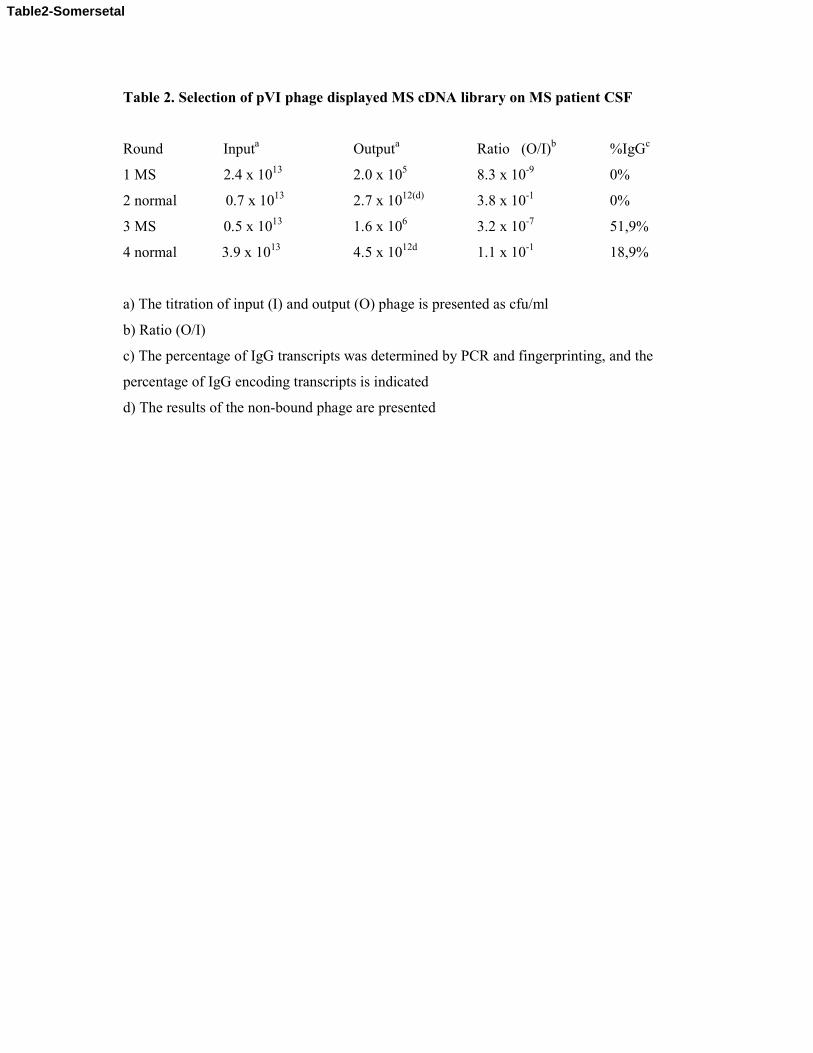
Table 2. Selection of pVI phage displayed MS cDNA library on MS patient CSF
Round Inputa Outputa Ratio (O/I)b %IgGc
1 MS 2.4 x 10¹³ 2.0 x 10⁵ 8.3 x 10⁻⁹ 0%
2 normal 0.7 x 10¹³ 2.7 x 10¹²(d) 3.8 x 10⁻¹ 0%
3 MS 0.5 x 10¹³ 1.6 x 10⁶ 3.2 x 10⁻⁷ 51,9%
4 normal 3.9 x 10¹³ 4.5 x 10¹²d 1.1 x 10⁻¹ 18,9%
a) The titration of input (I) and output (O) phage is presented as cfu/ml
b) Ratio (O/I)
c) The percentage of IgG transcripts was determined by PCR and fingerprinting, and the
percentage of IgG encoding transcripts is indicated
d) The results of the non-bound phage are presented
Table2-Somersetal