Chemokine receptor repertoire reflects mature T-cell lymphoproliferative disorder clinical...
7
Chemokine receptor repertoire reflects mature T-cell lymphoproliferative disorder clinical presentation João Moura a, ⁎, João Rodrigues b , Ana Helena Santos a , Maria dos Anjos Teixeira a , Maria Luís Queirós a , Marlene Santos a , Marta Gonçalves a , Sónia Fonseca a , Carla Laranjeira c , António Silva Rodrigues d , Esmeraldina Correia Júnior e , Fernanda Ribeiro d , Maria João Acosta e , Margarida Lima a a Laboratório de Citometria, Serviço de Hematologia Clínica, Centro Hospitalar do Porto (CHP), Hospital Geral de Santo António (HGSA), Rua D Manuel II, s/n, 4099-001 Porto, Portugal b Unidade de Biologia Molecular, Serviço de Hematologia Clínica, Centro Hospitalar do Porto (CHP), Hospital Geral de Santo António (HGSA), Porto, Portugal c Serviço de Hematologia Clínica, Centro Hospitalar do Porto (CHP), Hospital Geral de Santo António (HGSA), Porto, Portugal d Serviço de Hematologia, Hospital de Santo António dos Capuchos, Lisboa, Portugal e Laboratório de Hematologia, Centro Hospitalar de Lisboa Ocidental EPE, Hospital Santa Cruz, Lisboa, Portugal abstract article info Article history: Submitted 11 August 2008 Available online xxxx (Communicated by M. Lichtman, M.D., 25 August 2008) Keywords: Lymphoproliferative disorders Chemokine receptors T-cell lymphoma T-cell leukemia The World Health Organization classification of mature T-cell lymphoproliferative disorders, combines clinical, morphological and immunophenotypic data. The latter is a major contributor to the classification, as well as to the understanding of the malignant T-cell behavior. The fact that T-cell migration is regulated by chemokines should, in theory, enable us to identify tissue tropism and organ involvement by neoplastic T-cells by monitoring chemokine receptor surface expression. To address this issue we compared the expression of several early and late inflammatory, homeostatic, and organ specific chemokine receptors on blood T-cells from normal individuals and patients with T-cell large granular lymphocytic leukemia and peripheral T-cell lymphoma. T-cell large granular lymphocytic leukemia cells mainly express late inflammatory chemokine receptors (CXCR1 and CXCR2), whereas peripheral T-cell lymphoma cells usually express one or more organ homing receptors (CCR4, CCR6 and CCR7). Nevertheless, no clear correlation was found between CCR4 and CCR7 expression and skin and lymph node involvement, respectively. Compared to their normal counterparts, lymphoma T-cells displayed an exaggerated CCR4 expression, whereas leukemic T-cells had abnormally high CXCR1 and CXCR2 expression. Further analysis revealed that, in leukemia patients, the percentage of neoplastic cells expressing CCR5 correlates directly with lymphocytosis. In addition, in the case of CD8 T-cell leukemia patients, an inverse correlation with neutropenia was found. In lymphoma patients, higher CCR4 and CCR7 expression is accompanied by lower to absent CCR5 expression. © 2008 Elsevier Inc. All rights reserved. Introduction T-cell lymphoproliferative disorders (LPD) are uncommon dis- eases, arising from an abnormal proliferation or accumulation of neoplastic mature T-cells. They are broadly divided into leukemia or lymphoma depending on their presentation at the time of diagnosis. The World Health Organization (WHO) classifies mature T-cell neoplasms according to clinical, morphological and phenotypic characteristics. Some of the most representative groups of T-cell LPD are the large granular lymphocytic leukemia (LGLL) and the peripheral T-cell lymphoma (PTCL). PTCLs are usually sub-classified into nodal and extra-nodal lymphomas [1]. While the former affect the lymph nodes at diagnosis, the latter is evident in other organs and tissues, such as the skin, the gastrointestinal and the respiratory tract. Most nodal and extra-nodal cutaneous T-cell lymphoma cases arise on T-cell receptor α/β + CD4 + T-cells, and cutaneous T-cell lymphomas account for the majority of extra-nodal PTCLs [2]. T-cell LGLL, one of the most common T-cell leukemias, is characterized by the proliferation of monoclonal large granular lymphocytes (LGL) in the blood, usually of the CD8 subset, and is diagnosed mainly based on the morphologic and phenotypic characteristics of the monoclonal T-cell population, including the absence of the CD28 co-stimulatory receptor, the expression of NK- cell associated molecules and the presence of cytotoxic granules containing perforin and granzymes [3,4]. CD8 + T-cell LGLL usually is accompanied by neutropenia and other cytopenias, whereas CD4 + Blood Cells, Molecules, and Diseases xxx (2008) xxx–xxx ⁎ Corresponding author. E-mail address: [email protected] (J. Moura). YBCMD-01242; No. of pages: 7; 4C: 1079-9796/$ – see front matter © 2008 Elsevier Inc. All rights reserved. doi:10.1016/j.bcmd.2008.08.002 Contents lists available at ScienceDirect Blood Cells, Molecules, and Diseases journal homepage: www.elsevier.com/locate/ybcmd ARTICLE IN PRESS Please cite this article as: J. Moura, et al., Chemokine receptor repertoire reflects mature T-cell lymphoproliferative disorder clinical presentation, Blood Cell. Mol. Dis. (2008), doi:10.1016/j.bcmd.2008.08.002
-
Upload
independent -
Category
Documents
-
view
0 -
download
0
Transcript of Chemokine receptor repertoire reflects mature T-cell lymphoproliferative disorder clinical...

Blood Cells, Molecules, and Diseases xxx (2008) xxx–xxx
YBCMD-01242; No. of pages: 7; 4C:
Contents lists available at ScienceDirect
Blood Cells, Molecules, and Diseases
j ourna l homepage: www.e lsev ie r.com/ locate /ybcmd
ARTICLE IN PRESS
Chemokine receptor repertoire reflects mature T-cell lymphoproliferative disorderclinical presentation
João Moura a,⁎, João Rodrigues b, Ana Helena Santos a, Maria dos Anjos Teixeira a, Maria Luís Queirós a,Marlene Santos a, Marta Gonçalves a, Sónia Fonseca a, Carla Laranjeira c, António Silva Rodrigues d,Esmeraldina Correia Júnior e, Fernanda Ribeiro d, Maria João Acosta e, Margarida Lima a
a Laboratório de Citometria, Serviço de Hematologia Clínica, Centro Hospitalar do Porto (CHP), Hospital Geral de Santo António (HGSA), Rua D Manuel II, s/n, 4099-001 Porto, Portugalb Unidade de Biologia Molecular, Serviço de Hematologia Clínica, Centro Hospitalar do Porto (CHP), Hospital Geral de Santo António (HGSA), Porto, Portugalc Serviço de Hematologia Clínica, Centro Hospitalar do Porto (CHP), Hospital Geral de Santo António (HGSA), Porto, Portugald Serviço de Hematologia, Hospital de Santo António dos Capuchos, Lisboa, Portugale Laboratório de Hematologia, Centro Hospitalar de Lisboa Ocidental EPE, Hospital Santa Cruz, Lisboa, Portugal
⁎ Corresponding author.E-mail address: [email protected] (J. Moura).
1079-9796/$ – see front matter © 2008 Elsevier Inc. Aldoi:10.1016/j.bcmd.2008.08.002
Please cite this article as: J. Moura, et apresentation, Blood Cell. Mol. Dis. (2008), d
a b s t r a c t
a r t i c l e i n f oArticle history:
The World Health Organiz Submitted 11 August 2008Available online xxxx(Communicated by M. Lichtman, M.D.,25 August 2008)
Keywords:Lymphoproliferative disordersChemokine receptorsT-cell lymphomaT-cell leukemia
ation classification of mature T-cell lymphoproliferative disorders, combinesclinical, morphological and immunophenotypic data. The latter is a major contributor to the classification, aswell as to the understanding of the malignant T-cell behavior.The fact that T-cell migration is regulated by chemokines should, in theory, enable us to identify tissuetropism and organ involvement by neoplastic T-cells by monitoring chemokine receptor surface expression.To address this issue we compared the expression of several early and late inflammatory, homeostatic, andorgan specific chemokine receptors on blood T-cells from normal individuals and patients with T-cell largegranular lymphocytic leukemia and peripheral T-cell lymphoma.T-cell large granular lymphocytic leukemia cells mainly express late inflammatory chemokine receptors(CXCR1 and CXCR2), whereas peripheral T-cell lymphoma cells usually express one or more organ homingreceptors (CCR4, CCR6 and CCR7). Nevertheless, no clear correlation was found between CCR4 and CCR7expression and skin and lymph node involvement, respectively. Compared to their normal counterparts,lymphoma T-cells displayed an exaggerated CCR4 expression, whereas leukemic T-cells had abnormally highCXCR1 and CXCR2 expression.Further analysis revealed that, in leukemia patients, the percentage of neoplastic cells expressing CCR5correlates directly with lymphocytosis. In addition, in the case of CD8 T-cell leukemia patients, an inversecorrelation with neutropenia was found. In lymphoma patients, higher CCR4 and CCR7 expression isaccompanied by lower to absent CCR5 expression.
© 2008 Elsevier Inc. All rights reserved.
Introduction
T-cell lymphoproliferative disorders (LPD) are uncommon dis-eases, arising from an abnormal proliferation or accumulation ofneoplastic mature T-cells. They are broadly divided into leukemia orlymphoma depending on their presentation at the time of diagnosis.The World Health Organization (WHO) classifies mature T-cellneoplasms according to clinical, morphological and phenotypiccharacteristics. Some of the most representative groups of T-cell LPDare the large granular lymphocytic leukemia (LGLL) and the peripheralT-cell lymphoma (PTCL).
l rights reserved.
l., Chemokine receptor repeoi:10.1016/j.bcmd.2008.08.0
PTCLs are usually sub-classified into nodal and extra-nodallymphomas [1]. While the former affect the lymph nodes at diagnosis,the latter is evident in other organs and tissues, such as the skin, thegastrointestinal and the respiratory tract. Most nodal and extra-nodalcutaneous T-cell lymphoma cases arise on T-cell receptor α/β+ CD4+
T-cells, and cutaneous T-cell lymphomas account for the majority ofextra-nodal PTCLs [2].
T-cell LGLL, one of the most common T-cell leukemias, ischaracterized by the proliferation of monoclonal large granularlymphocytes (LGL) in the blood, usually of the CD8 subset, and isdiagnosed mainly based on the morphologic and phenotypiccharacteristics of the monoclonal T-cell population, including theabsence of the CD28 co-stimulatory receptor, the expression of NK-cell associated molecules and the presence of cytotoxic granulescontaining perforin and granzymes [3,4]. CD8+ T-cell LGLL usually isaccompanied by neutropenia and other cytopenias, whereas CD4+
rtoire reflects mature T-cell lymphoproliferative disorder clinical02
Table 1Specificities, clones and sources of the monoclonal antibodies used
Specificity Clone Conjugatea Sourceb
CD3 SK7 PerCP BDCD4 SK3 APC BDCD7 CLB-3A1/1,7F3 FITC CLBCD8 SK1 APC BDCD11c SHCL-3 PE BDCD27 9F4 FITC CLBCD28 CD28.2 PC5 IOTCD57 HNK-1 FITC BDCCR4 1G1 PE PHCCR5 3A9 PE PHCCR6 11A9 PE PHCCR7 3D12 PE PHCXCR1 5A12 PE PHCXCR2 6C6 PE PHCXCR3 1C6 PE PHCXCR4 12G5 PE PH
a APC — Allophycocyanin; FITC — fluorescein isothiocyanate; PC5 — Cy5-Phycoery-thrin; PerCP — Peridin–chlorophyll–protein complex.
b IOT — Immunotech, Marseille, France; BD — Becton-Dickinson, San José, CA, USA;CLB — Sanquin, Amsterdam, The Netherlands; PH — Pharmingen, San Diego, CA, USA.
2 J. Moura et al. / Blood Cells, Molecules, and Diseases xxx (2008) xxx–xxx
ARTICLE IN PRESS
T-cell LGLL frequently is associated with other hematological or non-hematological neoplasms [3]. Except for splenomegaly, which isrelatively frequent in patients with CD8+ T-cell LGLL, other organenlargement is rarely observed and, the marrow and spleen are theprincipal sites of accumulation of LGLL T-cells [4]. Unlike T-cellprolymphocytic leukemia [5], LGLL doesn't display any recurrentcytogenetic abnormality, nor does it usually result in high bloodlymphocyte counts [6].
These diverse characteristics of T-cell diseases make the differ-ential diagnosis difficult and demand for a different and betterapproach to classification.
Over the years, the classification systems have evolved from purelymorphologic systems combined with prognostic categories (WorkingFormulation) to classification systems which integrate clinical,biological and laboratorial data (REAL, WHO and EORTC classifica-tions) [7,8].
T lymphoblastic lymphoma/leukemia originates from the stages ofT-cell differentiation in the thymus and peripheral T-cell neoplasmsoriginate from post-thymic mature T-cells. T-cell prolymphocyticleukemia is thought to originate from naive T-cells whereas PTCL andLGLL originate from various stages of antigen-dependent T-cellactivation, with LGLL resembling the phenotype of terminallyactivated cytotoxic T-cells [7]. The fact that the normal blood T-cellpopulation is a mixture of cells in different activation stages, fromnaïve to effector, in conjunction with their resemblance to patholo-gical T-cells, makes the identification of neoplastic T-cells difficult [9].The same line of thought applies to migration in that lymphoidneoplasms should follow the homing patterns of their normalcounterparts, despite the fact that no clear correlation has yet beenmade between the CR repertoire and cell activation on normal T-cells.
To test this hypothesis we looked at chemokine receptor (CR)expression, since T-cell traffic can be indirectly assessed by theexpression of these CRs [10,11].
For this, we compared the expression of organ specific (CCR4, CCR6and CCR7) [12–14], early inflammatory, Th1 related (CCR5 and CXCR3)[15,16] late inflammatory (CXCR1 and CXCR2) [17], and homeostatic(CXCR4) [18] CRs between the abnormal peripheral blood (PB) T-cellsfrom patients with CD4+ PTCL and both CD4+ and CD8+ LGLL patients.We also compared the CR repertoire from the abnormal T-cells ofthese LPD with normal PB T-cells with an equivalent phenotype.
Materials and methods
Patients and controls
The CR repertoire was studied on the abnormal PB T-cells from 18CD4+ PTCL,10 CD4+ LGLL and 18 CD8+ LGLL patients and twelve normalage-matched adult individuals (blood donors) were used as controls.
All patients had normal hemoglobin values (11–13 g/dL), as well asnormal platelet counts (150–300×103/μL). White cell counts wereusually high (5–25×103/μL) due to lymphocytosis caused by T-cellproliferation. The majority of PTCL patients had either lymph node orskin infiltration by the neoplastic T-cells. The percentage of abnormalT-cells per total lymphocytes was always above 15%.
T-cell LPD was diagnosed and classified based on clinical andlaboratorial data according to the WHO schema [7]. Abnormal T-cellidentification was accessed by aberrant expression of cell-surfacemarkers by flow cytometry [3]. T-cell clonality was accessed by at leastone of the two following methods: a) expression of a single family ofthe TCR beta chain accessed by flow cytometry [3] and, or b)monoclonal TCR gamma and/or beta chain genes rearrangementaccessed by molecular PCR-based studies [19].
Samples
PB samples were collected on EDTA-K3 containing tubes.
Please cite this article as: J. Moura, et al., Chemokine receptor repepresentation, Blood Cell. Mol. Dis. (2008), doi:10.1016/j.bcmd.2008.08.0
Four-color flow cytometry
Immunophenotypic analysis of surface antigen expression on PBT-cells was performed in all cases using a stain-and-then-lyse four-color direct immunofluorescence technique, as previously describedin detail [3]. Briefly, 100 μl of whole blood containing 1–2×10⁶ cellswas incubated for 15 min at room temperature with antigen-specificfluorochrome-conjugated monoclonal antibodies (mAb), followed byerythrocyte lysis and cell fixation using FACS lysing solution(Becton-Dickinson BioSciences, BD, San José, CA USA), according tomanufacturer instructions. The mAb indicated in Table 1 were usedin different combinations in order to ensure the best selection of theabnormal T-cell population. In accordance, allophycocyanin con-jugated anti-CD4 or anti CD8 mAb (for CD4+ and CD8+ T-cell LPD,respectively) were combined with Phycoerythrin (PE) conjugatedanti-CR mAb in all cases. The specificity of the other mAb used wasvariable, depending on the best combination to identify thephenotypically abnormal T-cells in blood. In accordance, Cy5-Phycoerythrin (PC5) conjugated anti-CD28 and fluorescein (FITC)-conjugated anti-CD27 mAb were used in all LGLL cases, whereasPeridin–chlorophyll–protein complex. (PerCP) conjugated anti-CD3and FITC conjugated anti-CD7 mAb was the preferred combinationto identify PTCL cells. CD8+ LGLL cells were further subdivided intothree groups according to their CD11c and CD57 expression pattern(CD11c+CD57−, CD11c+CD57+ and CD11c−CD57+).
CD4+ and CD8+ LGLL cells were selected based on the absence ofCD27 and CD28 expression whereas CD4+ PTCL cells were identifiedbased on their abnormal light scatter characteristics and abnormallevels of CD3 and/or CD7 expression.
All experiments were done on a FACSCalibur cytometer (BDBiosciences), using CellQuest software version 3.1 (BD Biosciences)for sample acquisition and Paint-a-gate Pro software (BD Biosciences)for data analysis.
Comparison between neoplastic T-cells and their normal counterparts
Normal terminallyactivatedCD4+CD27−CD28− andCD8+CD27−CD28−
T-cells were considered as the normal counterparts for CD4+ and CD8+
LGLL T-cells.CD4+ PTCL cells have no clear homology to any specific subset of
normal PB CD4+ T-cells except for the fact that they are always CD28+
and usually express activation-related molecules [20]. Consideringthese two aspects normal PB CD4+CD28+CD45RO+ T-cells were used asnormal counterparts for CD4+ PTCL cells in this study.
rtoire reflects mature T-cell lymphoproliferative disorder clinical02
3J. Moura et al. / Blood Cells, Molecules, and Diseases xxx (2008) xxx–xxx
ARTICLE IN PRESS
Statistical analysis
All statistical analysis was done with Mann–Whitney U test.Differences were considered significant for pb0.05.
Ethical implications
All samples were collected after informed approval, and the workwas approved by the local Ethical Committee.
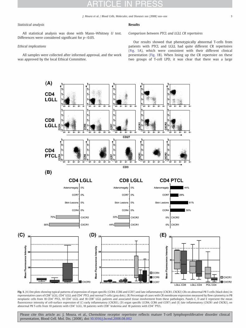
Fig. 1. (A) Dot plots showing typical patterns of expression of organ specific (CCR4, CCR6 andrepresentative cases of CD8+ LGLL, CD4+ LGLL and CD4+ PTCL and normal T-cells (grey dots). (neoplastic cells from 18 CD4+ PTCL, 10 CD4+ LGLL and 18 CD8+ LGLL patients and associatfluorescence intensity of cell-surface expression of (C) early inflammatory (CXCR3), (D) orgabnormal PB T-cells from 10 patients with CD4+ LGLL, 18 patients with CD8+ leukemia and
Please cite this article as: J. Moura, et al., Chemokine receptor repepresentation, Blood Cell. Mol. Dis. (2008), doi:10.1016/j.bcmd.2008.08.0
Results
Comparison between PTCL and LGLL CR repertoires
Our results showed that phenotypically abnormal T-cells frompatients with PTCL and LGLL had quite different CR repertoires(Fig. 1A), which were consistent with their different clinicalpresentation (Fig. 1B). When lining up the CR repertoire on thesetwo groups of T-cell LPD, it was clear that there was a large
CCR7) and late inflammatory (CXCR1, CXCR2) CRs on abnormal PB T-cells (black dots) inB) Percentage of cases with CR membrane expression measured by flow cytometry in PBed tissue involvement from these pathologies. Panels C, D and E represent the meanan specific (CCR4, CCR6 and CCR7) and (E) late inflammatory (CXCR1 and CXCR2), on18 patients with CD4+ PTCL.
rtoire reflects mature T-cell lymphoproliferative disorder clinical02
4 J. Moura et al. / Blood Cells, Molecules, and Diseases xxx (2008) xxx–xxx
ARTICLE IN PRESS
difference between PTCL and LGLL cells, concerning both thepercentage of cases expressing each of the CRs (Fig. 1B) and theirexpression intensity (Figs. 1C, D and E). PTCL cells usually expresseda variety of organ specific CRs, including CCR4, CCR6 and CCR7,whereas LGLL cells expressed the two late inflammatory IL-8receptors (CXCR1 and CXCR2).
Abnormal T-cells from the majority (78%) of patients with PTCLexpressed oneormore organ specific CRs,withhalf expressingCCR4 andnearly 1/3 expressing CCR6 or CCR7, which correlates with the highincidence of skin lesions and lymphnodeenlargementobserved in thosepatients (Fig. 1B). Nevertheless, no clear correlationwas found betweenCCR7 expression and lymph node involvement, either between CCR4expression and skin lesions. Actually, the neoplastic T-cells from 2 out of8 PTCL patients with lymphadenopathy had no CCR7 expression and T-cells from3 out of 12 patients with skin lesions had no CCR4 expression.PTCL cells have no late inflammatory CR expression.
On the other hand, CXCR1 and CXCR2 were expressed in themajority of CD4+ (90% and 70% respectively) and in a large fractionof CD8+ (44% and 33%, respectively) LGLL cases. In accordance tothe absence of skin, lymph node or other organ infiltrationobserved in LGLL patients (Fig. 1B), LGLL cells never expressedorgan specific CRs.
Early inflammatory, Th1-related CRs (CCR5 and CXCR3) wereexpressed in a fraction of both PTCL (28% and 50% respectively) andLGLL cells (10% and 26% respectively), with no statistically significantdifferences, except for a higher CXCR3 mean fluorescence intensity inPTCL (Fig. 1C), as compared to LGLL (pb0.05). No significant differencein CXCR4 expression was found.
Similarities and differences between the CR expression on neoplasticT-cells and their normal counterparts
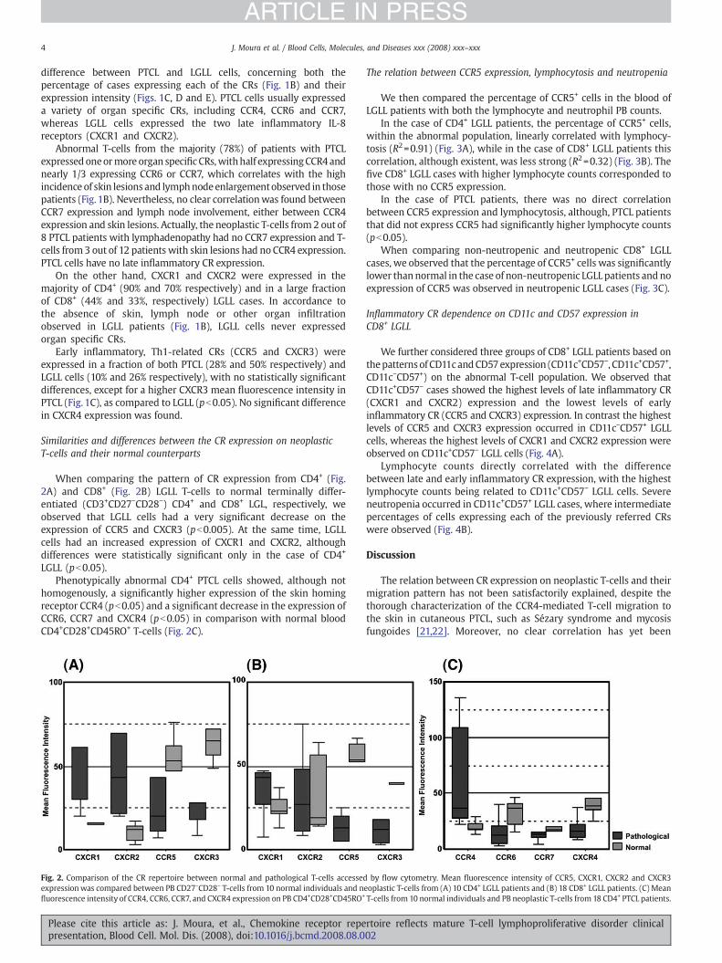
When comparing the pattern of CR expression from CD4+ (Fig.2A) and CD8+ (Fig. 2B) LGLL T-cells to normal terminally differ-entiated (CD3+CD27−CD28−) CD4+ and CD8+ LGL, respectively, weobserved that LGLL cells had a very significant decrease on theexpression of CCR5 and CXCR3 (pb0.005). At the same time, LGLLcells had an increased expression of CXCR1 and CXCR2, althoughdifferences were statistically significant only in the case of CD4+
LGLL (pb0.05).Phenotypically abnormal CD4+ PTCL cells showed, although not
homogenously, a significantly higher expression of the skin homingreceptor CCR4 (pb0.05) and a significant decrease in the expression ofCCR6, CCR7 and CXCR4 (pb0.05) in comparison with normal bloodCD4+CD28+CD45RO+ T-cells (Fig. 2C).
Fig. 2. Comparison of the CR repertoire between normal and pathological T-cells accessedexpressionwas compared between PB CD27−CD28− T-cells from 10 normal individuals and nfluorescence intensity of CCR4, CCR6, CCR7, and CXCR4 expression on PB CD4+CD28+CD45RO+
Please cite this article as: J. Moura, et al., Chemokine receptor repepresentation, Blood Cell. Mol. Dis. (2008), doi:10.1016/j.bcmd.2008.08.0
The relation between CCR5 expression, lymphocytosis and neutropenia
We then compared the percentage of CCR5+ cells in the blood ofLGLL patients with both the lymphocyte and neutrophil PB counts.
In the case of CD4+ LGLL patients, the percentage of CCR5+ cells,within the abnormal population, linearly correlated with lymphocy-tosis (R2=0.91) (Fig. 3A), while in the case of CD8+ LGLL patients thiscorrelation, although existent, was less strong (R2=0.32) (Fig. 3B). Thefive CD8+ LGLL cases with higher lymphocyte counts corresponded tothose with no CCR5 expression.
In the case of PTCL patients, there was no direct correlationbetween CCR5 expression and lymphocytosis, although, PTCL patientsthat did not express CCR5 had significantly higher lymphocyte counts(pb0.05).
When comparing non-neutropenic and neutropenic CD8+ LGLLcases, we observed that the percentage of CCR5+ cells was significantlylower thannormal in the case of non-neutropenic LGLL patients and noexpression of CCR5 was observed in neutropenic LGLL cases (Fig. 3C).
Inflammatory CR dependence on CD11c and CD57 expression inCD8+ LGLL
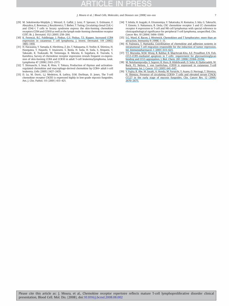
We further considered three groups of CD8+ LGLL patients based onthepatternsof CD11candCD57expression (CD11c+CD57−, CD11c+CD57+,CD11c−CD57+) on the abnormal T-cell population. We observed thatCD11c+CD57− cases showed the highest levels of late inflammatory CR(CXCR1 and CXCR2) expression and the lowest levels of earlyinflammatory CR (CCR5 and CXCR3) expression. In contrast the highestlevels of CCR5 and CXCR3 expression occurred in CD11c−CD57+ LGLLcells, whereas the highest levels of CXCR1 and CXCR2 expression wereobserved on CD11c+CD57− LGLL cells (Fig. 4A).
Lymphocyte counts directly correlated with the differencebetween late and early inflammatory CR expression, with the highestlymphocyte counts being related to CD11c+CD57− LGLL cells. Severeneutropenia occurred in CD11c+CD57+ LGLL cases, where intermediatepercentages of cells expressing each of the previously referred CRswere observed (Fig. 4B).
Discussion
The relation between CR expression on neoplastic T-cells and theirmigration pattern has not been satisfactorily explained, despite thethorough characterization of the CCR4-mediated T-cell migration tothe skin in cutaneous PTCL, such as Sézary syndrome and mycosisfungoides [21,22]. Moreover, no clear correlation has yet been
by flow cytometry. Mean fluorescence intensity of CCR5, CXCR1, CXCR2 and CXCR3eoplastic T-cells from (A) 10 CD4+ LGLL patients and (B) 18 CD8+ LGLL patients. (C) MeanT-cells from 10 normal individuals and PB neoplastic T-cells from 18 CD4+ PTCL patients.
rtoire reflects mature T-cell lymphoproliferative disorder clinical02
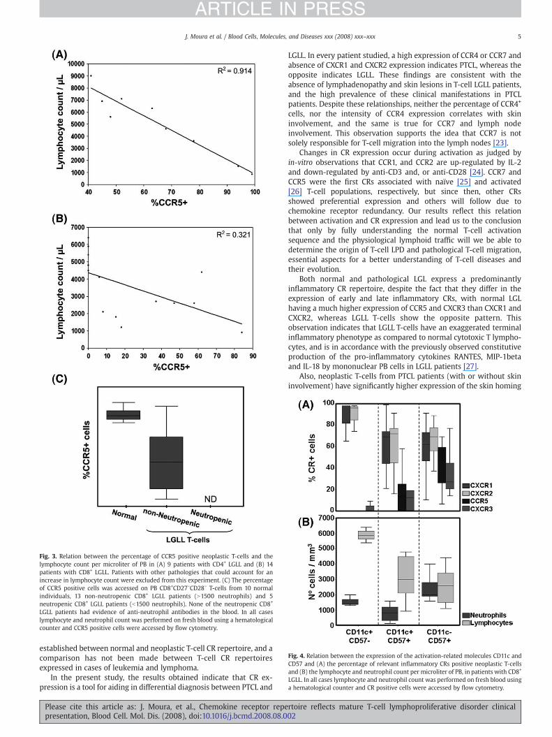
Fig. 3. Relation between the percentage of CCR5 positive neoplastic T-cells and thelymphocyte count per microliter of PB in (A) 9 patients with CD4+ LGLL and (B) 14patients with CD8+ LGLL. Patients with other pathologies that could account for anincrease in lymphocyte count were excluded from this experiment. (C) The percentageof CCR5 positive cells was accessed on PB CD8+CD27−CD28− T-cells from 10 normalindividuals, 13 non-neutropenic CD8+ LGLL patients (N1500 neutrophils) and 5neutropenic CD8+ LGLL patients (b1500 neutrophils). None of the neutropenic CD8+
LGLL patients had evidence of anti-neutrophil antibodies in the blood. In all caseslymphocyte and neutrophil count was performed on fresh blood using a hematologicalcounter and CCR5 positive cells were accessed by flow cytometry.
Fig. 4. Relation between the expression of the activation-related molecules CD11c andCD57 and (A) the percentage of relevant inflammatory CRs positive neoplastic T-cellsand (B) the lymphocyte and neutrophil count per microliter of PB, in patients with CD8+
LGLL. In all cases lymphocyte and neutrophil count was performed on fresh blood usinga hematological counter and CR positive cells were accessed by flow cytometry.
5J. Moura et al. / Blood Cells, Molecules, and Diseases xxx (2008) xxx–xxx
ARTICLE IN PRESS
established between normal and neoplastic T-cell CR repertoire, and acomparison has not been made between T-cell CR repertoiresexpressed in cases of leukemia and lymphoma.
In the present study, the results obtained indicate that CR ex-pression is a tool for aiding in differential diagnosis between PTCL and
Please cite this article as: J. Moura, et al., Chemokine receptor repepresentation, Blood Cell. Mol. Dis. (2008), doi:10.1016/j.bcmd.2008.08.0
LGLL. In every patient studied, a high expression of CCR4 or CCR7 andabsence of CXCR1 and CXCR2 expression indicates PTCL, whereas theopposite indicates LGLL. These findings are consistent with theabsence of lymphadenopathy and skin lesions in T-cell LGLL patients,and the high prevalence of these clinical manifestations in PTCLpatients. Despite these relationships, neither the percentage of CCR4+
cells, nor the intensity of CCR4 expression correlates with skininvolvement, and the same is true for CCR7 and lymph nodeinvolvement. This observation supports the idea that CCR7 is notsolely responsible for T-cell migration into the lymph nodes [23].
Changes in CR expression occur during activation as judged byin-vitro observations that CCR1, and CCR2 are up-regulated by IL-2and down-regulated by anti-CD3 and, or anti-CD28 [24]. CCR7 andCCR5 were the first CRs associated with naïve [25] and activated[26] T-cell populations, respectively, but since then, other CRsshowed preferential expression and others will follow due tochemokine receptor redundancy. Our results reflect this relationbetween activation and CR expression and lead us to the conclusionthat only by fully understanding the normal T-cell activationsequence and the physiological lymphoid traffic will we be able todetermine the origin of T-cell LPD and pathological T-cell migration,essential aspects for a better understanding of T-cell diseases andtheir evolution.
Both normal and pathological LGL express a predominantlyinflammatory CR repertoire, despite the fact that they differ in theexpression of early and late inflammatory CRs, with normal LGLhaving a much higher expression of CCR5 and CXCR3 than CXCR1 andCXCR2, whereas LGLL T-cells show the opposite pattern. Thisobservation indicates that LGLL T-cells have an exaggerated terminalinflammatory phenotype as compared to normal cytotoxic T lympho-cytes, and is in accordance with the previously observed constitutiveproduction of the pro-inflammatory cytokines RANTES, MIP-1betaand IL-18 by mononuclear PB cells in LGLL patients [27].
Also, neoplastic T-cells from PTCL patients (with or without skininvolvement) have significantly higher expression of the skin homing
rtoire reflects mature T-cell lymphoproliferative disorder clinical02

6 J. Moura et al. / Blood Cells, Molecules, and Diseases xxx (2008) xxx–xxx
ARTICLE IN PRESS
receptor CCR4 when compared to normal blood T-cells with the samephenotype (CD4+CD28+CD45RO+).
Increased CCR4 expression is in accordance with other studies thatindicate CCR4 as being responsible for the skin homing potential ofSézary syndrome [28,29] and other cutaneous lymphoma cells [30],and that this CR is also usually expressed in other PTCLs with skininvolvement [21,31]. Moreover, as confirmed in this paper, lymphomacells from mycosis fungoides patients [32,33] and other PTCL [34]express CCR4 with early inflammatory, Th1-related CR expression,namely CCR5 and CXCR3, supporting the idea that PTCL are derivedfrom cells with an activated phenotype. In addition, when comparedto the equivalent normal T lymphocytes, PTCL cells have infrequentand usually lower expression of CCR6 and CCR7, which cannot solelybe explained by the predominance of cutaneous T-cell lymphomasover nodal PTCL patients in this study (61% of patients with skinlesions versus 44% of patients with lymph node involvement) becauseeven patients with lymphadenopathy caused by PTCL cells display lessCCR7 on their surface than normal counterparts.
The clear inverse correlation between the percentage of neoplasticcells expressing CCR5 and lymphocyte counts in both LGLL and PTCLpatients is in accordance with a possible physiological role of CCR5 inthe regulation of T-cell proliferation and/or death, as well as T-celladhesion to endothelial cells and consequent migration to theinflamed tissue [35–37]. In contrast, the correlation between CCR5expression and neutrophil counts in CD8+ LGLL patients, whichexpress preferentially the late rather than the early inflammatory CRs,might be explained by a cytolytic action of terminally activated CD8+
cytotoxic lymphocytes on neutrophils.Taken together these two observations suggest that, in the case of
CD8+ LGLL patients, the decrease in the percentage of CCR5+ neoplasticcells is implicated in the development of neutropenia and lympho-cytosis, which are two useful parameters for monitoring LGLL. Thisobservation is important since it demonstrates a relation between theactivation status of LGLL T-cells and both neutropenia and lympho-cytosis, which in turn can be monitored by CCR5 expression.
CR expression is a determinant for the clinical presentation of T-cellLPD thus it is reasonable to assume that by monitoring the expressionof these and other organ specific CRs not addressed in this study, suchas CCR10 [38,39], we will be able to follow disease manifestations andpredict organ involvement during the course of the disease. In fact,preliminary data suggest that it is possible to identify a group ofpatients whose neoplastic T-cells only express CCR4, which have themost aggressive clinical behavior (data not shown).
This makes the evaluation of the CR repertoire by flow cytometryuseful for the classification of T-cell LPD, and especially important forestablishing a solid premise for the distinction between leukemia andlymphoma. Moreover, the knowledge of the specificities of the CRrepertoire on the different T-cell LPD may play an important role onthe future anti-chemokine receptor treatment of these neoplasticdisorders.
Acknowledgments
We acknowledge Professor Paula Ferreira for her critical discussionon the present work.
This work was supported by a grant from “ForumHematológico doNorte” and from “Associação Portuguesa Contra a Leucemia”.
João Moura is supported by a PhD grant (SFRH/BD/34471/2007)from Fundação para a Ciência e Tecnologia (FCT).
References
[1] E.S. Jaffe, Pathobiology of peripheral T-cell lymphomas, Hematology 2006 (2006)317–322.
[2] S.T. Rosen, C. Querfeld, Primary cutaneous T-Cell lymphomas, Hematology 2006(2006) 323–330.
Please cite this article as: J. Moura, et al., Chemokine receptor repepresentation, Blood Cell. Mol. Dis. (2008), doi:10.1016/j.bcmd.2008.08.0
[3] M. Lima, J. Almeida, M. dos Anjos Teixeira, M.d.C. Alguero, A.H. Santos, A.Balanzategui, M.L. Queiros, P. Barcena, A. Izarra, S. Fonseca, C. Bueno, B. Justica, M.Gonzalez, J.F. San Miguel, A. Orfao, TCR{alpha}{beta}+/CD4+ large granularlymphocytosis: a new clonal T-cell lymphoproliferative disorder, Am. J. Pathol.163 (2003) 763–771.
[4] T. Lamy, T.P. Loughran Jr., Current concepts: large granular lymphocyte leukemia,Blood Rev. 13 (1999) 230–240.
[5] C.M. Magro, C.D. Morrison, N. Heerema, P. Porcu, N. Sroa, A.C. Deng, T-cellprolymphocytic leukemia: an aggressive T cell malignancy with frequentcutaneous tropism, J. Am. Acad. Dermatol. 55 (2006) 467.
[6] E. Lazaro, O. Caubet, F. Menard, J.L. Pellegrin, J.F. Viallard, [Large granularlymphocyte leukemia], Presse Med. 36 (2007) 1694–1700.
[7] N.L. Harris, E.S. Jaffe, J. Diebold, G. Flandrin, H.K. Muller-Hermelink, J. Vardiman,T.A. Lister, C.D. Bloomfield, The World Health Organization classification ofneoplastic diseases of the hematopoietic and lymphoid tissues. Report of theClinical Advisory Committee meeting, Airlie House, Virginia, November, 1997,Ann. Oncol. 10 (1999) 1419–1432.
[8] R. Willemze, E.S. Jaffe, G. Burg, L. Cerroni, E. Berti, S.H. Swerdlow, E. Ralfkiaer, S.Chimenti, J.L. Diaz-Perez, L.M. Duncan, F. Grange, N.L. Harris, W. Kempf, H. Kerl, M.Kurrer, R. Knobler, N. Pimpinelli, C. Sander, M. Santucci, W. Sterry, M.H. Vermeer, J.Wechsler, S. Whittaker, C.J. Meijer, WHO-EORTC classification for cutaneouslymphomas, Blood 105 (2005) 3768–3785.
[9] E. Amyes, A.J. McMichael, M.F. Callan, Human CD4+ T cells are predominantlydistributed among six phenotypically and functionally distinct subsets, J.Immunol. 175 (2005) 5765–5773.
[10] B. Moser, M. Wolf, A. Walz, P. Loetscher, Chemokines: multiple levels of leukocytemigration control⁎, Trends Immunol. 25 (2004) 75.
[11] S.J. Ono, T. Nakamura, D. Miyazaki, M. Ohbayashi, M. Dawson, M. Toda,Chemokines: roles in leukocyte development, trafficking, and effector function,J. Allergy Clin. Immunol. 111 (2003) 1185.
[12] Y. Reiss, A.E. Proudfoot, C.A. Power, J.J. Campbell, E.C. Butcher, CC chemokinereceptor (CCR)4 and the CCR10 ligand cutaneous T cell-attracting chemokine(CTACK) in lymphocyte trafficking to inflamed skin, J. Exp. Med. 194 (2001)1541–1547.
[13] E. Schutyser, S. Struyf, J. Van Damme, The CC chemokine CCL20 and its receptorCCR6, Cytokine Growth Factor Rev. 14 (2003) 409.
[14] C. Potsch, D. Vohringer, H. Pircher, Distinct migration patterns of naive and effectorCD8 T cells in the spleen: correlation with CCR7 receptor expression andchemokine reactivity, Eur. J. Immunol. 29 (1999) 3562–3570.
[15] S. Qin, J.B. Rottman, P. Myers, N. Kassam, M. Weinblatt, M. Loetscher, A.E. Koch, B.Moser, C.R. Mackay, The chemokine receptors CXCR3 and CCR5 mark subsets of Tcells associated with certain inflammatory reactions, J. Clin. Invest. 101 (1998)746–754.
[16] C.H. Kim, L. Rott, E.J. Kunkel, M.C. Genovese, D.P. Andrew, L. Wu, E.C. Butcher, Rulesof chemokine receptor association with T cell polarization in vivo, J. Clin. Invest.108 (2001) 1331–1339.
[17] C. Murdoch, A. Finn, Chemokine receptors and their role in inflammation andinfectious diseases, Blood 95 (2000) 3032–3043.
[18] D.J. Campbell, C.H. Kim, E.C. Butcher, Chemokines in the systemic organization ofimmunity, Immunol. Rev. 195 (2003) 58–71.
[19] J.J. van Dongen, A.W. Langerak, M. Bruggemann, P.A. Evans, M. Hummel, F.L.Lavender, E. Delabesse, F. Davi, E. Schuuring, R. Garcia-Sanz, J.H. van Krieken, J.Droese, D. Gonzalez, C. Bastard, H.E. White, M. Spaargaren, M. Gonzalez, A.Parreira, J.L. Smith, G.J. Morgan, M. Kneba, E.A. Macintyre, Design andstandardization of PCR primers and protocols for detection of clonal immunoglo-bulin and T-cell receptor gene recombinations in suspect lymphoproliferations:report of the BIOMED-2 Concerted Action BMH4-CT98-3936, Leukemia 17 (2003)2257–2317.
[20] T. Rudiger, E. Geissinger, H.K. Muller-Hermelink, ‘Normal counterparts’ of nodalperipheral T-cell lymphoma, Hematol. Oncol. 24 (2006) 175–180.
[21] T. Ishida, A. Utsunomiya, S. Iida, H. Inagaki, Y. Takatsuka, S. Kusumoto, G. Takeuchi,S. Shimizu, M. Ito, H. Komatsu, A. Wakita, T. Eimoto, K. Matsushima, R. Ueda,Clinical Significance of CCR4 expression in adult T-Cell leukemia/lymphoma: itsclose association with skin involvement and unfavorable outcome, Clin. CancerRes. 9 (2003) 3625–3634.
[22] M.G. Narducci, E. Scala, A. Bresin, E. Caprini, M.C. Picchio, D. Remotti, G. Ragone, F.Nasorri, M. Frontani, D. Arcelli, S. Volinia, G.A. Lombardo, G. Baliva, M. Napolitano,G. Russo, Skin homing of Sezary cells involves SDF-1-CXCR4 signaling and down-regulation of CD26/dipeptidylpeptidase IV, Blood 107 (2006) 1108–1115.
[23] L.M. Ebert, P. Schaerli, B. Moser, Chemokine-mediated control of T cell traffic inlymphoid and peripheral tissues, Mol. Immunol. 42 (2005) 799–809.
[24] P. Loetscher, M. Seitz, M. Baggiolini, B. Moser, Interleukin-2 regulates CCchemokine receptor expression and chemotactic responsiveness in T lympho-cytes, J. Exp. Med. 184 (1996) 569–577.
[25] J.J. Campbell, K.E. Murphy, E.J. Kunkel, C.E. Brightling, D. Soler, Z. Shen, J. Boisvert,H.B. Greenberg, M.A. Vierra, S.B. Goodman, M.C. Genovese, A.J. Wardlaw, E.C.Butcher, L. Wu, CCR7 expression and memory T cell diversity in humans, J.Immunol. 166 (2001) 877–884.
[26] C.R. Mackay, Immunological memory, Adv. Immunol. 53 (1993) 217–265.[27] R. Kothapalli, S.B. Nyland, I. Kusmartseva, R.D. Bailey, T.M. McKeown, T.P. Loughran
Jr., Constitutive production of proinflammatory cytokines RANTES, MIP-1beta andIL-18 characterizes LGL leukemia, Int. J. Oncol. 26 (2005) 529–535.
[28] M.T. Fierro, A. Comessatti, P. Quaglino, M. Ortoncelli, S. Osella Abate, R. Ponti, M.Novelli, M.G. Bernengo, Expression pattern of chemokine receptors andchemokine release in inflammatory erythroderma and Sezary syndrome,Dermatology 213 (2006) 284–292.
rtoire reflects mature T-cell lymphoproliferative disorder clinical02
7J. Moura et al. / Blood Cells, Molecules, and Diseases xxx (2008) xxx–xxx
ARTICLE IN PRESS
[29] M. Sokolowska-Wojdylo, J. Wenzel, E. Gaffal, J. Lenz, P. Speuser, S. Erdmann, F.Abuzahra, E. Bowman, J. Roszkiewicz, T. Bieber, T. Tuting, Circulating clonal CLA(+)and CD4(+) T cells in Sezary syndrome express the skin-homing chemokinereceptors CCR4 and CCR10 as well as the lymph node-homing chemokine receptorCCR7, Br. J. Dermatol. 152 (2005) 258–264.
[30] K. Ferenczi, R.C. Fuhlbrigge, J. Pinkus, G.S. Pinkus, T.S. Kupper, Increased CCR4expression in cutaneous T cell lymphoma, J. Invest. Dermatol. 119 (2002)1405–1410.
[31] H. Harasawa, Y. Yamada, K. Hieshima, Z. Jin, T. Nakayama, O. Yoshie, K. Shimizu, H.Hasegawa, T. Hayashi, Y. Imaizumi, S. Ikeda, H. Soda, H. Soda, S. Atogami, Y.Takasaki, K. Tsukasaki, M. Tomonaga, K. Murata, K. Sugahara, K. Tsuruda, S.Kamihira, Survey of chemokine receptor expression reveals frequent co-expres-sion of skin-homing CCR4 and CCR10 in adult T-cell leukemia/lymphoma, Leuk.Lymphoma 47 (2006) 2163–2173.
[32] T. Shimauchi, S. Imai, R. Hino, Y. Tokura, Production of thymus and activation-regulated chemokine and macrophage-derived chemokine by CCR4+ adult t-cellleukemia, Cells (2005) 2427–2435.
[33] D. Lu, M. Duvic, L.J. Medeiros, R. Luthra, D.M. Dorfman, D. Jones, The T-cellchemokine receptor CXCR3 is expressed highly in low-grade mycosis fungoides,Am. J. Clin. Pathol. 115 (2001) 413–421.
Please cite this article as: J. Moura, et al., Chemokine receptor repepresentation, Blood Cell. Mol. Dis. (2008), doi:10.1016/j.bcmd.2008.08.0
[34] T. Ishida, H. Inagaki, A. Utsunomiya, Y. Takatsuka, H. Komatsu, S. Iida, G. Takeuchi,T. Eimoto, S. Nakamura, R. Ueda, CXC chemokine receptor 3 and CC chemokinereceptor 4 expression in T-cell and NK-cell lymphomas with special reference toclinicopathological significance for peripheral T-cell lymphoma, unspecified, Clin.Cancer Res. 10 (2004) 5494–5500.
[35] S.G. Ward, K. Bacon, J. Westwick, Chemokines and T lymphocytes: more than anattraction, Immunity 9 (1998) 1–11.
[36] H. Fujiwara, T. Hamaoka, Coordination of chemokine and adhesion systems inintratumoral T cell migration responsible for the induction of tumor regression,Int. Immunopharmacol. 1 (2001) 613–623.
[37] T.T. Murooka, M.M. Wong, R. Rahbar, B. Majchrzak-Kita, A.E. Proudfoot, E.N. Fish,CCL5-CCR5-mediated apoptosis in T cells: requirement for glycosaminoglycanbinding and CCL5 aggregation, J. Biol. Chem. 281 (2006) 25184–25194.
[38] M. Notohamiprodjo, S. Segerer, R. Huss, B. Hildebrandt, D. Soler, R. Djafarzadeh, W.Buck, P.J. Nelson, I. von Luettichau, CCR10 is expressed in cutaneous T-celllymphoma, Int. J. Cancer 115 (2005) 641–647.
[39] Y. Fujita, R. Abe, M. Sasaki, A. Honda, M. Furuichi, Y. Asano, O. Norisugi, T. Shimizu,H. Shimizu, Presence of circulating CCR10+ T cells and elevated serum CTACK/CCL27 in the early stage of mycosis fungoides, Clin. Cancer Res. 12 (2006)2670–2675.
rtoire reflects mature T-cell lymphoproliferative disorder clinical02