Risk based serological survey of Rift Valley fever in Tunisia ...
7
HAL Id: hal-03460875 https://hal.inrae.fr/hal-03460875 Submitted on 1 Dec 2021 HAL is a multi-disciplinary open access archive for the deposit and dissemination of sci- entific research documents, whether they are pub- lished or not. The documents may come from teaching and research institutions in France or abroad, or from public or private research centers. L’archive ouverte pluridisciplinaire HAL, est destinée au dépôt et à la diffusion de documents scientifiques de niveau recherche, publiés ou non, émanant des établissements d’enseignement et de recherche français ou étrangers, des laboratoires publics ou privés. Distributed under a Creative Commons Attribution| 4.0 International License Risk based serological survey of Rift Valley fever in Tunisia (2017–2018) Sana Kalthoum, Elena Arsevska, Kaouther Guesmi, Aymen Mamlouk, Jamel Cherni, Monia Lachtar, Raja Gharbi, Bassem Bel Haj Mohamed, Wiem Khalfaoui, Anissa Dhaouadi, et al. To cite this version: Sana Kalthoum, Elena Arsevska, Kaouther Guesmi, Aymen Mamlouk, Jamel Cherni, et al.. Risk based serological survey of Rift Valley fever in Tunisia (2017–2018). Heliyon, Elsevier 2021, 7 (9), pp.e07932. 10.1016/j.heliyon.2021.e07932. hal-03460875
-
Upload
khangminh22 -
Category
Documents
-
view
1 -
download
0
Transcript of Risk based serological survey of Rift Valley fever in Tunisia ...
HAL Id: hal-03460875https://hal.inrae.fr/hal-03460875
Submitted on 1 Dec 2021
HAL is a multi-disciplinary open accessarchive for the deposit and dissemination of sci-entific research documents, whether they are pub-lished or not. The documents may come fromteaching and research institutions in France orabroad, or from public or private research centers.
L’archive ouverte pluridisciplinaire HAL, estdestinée au dépôt et à la diffusion de documentsscientifiques de niveau recherche, publiés ou non,émanant des établissements d’enseignement et derecherche français ou étrangers, des laboratoirespublics ou privés.
Distributed under a Creative Commons Attribution| 4.0 International License
Risk based serological survey of Rift Valley fever inTunisia (2017–2018)
Sana Kalthoum, Elena Arsevska, Kaouther Guesmi, Aymen Mamlouk, JamelCherni, Monia Lachtar, Raja Gharbi, Bassem Bel Haj Mohamed, Wiem
Khalfaoui, Anissa Dhaouadi, et al.
To cite this version:Sana Kalthoum, Elena Arsevska, Kaouther Guesmi, Aymen Mamlouk, Jamel Cherni, et al.. Riskbased serological survey of Rift Valley fever in Tunisia (2017–2018). Heliyon, Elsevier 2021, 7 (9),pp.e07932. �10.1016/j.heliyon.2021.e07932�. �hal-03460875�
Heliyon 7 (2021) e07932
Contents lists available at ScienceDirect
Heliyon
journal homepage: www.cell.com/heliyon
Research article
Risk based serological survey of Rift Valley fever in Tunisia (2017–2018)
Sana Kalthoum a,*, Elena Arsevska b,c, Kaouther Guesmi a, Aymen Mamlouk d, Jamel Cherni a,Monia lachtar a, Raja Gharbi a, Bassem Bel Haj Mohamed a, Wiem Khalfaoui a, Anissa Dhaouadi a,Mohamed Naceur Baccar a, Haikel Hajlaoui a, Samia Mzoughi a, Ch�edia Seghaier a, Lilia Messadi d,Malek Zrelli e, Soufien Sghaier f, Catherine Cetre-Sossah c,g, Pascal Hendrikx b,c,C�ecile Squarzoni-Diaw b,g
a National Center of Zoosanitary Vigilance, Tunis, Tunisiab CIRAD, UMR ASTRE, F-34395 Montpellier, Francec ASTRE, Univ Montpellier, CIRAD, INRAE, Montpellier, Franced Service de Microbiologie et Immunologie, Ecole Nationale de M�edecine V�et�erinaire de Sidi Thabet, Univ. Manouba, Sidi Thabet, Tunisiae Minist�ere de l’Agriculture, Direction G�en�erale des Services V�et�erinaires, Tunis, Tunisiaf Institut de la Recherche V�et�erinaire de Tunisie (IRVT), Tunis, Tunisiag CIRAD, UMR ASTRE, F-97490 Sainte Clotilde, La R�eunion, France
A R T I C L E I N F O
Keywords:QRA methodologyRisk mappingSurveyTunisiaRift valley feverSmall ruminantsCamels
* Corresponding author.E-mail address: [email protected] (S. K
https://doi.org/10.1016/j.heliyon.2021.e07932Received 17 February 2021; Received in revised fo2405-8440/© 2021 The Authors. Published by Else
A B S T R A C T
Rift Valley fever (RVF) has been reported in the sub-Saharan region of Africa, Egypt and Arabian Peninsula -Yemen and Saudi Arabia, over the past 20 years and is a threat to both the animal and human populations inTunisia. Tunisia is considered as a high-risk country for the introduction of RVF due to the informal movements ofdiseased animals already reported in the neighboring countries. The objective of this study was to assess the statusof RVF in small ruminants and camels in Tunisia. A risk-based serological survey was conducted to evaluate thepresence of RVF based on spatial qualitative risk analysis (SQRA). Samples were collected from small ruminants(sheep and goats) (n ¼ 1,114), and camels (n ¼ 173) samples, belonging to 18 breeders in 14 governoratesbetween November 2017 and January 2018. Samples were tested using an RVF specific multispecies competitiveELISA. Out of the 1,287 samples tested for the presence of RVF IgG antibodies by ELISA, only one positive sample0.07% (1/1 287) was detected but not confirmed with the virus neutralization test (VNT) used for confirmation.So far, no RVF outbreaks have been reported in Tunisia and our study confirmed the absence of RVF in livestockup to January 2018. Further investigations are needed to confirm the RVF-free status of Tunisia today.
1. Introduction
Rift Valley fever (RVF) is a mosquito-borne zoonosis that affectshumans and domestic ruminants (camelids, cattle, goats, and sheep) [1]caused by a virus of the Phlebovirus genus that belongs to the Phenuivir-idae family. The virus was identified for the first time in 1930 in the RiftValley in Kenya [2, 3]. Humans are infected by the RVF virus (RVFV)through contact with the blood or organs of infected animals duringslaughter, or when handling infected animals, or through the ingestion ofcontaminated meat and raw milk [4]. Thus, staff working in slaughter-houses, laboratories and hospitals are the most exposed [5]. However,mosquitoes are the main vectors involved in the spread of RVFV duringepidemics. The RVFV has been isolated from at least 40 mosquito speciesbelonging to eight genera (mainly Aedes spp. and Culex spp.) [6, 7] when
althoum).
rm 28 March 2021; Accepted 1 Svier Ltd. This is an open access a
feeding on viremic animals. Infected females of Aedes spp. are known totransmit the virus to their progeny, via desiccated eggs that are resistantto drought, thus maintaining the viral life cycle [8].
The feeding activities of these mosquitoes rely mainly on environ-mental and climatic factors (rainfall, temperature) and outbreaks arelikely to occur during heavy rainfall events in areas susceptible toflooding [9]. The mode of transmission varies with the ecosystem. Forexample, the most recent epidemics in Mayotte and Senegal showed thatdepending on the environmental context and on the typology of thefarms, transmission of the vector or transmission linked to direct contactbetween herds and between animals can be of varying importance [10].In infected livestock, the most common clinical signs are fever, massiveabortions, high morbidity and mortality among young animals [11]. Inhumans, RVF causes a febrile and a hemorrhagic syndrome (epistaxis,
eptember 2021rticle under the CC BY license (http://creativecommons.org/licenses/by/4.0/).
S. Kalthoum et al. Heliyon 7 (2021) e07932
hemoptysis, melena, hematemesis, gingival bleeding, bruising), and insevere cases, death [12].
The geographical distribution of RVF indicates that until 2000, thedisease was limited to sub-Saharan Africa before expanding to theArabian Peninsula [13]. As far as North African countries are concerned,Egypt experienced extensive outbreaks in 1977–78 and it is believed thatthe virus was introduced from Sudan through the Aswan dam [14].Smaller epidemics occurred in 1993–94, 1996–97, followed by a largeroutbreak in 2003. Serological surveys in animals and humans revealedthe enzootic profile of the disease in Egypt [15]. In December 2019, Libyareported several RVF outbreaks in the southern part of the country [16].As far as the North Africa are concerned, in 2008 and 2009, serologicalstudies were conducted in Sahrawi refugee camps (Tindouf Province) onthe south-western border with western Sahara (Algeria), in Mauritania,and in southern Morocco, in ruminants and human populations and RVFspecific IgG antibodies were detected in camels and goats [17, 18].
In Tunisia, a serological survey was carried out in 2014 in the Centreof Tunisia (governorates of Sfax, Mahdia and Sousse) and revealed thepresence of RVF specific IgG antibodies in human samples despite theirabsence in samples from febrile patients and slaughterhouse workers[19]. Additional RVF focused seroprevalence studies conducted on ani-mal samples such as dromedaries in 2017 [20], goats and sheep in2006–2007 [21] did not confirm active circulation of RVF in Tunisia.However, a study by Selmi et al. using targeted sampling reported 34%seroprevalence in camel populations in the southern governorates ofTunisia. This result could be explained by the fact that sampled camelsmay originate from illegal trade (Sudan, Chad and Niger), and may havebeen introduced into Tunisia through Libya [22].
Recent studies in Tunisia demonstrated that climatic factors mightinfluence the distribution and abundance of the mosquitoes that transmitRVFV [23]. The mean temperature of the warmest quarter, the meantemperature of the coldest quarter, isothermally, and annual precipita-tion are considered to be the most significant climatic factors that in-fluence vector distribution in risk areas [23]. The intensification ofanimal trade has also been shown to increase the risk of RVFV intro-duction and spread, and hence emergence in previously unaffected ter-ritories [24]. The latest epidemics have shown that, depending onenvironmental and livestock conditions, vector and direct transmissionaffect the magnitude of the epidemic differently [10]. Ahigh-performance surveillance system is required for early detection ofRVF outbreaks to avoid the economic losses that can result from emer-gence of the disease. The risk-based surveillance approach (RBS) can beused to improve detection of RVF as it is more sensitive, would providehigher positive predictive values, and enable more effective and efficientallocation of resources in countries with limited resources [25]. Indeed,this method considerably reduces the number of areas to be surveyed andhence the cost of targeted surveillance [25]. In Tunisia, a risk-basedsurveillance approach was implemented for foot and mouth disease(FMD) in 2017/2018 completed by a spatial risk analysis. A serologicalsurvey conducted in very-high and high-risk imadas helped estimateantibody prevalence to FMD [26]. In fact, Tunisia is at permanent risk ofthe introduction of several vector and non-vector borne infectious dis-eases, including RVF, due the illegal movement of small ruminants that ismost intense during religious events [27].
The aim of this study was thus to investigate the circulation of RVF inthe small ruminant and camel populations in Tunisia, using a serologicalcross-sectional survey in areas considered as high and very high-riskareas for the introduction of RVF.
2. Materials and methods
2.1. Period of study and risk-based survey
Tunisia is located in the northern eastern part of Africa between lat-itudes 30� and 38�N, and longitudes 7� and 12�E, covers 163,610 squarekm and had 11.7 million inhabitants in 2019 [28]. Administratively, it is
2
organized in 24 governorates and 2,075 imadas. A risk-based surveytargeting small ruminants and camels in the high and very-high risk areaswas conducted in winter, i.e., between November 24, 2017 and January30, 2018. The study area was represented by 23 randomly sampledimadas out of 841 imadas classified as high and very-high risk of exposureto RVF using a qualitative risk assessment (QRA) method [29]. Thesurvey was limited to the two strata (very-high and high risk) forfinancial reasons.
The risk factors used to characterize the different levels of risk ofexposure to RVFV included:
a) Ruminant density (number of animals per km2) [30],b) Accessibility to other imadas (average travel time in minutes) [31],c) Frequency of national and cross-border ruminant movements [32],d) Presence/absence of permanent/temporary lakes and rivers, i.e.,
water bodies [33],e) Abundance of five of the known competent mosquito vectors in
Tunisia, i.e. Culex pipiens, Aedes vexans, Aedes aegypti, Anophelesgambiae, Culex quinquefasciatus [34].
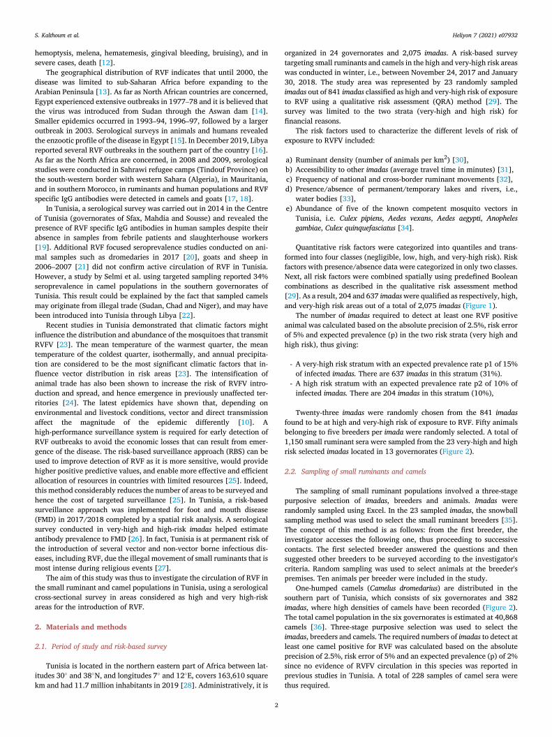
Quantitative risk factors were categorized into quantiles and trans-formed into four classes (negligible, low, high, and very-high risk). Riskfactors with presence/absence data were categorized in only two classes.Next, all risk factors were combined spatially using predefined Booleancombinations as described in the qualitative risk assessment method[29]. As a result, 204 and 637 imadaswere qualified as respectively, high,and very-high risk areas out of a total of 2,075 imadas (Figure 1).
The number of imadas required to detect at least one RVF positiveanimal was calculated based on the absolute precision of 2.5%, risk errorof 5% and expected prevalence (p) in the two risk strata (very high andhigh risk), thus giving:
- A very-high risk stratum with an expected prevalence rate p1 of 15%of infected imadas. There are 637 imadas in this stratum (31%).
- A high risk stratum with an expected prevalence rate p2 of 10% ofinfected imadas. There are 204 imadas in this stratum (10%),
Twenty-three imadas were randomly chosen from the 841 imadasfound to be at high and very-high risk of exposure to RVF. Fifty animalsbelonging to five breeders per imada were randomly selected. A total of1,150 small ruminant sera were sampled from the 23 very-high and highrisk selected imadas located in 13 governorates (Figure 2).
2.2. Sampling of small ruminants and camels
The sampling of small ruminant populations involved a three-stagepurposive selection of imadas, breeders and animals. Imadas wererandomly sampled using Excel. In the 23 sampled imadas, the snowballsampling method was used to select the small ruminant breeders [35].The concept of this method is as follows: from the first breeder, theinvestigator accesses the following one, thus proceeding to successivecontacts. The first selected breeder answered the questions and thensuggested other breeders to be surveyed according to the investigator'scriteria. Random sampling was used to select animals at the breeder'spremises. Ten animals per breeder were included in the study.
One-humped camels (Camelus dromedarius) are distributed in thesouthern part of Tunisia, which consists of six governorates and 382imadas, where high densities of camels have been recorded (Figure 2).The total camel population in the six governorates is estimated at 40,868camels [36]. Three-stage purposive selection was used to select theimadas, breeders and camels. The required numbers of imadas to detect atleast one camel positive for RVF was calculated based on the absoluteprecision of 2.5%, risk error of 5% and an expected prevalence (p) of 2%since no evidence of RVFV circulation in this species was reported inprevious studies in Tunisia. A total of 228 samples of camel sera werethus required.
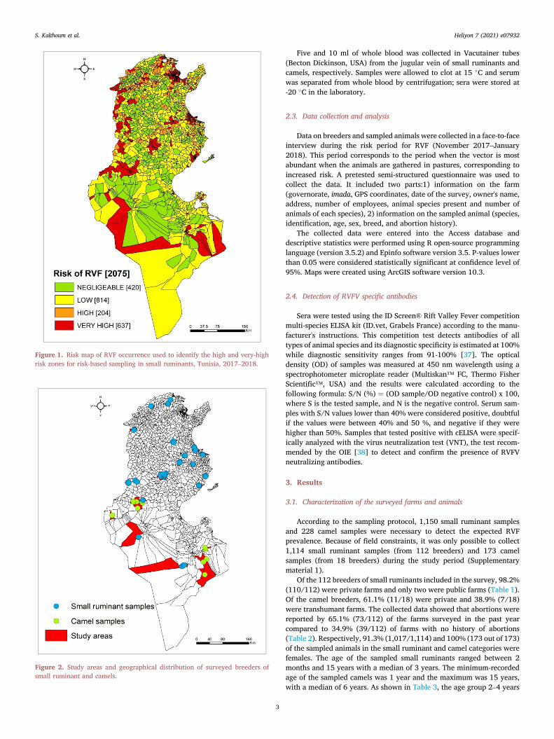
Figure 1. Risk map of RVF occurrence used to identify the high and very-highrisk zones for risk-based sampling in small ruminants, Tunisia, 2017–2018.
Figure 2. Study areas and geographical distribution of surveyed breeders ofsmall ruminant and camels.
S. Kalthoum et al. Heliyon 7 (2021) e07932
3
Five and 10 ml of whole blood was collected in Vacutainer tubes(Becton Dickinson, USA) from the jugular vein of small ruminants andcamels, respectively. Samples were allowed to clot at 15 �C and serumwas separated from whole blood by centrifugation; sera were stored at-20 �C in the laboratory.
2.3. Data collection and analysis
Data on breeders and sampled animals were collected in a face-to-faceinterview during the risk period for RVF (November 2017–January2018). This period corresponds to the period when the vector is mostabundant when the animals are gathered in pastures, corresponding toincreased risk. A pretested semi-structured questionnaire was used tocollect the data. It included two parts:1) information on the farm(governorate, imada, GPS coordinates, date of the survey, owner's name,address, number of employees, animal species present and number ofanimals of each species), 2) information on the sampled animal (species,identification, age, sex, breed, and abortion history).
The collected data were entered into the Access database anddescriptive statistics were performed using R open-source programminglanguage (version 3.5.2) and Epinfo software version 3.5. P-values lowerthan 0.05 were considered statistically significant at confidence level of95%. Maps were created using ArcGIS software version 10.3.
2.4. Detection of RVFV specific antibodies
Sera were tested using the ID Screen® Rift Valley Fever competitionmulti-species ELISA kit (ID.vet, Grabels France) according to the manu-facturer's instructions. This competition test detects antibodies of alltypes of animal species and its diagnostic specificity is estimated at 100%while diagnostic sensitivity ranges from 91-100% [37]. The opticaldensity (OD) of samples was measured at 450 nm wavelength using aspectrophotometer microplate reader (Multiskan™ FC, Thermo FisherScientific™, USA) and the results were calculated according to thefollowing formula: S/N (%) ¼ (OD sample/OD negative control) x 100,where S is the tested sample, and N is the negative control. Serum sam-ples with S/N values lower than 40% were considered positive, doubtfulif the values were between 40% and 50 %, and negative if they werehigher than 50%. Samples that tested positive with cELISA were specif-ically analyzed with the virus neutralization test (VNT), the test recom-mended by the OIE [38] to detect and confirm the presence of RVFVneutralizing antibodies.
3. Results
3.1. Characterization of the surveyed farms and animals
According to the sampling protocol, 1,150 small ruminant samplesand 228 camel samples were necessary to detect the expected RVFprevalence. Because of field constraints, it was only possible to collect1,114 small ruminant samples (from 112 breeders) and 173 camelsamples (from 18 breeders) during the study period (Supplementarymaterial 1).
Of the 112 breeders of small ruminants included in the survey, 98.2%(110/112) were private farms and only two were public farms (Table 1).Of the camel breeders, 61.1% (11/18) were private and 38.9% (7/18)were transhumant farms. The collected data showed that abortions werereported by 65.1% (73/112) of the farms surveyed in the past yearcompared to 34.9% (39/112) of farms with no history of abortions(Table 2). Respectively, 91.3% (1,017/1,114) and 100% (173 out of 173)of the sampled animals in the small ruminant and camel categories werefemales. The age of the sampled small ruminants ranged between 2months and 15 years with a median of 3 years. The minimum-recordedage of the sampled camels was 1 year and the maximum was 15 years,with a median of 6 years. As shown in Table 3, the age group 2–4 years
Table 1. Categorization of the surveyed farms (public and private).
Type of farms Small ruminants Camels
Public 2 (1.8%) 0 (0%)
Private 110 (98.2%) 11 (61.1%)
Transhumant 0 (0%) 7 (38.9%)
Total 112 18
Table 2. Occurrence of abortion on the surveyed farms.
Occurrence of abortion in thepast year
Number of small ruminantfarms
Number of camelsfarms
No 39 (34.9%) 8 (44.4%)
Yes 73 (65.1%) 10 (55.6%)
Total 112 100
S. Kalthoum et al. Heliyon 7 (2021) e07932
was the most common, 54% (607/1,114), among the small ruminants.Among the camels, the most common age group was 9–15 years (37.4%).
3.2. Detection of RVFV specific antibodies
Of the 1,287 samples tested for the presence of RVF antibodies, onlyone positive sample (0.07%) was detected in a 3-year-old ewe from theimada of Nefza East (governorate of Beja located in northeasternTunisia). The sheep belonged to a farm located at a distance of 1.5 kmfrom a dam and 0.12 km from a natural waterway with abortion reportedon the farm (Figure 3). The sample tested positive with ELISA butnegative with the virus neutralization confirmatory test (VNT).
4. Discussion
The aim of this study was to investigate the circulation of RVFV insmall ruminant and camel populations in Tunisia using a risk-basedsampling method, supported by a spatial qualitative risk analysis. RVFis known to be a vector-borne and zoonotic disease and its emergencedepends on climatic factors, mainly rainfall, and is mainly transmitted byAedes and Culex [39, 40]. Recent changes in the epidemiological situationof RVF have often been linked to the increasing density in sheep andcattle, animal mobility, intensification of the livestock trade and climaticfactors [41]. Previous studies in Tunisia revealed ubiquitous distributionof Culex and Aedes species [42]. Other studies demonstrated the RVFcompetence of Culex pipiens tested using ZH548 and Clone 13 viral strainsunder laboratory conditions and reported a transmission infection rate of14.7%. This species would likely involved in the spread of RVFV if thevirus were introduced in Tunisia [43]. The poorly controlled cross-bordermovement with neighboring countries (Algeria, Libya) in the presence ofthe potential vectors puts Tunisia at permanent and high risk of theintroduction and spread of RVF [43].
Several studies that assess the risk of introduction and the spread ofRVF in Tunisia, revealed that the northern and central-eastern regions arelikely to be the most suitable regions for RVF incursion and epizooticoccurrence [44] but no risk-based serological survey was undertakenbased on the results of the risk assessment to confirm this hypothesis.
Table 3. Age classification of the animals in the serological survey of RVF.
Age class Small ruminants Camels
�2 years 334 (29.9%) 51 (29.8%)
(2–4] 607 (54.5%) 13 (7.6%)
(4–9] 157 (14.09%) 43 (25.1%)
(9–15] 16 (1.4%) 64 (37.4%)
Total 1114 (100%) 171 (100%)
4
Our study is the first investigation of RVFV circulation in Tunisiausing RBS methodology. Samples were selected based on the risk ofexposure to RVF including the risk of introduction and risk of spread ofRVF (animal density, vector abundance, accessibility of imadas andmovements of small ruminants at national level) [29]. The RBS approachwas used in this study improve surveillance of RVF in Tunisia. To confirmfreedom from the disease, veterinary services could reduce the normallyrequired sample size using risk-based samples, especially when financialresources are limited [45, 46].
One limitation of our study was that samples came from farms wherethe animals were not identified, meaning tracing was not possible in thecase of positive results.
The RVF serological investigations conducted in this study revealedthe presence of only one seropositive sheep despite the use of a highlysensitive and specific ELISA. This sample was negative using the viralneutralization test (VNT), thereby confirming RVF was not circulating inTunisia in 2017–2018 among the animals sampled. The sheep farmwhere the cELISA positive sample was detected is located in Nefza(governorate of Beja) and abortions were already reported in this farmbefore the survey. Further epidemiological investigations in this imadaare thus needed to confirm its RVF free status.
Regarding camel sampling, the required number of 228 samples wasnot reached due to the free-living livestock system where the herdsremain for a long period without herders and are waited for at fixedwater points, often in the birthing season or in the period of health checks[47]. In a previous study conducted in Tunisia by Fakhfakh et al. (2006and 2007), 610 samples randomly collected from animals near watersources were all found to be RVF seronegative [21]. In 2017, Ben Hassineet al. did not find any RVF seropositive samples in camels (n¼ 118) in thesouthern region of Tunisia [20]. However, a serological survey carriedout in the summer of 2014 in the governorates of Sousse, Sfax andMahdia (east-central Tunisia) indicated that among the 181 sera ofhuman patients suffering from a febrile episode, 14 were RVF IgM pos-itive. This result pointed to recent circulation of the RVF virus in Tunisia.The authors reported they were unable to establish a link between theseemployees and the slaughter of animals directly imported from abroad[19]. Recently, Selmi et al. demonstrated that 162 camels out of 470 werefound to be antibody positive to RVF using the same competitive ELISAand suggested that other tests should be carried out, such as the virusneutralization test to confirm the presence of this disease in Tunisia [22].The presence of positive camels could be explained by the fact that ani-mals with RVFV antibodies may originate from different sources (illegaltrade, different livestock markets) [22]. Based on this result, the circu-lation of RVFV in Tunisia requires additional confirmatory tests such asthe viral neutralization test (VNT), to confirm the presence or the absenceof the disease in ruminants in some of the governorates in Tunisiabordering other countries involving possible illegal animal movements.
In the present study, most of the small ruminants sampled weresedentary and thus not at risk of exposure to the introduction of thedisease through commercial movements and are not mixed with otherherds, which would increase the probability of infection. However, thisresult should be interpreted with caution since RVF was detected inAlgeria in 2008 andMorocco in 2009 [48], and zones that are suitable forthe RVF vector were identified [49]. In this context, we recommendfurther serological investigations of farms that report abortions and highoffspring mortality in camels and small ruminants, especially in thegovernorates that border Libya and Algeria. We also suggest imple-menting the RBS in addition to conventional surveillance (event andactive surveillance) in Tunisia. This type of surveillance should target theborder sectors and zones with uncontrolled animal movement. The RBSneeds to be conducted in farms raising non-sedentary small ruminants(with abortions and high offspring mortality) or in camels in thebordering governorates, during the high risk periods of the RVF occur-rence due to vector abundance (in autumn when there is a significantincrease in rainfall and during the Eid el-Kebir religious festival) [50, 51].We are of the opinion that implementing the RBS test in Tunisia in
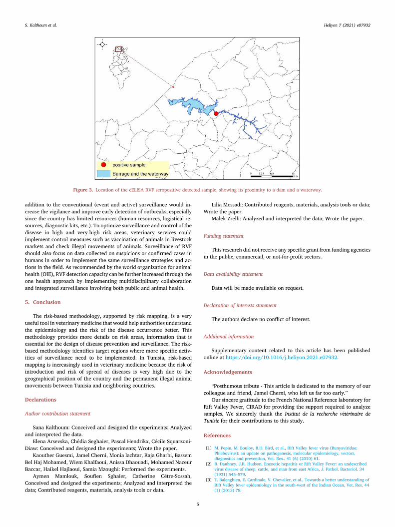
Figure 3. Location of the cELISA RVF seropositive detected sample, showing its proximity to a dam and a waterway.
S. Kalthoum et al. Heliyon 7 (2021) e07932
addition to the conventional (event and active) surveillance would in-crease the vigilance and improve early detection of outbreaks, especiallysince the country has limited resources (human resources, logistical re-sources, diagnostic kits, etc.). To optimize surveillance and control of thedisease in high and very-high risk areas, veterinary services couldimplement control measures such as vaccination of animals in livestockmarkets and check illegal movements of animals. Surveillance of RVFshould also focus on data collected on suspicions or confirmed cases inhumans in order to implement the same surveillance strategies and ac-tions in the field. As recommended by the world organization for animalhealth (OIE), RVF detection capacity can be further increased through theone health approach by implementing multidisciplinary collaborationand integrated surveillance involving both public and animal health.
5. Conclusion
The risk-based methodology, supported by risk mapping, is a veryuseful tool in veterinarymedicine that would help authorities understandthe epidemiology and the risk of the disease occurrence better. Thismethodology provides more details on risk areas, information that isessential for the design of disease prevention and surveillance. The risk-based methodology identifies target regions where more specific activ-ities of surveillance need to be implemented. In Tunisia, risk-basedmapping is increasingly used in veterinary medicine because the risk ofintroduction and risk of spread of diseases is very high due to thegeographical position of the country and the permanent illegal animalmovements between Tunisia and neighboring countries.
Declarations
Author contribution statement
Sana Kalthoum: Conceived and designed the experiments; Analyzedand interpreted the data.
Elena Arsevska, Ch�edia Seghaier, Pascal Hendrikx, C�ecile Squarzoni-Diaw: Conceived and designed the experiments; Wrote the paper.
Kaouther Guesmi, Jamel Cherni, Monia lachtar, Raja Gharbi, BassemBel Haj Mohamed, Wiem Khalfaoui, Anissa Dhaouadi, Mohamed NaceurBaccar, Haikel Hajlaoui, Samia Mzoughi: Performed the experiments.
Aymen Mamlouk, Soufien Sghaier, Catherine Cetre-Sossah,Conceived and designed the experiments; Analyzed and interpreted thedata; Contributed reagents, materials, analysis tools or data.
5
Lilia Messadi: Contributed reagents, materials, analysis tools or data;Wrote the paper.
Malek Zrelli: Analyzed and interpreted the data; Wrote the paper.
Funding statement
This research did not receive any specific grant from funding agenciesin the public, commercial, or not-for-profit sectors.
Data availability statement
Data will be made available on request.
Declaration of interests statement
The authors declare no conflict of interest.
Additional information
Supplementary content related to this article has been publishedonline at https://doi.org/10.1016/j.heliyon.2021.e07932.
Acknowledgements
“Posthumous tribute - This article is dedicated to the memory of ourcolleague and friend, Jamel Cherni, who left us far too early.”
Our sincere gratitude to the French National Reference laboratory forRift Valley Fever, CIRAD for providing the support required to analyzesamples. We sincerely thank the Institut de la recherche v�et�erinaire deTunisie for their contributions to this study.
References
[1] M. Pepin, M. Bouloy, B.H. Bird, et al., Rift Valley fever virus (Bunyaviridae:Phlebovirus): an update on pathogenesis, molecular epidemiology, vectors,diagnostics and prevention, Vet. Res.. 41 (6) (2010) 61.
[2] R. Daubney, J.R. Hudson, Enzootic hepatitis or Rift Valley Fever: an undescribedvirus disease of sheep, cattle, and man from east Africa, J. Pathol. Bacteriol. 34(1931) 545–579.
[3] T. Balenghien, E. Cardinale, V. Chevalier, et al., Towards a better understanding ofRift Valley fever epidemiology in the south-west of the Indian Ocean, Vet. Res. 44(1) (2013) 78.
S. Kalthoum et al. Heliyon 7 (2021) e07932
[4] The National Institute for Communicable Diseases (NICD), Healthcare WorkersGuidelines on Rift Valley Fever, January 2020. https://www.nicd.ac.za/wp-content/uploads/2020/01/RVF_2020_Guidelines_for_HCWs_oh_ak_200120_oh_vm.pdf.
[5] A.H. Turkistany, A.G. Mohamed, N. Al-Hamdan, Seroprevalence of Rift Valley feveramong slaughterhouse personnel in Makkah during Hajj, J. Fam. Commun. Med. 8(3) (2001) 53–57.
[6] M.O. Nanyingi, P. Munyua, S.G. Kiama, et al., A systematic review of Rift ValleyFever epidemiology 1931-2014, Infect. Ecol. Epidemiol. 5 (2015) 28024.
[7] A.D. LaBeaud, S. Muiruri, L.J. Sutherland, et al., Postepidemic analysis of Rift Valleyfever virus transmission in northeastern Kenya: a village cohort study, PLoSNeglected Trop. Dis. 5 (8) (2011) e1265.
[8] N.A. Bergren, R.C. Kading, The ecological significance and implications of transovarialtransmission among the vector-borne bunyaviruses: a review, Insects 9 (4) (2018) 173.
[9] Y. Himeidan, Rift Valley fever: current challenges and future prospects, Res. Rep.Trop. Med. 7 (2016) 1–9.
[10] R. Metras, W.J. Edmunds, C. Youssouffi, et al., Estimation of Rift Valley fever virusspillover to humans during the Mayotte 2018–2019 epidemic, Proc. Natl. Acad. Sci.U. S. A. 117 (39) (2020) 24567–24574, pmid:32929025.
[11] G.H. Gerdes, Rift Valley fever.2004, Rev. Sci. Tech. Off. Int. Epiz. 23 (2) (2004)613–623.
[12] B.M. Boushab, F.Z. Fall-Malick, S.E. Ould Baba, et al., Severe human illness causedby Rift Valley fever virus in Mauritania, 2015, Open Forum Infect Dis. 3 (4) (2016)ofw200. Published 2016 Sep 27.
[13] F. Zakham, A. Alaoui, O. Vapalahti, Rift Valley fever in the Middle East North Africa(MENA) region, Curr. Trop. Med. Rep. 5 (2018) 257–263.
[14] J.M. Drake, A.N. Hassan, J.C. Beier, A statistical model of Rift Valley fever activityin Egypt, J. Vector Ecol. 38 (2) (2013) 251–259.
[15] M.A. Kenawy, Y.M. Abdel-Hamid, J.C. Beier, Rift Valley Fever in Egypt and otherAfrican countries: historical review, recent outbreaks and possibility of diseaseoccurrence in Egypt, Acta Trop. 181 (2018) 40–49.
[16] OIE, Event Summary: Rift Valley Fever, Libya, 2019. https://www.oie.int/wahis2/public/wahid.php/Reviewreport/Review/viewsummary?reportid=%2032934.
[17] A. Di Nardo, D. Rossi, S.M.L. Saleh, S.M. Lejlifa, S.J. Hamdi, A. Di Gennaro, et al.,Evidence of rift valley fever seroprevalence in the Sahrawi semi-nomadic pastoralistsystem, Western Sahara, BMC Vet. Res. 10 (2014) 92.
[18] M. El-Harrak, R. Martín-Folgar, F. Llorente, et al., Rift Valley and west nile virusantibodies in camels, North Africa, Emerg. Infect. Dis. 17 (12) (2011) 2372–2374.
[19] A. Bosworth, T. Ghabbari, S. Dowall, et al., Serologic evidence of exposure to RiftValley fever virus detected in Tunisia, New Microb. New Infect. 9 (2015) 1–7.
[20] T.B. Hassine, J. Amdouni, F. Monaco, et al., Emerging vector-borne diseases indromedaries in Tunisia: west Nile, bluetongue, epizootic haemorrhagic disease andRift Valley fever, Onderstepoort J. Vet. Res. 84 (2017) 1–3.
[21] E.Ayari-Fakhfakh,A.Ghram,A.Bouattour, et al., First serological investigationofpeste-des-petits-ruminants and Rift Valley fever in Tunisia, Vet. J. 187 (2011) 402–404.
[22] R. Selmi, A. Mamlouk, M. Ben Said, et al., First serological evidence of the RiftValley fever Phlebovirus in Tunisian camels, Acta Trop. 207 (2020) 105462.
[23] C.N. Mweya, L.E.G. Mboera, S.I. Kimera, Climate influence on emerging risk areas forRift Valley fever epidemics in Tanzania, Am. J. Trop. Med. Hyg. 97 (2017) 109–114.
[24] A. Apolloni, G. Nicolas, Coste, et al., Towards the description of livestock mobilityin Sahelian Africa: some results from a survey in Mauritania, PloS One 13 (2018),e0191565.
[25] K.D. St€ark, G. Regula, J. Hernandez, et al., Concepts for risk-based surveillance inthe field of veterinary medicine and veterinary public health: review of currentapproaches, BMC Health Serv. Res. 6 (2006) 20.
[26] S. Kalthoum, workshop « Analyse qualitative et cartographique des risques pourl’optimisation de la surveillance de la fi�evre aphteuse et des maladies prioritaires auMaghreb et Afrique de l’Ouest et du centre, Personal communication, 2018, 23 au27 avril.
[27] OIE, Animal movements in the run-up to religious festivals, in: OIE Bulletin, 2019[Internet]. [cited 2 Nov 2019]. Available: https://oiebulletin.com/?panorama=animal-movements-in-the-run-upto-religious-festivals.
6
[28] Institut National des Statistiques. Flash D�emographique [Internet], 2016.http://www.ins.tn/sites/default/files/publication/pdf/Bulletin%20n%C2%B01-2016-v3.pdf.
[29] C. Squarzoni-Diaw, E. Arsevska, S. Kalthoum, et al., Using a participatoryqualitative risk assessment to estimate the risk of introduction and spread oftransboundary animal diseases in scarce-data environments. Transboundary andEmerging Diseases, Transbound Emerg. Dis. (2020) 1–13, 00.
[30] T.P. Robinson, G.R.W. Wint, G. Conchedda, et al., Mapping the global distributionof livestock, PloS One 9 (5) (2014), e96084.
[31] A. Nelson, D.J. Weiss, J. van Etten, et al., A suite of global accessibility indicators,Sci. Data 6 (2019) 266.
[32] B. Martínez-L�opez, A.M. Perez, J.M. S�anchez-Vizcaíno, Social network analysis.Review of general concepts and use in preventive veterinary medicine, Transbound.Emerg. Dis. 56 (2009) 109–120.
[33] Free spatial data/DIVA-GIS. https://www.diva-gis.org/gdata.[34] Mosquito metadata. http://vectormap.si.edu/Mosquito_Metadata.htm.[35] J. Kirchherr, K. Charles, Enhancing the sample diversity of snowball samples:
recommendations from a research project on anti-dam movements in SoutheastAsia, PloS One 13 (8) (2018), e0201710.
[36] Office de D�eveloppement du Sud. http://www.ods.nat.tn/.[37] J. Kortekaas, J. Kant, R. Vloet, C. Cetre-Sossah, et al., European ring trial to evaluate
ELISAs for the diagnosis of infection with Rift Valley fever virus, J. Virol Methods187 (2013) 177–181.
[38] World Organisation for Animal Health (OIE), Rift Valley Fever. Chapter 3.1.18.Manual of Diagnostic Tests and Vaccines for Terrestrial Animals, 2018.
[39] A. Tran, A.G. Fall, B. Biteye, et al., Spatial modeling of mosquito vectors for RiftValley fever virus in northern Senegal: integrating satellite derived meteorologicalestimates in population dynamics models, Rem. Sens. 11 (2019) 1024.
[40] A. Tran, C. Trevennec, J. Lutwama, et al., Development and assessment of ageographic knowledge-based model for mapping suitable Areas for Rift Valley fevertransmission in eastern Africa, PLoS Neglected Trop. Dis. 10 (2016), e0004999.
[41] R. Sang, S. Arum, E. Chepkorir, et al., Distribution and abundance of key vectors ofRift Valley fever and other arboviruses in two ecologically distinct counties inKenya, PLoS Neglected Trop. Dis. 11 (2017), e0005341.
[42] A. Tabbabi, J. Daaboub, Mosquitoes (Diptera: Culicidae) in Tunisia, with particularattention to proven and potential vectors: a Review, J. Trop. Dis. 5 (2017).
[43] G. Krida, L. Diancourt, A. Bouattour, et al., Estimation du risque d’introduction duvirus de la fi�evre de la vall�ee du Rift en Tunisie par le moustique Culex pipiens, Bull.Soc. Pathol. Exot. 104 (2011) 250–259.
[44] E. Arsevska, J. Hellal, S. Mejri, et al., Identifying areas suitable for the occurrence ofRift Valley fever in north Africa: implications for surveillance, Transbound. Emerg.Dis. 63 (2016) 658–674.
[45] S. Blickenstorfer, H. Schwermer, M. Engels, et al., Using scenario tree modelling fortargeted herd sampling to substantiate freedom from disease, BMC Vet. Res. 7(2011) 49.
[46] Risksur, Best Practices for Risk-Based and Cost Effective Animal Health Surveillancein the European Union, 2015 [cited 14 Nov 2019]. Available online at: https://www.fp7-risksur.eu//progress/best-practice-document.
[47] M. Ould Ahmed, Caract�erisation de la population des dromadaires (Camelusdromedarius) en Tunisie. Th�ese de doctorat en Sciences Agronomiques, InstitutNational d’Agronomie de Tunisie, 172p. [cited 14 Nov 2019]. Available: http://camelides.cirad.fr/fr/science/these_ouldahmed.html.
[48] E. Arsevska, R. Lancelot, A.B. El Mamy, et al., Situation �epid�emiologique de la fi�evrede la Vall�ee du Rift en Afrique de l’Ouest et du Nord, Bull Epid�emiologique 74(2016) 25–29. Available: http://agritrop.cirad.fr/580830/.
[49] W. Ben Ayed, F. Amraoui, Y. M’ghirbi, et al., A survey of Aedes (Diptera: Culicidae)mosquitoes in Tunisia and the potential role of Aedes detritus and Aedes caspius inthe transmission of zika virus, J. Med. Entomol. 56 (2019) 1377–1383.
[50] EFSA, Scientific opinion on Rift Valley fever, EFSA J. 11 (2013) 3180.[51] Y. Xiao, J.C. Beier, R.S. Cantrell, et al., Modelling the effects of seasonality and
socioeconomic impact on the transmission of rift valley Fever virus, PLoS NeglectedTrop. Dis. 9 (1) (2015) e3388.