The ???Primary??? Antiphospholipid Syndrome: Major Clinical and Serological Features
Serological Evidence of Flaviviruses and Alphavirusesin Livestock and Wildlife in Trinidad
Nadin N. Thompson,1 Albert J. Auguste,2,3 Dane Coombs,4 Bradley J. Blitvich,6 Christine V.F. Carrington,4
Amelia P. Travassos da Rosa,3 Eryu Wang,3 Dave D. Chadee,5 Michael A. Drebot,7
Robert B. Tesh,3 Scott C. Weaver,3 and Abiodun A. Adesiyun1
Abstract
Seroprevalence rates of selected arboviruses in animal populations in Trinidad were determined using serumsamples collected between 2006 and 2009 from horses (n = 506), cattle (n = 163), sheep (n = 198), goats (n = 82), pigs(n = 184), birds (n = 140), rodents (n = 116), and other vertebrates (n = 23). The sera were screened for antibodies toWest Nile virus (WNV), St. Louis encephalitis virus (SLEV), Ilheus virus (ILHV), Bussuquara virus (BSQV),Venezuelan equine encephalitis virus (VEEV), eastern equine encephalitis virus (EEEV), and western equineencephalitis virus (WEEV), using hemagglutination inhibition assay (HIA) and epitope-blocking enzyme-linkedimmunosorbent assays (ELISA). Antibodies to SLEV were detected in a total of 49 (9.7%) horses, 8 (4.9%) cattle, 1(1.2%) goat, 2 (1.4%) wild birds, and 3 (2.2%) wild rodents by both methods. In contrast, antibodies to EEEV,VEEV, and WNV were detected only in horses, at rates of 4.3%, 0.8%, and 17.2%, respectively, by ELISA, andIgM capture ELISA was WNV-positive in 3 (0.6%) of these sera. Among locally bred unvaccinated horses thathad never left Trinidad, seroprevalence rates against WNV were 12.1% and 17.2% by ELISA and HIA, re-spectively. The presence of WNV- and SLEV-specific antibodies in a representative sample of horse sera thatwere both ELISA- and HIA-seropositive was confirmed by plaque reduction neutralization testing (PRNT).Antibodies to ILHV and BSQV were not detected in any of the serum samples tested (i.e., sera from horses, otherlivestock, and wild birds in the case of ILHV, and wild mammals in the case of BSQV). The data indicate thepresence of WNV in Trinidad, and continuing low-level circulation of SLEV, EEEV, and VEEV.
Key Words: Alphavirus—Arbovirus—Eastern equine encephalitis virus—Flavivirus—Seroprevalence—St. Louisencephalitis virus—Trinidad—Venezuelan equine encephalitis virus—West Nile virus.
Introduction
Between 1959 and 1969, the Trinidad Regional VirusLaboratory (TRVL), in collaboration with the Rockefeller
Foundation, demonstrated through viral isolation andserology the existence of several arboviruses in human andanimal populations in the Caribbean region, including Trini-dad. The work done in Trinidad provided valuable informa-tion on circulating viruses (Aitken et al. 1956, 1960, 1964,1968a; Jonkers et al. 1968a; Price, 1978b), taxonomic classifi-cation of the vectors (Aitken et al. 1968c), and their associated
ecological characteristics (Downs et al. 1968; Jonkers et al.1966). These studies resulted in the identification and char-acterization of hundreds of arboviral isolates, primarily fromthe Bush Bush Forest, which is an approximately 0.85-sq kmregion within the Nariva swamp in southeastern Trinidad(Aitken et al. 1968c; Jonkers et al. 1968a, 1968b; Worth et al.1968). With the exception of the aforementioned studies, therehave been only a few serological studies in the 1970s and early1980s (Price 1978a, 1978b; Tikasingh et al. 1983), and morerecently, mosquito surveillance studies and opportunisticsampling of carcasses of Alouatta seniculus monkeys
1School of Veterinary Medicine, 2Department of Preclinical Sciences, Faculty of Medical Sciences, and 5Faculty of Science and Agriculture,The University of the West Indies, St. Augustine, Trinidad and Tobago.
3Institute for Human Infections and Immunity and Center for Tropical Diseases, University of Texas Medical Branch, Galveston, Texas.4DKC Veterinary Services, 3 Bridle Hill, Smith’s, FL03, Bermuda.6Department of Veterinary Microbiology and Preventive Medicine, College of Veterinary Medicine, Iowa State University, Ames, Iowa.7Zoonotic Diseases and Special Pathogens, National Microbiology Laboratory, Public Health Agency of Canada, Winnipeg, Manitoba,
Canada.
VECTOR-BORNE AND ZOONOTIC DISEASESVolume 12, Number 11, 2012ª Mary Ann Liebert, Inc.DOI: 10.1089/vbz.2012.0959
969
confirmed the continued circulation of VEE subytpe IIIAviruses (Mucambo virus; MUCV), several orthobunyaviruses,and Yellow fever virus (YFV) (Auguste et al. 2009, 2010a,2010b).
Despite the extensive surveillance performed in Trinidad,the role of rodents, other mammals, and birds in the main-tenance and spread of these viruses locally is unclear. It is alsonot clear to what extent the livestock and wildlife populationsin Trinidad are affected or potentially contribute to amplifi-cation, as routine surveillance and virological assays on sicklivestock are rarely performed and reported.
Given the significant changes in human demographicssince the comprehensive studies in the 1950s and 1960s, weperformed a serological survey for flaviviruses and alpha-viruses of veterinary public health importance in the Amer-icas. Sera from livestock (horses, cattle, sheep, goats, andpigs), birds, rodents, and other wildlife in Trinidad werecollected over a 2-year period, and subsequently screened forantibodies to West Nile (WNV), St. Louis encephalitis (SLEV),Ilheus (ILHV), Bussuquara (BSQV), Venezuelan equine en-cephalitis (VEEV), eastern equine encephalitis (EEEV), andwestern equine encephalitis viruses (WEEV), using the hem-agglutination inhibition assay (HIA), a traditional diagnosticmethod for detecting arboviral antibodies (Clarke and Casals,1958), and a more recently developed epitope-blockingenzyme-linked immunosorbent assay (ELISA) (Blitvich et al.2003a, 2003c; Hall et al. 1995).
Materials and Methods
Sampling strategies
Horses. Horses were selected from lists of registeredhorses from the Trinidad and Tobago Racing Authority(TTRA), and the Trinidad and Tobago Equestrian Society(TTES), mainly by stratified random sampling according togender and age, in categories of age < 1 year, age 1–5 years,age 6–10 years, and age > 10 years. Blood samples were col-lected from the jugular veins of 506 horses (200 male and 306female), from stud farms, equestrian facilities, and racetracks,at 25 locations within 20 geographic areas (Fig. 1a). Demo-graphic data for the horses and stables were also noted. Of the506 horses, 5.3% (n = 27) were < 1 year old, 40.7% (n = 206)were 1–5 years of age, 31.0% (n = 157) were 6–10 years of age,15.3% (n = 77) were older than 10 years, and the ages could notbe determined for 7.7% (n = 39). Also, 121 (23.9%) were im-ported (foreign-bred), 341 (67.4%) were locally-bred, and theorigin of 44 (8.7%) was uncertain. Finally, 36 (7.1%) horseswere from rural towns, while 470 (92.9%) originated fromurban areas (i.e., regions with ‡ 200 persons per square kilo-meter; Central Statistical Office, 1998).
FIG. 1. (a) Geographic distribution of horse samples inTrinidad, and (b) geographic distribution of alphavirus-positive horses in Trinidad. The VEEV-positive locationswere also EEEV-seropositive. (c) Geographic distribution ofSLEV-positive horses in Trinidad. (d) Geographic distribu-tion of WNV-positive horses in Trinidad. Spatial distributionwas documented and analyzed with Arcmap 9.2, ArcGIS 9(ESRI, Redlands, CA; SLEV, St. Louis encephalitis virus;WNV, West Nile virus; VEEV, Venezuelan equine encepha-litis virus; EEEV, eastern equine encephalitis). Color imagesavailable online at www.liebertpub.com/vbz
‰
970 THOMPSON ET AL.
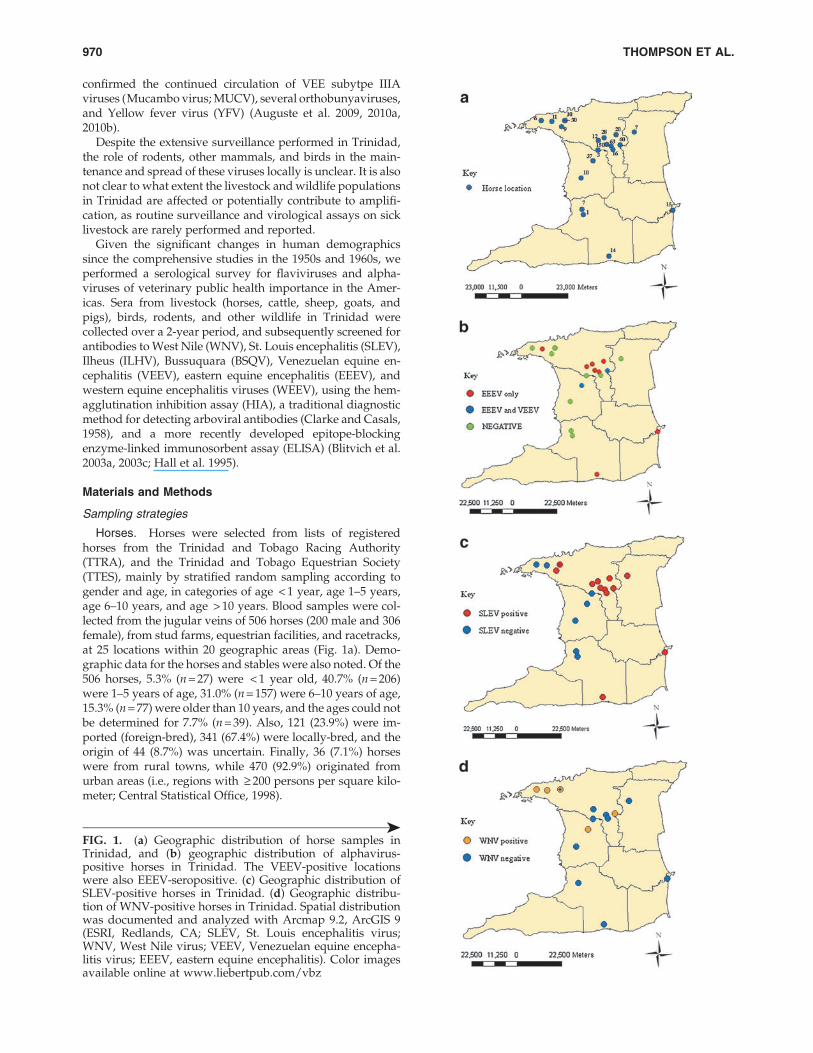
Livestock. A list of farms was prepared using informationfrom the Ministry of Agriculture, Land and Marine Resources(MALMR) and Animal Health Assistants (AHAs) that usuallyassist government veterinarians. Small ruminant and pig farmswere grouped by size as follows: for small ruminants, £ 15 ani-mals = small, 16–49 animals = medium, and ‡ 50 animals = large;for pigs, £ 25 animals = small, 26–50 animals = medium, and ‡ 50animals = large. For cattle, small farms were those with fewerthan 20 animals, medium farms had 21–39 animals, and largefarms were those with 40 or more animals. A systematic randomsampling protocol was then used to select the farms. Within eachof the cattle and small ruminant farms selected, stratified ran-dom sampling according to age, gender, and species (in the caseof small ruminant farms) was used. Where farmers from selectedfarms declined to participate in the study, the next scheduledfarm was substituted and a restart of the sequence was done.Medium and large pig farms were sampled randomly; however,small pig farms were sampled conveniently, as the majority onthe compiled list were inactive. Figure 2a illustrates the locationsof the livestock sampling sites. Blood samples were collectedfrom the jugular veins of small ruminants and cattle, and of thejugular vein or anterior vena cava of pigs.
Wildlife. Wild animals were sampled from the EmperorValley Zoo in Trinidad, from wildlife farms, or by trapping inforested areas. Supplementary Table S1 (see online supple-mentary material at http://www.liebertpub.com) shows thespecies and numbers of animals sampled. Systematic randomsampling was used for the selection of wildlife farms whereanimals were then sampled according to gender and growthstage (i.e., juvenile or adult). Free-living rats (Rattus spp.) werealso specifically targeted and trapped using humane metalcage traps at various locations in close proximity to homes,public buildings and markets.
Avian sera were obtained from confiscated birds at theWildlife Section of the Ministry of Agriculture, Land andMarine Resources, from birds collected in the wild, and fromcaptive birds at the Emperor Valley Zoo. Collection of wildbirds was carried out using hand nets and mist nets(35 · 35 mm), after which the birds were bled by cutaneousulnar or jugular venipuncture and then released. Supple-mentary Table S1 (see online supplementary material athttp://www.liebertpub.com) shows the avian species sam-pled, including their migrant status in Trinidad.
Two forested areas, the Aripo Savannah Scientific Reserveand the Bush Bush Forest in Nariva, were chosen for wildlifetrapping based on the results of previous and ongoing arbo-virus surveillance studies (Aitken et al. 1968b, 1968c; Jonkerset al. 1968a; Auguste et al. 2009). In Aripo, trapping was doneweekly from November to December 2007 and February toApril 2008, whereas in the Bush Bush Forest, trapping wasdone daily between April and August 2008. A total of 11 traps
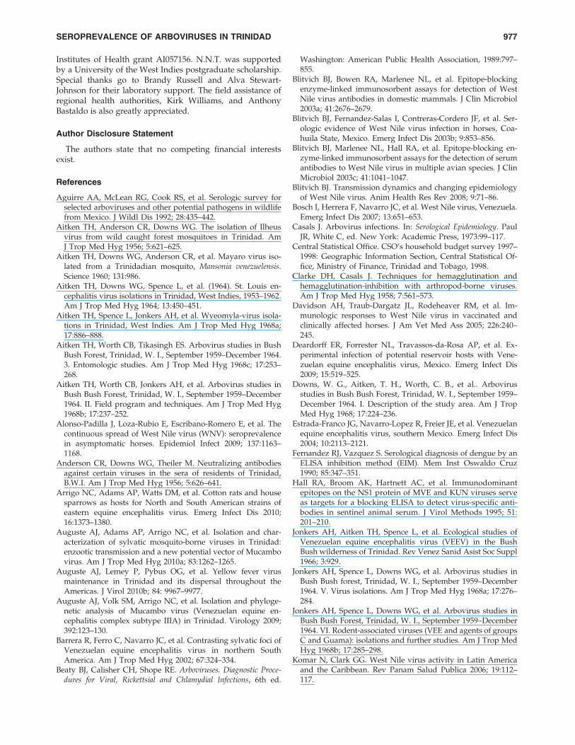
FIG. 2. (a) Geographic distribution of livestock samples inTrinidad. (b) Geographic distribution of rodent and wildlifesamples in Trinidad. (c) Geographic distribution of SLEV-positive livestock in Trinidad. (d) Geographic distribution ofSt. Louis encephalitis virus-positive rodents and wildlife inTrinidad. Spatial distribution was documented and analyzedwith Arcmap 9.2, ArcGIS 9. Color images available online atwww.liebertpub.com/vbz
‰
SEROPREVALENCE OF ARBOVIRUSES IN TRINIDAD 971
(9 measuring 16† · 5† · 5† and 2 being 32† · 10† · 12†) were setfrom 4 pm to 6 pm, and removed or re-set between 6 am and 9am the following morning. Baits used were peanut butter,cheese, ripe bananas, bread, salt fish, smoked herring, coco-nut, and seasonal fruits, depending on the target species(Aitken et al. 1968b; Barrera et al. 2002).
Hemagglutination inhibition assay (HIA)
HIA was performed as described in Beaty and associates(Beaty et al. 1989), using sucrose acetone antigens that hadbeen previously prepared and stored at - 70�C. For VEEV,WEEV, and EEEV, the strains used were TC-83 (subtype I-AB), McMillan, and North American NJ/60, respectively. Forthe flaviviruses, SLEV (T-35573), WNV (T-36296), and ILHV(BeH 7445) were used for testing sera from all species exceptrodents and other non-avian wildlife. In addition, BSQVstrain BeAn 4116 was used for rodents and other non-avianwildlife. Seroprevalence due to specific antibodies was de-termined by the demonstration of a fourfold or greater dif-ference in antibody titers caused by the various antigens. Apositive control serum titrated to its endpoint was includedfor each antigen.
Epitope-blocking enzyme-linked immunosorbentassay (ELISA)
An identification algorithm of murine monoclonal anti-bodies (MAbs) for subtyping EEEV (Roehrig et al. 1990) wasmodified and the following MAbs were used: MAb 5B4D-6,specific for detecting VEEV TC-83 vaccine strain, and MAb1B1C-4 cross-reactive for North American (NJ/60) and SouthAmerican (BeAn 5122) EEEV (both from Chemicon, Teme-cula, CA). Flaviviral antibodies included WNV MAb 3.1112Gand SLEV MAb 6B5A-2 (both from Chemicon), and a genericFlavivirus MAb 6B6C-1 (InBios International, Inc., Seattle,WA). MAb 6B6C-1 was used to test horse sera only. Flavivirusantigens used were SLEV strain TBH-28 and WNV NY-99-35261 strain. ELISAs to detect flavivirus antibodies wereperformed as described in Blitvich and colleagues (Blitvichet al. 2003c). Alphavirus antigens used were TC-83 (subtype 1)and North American (NJ/60) for VEEV and EEEV, respec-tively. Optimal concentrations of viral antigens were first ti-trated according to Wang and associates (Wang et al. 2005).ELISAs to detect alphavirus antibodies were performed ac-cording to Wang and associates (Wang et al. 2005) with minormodifications.
Seropositivities were calculated using the formula: % in-hibition = 100 – {(TS – B)/(CS – B)} · 100, where TS is the op-tical density of test sera, CS is the optical density of controlsera, and B is the background optical density (Hall et al. 1995).Each sample was tested in duplicate so an average opticaldensity was first calculated. An inhibition value of 30% ormore was considered seropositive (Blitvich et al. 2003a,2003b). Calculated percentage inhibition values that resultedin values below zero (negative) were recorded as zero. Due tothe limited volume of serum collected from the small animals,particularly birds, some sera were tested only once by ELISA.
IgM capture ELISA for West Nile virus
Immunoglobulin M (IgM) West Nile Detect� IgM captureELISA (InBios International, Inc.) was used to detect anti-
bodies to WNV in horse sera as stipulated by the manufac-turer and using the test controls provided to determine theimmune status ratio (ISR). ISR was calculated for the positiveand negative controls by dividing the OD450nm of the WNRAby OD450mm of the normal cell antigen. An ISR for test serum£ 2 was considered negative, ‡ 3.0 as positive, and between2.0 and 3.0 were deemed equivocal. All positive and equivocalsamples were repeated.
Plaque reduction neutralization test (PRNT)
The ability of seropositive sera to neutralize WNV (NY-99strain), SLEV (TBH-28 strain), VEEV (FSL190 strain), andEEEV (Trinidad 1959 strain), was determined by PRNT. WNVand SLEV PRNTs were performed as described by Blitvichand associates (Blitvich et al. 2003c), with the exception thatplaques for SLEV were counted 8 days post-infection. VEEVand EEEV PRNTs were performed as previously described(Beaty et al. 1989).
Ethical approval and permits
This study was approved by the Ethics Committee of theFaculty of Medical Sciences at the University of the West In-dies, St. Augustine Campus, prior to commencement. Permitswere obtained from the Wildlife Section of the Ministry ofAgriculture, Land and Marine Resources for capture of ani-mals for the study.
Results
Horses
Seroprevalence rates of EEEV, VEEV, WNV, and SLEV, ateach of the sampled locations are shown in Table 1. Figure 1shows all of the horse sampling locations and the locationswhere VEEV-, EEEV-, SLEV-, and WNV-seropositive horseswere detected. Based on the combined ELISA and HIA resultsthere were 26 (5.1%) EEEV-seropositive horses, 4 (0.8%)VEEV-seropositive animals, and 7 (1.4%) samples that ex-hibited cross-reactivity within the Alphavirus genus (i.e., withat least a fourfold difference), such that their identities couldnot be determined. Of the 26 horses ELISA-seropositive forEEEV, 21 (80.8%) were from stud farms, and 5 (19.2%) werefrom race tracks, making the seropositive rates at these loca-tions 6.7% (21 out of 315 horses), and 3.3% (5 out of 150),respectively. All 22 HIA-seropositive horses were detectedwith ELISA including 19 (6.0%) of 315 stud farm horses, and3 (2.0%) of 150 racetrack horses. All four (1.3%) VEEV-seropositive horses originated from stud farms. Of the 26EEEV-seropositive horses, 16 (61.2%), 5 (19.2%), and 4(15.4%), were aged 6–10, 1–5, and > 10 years, respectively.Twenty EEEV-seropositive horses were imported and 6 werelocally bred. PRNT80 performed on EEEV- and VEEV-sero-positive horses confirmed the presence of VEEV-neutralizingantibodies in 2 of 4 samples tested, and EEEV-neutralizingantibodies in 7 of 26 samples tested (antibody titers > 20). Ofthe 58 horses that had not been vaccinated against WNV andwere locally bred, a total of 11 (19.0%) were seropositive forWNV (Table 2), with 7 (12.1%) seropositive for WNV byELISA, and 10 (17.2%) by HIA.
Of the 506 horses studied, 49 (9.7%) were seropositive forSLEV by epitope-blocking ELISA, compared with 13 (2.6%)that were positive by HIA (Table 3), with 12/13 HIA-positives
972 THOMPSON ET AL.
Table 1. Seropositivity for Arboviral Antibodies in Horses by ELISA and HIA
No. (%) positive forflaviviral antibodies
No. (%) positive foralphaviral antibodies
No. of horses testedSLEV WNVb EEEV VEEV
Locationtype Locationa
SLEV, EEEVand VEEV WNV ELISA HIA ELISA HIA ELISA HIA ELISA HIA
Stud farm Arima 63 1 7 (11.1) 1 (1.6) 0 (0.0) 0 (0.0) 9 (14.3) 9 (14.3) 0 (0.0) 0 (0.0)Santa Cruz 30 3 5 (16.7) 0 (0.0) 3 (100) 3 (100) 0 (0.0) 0 (0.0) 0 (0.0) 0 (0.0)D’Abadie 28 4 3 (10.7) 0 (0.0) 0 (0.0) 0 (0.0) 1 (3.6) 2 (7.1) 0 (0.0) 0 (0.0)Freeport 10 2 0 (0.0) 0 (0.0) 0 (0.0) 0 (0.0) 0 (0.0) 0 (0.0) 0 (0.0) 0 (0.0)Chaguanas 37 4 0 (0.0) 0 (0.0) 0 (0.0) 1 (25.0) 1 (2.7) 0 (0.0) 2 (5.4) 0 (0.0)Wallerfield 60 13 0 (0.0) 4 (6.7) 1 (7.7) 2 (15.4) 6 (10.0) 5 (8.3) 2 (3.3) 0 (0.0)Heights of
Guanapo20 0 6 (30.0) 0 (0.0) 0 (0.0) 0 (0.0) 1 (5.0) 0 (0.0) 0 (0.0) 0 (0.0)
Arouca 12 8 3 (25.0) 0 (0.0) 0 (0.0) 0 (0.0) 0 (0.0) 1 (8.3) 0 (0.0) 0 (0.0)Petit Valley 11 2 0 (0.0) 0 (0.0) 2 (100) 2 (100) 1 (9.1) 1 (9.1) 0 (0.0) 0 (0.0)Mayaro 15 1 5 (33.3) 1 (6.7) 0 (0.0) 0 (0.0) 1 (6.7) 0 (0.0) 0 (0.0) 0 (0.0)Moruga 14 2 1 (7.1) 2 (14.3) 0 (0.0) 0 (0.0) 1 (7.1) 1 (7.1) 0 (0.0) 0 (0.0)Gasparillo 7 0 0 (0.0) 0 (0.0) 0 (0.0) 0 (0.0) 0 (0.0) 0 (0.0) 0 (0.0) 0 (0.0)Princes Town 1 1 0 (0.0) 0 (0.0) 0 (0.0) 0 (0.0) 0 (0.0) 0 (0.0) 0 (0.0) 0 (0.0)Valencia 7 2 1 (14.3) 1 (14.3) 0 (0.0) 0 (0.0) 0 (0.0) 0 (0.0) 0 (0.0) 0 (0.0)Subtotal 315 43 31 (9.8) 9 (2.9) 6 (14.0) 8 (18.6) 21 (6.7) 19 (6.0) 4 (1.3) 0 (0.0)
Equestrianfacility
Chaguaramas 6 4 0 (0.0) 0 (0.0) 1 (25.0) 0 (0.0) 0 (0.0) 0 (0.0) 0 (0.0) 0 (0.0)
Santa Cruz 10 0 3 (30.0) 0 (0.0) 0 (0.0) 0 (0.0) 0 (0.0) 0 (0.0) 0 (0.0) 0 (0.0)Piarco 3 2 0 (0.0) 0 (0.0) 0 (0.0) 0 (0.0) 0 (0.0) 0 (0.0) 0 (0.0) 0 (0.0)St. Anns 6 0 2 (22.2) 0 (0.0) 0 (0.0) 0 (0.0) 0 (0.0) 0 (0.0) 0 (0.0) 0 (0.0)Guanapo 16 3 3 (18.8) 1 (6.3) 0 (0.0) 0 (0.0) 0 (0.0) 0 (0.0) 0 (0.0) 0 (0.0)Subtotal 41 9 8 (19.5) 1 (2.4) 1 (11.1) 0 (0.0) 0 (0.0) 0 (0.0) 0 (0.0) 0 (0.0)
Race track Arima 150 6 10 (6.7) 3 (2.0) 0 (0.0) 2 (33.3) 5 (3.3) 3 (2.0) 0 (0.0) 0 (0.0)Total 506 58 49 (9.7) 13 (2.6) 7 (12.1) 10 (17.2) 26 (5.1) 22 (4.3) 4 (0.8) 0 (0.0)
aResults of 4 locations were merged due to overlap of global positioning system points.bIncludes only unvaccinated and locally-bred horses.ELISA, enzyme-linked immunosorbent assay; HIA, hemagglutination inhibition assay; SLEV, St. Louis encephalitis virus; EEEV, eastern
equine encephalitis virus; VEEV, Venezuelan equine encephalitis virus; WNV, West Nile virus.
Table 2. Unvaccinated and Locally-Bred Horses Positive for Antibodies to WNV, SLEV, ILHV, and FLAV
HIA titer Epitope b-ELISA (%I)a
SampleID
Localityby town Gender
Agegroup (y) WNV SLEV ILHV FLAV WNV SLEV
WNVcaptureIgMb
WNVPRNTtiterc
SLEVPRNTtiterd
1C11H Chaguanas F 1–5 1:40 0 0 55.2 21.4 4.1 1.0 160 < 201CH3H Chaguaramas M > 10 0 0 0 39.0 36.4 1.6 NT NT NT1O7H Wallerfield F 6–10 1:320 1:20 0 87.7 0 26.3 1.4 320 < 201OR2H Wallerfield F Unknown 1:320 1:20 0 90.3 83 19.7 1.1 > 640 < 201P1H Petit Valley F 1–5 1:320 1:40 1:20 62.1 47.9 10.6 2.6 NT NT1P3H Petit Valley F 6–10 1:320 1:40 1:40 73.7 43.2 29.5 1.2 NT NT1S3H Santa Cruz M 1–5 1:2560 1:160 1:160 94.9 80.7 23.3 16.3 > 640 1601S4H Santa Cruz M 1–5 1:2560 1:80 1:40 93.5 86.9 23.3 2.7 > 640 401S5H Santa Cruz M 1–5 1:1280 1:160 1:80 81.3 75.2 2.7 3.0 > 640 403RT5H Arima F 1–5 1:40 0 0 43.7 0 29.4 1.1 160 < 203RT19H Arima F 1–5 1:160 1:20 0 41.4 0 21.5 1.3 320 20
aInterpretation for epitope-blocking ELISA: < 30% inhibition (%I) is antibody-negative, and > 30% inhibition is antibody-positive.bWest Nile immunoglobulin type M capture ELISA interpretation: < 2.0 is antibody-negative, 2.0–3.0 is equivocal, and > 3.0 is antibody-
positive.cPlaque reduction neutralization test (PRNT) for WNV (titers are expressed as the reciprocal of the serum dilution yielding 90% reduction
in the number of plaques).dPRNT for SLEV (titers are expressed as the reciprocal of the serum dilution yielding 90% reduction in the number of plaques).ELISA, enzyme-linked immunosorbent assay; HIA, hemagglutination inhibition assay; SLEV, St. Louis encephalitis virus; EEEV, eastern
equine encephalitis virus; VEEV, Venezuelan equine encephalitis virus; WNV, West Nile virus; ILHV, Ilheus virus; NT, not tested.
SEROPREVALENCE OF ARBOVIRUSES IN TRINIDAD 973
also being ELISA-positive. The summary of location types, agegroups, gender, and origin of SLEV-positive horses by ELISA isshown in Table 3. Of the 49 seropositive animals, 45 (91.8%)were from urban areas, and 4 (8.2%) were from rural areas.
A representative sample of 18 horse sera (8 WNV and 10SLEV samples) was tested by PRNT for neutralization anti-bodies. WNV was confirmed in all 8 samples by PRNT90, withantibody titers > 160 ( > 640 in 4/10 samples; titers are ex-pressed as the reciprocal of the serum dilution yielding 90%reduction in the number of plaques), and SLEV was con-firmed in 9 of 10 samples, with antibody titers > 40 (using a1:20 cut-off and/or a greater than fourfold difference in an-tibody titer between the viruses tested; Table 4). Cross-PRNTswere performed on these 18 samples for confirmation.PRNT80 (titers expressed as the reciprocal of the serum dilu-tion yielding 80% reduction in the number of plaques) werealso performed to confirm infection with EEEV and VEEVwith antibody titers > 20, but cross-PRNTs were not done aswith WNV and SLEV. Six of the 22 EEEV samples tested werepositive by PRNT, and none of the four VEEV samples testedhad neutralizing antibodies to this virus.
Livestock and wildlife
Of the 906 sera representing livestock, birds, wild mam-mals, and reptiles, all were negative for antibodies to EEEV,
VEEV, and WEEV. Figure 2 illustrates the locations of the rats,other wild mammals, and reptiles sampled. Eight (4.9%) of163 cattle were seropositive for SLEV in HIAs performed withT-35573 antigen, but were all negative in ELISAs performedwith the same antigen. Two animals seropositive for SLEVwere aged 1–5 years, and 6 were older than 5 years.
Of 82 goat sera tested, 1 (1.2%) was seropositive for SLEVby HIA, but all were negative by ELISA. All sheep tested forantibodies to SLEV by both methods were negative. All titersdetermined in cattle and goat sera were 1:20 by HIA. Overall,the seroprevalence rate of SLEV in livestock was 2.0% (9 of443) by HIA. With the exception of horses, no other livestockspecies were seropositive for WNV or ILHV by either sero-logical method.
Two (1.4%) of 140 wild bird sera were seropositive forSLEV by ELISA. Of the 98 birds also tested by HIA only 1(1.0%) was seropositive. One bird, a 20-year old captive tur-key vulture, was seropositive by both methods. The ELISAinhibition value was 35.3%, while the HIA titer was 1:40 forthis bird. The other positive bird was a fledgling rock pigeonwith an ELISA inhibition value of 31.4%.
Three (2.2%) rodents of 139 tested by ELISA were positivefor antibodies to SLEV TBH-28 antigen: a juvenile agouti(Dasyprocta leporina) and two rats (Rattus spp.) with inhibitionvalues of 38.0%, 54.9%, and 55.9%, respectively. The ELISA-positive agouti was also positive by HIA, with a high specific
Table 3. Frequency of SLEV-Specific Antibodies by ELISA in Horses by Age, Gender, and Origin
Frequency of SLEV antibodies Frequency of SLEV antibodies
Locationtype
CategoryAge (y)
No.tested
No. (%)positive
by ELISAMale
(n = 89)Female
(n = 226)Foreign
bred (n = 89)Locally
bred (n = 207)NA
(n = 19)
Stud farm < 1 27 2 (7.4) 12 1 (0.7) 15 1 (6.7) 1 0 (0.0) 26 2 (7.7) 0 0 (0.0)1–5 83 8 (9.6) 37 4 (10.8) 46 4 (8.7) 23 4 (17.4) 56 4 (7.1) 4 0 (0.0)
6–10 128 17 (13.3) 31 6 (19.4) 97 11 (1.0) 42 5 (11.9) 82 12 (14.6) 4 0 (0.0)> 10 60 4 (6.7) 5 0 (0.0) 55 4 (7.3) 21 1 (4.8) 39 3 (7.7) 0 0 (0.0)NA 17 0 (0.0) 4 0 (0.0) 13 0 (0.0) 2 0 (0.0) 4 0 (0.0) 11 0 (0.0)
Subtotal 315 31 (9.8) 89 11 (12.4) 226 20 (8.8) 89 10 (11.2) 207 21 (10.1) 19 0 (0.0)
Frequency of SLEV antibodies Frequency of SLEV antibodies
Equestrianfacility
CategoryAge (y)
No.tested
No. (%)positive
Male(n = 29)
Female(n = 12)
Foreignbred (n = 8)
Locallybred (n = 32)
NA(n = 1)
< 1 0 0 (0.0) 0 0 (0.0) 0 0 (0.0) 0 0 (0.0) 0 0 (0.0) 0 0 (0.0)1–5 10 2 (20.0) 7 2 (28.6) 3 0 (0.0) 1 0 (0.0) 9 2 (22.2) 0 0 (0.0)
6–10 14 2 (14.3) 10 1 (10.0) 4 1 (25.0) 4 0 (0.0) 10 2 (20.0) 0 0 (0.0)> 10 16 4 (23.5) 11 4 (36.4) 5 0 (0.0) 3 2 (66.7) 13 2 (15.4) 0 0 (0.0)NA 1 0 (0.0) 1 0 (0.0) 0 0 (0.0) 0 0 (0.0) 0 0 (0.0) 1 0 (0.0)
Subtotal 41 8 (19.5) 29 7 (24.1) 12 1 (8.3) 8 2 (25.0) 32 6 (18.8) 1 0 (0.0)
Frequency of SLEV antibodies Frequency of SLEV antibodies
Racetrack
CategoryAge (y)
No.tested
No. (%)positive
Male(n = 82)
Female(n = 68)
Foreignbred (n = 24)
Locallybred (n = 102)
NA(n = 24)
< 1 0 0 (0.0) 0 0 (0.0) 0 0 (0.0) 0 0 (0.0) 0 0 (0.0) 0 0 (0.0)1–5 113 7 (6.2) 70 5 (7.1) 43 2 (4.7) 19 3 (15.8) 90 4 (4.4) 4 0 (0.0)
6–10 15 0 (0.0) 12 0 (0.0) 3 0 (0.0) 4 0 (0.0) 11 0 (0.0) 0 0 (0.0)> 10 1 0 (0.0) 0 0 (0.0) 1 0 (0.0) 1 0 (0.0) 0 0 (0.0) 0 0 (0.0)NA 21 3 (14.3) 0 0 (0.0) 21 3 (14.3) 0 0 (0.0) 1 0 (0.0) 20 3 (15.0)
Subtotal 150 10 (6.7) 82 5 (6.1) 68 5 (7.4) 24 3 (12.5) 102 4 (3.9) 24 3 (12.5)Total 506 49 (9.7) 200 23 (11.5) 306 27 (8.8) 121 15 (12.4) 341 31 (9.0) 44 4 (8.9)
SLEV, St. Louis encephalitis virus; ELISA, enzyme-linked immunosorbent assay; NA, not applicable due to unknown status.
974 THOMPSON ET AL.
titer of 1:1320. No wildlife species was seropositive for WNV,ILHV, or BSQV, and no reptile was seropositive for any of theviruses tested.
Discussion
We determined the current seroprevalence rates of four ar-boviruses in Trinidad that are of public health importance inthe Americas. With the exception of WNV, ELISA detectedmore potential seropositives than HIA for both the flavivirusesand alphaviruses tested. Previous researchers (Fernandez andVazquez 1990; Shi and Wong 2003) have shown that theblocking ELISA is more sensitive than the HIA, althoughblocking ELISAs may occasionally show lower sensitivity be-cause there is reliance on only one epitope for antibody bind-ing, and some individuals may not produce antibodies againstthat specific epitope (Wang et al. 2005). Nonetheless, theepitope-blocking ELISA has the major advantage of beingspecies-independent for the reagents required. Our findingssuggest the presence of all four viruses in horse populationsacross the island in both urban and rural areas. Importantly,while SLEV-specific antibodies were detected in a wide varietyof species (domestic and wildlife), EEEV-, VEEV-, and WNV-specific antibodies were only detected in horses.
In the case of EEEV, the estimated seroprevalence ratesamong horses of 4.3% by HIA and 5.1% by ELISA were closeto the rate of 4.5% determined in a 1954 survey of 22 horses inTrinidad (Anderson et al. 1956). However, as vaccinationagainst EEEV is not a requirement in Trinidad and Tobago,we cannot exclude the possibility that some of the foreign-bred horses in our study may have been seropositive as aresult of vaccination in their country of origin. Serologicaltesting using South American strains (which were unavailablefor the current study) could confirm local versus foreign ex-posure. Nonetheless, our results indicate that horses continueto be exposed to EEEV at low levels in Trinidad, since 5 (1.5%)of the 344 locally-bred animals were seropositive and hadnever left Trinidad.
Our data also indicate low-level circulation of VEEV, as theestimated seroprevalence rate for antibodies to the virus
among locally-bred horses that have never left Trinidad was0.8%. The serological approach used (TC-83 antigen for HIAand subtype I-specific MAb for ELISA) identifies all subtype IVEEV, but it was not possible to determine the correspondingstrains within subtype I. Given the evidence for the exposureof local horses, investigations of clinically-ill horses with en-cephalitis should consider EEEV and VEEV in the differential,particularly since equine clinical cases were recently reported(OIE 2009) from Central America.
Both VEEV and EEEV have previously been detected se-rologically in non-equid species in Trinidad, so the failure todetect antibodies in the current study may indicate that therodents and bird species sampled are not preferred reservoirspecies for these two viruses. For example, probable VEEVreservoir hosts such as Proechimys spp. ( Jonkers et al. 1968b;Barrera et al. 2002), Zygodontomys spp., and Orzyomys spp.( Jonkers et al. 1968b; Deardorff et al. 2009), Cathartes aura(Aguirre et al. 1992), and other birds and small rodent specieswhich were previously reported to be VEEV-seropositive(Scherer et al. 1966; Jonkers et al. 1968b; Aguirre et al. 1992;Estrada-Franco et al. 2004; Navarro et al. 2005), were notamong the species trapped and tested in the current study.Given the short period of time during which the traps wereset, it is possible that certain host or reservoir species mayhave been missed by this trapping system. With regard toEEEV, some avian species that were previously recorded to beEEEV-seropositive in Trinidad (Tikasingh, 1968, 1973, 2004)were negative in the current study. Considering reports thatboth birds and rodents become viremic after EEEV infection(Arrigo et al. 2010), a larger sample of avian and rodent sera,as well as a more focused survey in EEEV- and VEEV-positivelocations, may prove useful to gain further insight into thelocal circulation and maintenance of these viruses.
Our results indicate that 12.1% and 17.2% of locally-bredunvaccinated horses (n = 58) were WNV-seropositive byepitope-blocking ELISA and HIA, respectively. These ratesare significantly higher than the 4.0% (8 of 200 unvaccinatedhorses) observed in a previously unpublished study (referredto in Komar and Clark, 2006) in 2004 in Trinidad. It is alsohigher than the rate reported for horses in Venezuela (4.5%;
Table 4. Locally-Bred Horses Seropositive for Antibodies to St. Louis Encephalitis Virus
HIA titer Epitope b-ELISA (%I)a
No. Sample IDLocalityby town Gender
Agegroup (y) WNV SLEV ILHV FLAV WNV SLEV
SLEVPRNT90
titerb
WNVPRNT90
titerc
1 1MG4H Moruga F > 5 0 1:40 0 60.8 0 31.0 > 640 < 202 1MG9H Moruga F 1–5 0 1:20 0 31.7 0 32.0 80 < 203 1MY7H Mayaro F 1–5 0 1:40 0 42.2 0 20.5 160 < 204 1OR10H Oropune F > 5 0 1:80 0 39.1 0 0 160 < 205 1OR17H Oropune F > 5 0 1:40 0 30.2 19.7 9.6 40 406 1V2H Valencia F > 5 1:40 1:160 1:40 46.1 0 37.5 > 640 207 4RT16H Arima M 1–5 0 1:20 0 38.5 0 19.5 80 208 4RT7H Arima F 1–5 0 1:20 0 33.2 0 36.1 40 < 209 5RT10H Arima M 1–5 0 1:20 0 31.1 0 15.5 40 < 20
10 1GU4H Guanapo F > 5 0 1:40 0 33.3 0 13.0 < 20 < 20
aInterpretation for epitope-blocking ELISA: < 30% inhibition (%I) is antibody-negative, and > 30% inhibition is antibody-positive.bPlaque reduction neutralization test (PRNT) for SLEV (titers are expressed as the reciprocal of the serum dilution yielding 90% reduction
in the number of plaques).cPlaque reduction neutralization test for WNV (titers are expressed as the reciprocal of the serum dilution yielding 90% reduction in the
number of plaques).ELISA, enzyme-linked immunosorbent assay; HIA, hemagglutination inhibition assay; SLEV, St. Louis encephalitis virus; WNV, West Nile
virus; ILHV, Ilheus virus.
SEROPREVALENCE OF ARBOVIRUSES IN TRINIDAD 975
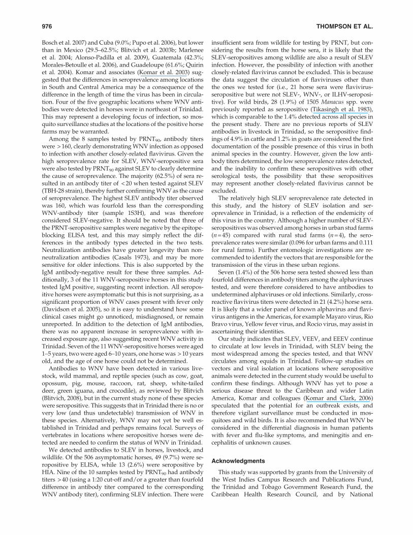
Bosch et al. 2007) and Cuba (9.0%; Pupo et al. 2006), but lowerthan in Mexico (29.5–62.5%; Blitvich et al. 2003b; Marleneeet al. 2004; Alonso-Padilla et al. 2009), Guatemala (42.3%;Morales-Betoulle et al. 2006), and Guadeloupe (61.6%; Quirinet al. 2004). Komar and associates (Komar et al. 2003) sug-gested that the differences in seroprevalence among locationsin South and Central America may be a consequence of thedifference in the length of time the virus has been in circula-tion. Four of the five geographic locations where WNV anti-bodies were detected in horses were in northeast of Trinidad.This may represent a developing focus of infection, so mos-quito surveillance studies at the locations of the positive horsefarms may be warranted.
Among the 8 samples tested by PRNT90, antibody titerswere > 160, clearly demonstrating WNV infection as opposedto infection with another closely-related flavivirus. Given thehigh seroprevalence rate for SLEV, WNV-seropositive serawere also tested by PRNT90 against SLEV to clearly determinethe cause of seroprevalence. The majority (62.5%) of sera re-sulted in an antibody titer of < 20 when tested against SLEV(TBH-28 strain), thereby further confirming WNV as the causeof seroprevalence. The highest SLEV antibody titer observedwas 160, which was fourfold less than the correspondingWNV-antibody titer (sample 1S3H), and was thereforeconsidered SLEV-negative. It should be noted that three ofthe PRNT-seropositive samples were negative by the epitope-blocking ELISA test, and this may simply reflect the dif-ferences in the antibody types detected in the two tests.Neutralization antibodies have greater longevity than non-neutralization antibodies (Casals 1973), and may be moresensitive for older infections. This is also supported by theIgM antibody-negative result for these three samples. Ad-ditionally, 3 of the 11 WNV-seropositive horses in this studytested IgM positive, suggesting recent infection. All seropos-itive horses were asymptomatic but this is not surprising, as asignificant proportion of WNV cases present with fever only(Davidson et al. 2005), so it is easy to understand how someclinical cases might go unnoticed, misdiagnosed, or remainunreported. In addition to the detection of IgM antibodies,there was no apparent increase in seroprevalence with in-creased exposure age, also suggesting recent WNV activity inTrinidad. Seven of the 11 WNV-seropositive horses were aged1–5 years, two were aged 6–10 years, one horse was > 10 yearsold, and the age of one horse could not be determined.
Antibodies to WNV have been detected in various live-stock, wild mammal, and reptile species (such as cow, goat,opossum, pig, mouse, raccoon, rat, sheep, white-taileddeer, green iguana, and crocodile), as reviewed by Blitvich(Blitvich, 2008), but in the current study none of these specieswere seropositive. This suggests that in Trinidad there is no orvery low (and thus undetectable) transmission of WNV inthese species. Alternatively, WNV may not yet be well es-tablished in Trinidad and perhaps remains focal. Surveys ofvertebrates in locations where seropositive horses were de-tected are needed to confirm the status of WNV in Trinidad.
We detected antibodies to SLEV in horses, livestock, andwildlife. Of the 506 asymptomatic horses, 49 (9.7%) were se-ropositive by ELISA, while 13 (2.6%) were seropositive byHIA. Nine of the 10 samples tested by PRNT90 had antibodytiters > 40 (using a 1:20 cut-off and/or a greater than fourfolddifference in antibody titer compared to the correspondingWNV antibody titer), confirming SLEV infection. There were
insufficient sera from wildlife for testing by PRNT, but con-sidering the results from the horse sera, it is likely that theSLEV-seropositives among wildlife are also a result of SLEVinfection. However, the possibility of infection with anotherclosely-related flavivirus cannot be excluded. This is becausethe data suggest the circulation of flaviviruses other thanthe ones we tested for (i.e., 21 horse sera were flavivirus-seropositive but were not SLEV-, WNV-, or ILHV-seroposi-tive). For wild birds, 28 (1.9%) of 1505 Manacus spp. werepreviously reported as seropositive (Tikasingh et al. 1983),which is comparable to the 1.4% detected across all species inthe present study. There are no previous reports of SLEVantibodies in livestock in Trinidad, so the seropositive find-ings of 4.9% in cattle and 1.2% in goats are considered the firstdocumentation of the possible presence of this virus in bothanimal species in the country. However, given the low anti-body titers determined, the low seroprevalence rates detected,and the inability to confirm these seropositives with otherserological tests, the possibility that these seropositivesmay represent another closely-related flavivirus cannot beexcluded.
The relatively high SLEV seroprevalence rate detected inthis study, and the history of SLEV isolation and ser-oprevalence in Trinidad, is a reflection of the endemicity ofthis virus in the country. Although a higher number of SLEV-seropositives was observed among horses in urban stud farms(n = 45) compared with rural stud farms (n = 4), the sero-prevalence rates were similar (0.096 for urban farms and 0.111for rural farms). Further entomologic investigations are re-commended to identify the vectors that are responsible for thetransmission of the virus in these urban regions.
Seven (1.4%) of the 506 horse sera tested showed less thanfourfold differences in antibody titers among the alphavirusestested, and were therefore considered to have antibodies toundetermined alphaviruses or old infections. Similarly, cross-reactive flavivirus titers were detected in 21 (4.2%) horse sera.It is likely that a wider panel of known alphavirus and flavi-virus antigens in the Americas, for example Mayaro virus, RioBravo virus, Yellow fever virus, and Rocio virus, may assist inascertaining their identities.
Our study indicates that SLEV, VEEV, and EEEV continueto circulate at low levels in Trinidad, with SLEV being themost widespread among the species tested, and that WNVcirculates among equids in Trinidad. Follow-up studies onvectors and viral isolation at locations where seropositiveanimals were detected in the current study would be useful toconfirm these findings. Although WNV has yet to pose aserious disease threat to the Caribbean and wider LatinAmerica, Komar and colleagues (Komar and Clark, 2006)speculated that the potential for an outbreak exists, andtherefore vigilant surveillance must be conducted in mos-quitoes and wild birds. It is also recommended that WNV beconsidered in the differential diagnosis in human patientswith fever and flu-like symptoms, and meningitis and en-cephalitis of unknown causes.
Acknowledgments
This study was supported by grants from the University ofthe West Indies Campus Research and Publications Fund,the Trinidad and Tobago Government Research Fund, theCaribbean Health Research Council, and by National
976 THOMPSON ET AL.
Institutes of Health grant AI057156. N.N.T. was supportedby a University of the West Indies postgraduate scholarship.Special thanks go to Brandy Russell and Alva Stewart-Johnson for their laboratory support. The field assistance ofregional health authorities, Kirk Williams, and AnthonyBastaldo is also greatly appreciated.
Author Disclosure Statement
The authors state that no competing financial interestsexist.
References
Aguirre AA, McLean RG, Cook RS, et al. Serologic survey forselected arboviruses and other potential pathogens in wildlifefrom Mexico. J Wildl Dis 1992; 28:435–442.
Aitken TH, Anderson CR, Downs WG. The isolation of Ilheusvirus from wild caught forest mosquitoes in Trinidad. AmJ Trop Med Hyg 1956; 5:621–625.
Aitken TH, Downs WG, Anderson CR, et al. Mayaro virus iso-lated from a Trinidadian mosquito, Mansonia venezuelensis.Science 1960; 131:986.
Aitken TH, Downs WG, Spence L, et al. (1964). St. Louis en-cephalitis virus isolations in Trinidad, West Indies, 1953–1962.Am J Trop Med Hyg 1964; 13:450–451.
Aitken TH, Spence L, Jonkers AH, et al. Wyeomyla-virus isola-tions in Trinidad, West Indies. Am J Trop Med Hyg 1968a;17:886–888.
Aitken TH, Worth CB, Tikasingh ES. Arbovirus studies in BushBush Forest, Trinidad, W. I., September 1959–December 1964.3. Entomologic studies. Am J Trop Med Hyg 1968c; 17:253–268.
Aitken TH, Worth CB, Jonkers AH, et al. Arbovirus studies inBush Bush Forest, Trinidad, W. I., September 1959–December1964. II. Field program and techniques. Am J Trop Med Hyg1968b; 17:237–252.
Alonso-Padilla J, Loza-Rubio E, Escribano-Romero E, et al. Thecontinuous spread of West Nile virus (WNV): seroprevalencein asymptomatic horses. Epidemiol Infect 2009; 137:1163–1168.
Anderson CR, Downs WG, Theiler M. Neutralizing antibodiesagainst certain viruses in the sera of residents of Trinidad,B.W.I. Am J Trop Med Hyg 1956; 5:626–641.
Arrigo NC, Adams AP, Watts DM, et al. Cotton rats and housesparrows as hosts for North and South American strains ofeastern equine encephalitis virus. Emerg Infect Dis 2010;16:1373–1380.
Auguste AJ, Adams AP, Arrigo NC, et al. Isolation and char-acterization of sylvatic mosquito-borne viruses in Trinidad:enzootic transmission and a new potential vector of Mucambovirus. Am J Trop Med Hyg 2010a; 83:1262–1265.
Auguste AJ, Lemey P, Pybus OG, et al. Yellow fever virusmaintenance in Trinidad and its dispersal throughout theAmericas. J Virol 2010b; 84: 9967–9977.
Auguste AJ, Volk SM, Arrigo NC, et al. Isolation and phyloge-netic analysis of Mucambo virus (Venezuelan equine en-cephalitis complex subtype IIIA) in Trinidad. Virology 2009;392:123–130.
Barrera R, Ferro C, Navarro JC, et al. Contrasting sylvatic foci ofVenezuelan equine encephalitis virus in northern SouthAmerica. Am J Trop Med Hyg 2002; 67:324–334.
Beaty BJ, Calisher CH, Shope RE. Arboviruses. Diagnostic Proce-dures for Viral, Rickettsial and Chlamydial Infections, 6th ed.
Washington: American Public Health Association, 1989:797–855.
Blitvich BJ, Bowen RA, Marlenee NL, et al. Epitope-blockingenzyme-linked immunosorbent assays for detection of WestNile virus antibodies in domestic mammals. J Clin Microbiol2003a; 41:2676–2679.
Blitvich BJ, Fernandez-Salas I, Contreras-Cordero JF, et al. Ser-ologic evidence of West Nile virus infection in horses, Coa-huila State, Mexico. Emerg Infect Dis 2003b; 9:853–856.
Blitvich BJ, Marlenee NL, Hall RA, et al. Epitope-blocking en-zyme-linked immunosorbent assays for the detection of serumantibodies to West Nile virus in multiple avian species. J ClinMicrobiol 2003c; 41:1041–1047.
Blitvich BJ. Transmission dynamics and changing epidemiologyof West Nile virus. Anim Health Res Rev 2008; 9:71–86.
Bosch I, Herrera F, Navarro JC, et al. West Nile virus, Venezuela.Emerg Infect Dis 2007; 13:651–653.
Casals J. Arbovirus infections. In: Serological Epidemiology. PaulJR, White C, ed. New York: Academic Press, 1973:99–117.
Central Statistical Office. CSO’s household budget survey 1997–1998: Geographic Information Section, Central Statistical Of-fice, Ministry of Finance, Trinidad and Tobago, 1998.
Clarke DH, Casals J. Techniques for hemagglutination andhemagglutination-inhibition with arthropod-borne viruses.Am J Trop Med Hyg 1958; 7:561–573.
Davidson AH, Traub-Dargatz JL, Rodeheaver RM, et al. Im-munologic responses to West Nile virus in vaccinated andclinically affected horses. J Am Vet Med Ass 2005; 226:240–245.
Deardorff ER, Forrester NL, Travassos-da-Rosa AP, et al. Ex-perimental infection of potential reservoir hosts with Vene-zuelan equine encephalitis virus, Mexico. Emerg Infect Dis2009; 15:519–525.
Downs, W. G., Aitken, T. H., Worth, C. B., et al.. Arbovirusstudies in Bush Bush Forest, Trinidad, W. I., September 1959–December 1964. I. Description of the study area. Am J TropMed Hyg 1968; 17:224–236.
Estrada-Franco JG, Navarro-Lopez R, Freier JE, et al. Venezuelanequine encephalitis virus, southern Mexico. Emerg Infect Dis2004; 10:2113–2121.
Fernandez RJ, Vazquez S. Serological diagnosis of dengue by anELISA inhibition method (EIM). Mem Inst Oswaldo Cruz1990; 85:347–351.
Hall RA, Broom AK, Hartnett AC, et al. Immunodominantepitopes on the NS1 protein of MVE and KUN viruses serveas targets for a blocking ELISA to detect virus-specific anti-bodies in sentinel animal serum. J Virol Methods 1995; 51:201–210.
Jonkers AH, Aitken TH, Spence L, et al. Ecological studies ofVenezuelan equine encephalitis virus (VEEV) in the BushBush wilderness of Trinidad. Rev Venez Sanid Asist Soc Suppl1966; 3:929.
Jonkers AH, Spence L, Downs WG, et al. Arbovirus studies inBush Bush forest, Trinidad, W. I., September 1959–December1964. V. Virus isolations. Am J Trop Med Hyg 1968a; 17:276–284.
Jonkers AH, Spence L, Downs WG, et al. Arbovirus studies inBush Bush Forest, Trinidad, W. I., September 1959–December1964. VI. Rodent-associated viruses (VEE and agents of groupsC and Guama): isolations and further studies. Am J Trop MedHyg 1968b; 17:285–298.
Komar N, Clark GG. West Nile virus activity in Latin Americaand the Caribbean. Rev Panam Salud Publica 2006; 19:112–117.
SEROPREVALENCE OF ARBOVIRUSES IN TRINIDAD 977
Komar N, Langevin S, Hinten S, et al. Experimental infection ofNorth American birds with the New York 1999 strain of WestNile virus. Emerg Infect Dis 2003; 9:311–322.
Marlenee NL, Lorono-Pino MA, Beaty BJ, et al. Detection ofantibodies to West Nile and Saint Louis encephalitis viruses inhorses. Salud Publica Mex 2004; 46:373–375.
Morales-Betoulle ME, Morales H, Blitvich BJ, et al. West Nilevirus in horses, Guatemala. Emerg Infect Dis 2006; 12:1038–1039.
Navarro JC, Medina G, Vasquez C, et al. Postepizootic persis-tence of Venezuelan equine encephalitis virus, Venezuela.Emerg Infect Dis 2005; 11:1907–1915.
OIE. Venezuelan equine encephalomyelitis, Costa Rica. WorldAnimal Health Information Database Interface: Animal HealthInformation, 2009.
Price JL. Isolation of Rio Bravo and a hitherto undescribed agent,Tamana bat virus, from insectivorous bats in Trinidad, withserological evidence of infection in bats and man. Am J TropMed Hyg 1978a; 27(1 Pt 1):153–161.
Price JL. Serological evidence of infection of Tacaribe virus andarboviruses in Trinidadian bats. Am J Trop Med Hyg 1978b;27(1 Pt 1):162–167.
Pupo M, Guzman MG, Fernandez R, et al. West Nile Virus in-fection in humans and horses, Cuba. Emerg Infect Dis 2006;12:1022–1024.
Quirin R, Salas M, Zientara S, et al. West Nile virus, Guade-loupe. Emerg Infect Dis 2004; 10:706–708.
Roehrig JT, Hunt AR, Chang GJ, et al. Identification of mono-clonal antibodies capable of differentiating antigenic varietiesof eastern equine encephalitis viruses. Am J Trop Med Hyg1990; 42:394–398.
Scherer WF, Sainz CC, De Mucha Macias J, et al. Serologic sur-vey for neutralizing antibodies to eastern equine and westernequine encephalitis viruses in man, wild birds and swine in
southern Mexico during 1961. Am J Trop Med Hyg 1966;15:211–218.
Shi PY, Wong SJ. Serologic diagnosis of West Nile virus infec-tion. Expert Rev Mol Diagn 2003; 3:733–741.
Tikasingh ES, Aitken TH, Butcher L, et al. Epidemiologic in-vestigations relating to a case of eastern equine encephalitis ina Trinidadian horse. West Indian Med J 1968; 17:90–95.
Tikasingh ES. Eastern equine encephalitis virus activity inTrinidad: A review. J Caribbean Veterinary Med Assn 2004;4:18–23.
Tikasingh ES, Jonkers AH, Spence L, et al. Arbovirus studies inan evergreen seasonal marsh forest in Trinidad, West Indies.West Indian Med J 1983; 32:223–231.
Tikasingh ES, Worth CB, Jonkers AH, et al. A three-yearsurveillance of eastern equine encephalitis virus activity inTrinidad. West Indian Med J 1973; 22:24–31.
Wang E, Paessler S, Aguilar PV, et al. A novel, rapid assay fordetection and differentiation of serotype-specific antibodies toVenezuelan equine encephalitis complex alphaviruses. AmJ Trop Med Hyg 2005; 72:805–810.
Worth CB, Downs WG, Aitken TH, et al. Arbovirus studies inBush Bush forest, Trinidad, W. I., September 1959–December1964. IV. Vertebrate populations. Am J Trop Med Hyg 1968;17:269–275.
Address correspondence to:Abiodun A. Adesiyun
School of Veterinary MedicineFaculty of Medical Sciences
The University of the West IndiesSt. Augustine
Republic of Trinidad and Tobago
E-mail: [email protected].
978 THOMPSON ET AL.
Copyright © 2022 FDOKUMEN