The ???Primary??? Antiphospholipid Syndrome: Major Clinical and Serological Features

Journal of Immunological Methods 244 (2000) 69–80www.elsevier.nl / locate / jim
Isolation and serological analyses of fungal melaninsa b c d ,1´ ´Angel L. Rosas , Joshua D. Nosanchuk , Beatriz L. Gomez , William A. Edens ,
d a,b ,*Joan M. Henson , Arturo CasadevallaDepartment of Microbiology and Immunology, Albert Einstein College of Medicine, Bronx, NY 10461, USA
bDivision of Infectious Diseases of the Department of Medicine, Albert Einstein College of Medicine, Bronx, NY 10461, USAcDermatology Department, St. John’s Institute of Dermatology, Guy’s Hospital, Guy’s, Kings and St. Thomas Medical Schools,
London SE1 9RT, UKdMicrobiology Department, Montana State University, Bozeman, MT 59717, USA
Received 21 April 2000; received in revised form 7 June 2000; accepted 9 June 2000
Abstract
Melanins are notoriously difficult to work with because of their unique physical and chemical properties. The study ofmelanins is hampered by the scarcity of melanin-specific reagents and serological techniques. In this study we describemodifications to the standard method for the isolation of melanins from in vitro-melanized fungal cells and detail theoptimization of serological techniques for the study of melanin compounds. The isolation procedure involves the digestion ofmelanized cells with a combination of proteolytic and glycolytic enzymes, denaturant, organic extractions, and boiling in 6.0M HCl. Elemental quantitative analyses suggest that this procedure does not significantly affect the relative elementalcomposition of melanins. For the serological assays, our goal was to achieve a homogenous distribution of melanin particleson a solid support to maximize their recognition by melanin-binding antibodies. The results from enzyme-linkedimmunosorbent assays (ELISAs) demonstrate that melanins, in general, disperse more efficiently on, and adhere better to,medium-binding polystyrene surfaces, especially in the presence of trace amounts of salt. Blocking the melanin-coated
ELISA plates with the commercially available SuperBlock Blocking Buffer for 4 h was more efficient at reducingnon-specific binding of a negative control monoclonal antibody (mAb) compared to blocking with 2% bovine serum albumin(BSA) and 5% milk. Increasing the ionic strength of the antibody solutions reduced binding to the melanins, indicating thatbinding is in part mediated by electrostatic interactions. These conditions were also applied to immunofluorescence (IF)analyses of melanins, and the results were consistent with those obtained by ELISA. 2000 Elsevier Science B.V. Allrights reserved.
Keywords: Melanins; Isolation; Quantitative elemental analysis; ELISA; Immunofluorescence; Monoclonal antibodies
Abbreviations: ELISA, enzyme-linked immunosorbent assay; IF, immunofluorescence; SEM, scanning electron microscopy; mAbs,monoclonal antibodies; DOPA, 3,4-dihydroxyphenylalanine; DHN, 1,8-dihydroxynaphthalene; BSA, bovine serum albumin; PBS, phos-phate-buffered saline
*Corresponding author. Tel.: 11-718-430-3665; fax: 11-718-430-8701.E-mail address: [email protected] (A. Casadevall).1Present address: Department of Biochemistry, School of Medicine, Emory University, Atlanta, Georgia 30322, USA.
0022-1759/00/$ – see front matter 2000 Elsevier Science B.V. All rights reserved.PI I : S0022-1759( 00 )00255-6

´70 A.L. Rosas et al. / Journal of Immunological Methods 244 (2000) 69 –80
1. Introduction insolubility in polar and most organic solvents,relative resistance to degradation by acids, degra-
Melanins are negatively-charged, hydrophobic dation and solubilization by hot concentrated alkalinepigments of high molecular weight that are formed solutions, and bleaching by oxidizing agents (re-by the oxidative polymerization of phenolic and/or viewed by Butler & Day, 1998).indolic compounds (reviewed by Wheeler & Bell, The analysis of naturally occuring melanins is1988; Butler & Day, 1998). These pigments are hampered by the scarcity of melanin-specific re-usually black or brown, although red, yellow, purple, agents and serological techniques. Previous studiesblue and green melanins also exist, depending on the have used the Masson–Fontana silver stain to detectsubstrate(s) from which they are formed (reviewed melanin in pathogenic organisms (Kwon-Chung etby Wheeler & Bell, 1988). Melanins are synthesized al., 1981) and human tissue sections (Warkel et al.,by organisms of all biological kingdoms and are 1980), but the reaction is not specific for melanins. Ainvolved in a wide variety of processes, including modified Warthin–Starry silver staining procedure atcamouflage, sex appeal, and protection against UV pH 3.2 has been successfully employed to detectradiation (reviewed by Hill, 1992). Melanins have melanization of human melanoma tissue sectionsbeen implicated in microbial pathogenesis (reviewed (Warkel et al., 1980) and has proven to be moreby Wheeler & Bell, 1988; Butler & Day, 1998), in specific and sensitive than the Masson–Fontanapart because of their ability to bind and neutralize stain. Our laboratory has developed melanin-bindingoxidants (Jacobson & Emery, 1991; Wang & peptides (Nosanchuk et al., 1999) and monoclonalCasadevall, 1994a), microbicidal peptides (Doering antibodies (mAbs) (Rosas et al., 2000) to studyet al., 1999), and antimicrobial drugs (Wang & melanization of the pathogenic fungus CryptococcusCasadevall, 1994b). They have also been implicated neoformans. Both reagents react with melanins andin the pathogenesis of malignant melanoma (re- melanized cells, but not with non-melanized cells.viewed by Hill, 1991), Parkinson’s disease (reviewed The melanin-binding peptides are characterized by aby d’Ischia & Prota, 1997), traumatic anterior high proportion of positively-charged and aromaticchamber uveitis (Kaya et al., 1992), and vitiligo residues, suggesting that binding to melanins in-(Langhof et al., 1965). Recent studies have shown volves both electrostatic and hydrophobic interac-that melanins are highly immunogenic (Nosanchuk tions. The mAbs to melanin have strong reactivityet al., 1998) and have anti-inflammatory properties with a variety of melanins, suggesting that melanins(Avramidis et al., 1998; Mohagheghpour et al., from different sources share antigenic epitopes. We2000). have used enzyme-linked immunosorbent assays
Despite their ubiquity in nature, relatively little is (ELISAs) and immunofluorescence (IF) analyses toknown about the structure of melanins. In fact, no determine the presence of melanin-binding antibo-structure of any naturally occurring melanin has been dies in immunized (Nosanchuk et al., 1998) andsolved to date. This is due in part to the fact that infected (Nosanchuk et al., 1999) mice. However,most melanins are highly insoluble, which makes the these techniques are difficult because melanins aredetermination of their molecular weights and struc- insoluble, their surfaces have both hydrophobic andtures very difficult by conventional biochemical and charged properties, and have high affinity for chemi-biophysical techniques. Melanins are also often cals (Larsson, 1993) and proteins (Doering et al.,tightly associated with other cellular components, 1999).which can only be removed with great difficulty by In this study we describe modifications to theprolonged treatment with hot concentrated acids, and method by Wang et al. (1996) for the isolation ofit is not known what effect, if any, this has on the melanins from in vitro-melanized fungal cells. Westructure of melanins. Because of the lack of chemi- also describe the optimization of serological tech-cal tests to classify them, there is still controversy niques for the study of melanins, such as selection ofabout the precise definition of a melanin (reviewed solid support for ELISAs and conditions to induceby Butler & Day, 1998). Compounds are provision- optimal dispersion and adherence of melanin par-ally classified as melanins based on dark color, ticles to surfaces, the efficiency of various blocking

A.L. Rosas et al. / Journal of Immunological Methods 244 (2000) 69 –80 71
solutions to prevent or reduce non-specific binding to and 0.5% SDS; pH 7.8) for 4 h at 658C, washed withmelanins in ELISAs and IF analyses, and the effect PBS, and extracted three times with chloroform.of the ionic strength of the solvent on the binding of After washing with PBS, the particulate was boiledantibodies to melanins. in 6.0 M HCl for 1 h to hydrolyze cellular con-
(The data in this paper are from a thesis to be taminants that may still be associated with the´submitted by Angel L. Rosas in partial fulfillment of melanin. Melanin particles were collected by cen-
the requirements for the degree of doctor of trifugation, washed extensively with PBS, andphilosophy in the Sue Golding Graduate Division of dialyzed against distilled water for 10 days.Medical Sciences, Albert Einstein College of Medi- A. niger was grown on Sabouraud dextrose agarcine, Yeshiva University, Bronx, NY.) (pH 5.6) (Difco Laboratories, Detroit, MI) for 5
months at 308C to allow heavy melanization of theconidia. A. niger conidial melanin was isolated asdescribed above.
2. Materials and methodsSynthetic melanin (lot [: 10H02926) and melanin
from the cuttlefish Sepia officinalis (lot [:2.1. Chemical treatment of melanins 103H1023) were purchased from Sigma Chemical
Co. Synthetic melanin is a DOPA-melanin preparedMelanins were extracted from the human patho- by oxidation of tyrosine in the presence of H O . S.2 2
genic fungus Cryptococcus neoformans (strains officinalis melanin is a DOPA-melanin extracted24067 and F7) and conidia from Aspergillus niger from the ink glands of the cuttlefish as described byJ9901. C. neoformans 24067 and A. niger J9901 Zeise et al. (1992). Samples of these melanins werewere purchased from the American Type Culture boiled in 6.0 M HCl for 1 h to compare the effect ofCollection (Rockville, MD). C. neoformans F7 is a acid treatment on their relative elemental composi-poorly encapsulated pseudohyphal mutant of strain tion.24067 (Fries et al., 1999). All melanins were washed extensively and sus-
C. neoformans was grown in a defined minimal pended in deionized, distilled water (BioWhittakermedium (15.0 mM glucose, 10.0 mM MgSO , 29.4 Molecular Applications Inc., Walkersville, MD), and4
mM KH PO , 13.0 mM glycine, and 3.0 mM lyophilized with a Flexi-Dry microprocessor (FTS2 4
vitamin B ; pH 5.5) in the presence of 1.0 mM Systems, Inc., Stone Ridge, NY) prior to quantitative1
L-dopa (Sigma Chemical Co., St. Louis, MO) for 21 elemental analyses. For serological analyses,days at 308C in a rotary shaker at 150 rpm. Melan- melanins were suspended in deionized, distilledized cells were collected by centrifugation at 3000 water, with or without prior lyophilization.rpm for 30 min, washed with phosphate-bufferedsaline (PBS), and suspended in 1.0 M sorbitol–0.1M sodium citrate (pH 5.5). Cell wall–lysing en- 2.2. Quantitative elemental analyses of melaninszymes (Sigma Chemical Co.), which contain pro-tease, cellulase and chitinase activities, were added at Carbon, nitrogen, and oxygen analyses on the10 mg/ml and the suspension was incubated over- lyophilized melanin samples were performed bynight at 308C to generate protoplasts. The protoplasts Quantitative Technologies Inc. (Whitehouse, NJ).were collected by centrifugation, washed with PBS, Briefly, melanin samples were converted into gasesand incubated in 4.0 M guanidine thiocyanate (de- such as CO , H O, and N by combustion. The2 2 2
naturant) for 12 h at room temperature with frequent product gases were separated under steady-statevortexing. A dark particulate material consisting of conditions and the percentage of each element in themelanin and cellular debris was collected by cen- samples was measured as a function of thermaltrifugation and washed with PBS. The particulate conductivity. C:N:O ratios were calculated by divid-was then treated with 1.0 mg/ml Proteinase K ing the percentage of each element in the samples by(Roche Molecular Biochemicals, Indianapolis, IN) their respective atomic weights. The values reported(reaction buffer was 10.0 mM Tris, 1.0 mM CaCl , are from one of two independent analyses.2

´72 A.L. Rosas et al. / Journal of Immunological Methods 244 (2000) 69 –80
2.3. Scanning Electron Microscopy (SEM.) 2.7. ELISA plates
Melanized fungal cells and melanin particles were The following brands of plates were tested forincubated in 2.5% gluteraldehyde for 1 h at room dispersion and adherence of melanin particles to theirtemperature and applied to poly-L-lysine-coated surfaces: (a) Corning polystyrene plates (classified asslides (Sigma Chemical Co.). The samples were medium- and high-binding surfaces) (Corning Glassdehydrated by serial incubations in solutions of Works, Corning, NY); (b) Falcon 96-well, flat-bot-increasing ethanol concentration, dried in a Tousimis tom EIA/RIA polystyrene plates (Becton DickinsonSamdri-790 Critical Point Drier (Tousimis Research Labware, Lincoln Park, NJ); (c) Nunc-Immuno PlateCo., Rockville, MD), coated with gold-palladium in MaxiSorp (Nalge Nunc International, Rochester,a Denton Vacuum Desk-1 Sputter Coater (Denton NY); and (d) Falcon tissue culture plates treated byVacuum Inc., Cherry Hill, NJ), and examined using a vacuum gas plasma (Becton Dickinson Labware,JEOL JSM-6400 scanning electron microscope Franklin Lakes, NJ). Prior to addition of melanin,(JEOL, Tokyo, Japan). half of each plate was incubated with 100 ml per well
of 0.01% poly-L-lysine (mol. wt. 70,000–150,000)2.4. Antibodies (Sigma Chemical Co.) for 1 h at room temperature,
washed extensively with deionized, distilled water,MAbs 6D2 and 11B11 (mk) to melanin have been and left drying overnight at room temperature. The
described (Rosas et al., 2000). MAb 5C11 (mk) to other half of the plate was left untreated.mycobacterial lipoarabinomannan (Glatman-Freed-man et al., 1996) was used as an isotype-matched 2.8. Melanin ELISAsnegative control. The mAbs were purified fromconcentrated cell culture supernatants by ultralinked- To analyze the distribution of melanin particles on
5 6mannan binding protein chromatography (Pierce, solid surface, fungal melanins (5310 –5310Rockville, MD) according to the manufacturer’s melanin particles, which are equivalent to 1.5–15 mginstructions and their concentrations were determined of dry melanin), and 20–100 mg of synthetic and S.by ELISA relative to purified murine polyclonal IgM officinalis melanins were suspended in water and(1.0 mg/ml) (ICN Biomedicals, Aurora, OH). MAbs plated in each well of the plates for 2–3 days atwere suspended in PBS with 0.02% azide at 1.0 room temperature to allow the melanin particles tomg/ml. Antibody solutions were kept at 2208C until dry and adhere to the surface. In some cases, theuse. melanin particles were heat-fixed to the surface by
incubating the plates at 608C for 30 min.2.5. Antibody solutions For the melanin ELISAs, Corning polystyrene
medium-binding plates were covered with 15 mg ofFor ELISAs and IF analyses, the antibodies were fungal melanin or 100 mg of S. officinalis melanin
diluted in PBS (0.14 M NaCl, 0.0027 M KCl, 0.0015 per well and then blocked to prevent or reduceM KH PO , and 0.0085 M Na HPO ; pH 7.4), PBS non-specific binding using the solutions described2 4 2 4
(0.28 M NaCl), or PBS (0.56 M NaCl). above. The plates were washed three times with0.1% Tween-20 in Tris-buffered saline (TBS) after
2.6. Blocking solutions each incubation. Antibodies were diluted in solutionsof different salt concentrations (i.e., PBS [0.14 M
The following blocking solutions and incubation NaCl, 0.28 M NaCl, or 0.56 M NaCl]) to 10 mg/ml,times were used in ELISAs and IF analyses of added to the wells of the melanin-coated plates, andmelanins at room temperature: (a) 2% bovine serum incubated for 1.5 h at 378C. After washing, a 1:1000albumin (BSA) in PBS for 2 h; (b) 2% BSA in PBS dilution of alkaline phosphatase-conjugated goatfor 2 h followed by incubation with 5% powdered anti-mouse IgM (Southern Biotechnologies As-
milk in water for 2 h; and (c) SuperBlock Blocking sociates Inc., Birmingham, AL) was added to theBuffer in PBS (Pierce, Rockville, IL) for 2 or 4 h. wells and incubated for 1.5 h at 378C. Antibody

A.L. Rosas et al. / Journal of Immunological Methods 244 (2000) 69 –80 73
binding was detected by addition of p-nitrophenyl mouse IgM (Southern Biotechnologies Associatesphosphate (Sigma Chemical Co.) (reaction buffer Inc.) for 1.5 h at 378C. Samples were then washedwas 1.0 mM MgCl and 50.0 mM Na CO ; pH 9.8). with PBS to eliminate unbound antibody. A mount-2 2 3
After 30 min, the solutions were transferred to clear ing solution (50% glycerol, 50 PBS, and 0.1 Mplates and the optical densities were measured at 405 N-propyl gallate) and coverslip were applied to thenm with a Ceres 900HDi EIA Workstation (Bio-Tek melanins fixed to the slides, and the samples wereInstruments, Inc., Winooski, VT). Each value is the examined using an Olympus AX70 microscopemean6standard deviation of four independent mea- (Olympus America Inc., Melville, NY) with asurements from one experiment. Each ELISA was fluorescein isothiocyanate filter at a magnification ofdone twice. P-values were calculated with Student’s 3250. Melanins in suspension were collected byt-test using Primer of Statistics: The Program Version centrifugation for 10 min at 1200 rpm, suspended in3.0 (McGraw–Hill Inc., New York, NY) for com- the mountain solution, and then applied to the slides.parison in melanin binding of mAbs 6D2 and 11B11 IF analyses were also performed with paraffin-and polyclonal IgM to that of negative control mAb embedded tissue sections from 12 C. neoformans-5C11. P-values less than 0.05 were considered infected BALB/c mouse brains. C. neoformanssignificant. infections were done as described (Rosas et al.,
Melanin ELISAs were also performed with 1,8- 2000). Mice were housed in the animal facility ofdihydroxynaphthalene (DHN)-melanin catalyzed by our institution and all experimental procedureslaccase from Gaeumannomyces graminis var. tritici. adhered to protocols approved by the Animal CareLaccase was purified as described by Edens et al. and Use Committee at Albert Einstein College of(1999), and 3000 units of pure laccase were incu- Medicine. After seven days of infection, the brainsbated in a 2.0 mM solution of DHN (in 50% ethanol were collected and sectioned. Tissue sections, 4 mmv /v) overnight with constant stirring. A unit of thick were deparaffinized in xylenes and rehydratedlaccase is defined as the amount of laccase needed to by serial incubations in solutions of decreasingoxidize 1.0 mmol of 2,6-dimethoxyphenol per min- ethanol concentration. The samples were treated withute. The majority of the ethanol was then removed 20 mg/ml Proteinase K for 1 h at room temperatureby rotary evaporation and final traces were elimi- and then heated in 10 mM citric acid in a microwavenated by normal evaporation for 24 h. The DHN- oven for 5 min. Tissue sections were blocked with
melanin suspension was then diluted 1:2, 1:4, and SuperBlock Blocking Buffer in PBS for 4 h,1:8. A total of 50 ml of the melanin suspensions were incubated with mAb solutions (10 mg/ml) of differ-plated in each well of the plates to determine which ent salt concentrations, and treated as describeddilution gave the best distribution of the melanin. above.Melanin ELISAs were performed only with the 1:4dilution on Corning polystyrene medium-bindingplates as described above. 3. Results
2.9. IF analyses of melanins 3.1. Elemental analyses of melanins
6Approximately 10 C. neoformans and A. niger In vitro-melanized fungal cells were treated with amelanin particles (approximately 3 mg of dry combination of proteolytic and glycolytic enzymes,melanin) were either fixed to poly-L-lysine-coated denaturant, chloroform extractions, and boiling in 6.0slides or suspended in PBS. The melanins were then M HCl for 1 h to isolate melanin from other cellularblocked as described above and then incubated with components. This treatment is a modification of a10 mg/ml of mAbs 6D2, 11B11, or 5C11 in solu- previous protocol used to isolate melanin from intions of different salt concentrations as described vitro-melanized C. neoformans cells (Wang et al.,above for 1.5 h at 378C. Samples were washed three 1996). The melanin particles were referred to astimes with PBS and incubated with a 1:100 dilution ‘‘ghosts’’ since they retained the round shape ofof fluorescein isothiocyanate-conjugated goat anti- melanized C. neoformans cells. Our results demon-

´74 A.L. Rosas et al. / Journal of Immunological Methods 244 (2000) 69 –80
Table 1strate that melanin ‘‘ghosts’’ not only retain theElemental quantitative analyses of melaninsshape but also the surface features of the cells fromMelanin C:N:Owhich they were isolated, including budding scars
(Fig. 1). Elemental quantitative analyses were per- synthetic DOPA-melanin 9:1:2formed to determine the relative elemental com- synthetic DOPA-melanin (treated with HCl) 9:1:2
DOPA-melanin from S. officinalis 7:1:2position of the melanins (Table 1). The C:N:O ratiosDOPA-melanin from S. officinalis (treated with HCl) 8:1:3of acid-treated synthetic melanin were not altered inDOPA-melanin from C. neoformans 24067 16:2:5
comparison to the untreated sample (9C:1N:2C), melanin from A. niger J9901 conidia 29:2:6whereas only a slight difference was observed be-tween acid-treated and untreated S. officinalismelanin samples (8C:1N:3O for acid-treated vs. tify that which provided the best surface for disper-7C:1N:2O for untreated). This experiment was done sion and attachment of melanin particles. In general,twice for each melanin and the results were con- Corning polystyrene medium-binding plates gave thesistent. best dispersion of melanin particles on the surface of
the wells. Except for tissue culture plates, treatment3.2. ELISAs with poly-L-lysine prior to addition of melanin did
not improve the distribution of the melanin particlesSeveral brands of microtiter 96-well plates of on the surface of the wells.
different chemical compositions were tested to iden- Melanins differed in their ability to disperse and
Fig. 1. SEM of melanized C. neoformans 24067 (A), C. neoformans F7 (B), and A. niger J9901 conidia (C). Panels (D), (E), and (F)represent their respective melanin particles (‘‘ghosts’’). Arrow on C. neoformans melanin ‘‘ghost’’ denotes a budding scar.
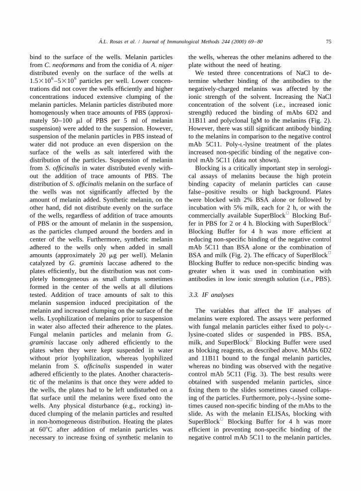
A.L. Rosas et al. / Journal of Immunological Methods 244 (2000) 69 –80 75
bind to the surface of the wells. Melanin particles the wells, whereas the other melanins adhered to thefrom C. neoformans and from the conidia of A. niger plate without the need of heating.distributed evenly on the surface of the wells at We tested three concentrations of NaCl to de-
6 61.5310 –5310 particles per well. Lower concen- termine whether binding of the antibodies to thetrations did not cover the wells efficiently and higher negatively-charged melanins was affected by theconcentrations induced extensive clumping of the ionic strength of the solvent. Increasing the NaClmelanin particles. Melanin particles distributed more concentration of the solvent (i.e., increased ionichomogenously when trace amounts of PBS (approxi- strength) reduced the binding of mAbs 6D2 andmately 50–100 ml of PBS per 5 ml of melanin 11B11 and polyclonal IgM to the melanins (Fig. 2).suspension) were added to the suspension. However, However, there was still significant antibody bindingsuspension of the melanin particles in PBS instead of to the melanins in comparison to the negative controlwater did not produce an even dispersion on the mAb 5C11. Poly-L-lysine treatment of the platessurface of the wells as salt interfered with the increased non-specific binding of the negative con-distribution of the particles. Suspension of melanin trol mAb 5C11 (data not shown).from S. officinalis in water distributed evenly with- Blocking is a critically important step in serologi-out the addition of trace amounts of PBS. The cal assays of melanins because the high proteindistribution of S. officinalis melanin on the surface of binding capacity of melanin particles can causethe wells was not significantly affected by the false–positive results or high background. Platesamount of melanin added. Synthetic melanin, on the were blocked with 2% BSA alone or followed byother hand, did not distribute evenly on the surface incubation with 5% milk, each for 2 h, or with the
of the wells, regardless of addition of trace amounts commercially available SuperBlock Blocking Buf-of PBS or the amount of melanin in the suspension, fer in PBS for 2 or 4 h. Blocking with SuperBlock
as the particles clumped around the borders and in Blocking Buffer for 4 h was more efficient atcenter of the wells. Furthermore, synthetic melanin reducing non-specific binding of the negative controladhered to the wells only when added in small mAb 5C11 than BSA alone or the combination of
amounts (approximately 20 mg per well). Melanin BSA and milk (Fig. 2). The efficacy of SuperBlockcatalyzed by G. graminis laccase adhered to the Blocking Buffer to reduce non-specific binding wasplates efficiently, but the distribution was not com- greater when it was used in combination withpletely homogeneous as small clumps sometimes antibodies in low ionic strength solution (i.e., PBS).formed in the center of the wells at all dilutionstested. Addition of trace amounts of salt to this 3.3. IF analysesmelanin suspension induced precipitation of themelanin and increased clumping on the surface of the The variables that affect the IF analyses ofwells. Lyophilization of melanins prior to suspension melanins were explored. The assays were performedin water also affected their adherence to the plates. with fungal melanin particles either fixed to poly-L-Fungal melanin particles and melanin from G. lysine-coated slides or suspended in PBS. BSA,
graminis laccase only adhered efficiently to the milk, and SuperBlock Blocking Buffer were usedplates when they were kept suspended in water as blocking reagents, as described above. MAbs 6D2without prior lyophilization, whereas lyophilized and 11B11 bound to the fungal melanin particles,melanin from S. officinalis suspended in water whereas no binding was observed with the negativeadhered efficiently to the plates. Another characteris- control mAb 5C11 (Fig. 3). The best results weretic of the melanins is that once they were added to obtained with suspended melanin particles, sincethe wells, the plates had to be left undisturbed on a fixing them to the slides sometimes caused collaps-flat surface until the melanins were fixed onto the ing of the particles. Furthermore, poly-L-lysine some-wells. Any physical disturbance (e.g., rocking) in- times caused non-specific binding of the mAbs to theduced clumping of the melanin particles and resulted slide. As with the melanin ELISAs, blocking with
in non-homogeneous distribution. Heating the plates SuperBlock Blocking Buffer for 4 h was moreat 608C after addition of melanin particles was efficient in preventing non-specific binding of thenecessary to increase fixing of synthetic melanin to negative control mAb 5C11 to the melanin particles.

´76 A.L. Rosas et al. / Journal of Immunological Methods 244 (2000) 69 –80
Fig. 2. Binding of antibodies to C. neoformans 24067 melanin (15 mg/well) by ELISA after blocking with 2% BSA for 2 h (A), 2% BSA followed by 5% milk for 2 h each (B), SuperBlock Blocking Buffer in PBS for 2 h (C), or SuperBlock Blocking Buffer in PBS for 4 h
(D). The antibodies were diluted in solutions of different salt concentrations at 10 mg/ml. Each value is the mean6deviation of fourmeasurements from one of two independent experiments. P-values were calculated with Student’s t-test for comparison in melanin bindingof mAbs 6D2 and 11B11 and polyclonal IgM to that of negative control mAb 5C11. P-values were less than 0.05 for all comparisons.Similar results were obtained with A. niger J9901 conidial melanin (15 mg/well), S. officinalis DOPA-melanin (100 mg/well), and a 1:4dilution of DHN-melanin suspension from G. graminis.
We observed strong binding when the mAbs to Binding of the mAbs to melanin was observed whenmelanin were incubated in PBS, but binding was incubated in PBS, but was almost abolished whengreatly reduced when the mAbs were incubated in the ionic strength of the solutions was increased.solutions of higher ionic strength.
Results from IF analyses of C. neoformans-infec-ted BALB/c mouse brain tissues also demonstrated 4. Discussionbinding of mAbs 6D2 and 11B11 to cell wall-associated melanin (Fig. 4), whereas no binding was In this study we employed a combination ofobserved with the negative control mAb 5C11. proteolytic and glycolytic enzymes, denaturant, chlo-

A.L. Rosas et al. / Journal of Immunological Methods 244 (2000) 69 –80 77
Fig. 3. Light and fluorescent microscopy of C. neoformans 24067 melanin ‘‘ghosts’’ (A), C. neoformans F7 melanin ‘‘ghosts’’ (B), and A.niger conidial melanin ‘‘ghosts’’ (C) incubated with 10 mg/ml of mAb 6D2 in PBS. Original magnification: 3250.
Fig. 4. Light and fluorescent microscopy of C. neoformans 24067-infected BALB/c mouse brain tissue incubated with 10 mg/ml of mAb6D2 in PBS 7 days after infection. Original magnification: 3250.

´78 A.L. Rosas et al. / Journal of Immunological Methods 244 (2000) 69 –80
roform extractions, and boiling in 6.0 M HCl to extracted from the conidia of A. niger J9901isolate melanin from in vitro-melanized fungal cells, (29C:2N:6O) differ from to those reported by Ray &and analyzed the effect of acid treatment on the Eakin (1975). Those investigators reportedelemental composition of melanins. Digestion of 16C:1N:8O ratios for conidial melanin extracted bymelanized fungal cells produced melanin particles steaming pads of melanized A. niger conidia in 0.1(‘‘ghosts’’) that retained the shape and structural M NaOH for 20 min. The discrepancy in elementalfeatures of the cells from which they were isolated. composition may be due to differences in the A.This feature of microbial melanins could be useful in niger strains, culture conditions, and melanin ex-distinguishing them from melanins of different bio- traction procedures used in these studies. Heating inlogical origin and other compounds that may be 0.1 M NaOH for 20 min may not be sufficient toresistant to the digestion procedure. Acid treatment remove all the impurities associated with thedid not alter the relative elemental composition of melanin. Alkali treatment could have affected thesynthetic melanin, but slightly altered the elemental elemental composition of the melanin since strongcomposition of S. officinalis melanin. The differ- bases are known to dissolve melanins (reviewed byences observed in the C:N:O ratios between acid- Butler & Day, 1998).treated and untreated S. officinalis melanin may The highly hydrophobic nature of melanins thatreflect elimination of impurities from the melanin induces them to clump can interfere with the forma-and/or minor changes in the elemental composition tion of uniform layers on glass and plastic surfaces.of the melanin induced by the acid treatment. These Polystyrene is a hydrophobic polymer formed byresults suggest that boiling in 6.0 M HCl for 1 h does chain-reaction polymerization of styrene unitsnot significantly alter the elemental composition of (H C=CHC H ), and it is likely that melanins2 6 5
melanins while degrading cellular contaminants that adhere to this polymer by hydrophobic interactions.may be associated with the melanin particles. The We tested several brands of ELISA plates to de-C:N:O ratios of C. neoformans 20467 melanin termine which gave the best dispersion of melanindetermined in this study (16C:2N:5O) differ from particles over the surface of the wells. Melaninthose reported by Wang et al. (1996) and Nosanchuk particles distributed best on medium-binding poly-et al. (1998) (reported ratios of 12C:1N and styrene Corning ELISA plates, whereas small clumps12.3C:1N, respectively). A plausible explanation for occurred on other plates of slightly different chemi-the discrepancy is that the procedure described here cal composition. Another problem with melanins isinvolves the use of Proteinase K under conditions of their high affinity for many compounds, includingoptimal proteolytic activity and several chloroform proteins (Doering et al., 1999), which may causeextractions that might help in the removal of non-specific binding. To avoid or reduce non-specificmelanin-associated substances. DOPA-melanins are binding of antibodies to the melanin particles, wesynthesized via a common pathway from tyrosine or tested different blocking solutions, including 2%dopa (reviewed by Wheeler & Bell, 1988). The BSA, 5% milk, and the commercially available
differences observed in the C:N:O ratios between the SuperBlock Blocking Buffer in PBS. In serologicalvarious DOPA-melanins (synthetic, C. neoformans, analyses, blocking of melanins with SuperBlock
and S. officinalis melanins) may reflect heterogeneity Blocking Buffer for 4 h proved to be the mostin polymerization of the various intermediate struc- efficient in reducing non-specific binding.tures of the melanogenic pathway. Furthermore, Binding of antibodies to the melanins was affectedduring formation of synthetic melanin, chemical by the ionic strength of the antibody solutions. Highoxidants such as H O induce other modes of salt concentrations reduced binding of the antibodies,2 2
polymerization (Prota, 1988) as well as degradation which indicates that electrostatic interactions play aof some of the key intermediates of the natural significant role in the binding of the antibodies to themelanogenic pathway (reviewed by Stravs-Mombelli melanins.& Wyler, 1985); therefore, the final product is likely In summary, because of their complex nature,to consist of a mixture of various polymerized conditions have to be optimized to study eachstructures. The C:N:O ratios we obtained for melanin melanin compound individually. Serological assays

A.L. Rosas et al. / Journal of Immunological Methods 244 (2000) 69 –80 79
Purification and characterization of a secreted laccase ofprovide a means to study the antigenic compositionGaeumannomyces graminis var. tritici. Appl. Environ. Mi-of melanins and the antibody response to melanin incrobiol. 65, 3071.
various conditions. We have reported that the mAbs Fries, B.C., Goldman, D.L., Cherniak, R., Ju, R., Casadevall, A.,generated against C. neoformans DOPA-melanin 1999. Phenotypic switching in Cryptococcus neoformans
results in changes in cellular morphology and glucoronox-were cross-reactive with other types of melaninsylomannan structure. Infect. Immun. 67, 6076.(Rosas et al., 2000), suggesting that melanins from
Glatman-Freedman, A., Martin, J.M., Rifka, P.F., Bloom, B.R.,different sources share antigenic epitopes. These Casadevall, A., 1996. Monoclonal antibodies to surface an-mAbs have been successfully used to demonstrate tigens of Mycobacterium tuberculosis and their use in a
modified enzyme-linked immunosorbent spot assay for de-the presence of melanin or melanin-like compoundstection of mycobacteria. J. Clin. Microbiol. 34, 2795.in C. neoformans during infections in rodents (Rosas
Hill, H.Z., 1991. Melanins in the photobiology of skin cancer andet al., 2000) and are currently being used to de-the radiobiology of melanomas. In: Wilson, S.H. (Ed.), Cancer
termine melanization of other pathogenic fungi in Biology and Biosynthesis. Telford Press, Caldwell, NJ, p. 31.vitro and in vivo. However, the physical properties Hill, H.Z., 1992. The function of melanin or six blind people
examine an elephant. BioEssays 14, 49.of melanins make serological assays difficult. We areJacobson, E.S., Emery, H.S., 1991. Catecholamine uptake, melani-hopeful that the detailed methodology described here
zation, and oxygen toxicity in Cryptococcus neoformans. J.will provide useful methods for the study of antibody Bacteriol. 173, 401.responses to melanin and proof useful in the Kaya, M., Edward, D.P., Tessler, H., Hendricks, R.L., 1992.serological analysis of melanin-related diseases. Augmentation of intraoccular inflammation by melanin. Invest.
Opthalmol. Vis. Sci. 33, 522.Kwon-Chung, K.J., Hill, W.B., Bennett, J.E., 1981. New, special
stain for histopathological diagnosis of cryptococcosis. J. Clin.Acknowledgements Microbiol. 13, 383.
Langhof, H., Feuerstein, M., Schabinski, G., 1965. Melaninan-Arturo Casadevall is supported by NIH grants tikorperbildung bei vitiligo. Der Hautarzt 16, 209.
Larsson, B.S., 1993. Interaction between chemicals and melanin.AI33774, AI13342, and HL59842, and is a recipientPigment Cell Res. 6, 127.of a Burroughs Wellcome Fund Scholar Award in
Mohagheghpour, N., Waleh, N., Garger, S.J., Dousman, L., Grill,´Experimental Therapeutics. Angel L. Rosas is sup- ´L.K., Tuse, D., 2000. Synthetic melanin suppresses productionported by NIH grant 5T32GM07491. Beatriz L. of proinflammatory cytokines. Cell. Immunol. 199, 25.
´Gomez is supported by the UNESCO-ASM 1999 Nosanchuk, J.D., Rosas, A.L., Casadevall, A., 1998. The antibodyresponse to fungal melanin in mice. J. Immunol. 160, 6026.Travel Award. Joshua D. Nosanchuk is supported by
Nosanchuk, J.D., Valadon, P., Feldmesser, M., Casadevall, A.,NIH grant AI01489. William A. Edens and Joan M.1999. Melanization of Cryptococcus neoformans in murine
Henson are supported by the U.S. Army Research infection. Mol. Cell. Biol. 19, 745.Office grant DAAH04-96-1-0194 and the National Prota, G., 1988. Progress in the chemistry of melanins and relatedScience Foundation grant MCB-9977922. metabolites. Med. Res. Rev. 8, 525.
Ray, A.C., Eakin, R.E., 1975. Studies on the biosynthesis ofaspergillin by Aspergillus niger. Appl. Microbiol. 30, 909.
Rosas, A.L., Nosanchuk, J.D., Feldmesser, M., Cox, G.M.,References McDade, H.C., Casadevall, A., 2000. Synthesis of polymer-
ized melanin by Cryptococcus neoformans in infected rodents.Avramidis, N., Kourounakis, A., Hadjipetrou, L., Senchuk, V., Infect. Immun. 68, 2845.
1998. Anti-inflammatory and immunomodulating properties of Stravs-Mombelli, L., Wyler, H., 1985. Reinvestigation of thegrape melanin: inhibitory effects on paw edema and adjuvant formation of dopa-melanin: new aspects of the autoxidation ofinduced disease. Arzneim.-Forsch. /Drug Res. 48, 764. dopa. In: Bagnara, J., Klaus, S.N., Paul, E., Schartl, M. (Eds.),
Butler, M., Day, A.W., 1998. Fungal melanins: a review. Can. J. Pigment Cell. Biological, Molecular and Clinical Aspects ofMicrobiol. 44, 1115. Pigmentation. University of Tokyo Press, T, okyo, p. 69.
d’Ischia, M., Prota, G., 1997. Biosynthesis, structure, and function Wang, Y., Casadevall, A., 1994a. Susceptibility of melanized andof neuromelanin and its relation to Parkinson’s disease: a nonmelanized Cryptococcus neoformans to nitrogen- andcritical update. Pigment Cell Res. 10, 370. oxygen-derived oxidants. Infect. Immun. 62, 3004.
Doering, T.L., Nosanchuk, J.D., Roberts, W.K., Casadevall, A., Wang, Y., Casadevall, A., 1994b. Growth of Cryptococcus neofor-1999. Melanin as a potential cryptococcal defence against mans in presence of L-dopa decreases its susceptibility tomicrobicidal proteins. Med. Mycol. 37, 175. amphotericin B. Antimicrob. Agents Chemother. 38, 2648.
Edens, W.A., Goins, T.Q., Dooley, D., Henson, J.M., 1999. Wang, Y., Aisen, P., Casadevall, A., 1996. Melanin, melanin

´80 A.L. Rosas et al. / Journal of Immunological Methods 244 (2000) 69 –80
‘‘ghosts,’’ and melanin composition in Cryptococcus neofor- Wheeler, M., Bell, A.A., 1988. Melanins and their importance inmans. Infect. Immun. 64, 2420. pathogenic fungi. Curr. Top. Med. Mycol. 2, 338.
Warkel, R.L., Luna, L.G., Helwig, E.B., 1980. A modified Zeise, L., Murr, B.L., Chedekel, M.R., 1992. Melanin standardWarthin–Starry procedure at low pH for melanin. Amer. J. method: particle description. Pigment Cell Res. 5, 132.Clin. Pathol. 73, 812.
Copyright © 2022 FDOKUMEN