Fungal Planet description sheets: 281–319
78
© 2014 Naturalis Biodiversity Center & Centraalbureau voor Schimmelcultures You are free to share - to copy, distribute and transmit the work, under the following conditions: Attribution: You must attribute the work in the manner specified by the author or licensor (but not in any way that suggests that they endorse you or your use of the work). Non-commercial: You may not use this work for commercial purposes. No derivative works: You may not alter, transform, or build upon this work. For any reuse or distribution, you must make clear to others the license terms of this work, which can be found at http://creativecommons.org/licenses/by-nc-nd/3.0/legalcode. Any of the above conditions can be waived if you get permission from the copyright holder. Nothing in this license impairs or restricts the author’s moral rights. Persoonia 33, 2014: 212 – 289 www.ingentaconnect.com/content/nhn/pimj http://dx.doi.org/10.3767/003158514X685680 RESEARCH ARTICLE Fungal Planet description sheets: 281– 319 P.W. Crous 1 , M.J. Wingfield 2 , R.K. Schumacher 3 , B.A. Summerell 4 , A. Giraldo 5 , J. Gené 5 , J. Guarro 5 , D.N. Wanasinghe 6,7,8 , K.D. Hyde 6,7,8 , E. Camporesi 9,10 , E.B. Gareth Jones 11 , K.M. Thambugala 8,12 , E.F. Malysheva 13 , V.F. Malysheva 13 , K. Acharya 14 , J. Álvarez 15 , P. Alvarado 16 , A. Assefa 17 , C.W. Barnes 18 , J.S. Bartlett 19 , R.A. Blanchette 20 , T.I. Burgess 21 , J.R. Carlavilla 15 , M.P.A. Coetzee 22 , U. Damm 23 , C.A. Decock 24 , A. den Breeÿen 25 , B. de Vries 26 , A.K. Dutta 14 , D.G. Holdom 19 , S. Rooney-Latham 27 , J.L. Manjón 15 , S. Marincowitz 22 , M. Mirabolfathy 28 , G. Moreno 15 , C. Nakashima 29 , M. Papizadeh 30 , S.A. Shahzadeh Fazeli 30 , M.A. Amoozegar 30 , M.K. Romberg 31 , R.G. Shivas 19 , J.A. Stalpers 1 , B. Stielow 1 , M.J.C. Stukely 32 , W.J. Swart 33 , Y.P. Tan 19 , M. van der Bank 34 , A.R. Wood 25 , Y. Zhang 35 , J.Z. Groenewald 1 Key words ITS DNA barcodes LSU novel fungal species systematics Abstract Novel species of fungi described in the present study include the following from South Africa: Alanphil- lipsia aloeicola from Aloe sp., Arxiella dolichandrae from Dolichandra unguiscati, Ganoderma austroafricanum from Jacaranda mimosifolia, Phacidiella podocarpi and Phaeosphaeria podocarpi from Podocarpus latifolius, Phyllosticta mimusopisicola from Mimusops zeyheri and Sphaerulina pelargonii from Pelargonium sp. Furthermore, Barssia maroccana is described from Cedrus atlantica (Morocco), Codinaea pini from Pinus patula (Uganda), Crucellispo- riopsis marquesiae from Marquesia acuminata (Zambia), Dinemasporium ipomoeae from Ipomoea pes-caprae (Vietnam), Diaporthe phragmitis from Phragmites australis (China), Marasmius vladimirii from leaf litter (India), Melanconium hedericola from Hedera helix (Spain), Pluteus albotomentosus and Pluteus extremiorientalis from a mixed forest (Russia), Rachicladosporium eucalypti from Eucalyptus globulus (Ethiopia), Sistotrema epiphyllum from dead leaves of Fagus sylvatica in a forest (The Netherlands), Stagonospora chrysopyla from Scirpus microcarpus (USA) and Trichomerium dioscoreae from Dioscorea sp. (Japan). Novel species from Australia include: Corynespora endiandrae from Endiandra introrsa, Gonatophragmium triuniae from Triunia youngiana, Penicillium coccotrypicola from Archontophoenix cunninghamiana and Phytophthora moyootj from soil. Novelties from Iran include Neocama- rosporium chichastianum from soil and Seimatosporium pistaciae from Pistacia vera. Xenosonderhenia eucalypti and Zasmidium eucalyptigenum are newly described from Eucalyptus urophylla in Indonesia. Diaporthe acaciarum and Roussoella acacia are newly described from Acacia tortilis in Tanzania. New species from Italy include Comoclathris spartii from Spartium junceum and Phoma tamaricicola from Tamarix gallica. Novel genera include (Ascomycetes): Acremoniopsis from forest soil and Collarina from water sediments (Spain), Phellinocrescentia from a Phellinus sp. (French Guiana), Neobambusicola from Strelitzia nicolai (South Africa), Neocladophialophora from Quercus robur (Germany), Neophysalospora from Corymbia henryi (Mozambique) and Xenophaeosphaeria from Grewia sp. (Tanzania). Morphological and culture characteristics along with ITS DNA barcodes are provided for all taxa. Article info Received: 1 October 2014; Accepted: 18 October 2014; Published: 24 November 2014. Acknowledgements Alejandra Giraldo acknowledges support from the Spanish Ministerio de Economía y Competitividad, grant CGL 2011-27185. Ekaterina F. Malysheva & Vera F. Malysheva thank DrsA. Kovalenko and N. Psurtseva (Komarov Botanical Institute) for help in collecting specimens. Financial support was provided by the Russian Foundation for Basic Research (projects 13-04-00838a and 12-04-33018). Gabriel H. Moreno acknowledges financial support from the Spanish grant (project RTA2012- 00007-00-00) and to Dr L. Monje and Mr A. Pueblas of the Department of Drawing and Scientific Photography at the Alcalá University for his help in the digital preparation of the photographs, and to Dr J. Rejos, curator of the AH herbarium for his assistance with the specimens examined in the present study. We also thank the technical staff,A. van Iperen (cultures), M. Vermaas (photographic plates), and M. Starink-Willemse (DNA isolation, amplification and sequencing) for their invaluable assistance. Cony Decock acknowledges the financial support received from the Belgian State – Belgian Federal Sci- ence Policy through the BCCM research programme and the FNRS / FRFC (convention FRFC 2.4544.10).
-
Upload
independent -
Category
Documents
-
view
1 -
download
0
Transcript of Fungal Planet description sheets: 281–319

© 2014 Naturalis Biodiversity Center & Centraalbureau voor Schimmelcultures
You are free to share - to copy, distribute and transmit the work, under the following conditions:Attribution: Youmustattributetheworkinthemannerspecifiedbytheauthororlicensor(butnotinanywaythatsuggeststhattheyendorseyouoryouruseofthework).Non-commercial: Youmaynotusethisworkforcommercialpurposes.Noderivativeworks: Youmaynotalter,transform,orbuilduponthiswork.Foranyreuseordistribution,youmustmakecleartoothersthelicensetermsofthiswork,whichcanbefoundathttp://creativecommons.org/licenses/by-nc-nd/3.0/legalcode.Anyoftheaboveconditionscanbewaivedifyougetpermissionfromthecopyrightholder.Nothinginthislicenseimpairsorrestrictstheauthor’smoralrights.
Persoonia33,2014:212–289www.ingentaconnect.com/content/nhn/pimj http://dx.doi.org/10.3767/003158514X685680RESEARCH ARTICLE
Fungal Planet description sheets: 281–319P.W.Crous1,M.J.Wingfield2,R.K.Schumacher3,B.A.Summerell4,A.Giraldo5, J.Gené5,J.Guarro5,D.N.Wanasinghe6,7,8,K.D.Hyde6,7,8,E.Camporesi 9,10, E.B.GarethJones11,K.M.Thambugala8,12,E.F.Malysheva13,V.F.Malysheva13, K.Acharya14,J.Álvarez15,P.Alvarado16,A.Assefa17,C.W.Barnes18,J.S.Bartlett19, R.A.Blanchette 20,T.I.Burgess 21,J.R.Carlavilla15,M.P.A.Coetzee 22,U.Damm23, C.A.Decock 24,A.denBreeÿen25,B.deVries26,A.K.Dutta14,D.G.Holdom19, S.Rooney-Latham27,J.L.Manjón15,S.Marincowitz 22,M.Mirabolfathy28, G.Moreno15, C.Nakashima29,M.Papizadeh30,S.A.ShahzadehFazeli 30,M.A.Amoozegar30, M.K.Romberg31,R.G.Shivas19,J.A.Stalpers1,B.Stielow1,M.J.C.Stukely32,
W.J.Swart 33,Y.P.Tan19,M.vanderBank34,A.R.Wood25,Y.Zhang35, J.Z.Groenewald1
Key words
ITSDNAbarcodesLSUnovel fungal speciessystematics
Abstract Novel species of fungi described in the present study include the following from South Africa: Alanphil-lipsia aloeicola from Aloesp.,Arxiella dolichandrae from Dolichandra unguiscati, Ganoderma austroafricanum from Jacaranda mimosifolia, Phacidiella podocarpi and Phaeosphaeria podocarpi from Podocarpus latifolius, Phyllosticta mimusopisicola from Mimusops zeyheri and Sphaerulina pelargonii from Pelargoniumsp.Furthermore,Barssia maroccana is described from Cedrus atlantica(Morocco),Codinaea pini from Pinus patula(Uganda),Crucellispo-riopsis marquesiae from Marquesia acuminata (Zambia),Dinemasporium ipomoeae from Ipomoea pes-caprae (Vietnam),Diaporthe phragmitis from Phragmites australis (China),Marasmius vladimirii from leaf litter (India),Melanconium hedericola from Hedera helix(Spain),Pluteus albotomentosus and Pluteus extremiorientalis from a mixedforest(Russia),Rachicladosporium eucalypti from Eucalyptus globulus(Ethiopia),Sistotrema epiphyllum from dead leaves of Fagus sylvaticainaforest(TheNetherlands),Stagonospora chrysopyla from Scirpus microcarpus (USA)andTrichomerium dioscoreae from Dioscoreasp.(Japan).NovelspeciesfromAustraliainclude:Corynespora endiandrae from Endiandra introrsa, Gonatophragmium triuniae from Triunia youngiana, Penicillium coccotrypicola from Archontophoenix cunninghamiana and Phytophthora moyootjfromsoil.NoveltiesfromIranincludeNeocama-rosporium chichastianum from soil and Seimatosporium pistaciae from Pistacia vera. Xenosonderhenia eucalypti and Zasmidium eucalyptigenum are newly described from Eucalyptus urophyllainIndonesia.Diaporthe acaciarum and Roussoella acacia are newly described from Acacia tortilis inTanzania.NewspeciesfromItalyincludeComoclathris spartii from Spartium junceum and Phoma tamaricicola from Tamarix gallica.Novelgenerainclude(Ascomycetes):Acremoniopsis from forest soil and Collarinafromwatersediments(Spain),Phellinocrescentia from a Phellinussp. (FrenchGuiana),Neobambusicola from Strelitzia nicolai(SouthAfrica),Neocladophialophora from Quercus robur (Germany),Neophysalospora from Corymbia henryi (Mozambique) andXenophaeosphaeria from Grewia sp.(Tanzania).MorphologicalandculturecharacteristicsalongwithITSDNAbarcodesareprovidedforalltaxa.
Article infoReceived:1October2014;Accepted:18October2014;Published:24November2014.
AcknowledgementsAlejandraGiraldoacknowledges support from theSpanishMinisteriodeEconomíayCompetitividad,grantCGL2011-27185.EkaterinaF.Malysheva&VeraF.MalyshevathankDrsA.KovalenkoandN.Psurtseva(KomarovBotanicalInstitute)forhelpincollectingspecimens.Financial support was provided by the Russian Foundation for Basic Research (projects 13-04-00838aand12-04-33018).GabrielH.MorenoacknowledgesfinancialsupportfromtheSpanishgrant(projectRTA2012-00007-00-00)andtoDrL.MonjeandMrA.PueblasoftheDepartmentofDrawingandScientificPhotographyattheAlcaláUniversityforhishelpin
thedigitalpreparationofthephotographs,andtoDrJ.Rejos,curatoroftheAHherbariumforhisassistancewiththespecimensexaminedinthepresentstudy.Wealsothankthetechnicalstaff,A.vanIperen(cultures),M.Vermaas(photographicplates),andM.Starink-Willemse(DNAisolation,amplificationandsequencing)fortheirinvaluableassistance.ConyDecockacknowledgesthefinancialsupportreceivedfromtheBelgianState–BelgianFederalSci-ence Policy through the BCCM research programme and the FNRS / FRFC (conventionFRFC2.4544.10).

213Fungal Planet description sheets
© 2014 Naturalis Biodiversity Center & Centraalbureau voor Schimmelcultures
1 CBS-KNAWFungalBiodiversityCentre,P.O.Box85167,3508ADUtrecht,TheNetherlands;correspondingauthore-mail:[email protected].
2 Forestry andAgricultural Biotechnology Institute (FABI),University ofPretoria,P.BagX20,Pretoria,0028,SouthAfrica.
3 Hölderlinstraße25,15517Fürstenwalde/Spree,Germany.4 RoyalBotanicGardensandDomainTrust,Mrs.MacquariesRoad,Sydney,NSW2000,Australia.
5 MycologyUnit,MedicalSchoolandIISPV,UniversitatRoviraiVirgili(URV),SantLlorenç21,43201Reus,Tarragona,Spain.
6 WorldAgroforestryCentreEastandCentralAsiaOffice,132LanheiRoad,Kunming650201,China.
7 KeyLaboratoryforPlantBiodiversityandBiogeographyofEastAsia(KLPB),Kunming Institute ofBotany,ChineseAcademyofScience,Kunming 650201,YunnanChina.
8 InstituteofExcellenceinFungalResearchandSchoolofScience,MaeFahLuangUniversity,ChiangRai57100,Thailand.
9 A.M.B.GruppoMicologicoForlivese‘AntonioCicognani’,ViaRoma18,For-lì,ItalyandA.M.B.CircoloMicologico‘GiovanniCarini’,C.P.314,Brescia, Italy.
10 SocietàpergliStudiNaturalisticidellaRomagna,C.P.144,Bagnacavallo(RA),Italy.
11 DepartmentofBotanyandMicrobiology,CollegeofScience,KingSaudiUniversity,Riyadh,SaudiArabia.
12 GuizhouKeyLaboratoryofAgriculturalBiotechnology,GuizhouAcademyofAgriculturalSciences,XiaoheDistrict,GuiyangCity,GuizhouProvince550006,People’sRepublicofChina.
13 KomarovBotanicalInstituteoftheRussianAcademyofSciences,Prof.PopovSt.2,RUS-197376,SaintPetersburg,Russia.
14 MolecularandAppliedMycologyandPlantPathologyLaboratory,Depart-ment ofBotany,University ofCalcutta, 35,BallygungeCircularRoad,Kolkata700019,WestBengal,India.
15 DepartamentodeCienciasdelaVida(ÁreadeBotánica),UniversidaddeAlcalá,E-28805AlcaládeHenares,Spain.
16 ALVALAB,LaRochela47,E-39012,Santander,Spain.17 DepartmentofBiology,MadawalabuUniversity,P.O.Box247,BaleRobe,Ethiopia.
18 Centro de Investigación,Estudios yDesarrollo de Ingeniería (CIEDI),FacultaddeIngenieríasyCienciasAgropecuarias(FICA),UniversidaddeLasAméricas,CalleJoséQueris/nentreAv.GranadosyAv.EloyAlfaro,Quito,Ecuador.
19 BiosecurityQueensland,EcosciencesPrecinct,DepartmentofAgriculture,FisheriesandForestry,DuttonPark4102,Queensland,Australia.
20 UniversityofMinnesota,495BorlaugHall,1991UpperBufordCircle,St.Paul,MN55108,USA.
21 Centre for Phytophthora Science and Management, Murdoch University, 90SouthStreet,Murdoch,WA6150,Australia.
22 DepartmentofGenetics,CentreofExcellenceinTreeHealthBiotechnology(CTHB),ForestryandAgriculturalBiotechnologyInstitute(FABI),FacultyofNaturalandAgriculturalSciences,UniversityofPretoria,P.BagX20,Pretoria,0028,SouthAfrica.
23 SenckenbergMuseumofNaturalHistoryGörlitz,PF300154, 02806Görlitz,Germany.
24 Mycothèquedel’UniversitécatholiquedeLouvain(MUCL,BCCMTM),EarthandLifeInstitute–ELIM–Mycology,UniversitécatholiquedeLouvain,CroixduSud2bteL7.05.06,B-1348Louvain-la-Neuve,Belgium.
25 ARC–PlantProtectionResearchInstitute,P.BagX5017,Stellenbosch7599,SouthAfrica.
26 Roerdomplaan222,7905ELHoogeveen,TheNetherlands.27 CaliforniaDepartmentofFoodandAgriculture,3294MeadowviewRoad,Sacramento,CA95832,USA.
28 IranianResearchInstituteofPlantProtection,Tehran,Iran.29 GraduateSchoolofBioresources,MieUniversity,1577Kurima-machiya,Tsu,Mie514-8507,Japan.
30 IranianBiologicalResourceCenter(IBRC),AcademicCenterforEduca-tion,Culture&Research(ACECR)Tehran,Iran.
31 USDAAPHISPPQNIS,10300BaltimoreAve,Beltsville,MD20705,USA.32 ScienceDivision,Department ofParksandWildlife, LockedBag104,BentleyDeliveryCentre,WA6983,Australia.
33 DepartmentofPlantSciences,UniversityoftheFreeState,P.O.Box339,Bloemfontein9300,SouthAfrica.
34 DepartmentofBotanyandPlantBiotechnology,UniversityofJohannes-burg,P.O.Box524,AucklandPark,2006,SouthAfrica.
35 InstituteofMicrobiology,BeijingForestryUniversity,P.O.Box61,Beijing100083,PRChina.
ASCOMYCOTADothideomycetes Acrospermales, Acrospermaceae Gonatophragmium triuniae Botryosphaeriales, Botryosphaeriaceae Alanphillipsia aloeicola Botryosphaeriales, Phyllostictaceae Phyllosticta mangiferae-indica Phyllosticta mimusopisicola Phyllosticta rubella Capnodiales, Cladosporiaceae Rachicladosporium eucalypti Capnodiales, Mycosphaerellaceae Sphaerulina pelargonii Xenosonderhenia eucalypti Zasmidium eucalyptigenum Incertae sedis Arxiella dolichandrae Pleosporomycetidae, Pleosporales, Incertae sedis Phellinocrescentia guianensis Pleosporomycetidae, Pleosporales, Incertae sedis, Corynesporascaceae Corynespora endiandrae Pleosporomycetidae, Pleosporales, Incertae sedis, Roussoellaceae Roussoella acaciae Pleosporomycetidae, Pleosporales, Massarineae, Bambusicolaceae Neobambusicola strelitziae Pleosporomycetidae, Pleosporales, Massarineae, Massarinaceae Stagonospora chrysopyla Pleosporomycetidae, Pleosporales, Pleosporineae, Didymellaceae Phoma tamaricicola
Pleosporomycetidae, Pleosporales, Pleosporineae, Phaeosphaeriaceae Phaeosphaeria podocarpi Xenophaeosphaeria grewiae Pleosporomycetidae, Pleosporales, Pleosporineae, Pleosporaceae Comoclathris spartii Neocamarosporium chichastianum
Eurotiomycetes Chaetothyriomycetidae, Chaetothyriales, Trichomeriaceae Trichomerium dioscoreae Eurotiomycetidae, Eurotiales, Trichocomaceae Penicillium coccotrypicola Incertae sedis Neocladophialophora quercina
Lecanoromycetes Ostropomycetidae, Ostropales, Incertae sedis Phacidiella podocarpi
Leotiomycetes Helotiales, Hyaloscyphaceae Crucellisporiopsis marquesiae
Pezizomycetes Pezizomycetidae, Pezizales, Helvellaceae Barssia maroccana
Sordariomycetes Hypocreomycetidae, Hypocreales, Clavicipitaceae Collarina aurantiaca Hypocreomycetidae, Hypocreales, Incertae sedis Acremoniopsis suttonii
Hypocreomycetidae, Incertae sedis Neophysalospora eucalypti Sordariomycetidae, Chaetosphaeriales, Chaetosphaeriaceae Codinaea pini Dinemasporium ipomoeae Sordariomycetidae, Diaporthales, Diaporthaceae Diaporthe acaciarum Diaporthe phragmitis Sordariomycetidae, Diaporthales, Melanconidaceae Melanconium hedericola Xylariomycetidae, Xylariales, Amphisphaeriaceae Seimatosporium pistaciae
BASIDIOMYCOTA Agaricomycetes, Agaricomycetidae, Agaricales, Marasmiaceae Marasmius vladimirii Agaricomycetes, Agaricomycetidae, Agaricales, Pluteaceae Pluteus albotomentosus Pluteus extremiorientalis Agaricomycetes, Agaricomycetidae, Polyporales, Ganodermataceae Ganoderma austroafricanum Agaricomycetes, Incertae sedis, Cantharellales, Hydnaceae Sistotrema epiphyllum
CHROMISTA Oomycota, Oomycetes, Pythiales, Pythiaceae Phytophthora moyootj
HIGHER ORDER CLASSIFICATION OF TAXONOMIC NOVELTIES

214 Persoonia–Volume33,2014
Crucellisporiopsis marquesiae

215Fungal Planet description sheets
© 2014 Naturalis Biodiversity Center & Centraalbureau voor Schimmelcultures
Etymology.NamereflectsthehostgenusMarquesia, from which the spe- cies was isolated.
Foliicolous.Conidiomata stromatic, scattered to gregarious, erumpent, erect, acervuloid to cup-shaped, up to 400 µm diam; basal stroma up to 100 µm deep, consisting of textura angula-ris, hyaline, thick-walled; excipulum of textura prismatica and textura intricata; cavity surrounded by sterile hyphae, hyaline, 3–6-septate,withobtuseends,upto150µmlong,2–2.5µmdiam.Conidiophores arising from conidiomatal cavity, septate, branched, hyaline, thin- and smooth-walled, branches fertile orendinginobtuselyrounded,sterilesetae,10–50×2–2.5µm.Conidiogenous cells integrated or discrete, subcylindrical, hyaline,smooth,8–15×2–2.5µm,withmucoidlayer;proliferat-inginconspicuouspercurrentlyatapex.Conidia tetra-radiate, mainaxiscylindrical,0–1-septate,cellsunequal,basenarrow,truncatewithmarginalfrill,hyaline,smooth,15–20×2–2.5µm,withtubular,unbranchedcentralappendage,1–3.5µmlong;arms3(–4),atdifferentapicallocionmainaxis,separatedbysepta, attenuated, septate, hyaline, smooth, not constricted at septa,(15–)30–40(–55)×1.5µm. Culture characteristics — Colonies reaching 12 mm diam after2wkat25°Cinthedark,erumpent,withmoderateaerialmyceliumandeven,lobedmargin.OnMEA,PDAandOAsur-facedirtywhitetobuff,reverseluteouswithpatchesofbuff.
Typus.Zambia,OM4142,-11.8173024.36443,ontwigsofMarquesia acu-mi nata(Dipterocarpaceae),24Feb.2013,M. van der Bank(holotypeCBSH-21977,cultureex-typeCPC22539=CBS138895;ITSsequenceGenBankKP004443,LSUsequenceGenBankKP004471,MycoBankMB810587).
FungalPlanet281–24November2014
Crucellisporiopsis marquesiae Crous, sp. nov. Notes—ThegenusCrucellisporiopsis was treated by Nag Raj(1993),whoacceptedthreespecies.Thegenusischarac-terised by having stromatic, acervuloid conidiomata, hyaline structures with conidiogenous cells giving rise to conidia via inconspicuous percurrent proliferation, and conidia with a sub- cylindricalcentralaxiswithbasalappendage,and4–5radiate,septate arms.Crucellisporiopsis marquesiae can be distin-guished from all three species based on its conidia having a basal appendage, and the dimensions of its central axis, and lateral,3(–4)radiatingarms. ITS.BasedonamegablastsearchofNCBIsGenBanknu-cleotidedatabase,theclosesthitsusingtheITSsequenceareCrucellisporium umtamvunae (GenBankGU291797;Identities=546/560(98%),Gaps=1/560(0%)),Lachnum varians (Gen-BankAB481267;Identities=465/511(91%),Gaps=8/511(1%))andLachnellula tricolor (GenBankKC464643;Identities=488/541(90%),Gaps=8/541(1%)). LSU.BasedonamegablastsearchofNCBIsGenBanknu-cleotidedatabase,theclosesthitsusingtheLSUsequenceareLachnellula suecica (GenBankKC492980;Identities=788/809(97%),nogaps),Lachnellula flavovirens (GenBankKC492975;Identities=788/809(97%),nogaps)andLachnum cf. bicolor (GenBankAY544674;Identities=788/809(97%),nogaps).
Colour illustrations.Marquesia acuminatainZambia;conidiomata,coni-diogenouscellsandconidia.Scalebars:conidiomata=400µm,allothers= 10µm.
PedroW.Crous&JohannesZ.Groenewald,CBS-KNAWFungalBiodiversityCentre,P.O.Box85167,3508ADUtrecht,TheNetherlands;
e-mail:[email protected]&[email protected],DepartmentofBotanyandPlantBiotechnology,UniversityofJohannesburg,
P.O.Box524,AucklandPark,2006,SouthAfrica;e-mail:[email protected]

216 Persoonia–Volume33,2014
Alanphillipsia aloeicola

217Fungal Planet description sheets
© 2014 Naturalis Biodiversity Center & Centraalbureau voor Schimmelcultures
Etymology.NamereflectsthehostgenusAloe, from which the species was isolated.
Conidiomatapycnidial,erumpent,brown,subglobose,upto350µmdiamwithcentralostiole;wallof6–8layersofthick-walled,brown textura angularis.Conidiophores reduced to conidio- genouscells.Conidiogenous cells lining the inner cavity, hyaline, smooth,ampulliform,15–25×4–6µm,proliferatingseveraltimespercurrentlyatapex.Paraphyses intermingled among co-nidiogenous cells, hyaline, smooth, subcylindrical, unbranched, septate,upto80µmlong,4–6µmdiam.Conidia solitary, thick-walled,guttulate,initiallyhyaline,becomingpalebrown,finelyverruculose,withlongitudinalstriations(whenmature)alongthelengthof itsbody,(25–)30–35(–42)×(10–)12–14(–17)µm,clavatetosubcylindrical,apexobtuse,basetruncate,4–6µmdiam,withmarginalfrillupto2µmlong.Spermatial state developinginsameconidioma.Spermatophores tightly aggre-gated,hyaline,smooth,branched,subcylindrical,15–25×3–4µm.Spermatogenous cellsterminal,subcylindrical,8–12×2–3µm.Spermatiahyaline,smooth,subcylindrical,3–6×2µm. Culture characteristics — Colonies reaching 40 mm diam after2wkat25°Cinthedark.OnMEAflat,spreadingwithsparse aerial mycelium and lobed, feathery margins; surface olivaceous-greyincentre,outerregiondirtywhite.OnOAandPDAolivaceous-greywithadirtywhiteouterregion.
Typus. South africa,WesternCapeprovince,Clanwilliam,Ramskop,onAloesp.(Aloaceae),Sept.2013,M.J. Wingfield(holotypeCBSH-21978,cul-tureex-typeCPC23674=CBS138896;ITSsequenceGenBankKP004444,LSUsequenceGenBankKP004472,MycoBankMB810590).
Notes—ThegenusAlanphillipsia(Botryosphaeriaceae, see Phillipsetal.2013)wasrecentlyintroducedtoaccommodatefour species that are aplosporella-like in morphology, but have conidiawithahyalineouterlayer.Ofthethreespeciesknownfrom Aloe, A. aloeicola is most similar to A. aloetica in morphol-ogy, but distinct in that conidia of A. aloeicola(25–)30–35(–42)× (10–)12–14(–17) µmarewider than thoseofA. aloetica (20–)30–33(–35)×(5–)6(–7)μm(Crousetal.2013). ITS.BasedonamegablastsearchofNCBIsGenBanknu-cleotidedatabase,theclosesthitsusingtheITSsequenceareAlanphillipsia aloetica (GenBankKF777139;Identities=568/ 571(99%),Gaps=2/571(0%)),Alanphillipsia aloeigena (Gen- BankKF777137;Identities=564/571(99%),Gaps=4/571(0%))andAlanphillipsia aloes (GenBankKF777138;Identities=547/566(97%),Gaps=9/566(1%)). LSU.BasedonamegablastsearchofNCBIsGenBanknu- cleotidedatabase, theclosesthitsusing theLSUsequenceare Alanphillipsia aloetica (GenBankKF777195; Identities=811/812(99%),nogaps),Alanphillipsia aloeigena (GenBankKF777193;Identities=783/784(99%),nogaps)andAlanphil-lipsia aloes (GenBankKF777194;Identities=810/812(99%),nogaps).
FungalPlanet282–24November2014
Alanphillipsia aloeicola Crous, sp. nov.
Colour illustrations.Aloe sp. inClanwilliam; conidiogenous cellswithconidia,spermatophoresandspermatia.Scalebars=10µm.
PedroW.Crous&JohannesZ.Groenewald,CBS-KNAWFungalBiodiversityCentre,P.O.Box85167,3508ADUtrecht,TheNetherlands;
e-mail:[email protected]&[email protected],ForestryandAgriculturalBiotechnologyInstitute(FABI),UniversityofPretoria,
P.BagX20,Pretoria,0028,SouthAfrica;e-mail:[email protected]

218 Persoonia–Volume33,2014
Diaporthe phragmitis
219Fungal Planet description sheets
© 2014 Naturalis Biodiversity Center & Centraalbureau voor Schimmelcultures
Etymology.NamereflectsthehostgenusPhragmites, from which the species was isolated.
SporulatingonPNA.Conidiomata pycnidial, globose, up to 250µmdiam,black,erumpent,exudingcreamyconidialdrop-letsfromcentralostioles;wallsconsistingof3–6layersofme-dium brown textura angularis.Conidiophores hyaline, smooth, 1–3-septate,rarelybranched,denselyaggregated,cylindrical,straight to sinuous, 20–30× 3–4µm.Conidiogenous cells 10–17×2–2.5µm,phialidic,cylindrical,terminalandintercalary,withslightapicaltaper,1–1.5µmdiam,withvisiblepericlinalthickening;collaretteprominentlyflared,upto3µmlong.Para-physesnotobserved.Alpha conidia aseptate, hyaline, smooth, multi- or bi-guttulate, fusoid to ellipsoid, tapering towards both ends,straightapexsubobtuse,basesubtruncate,(6–)7–8(–8.5)×(2–)2.5(–3)µm.Gamma and beta conidianotobserved. Culture characteristics — Colonies covering dish after 2 wk at25°C in thedark.OnMEA flat,spreadingwithmoderateaerial mycelium and lobed, feathery margins; surface dirty white,reverseapricot.OnOAandPDAdirtywhite.
Typus. china,Beijing,FragrantHill,N39°59'18.4"E116°11'25",onPhrag-mites australis(Poaceae),31Aug.2013,P.W. Crous & Y. Zhang(holotypeCBSH-21979,cultureex-typeCPC23607=CBS138897;ITSsequenceGenBankKP004445,LSUsequenceGenBankKP004473,HISsequenceGenBankKP004503,TUBsequenceGenBankKP004507,MycoBankMB810588).
FungalPlanet283–24November2014
Diaporthe phragmitis Crous, sp. nov. Notes — Diaporthe phragmitis was isolated as endophyte from leaves of Phragmites australis. Phylogenetically, it is similar to species such as P.cotoneastri, P. juglandica and P. vaccinii basedonDNAsequencedataoftheITSgene,butcanbedis-tinguishedfromthesetaxabasedonotherloci(Lombardetal.2014). ITS.BasedonamegablastsearchofNCBIsGenBanknu-cleotidedatabase,theclosesthitsusingtheITSsequencearePhomopsis vaccinii (GenBankKJ739481;Identities=561/567(99%),nogaps),Phomopsis juglandina (GenBankKC242236;Identities=530/536(99%),Gaps=1/536(0%))andDiaporthe cotoneastri (GenBankKJ609015;Identities=564/572(99%),Gaps=2/572(0%)). LSU.BasedonamegablastsearchofNCBIsGenBanknu- cleotidedatabase, theclosesthitsusing theLSUsequenceare Diaporthe eres (GenBankAF362565;Identities=794/794(100%),nogaps),Diaporthe maytenicola (GenBankKF777210;Identities=793/794(99%),nogaps)andPhomopsis vaccinii (GenBankAF439630;Identities=793/794(99%),nogaps). HIS.ClosesthitsusingtheHISsequencehadhighestsimi-laritytonumeroussequencesofDiaporthe eres (e.g.GenBankKJ420886;Identities=319/319(100%),nogaps),aswellashits with Diaporthe cf. nobilis (GenBankKC343635;Identities=319/319(100%),nogaps)andDiaporthe nitschkei (GenBankKJ420875;Identities=317/319(99%),nogaps). TUB.BasedonamegablastsearchofNCBIsGenBanknu-cleotidedatabase,theclosesthitsusingtheTUBsequenceareDiaporthe‘sp.YY-2013’(anunpublishedspeciesfromjujubeinChina;GenBankKF600610;Identities=773/785(98%),Gaps=1/785(0%)),Diaporthe cf. nobilis (GenBankKC344115;Iden- tities=692/697(99%),nogaps)andDiaporthe bicincta (Gen-BankKC344102;Identities=688/697(99%),nogaps).
Colour illustrations. FragrantHill,Beijing; conidiomata, conidiophoresandconidia.Scalebar=10µm.
PedroW.Crous&JohannesZ.Groenewald,CBS-KNAWFungalBiodiversityCentre,P.O.Box85167,3508ADUtrecht,TheNetherlands;
e-mail:[email protected]&[email protected],InstituteofMicrobiology,BeijingForestryUniversity,P.O.Box61,Beijing100083,PRChina;
e-mail:[email protected]

220 Persoonia–Volume33,2014
Dinemasporium ipomoeae

221Fungal Planet description sheets
© 2014 Naturalis Biodiversity Center & Centraalbureau voor Schimmelcultures
Etymology.NamereflectsthehostgenusIpomoea, from which the spe-cies was isolated.
Conidiomata stromatic, scatteredor aggregated, superficial,palebrown,cupulate,unilocular,globose,upto250μmdiam,setose with a central crystalline conidial mass on PNA; basal stroma of textura angularis,layer20–30µmthick.Setae of two types.TypeAbrowntoblack,simple,subulatewithacuteapex,unbranched,smooth,thick-walled,upto6-septate,50–200× 5–8μm,1µmwideatacuteapex,arisingfrombasalstromaorlateralfromexcipulum.TypeBsetatepalebrown,flexuous,septate,upto100µmlong,1.5–2µmdiam.Conidiophores lining thebasalstroma,1–2-septate,sparinglybranched,cylindrical,thin-walled,smooth,basepalebrown,apexhyaline,15–20× 2–3µm.Conidiogenous cells determinate, phialidic with peri-clinalthickening,hyaline,smooth,subcylindrical,8–12×2–2.5µm.Conidia hyaline, aseptate, thin-walled, smooth, fusoid-ellipsoid,straight,endsacutelyrounded,guttulate,(7–)8(–9)×(2.5–)3(–3.5)μm,withthree,unbranched,flexuous,centric,tubularappendagesateachend,3–5µm. Culturecharacteristics—Coloniesafter2wkat25°Cinthedark spreading, flat, with sparse to moderate aerial mycelium andfeatherymargins.OnMEAsurfacewhite,reversewhitetoochreous.OnOAbuff.OnPDAsurfacedirtywhite,reversebuff.
Typus. Vietnam,CanDaoIslands,ConSon,seashore,onleavesofIpo-moea pes-caprae(Convolvulaceae),12Dec.2012,U. Damm(holotypeCBSH-21980,cultureex-typeCPC21885=CBS138898;ITSsequenceGenBankKP004446,LSUsequenceGenBankKP004474,MycoBankMB810589).
Notes—ThegenusDinemasporium and allied genera were recentlytreatedinseparatestudies(Crousetal.2012b,2014,Hashimotoetal.2014),inwhichDiarimella and Stauronema were reduced to synonymy under Dinemasporium.Inconidio-mata of Dinemasporium ipomoeae, dehiscence by a longitudinal raphe was not seen, but the conidial appendages and two types of setae suggest that this is a member of the genus Diarimella sensuSutton(1980).ThisaddsfurthersupporttoreduceDia-rimella to synonymy with Dinemasporium (Hashimotoet al.2014).Dinemasporium ipomoeae is phylogenetically distinct fromothermembers. ITS.BasedonamegablastsearchofNCBIsGenBanknu- cleotide database, the closest hits using the ITS sequenceare Dinemasporium polygonum (GenBankJQ889276;Identi-ties=428/445(96%),Gaps=8/445(1%)),Dinemasporium americana (GenBankJQ889274;Identities=474/509(93%),Gaps=13/509(2%))andDinemasporium strigosum (GenBankJQ889283;Identities=521/560(93%),Gaps=16/560(2%)). LSU.BasedonamegablastsearchofNCBIsGenBanknu-cleotidedatabase,theclosesthitsusingtheLSUsequenceareDinemasporium polygonum (GenBankJQ889292;Identities=788/793(99%),nogaps),Dinemasporium morbidum (Gen- BankJQ889297; Identities=786/793(99%),nogaps)andDinemasporium pseudostrigosum (GenBankJQ889295;Identi-ties=786/793(99%),nogaps).
FungalPlanet284–24November2014
Dinemasporium ipomoeae Crous, sp. nov.
Colour illustrations.ScenictreefromCanDaoIslands,Vietnam;conidio-mataonCLA,setae,conidiogenouscellsandconidia.Scalebars=10µm.
PedroW.Crous&JohannesZ.Groenewald,CBS-KNAWFungalBiodiversityCentre,P.O.Box85167,3508ADUtrecht,TheNetherlands;
e-mail:[email protected]&[email protected],SenckenbergMuseumofNaturalHistoryGörlitz,PF300154,02806Görlitz,Germany;
e-mail:[email protected]

222 Persoonia–Volume33,2014
Phyllosticta mimusopisicola

223Fungal Planet description sheets
© 2014 Naturalis Biodiversity Center & Centraalbureau voor Schimmelcultures
Etymology.Name reflects thehost genusMimusops, from which the species was isolated.
Leaf spots brown, amphigenous, subcircular, associated with leafmargins, up to 2 cmdiam.Conidiomata pycnidial, soli-tary,black,erumpent,globose,exudingcolourlesstoopaqueconidial masses; pycnidiaupto150µmdiam;pycnidialwallof several layers of textura angularis,upto30µmthick;innerwall of hyaline textura angularis.Ostiolecentral,upto10–20µmdiam.Conidiophores subcylindrical to ampulliform, reduced toconidiogenouscells,orwith1–2supportingcells,attimesbranched at base, 20–30× 5–7 µm.Conidiogenous cells terminal, subcylindrical, hyaline, smooth, coated in a mucoid layer,7–15×2.5–3µm;proliferatingseveraltimespercurrentlynearapex.Conidia10–11(–12)×(5.5–)6–6.5(–7)µm,solitary,hyaline, aseptate, thin- and smooth-walled, coarsely guttulate, or with a single large central guttule, ellipsoid to obovoid, taper-ingtowardsanarrowtruncatebase,2.5–3µmdiam,enclosedina thin,persistentmucoidsheath,1–2µmthickandbear-ingahyaline,apicalmucoidappendage, (8–)17–25(–35)× 1.5(–2)µm,flexible,unbranched,taperingtowardsanacutetip.Spermatogoniaresemblingconidiomata.Spermatia hyaline, smooth, subcylindrical with obtuse apex and truncate base, 7–15×1.5–2µm. Culture characteristics — Colonies flat, spreading with sparse aerialmycelium,andfeathery,lobatemargins.OnPDAsurfacegreenishblack,reverseiron-grey.OnOAsurfaceiron-grey.OnMEA surface olivaceous-grey in centre, pale olivaceous-grey in outerregion,olivaceous-greyunderneath.
Typus. South africa, Limpopo province, Klein KaribaATKV resort,S24°50'11.6"E28°19'55.6",onleavesofMimusops zeyheri(Sapotaceae),22Jan.2013,P.W. Crous & W.J. Swart(holotypeCBSH-21981,cultureex-typeCPC22063=CBS138899;ITSsequenceGenBankKP004447,LSUsequenceGenBankKP004475,MycoBankMB810591).
Notes — Several species of Phyllosticta have been de-scribed from Mimusops, namely P. mimusopsidisHenn.,whichturned out to be a species of Phomopsis, P. mimusopsidis Cufino,whichappearstobeaspeciesofPhoma, along with P. mimusopsidis-elengi(vanderAa&Vanev2002).Asfaraswe are aware, Phyllosticta mimusopisicolaisthusthefirsttruespecies of Phyllosticta reported from Mimusops.
Inarecentphylogeneticre-evaluationofthegenusPhyllosticta (Wikeeetal.2013),twonomenclaturalerrorsweremadethatneed to be corrected, namely P. rubraBerl.&Voglino(1886)was added to the MycoBank repository after the deposit of P. rubraWikee&Crous (2013), rendering the latter invalid,while the name P. mangifera-indicaWikee,Crous,K.D.Hyde&McKenziewasneverdepositedinMycoBank.
Phyllosticta rubellaWikee&Crous, nom. nov. — MycoBank MB810592
≡Phyllosticta rubraWikee&Crous,Stud.Mycol.76:25.2013 (nom.illegit.,Art.53.1),nonP. rubraBerl.&Voglino(1886).
Descriptionandillustration:Wikeeetal.(2013).
Specimen examined.USA,Missouri,onAcer rubrum,July1999,G. Carroll (holotypeCBSH-21398,cultureex-typeCBS111635).
Phyllosticta mangiferae-indicaeWikee,Crous,K.D.Hyde&McKenzie,sp. nov.––MycoBankMB810593
≡Phyllosticta mangifera-indicaWikee,Crous,K.D.Hyde&McKenzie,Stud.Mycol.76:18.2013(nom.illegit.,Art.42.1).
Descriptionandillustration:Wikeeetal.(2013).
Specimen examined.thailand, Chiangrai, Nanglae, on healthy leaf of Mangifera indica,July2011,S. Wikee(holotypeMFU13-0108;ex-typecultureCPC20274=MFLUCC10-0029=CBS136061).
ITS.BasedonamegablastsearchofNCBIsGenBanknu-cleotidedatabase,theclosesthitsusingtheITSsequencearePhyllosticta podocarpicola(GenBankKF206173;Identities=389/409(95%),Gaps=7/409(1%)),Phyllosticta cornicola (GenBankKF170307; Identities= 384/409 (94%),Gaps=9/409 (2%))andPhyllosticta minima (GenBankKF766216;Identities=384/409(94%),Gaps=6/409(1%)). LSU.BasedonamegablastsearchofNCBIsGenBanknu- cleotidedatabase, theclosesthitsusing theLSUsequenceare Phyllosticta philoprina (GenBankKF766342; Identities=762/773 (99%), no gaps),Guignardia rhodorae (GenBankKF206292;Identities=745/756(99%),nogaps)andPhyllo-sticta foliorum (GenBankKF206287; Identities = 745/756(99%),nogaps).
FungalPlanet285–24November2014
Phyllosticta mimusopisicola Crous&W.J.Swart,sp. nov.
Colour illustrations.Mimusops zeyheriattheKleinKaribaATKVresort;conidiomata,conidiophores,conidiaandspermatia.Scalebars=10µm.
PedroW.Crous&JohannesZ.Groenewald,CBS-KNAWFungalBiodiversityCentre,P.O.Box85167,3508ADUtrecht,TheNetherlands;
e-mail:[email protected]&[email protected],DepartmentofPlantSciences,UniversityoftheFreeState,P.O.Box339,Bloemfontein9300,SouthAfrica;e-mail:[email protected]

224 Persoonia–Volume33,2014
Rachicladosporium eucalypti

225Fungal Planet description sheets
© 2014 Naturalis Biodiversity Center & Centraalbureau voor Schimmelcultures
Etymology.Namereflects thehostgenusEucalyptus, from which the species was isolated.
Leaf spots brown, amphigenous, subcircular to irregular, up to 15mmdiam.Colonieshomothallic,sporulatingonOA.Asco-matapseudothecial,erumpent,upto90µmdiam,withcentralostiole;wall of 3–6 layers of brown textura angularis.Asci fasciculate,bitunicate,subsessile,hyaline,smooth,8-spored,narrowly obovoid, with minute apical chamber, 1 µm diam, 23–40× 7–12µm.Pseudoparaphyses absent.Ascospores hyaline, smooth, guttulate, fusoid-ellipsoid, widest in middle of apical cell, tapering towards both ends, constricted at median septum, (10–)11–12× 3(–3.5)µm;ascosporesgerminatingfrombothends,frequentlywithlateralbranches,ascosporesbecomingdistorted,6–8µmdiam,brownandverruculose. Culture characteristics — Colonies reaching 12 mm diam after 2wk at 25°C in the dark, spreadingwithmoderateaerialmycelium,andeven,smoothmargins.OnMEAsurfaceolivaceous-grey,reverseiron-grey.OnPDAsurfacesmokegrey,reverseolivaceous-grey.OnOAsurfaceolivaceous-grey.
Typus. ethiopia,AddisAbaba,AddisAbabaBotanicalGarden,N09°05'16.2"E38°43'4.7",onleavesofEucalyptus globulus(Myrtaceae),24June2013,P.W. Crous & A. Assefa(holotypeCBSH-21982,cultureex-typeCPC23241=CBS138900;ITSsequenceGenBankKP004448,LSUsequenceGenBankKP004476,MycoBankMB810594).
Notes—ThegenusRachicladosporium was established for taxa associated with leaf spots that are cladosporium-like in morphology, but distinct in that they have conidiophores with an apical rachis, and conidia that are pigmented, occur in chains andhaveslightlythickenedhila(Crousetal.2007b).Rachiclado-sporium eucalyptiisthefirstspeciesinthegenuswithaknownsexualmorph,whichismycosphaerella-likeinmorphology. ITS.BasedonamegablastsearchofNCBIsGenBanknu-cleotidedatabase,theclosesthitsusingtheITSsequenceareRachicladosporium alpinum (GenBankKF309941;Identities=451/464(97%),Gaps=4/464(0%)),Rachicladosporium in-conspicuum (GenBankKF309939;Identities=451/464(97%),Gaps=4/464(0%))andRachicladosporium pini (GenBankJF951145;Identities=564/584(97%),Gaps=3/584(0%)). LSU.BasedonamegablastsearchofNCBIsGenBanknu-cleotidedatabase,theclosesthitsusingtheLSUsequenceareRachicladosporium alpinum (GenBankKF309988;Identities=705/707(99%),nogaps),Rachicladosporium pini (GenBankJF951165;Identities=756/759(99%),nogaps)andRachicla-dosporium luculiae (GenBankEU040237;Identities=756/759(99%),nogaps).
FungalPlanet286–24November2014
Rachicladosporium eucalypti Crous, sp. nov.
PedroW.Crous&JohannesZ.Groenewald,CBS-KNAWFungalBiodiversityCentre,P.O.Box85167,3508ADUtrecht,TheNetherlands;
e-mail:[email protected]&[email protected],DepartmentofBiology,MadawalabuUniversity,P.O.Box247,BaleRobe,Ethiopia;
e-mail:[email protected]
Colour illustrations.Eucalyptus globulus leaves at the Addis Ababa Bo-tanicalGarden,Ethiopia;ascomata,asciandgerminatingascospores.Scalebars=10µm.

226 Persoonia–Volume33,2014
Arxiella dolichandrae
227Fungal Planet description sheets
© 2014 Naturalis Biodiversity Center & Centraalbureau voor Schimmelcultures
Etymology.NamereflectsthehostgenusDolichandra, from which the species was isolated.
Conidiomata sporodochial, forming loose,brown,superficialsporodochiaonagarsurface,upto300µmdiam,consistingofbrown textura angularis to textura globulosa, that become fertile at the edges.Conidiogenous cells smooth, brown, globose tosomewhatelongated,4–6µmdiam,phialidic,withminutepericlinalthickening.Conidia solitary, hyaline, smooth, guttulate, reniform, medianly 1-septate, inner plane with apical and basal horn-likeappendagesfollowingcurvatureofinnerplane,2–3µmlong;conidia(incl.appendages)10–11×2.5–3µm,withaslightlyraisedhilum(0.5µmdiam)atthebasewherethebasalappendagejoinstheconidiumbody. Culture characteristics — Colonies flat, appressed, spread-ing with sparse aerial mycelium, surface folded with smooth, lobatemargin,reaching3cmdiamafter2wkat25°Cinthedark.OnMEAsurfacedirtywhite,reverseochreous.OnPDAsurfacedirtywhite,reversepaleluteous.OnOAsurfacepaleluteous.
Typus. South africa,KwaZulu-Natal,Pietermaritzburg,S29°37'50.95"E30°25'51.67", on leaves ofDolichandra unguiscati (Bignoniaceae), 24May2013,A. King(holotypeCBSH-21983,cultureex-typeCPC22951=CBS138853;ITSsequenceGenBankKP004449,LSUsequenceGenBankKP004477,MycoBankMB810595).
Notes—ThegenusArxiella was established for a fungus collected from leaf litter and soil under Acacia karroo in South Africa(Papendorf1967)andpresentlyincludestwospecies.Arxiella dolichandrae is distinct from these species by its co-nidialdimensions(A. terrestris,6–16×3–4.5µm;A. lunata, 10–17×3–4µm)(Papendorf1967,Ruscoe1970). ITS.BasedonamegablastsearchofNCBIsGenBanknu- cleotide database, the closest hits using the ITS sequenceare Mycoleptodiscus terrestris (GenBankJN711860;Identities=363/420 (86%),Gaps=17/420 (4%)),Polychaeton citri (GenBankGU214649; Identities=445/538 (83%),Gaps=22/538(4%))andLeptoxyphium madagascariense (GenBankGQ303277;Identities=409/501(82%),Gaps=28/501(5%)). LSU.BasedonamegablastsearchofNCBIsGenBanknu- cleotidedatabase, theclosesthitsusing theLSUsequenceare Chlamydotubeufia huaikangplaensis (GenBankJN865198;Identities=722/809(89%),Gaps=5/809(0%)),Hysterium ver- miforme (GenBankGQ221897;Identities=719/810(89%),Gaps=6/810(0%))andChlamydotubeufia khunkornensis (Gen- BankJN865190;Identities=720/813(89%),Gaps=11/813(1%)).
FungalPlanet287–24November2014
Arxiella dolichandrae Crous, sp. nov.
Colour illustrations.SymptomaticleavesofDolichandra unguiscati; colo-niesonOA,conidiogenouscellsandconidia.Scalebars=10µm.
PedroW.Crous&JohannesZ.Groenewald,CBS-KNAWFungalBiodiversityCentre,P.O.Box85167,3508ADUtrecht,TheNetherlands;
e-mail:[email protected]&[email protected]ÿen,ARC–PlantProtectionResearchInstitute,P.BagX5017,Stellenbosch7599,SouthAfrica;
e-mail: [email protected]

228 Persoonia–Volume33,2014
Corynespora endiandrae

229Fungal Planet description sheets
© 2014 Naturalis Biodiversity Center & Centraalbureau voor Schimmelcultures
Etymology.Name reflects thehost genusEndiandra, from which the species was isolated.
Mycelium consisting of hyaline, smooth, branched, septate, 3–4µmdiamhyphae.Conidiophores solitary, erect, straight to flexuous, subcylindrical, unbranched, brown, thick-walled, finelyroughened,basebulbous, lackingrhizoids,10–12µmdiam,stipe200–300×5–7µm,8–16-septate.Conidiogenous cells integrated, terminal and lateral, monotretic, subcylindrical, brown, finely roughened, slightly darkened,2µmdiam.Co-nidiaobclavate,solitaryorinshortchains(2–3),thick-walled,brown,finelyroughened,3(–4)distoseptate,(35–)37–45(–57)×(7–)8(–9)µm;hiladarkened,thickened,2.5–3.5µmdiam. Culture characteristics — Colonies reaching 20 mm diam after2wkat25°Cinthedark,withmoderateaerialmyceliumandsmooth,evenmargins.OnMEA,PDAandOAsurfaceandreversedirtywhite.
Typus.auStralia,NewSouthWales,NightcapNationalPark,S28.33.918E153.20.228,on leavesofEndiandra introrsa (Lauraceae),9Mar.2013,B.A. Summerell (holotypeCBSH-21984, culture ex-typeCPC22194=CBS138902;ITSsequenceGenBankKP004450,LSUsequenceGenBankKP004478,MycoBankMB810596).
Notes — Species of Corynespora are commonly associated withleafspotsasnecrotrophicpathogens.Specieshavemainlybeen described based on host association, and the genus is inneedofrevision.Nospecieshavethusfarbeenrecordedon Endiandra,andbasedonthekeyprovidedbySiboeetal.(1999),C. endiandraeappearstorepresentanoveltaxon. ITS.BasedonamegablastsearchofNCBIsGenBanknu-cleotidedatabase,theclosesthitsusingtheITSsequenceareHelminthosporium velutinum(GenBankJN198435;Identities=453/505(90%),Gaps=9/505(1%)),Helminthosporium so-lani (GenBankKC106739;Identities=501/560(89%),Gaps=13/560(2%))andHelminthosporium chlorophorae (GenBankAF120259;Identities=422/475(89%),Gaps=16/475(3%)). LSU.BasedonamegablastsearchofNCBIsGenBanknu- cleotidedatabase, theclosesthitsusing theLSUsequenceare Corynespora leucadendri (GenBankKF251654;Identities=806/819(98%),nogaps),Corynespora olivacea (GenBankJQ044448;Identities=806/820(98%),Gaps=1/820(0%))and Byssothecium circinans (GenBankGU205217;Identities=802/819(98%),nogaps).
FungalPlanet288–24November2014
Corynespora endiandrae Crous & Summerell, sp. nov.
Colour illustrations.NightcapNationalPark,Australia;conidiophoresandconidia.Scalebars=10µm.
PedroW.Crous&JohannesZ.Groenewald,CBS-KNAWFungalBiodiversityCentre,P.O.Box85167,3508ADUtrecht,TheNetherlands;
e-mail:[email protected]&[email protected],RoyalBotanicGardensandDomainTrust,Mrs.MacquariesRoad,Sydney,NSW2000,Australia;
e-mail:[email protected]

230 Persoonia–Volume33,2014
Gonatophragmium triuniae

231Fungal Planet description sheets
© 2014 Naturalis Biodiversity Center & Centraalbureau voor Schimmelcultures
Etymology.NamereflectsthehostgenusTriunia, from which the species was isolated.
Mycelium consistingof hyaline, septate, branched, 2–3µmdiamhyphae.Conidiophores solitary, macronematous, erect, arising fromsuperficialhyphae,straight to flexuous,T-cellatbaseslightlyswollen(upto7µmdiam)ornot,stipe200–280µmlong,4–5µmdiamatthebase,4–7-septate,brown,smooth,thin-walled, branched in upper part.Primary branchespalebrown,verruculose,subcylindrical,aseptate,25–35×3–4µm,givingriseto1–2secondarybranches,palebrown,subcylindri-cal,aseptate,15–20×3–4µm.Secondarybranchesgivingrisetoaconidiogenousregionconsistingof3–4subcylindricalcells,palebrown,finelyverruculosetosmooth,eachcellwithan upper fertile region consisting of aggregated denticulate loci, 0.5µmlong,1µmdiam,darkenedandthickened;attimescellsalsohaveafertilelateralbranch,13–20×3–3.5µm.Conidia solitary, clavate, pale brown, guttulate, roughened, apex ob-tuse, lower part attenuating towards truncate base, 1 µm diam; conidia 1-septate, slightly constricted at septum, straight to slightlycurved,apicalcell5–6µmlong,basalcell7–8µmlong,conidia(10–)12–14(–15)×(3.5–)4(–4.5)µm(apicalcellrarelydevelopingasecondseptum);hila0.5–1µmdiam,somewhatdarkenedandthickened. Culturecharacteristics—Colonies reaching15mmdiamafter2wkat25°Cinthedark,withmoderateaerialmyceliumandsmooth,evenmargins.OnMEAsurfaceochreous,reverseumber.OnPDAsurfaceluteoustobuff,withdiffuse,luteouspigment,butumberinreverse.OnOAsurfacedirtywhitewithdiffusebuffpigment.
Typus.auStralia,NewSouthWales,NightcapNationalPark,S28.38.413E153.20.179,on leavesofTriunia youngiana (Proteaceae),9Mar.2013,B.A. Summerell (holotypeCBSH-21985,cultureex-typeCPC22191,22192=CBS138901; ITSsequenceofCPC22191,GenBankKP004451,LSUsequenceGenBankKP004479,MycoBankMB810597).
Notes — Species of Gonatophragmium are commonly asso-ciatedwithleafspotsonawiderangeofhosts(Ellis1971,1976,Braun&Hill2008).Oftheapproximately15speciespresentlyknown to occur in the genus, G. triuniae is easily distinguished basedonitssmall,1-septateconidia.Itisalsotheonlyspeciesthus far reported from Triunia. ITS.BasedonamegablastsearchofNCBIsGenBanknu- cleotidedatabase,theclosesthitsusingtheITSsequenceareArthothelium spectabile (GenBankAF138814;Identities=446/ 469(95%),Gaps=9/469(1%)),Phaeodactylium stadleri (Gen- BankHF678526;Identities=317/369(86%),Gaps=8/369(2%))andRadulidium subulatum (GenBankEU041790;Identi-ties=436/544(80%),Gaps=36/544(6%)). LSU.BasedonamegablastsearchofNCBIsGenBanknu- cleotidedatabase, theclosesthitsusing theLSUsequenceare Acrospermum adeanum (GenBankEU940104;Identities= 758/800(95%),nogaps),Pseudovirgaria grisea (GenBankJF957610;Identities=780/827(94%),Gaps=2/827(0%))and Pseudovirgaria hyperparasitica (GenBankEU041822;Iden- tities=780/827(94%),Gaps=2/827(0%)).
FungalPlanet289–24November2014
Gonatophragmium triuniae Crous & Summerell, sp. nov.
Colour illustrations.NightcapNationalPark,Australia; conidiophores,conidiogenouscellsandconidia.Scalebars=10µm.
PedroW.Crous&JohannesZ.Groenewald,CBS-KNAWFungalBiodiversityCentre,P.O.Box85167,3508ADUtrecht,TheNetherlands;
e-mail:[email protected]&[email protected],RoyalBotanicGardensandDomainTrust,Mrs.MacquariesRoad,Sydney,NSW2000,Australia;
e-mail:[email protected]

232 Persoonia–Volume33,2014
Phaeosphaeria podocarpi & Phacidiella podocarpi
233Fungal Planet description sheets
© 2014 Naturalis Biodiversity Center & Centraalbureau voor Schimmelcultures
Etymology.NamereflectsthehostgenusPodocarpus, from which the species was isolated.
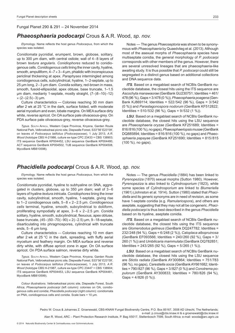
Conidiomata pycnidial, erumpent, brown, globose, solitary, upto300µmdiam,withcentralostiole;wallof4–8layersofbrown textura angularis.Conidiophores reduced to conidio-genouscells.Conidiogenous cells lining the inner cavity, hyaline, smooth,ampulliform,4–7×3–4µm,phialidicwithinconspicuouspericlinalthickeningatapex.Paraphyses intermingled among conidiogenouscells,subcylindrical,hyaline,1–2-septate,upto25µmlong,2–3µmdiam.Conidia solitary, red-brown in mass, smooth,fusoid-ellipsoidal,apexobtuse,basetruncate,1–1.5µmdiam,medianly1-septate,mostlystraight,(7–)8–10(–12)×(2–)2.5(–3)µm. Culturecharacteristics—Colonies reaching30mmdiamafter2wkat25°Cinthedark,surfacefolded,withmoderateaerialmyceliumandeven,lobatemargins.OnMEAsurfacedirtywhite,reverseapricot.OnOAsurfacepaleolivaceous-grey.OnPDAsurfacepaleolivaceous-grey,reverseolivaceous-grey.
Typus. South africa,WesternCapeProvince,Knysna,GardenRouteNationalPark,Velbroeksdraaipicnicsite,DiepwalleForest,S33°56'E23°09',on leaves of Podocarpus latifolius (Podocarpaceae), 1 July 2013,A.R. Wood(holotypeCBSH-21986,cultureex-typeCPC23433=CBS138903;ITSsequenceGenBankKP004452,LSUsequenceGenBankKP004480,ACTsequenceGenBankKP004502,TUBsequenceGenBankKP004508,MycoBankMB810598).
Notes—ThegenusPhaeoseptoria was shown to be synony-mous with PhaeosphaeriabyQuaedvliegetal.(2013).Althoughmost of the asexual morphs of Phaeosphaeria species have multiseptate conidia, the general morphology of P. podocarpi correspondswithothermembersofthegenus.However,thereare several unresolved lineages that are phaeosphaeria-like awaitingstudy.ItisthuspossiblethatP. podocarpi could still be segregated in a distinct genus based on additional collections andDNAsequencedata. ITS.BasedonamegablastsearchofNCBIsGenBanknu-cleotidedatabase,theclosesthitsusingtheITSsequenceareAscochyta manawaorae(GenBankGU230751;Identities=461/ 478(96%),Gaps=3/478(0%)),Phaeosphaeria poagena (Gen-BankKJ869114;Identities=522/542(96%),Gaps=3/542(0%))andParastagonospora nodorum (GenBankKF512822; Identities=510/532(96%),Gaps=9/532(1%)). LSU.BasedonamegablastsearchofNCBIsGenBanknu- cleotidedatabase, theclosesthitsusing theLSUsequenceare Phaeosphaeria oryzae (GenBankKF251689;Identities=816/816(100%),nogaps),Phaeosphaeriopsis musae (GenBankDQ885894;Identities=816/816(100%),nogaps)andPhaeo- sphaeria papayae (GenBankKF251690;Identities=815/815(100%),nogaps).
FungalPlanet290&291–24November2014
Phaeosphaeria podocarpi Crous&A.R.Wood,sp. nov.
Phacidiella podocarpi Crous&A.R.Wood,sp. nov.
Colour illustrations.Velbroeksdraaipicnicsite,DiepwalleForest,SouthAfrica; Phaeosphaeria podocarpi (left column): colonies onOA, conidio-genous cells and conidia; Phacidiella podocarpi (rightcolumn):conidiomataonPNA,conidiogenouscellsandconidia.Scalebars=10µm.
PedroW.Crous&JohannesZ.Groenewald,CBS-KNAWFungalBiodiversityCentre,P.O.Box85167,3508ADUtrecht,TheNetherlands;e-mail:[email protected]&[email protected]
AlanR.Wood,ARC–PlantProtectionResearchInstitute,P.BagX5017,Stellenbosch7599,SouthAfrica;e-mail:[email protected]
Etymology.NamereflectsthehostgenusPodocarpus, from which the species was isolated.
Conidiomata pycnidial, hyaline to subhyaline on SNA, aggre-gated inclusters,globose,up to350µmdiam;wallof3–6layers of hyaline textura intricata.Conidiophores lining the inner cavity, subcylindrical, smooth, hyaline, 1-septate, giving rise to1–2conidiogenouscells,5–8×2–2.5µm.Conidiogenous cells terminal, hyaline, smooth, subcylindrical to doliiform, proliferatingsympodiallyatapex,3–6×2–2.5µm.Conidia solitary, hyaline, smooth, subcylindrical, flexuous, apex obtuse, basetruncate,(45–)50–70(–90)×2(–2.5)µm,9–18-septate,disarticulating into phragmospores, cylindrical with truncate ends,5–6µmlong. Culture characteristics — Colonies reaching 10 mm diam after 2wkat 25°C in the dark, spreading,with fluffy aerialmyceliumandfeatherymargin.OnMEAsurfaceandreversedirtywhite,withdiffuseapricotzoneinagar.OnOAsurfaceapricot.OnPDAsurfacesalmon,reversedirtywhite.
Typus. South africa,WesternCapeProvince,Knysna,GardenRouteNationalPark,Velbroeksdraaipicnicsite,DiepwalleForest,S33°56'E23°09',on leaves of Podocarpus latifolius (Podocarpaceae), 1 July 2013,A.R. Wood(holotypeCBSH-21987,cultureex-typeCPC23447=CBS138904;ITSsequenceGenBankKP004453,LSUsequenceGenBankKP004481,MycoBankMB810599).
Notes—ThegenusPhacidiella(1884)hasbeenlinkedtoPyrenopeziza(1870)sexualmorphs(Sutton1980).However,Pyrenopeziza is also linked to Cylindrosporium(1823),whilesome species of Cylindrosporium are linked to Blumeriella (1961)(Johnstonetal.1914).Sutton(1980)statedthatPhaci-diella and its generic synonyms are in need of revision, as some have1-septateconidia(e.g.Ramulariospora),andothersareaseptate,suggestingthattheymaynotallbecongeneric.Phaci-diella podocarpi is thus best described in the genus Phacidiella basedonitshyaline,aseptateconidia. ITS.BasedonamegablastsearchofNCBIsGenBanknu- cleotide database, the closest hits using the ITS sequenceare Glomerobolus gelineus(GenBankDQ247782;Identities= 232/248(94%),Gaps=6/248(2%)),Caloplaca alboprui nosa (GenBankEF093566;Identities=240/260(92%),Gaps=3/ 260(1%))andUmbilicaria mammulata (GenBankDQ782851; Identities=243/265(92%),Gaps=5/265(1%)). LSU.BasedonamegablastsearchofNCBIsGenBanknu- cleotidedatabase, theclosesthitsusing theLSUsequenceare Stictis radiata (GenBankAY300864;Identities=751/783(96%),nogaps),Carestiella socia (GenBankAY661682;Identi-ties=790/827(96%),Gaps=3/827(0%))andConotrema po- pulorum (GenBankAY300833; Identities= 780/826 (94%),Gaps=4/826(0%)).

234 Persoonia–Volume33,2014
Phellinocrescentia guianensis

235Fungal Planet description sheets
© 2014 Naturalis Biodiversity Center & Centraalbureau voor Schimmelcultures
FungalPlanet292–24November2014
Phellinocrescentia Crous & Decock, gen. nov. Etymology. L. =crescit, growing on, referring to its ecological habit, growing on Phellinus.
Conidiomata pycnidial, globose, solitary or aggregated, uni- to multilocular,withcentralostiole;wallconsistingof3–6layersofbrown textura angularis; outer surface covered in brown, warty hyphae.Conidiophores reduced to conidiogenous cells or a sup-portingcell.Conidiogenous cells lining the inner cavity, hyaline,
Etymology.Namereflectsthelocality,FrenchGuiana,wherethisspecieswascollected.
Conidiomata pycnidial, globose, solitary or aggregated, uni- to multilocular, up to 220 µm diam, with central ostiole; wall consistingof3–6layersofbrowntextura angularis, becoming hyaline towards inner centrum; outer surface covered in brown, wartyhyphae,3–4µmdiam.Conidiophores reduced to conidio-genouscellsorasupportingcell.Conidiogenous cells lining the inner cavity, hyaline, smooth, tightly aggregated, subcylindrical, straighttocurved,phialidicwithpericlinalthickening,5–12× 1.5µm.Conidia aseptate, solitary, hyaline, smooth, guttulate, thin-walled,ellipsoidtotear-drop-shaped,(2.5–)3(–4)×1.5µm. Culturecharacteristics—Coloniesreaching7mmdiamafter2wkat25°C in thedark,surfacefolded,withsparseaerialmyceliumandeven,lobedmargins.OnMEAsurfaceamixtureofdirtywhiteandolivaceous-grey,reverseolivaceous-grey.OnOAandPDAolivaceous-grey.
Typus. french Guiana,onpolyporeNo.742(Phellinussp.),12July2013, C. Decock(holotypeCBSH-21988,cultureex-typeCPC23600=CBS138913; ITSsequenceGenBankKP004454,LSUsequenceGenBankKP004482,MycoBankMB810601).
smooth, tightly aggregated, subcylindrical, straight to curved, phialidicwith periclinal thickening.Conidia aseptate, soli- tary, hyaline, smooth, guttulate, thin-walled, ellipsoid to tear- drop-shaped.
Type species.Phellinocrescentia guianensis. MycoBankMB810600.
Phellinocrescentia guianensis Crous & Decock, sp. nov. Notes—Thestrainwasfoundgrowingonthesporocarpofa Phellinussp.andisolatedbyplatingitonmaltagar.Phel-linocrescentia guianensis is phoma-like in morphology, but distinct in having solitary or aggregated, uni- to multilocular conidiomata, and ellipsoid to tear-drop-shaped conidia. Italsohasauniqueecologicalhabit,growingonbasidiocarpsof a Phellinussp.Itwasnotpossibletoassignagenustothisfungus based on phylogenetic inference and the new genus, Phellinocrescentia,isintroducedtoaccommodateit. ITS.BasedonamegablastsearchofNCBIsGenBanknu- cleotidedatabase,theclosesthitsusingtheITSsequenceareDidymosphaeria futilis (GenBankEU552123;Identities=470/ 574(82%),Gaps=38/574(6%)),Funbolia dimorpha (Gen-BankJF951136;Identities=362/424(85%),Gaps=15/424(3%))andGeomyces pannorum var. asperulatus (GenBankAJ938166;Identities=329/395(83%),Gaps=25/395(6%)). LSU.BasedonamegablastsearchofNCBIsGenBanknu- cleotidedatabase, theclosesthitsusing theLSUsequenceare Pseudopassalora gouriqua (GenBankJN712565;Identities= 743/790 (94%), nogaps),Heleiosa barbatula (GenBankGU479787;Identities=735/793(93%),Gaps=5/793(0%))and Funbolia dimorpha (GenBankJF951156;Identities=729/790(92%),Gaps=1/790(0%)).
PedroW.Crous&JohannesZ.Groenewald,CBS-KNAWFungalBiodiversityCentre,P.O.Box85167,3508ADUtrecht,TheNetherlands;
e-mail:[email protected]&[email protected],Mycothèquedel’UniversitécatholiquedeLouvain(MUCL,BCCMTM),EarthandLifeInstitute–ELIM–Mycology,
UniversitécatholiquedeLouvain,CroixduSud2bteL7.05.06,B-1348Louvain-la-Neuve,Belgium;e-mail:[email protected]
Colour illustrations.BasidiomataofPhellinussp.onadeadstandingtreeinFrenchGuiana;conidiomatainagar,wartyhyphae,conidiogenouscellsandconidia.Scalebars=10µm.

236 Persoonia–Volume33,2014
Neocamarosporium chichastianum

237Fungal Planet description sheets
© 2014 Naturalis Biodiversity Center & Centraalbureau voor Schimmelcultures
Etymology.Namereflectsthelocation,LakeUrmia(formerlyknownasChichast),fromwhichthespecieswasisolated.
Conidiomata pycnidial, solitary, uniloculate, black, up to 200 µmdiam,with1–3papillatenecks(upto150µmdiam),withcentralostioles5–10µmdiam.Conidiophores reduced to co-nidiogenouscells.Conidiogenous cells lining the inner cavity, hyaline,smooth,subcylindrical,7–15×4–5µm,proliferatingpercurrentlyatapex.Paraphyses intermingled between coni-diogenouscells,hyaline,smooth,subcylindrical,1–2-septatewithobtuseends,upto35µmlong,4–5µmdiam.Conidia solitary,brown,finelyroughened,ellipsoid,widest inmiddle,apex obtuse, muriformly septate, thick-walled, base truncate, 2–3µmdiam,(11–)15–19(–22)×(6–)8–9(–11)µm;3trans-versesepta,1–2obliqueorverticalsepta. Culture characteristics — Colonies flat, spreading with sparse aerialmycelia.OnOAsurfaceolivaceous-grey.OnMEAsur-facepaleolivaceous-greytoolivaceous-grey.Optimumgrowthoccurredat25°C,butthefungusgrewat15°Cupto35°C.Furthermore,optimumgrowthwasrecordedatpHvaluesbe-tween5.5and6.5,althoughitcouldgrowatabroadrangeofpHvalues(4–10).
Typus.iran,LakeUrmia,soil,2011,M. Papizadeh & M.R. Soudi(holo- typeCBSH-21989,cultureex-typeIBRC-M30126=CBS137502;ITSse- quenceGenBankKP004455,LSUsequenceGenBankKP004483,Myco-BankMB810602).
Notes — Neocamarosporium chichastianum clusters with N. goegapense, the type species of the genus Neocamaro-sporium, which is morphologically similar to the genus Camaro-sporium based on its pycnidial conidiomata, hyaline, percurrently proliferating conidiogenous cells, and brown, muriformly septate conidia(Crousetal.2014).Neocamarosporium chichastianum is the second species described in this genus, and interestingly has paraphyses, which were not observed in N.goegapense. ITS.BasedonamegablastsearchofNCBIsGenBanknu- cleotidedatabase,theclosesthitsusingtheITSsequenceareNeocamarosporium goegapense(GenBankKJ869163;Identi-ties=550/579(95%),Gaps=5/579(0%)),Phoma betae (Gen- BankKC460811;Identities=463/493(94%),Gaps=7/493(1%))andAscochyta obiones (GenBankGU230752;Identities=463/498(93%),Gaps=4/498(0%)).OurITSsequenceis98%(517/529)identicaltothesequenceofChaetosphaerone-ma hispidulum CBS826.88inQ-bank(www.q-bank.eu). LSU.BasedonamegablastsearchofNCBIsGenBanknu- cleotidedatabase, theclosesthitsusing theLSUsequenceare Neocamarosporium goegapense (GenBankKJ869220;Identities=804/806(99%),nogaps),Chaetosphaeronema his-pidulum (GenBankEU754145;Identities=848/851(99%),nogaps)andConiothyrium obiones (GenBankDQ678054;Iden- tities=849/853(99%),nogaps).
FungalPlanet293–24November2014
Neocamarosporium chichastianum Papizadeh,Crous,ShahzadehFazeli &Amoozegar,sp. nov.
PedroW.Crous&JohannesZ.Groenewald,CBS-KNAWFungalBiodiversityCentre,P.O.Box85167,3508ADUtrecht,TheNetherlands;
e-mail:[email protected]&[email protected],SeyedAbolhassanShahzadehFazeli&MohammadAliAmoozegar,
MicroorganismsBank,IranianBiologicalResourceCenter(IBRC),AcademicCenterforEducation,Culture&Research(ACECR)Tehran,Iran e-mail: [email protected], [email protected]&[email protected]
Colour illustrations.LakeUrmiainIran;conidiomataonOAandPNA,conidiomatalneck,conidiogenouscellsandconidia.Scalebars=10µm.
238 Persoonia–Volume33,2014
Sphaerulina pelargonii

239Fungal Planet description sheets
© 2014 Naturalis Biodiversity Center & Centraalbureau voor Schimmelcultures
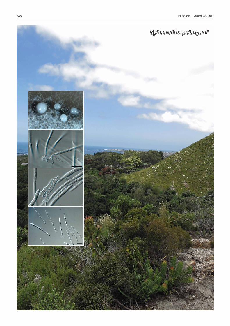
Etymology.NamereflectsthehostgenusPelargonium, from which this specieswasisolated.
SporulatingonSNA.Conidiomata pycnidial, brown, separate, immersed toerumpent,globose,up to150µmdiam,exud-ing a creamy crystalline conidial mass via a central ostiole; wallof3–4layersofbrowntextura angularis.Conidiophores reduced to conidiogenous cells.Conidiogenous cells hya-line, at times pale brown, smooth, subcylindrical, straight to geniculous-sinuous,7–15×3–5µm,proliferatingsympodially.Conidiahyaline,smooth,guttulate,filiform,narrowlyobclavate,apex subobtusely rounded, base long obconically truncate, (1–)3–4-septate,(15–)28–45(–60)×1.5–2(–2.5)µm. Culture characteristics — Colonies on PDA, MEA and OA spreading with sparse to moderate aerial mycelium, and smooth, lobatemargins,reaching20mmdiamafter2wkat25°Cinthedark.OnMEAsurfaceolivaceous-greywithapricotsporu-lation,iron-greyunderneath.OnPDAsurfaceolivaceous-greywithstrandsofdirtywhitemycelium,reverseiron-grey.OnOAsurfacedirtywhite.
Typus. South africa,WesternCapeProvince,Betty’sBay,HaroldPorterNationalBotanicalGarden,on leavesofPelargoniumsp. (Geraniaceae),15Jan.2014,P.W. Crous & M.J. Wingfield(holotypeCBSH-21990,cultureex-typeCPC24151=CBS138857; ITSsequenceGenBankKP004456,LSUsequenceGenBankKP004484,TEFsequenceGenBankKP004506,MycoBankMB810603).
Notes—ThegenusSphaerulina was shown to have septo-ria-likeasexualmorphsbyQuaedvliegetal.(2013),severalofwhichwereeitherendophytesorimportantplantpathogens.Al-though Sphaerulina pelargonii was associated with small, brown leaf spots on Pelargonium, inoculation studies have not been conductedtoconfirmitspathogenicity.Thisisthefirstspeciesof Sphaerulinareportedonthishost.SimilartaxareportedfromPelargonium include Septoria pelargonii(conidia3–5-septate,40–60×2–2.5µm),Septoria canberrica(conidia1–3-septate,12–30(–36)×1.5–2µm),Septoria geranii(conidia35–50× 1µm)andSeptoria geranii-nodosi (conidia50–65×2µm).Species of Septoria that are known from culture were recently treatedindetailbyVerkleyetal.(2013). ITS.BasedonamegablastsearchofNCBIsGenBanknu-cleotidedatabase,theclosesthitsusingtheITSsequenceareSphaerulina rhododendricola (GenBankKF777187;Identities=600/614(98%),Gaps=8/614(1%)),Mycosphaerella ribis (Gen- BankEU167588;Identities=634/649(98%),Gaps=5/649(0%)) and Pseudocercosporella chaenomelis (GenBankJQ793663;Identities=573/587(98%),Gaps=8/587(1%)). LSU.BasedonamegablastsearchofNCBIsGenBanknu-cleotidedatabase,theclosesthitsusingtheLSUsequenceareSphaerulina rhododendricola (GenBankKF779493;Identities=834/836(99%),nogaps),Pseudocercosporella chaenomelis (GenBankGU253834;Identities=826/828(99%),nogaps)and Sphaerulina azaleae (GenBankKF252105;Identities=823/ 825(99%),nogaps). TEF.BasedonamegablastsearchofNCBIsGenBanknu- cleotidedatabase, theclosesthitsusing theTEFsequenceare Sphaerulina rhabdoclinis(GenBankKF253578;Identities =344/382(90%),Gaps=9/382(2%)),Sphaerulina amelan-chier (GenBankKF253545;Identities=344/382(90%),Gaps=9/382(2%))andSphaerulina menispermi (GenBankKF253565; Identities=343/381(90%),Gaps=10/381(2%)).
FungalPlanet294–24November2014
Sphaerulina pelargonii Crous&M.J.Wingf.,sp. nov.
Colour illustrations.HaroldPorterNationalBotanicalGarden,Betty’sBay,SouthAfrica;conidiomataonOA,conidiogenouscellsandconidia.Scalebars=10µm.
PedroW.Crous&JohannesZ.Groenewald,CBS-KNAWFungalBiodiversityCentre,P.O.Box85167,3508ADUtrecht,TheNetherlands;
e-mail:[email protected]&[email protected],ForestryandAgriculturalBiotechnologyInstitute(FABI),UniversityofPretoria,
P.BagX20,Pretoria,0028,SouthAfrica;e-mail:[email protected]

240 Persoonia–Volume33,2014
Xenosonderhenia eucalypti& Zasmidium eucalyptigenum

241Fungal Planet description sheets
© 2014 Naturalis Biodiversity Center & Centraalbureau voor Schimmelcultures
Etymology.NamereflectsthehostgenusEucalyptus, from which this specieswasisolated.
Leaf spotsamphigenous,darkbrown,10–20mmdiam,withdarkbrownborder.Co-occurringonleafspotswithZasmidium eucalyptigenum.Ascomata hypophyllous, black erumpent, globose, solitary, up to 110 µm diam, with central ostiole; wall of 2–3 layers of brown textura angularis.Asci fasciculate, bitunicate,subsessile,hyaline,smooth,8-spored,obovoid toellipsoid, aparaphysate, straight to slightly curved, 35–45× 10–12µm.Pseudoparaphysesabsent.Ascospores tri- to multi-seriate, hyaline, smooth, fusoid-ellipsoid, widest in apical cell, one third from apex, tapering towards both ends, not constricted atmedianseptum,(17–)18–20(–22)×(3–)4µm;ascosporesgerminating from both ends, not constricting or distorting, re-maininghyaline,4–5µmdiam;germtubesdevelopingnumer-ouslateralbranches. Culture characteristics — Colonies spreading, erumpent with moderate aerial mycelium and smooth, lobate margins, reaching20mmdiamonPDA,MEAandOAafter2wkat25°Cinthedark.OnMEAsurfacepaleluteuswithpatchesofdirtywhite,reversesienna.OnOAsurfacesaffron.OnPDAsurfacesaffron,reversepaleluteus.
Typus. moZambique, Forestas de Niassa, leaf spots of Eucalyptus urophylla (Myrtaceae),2Feb.2014,M.J. Wingfield(holotypeCBSH-21991,cultureex-typeCPC24247=CBS138858; ITSsequenceGenBankKP004457,LSUsequenceGenBankKP004485,MycoBankMB810604).
Notes — Xenosonderhenia eucalypti appears to represent an undescribed genus in the Mycosphaerellaceae.Itclusterswith‘Mycosphaerella’elaeocarpi that lacks an asexual morph and Xenosonderhenia syzygii, which lacks a sexual morph (Crousetal.2012a).Becausethistaxonisclearlynotaspeciesof Mycosphaerellas.str.,whichhasRamularia asexual morphs (Verkleyetal.2004,Crousetal.2009b),wetentativelyplaceit in the genus Xenosonderhenia,pendingfurthercollections. ITS.BasedonamegablastsearchofNCBIsGenBanknu- cleotide database, the closest hits using the ITS sequenceare Xenosonderhenia syzygii (GenBankJX069872;Identities =506/525(96%),Gaps=3/525(0%)),Mycosphaerella elon-gata (GenBankEF394833;Identities=492/520(95%),Gaps= 4 /520 (0%)) andMycosphaerella elaeocarpi (GenBankEU040212;Identities=514/547(94%),Gaps=7/547(1%)).
FungalPlanet295&296–24November2014
Xenosonderhenia eucalypti Crous&M.J.Wingf.,sp. nov.
Zasmidium eucalyptigenum Crous&M.J.Wingf.,sp. nov.
Colour illustrations.LeafspotsonEucalyptus urophylla.Xenosonderhe-nia eucalypti (leftcolumn):leafspot,asciwithascosporesandgerminatingascospores; Zasmidium eucalyptigenum(rightcolumn):leafspots,conidio-phoreandconidia,ascosporesandgerminatingascospores.Scalebars=10µm.
PedroW.Crous&JohannesZ.Groenewald,CBS-KNAWFungalBiodiversityCentre,P.O.Box85167,3508ADUtrecht,TheNetherlands;e-mail:[email protected]&[email protected]
MichaelJ.Wingfield,ForestryandAgriculturalBiotechnologyInstitute(FABI),UniversityofPretoria,P.BagX20,Pretoria,0028,SouthAfrica;e-mail: [email protected]
Etymology.NamereflectsthehostgenusEucalyptus, from which this spe- cieswasisolated.
Co-occurring on leaf spots with Xenosonderhenia eucalypti.Ascomata hypophyllous, black erumpent, globose, solitary, upto100µmdiam,withcentralostiole;wallof2–3layersofbrown textura angularis.Asci fasciculate, bitunicate, subsessile, hyaline,smooth,8-spored,obovoidtoellipsoid,aparaphysate,straighttoslightlycurved,25–40×8–10µm.Pseudoparaphyses absent.Ascospores hyaline, smooth, fusoid-ellipsoid, widest in middle of apical cell, tapering towards both ends, constricted atmedianseptum,13–16×(2.5–)3.5–4µm;ascosporesger-minating from both ends, becoming constricted, but remaining hyalineandsmooth,4–6µmdiam,developinglateralbranches.Mycelium brown, verruculose, typical of Zasmidium asexual morph.Conidiophoresbrown,verruculose,solitaryonsuperficialhyphae,erect,branchedornot,upto50µmtall,3–4µmdiam,1–3-septate.Conidiogenous cells terminal or intercalary, with severalthickened,darkened,refractivescars,1µmdiam.Coni-dia brown, verruculose, straight to curved, solitary or in branched chains, subcylindrical, apex obtuse, base tapering to a truncate hilum,1–1.5µmdiam,1–9-septate,30–120×(2.5–)3µm.
Culture characteristics — Colonies erumpent, spreading, folded, with mucoid exudate and sparse to moderate aerial mycelium,andsmooth, lobedmargins, reaching3cmdiamafter2wkat25°Cinthedark.Cultureissterile..OnOAsurfaceolivaceous-grey.OnMEAsurfaceolivaceous-greywithpatchesofpaleolivaceous-grey.OnPDAsurfaceolivaceous-greywithpatchesofpaleolivaceous-grey,iron-greyinreverse.
Typus. moZambique, Forestas de Niassa, leaf spots of Eucalyptus urophylla (Myrtaceae),2Feb.2014,M.J. Wingfield(holotypeCBSH-21992,cultureex-typeCPC24251=CBS138860; ITSsequenceGenBankKP004458,LSUsequenceGenBankKP004486,MycoBankMB810605).
Notes—ThegenusMycosphaerellaispolyphyletic(Crousetal.2007a),andZasmidium is the oldest name to accommodate stenella-like taxa clustering in the Mycosphaerellaceae(Arzan-louetal.2007).Severalspecieshavethusfarbeendescribedfrom Eucalyptus(Crousetal.2009a,b,Braunetal.2010),allof which are phylogenetically distinct from Z. eucalyptigenum. ITS.BasedonamegablastsearchofNCBIsGenBanknu- cleotide database, the closest hits using the ITS sequenceare Zasmidium rothmanniae (GenBankKJ869135;Identities=461/480(96%),Gaps=7/480(1%)),Periconiella arcuata (Gen- BankEU041779;Identities=427/449(95%),Gaps=6/449(1%))andMycosphaerella pseudovespa (GenBankDQ530216; Identities=432/457(95%),Gaps=6/457(1%)).

242 Persoonia–Volume33,2014
Diaporthe acaciarum

243Fungal Planet description sheets
© 2014 Naturalis Biodiversity Center & Centraalbureau voor Schimmelcultures
Etymology.NamereflectsthehostgenusAcacia, from which this species wasisolated.
SporulatingonPNA.Conidiomatapycnidial,globose,upto300µm diam, black, erumpent, exuding creamy conidial droplets fromcentral ostioles;walls consisting of 3–6 layers ofme-dium brown textura angularis.Conidiophores hyaline, smooth, 2–3-septate,branched,denselyaggregated,cylindrical,straighttosinuous,20–30×2.5–4µm.Conidiogenous cells15–25× 2–3µm,phialidic,cylindrical,terminalandintercalary,withslightapicaltaper,1–1.5µmdiam,withvisiblepericlinalthickenening;collarettenotflared,upto2µmlong.Paraphysesnotobserved.Alpha conidia(6–)6.5–7(–7.5)×(2–)2.5(–3)µm,aseptate,hya-line, smooth, bi-guttulate, fusoid-ellipsoid, tapering towards both ends,straightapexsubobtuse,basesubtruncate,1µmdiam.Beta conidia spindle-shaped, aseptate, smooth, hyaline, apex acutely rounded, base truncate, tapering from the lower third towardsthebase,(20–)25–35(–40)×1.5(–2)µm. Culture characteristics — Colonies covering the dish after 2wkat25°Cinthedark,withsparsetomoderateaerialmy-celium.OnMEA,PDAandOAsurfacedirtywhitewithpatchesofgrey-olivaceous,reversedirtywhitewithpatchesofsienna.
Typus. tanZania, Serengeti, on thorns of Acacia tortilis(Fabaceae),Feb.2014, M.J. Wingfield(holotypeCBSH-21994,cultureex-typeCPC24324=CBS138862;ITSsequenceGenBankKP004460,LSUsequenceGenBankKP004488,HISsequenceGenBankKP004504,TUBsequenceGenBankKP004509,MycoBankMB810606).
Notes — No Diaporthe(incl.Phomopsis)specieshavebeendescribed from Acacia tortilis(Uecker1988,Gomesetal.2013). Phylogenetically, D. acaciarum is closely related to several speciesbasedonITS(seebelow),butitcanbedistinguishedfromthembasedonTUBsequencedata. ITS.BasedonamegablastsearchofNCBIsGenBanknu- cleotidedatabase,theclosesthitsusingtheITSsequenceare Phomopsis chimonanthi (GenBankKF746059;Identities=505/ 513(98%),Gaps=5/513(0%)),Diaporthe helianthi (GenBankJQ936257;Identities=546/556(98%),Gaps=1/556(0%))and Diaporthe infecunda (GenBankKF939614;Identities=525/ 536(98%),Gaps=4/536(0%)). LSU.BasedonamegablastsearchofNCBIsGenBanknu- cleotidedatabase,theclosesthitsusingtheLSUsequenceareDiaporthe leucospermi (GenBankJN712524;Identities=836/ 839(99%),nogaps),Taeniolella alta (GenBankDQ377938;Iden- tities=834/839(99%),nogaps)andDiaporthe arctii (GenBankAF362562;Identities=831/836(99%),nogaps). HIS.ClosesthitsusingtheHISsequencehadhighestsimi-larity to Diaporthe infecunda (GenBankKC343613;Identities=359/369(97%),Gaps=1/ 369(0%)),Diaporthe terebinthifolii (GenBankKC343702;Identities=353/371(95%),Gaps=6/371 (1%))andDiaporthe melonis (GenBankKC343626;Identities=351/373(94%),Gaps=5/373(1%)). TUB.ClosesthitsusingtheTUBsequencehadhighestsimi-larity to Diaporthe infecunda (GenBankKF939619;Identities=753/783(96%),Gaps=4/783(0%)),Diaporthe beilharziae (Gen- BankKF170921;Identities=743/777(96%),Gaps=1/777(0%))andDiaporthe terebinthifolii (GenBankKC344186;Identi-ties=662/708(94%),Gaps=1/708(0%)).
FungalPlanet297–24November2014
Diaporthe acaciarumCrous&M.J.Wingf.,sp. nov.
PedroW.Crous&JohannesZ.Groenewald,CBS-KNAWFungalBiodiversityCentre,P.O.Box85167,3508ADUtrecht,TheNetherlands;
e-mail:[email protected]&[email protected],ForestryandAgriculturalBiotechnologyInstitute(FABI),UniversityofPretoria,
P.BagX20,Pretoria,0028,SouthAfrica;e-mail: [email protected]
Colour illustrations.LeopardinAcacia tortilistreeintheSerengeti,Tan-zania;conidiomataonPNA,conidiogenouscells,alphaandbetaconidia.Scalebars=10µm.
244 Persoonia–Volume33,2014
Melanconium hedericola

245Fungal Planet description sheets
© 2014 Naturalis Biodiversity Center & Centraalbureau voor Schimmelcultures
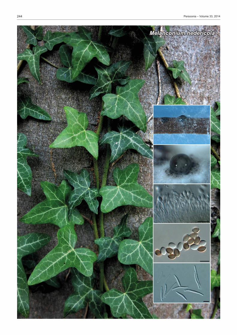
Etymology.NamereflectsthehostgenusHedera, from which this species wasisolated.
Conidiomata pycnidial single or in densely crowded groups under loosenedbark, superficial on a black stromatic layerwhich continues as a black stromatic line deep in the wood, ± pyroid with flattened base, black, rough, soft, distinctly thick, ostiolum central and indistinct; periphyses not seen, with a few setaeontheoutersiteof theperidium.Setae 1-celled, stiff, pointed, basally enlarged and flattened, brown, thick-walled, smooth,eguttulate,upto26µmlong.Peridium multi-layered, consisting of a red-brown textura epidermoidea(outerlayer)and hyaline textura angularis-prismatica (inner layer), cellsthick-walled,smoothandeguttulate.SporulatingonPNA.Coni- diomata pycnidial, globose, up to 600µmdiam, black, im-mersed, exuding creamy conidial droplets from central ostioles, or developing1–4black necks onOA (not onPNA);wallsconsistingof3–6layersofmediumbrowntextura angularis.Conidiophoreshyaline,smooth,1–2-septate,branchedbelow,denselyaggregated,cylindrical,straighttosinuous,20–40× 3–4µm.Conidiogenous cells10–17×2–3µm,phialidic,cy-lindrical, terminal and intercalary, with slight apical taper, 2 µm diam, with visible periclinal thickening; collarette flared, up to 5µmlong.Paraphysesnotobserved.Alpha conidia(6–)7(–8)×(3.5–)4(–4.5)µm,aseptate,hyaline,smooth,ellipsoidwithlargecentralguttule,becomingbrownwithage.Beta conidia spindle-shaped, aseptate, smooth, hyaline, guttulate, apex bluntly rounded, base truncate, tapering from the middle to-wardsthebase,(17–)18–20(–25)×2.5(–3)µm. Culture characteristics — Colonies flat, spreading, cover-ingthedishafter2wkat25°Cinthedark,withsparseaerialmycelium.OnMEA,PDAandOAsurfaceumberwithpatchesof dirty white and iron-grey; reverse umber with patches of dirty whiteandiron-grey.
Typus. Spain,Sarasibar(Navarra),onbranchofHedera helix(Araliaceae),26Jan.2014,S. Garcia(holotypeCBSH-21995,cultureex-typeCPC24278=CBS138863;ITSsequenceGenBankKP004461,LSUsequenceGenBankKP004489,HISsequenceGenBankKP004505,TUBsequenceGenBankKP004510,MycoBankMB810607).
Notes — Although Melanconium hedericola clusters within the genus Diaporthe, theLSUregion lacksresolutionwithinthe Diaporthales. WehavethuschosentodescribeitinMe-nanconium based on the ellipsoid alpha conidia that turn brown with age, and their characteristic large, central guttules (Sutton1980).However,Melanconium is known to not have species with beta conidia, which suggests that M. hedericola mightrepresentanunknowngenuswithinthiscomplex.Fur-therstudiesandcollectionsarerequiredbeforethisquestioncanberesolved. Previously published taxa on Hedera include Coniothyrium hederae and its possible synonym, Melanconium hederae,whichhavesimilaralphaconidia(6–8×4.5–6µm),butthatlackbetaconidia. ITS.BasedonamegablastsearchofNCBIsGenBanknu- cleotidedatabase,theclosesthitsusingtheITSsequencearePhomopsis columnaris(GenBankFN394688;Identities=528/ 541(98%),Gaps=2/541(0%)),Diaporthe endophytica (Gen- BankAB899789;Identities=566/583(97%),Gaps=7/583(1%))andDiaporthe phaseolorum (GenBankJQ936148;Iden-tities=565/583(97%),Gaps=7/583(1%)). LSU.BasedonamegablastsearchofNCBIsGenBanknu- cleotidedatabase,theclosesthitsusingtheLSUsequencearePhomopsis columnaris (GenBankAF439627;Identities=834/ 840(99%),nogaps),Diaporthe ambigua (GenBankJQ862833; Identities=821/828(99%),nogaps)andPhomop sis sclero-tioides (GenBankAF439628;Identities=831/840(99%),nogaps). HIS.BasedonamegablastsearchofNCBIsGenBanknu-cleotidedatabase,theclosesthitsusingtheHISsequenceareDiaporthe longispora(GenBankKC343619;Identities=360/ 378(95%),Gaps=5/378(1%)),Diaporthe sclerotioides (Gen-BankKC343678;Identities=359/381(94%),Gaps=4/381(1%))andDiaporthe ‘sp.4’ (GenBankKC343690;Identities=356/378(94%),Gaps=5/378(1%)). TUB.BasedonamegablastsearchofNCBIsGenBanknu- cleotidedatabase,theclosesthitsusingtheTUBsequenceareDiaporthe longispora(GenBankKC344103;Identities=398/ 420(95%),Gaps=2/420(0%)),Diaporthe sclerotioides (Gen-BankKC344161;Identities=391/416(94%),Gaps=2/416(0%))andDiaporthe scabra (GenBankHQ450372;Identities=418/450(93%),Gaps=2/450(0%)).
FungalPlanet298–24November2014
Melanconium hedericola Crous&R.K.Schumach.,sp. nov.
PedroW.Crous&JohannesZ.Groenewald,CBS-KNAWFungalBiodiversityCentre,P.O.Box85167,3508ADUtrecht,TheNetherlands;
e-mail:[email protected]&[email protected]éK.Schumacher,Hölderlinstraße25,15517Fürstenwalde/Spree,Germany;e-mail:[email protected]
Colour illustrations.Hedera helix growing along a tree trunk; conidiomata onPNAandOA,conidiogenouscells,alphaandbetaconidia.Scalebars=10µm.

246 Persoonia–Volume33,2014
Neophysalospora eucalypti

247Fungal Planet description sheets
© 2014 Naturalis Biodiversity Center & Centraalbureau voor Schimmelcultures
Etymology.Namereflectsthefactthatthegenusismorphologicallysimilarto the genus Physalospora.
Endophyticandplantpathogenic. Ascomata globose, solitary, brown,immersed,withcentralostiole;wallof2–3layersofbrowntextura angularis.Asci cylindrical, hyaline, stipitate, unitunicate withapicalmechanismstaininginMelzer’sreagent,ascosporesuniseriatebutoverlapping,with8ascosporesperascus.Para-physes intermingledamongasci,hyaline,septate,branched.Ascospores hyaline, smooth, guttulate, fusoid-ellipsoid, with acutelyroundedends.Conidiomata globose, solitary to aggre-
gated,brown,withcentralostiole;wallof2–3layersofbrowntextura angularis.Conidiophores lining the inner conidiomatal wall, subcylindrical, straight to curved, branched, septate, or reduced to conidiogenous cells, hyaline to pale brown, smooth orfinelyverruculose.Conidiogenous cells ampulliform to sub-cylindrical, hyaline, smooth, terminal or intercalary, phialidic with flaredcollaretteattheapex.Conidia solitary, hyaline, smooth, subcylindrical, curved, with obtuse apex and truncate base, thick-walled.
Type species.Neophysalospora eucalypti. MycoBankMB810608.
FungalPlanet299–24November2014
Neophysalospora Crous&M.J.Wingf.,gen. nov.
Neophysalospora eucalypti Crous&M.J.Wingf.,sp. nov.
PedroW.Crous&JohannesZ.Groenewald,CBS-KNAWFungalBiodiversityCentre,P.O.Box85167,3508ADUtrecht,TheNetherlands;
e-mail:[email protected]&[email protected],ForestryandAgriculturalBiotechnologyInstitute(FABI),UniversityofPretoria,
P.BagX20,Pretoria,0028,SouthAfrica;e-mail:[email protected]
Colour illustrations.Corymbia henryileavesinMozambique;conidiomataon SNA, asci and ascospores, conidiogenous cells, germinating ascospores andconidia.Scalebars=10µm.
Etymology.NamereflectsthehostgenusEucalyptus, from which this specieswasisolated.
Associated with brown leaf spots in plantations, and cutting rot innurseries.Ascomataglobose,solitary,brown,upto250µmdiam,withcentralostiole,upto80µmdiam;wallof2–3layersof brown textura angularis.Asci cylindrical, hyaline, stipitate, unitunicatewithapicalmechanismstaininginMelzer’sreagent,ascosporesuniseriatebutoverlapping,with8ascosporesperascus,70–100× 6–8µm.Paraphyses intermingled among asci,hyaline,septate,branched,2–3µmdiam.Ascospores hyaline, smooth, guttulate, fusoid-ellipsoid, widest in middle, ta-peringtoacutelyroundedends,(13–)15–17(–19)×(4–)4.5(–5)µm.Ascospores germinate from one end, remain hyaline,fusoid-ellipsoid,15–17×7µm.Conidiomata globose, solitary to aggregated, brown, up to 200 µm diam, with central ostiole; wall of2–3layersofbrowntextura angularis.Conidiophores lining the inner conidiomatal wall, subcylindrical, straight to curved, branched,1–3-septate,orreducedtoconidiogenouscells,hya-linetopalebrown,smoothorfinelyverruculose,10–30×2.5–4µm.Conidiogenous cells ampulliform to subcylindrical, hyaline, smooth,terminalorintercalary,7–12×1.5–3µm,phialidicwithflaredcollaretteattheapex,1.5–2µmdiam.Conidia solitary, hyaline, smooth, subcylindrical, curved, with obtuse apex and truncatebase,thick-walled,(13–)14–15(–16)×1.5µm. Culture characteristics — Colonies spreading with sparse, appressedaerialmycelium,reaching6mmdiamafter2wkat25°Cinthedark;surfacefoldedwithfeathery,lobatemargin.On MEA surface flesh with patches of saffron, reverse red with patchesofcoral.OnOAsurfaceredwithpatchesofpeachandsaffron.OnPDAsurfacewhite,reversesaffron.
Typus. moZambique, on leaves of Corymbia henryi(Myrtaceae),1Feb.2014, M.J. Wingfield (holotypeCBSH-21996,cultureex-typeCPC24209=CBS138864;ITSsequenceGenBankKP004462,LSUsequenceGenBankKP004490,MycoBankMB810609).–South africa, Mpumalanga province, Piet Retief, forestry nursery, cutting rot of Eucalyptus grandis × camaldulensis, 9Jan.1989,P.W. Crous,CPC123=CBS110740(ITS,LSUsequenceGen-BankKP031106,KP031108),CPC124=CBS111123(ITS,LSUsequenceGenBankKP031107,KP031109)(specimenalsodepositedinIMI,nowKew).
Notes—ThegenusPhysalospora(Hyponectriaceae)ispoly- phyletic,andinneedoftaxonomicrevision.Neophysalospora eucalypti is reminiscent of Clypeophysalospora latitans and to some extent also Plectosphaera eucalypti (=Phyllachora eucalypti ).Clypeophysalospora latitans is a saprobe or weak pathogen that is commonly encountered on living eucalypt leavesandleaf litter(Crousetal.1990).Ithasamelanisedclypeus,andunitunicateascithatstraininMelzer’sreagent,anddoesnotproduceanasexualmorphinculture.Plectosphaera eucalypti has black ascomata embedded in the leaf tissue, with a melanised pseudoclypeus, forming circular to irregular colo-niesontheleafsurface.Ithasbitunicateasciandascosporeswitaprominentmucoidsheath.Infectionsareusuallyvisibleononlyonesideoftheleafsurface(Pascoe1990,Parketal.2000). ITS.BasedonamegablastsearchofNCBIsGenBanknu-cleotidedatabase,theclosesthitsusingtheITSsequenceareBagadiella lunata (GenBankGQ303269;Identities=586/625(94%),Gaps=15/625(2%)),Bagadiella koalae (GenBankJF951142;Identities=586/627(93%),Gaps=14/627(2%))andBagadiella victoriae (GenBankJF951141;Identities=584/627 (93%),Gaps=12/627(1%)). LSU.BasedonamegablastsearchofNCBIsGenBanknu- cleotidedatabase, theclosesthitsusing theLSUsequenceare Plectosphaera eucalypti (GenBankDQ923538;Identities=821/840(98%),Gaps=2/840(0%)),Bagadiella victoriae (GenBankJF951161;Identities=815/839(97%),nogaps)andBagadiella lunata (GenBankGQ303300;Identities=815/839(97%),nogaps).

248 Persoonia–Volume33,2014
Seimatosporium pistaciae

249Fungal Planet description sheets
© 2014 Naturalis Biodiversity Center & Centraalbureau voor Schimmelcultures
Etymology.NamereflectsthehostgenusPistacia, from which this spe-cieswasisolated.
Foliicolous.Conidiomata pycnidioid, separate to gregarious, becomingerumpent,ovaltoelongate,upto150µmdiam.Coni-diophoresarisingfromacentralstroma,hyaline,3–4-septate,branched, subcylindrical, 20–45× 3–4µm.Conidiogenous cells terminal and intercalary, hyaline, smooth, subcylindrical, straighttosomewhatcurved,10–15×2–2.5µm,proliferatinginconspicuouslypercurrentlyatapex.Conidia ellipsoid to fusoid, 3-septate,smooth,notconstrictedatsepta,twomediancellsmediumbrown,basalandapicalcellhyaline,granular,(15–)17– 20(–22)×(4–)4.5(–5)µm,apicalcellobtuselyroundedwithapicalappendagesingle,unbranched,filiform,flexuous,10–14µm;basalappendagesingle,unbranched,filiform, flexuous,excentric,12–20µm. Culture characteristics — Colonies spreading, appressed with moderate aerial mycelium and smooth margin, reaching 7cmdiamafter2wkat25°C in thedark.OnMEAsurfacepeach with patches of olivaceous-grey due to sporulation, and diffuseredpigment,reversesimilar.OnPDAsurfacedirtywhitewith patches of luteous and olivaceous-grey, reverse salmon withpatchesofolivaceous-grey.OnOAsurfacesalmonwithpatchesofgrey-olivaceous.
Typus. iran, Saveh, on buds of Pistacia vera(Anacardiaceae),29Apr.2014, M. Mirabolfathy (holotypeCBSH-21997,cultureex-typeCPC24455=CBS138865;CPC24455ITSsequenceGenBankKP004463,CPC24455LSUsequenceGenBankKP004491,CPC24457ITSsequenceGenBankKP004464,CPC24457LSUsequenceGenBankKP004492,MycoBankMB810610).
Notes—ThegenusSeimatosporium(1833)islinkedtosex-ual morphs in Discostroma(1909).Becausetheformergenusisbetter established in literature, and represents the older name with many more species, it has preference over Discostroma.As far as we are aware, no species of Seimatosporium have been described from Pistacia.OfthespeciestreatedbyNagRaj(1993),S.pistaciae morphologically most closely matches S. lonicerae(conidia9–16×3.5–5µm)andS. rosae (conidia12.5–16.5×3.5–4µm),butcanbedistinguishedbasedonitslargerconidialdimensions. ITS.BasedonamegablastsearchofNCBIsGenBanknu- cleotidedatabase,theclosesthitsusingtheITSsequenceare Discostroma fuscellum (GenBankJF320818;Identities=559/ 566(99%),Gaps=2/566(0%)),Seimatosporium parasiticum (GenBankAB594808;Identities=542/551(98%),nogaps)and Seimatosporium discosioides (GenBankAB594800;Identities=544/555(98%),Gaps=4/555(0%)). LSU.Based on amegablast search ofNCBIsGenBanknucleotidedatabase,theclosesthitsusingtheLSUsequence are Discostroma botan (GenBankDQ368629;Identities=826/ 830(99%),nogaps),Seimatosporium parasiticum (GenBankAB593741;Identities=795/799(99%),nogaps)andDisco-stroma fuscellum (GenBankAB593726;Identities=795/799(99%),nogaps).
FungalPlanet300–24November2014
Seimatosporium pistaciae Crous&Mirab.,sp. nov.
PedroW.Crous&JohannesZ.Groenewald,CBS-KNAWFungalBiodiversityCentre,P.O.Box85167,3508ADUtrecht,TheNetherlands;
e-mail:[email protected]&[email protected],IranianResearchInstituteofPlantProtection,Tehran,Iran;
e-mail:[email protected]
Colour illustrations.Pistacia vera trees; conidiomata on PDA, conidio-phoresandconidia.Scalebars=10µm.

250 Persoonia–Volume33,2014
Codinaea pini

251Fungal Planet description sheets
© 2014 Naturalis Biodiversity Center & Centraalbureau voor Schimmelcultures
Etymology.NamereflectsthehostgenusPinus, from which this species wasisolated.
Conidiophoreserect,brown,flexuous,finelyverruculous,ap-pearingsomewhatgranular,unbranched,arisingfromsuperficialmycelium,mononematous,macronematous,cylindrical,1–3- septate,30–100× 2.5–4µm.Conidiogenous cells terminal, pale to medium brown, mono- to rarely polyphialidic, with one lateralaperture,25–60×3–3.5µm;collarettefunnel-shaped,3–4µmdiam,2–3µmdeep,palebrown.Conidia(12–)13–15× (2–)2.5(–3)µm,solitary,aggregatinginaglobosemucoidmass,hyaline, smooth, granular, fusoid, slightly curved or straight, widest in middle, tapering towards acute apices that give rise tosetulaeateachend,8–9(–10)µm. Culture characteristics — Colonies flat, appressed, spread-ing, with sparse aerial mycelium and smooth, even, lobate margin,reaching35mmdiamafter2wkat25°Cinthedark.OnOAsurfaceiron-greyincentre,crystallineinouterregion.On MEA surface olivaceous-grey, pale olivaceous-grey in outer region,olivaceous-greyinreverse.OnPDAsurfaceolivaceous-grey in centre, pale olivaceous-grey in outer region; in reverse olivaceous-greyincentre,paleolivaceous-greyinouterregion.
Typus. uGanda, on dead needles of Pinus patula(Pinaceae),Jan.2014,M.J. Wingfield(holotypeCBSH-21998,cultureex-typeCPC24400=CBS138866; ITS sequenceGenBankKP004465, LSU sequenceGenBankKP004493,MycoBankMB810611).
Notes—ThespeciesisdescribedinthegenusCodinaea becauseRéblová&Winka(2000)suggestedseparatingCodi-naea(setulateconidia)fromDictyochaeta(asetulateconidia).BasedonthekeysprovidedbyKuthubutheen&Nawawi(1991)andWhittonetal.(2000),C. pini appears to be distinct from pre-sentlyknowntaxa.Phylogenetically,itispartoftheC. simplex speciescomplex(Hughes&Kendrick1968). ITS.BasedonamegablastsearchofNCBIsGenBanknu- cleotidedatabase,theclosesthitsusingtheITSsequenceareDictyochaeta simplex (GenBankEF029193;Identities=472/ 515(92%),Gaps=21/515(4%)),Pseudolachnella guaviyunis (GenBankKJ834524;Identities=493/548(90%),Gaps=27/ 548(4%))andDictyochaeta fertilis (GenBankAF178540;Iden-tities=463/507(91%),Gaps=23/507(4%)). LSU.BasedonamegablastsearchofNCBIsGenBanknu- cleotidedatabase,theclosesthitsusingtheLSUsequenceareDictyochaeta simplex (GenBankAF178559;Identities=827/ 832(99%),Gaps=1/832(0%)),Rattania setulifera (GenBankHM171322;Identities=811/838(97%),Gaps=1/838(0%))and Pseudolachnella guaviyunis (GenBankKJ834525;Identi-ties=803/831(97%),Gaps=1/831(0%)).
FungalPlanet301–24November2014
Codinaea pini Crous&M.J.Wingf.,sp. nov.
PedroW.Crous&JohannesZ.Groenewald,CBS-KNAWFungalBiodiversityCentre,P.O.Box85167,3508ADUtrecht,TheNetherlands;
e-mail:[email protected]&[email protected],ForestryandAgriculturalBiotechnologyInstitute(FABI),UniversityofPretoria,
P.BagX20,Pretoria,0028,SouthAfrica;e-mail:[email protected]
Colour illustrations.Pinus patula plantation in Uganda; colonies sporulat-ingonPNAandSNA,conidiophoresandconidia.Scalebars=10µm.

252 Persoonia–Volume33,2014
Xenophaeosphaeria grewiae

253Fungal Planet description sheets
© 2014 Naturalis Biodiversity Center & Centraalbureau voor Schimmelcultures
Etymology.NamereflectsamorphologicalsimilaritytoPhaeosphaeria.
Caulicolous.Ascomata solitary to gregarious, immersed, open-ing via a central ostiole, somewhat papillate, globose, brown; wallof3–4layersofbrowntextura angularis.Pseudoparaphy-ses intermingled among asci, hyaline, smooth, septate, hyphal-like,anastomosing.Asci clavate to fusoid-ellipsoid, fasciculate, short-stipitate with apical chamber, bitunicate, ascospores uni- to
biseriate,overlapping,8-spored.Ascospores brown, guttulate, smooth, fusoid-ellipsoid, straight, apex obtusely rounded, base subobtusely rounded, medianly euseptate, but each cell contains 2–4distosepta.
Type species.Xenophaeosphaeria grewiae. MycoBankMB810612.
FungalPlanet302–24November2014
Xenophaeosphaeria Crous&M.J.Wingf.,gen. nov.
Etymology.NamereflectsthehostgenusGrewia, from which this species wasisolated.
Caulicolous.Ascomata solitary to gregarious, immersed, opening via a central ostiole, up to 100 µm diam, somewhat papillate; as-comataupto350µmdiam,globose,brown;wallof3–4layersofbrown textura angularis.Pseudoparaphyses intermingled among asci,hyaline,smooth,septate,hyphal-like,anastomosing,3–5µmdiam,frequentlyconstrictedatsepta.Asci clavate to fusoid-ellipsoid, fasciculate,short-stipitatewithapicalchamber,3–4µm diam, bitunicate, ascospores uni- to biseriate, overlapping, 8-spored,80–120×12–15µm.Ascospores brown, guttulate, smooth, fusoid-ellipsoid, straight, apex obtusely rounded, base subobtusely rounded, 1-euseptate, apical cell (12–15 µm)shorterthanbasalcell(15–20µm),widestattopofbasalcell,but each cell contains 2–4distosepta, (28–)32–37(–40)× (6–)7(–8)µm. Culture characteristics — Colonies appressed, spreading with sparse aerial mycelium and smooth, even, lobate margins, reach- ing20mmdiamafter2wkat25°Cinthedark.OnPDAsurfacegreyish sepia with patches of mouse grey, reverse greyish se-pia.OnMEAsurfacegreyishsepiawithpatchesofdirtywhite,reversegreyishsepia.OnOAsurfacemousegreywithpatchesofredinouterzone.
Typus. tanZania,MasekLake,ontwigsofGrewiasp.(Malvaceae),Feb.2014, M.J. Wingfield(holotypeCBSH-21999,cultureex-typeCPC24398=CBS138867;ITSsequenceGenBankKP004466,LSUsequenceGenBankKP004494,MycoBankMB810613).
Xenophaeosphaeria grewiae Crous&M.J.Wingf.,sp. nov. Notes — Xenophaeosphaeria represents a novel genus in the Phaeosphaeriaceae(Zhangetal.2009,2012).Thisresem-bles genera such as Neomassariosphaeria and Neophaeo-sphaeria, but is distinct with regards to the development of its ascosporeseptation.Xenophaeosphaeria grewiae sporulates readilyinculture,andisnotassociatedwithanasexualmorph. ITS.BasedonamegablastsearchofNCBIsGenBanknu- cleotide database, the closest hits using the ITS sequenceare Setophoma vernoniae (GenBankKJ869141; Identities=423/481(88%),Gaps=15/481(3%)),Ophiobolus dissemi-nans (GenBankKM014664;Identities=424/483(88%),Gaps=15/483(3%))andChaetosphaeronema hispidulum (Gen- BankKF871469;Identities=423/483(88%),Gaps=16/483(3%)). LSU.BasedonamegablastsearchofNCBIsGenBanknu- cleotidedatabase, theclosesthitsusing theLSUsequenceare Phaeosphaeria juncicola (GenBankKF251686;Identities=786/810(97%),Gaps=2/810(0%)),Coniothyrium concentri-cum (GenBankEU754152;Identities=786/810(97%),Gaps=2/810(0%))andLeptospora rubella (GenBankDQ195792;Identities=86/810(97%),Gaps=2/810(0%)).
PedroW.Crous&JohannesZ.Groenewald,CBS-KNAWFungalBiodiversityCentre,P.O.Box85167,3508ADUtrecht,TheNetherlands;
e-mail:[email protected]&[email protected],ForestryandAgriculturalBiotechnologyInstitute(FABI),UniversityofPretoria,
P.BagX20,Pretoria,0028,SouthAfrica;e-mail:[email protected]
Colour illustrations.Grewiasp.inTanzania;colonysporulatingonOA,ostiolarregion,asciandascospores.Scalebars=10µm.
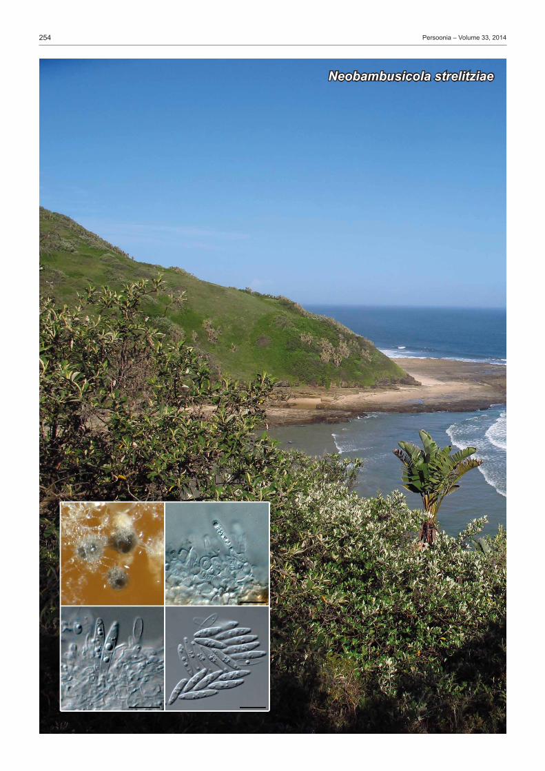
254 Persoonia–Volume33,2014
Neobambusicola strelitziae

255Fungal Planet description sheets
© 2014 Naturalis Biodiversity Center & Centraalbureau voor Schimmelcultures
Etymology.Name reflects themorphologicalsimilaritywith thegenusBambusicola.
Conidiomata separate or aggregated, erumpent, globose, dark brown,openingviacentralostiole;wallof3–6layersofbrowntextura angularis.Conidiophores reduced to conidiogenous cells.Conidiogenous cells hyaline, smooth, subcylindrical to ampulliform, phialidic, proliferating percurrently at apex, or with prominentpericlinalthickening.Conidia solitary, hyaline, smooth (becomingolivaceouswithage),prominentlyguttulate,medianly1-septate, constricted at septum, fusoid-ellipsoid, apex bluntly
subobtusely rounded, tapering to a distinctly truncate base, mostlystraight,butattimesslightlycurved.Microconidial state occurringinsameconidioma.Microconidiogenous cells hyaline, smooth, doliiform to subcylindrical, proliferating percurrently at apex.Microconidia solitary, hyaline, smooth, guttulate to granular, aseptate, subglobose to subcylindrical, apex obtusely rounded,basetruncate.
Type species.Neobambusicola strelitziae. MycoBankMB810614.
FungalPlanet303–24November2014
Neobambusicola Crous&M.J.Wingf.,gen. nov.
Neobambusicola strelitziae Crous&M.J.Wingf.,sp. nov.
PedroW.Crous&JohannesZ.Groenewald,CBS-KNAWFungalBiodiversityCentre,P.O.Box85167,3508ADUtrecht,TheNetherlands;
e-mail:[email protected]&[email protected],ForestryandAgriculturalBiotechnologyInstitute(FABI),UniversityofPretoria,
P.BagX20,Pretoria,0028,SouthAfrica;e-mail:[email protected]
Colour illustrations.Strelitzia nicolaiatHagaHaga,EasternCapeProv-ince,SouthAfrica;colonysporulatingonOA,conidiogenouscellsandconidia.Scalebars=10µm.
Etymology.NamereflectsthehostgenusStrelitzia, from which this spe-cieswasisolated.
Conidiomata separate or aggregated, erumpent, globose, dark brown, up to 200 µm diam, opening via central ostiole; wall of 3–6layersofbrowntextura angularis.Conidiophores reduced toconidiogenouscells.Conidiogenous cells hyaline, smooth, subcylindricaltoampulliform,7–15×3–5µm,phialidic,proli-ferating percurrently at apex, or with prominent periclinal thick-ening.Conidiasolitary,hyaline,smooth(becomingolivaceouswithage),prominentlyguttulate,medianly1-septate,constrictedat septum, fusoid-ellipsoid, apex bluntly subobtusely rounded, tapering to a distinctly truncate base, 2 µm diam, mostly straight, butattimesslightlycurved,(15–)17–19(–21)×(3–)3.5(–4)µm.Microconidial stateoccurringinsameconidioma.Microconidio-genous cellshyaline,smooth,doliiformtosubcylindrical,3–6× 3–5µm,proliferatingpercurrentlyatapex.Microconidia solitary, hyaline, smooth, guttulate to granular, aseptate, subglobose to subcylindrical,3–7×3–4µm,apexobtusely rounded,basetruncate,2–2.5µmdiam. Culture characteristics — Colonies erumpent with sparse aerial mycelium and smooth, even, lobate margin, reaching 15mmdiamafter2wkat25°Cinthedark.OnMEAsurfacedirtywhitewithluteousincentre,reverserusttored.OnOAsurfacedirtywhitewithdiffuseluteouspigment.OnPDAsurfacepale luteous with diffuse luteous pigment, reverse orange with diffuseluteouspigment.
Typus. South africa,EasternCapeProvince,HagaHaga,onleavesofStrelitzia nicolai (Strelitziaceae),Dec.2013,M.J. Wingfield(holotypeCBSH-22000,cultureex-typeCPC24182=CBS138869;ITSsequenceGenBankKP004467,LSUsequenceGenBankKP004495,MycoBankMB810615).
Notes — Neobambusicola strelitziae was isolated from ne- crotic leaf tissue associated with infections of Phyllachora stre-litziae.The latter funguscauseswell-definedsubcircular leafspots,3–10mmdiam,withepiphyllousblackascostromata.Asthese leaf spots get older and enlarge, conidiomata are observed surroundingtheascomatainthedeadleaftissue.Doidge(1942)commented on 2-celled conidia of a potential hyperparasite invading old ascomata of P. strelitziae, which we suspect is Neo- bambusicola strelitziae.Furthercollectionsarerequired,how-ever,toresolvetherelationshipbetweenthesetwospecies.Neobambusicola resembles the genus Bambusicola in having reduced conidiophores with percurent proliferation, and conidia thatturnpalebrownatmaturity(Daietal.2012,Hydeetal.2013).However,Neobambusicola is distinct in that it does not have pycnothyrial conidiomata, and its conidia are fusoid-ellipsoid,ratherthancylindrical. ITS.BasedonamegablastsearchofNCBIsGenBanknu-cleotidedatabase,theclosesthitsusingtheITSsequenceareMicrodiplodia hawaiiensis (GenBankGU361956; Identities=391/461(85%),Gaps=24/461(5%)),Camarographium kore-anum (GenBankJQ044432;Identities=413/494(84%),Gaps=28/494(5%))andParaconiothyrium hawaiiense (GenBankKF177681;Identities=390/461(85%),Gaps=24/461(5%)). LSU.BasedonamegablastsearchofNCBIsGenBanknu- cleotidedatabase, theclosesthitsusing theLSUsequenceare Bambusicola irregulispora (GenBankJX442036;Identities=783/809(97%),Gaps=3/809(0%)),Bambusicola mas- sarinia (GenBank JX442037; Identities = 782/808 (97%),Gaps=2/808(0%))andBambusicola bambusae (GenBankJX442035;Identities=785/814(96%),Gaps=3/814(0%)).

256 Persoonia–Volume33,2014
Trichomerium dioscoreae

257Fungal Planet description sheets
© 2014 Naturalis Biodiversity Center & Centraalbureau voor Schimmelcultures
Etymology.Name reflects thehostgenusDioscorea, from which this specieswasisolated.
Mycelium consists of olivaceous-brown, septate, branched, smooth, 3–4 µmdiam hyphae.Conidiophores reduced to conidiogenouscells;conidiaarisingdirectlyfromhyphae.Coni-diogenous loci inconspicuous, truncate, somewhat erumpent, 1.5–2 µm diam.Conidia solitary, pale to medium brown, smooth,consistingofasubcylindricalbasalcell,1–3-septate,10–20×3–5µm,withtruncatehilum,2µmdiam,givingriseto 2–3 lateral arms froma central cell; arms 1–2-septate,subcylindricalwithobtuselyroundedends,12–25×3–6µm. Culture characteristics — Colonies erumpent, spreading, surface folded, with sparse to moderate aerial mycelium and even,lobatemargins,reaching35mmdiamafter2wkat25°Cinthedark.OnMEA,PDAandOAsurfaceiron-greytofuscous-black,reversefuscous-black.
Typus. Japan,Iwate,Morioka,Koma,onleavesofDioscoreasp.(Diosco-reaceae),10Sept.2013,C. Nakashima (holotypeCBSH-22001,cultureex-typeCPC24259=CBS138870; ITSsequenceGenBankKP004468,LSUsequenceGenBankKP004496,MycoBankMB810616).
Notes—ThegenusTrichomerium(1918)wasrecentlystud-iedbyChomnuntietal.(2012),whoestablishedthenewfamily,Trichomeriaceaetoaccommodateit.Althoughtheseauthorsfocused on the sexual morph, they did note that it possibly had Tripospermum(1918)asexualmorphs.Thepresentcollectionof a Tripospermum morph that clusters in Trichomerium, thus conformsthisassociation. ITS.BasedonamegablastsearchofNCBIsGenBanknu- cleotidedatabase,theclosesthitsusingtheITSsequenceareTrichomerium deniqulatum (GenBankJX313654;Identities=627/672(93%),Gaps=20/672(2%)),Trichomerium gleo-sporum (GenBank JX313656; Identities = 628/684 (92%),Gaps=31/684(4%))andTrichomerium foliicola (GenBankJX313655;Identities=628/684(92%),Gaps=31/684(4%)). LSU.BasedonamegablastsearchofNCBIsGenBanknu- cleotidedatabase,theclosesthitsusingtheLSUsequenceareTrichomerium foliicola (GenBankJX313659;Identities=834/ 845(99%),nogaps),Trichomerium deniqulatum (GenBankJX313660;Identities=828/839(99%),nogaps)andTricho-merium gleosporum (GenBankJX313662;Identities=831/843(99%),Gaps=1/843(0%)).
FungalPlanet304–24November2014
Trichomerium dioscoreae Crous&C.Nakash., sp. nov.
PedroW.Crous&JohannesZ.Groenewald,CBS-KNAWFungalBiodiversityCentre,P.O.Box85167,3508ADUtrecht,TheNetherlands;
e-mail:[email protected]&[email protected],GraduateSchoolofBioresources,MieUniversity,1577Kurima-machiya,
Tsu,Mie514-8507,Japan;e-mail:[email protected]
Colour illustrations.LeafofDioscoreasp.infectedwithDistocercospora pachyderma and co-colonised by Trichomerium dioscoreae; conidiogenous lociandconidia.Scalebars=10µm.

258 Persoonia–Volume33,2014
Roussoella acaciae

259Fungal Planet description sheets
© 2014 Naturalis Biodiversity Center & Centraalbureau voor Schimmelcultures
Etymology.NamereflectsthehostgenusAcacia, from which this species wasisolated.
Conidiomata eustromatic, multilocular, separate, globose, im-mersed, brown, up to 200 µm diam, opening via central ostiole, exudingabrownconidialmass;wallof3–6layersofbrowntextura angularis.Conidiophores reduced to conidiogenous cells.Conidiogenous cells lining the inner cavity, hyaline, smooth,ampulliformtodoliiform,3–7×5–7µm,withprominentpericlinal thickening at apex, or with tightly aggregated percurrent proliferationsatapex.Conidia solitary, pale to medium brown, smooth, guttulate, subcylindrical, straight to slightly curved, apexobtuse,basetruncate,2–3µmdiam,mostlycentral,butat timesalsodisplacedlaterally,(5–)6–7(–10)×(2–)2.5–3µm. Culture characteristics — Colonies flat, spreading, with sparse aerial mycelium and even, smooth margin, reaching 6mmdiamafter2wkat25°Cinthedark.OnMEAsurfaceolivaceous-grey with patches of pale luteous, reverse sienna in centre,orangeinouterregion.OnOAolivaceous-greyincentre,withdirtywhiteinouterregion.OnPDAcentreolivaceous-greyonsurfaceandreverse.
Typus. tanZania, Serengeti, on leaves of Acacia tortilis(Fabaceae),Feb.2014, M.J. Wingfield(holotypeCBSH-22002,cultureex-typeCPC24314=CBS138873;ITSsequenceGenBankKP004469,LSUsequenceGenBankKP004497,MycoBankMB810617).
Notes — Members of the genus Roussoella (1888)(Rous-soellaceae;Liuetal.2014)mostlyoccuronmonocotyledons,thus the occurrence of R. acaciae on Acacia isunusual.Al-though we isolated only the Cytoplea(1885)asexualmorph,which is unknown for most species of Roussoella, the fungus on Acacia appears to be phylogenetically distinct from other membersofthegenus.BoththegeneraRoussoella and Cyto-pleaareinneedofrevision. ITS.BasedonamegablastsearchofNCBIsGenBanknu- cleotidedatabase,theclosesthitsusingtheITSsequenceareRoussoella chiangraina (GenBankKJ474828;Identities=360/ 395(91%),Gaps=17/395(4%)),Roussoella siamensis (Gen-BankKJ474837;Identities=352/387(91%),Gaps=16/387(4%))andArthopyrenia salicis (GenBankKM030296;Identities=353/390(91%),Gaps=17/390(4%)). LSU.BasedonamegablastsearchofNCBIsGenBanknu- cleotidedatabase,theclosesthitsusingtheLSUsequenceareRoussoella percutanea (GenBankKF366449;Identities=822/ 841(98%),Gaps=3/841(0%)),Sporidesmium australiense (GenBankDQ408554;Identities=827/847(98%),Gaps=2/ 847(0%))andRoussoella hysterioides (GenBankAB524622; Identities=809/829(98%),Gaps=2/829(0%)).
FungalPlanet305–24November2014
Roussoella acaciae Crous&M.J.Wingf. sp. nov.
Colour illustrations. Lion resting in anAcacia tortilis tree, Serengeti, Tanzania;conidiomatasporulatingonOA,conidiogenouscellsandconidia.Scalebars=10µm.
PedroW.Crous&JohannesZ.Groenewald,CBS-KNAWFungalBiodiversityCentre,P.O.Box85167,3508ADUtrecht,TheNetherlands;
e-mail:[email protected]&[email protected],ForestryandAgriculturalBiotechnologyInstitute(FABI),UniversityofPretoria,
P.BagX20,Pretoria,0028,SouthAfrica;e-mail:[email protected]

260 Persoonia–Volume33,2014
Neocladophialophora quercina

261Fungal Planet description sheets
© 2014 Naturalis Biodiversity Center & Centraalbureau voor Schimmelcultures
Etymology.NamereflectsitsmorphologicalsimilaritytothegenusClado-phialophora.
Mycelium consisting of hyaline, smooth, septate, branched, hyphae.Conidiophoresreducedtoconidiogenouscells.Coni-diogenous cells erect, subcylindrical, hyaline to subhyaline, mono-topolyphialidic;locitruncate.Conidia formed in long, rarely branched basipetal chains consisting of subcylindrical
conidia, apical conidium clavate to subglobose, other coni-diasubcylindrical,0–1-septate,somewhatconstrictedattheseptum, guttulate to granular, subhyaline; constricted at the truncatehila,somewhatdarkened.Olderconidiaappearpaleolivaceousinmass.
Type species.Neocladophialophora quercina. MycoBankMB810618.
FungalPlanet306–24November2014
Neocladophialophora Crous&R.K.Schumach.,gen. nov.
PedroW.Crous&JohannesZ.Groenewald,CBS-KNAWFungalBiodiversityCentre,P.O.Box85167,3508ADUtrecht,TheNetherlands;
e-mail:[email protected]&[email protected]éK.Schumacher,Hölderlinstraße25,15517Fürstenwalde/Spree,Germany;e-mail:[email protected]
Colour illustrations.Quercus robur tree; conidiophores and polyphialidic conidiogenouscellsgivingrisetobranchedchainsofconidia.Scalebars=10µm.
Neocladophialophora quercina Crous&R.K.Schumach.,sp. nov. Etymology.NamereflectsthehostgenusQuercus, from which this spe-cieswasisolated.
Mycelium consisting of hyaline, smooth, septate, branched, 1.5–2µmdiamhyphae.Conidiophores reduced to conidio-genouscells.Conidiogenous cells erect, subcylindrical, hyaline tosubhyaline,mono-topolyphialidic,10–30×3–4µm;locitruncate,1µmdiam.Conidia formed in long, rarely branched basipetal chains consisting of subcylindrical conidia (up to20perchain),apical conidiumclavate tosubglobose,otherconidiasubcylindrical,0–1-septate,somewhatconstrictedattheseptum,guttulatetogranular,subhyaline,(9–)13–15(–17)×(2.5–)3(–5)µm;constrictedatthetruncatehila,somewhatdarkened,0.5µmdiam.Olderconidiaappearpaleolivaceousinmass. Culture characteristics — Colonies spreading, appressed, surface folded with sparse aerial mycelium and smooth, lobate margin,reaching18mmdiamafter2wkat25°Cinthedark.OnMEAandPDAsurfaceandreverseochreous.OnOAsurfacepaleluteous.
Typus. Germany, on dead twig of Quercus robur(Fagaceae),23Feb.2014, R.K. Schumacher (holotypeCBSH-22003,cultureex-typeCPC24426=CBS138874;ITSsequenceGenBankKP004470,LSUsequenceGenBankKP004498,MycoBankMB810619).
Notes — Neocladophialophora is morphologically similar to Cladophialophora, Fusicladium and Polyscytalidium (seeCrouset al. 2007c,Benchet al. 2012), but different in thatconidiophores are hyaline, conidiogenous cells phialidic, and conidiaareconstrictedattheirslightlydarkenedhila.Further-more, conidia are pigmented in mass, and terminal conidia are frequentlyclavatetosubglobose. ITS.BasedonamegablastsearchofNCBIsGenBanknu-cleotidedatabase,theclosesthitsusingtheITSsequenceareTriscelophorus cf.acuminatus (GenBankKF730836;Identities=210/235(89%),Gaps=14/235(5%)),Triscelophorus mono-sporus (GenBankKF730840;Identities=184/193(95%),Gaps=1/193(0%))andHemibeltrania mitrata (GenBankEF029228;Identities=280/351(80%),Gaps=19/351(5%)). LSU.BasedonamegablastsearchofNCBIsGenBanknu-cleotidedatabase,theclosesthitsusingtheLSUsequenceareScolecobasidium tropicum (GenBankKF156102; Identities=687/786(87%),Gaps=14/786(1%)),Isthmolongispora ampul-liformis (GenBankEU107303;Identities=707/809(87%),Gaps=8/809(0%))andDactylaria humicola (GenBankEU107304;Identities=687/793(87%),Gaps=13/793(1%)).

262 Persoonia–Volume33,2014
Barssia maroccana

263Fungal Planet description sheets
© 2014 Naturalis Biodiversity Center & Centraalbureau voor Schimmelcultures
FungalPlanet307–24November2014
Barssia maroccana G.Moreno,Manjón, Carlavilla&P.Alvarado,sp. nov. Etymology.FromtheBerberMur N’Akush(‘landofGod’),whichispres-entlyknownasMorocco,thecountrywherethisspecieswascollected.
Hypogenous ascomata1.7–5.3×1.5–2.5cm(measurementstakenfromherbariummaterial),irregularlygloboseorsubglo-bose to elongated and flattened, more or less broadly lobed, reddish brown to dark reddish brown, sometimes with a rounded to irregularapicaldepression.Peridium covered with broad, roughlypolygonaldarkreddishbrownwarts;about140–200µmthick,formedbypseudo-parenchymaticcells,12–50µmdiam,thick-walled.Theoutermostcelllayersarereddishandhavedarkwalls,while thesebecome lighter inwards.Gleba whitishtopalepinkish,compactorfrequentlypresentingsmalllabyrinth-likecavities,withwell-definedsinuousveins,formedbyaprosenchymaticstructureofinterwovenhyphae8–15µmdiam.Sinuoseparaphysesnotwell-defined,5–7µmdiam.Asci clavate to broadly ellipsoid, indehiscent, immersed into the gleba,formingadefinitehymenium,hyaline,hardlyobservableinmatureascomata,8-spored,110–130×30–50µm.Asco-sporesellipsoidal,29–36×(16–)18–22µm,hyaline,smooth,not amyloid or dextrinoid, with an obtuse apex and a large oil droplet(L/ l=1.6–1.7).Smellandtastenotrecorded. Habitat&Distribution—So far foundonly underCedrus atlantica,atIfrane,Morocco,1760masl.
Typus.morocco,Azrou, province of Ifrane,Cedrus atlantica forest, 18Nov.2010,M.A. Sanz, J. Álvarez, P. Alvarado & J.L. Manjón(holotypeAH39117;ITSsequenceGenBankKM243649,LSUsequenceGenBankKM243655,MycoBankMB809666);Ifrane,Cedrus atlantica forest,18Nov.2010, M.Á. Sanz, P. Alvarado & J.L. Manjón,paratypeAH39116;ibid.,AH44221;Ifrane,Cedrus atlantica forest with some Quercus ilex species,1760m asl, J.L. Manjón, J. Álvarez-Jiménez & M.Á. Sanz,21Feb.2014,paratypeAH44099(ITS,LSUsequencesGenBankKM243648,KM243654).
Additional specimens examined.Balsamia vulgaris: italy, Reggio Emilia, Regnano, Quercus and Pinus mixed forest,450masl,11Dec.2005,A. Mon-tecchi,Amer2482=AH44222(ITS,LSUsequencesGenBankKM243645,KM243651);ReggioEmilia,Rio delleViole,Quercus pubescens forest, 350masl,9Dec.2002,A. Montecchi,Amer2404=AH44223(ITS,LSUsequencesGenBankKM243646,KM243652);ReggioEmilia,Montalvo,Quercus pubescens forest,350masl,21Apr.2003,A. Montecchi, Amer 2403=AH44224(ITS,LSUsequencesGenBankKM243647,KM243653).Balsamia polysperma: italy, Reggio Emilia, Monte Duro, Ostrya and conifers, 650masl,14Dec.1999,A. Montecchi,Amer2042=AH44225(ITS,LSUsequencesGenBankKM243650,KM243656).
Notes — Barssia maroccana is morphologically character-ised by its large and broad spores with obtuse apex, growing under Cedrus atlantica.ThedeviantphylogeneticplacementofthislineagewasfirstreportedbyAlvaradoetal.(2011).Bars-sia maroccana is very similar to Balsamia polysperma, but the
Colour illustrations.Morocco,Ifrane,forestofCedrus atlantica where the holotype was collected; ascomata; peridium and gleba; detail of the outermost layer of the peridium with pseudoparenchymatic structure; prosenchymatic gleba,asciandascospores;ascospores(holotypeAH39117).Scalebars=1 cm (ascomata), 100µm (cortex), 20µm (pseudoparenchymatic andprosenchymaticcells),10µm(ascusandspores).
GabrielMoreno,JuanR.Carlavilla,JulioÁlvarez&JoséL.Manjón,DepartamentodeCienciasdelaVida(ÁreadeBotánica),UniversidaddeAlcalá,E-28805AlcaládeHenares,Spain;
e-mail:[email protected],[email protected],[email protected]&[email protected],ALVALAB,LaRochela47,E-39012,Santander,Spain;
e-mail:[email protected]
latterhassmallerascomata0.5–2(–3)cmdiam,withnarrowerellipsoidalspores,18–25×9–16µm,L/ l=1.6–1.7,anddiffer-entecology(Montecchi&Sarasini2000).ThemonotypicgenusBarssiawas createdbyGilkey (1925) to accommodate theAmerican species B. oregonensis.ThisspecieswasoriginallyfoundinOregonandthePacificNorthwestoftheUSA(Trappe1979),butlaterreportsciteditalsoinPoland(Ławrynowicz&Skirgiełło1984).Barssia oregonensis differs from B. maroccana because of its ascomata being excavated with a deep apical depression covered by the peridium, and smaller spores about 24–29×14.5–17μm,L/I=1.6–1.7,andadifferentecology(Ławrynowicz&Skirgiełło1984).Theonlyotherspeciesinthegenus, Barssia yezomontana,with globose spores (Trappe1979),wascombinedintoBarssia from the monotypic genus Phymatomyces.Unfortunately,thetypespecimenofP. yezo- montanuswaslostinWorldWarII(Gilkey1961)anditisthere-forenotpossibletoconfirmthistaxonomicdecisionuntilthisJapanesetaxonisrecollected.Gilkey(1925)placedthegenusBarssia in the family Tuberace-ae,butlaterTrappe(1979)transferredittothefamilyBalsamia-ceae.Kimbroughetal.(1996)performedanultrastructuralstudyof BarssiaandconcludeditshouldbeclassifiedwithinthefamilyHelvellaceae.LaterPercudanietal.(1999)putBalsamia and Barssia back into the family Balsamiaceae, which was nested within Helvellaceae.Macro- andmicroscopical differencesbetween Balsamia and Barssiaareverysubtle.Gilkey(1925)highlights the apical depression observed in B. oregonensis, and compares it to the analogous structures present in Genea, Pseudobalsamia(currentlyconsideredasynonymofBalsamia),Pachyphloeus and Hydnotrya.ThesimilaritiesbetweenBars-sia and Balsamia are also commented on by Montecchi & Sarasini(2000).Glebalchambers,smoothsporesunderthelight microscope, and the presence of paraphyses are shared bybothgenera.Thepresentmoleculardataconfirmthatbothgenera are monophyletic, with the new species from Morocco being better accommodated within Barssia.
Phylogenetic tree of Helvellaceae species constructed with a maximumlikelihood(ML)analysisofLSUsequencesbyrunningRAxML-HPC2onXSEDEonlinev.8.0.24.Helvella lacunosa (KC122796andKC122771)istheoutgroup.Bootstrapsupportvalues≥75%aregivenabovebranches.Thephylogeneticposition of Barssia maroccana is indicated in bold.

264 Persoonia–Volume33,2014
Acremoniopsis suttonii
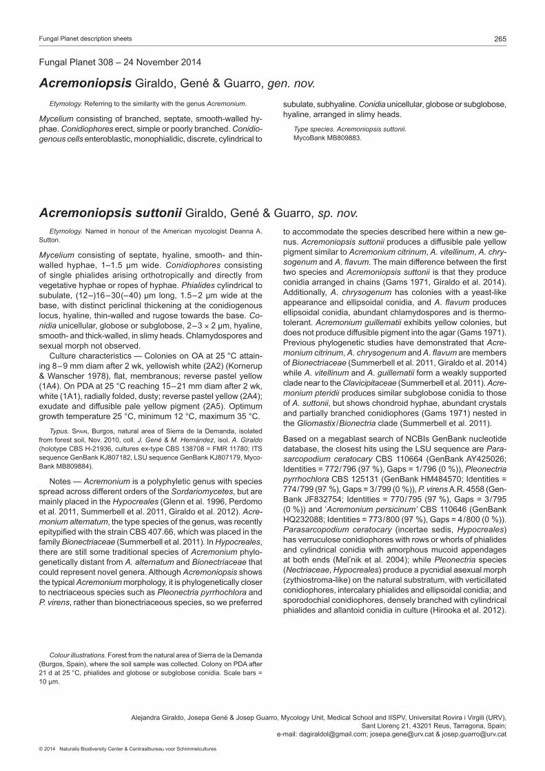
265Fungal Planet description sheets
© 2014 Naturalis Biodiversity Center & Centraalbureau voor Schimmelcultures
Etymology. Referring to the similarity with the genus Acremonium.
Mycelium consisting of branched, septate, smooth-walled hy-phae.Conidiophoreserect,simpleorpoorlybranched.Conidio-genous cells enteroblastic, monophialidic, discrete, cylindrical to
subulate,subhyaline.Conidia unicellular, globose or subglobose, hyaline,arrangedinslimyheads.
Type species. Acremoniopsis suttonii. MycoBankMB809883.
FungalPlanet308–24November2014
Acremoniopsis Giraldo,Gené&Guarro,gen. nov.
Etymology. Named in honour of theAmericanmycologistDeannaA.Sutton.
Mycelium consisting of septate, hyaline, smooth- and thin-walled hyphae, 1–1.5 µmwide.Conidiophores consisting of single phialides arising orthotropically and directly from vegetativehyphaeorropesofhyphae.Phialides cylindrical to subulate, (12–)16–30(–40)µm long,1.5–2µmwideat thebase, with distinct periclinal thickening at the conidiogenous locus,hyaline,thin-walledandrugosetowardsthebase.Co-nidiaunicellular,globoseorsubglobose,2–3× 2 µm, hyaline, smooth-andthick-walled,inslimyheads.Chlamydosporesandsexualmorphnotobserved. Culturecharacteristics—ColoniesonOAat25°Cattain-ing8–9mmdiamafter2wk,yellowishwhite(2A2)(Kornerup&Wanscher1978), flat,membranous;reversepastelyellow(1A4).OnPDAat25°Creaching15–21mmdiamafter2wk,white(1A1),radiallyfolded,dusty;reversepastelyellow(2A4);exudateanddiffusible pale yellowpigment (2A5).Optimumgrowthtemperature25°C,minimum12°C,maximum35°C.
Typus. Spain, Burgos, natural area of Sierra de la Demanda, isolated fromforestsoil,Nov.2010,coll.J. Gené & M. Hernández, isol. A. Giraldo (holotypeCBSH-21936,culturesex-typeCBS138708=FMR11780;ITSsequenceGenBankKJ807182,LSUsequenceGenBankKJ807179,Myco-BankMB809884).
Notes — Acremonium is a polyphyletic genus with species spread across different orders of the Sordariomycetes, but are mainly placed in the Hypocreales (Glennetal.1996,Perdomoetal.2011,Summerbelletal.2011,Giraldoetal.2012).Acre-monium alternatum, the type species of the genus, was recently epitypifiedwiththestrainCBS407.66,whichwasplacedinthefamily Bionectriaceae(Summerbelletal.2011). InHypocreales, there are still some traditional species of Acremonium phylo-genetically distant from A. alternatum and Bionectriaceae that couldrepresentnovelgenera.AlthoughAcremoniopsis shows the typical Acremonium morphology, it is phylogenetically closer to nectriaceous species such as Pleonectria pyrrhochlora and P. virens, rather than bionectriaceous species, so we preferred
Colour illustrations. Forest from the natural area of Sierra de la Demanda (Burgos,Spain),wherethesoilsamplewascollected.ColonyonPDAafter21dat25°C,phialidesandgloboseorsubgloboseconidia.Scalebars=10µm.
AlejandraGiraldo,JosepaGené&JosepGuarro,MycologyUnit,MedicalSchoolandIISPV,UniversitatRoviraiVirgili(URV),SantLlorenç21,43201Reus,Tarragona,Spain;
e-mail:[email protected];[email protected]&[email protected]
Acremoniopsis suttonii Giraldo,Gené&Guarro,sp. nov.to accommodate the species described here within a new ge-nus.Acremoniopsis suttonii produces a diffusible pale yellow pigment similar to Acremonium citrinum, A.vitellinum, A.chry-sogenum and A.flavum.Themaindifferencebetweenthefirsttwo species and Acremoniopsis suttonii is that they produce conidiaarrangedinchains(Gams1971,Giraldoetal.2014).Additionally, A.chrysogenum has colonies with a yeast-like appearance and ellipsoidal conidia, and A. flavum produces ellipsoidal conidia, abundant chlamydospores and is thermo-tolerant.Acremonium guillematii exhibits yellow colonies, but doesnotproducediffusiblepigmentintotheagar(Gams1971).Previous phylogenetic studies have demonstrated that Acre-monium citrinum, A.chrysogenum and A.flavum are members of Bio nectriaceae (Summerbelletal.2011,Giraldoetal.2014) while A.vitellinum and A.guillematii form a weakly supported clade near to the Clavicipitaceae (Summerbelletal.2011).Acre-monium pteridii produces similar subglobose conidia to those of A. suttonii, but shows chondroid hyphae, abundant crystals andpartiallybranchedconidiophores(Gams1971)nestedinthe Gliomastix /Bionectriaclade(Summerbelletal.2011).
BasedonamegablastsearchofNCBIsGenBanknucleotidedatabase,theclosesthitsusingtheLSUsequencearePara-sarcopodium ceratocaryCBS110664 (GenBankAY425026;Identities=772/796(97%),Gaps=1/796(0%)),Pleonectria pyrrhochlora CBS125131(GenBankHM484570;Identities=774/799(97%),Gaps=3/799(0%)),P. virens A.R.4558(Gen-BankJF832754;Identities=770/795(97%),Gaps=3/795(0%))and‘Acremonium persicinum’CBS110646(GenBankHQ232088;Identities=773/800(97%),Gaps=4/800(0%)).Parasarcopodium ceratocary (incertae sedis,Hypocreales)has verruculose conidiophores with rows or whorls of phialides and cylindrical conidia with amorphous mucoid appendages atbothends(Mel’niketal.2004);whilePleonectria species (Nectriaceae, Hypocreales)produceapycnidialasexualmorph(zythiostroma-like)onthenaturalsubstratum,withverticillatedconidiophores, intercalary phialides and ellipsoidal conidia; and sporodochial conidiophores, densely branched with cylindrical phialidesandallantoidconidiainculture(Hirookaetal.2012).

266 Persoonia–Volume33,2014
Marasmius vladimirii
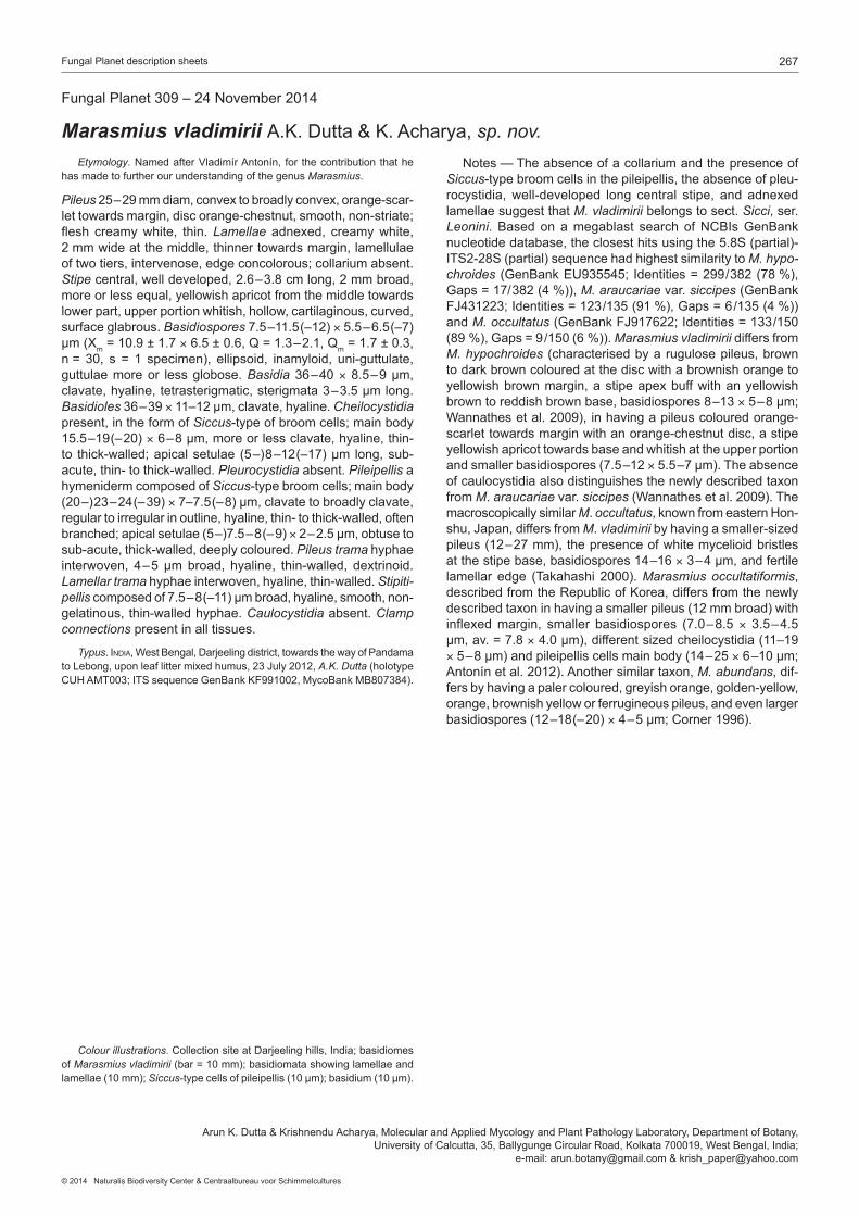
267Fungal Planet description sheets
© 2014 Naturalis Biodiversity Center & Centraalbureau voor Schimmelcultures
FungalPlanet309–24November2014
Marasmius vladimirii A.K.Dutta&K.Acharya,sp. nov. Etymology.NamedafterVladimírAntonín, for thecontribution thathehas made to further our understanding of the genus Marasmius.
Pileus25–29mmdiam,convextobroadlyconvex,orange-scar-let towards margin, disc orange-chestnut, smooth, non-striate; flesh creamywhite, thin.Lamellae adnexed, creamy white, 2 mm wide at the middle, thinner towards margin, lamellulae oftwotiers,intervenose,edgeconcolorous;collariumabsent.Stipecentral,welldeveloped,2.6–3.8cmlong,2mmbroad,moreorlessequal,yellowishapricotfromthemiddletowardslower part, upper portion whitish, hollow, cartilaginous, curved, surfaceglabrous.Basidiospores7.5–11.5(–12)×5.5–6.5(–7)µm(Xm=10.9±1.7×6.5±0.6,Q=1.3–2.1,Qm=1.7±0.3,n= 30, s = 1 specimen), ellipsoid, inamyloid, uni-guttulate,guttulaemoreor lessglobose.Basidia36–40×8.5–9µm,clavate,hyaline,tetrasterigmatic,sterigmata3–3.5µmlong.Basidioles36–39×11–12µm,clavate,hyaline.Cheilocystidia present, in the form of Siccus-type of broom cells; main body 15.5–19(–20)×6–8µm,moreorlessclavate,hyaline,thin-to thick-walled;apicalsetulae (5–)8–12(–17)µm long,sub-acute,thin-tothick-walled.Pleurocystidiaabsent.Pileipellis a hymeniderm composed of Siccus-type broom cells; main body (20–)23–24(–39)×7–7.5(–8)µm,clavatetobroadlyclavate,regular to irregular in outline, hyaline, thin- to thick-walled, often branched;apicalsetulae(5–)7.5–8(–9)×2–2.5µm,obtusetosub-acute,thick-walled,deeplycoloured.Pileus trama hyphae interwoven, 4–5µmbroad, hyaline, thin-walled, dextrinoid.Lamellar tramahyphaeinterwoven,hyaline,thin-walled.Stipiti-pelliscomposedof7.5–8(–11)µmbroad,hyaline,smooth,non- gelatinous,thin-walledhyphae.Caulocystidiaabsent.Clamp connectionspresentinalltissues.
Typus.india,WestBengal,Darjeelingdistrict,towardsthewayofPandamatoLebong,uponleaflittermixedhumus,23July2012,A.K. Dutta(holotypeCUHAMT003;ITSsequenceGenBankKF991002,MycoBankMB807384).
Colour illustrations.CollectionsiteatDarjeelinghills,India;basidiomesof Marasmius vladimirii(bar=10mm);basidiomatashowinglamellaeandlamellae(10mm);Siccus-typecellsofpileipellis(10µm);basidium(10µm).
ArunK.Dutta&KrishnenduAcharya,MolecularandAppliedMycologyandPlantPathologyLaboratory,DepartmentofBotany,UniversityofCalcutta,35,BallygungeCircularRoad,Kolkata700019,WestBengal,India;
e-mail:[email protected]&[email protected]
Notes—TheabsenceofacollariumandthepresenceofSiccus-type broom cells in the pileipellis, the absence of pleu-rocystidia, well-developed long central stipe, and adnexed lamellae suggest that M. vladimiriibelongstosect.Sicci,ser.Leonini. Basedonamegablast searchofNCBIsGenBanknucleotidedatabase,theclosesthitsusingthe5.8S(partial)-ITS2-28S(partial)sequencehadhighestsimilaritytoM. hypo-chroides (GenBankEU935545; Identities=299/382(78%),Gaps=17/382(4%)),M. araucariaevar.siccipes(GenBankFJ431223;Identities=123/135(91%),Gaps=6/135(4%))and M. occultatus(GenBankFJ917622;Identities=133/150(89%),Gaps=9/150(6%)).Marasmius vladimirii differs from M.hypochroides (characterisedbyarugulosepileus,brownto dark brown coloured at the disc with a brownish orange to yellowish brown margin, a stipe apex buff with an yellowish browntoreddishbrownbase,basidiospores8–13×5–8μm;Wannathesetal.2009),inhavingapileuscolouredorange-scarlet towards margin with an orange-chestnut disc, a stipe yellowish apricot towards base and whitish at the upper portion andsmallerbasidiospores(7.5–12×5.5–7µm).Theabsenceof caulocystidia also distinguishes the newly described taxon from M.araucariaevar.siccipes(Wannathesetal.2009).Themacroscopically similar M.occultatus,knownfromeasternHon-shu,Japan,differsfromM.vladimiriibyhavingasmaller-sizedpileus(12–27mm),thepresenceofwhitemycelioidbristlesatthestipebase,basidiospores14–16×3–4µm,andfertilelamellar edge (Takahashi 2000).Marasmius occultatiformis, describedfromtheRepublicofKorea,differsfromthenewlydescribedtaxoninhavingasmallerpileus(12mmbroad)withinflexedmargin, smaller basidiospores (7.0–8.5× 3.5–4.5μm,av.=7.8×4.0μm),differentsizedcheilocystidia(11–19×5–8μm)andpileipelliscellsmainbody(14–25×6–10μm;Antonínetal.2012).Anothersimilartaxon,M. abundans, dif-fers by having a paler coloured, greyish orange, golden-yellow, orange, brownish yellow or ferrugineous pileus, and even larger basidiospores(12–18(–20)×4–5μm;Corner1996).

268 Persoonia–Volume33,2014
Ganoderma austroafricanum

269Fungal Planet description sheets
© 2014 Naturalis Biodiversity Center & Centraalbureau voor Schimmelcultures
FungalPlanet310–24November2014
Ganoderma austroafricanumM.P.A.Coetzee,M.J.Wingf,Marinc.,Blanchette,sp. nov. Etymology.Namereflectsthegeographicaloriginfromwhichthefunguswascollected.
Mature basidiomata annual, pileate, sessile, dimidiate, pileus dark to reddish brown when dry, margin rounded, thickened, redbrownwhendry.Pore surfacesmooth,pores3–4permm,roundtoangular,radiallyelongated,150–390µmlong,85–245µmwide,dissepiments60–200µmdiam.Hyphal system dimi-tic,generativehyphae,usuallycollapsed,thin-walled,hyaline.Vegetative hyphaeobservedinthetubes.Tube trama intricate andagglutinatedskeletalhyphae4–8µmthick,withextremitiestaperingtoelongateapicesof2–2.5µmdiam,hyalinetopaleyellowish in5%KOH.Basidianotobserved.Basidiospores brown, subglobose with a truncate base, bitunicate, verrucu-lose,8–11×5.5–7µm.Chlamydosporeson2%maltextractagarmedium(MEA)ellipsoidalwithahyphalextensionatthebase, apex occasionally papillated, terminal or intercalary, 10.5–19.5×6.5–9.5µm. Culture characteristics — Colonies on2%MEAfertile,show-ingthebestgrowthreaching82mmat25°Cinthedarkin8d,thesecondbest62mmat30°C,37mmat20°C,18mmat15°C,nogrowthat35°C,circularwithentireedge,flat,felt-liketexture, white at all temperatures, with sporadic tint of yellow atinner20mmcircleat30°C;chlamydosporespresent.
Typus.South africa,Gauteng,Pretoria,Brooklyn,Jan.2014,onJaca-randa mimosifolia, M.J. Wingfield(holotypePREM61074,livingcultureex-typeCBS138724=CMW41454;ITSsequenceGenBankKM507324,LSUsequenceGenBankKM507325,MycoBankMB810411).
Colour illustrations.Jacaranda mimosifolia(Jacarandatree)growingasstreettreeinthesuburbofBrooklyn,Pretoria,SouthAfrica(background);crownofinfectedJacarandatreeshowingbranchdie-back;purpleflowersof Jacaranda tree;basidiocarps;basidiospores (10µm); skeletalhyphaerefractiveinphasecontrast(25µm);chlamydosporeson2%MEA(10µm).
MartinP.A.Coetzee,SeonjuMarincowitz&MichaelJ.Wingfield,DepartmentofGenetics,CentreofExcellenceinTreeHealthBiotechnology(CTHB),ForestryandAgriculturalBiotechnologyInstitute(FABI),FacultyofNaturalandAgriculturalSciences,UniversityofPretoria,Pretoria,0002,SouthAfrica;
e-mail:[email protected],[email protected]&[email protected],UniversityofMinnesota,495BorlaugHall,1991UpperBufordCircle,St.Paul,MN55108;
e-mail:[email protected],CentrodeInvestigación,EstudiosyDesarrollodeIngeniería(CIEDI),FacultaddeIngenieríasyCienciasAgropecuarias(FICA),
UniversidaddeLasAméricas,CalleJoséQueris/nentreAv.GranadosyAv.EloyAlfaro,Quito,Ecuador;e-mail:[email protected]
Notes — Ganoderma austroafricanum is the causal agent of rootandbuttrotdiseaseonlargenumbersofjacarandatreesinthesuburbofBrooklyn(Pretoria,SouthAfrica).ITSsequencecomparisons usingBlastn searches against sequences inGenBankyieldedtheclosesthitswithG. subamboinense var. laevisporum(JQ520205;querycover:100%,similarity98%).ItcanbedifferentiatedfromtheclosestrelatedsequencesofG. subamboinense var. laevisporumbasedon13nucleotidedifferences in the ITS sequence.Phylogenetic treesgener-atedfromITSsequencesplacedG. austroafricanum within a cladethatincludedsequencesfromGenBankrepresentingG. weberianum and G. stipitatum but with low bootstrap support (parsimonybootstrap=52%).Thelattergroupformedasistergrouptoamonophyleticclade(bootstrapsupport=92%)thatincludedsequencesfromGenBankrepresenting G. lobatum, G. lucidum, G. neojaponicum, G. oregonense and G. resina-ceum.

270 Persoonia–Volume33,2014
Collarina aurantiaca
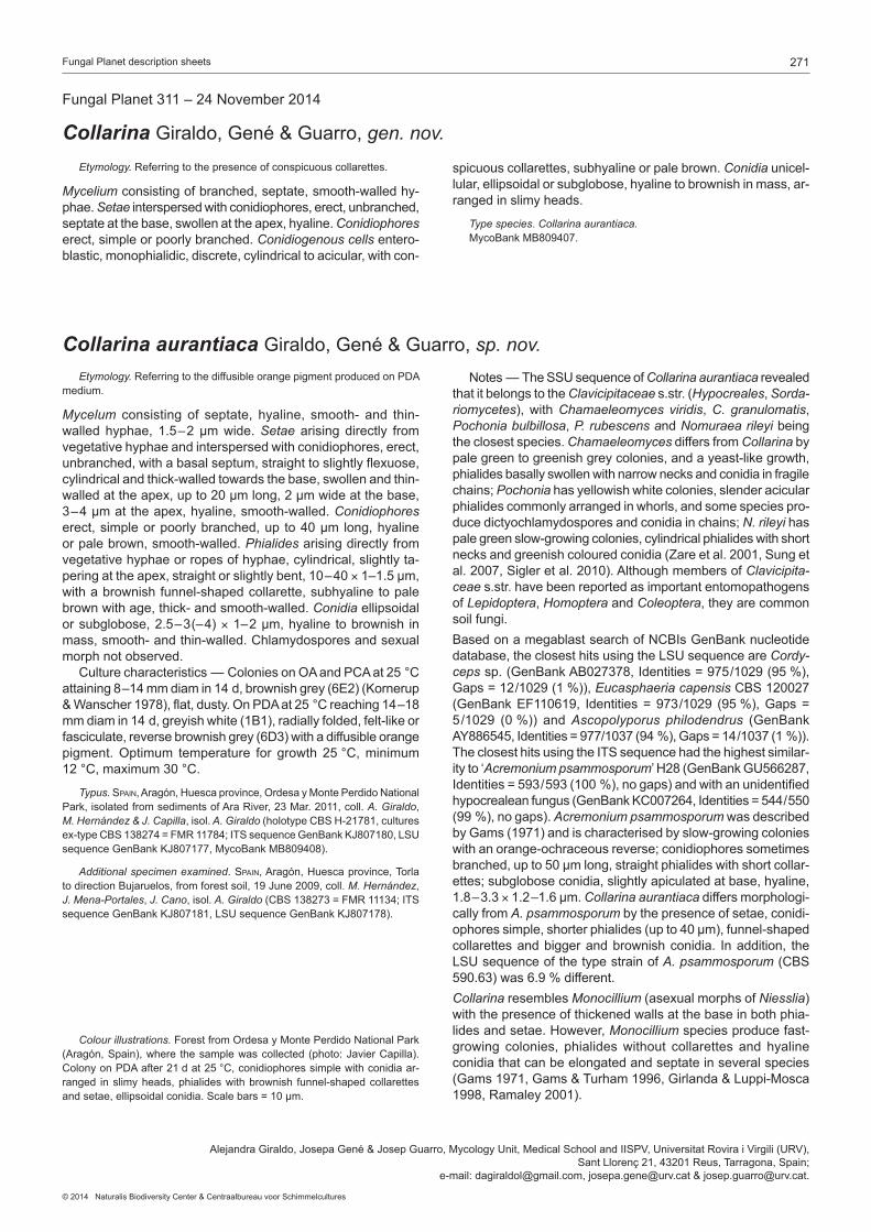
271Fungal Planet description sheets
© 2014 Naturalis Biodiversity Center & Centraalbureau voor Schimmelcultures
Etymology. Referringtothepresenceofconspicuouscollarettes.
Mycelium consisting of branched, septate, smooth-walled hy-phae.Setae interspersed with conidiophores, erect, unbranched, septateatthebase,swollenattheapex,hyaline.Conidiophores erect,simpleorpoorlybranched.Conidiogenous cells entero-blastic, monophialidic, discrete, cylindrical to acicular, with con-
spicuouscollarettes,subhyalineorpalebrown.Conidia unicel-lular, ellipsoidal or subglobose, hyaline to brownish in mass, ar- rangedinslimyheads.
Type species.Collarina aurantiaca. MycoBankMB809407.
FungalPlanet311–24November2014
Collarina Giraldo,Gené&Guarro,gen. nov.
Etymology. Referring to the diffusible orange pigment produced on PDA medium.
Mycelum consisting of septate, hyaline, smooth- and thin-walled hyphae, 1.5–2µmwide.Setae arising directly from vegetative hyphae and interspersed with conidiophores, erect, unbranched, with a basal septum, straight to slightly flexuose, cylindrical and thick-walled towards the base, swollen and thin-walled at the apex, up to 20 µm long, 2 µm wide at the base, 3–4µmattheapex,hyaline,smooth-walled.Conidiophores erect, simple or poorly branched, up to 40 µm long, hyaline orpalebrown,smooth-walled.Phialides arising directly from vegetative hyphae or ropes of hyphae, cylindrical, slightly ta-peringattheapex,straightorslightlybent,10–40×1–1.5µm,with a brownish funnel-shaped collarette, subhyaline to pale brownwithage,thick-andsmooth-walled.Conidia ellipsoidal orsubglobose,2.5–3(–4)×1–2µm,hyalinetobrownish inmass,smooth-andthin-walled.Chlamydosporesandsexualmorphnotobserved. Culturecharacteristics—ColoniesonOAandPCAat25°Cattaining8–14mmdiamin14d,brownishgrey(6E2)(Kornerup&Wanscher1978),flat,dusty.OnPDAat25°Creaching14–18mmdiamin14d,greyishwhite(1B1),radiallyfolded,felt-likeorfasciculate,reversebrownishgrey(6D3)withadiffusibleorangepigment.Optimum temperature for growth 25°C,minimum12°C,maximum30°C.
Typus. Spain,Aragón,Huescaprovince,OrdesayMontePerdidoNationalPark,isolatedfromsedimentsofAraRiver,23Mar.2011,coll.A. Giraldo, M. Hernández & J. Capilla, isol. A. Giraldo (holotypeCBSH-21781,culturesex-typeCBS138274=FMR11784;ITSsequenceGenBankKJ807180,LSUsequenceGenBankKJ807177,MycoBankMB809408).
Additional specimen examined.Spain,Aragón,Huescaprovince,TorlatodirectionBujaruelos,fromforestsoil,19June2009,coll.M. Hernández, J. Mena-Portales, J. Cano, isol. A. Giraldo(CBS138273=FMR11134;ITSsequenceGenBankKJ807181,LSUsequenceGenBankKJ807178).
Colour illustrations. Forest from Ordesa y Monte Perdido National Park (Aragón,Spain),where thesamplewascollected(photo:JavierCapilla).ColonyonPDAafter21dat25°C,conidiophoressimplewithconidiaar-ranged in slimy heads, phialides with brownish funnel-shaped collarettes andsetae,ellipsoidalconidia.Scalebars=10µm.
AlejandraGiraldo,JosepaGené&JosepGuarro,MycologyUnit,MedicalSchoolandIISPV,UniversitatRoviraiVirgili(URV),SantLlorenç21,43201Reus,Tarragona,Spain;
e-mail:[email protected],[email protected]&[email protected].
Collarina aurantiaca Giraldo,Gené&Guarro,sp. nov. Notes—TheSSUsequenceofCollarina aurantiaca revealed that it belongs to the Clavicipitaceaes.str.(Hypocreales, Sorda-riomycetes),withChamaeleomyces viridis, C. granulomatis, Pochonia bulbillosa, P. rubescens and Nomuraea rileyi being theclosestspecies.Chamaeleomyces differs from Collarina by pale green to greenish grey colonies, and a yeast-like growth, phialides basally swollen with narrow necks and conidia in fragile chains; Pochonia has yellowish white colonies, slender acicular phialides commonly arranged in whorls, and some species pro-duce dictyochlamydospores and conidia in chains; N. rileyi has pale green slow-growing colonies, cylindrical phialides with short necksandgreenishcolouredconidia(Zareetal.2001,Sungetal.2007,Sigleretal.2010).AlthoughmembersofClavicipita-ceaes.str.havebeenreportedasimportantentomopathogensof Lepidoptera, Homoptera and Coleoptera, they are common soilfungi.BasedonamegablastsearchofNCBIsGenBanknucleotide database,theclosesthitsusingtheLSUsequenceareCordy-cepssp.(GenBankAB027378,Identities=975/1029(95%),Gaps=12/1029(1%)),Eucasphaeria capensisCBS120027(GenBankEF110619, Identities=973/1029 (95%),Gaps=5/1029 (0%)) andAscopolyporus philodendrus (GenBankAY886545,Identities=977/1037(94%),Gaps=14/1037(1%)). TheclosesthitsusingtheITSsequencehadthehighestsimilar-ityto‘Acremonium psammosporum’H28(GenBankGU566287,Identities=593/593(100%),nogaps)andwithanunidentifiedhypocrealeanfungus(GenBankKC007264,Identities=544/550(99%),nogaps).Acremonium psammosporum was described byGams(1971)andischaracterisedbyslow-growingcolonieswith an orange-ochraceous reverse; conidiophores sometimes branched,upto50µmlong,straightphialideswithshortcollar-ettes; subglobose conidia, slightly apiculated at base, hyaline, 1.8–3.3×1.2–1.6µm.Collarina aurantiaca differs morphologi-cally from A. psammosporum by the presence of setae, conidi-ophoressimple,shorterphialides(upto40µm),funnel-shapedcollarettesandbiggerandbrownishconidia. Inaddition, theLSUsequenceofthetypestrainofA. psammosporum(CBS590.63)was6.9%different.Collarina resembles Monocillium(asexualmorphsofNiesslia)with the presence of thickened walls at the base in both phia-lidesandsetae.However,Monocillium species produce fast-growing colonies, phialides without collarettes and hyaline conidia that can be elongated and septate in several species (Gams1971,Gams&Turham1996,Girlanda&Luppi-Mosca1998,Ramaley2001).

272 Persoonia–Volume33,2014
Comoclathris spartii
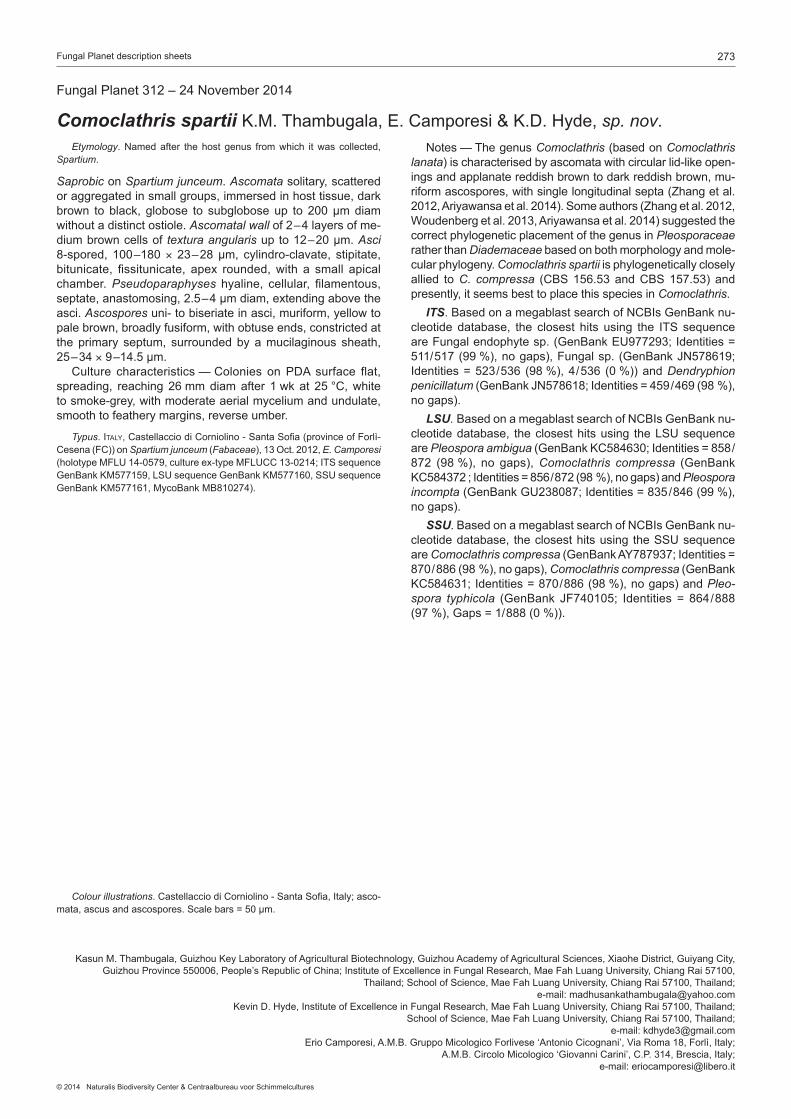
273Fungal Planet description sheets
© 2014 Naturalis Biodiversity Center & Centraalbureau voor Schimmelcultures
FungalPlanet312–24November2014
Comoclathris spartii K.M.Thambugala,E.Camporesi&K.D.Hyde,sp. nov.
Colour illustrations.CastellacciodiCorniolino-SantaSofia,Italy;asco-mata,ascusandascospores.Scalebars=50μm.
Etymology.Namedafter thehost genus fromwhich itwas collected,Spartium.
Saprobic on Spartium junceum.Ascomata solitary, scattered or aggregated in small groups, immersed in host tissue, dark brown to black, globose to subgloboseup to 200μmdiamwithoutadistinctostiole.Ascomatal wallof2–4layersofme-dium brown cells of textura angularisupto12–20μm.Asci 8-spored, 100–180× 23–28μm, cylindro-clavate, stipitate,bitunicate, fissitunicate, apex rounded,with a small apicalchamber.Pseudoparaphyses hyaline, cellular, filamentous,septate,anastomosing,2.5–4μmdiam,extendingabovetheasci.Ascospores uni- to biseriate in asci, muriform, yellow to pale brown, broadly fusiform, with obtuse ends, constricted at the primary septum, surrounded by a mucilaginous sheath, 25–34×9–14.5μm. Culture characteristics — Colonies on PDA surface flat, spreading, reaching26mmdiamafter 1wkat 25°C,whiteto smoke-grey, with moderate aerial mycelium and undulate, smoothtofeatherymargins,reverseumber.
Typus.italy,CastellacciodiCorniolino-SantaSofia(provinceofForlì-Cesena(FC))onSpartium junceum(Fabaceae),13Oct.2012,E. Camporesi (holotypeMFLU14-0579,cultureex-typeMFLUCC13-0214;ITSsequenceGenBankKM577159,LSUsequenceGenBankKM577160,SSUsequenceGenBankKM577161,MycoBankMB810274).
KasunM.Thambugala,GuizhouKeyLaboratoryofAgriculturalBiotechnology,GuizhouAcademyofAgriculturalSciences,XiaoheDistrict,GuiyangCity,GuizhouProvince550006,People’sRepublicofChina;InstituteofExcellenceinFungalResearch,MaeFahLuangUniversity,ChiangRai57100,
Thailand;SchoolofScience,MaeFahLuangUniversity,ChiangRai57100,Thailand;e-mail:[email protected]
KevinD.Hyde,InstituteofExcellenceinFungalResearch,MaeFahLuangUniversity,ChiangRai57100,Thailand;SchoolofScience,MaeFahLuangUniversity,ChiangRai57100,Thailand;
e-mail:[email protected],A.M.B.GruppoMicologicoForlivese‘AntonioCicognani’,ViaRoma18,Forlì,Italy;
A.M.B.CircoloMicologico‘GiovanniCarini’,C.P.314,Brescia,Italy;e-mail:[email protected]
Notes—ThegenusComoclathris(basedonComoclathris lanata)ischaracterisedbyascomatawithcircularlid-likeopen-ings and applanate reddish brown to dark reddish brown, mu-riformascospores,withsinglelongitudinalsepta(Zhangetal.2012,Ariyawansaetal.2014).Someauthors(Zhangetal.2012,Woudenbergetal.2013,Ariyawansaetal.2014)suggestedthecorrect phylogenetic placement of the genus in Pleosporaceae rather than Diademaceae based on both morphology and mole-cularphylogeny.Comoclathris spartii is phylogenetically closely allied to C. compressa (CBS156.53andCBS157.53) andpresently, it seems best to place this species in Comoclathris. ITS.BasedonamegablastsearchofNCBIsGenBanknu- cleotide database, the closest hits using the ITS sequenceareFungalendophytesp.(GenBankEU977293;Identities=511/517(99%),nogaps),Fungalsp.(GenBankJN578619;Identities=523/536 (98%),4/536 (0%))andDendryphion penicillatum(GenBankJN578618;Identities=459/469(98%),nogaps). LSU.BasedonamegablastsearchofNCBIsGenBanknu- cleotidedatabase, theclosesthitsusing theLSUsequenceare Pleospora ambigua(GenBankKC584630;Identities=858/ 872 (98%), no gaps),Comoclathris compressa (GenBankKC584372;Identities=856/872(98%),nogaps)andPleo spora incompta(GenBankGU238087;Identities=835/846(99%), nogaps). SSU.BasedonamegablastsearchofNCBIsGenBanknu- cleotidedatabase, theclosesthitsusingtheSSUsequenceare Comoclathris compressa(GenBankAY787937;Identities=870/886(98%),nogaps),Comoclathris compressa(GenBank KC584631;Identities=870/886(98%),nogaps)andPleo-spora typhicola (GenBank JF740105; Identities = 864/888(97%),Gaps=1/888(0%)).

274 Persoonia–Volume33,2014
Pluteus albotomentosus

275Fungal Planet description sheets
© 2014 Naturalis Biodiversity Center & Centraalbureau voor Schimmelcultures
FungalPlanet313–24November2014
Pluteus albotomentosusE.F.Malysheva&Malysheva,sp. nov. Etymology.Albotomentosus refers to the colour and character of the pileussurface.
Pileus50‒60mmdiam,hemisphericalbecomingapplanatewith low, broad umbo, pure white when fresh, yellowish white or cream (4A2‒A3) in herbarium; fine-tomentose over theentire surface, slightly flocculose towardsmargin.Lamellae free, crowded, broadly ventricose, white becoming pink with concolorousevenedges.Stipe 60× 8‒10mm,broadeningdownwards,with vaguebasal bulb up to 12‒15mmwide;solid,longitudinallyfibrillose,whitetoslightlyyellowishwhite,withwhitebasaltomentum.Contextwhite.Smellweak,sour-ish.Basidiospores 6.0‒6.6(‒6.8)× 5.3‒6.0(‒6.2) µm,Q=1.00‒1.21(‒1.28),Q*=1.11(n=30),subglobosetobroadlyellipsoid, some oviform, smooth, thin- to slightly thick-walled, hyaline, contentswith guttules.Basidia 23‒32× 6‒8 µm,4-spored,broadlyclavate.Pleurocystidiaabundant,27‒70× 11‒26µm,utriformtobroadlylageniformwithveryshortandwideneck,somebroadlyclavate,hyaline,thin-walled.Lamel-laeedgesterile.Cheilocystidia numerous, forming dense layer, 23‒65×5‒11µm,variableinshape,predominantlycylindrical,narrowlylageniformornarrowlyclavate,thin-walled,hyaline.Pileipellis a cutis, made up of undifferentiated cylindrical hyphae upto6‒10µmwide,thin-orslightlythick-walled,colourless.Stipitipellisacutisof5‒12µmwidehyalinehyphae,atlowerpart of stipe with scarce hyphae-like, cylindrical or narrowly clavatecaulocystidia,10‒30×4‒7µm. Clamp connections absentinalltissues. Habitat&Distribution—Solitaryonmossydecayedlogofdeciduoustree.FoundonceinthesouthofPrimoryeTerritory.
Typus.ruSSia,PrimoryeTerritory,‘LandoftheLeopard’NationalPark,watershed ofAnanjevka andGryaznayaRivers,mixed forest (Quercus mongolica, Carpinus cordata, Ulmus japonica, Abies nephrolepis),onmossylogofdeciduoustree,1Sept.2011,A. Kovalenko(holotypeLE289394;ITSsequenceGenBankKM658284,MycoBankMB810391).
EkaterinaF.Malysheva&VeraF.Malysheva,KomarovBotanicalInstituteoftheRussianAcademyofSciences,Prof.PopovSt.2,RUS-197376,SaintPetersburg,Russia;
e-mail: [email protected]&[email protected]
Colour illustrations.Russia,PrimoryeTerritory, ‘Landof theLeopard’National Park, area of the East Manchurian mountains, Manchurian mixed forests, where the holotype was collected; basidiocarp, pileipellis, basidio-spores,pleurocystidia,cheilocystidia(allfromholotype).Scalebars=1cm(basidiocarp),10µm(microscopicstructures).
Notes—Themacroscopicdescriptionisbasedonfreshba-sidiocarps from the original collection and the photos taken at thesite.ColourtermsareaccordingtoKornerup&Wanscher(1978).Microscopicobservationsandphotosweremadefromdriedmaterialmounted in5%KOHusinganAxioImagerA1lightmicroscope.Pluteus albotomentosusisquitedifferentfromtheotherwhite-coloured Pluteus species because of a combination of distinc-tive morphological characters, a scilicet distinctly tomentose pure white pileus, thin-walled utriform pleurocystidia, cylindrical cheilocystidiaandacutis-likepileipellis.Thepileipellisstructureand non-metuloid cystidia place P. albotomentosusinsect.His-pidoderma.BasedonthisitcanbecomparedtoP. ephebeus, which is easily distinguished by its dark basidiocarp colour, largersporesize(5.5‒8.5×4.5‒6.0(‒7.0)µmaccording toVellinga1990)anddifferentlyshapedcystidia.Themoleculardata(ITSsequence)indicatethatitiscloselyrelatedtoP. ephe-beusbutthepercentageofsimilaritybetweensequencesofthetwospeciesisonly95‒96%orless.

276 Persoonia–Volume33,2014
Pluteus extremiorientalis

277Fungal Planet description sheets
© 2014 Naturalis Biodiversity Center & Centraalbureau voor Schimmelcultures
FungalPlanet314–24November2014
Pluteus extremiorientalisE.F.Malysheva&Malysheva,sp. nov. Etymology.Namedafterthegeographicalareawhereitwascollected(RussianFarEast).
Pileus 10‒35mmdiam, firstly hemispherical, then convexbecoming applanate, commonly with obtuse low umbo; surface tomentose to squamulose – densely punctate-granulose atcentreandfibrillose-squamulose towardsmargin,oftenwithwhite context exhibited between rare fibrils or squamules;centre strongly venose-rugose; dark coloured, cocoa brown, leatherbrown(6E6),oftenwithreddishtint–chestnutbrown(6F7‒F8) or henna (7E8),with darker disc (7F7‒F8) andlightermarginbecauseofscarcityofsquamules;marginevenorrimose,slightlystriateornot,oftenincurved.Lamellae free, moderately crowded, ventricose, white becoming pink; edges even or slightly fimbriate, fuscous (brownish),more rarelyconcolorous.Stipe20‒55×1.5‒5mm,cylindricalorslightlybroadeningdownwards(upto7mm),butwithoutbasalbulb,longitudinally fibrillose,whitish at upper part, coveredwithdarkbrownfibrilsorsquamulesatlowerpart;basaltomentumwhite.Contextwhite.Smellindistinct.Basidiospores5.6‒7× 4.6‒5.7µm,Q=(1.02‒)1.04‒1.39(‒1.48),Q*=1.24(n=40),subglobose to broadly ellipsoid, some oviform, smooth, slightly thick-walled, hyaline, contents with one large or numerous smallguttules.Basidia20‒30×6‒9µm,4-spored,clavate.Pleurocystidia scattered, 35‒70× 10‒17(‒25) µm, broadlylageniform to utriform, some broadly fusiform to oviform, with pedicesandobtuseapices,thin-walled,hyaline.Lamellaeedgesterile.Cheilocystidianumerous,40‒90×10‒33µm,varyinginshape, predominantly lageniform with inflated body and narrow longneck(upto30µmlongand6µmwide),afewnarrowutri-form to clavate-cylindrical, thin-walled, with brown intracellular pigment(includingholotype),butinsomespecimenshyaline.Pileipellis a cystoderm, made up of spheropedunculate, pyri-form, broadly clavate cells in combination with lageniform or narrowlyfusiformelementswithacuteapices,20‒75(‒90)× 12‒30(‒35)µm,thin-orslightlythick-walled,withdarkbrownintracellularpigmentinvacuoles.Stipitipellis a cutis of hyaline thin-orslightlythick-walledcylindricalhyphae,5‒12µmwide,in stipe base with brown content and scattered clavate or cylindricalcaulocystidia,35‒70(‒100)×8‒15µm,oftenwithyellow-brownintracellularpigment.Clamp connections absent inpileipellis,butoccasionalinstipecontext. Habitat&Distribution—Scatteredtogregariousondecayingdeciduouswoodorsoil,infloodplainbroadleafforests.KnownfromtwolocalitiesintheRussianFarEast.
Typus.ruSSia,PrimoryeTerritory,UssuriyskyNatureReserve,vicinitiesofPeishulafieldstation,floodplainofKoryavayaRiver,broadleavedforest(withUlmus, Populus, Acer),onsoil,12Aug.2011,N. Psurtseva(holotypeLE262872;ITSsequenceGenBankKM658280,MycoBankMB810390).
EkaterinaF.Malysheva&VeraF.Malysheva,KomarovBotanicalInstituteoftheRussianAcademyofSciences,Prof.PopovSt.2,RUS-197376,SaintPetersburg,Russia;
e-mail: [email protected]&[email protected]
Additional specimens examined.ruSSia,PrimoryeTerritory,UssuriyskyNatureReserve,vicinitiesofPeishulafieldstation,floodplainofKoryavayaandSuvorovkaRivers,broadleavedforest(withUlmus, Populus, Acer),onwood of Ulmus, 12Aug.2011,A. Kovalenko, LE262871, ITSsequenceGenBankKM658279;ibid.,onsoil,12Aug.2011,E. Malysheva,LE303463,ITSsequenceGenBankKM658282;ibid.,Ulmus japonicaforest,onsoil,13Aug.2011,E. Malysheva,LE262865,ITSsequenceGenBankKM658281;PrimoryeTerritory,KedrovayaPadNatureReserve, valley ofKedrovayaRiver,floodplainbroadleavedforest(Quercus mongolica, Carpinus cordata, Tilia amurensis, Juglans mandshurica),ondecayingdeciduouswood,5Sept.2011, A. Kovalenko,LE303464,ITSsequenceGenBankKM658283.
Notes — Macroscopic descriptions are based on fresh basi- diocarps from the original collections and photos taken at the site.Colour terms are according toKornerup&Wanscher(1978).Microscopicobservationsandphotosweremadefromdriedmaterialmounted in5%KOHusinganAxioImagerA1lightmicroscope.Pluteus extremiorientalis is characterised by small to medium-sized,brownishcolouredandtomentose-squamulosepileus,fuscousedgesoflamellaeanddarkfibrilsonlowerpartofstipe.Microscopically, the pileipellis consists of two types of cystidioid elements(broadlyclavatetosphaeropedunculateandfusiform);cheilocystidia abundant, variable in pigmentation in different col- lections, but most contain brown intracellular pigment; pleuro-cystidianotrare,predominantlylageniformorutriform.Based on its pileipellis structure P. extremiorientalis is placed tosect.Cellulodermaandsubsect.Mixtini.TheITSsequencesfromthefivestudiedcollectionsare99‒100%identical,butmorphologicalcharactersdifferslightlybetweencollections–this mainly concerns the differences in colour of basidiocarps, densityofsquamulesorfibrilsonpileussurface,pigmentationofcheilocystidiaandsubstratepreferences.Pluteus extremiorientalis mostly resembles P. podospileus and P. seticepsvar.cystidiosus in pileus colouration, dark brown fibrils at stipe base, shape of pleurocystidia and pileipellisstructure(Minnis&Sundberg2010). Itcanbedistinguishedfromboth by the strong tomentose-squamulose surface ofthe pileus, brownish coloured lamellae edges, cheilocystidia shapeandsmallerelementsinthepileipellis.Themoleculardata(generatedITSsequences)confirmedthemorphologicaldifferences between all species discussed and supported the recognition of P. extremiorientalisasaseparatetaxon.
Colour illustrations.Russia,PrimoryeTerritory,UssuriyskyNatureReserve,southern spurs of the Sikhote-Alin mountains, liana coniferous-broadleaved forest; basidiocarps, pileipellis elements, basidiospores, pleuro cystidia, sterile edgeoflamella(withpigmentedcheilocystidia),stipitipellis(allfromholotype).Scalebars=1cm(basidiocarps),10µm(microscopicstructures).

278 Persoonia–Volume33,2014
Phytophthora moyootj

279Fungal Planet description sheets
© 2014 Naturalis Biodiversity Center & Centraalbureau voor Schimmelcultures
FungalPlanet315–24November2014
Phytophthora moyootj T.I.Burgess,sp. nov. Etymology. Named for the swamp or wetlands from which isolates were recovered(moyootj=swampcountryinthelocalAboriginalNyoongarlan-guage).
Sporangia produced abundantly in non-sterile soil extract; non-caducous, non-papillate with long unbranched sporangiophores, mostcommonlyovoid(63%),withbroadovoid(11.5%),limoni-form(9%),elongatedovoid(8.5%),globose(5%)andmouseshapes(3%)alsofound;39.6±10.8×26.5±4.2µm(overallrange18–73.3× 18.3–38.4µm), length/breadth ratio1.5±0.4.Sporangial proliferation in chains of internally proliferating sporangia,bothnestedandextended.Hyphal swellings absent although rarely sporangia failed to produce septa and these thencontinuedtogrowfromtheapextoproduce‘sporangiashaped’swellings.Chlamydosporesnotobserved.Gametangia not produced in single culture or when paired with A1 and A2 tester strains of P. cinnamomi.RadialgrowthratesonV8agaratoptimumtemperature(25–30°C)andnearthemaximumtemperature(32.5°C),5.5±0.5mm/dand0.8±0.1mm/d,respectively. Culture characteristics — Colonies have a stellate pattern on carrotagarandV8agarandafluffyirregularpatternonpotatodextroseagar.
Typus. auStralia,WesternAustralia,Walpole,mudfromvehicle,2012,collectedbyDepartmentofParksandWildlife(holotypeMURU469,culturesex-typeCBS138759=VHS27218; ITS sequenceGenBankKJ372256,ß-tubulin sequenceGenBank KJ372303, HSP90 sequenceGenBankKJ396730,coxIsequenceGenBankKJ396702,NADHsequenceGenBankKJ396681,LSUsequenceGenBankKP004501,MycoBankMB809152).
TreenaI.Burgess,CentreforPhytophthoraScienceandManagement,MurdochUniversity,90SouthStreet,Murdoch,WA6150,Australia;e-mail: [email protected]
MichaelJ.C.Stukely,ScienceDivision,DepartmentofParksandWildlife,LockedBag104,BentleyDeliveryCentre,WA6983,Australia;e-mail: [email protected]
Colour illustrations. TypicalnicheforrecoveryofP. moyootj; mature spo-rangia,ovoid,broadovoid,ovoidjustbeforereleaseofzoospores,nestedproliferation, empty sporangiawith trappedencysted zoospores, internalnestedandextendedproliferation.Scalebar=25µm.StellatecolonyonV8agar(T.I.Burgess).
Additional specimens examined.auStralia,WesternAustralia,FitzgeraldRiverNationalPark,baited fromsoil innativeheathland,2006,collectedbyDepartmentofParksandWildlife,VHS16108;Jarrahdale,isolatedfromwater from restored pit at mine site, 2012, D. Hüberli,DH103.
Notes — Phylogenetically, P. moyootj resides in a strongly sup- ported terminal clade and shares a common ancestor with P. fluvialis, P. litoralis and P. thermophila (Junget al. 2011,Crousetal.2011,2012a).Inamultigenephylogeny oftheITS,HSP90,BT,NADHand coxIgeneregions,P. moyootj differs from P. fluvialisby87bp(1.8%),P. litoralisby107bp(2.3%)and P. thermophilaby118bp(2.6%).Thesefourspecieshave all been isolated from waterways and wetlands in the south-west ofWesternAustralia. Phytophthora moyootj has a life strategy similar to P. litoralis and P. fluvialis, being sterile in culture and having abundant and continuous asexual multiplication chains of nested and extended internally proliferating sporangia.Phytophthora moyootj can be separated from these species because it lacks external proliferation of sporangia and hyphal swellings and it has lower optimum and maximum temperatures forgrowth.
Phytophthora amnicola CBS131652Phytophthora amnicola VHS19503
Phytophthora fluvialis CBS129424Phytophthora fluvialis VHS17350
Phytophthora litoralis VHS17085Phytophthora litoralis CBS127953
Phytophthora thermophila VHS7474Phytophthora thermophila CBS127954
Phytophthora gonapodyides H14-02Phytophthora gonapodyides MUCC776
Phytophthora lacustris HSA1959Phytophthora lacustris P245
Phytophthora moyootj DH103Phytophthora moyootj VHS16108Phytophthora moyootj VHS27218
1.00
1.00
1.00
1.00
1.00
1.00
1.00
0.651.00
1.00
1.00
1.00
1.00
1.00
BayesianinferencetreebasedonconcatenatedITS,BT,HSP90,coxI andNADHsequencealignment generated inMrBayesusingtheGTRsubstitutionmodel.Theposteriorprobabilityisshownatthenodes.Thespeciesdescribedhereisprintedinboldface.ThetreewasrootedtoP. humicola (notshown).ThealignmentandtreeareavailableinTreeBASE(SubmissionID15985).

280 Persoonia–Volume33,2014
Phoma tamaricicola

281Fungal Planet description sheets
© 2014 Naturalis Biodiversity Center & Centraalbureau voor Schimmelcultures
FungalPlanet316–24November2014
Phoma tamaricicola Wanasinghe,Camporesi,E.B.G.Jones&K.D.Hyde,sp. nov. Etymology.Namedafter thehost genus fromwhich itwas collected,Tamarix.
Saprobicondeadherbaceousbranches.Sexual state: Asco-mata120–150µmhigh,170–210µmdiam(x̅=132×196µm,n=10)slightlyerumpent,solitary,scattered,hardlyremovedfromthehostsubstrate,darkbrowntoblack,coriaceous.Pe-ridium10–20µmwideatthebase,20–25µmwideinsides,thick,comprising6–8layers,outer layerheavilypigmented,thick-walled, comprising blackish to dark brown cells of textura angularis, inner layer composed of hyaline thin-walled cells of textura angularis.Hamathecium comprising numerous, 2.3µm (n= 30)wide, filamentous, branched, septate, pseudo-paraphyses.Asci (70–110)×(10–20)µm(x̅=14.5×95µm, n=40),8-spored,bitunicate,fissitunicate,cylindricaltocylin-dric-clavate, pedicellate, thick-walled at the apex, with a minute ocularchamber.Ascospores(15–20)×(7–10)µm(x̅=18× 9µm,n=50),overlapping1–2-seriate,muriform,mostlyel-lipsoidal, 4–6 transverselyseptate,with3–4vertical septa,constricted at the septa, initially hyaline, becoming yellowish brown at maturity, conical and narrowly rounded at the ends, without amucilaginous sheath.Asexual state: Conidiomata superficialorimmersedintheagar,darkbrowntoblack,clothedwithwhitehyphal projections, 0.5–1.5mmdiam, simple, orcomplexwith severalmerging cavities. Conidiomatalwallcomposedofa30–45μmthickouterlayeranda35–60μmthick inner layer of textura angulariscells.Conidiogenous cells (4–7×3–4μm)discrete,assembledintoprotrudingmassesofcells,orintegratedinverycompactconidiophores.Conidia (4–7× 2.5–3.5μm)narrowlyellipsoidal or short-cylindrical,straight or slightly curved, rounded at both ends, 1-celled, with 1–2small,polarguttules,andwiththinandsmoothwallsthatarehyalineatsecession,becominglightbrown. Culturecharacteristics—ColoniesonPDAreaching30–35mm diam in 21 d, with abundant, fluffy grey aerial mycelium on surface,reversesmoke-grey. Knowndistribution—OndeadbranchesofTamarix gallica (Tamaricaceae),Italy.
Typus. italy, Forlì-Cesena Province, Ravaldino in Monte, dead and hanging branches of Tamarix gallica,15Jan.2014,E. Camporesi(holotypeMFLU14-0333,ex-typelivingculture=MFLUCC14-0602,ITSsequenceGenBankKM408753,LSUsequenceGenBankKM408754,SSUsequenceGenBankKM408755,MycoBankMB810072).
Colour illustrations.RavaldinoinMonte,Italy.Ascomataonhostsubstrate,section of ascoma, asci, ascospores, colonies on PDA; section of conidio-mata,conidiomatalwallandconidia.Scalebars=20μm.
DhanushkaN.Wanasinghe &KevinD.Hyde,WorldAgroforestryCentreEastandCentralAsiaOffice,132LanheiRoad,Kunming650201,China;KeyLaboratoryforPlantBiodiversityandBiogeographyofEastAsia(KLPB),KunmingInstituteofBotany,ChineseAcademyofScience,
Kunming650201,YunnanChina;InstituteofExcellenceinFungalResearchandSchoolofScience,MaeFahLuangUniversity,ChiangRai,57100,Thailand;
e-mail:[email protected]&[email protected],A.M.B.GruppoMicologicoForlivese‘AntonioCicognani’,ViaRoma18,Forlì,Italy;A.M.B.CircoloMicologico‘GiovanniCarini’,C.P.314,
Brescia,Italy;SocietàpergliStudiNaturalisticidellaRomagna,C.P.144,Bagnacavallo(RA),Italy;e-mail:[email protected]
E.B.GarethJones,DepartmentofBotanyandMicrobiology,CollegeofScience,KingSaudiUniversity,Riyadh,SaudiArabia;e-mail:[email protected]
Notes — Species belonging to the genus Phoma are im-portant plant pathogens (deGruyter et al. 2009,Aveskampetal.2010,Wijayawardeneetal.2014)andcharacterisedby‘hyaline,unicellularconidiathatmaybecomeseptateduetosecondary septation, phialidic, ampulliform to doliiform conidio-genouscellsand(sub)globose,glabroustopiloseorsetose,pseudoparenchymatousorscleroplectenchymatouspycnidia’(deGruyteretal.2010).Phoma was shown to be highly polyphyletic, and molecular based studies have shown that species are scattered throughout the Pleosporales(deGruyteretal.2009,2010,2013,Aveskampetal.2010),withPhoma herbarum clustering in Didymellaceae as the type species of Phoma(deGruyteretal.2013). ITS.BasedonamegablastsearchofNCBIsGenBanknu-cleotidedatabase, theclosesthitsusing theLSUsequencehad highest similarity to Ascochyta pisi(GenBankDQ678070;Identities=888/890(99%),nogaps),Peyronellaea prosopidis (GenBankKF777232;Identities=887/890(99%),nogaps)and Coniothyrium prosopidis(GenBankKF777205;Identities=887/890(99%),nogaps). ITS.BasedonamegablastsearchofNCBIsGenBanknu-cleotidedatabase, theclosesthitsusingtheITSsequencesare Microsphaeropsis proteae(GenBankJN712497;Identities=517/523(99%),Gaps=1/523(0%)),Phoma macrostoma (GenBankHM036611;Identities=514/519(99%),nogaps)andPhoma herbarum(GenBankFN868459;Identities=518/525 (99%),Gaps=3/525(0%)).
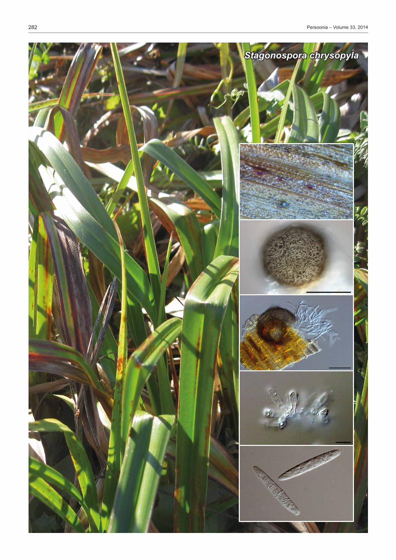
282 Persoonia–Volume33,2014
Stagonospora chrysopyla

283Fungal Planet description sheets
© 2014 Naturalis Biodiversity Center & Centraalbureau voor Schimmelcultures
FungalPlanet317–24November2014
Stagonospora chrysopyla Romberg&Rooney-Latham,sp. nov. Etymology. Named after the location where this fungus was collected, GoldenGateParkConservancy,SanFrancisco,California,USA,chryso =gold, pyla =gate.
Leaf spots spreading longitudinally along the leaf, angular, red-dishbrown,upto5cminlength,edgesindistinct.Conidiomata immersed,globosetosubglobose,upto250μmdiam,darkbrown,scattered,subepidermal;wallof3–6layersofbrowntextura intricata, ostiole indistinct, only present on abaxial sur-face,30–50µmdiam.Conidiophores reduced to conidiogenous cells.Conidiogenous cells hyaline, smooth, lining inner cavity, ampulliformtosubglobose,10–20×5–12μm.Conidia solitary, hyaline, smooth, cylindrical, straight, apex obtuse to subob-tuse,basetruncate,(3–)5–6(–7)-septate,(48–)50–65(–69)×(5–)7–8(–10)μm. Culture characteristics — Colonies flat, circular, with even margins, 50mmdiamafter 2wkat 20°C in dark onPDA.Aerialmyceliumsparse,cream-colouredatfirst,becomingpaleolivaceous-grey to olivaceous-grey, reverse olivaceous-grey (Rayner1970).
Typus. USA,California,MarinCounty,GoldenGateParkConservancy,on leaves of Scirpus microcarpus (Cyperaceae), 15 Jan. 2014,A. Shor (holotypeBPI892895,cultureex-typeCBS137792;ITSsequenceGenBankKM033942,TUBsequenceGenBankKM033943,MycoBankMB809083).
MeganK.Romberg,USDAAPHISPPQNIS,10300BaltimoreAve,Beltsville,MD20705;e-mail:[email protected]
SuzanneRooney-Latham,CaliforniaDepartmentofFoodandAgriculture,3294MeadowviewRoad,Sacramento,CA95832;e-mail: [email protected]
Colour illustrations.LeavesofScirpus microcarpusgrowingattheGoldenGateParkConservancy(photo:A.Shor);leafsurfacewithostiole,pycnidialsurface(bar=100µm),pycnidium(bar=100µm),conidiogenouscellsandconidia(bars=10µm).
Notes—Of themore than500names inStagonospora, fewerthan20arerepresentedintheNCBIGenBank.Showninthe table is a comparison of conidial measurements and number of septa for the species of Stagonospora currently described from Scirpus, as well as larger-spored Stagonosporaspp.de-scribed from hosts in the Cyperaceae that are not represented inGenBank(Tehon1933,Cunnell1957,Castellani&Germano1977).TheconidiaofStagonospora chrysopyla are larger than those of other species of Stagonospora on Scirpus and it is also thefirstStagonospora described from Scirpus microcarpus.Ofthe larger-spored Stagonospora on Cyperaceae, S. chrysopyla most resembles S. dolosa, but differs in having smaller and thin-nerconidiathatare5–6-septateratherthanmainly5-septate.TheconidialsizeofS. chrysopyla also overlaps with that of S. foliicola; however, the examined herbarium specimens of S. foliicola show a great deal of variation and the circumscription ofthisspeciesmayneedtobere-examined.TheITSregionof S. chrysopyla has90% identity toS. foliicola (GenBankKF251256). ITS.BasedonamegablastsearchofNCBIsGenBanknu-cleotidedatabase, theclosesthitsof the ITSsequencehadhighest similarity to S. paludosa (GenBankKF251257;Identities=493/518(95%))andS. pseudocaricis (GenBankKF251260;Identities=494/517(96%)).BothofthesespeciesdifferfromS. chrysopylainconidialsizeandreportedhost(Crousetal.2013,Quaedvliegetal.2013). TUB.BasedonamegablastsearchofNCBIsGenBanknu-cleotidedatabase,theclosesthitsoftheTUBsequencesareS. paludosa(GenBankKF252740,Identities=392/416(94%)) and S. pseudocaricis(GenBankKF252741,Identities=395/ 420(94%)).
Species Conidia(µm)1 No.ofsepta Specimen(s)examined Reportedhost(s)
Stagonospora aquatica 25–30×5–6 3 – ScirpusS. aquaticavar.sexseptata 32–35×5–6 6 – ScirpusS. chrysopyla 50–65 × 7–8 5–6 BPI 892895 ScirpusS. cylindrica 48–78×8–11.5 3(–4) – PhragmitesS. dolosa 60–70×10 5 BPI374901 PhragmitesS. elegans 52–84×8.5–14 (3–)4–6 – PhragmitesS. foliicola 35–90×5.5–9.5 6–12 BPI374911/BPI374909 VariousS. gigaspora 58–84×10–14 6–9 BPI374955 CarexS. maritima 16–20×4–6.5 2 – VariousS. scirpi 20–28×5–6.5 (3–)4–5 – ScirpusS. scirpicola 12–18×2.5–3 3 BPI375122 ScirpusS. scirpini 20 ×3–4 1–4 BPI375126 Scirpus1 ConidialmeasurementsaspresentedinspeciesdescriptionsinTehon(1933),Cunnell(1957),Castellani&Germano(1977)and/ordetermineduponexaminationofherbariumspecimens.
Table Comparison of conidial measurements and number of septa for selected species of Stagonospora.

284 Persoonia–Volume33,2014
Penicillium coccotrypicola

285Fungal Planet description sheets
© 2014 Naturalis Biodiversity Center & Centraalbureau voor Schimmelcultures
FungalPlanet318–24November2014
Penicillium coccotrypicola Holdom,Y.P.Tan&R.G.Shivas,sp. nov. Etymology.Derivedfromthegenericnameofthepalmseedborerweevil(Coccotrypes)thatformedthegalleriesfromwhichthefunguswasisolated.
Myceliumramifiedingalleriesofpalmseedborer(Coccotrypes carpophagus)scolytineweevilsthatinfestseedsofArchonto-phoenix cunninghamiana.Synnemataprotrudefromthefibrousseed husk, blue-grey, clavate, often furcated and slightly flat-tened, up to 1 cm high, solitary or clustered in small coralloid groups.Conidiophores in vivo borne on surface of synnemata; stipes30–120μm,verruculoseorfinelyroughenedtopartlysmooth, subhyaline to pale olivaceous; penicilli biverticillate or terverticillate,rarelymonoverticillate;2–5metulae,onemetulaoftenlargerthanothers,(9–)10.5–13.5(–16.5)×(2.5–)3.5× 4.5(–5)µm,smoothorfinelyroughened,subhyalinetopalegrey-olivaceous.Conidiogenous cells phialides, in verticils of 2–6,acerose,smooth,9–15×2–3.5µm,withadistinctneck,hyalinetosubhyaline.Conidiaglobosetosubglobose(2.5–)3×3.5(–4)µm,smooth,palegrey-olivaceous.Synnematapro-ducedinvitroonCzapekyeastextractagar(CYA)after14dinthedarkat25°C,withsimilarconidiophoresbornefromhy-phaewithstipesupto200µm;metulae(8.5–)9–11.5(–14.5)× 2.5–3.5(–4)µmandphialides(7–)8–10(–12)×2–2.5(–3)µm. Culturecharacteristics—(after7d in thedarkat25°C).OnCYAcolonies40–43mmdiam,velutinous,weaklysulcate,glaucous grey to greenish grey; tinged sienna at centre and white margins, apricot droplets exuded at centre and pale brown soluble pigment released into agar; cinnamon in reverse; no growthat5or37°C.Conidialmassbecomingcinnamononolderculturesonmostmedia; increasingCuandZnhadnoeffect; rare (<1%)germinationbutnogrowthat30°C.Onoatmealagar(OA)andpotatodextroseagar(PDA)colonies40–42mmdiam,with irregularconcentric ringsofgreenishgreysporulation,synnemataforminrings.On25%glycerolnitrateagarcolonies12–14mmdiamwithnosporulation.Oncreatinesucroseagarcolonies27–31mmdiamwithacidpro-duction.OnCzapekagar(CzA)colonies24–30mmdiam.Onmaltextractagar(Pitt1979)colonies29–30mm,sporulationsparse, soluble pigment not released, and colour of culture not changingtocinnamonwithage.Onyeastextractsucroseagarcolonies43–47mmdiam,nosporulation,primrosearoundadarkercentre.OnCYA+5%NaClcolonies26–28mmdiam.Onnitrite-sucroseagarcolonies27–30mmdiam.NogrowthonCzA+proprionicacid.OnCzA+sorbicandbenzoicacidscolonies5–11mmdiam.
Typus.auStralia,Queensland,BliBli,MaroochyWetlands,onseedsofArchontophoenix cunninghamiana and in galleries infested by Coccotrypes carpophagus,6July2013,D. Holdom & J. Hewett(holotypeBRIP59608;ITSsequenceGenBankKM605436,LSUsequenceGenBankKM605437,beta-tubulinsequenceGenBankKM605438,MycoBankMB810327).
Notes — Penicilliums.str.hasrecentlybeenredefinedasamonophyleticgenusbymultilocus(RPB1,RPB2,Tsr1andCct8)phylogeneticanalysis(Houbraken&Samson2011).Theformation of synnemata by species of Penicillium is uncommon (Pitt 1979,Seifert et al. 2004).Penicillium coccotrypicola is morphologically distinct from other species by having furcated synnemata.Penicillium coccotrypicolaproducedramifiedmy-celiuminthegalleriesofpalmseedborerweevils(Coccotrypes carpophagus: Coleoptera, Curculionidae, Scolytinae)inseedsof Archontophoenix cunninghamiana(Arecaceae).Thepalmseed borer weevils in the galleries were not colonised by P. coccotrypicola, which provides evidence that these ambrosia insectswerefarmingthefungus.Possiblemutualisticassocia-tions between Penicillium species and various scolytine weevil genera(Crypturgus, Dendroctonus, Hypothenemus, Ips, Pityo-genes and Tomicus)havebeennotedbefore(Petersonetal.2003,Giordanoetal.2013). ITS. BasedonamegablastsearchoftheNCBIGenBanknu-cleotidedatabase,theclosesthitsusingtheITSsequencearePenicillium mononematosum(ex-typestrainCBS172.87;Gen-BankJX997082;Identities=507/512(99%),Gaps=0(0%)),P. gladioli (ex-type strainNRRL939;GenBankDQ339568;Identities=658/665(99%),Gaps=1/665(0%)),P. confertum (ex-typestrainCBS171.87;GenBankJX997081;Identities=506/512(99%),Gaps0/512(0%))andP. flavigenum(ex-typestrainCBS419.89;GenBankJX997105;Identities=506/512(99%),Gaps=0/512(0%)). BT. BasedonamegablastsearchoftheNCBIGenBanknu- cleotidedatabase,theclosesthitsusingtheBTsequencearePenicillium expansum(ex-typestrainCBS325.48;GenBankJQ965099;Identities=585/634(92%),Gaps=8/634(1%))and P. sclerotigenum(ex-typestrainCBS101033;GenBankAY674393;Identities=429/467(92%),Gaps=6/467(1%)).
DavidG.Holdom,YuPeiTan,JustinS.Bartlett&RogerG.Shivas,BiosecurityQueensland,EcosciencesPrecinct,DepartmentofAgriculture,FisheriesandForestry,DuttonPark4102,Queensland,Australia
e-mail:[email protected],[email protected],[email protected]&[email protected]
Colour illustrations.Archontophoenix cunninghamiana at Mapleton Falls NationalParkQueensland;seedsofA. cunninghamia with synnemata of Penicillium coccotrypicola; 7-d-old cultures onCYA (left) andOA (right);synnemataonOA;conidiaandconidiophoresonCYAafter14d.Scalebars(fromtoptobottom)=1cm,1cm,1cm,1mm,10µm.

286 Persoonia–Volume33,2014
Sistotrema epiphyllum
287Fungal Planet description sheets
© 2014 Naturalis Biodiversity Center & Centraalbureau voor Schimmelcultures
FungalPlanet319–24November2014
Sistotrema epiphyllumStalpers,Stielow&B.deVries,sp. nov.
Colour illustrations.LeavesofFagus sylvatica in beech park; basidia, spores,clamps,basidiomeonleaf.Scalebars=5µm.
Etymology.Growingonleaves:epi=on,phyllon=leaf.
Basidiome resupinate, effused, arachnoid to pellicular and sepa-rable,70–100µmthick.Hymenium even, smooth, becoming moreorlesscontinuous,whitetoslightlygreyish.Margin not dif-ferentiated,hyphalstrandsabsent.Subiculumveryloose.Subi-cular hyphae hyaline to slightly yellowish brownish, thin-walled, 3.5–7(–8)µmwide,contentshyalinetogranular,individualcellssometimesswollen.Subhymenial hyphae hyaline, thin-walled, 3.5–4.5(–5)µmwide.Allseptawithprominentclamps.Cystidia absent.Basidia in small clusters, originating from a sprouting clamp,typicallyurniform,18–25×4.5–5.5µm,with4–6(–7)sterigmata.Sterigmata 3–4µm long.Spores hyaline, thin-walled, ellipsoid, sometimes slightly pip-shaped, flattened at the apicularside,(5.4–)5.7–6.5(–7)×3.3–4(–4.3)µm,notamyloid,notdextrinoid,notcyanophilous.Apiculusshortbutdistinct. Habitat&Distribution—Ontheundersideofbrown,fallenleaves of Fagus sylvatica, which were heaped up in a depres-sionintheterrain.Theaffectedleaveswerenotdirectlyonthesurfaceoftheleafstack,butlowerdown.
Typus.netherlandS,Duivekate,parkforest,20Apr.2013,J.A. Stalpers (holotypeCBSH-21517;leg.B. de Vries,paratypeBdV7510;ITSsequenceGenBankKM401576,MycoBankMB809154).
Notes — Sistotrema epiphyllum is characterised by the com- bination of even and pellicular hymenium and the ellipsoid-flattened spores in the rangeof 5.5–7× 3.3–4.3µm,whilealso the substrate is uncommon for Sistotremaspp.,whichareusually found on wood, or on old Ganoderma basidiomes.Theimpression here is that the species is actually soil-bound and fruits on more solid material, a situation also found in Tomen-tella.MoleculardatahaveprovenSistotrema to be a hetero-genous genus, a situation which has only been partly resolved yet.Basedontheavailablesequences,theclosestrelativeof S. epiphyllum is S. hypogaeum (Warcup&Talbot1962), re-peatedly isolated fromsoil inAdelaide,Australia. Itdiffers inthecylindricaltosubnavicularspores,(5–)7–9×2–2.5µm.Remarkably, the closest relatives to the S. epiphyllum clade belong to the genera Minimedusa and Burgoa, athelioid spe-cies with cylindrical, 4-spored basidia and characterised by the productionofbulbilsorsclerotioidbodiesinculture.Suchbodies(yellowishirregularsclerotium-likemass)arealsoreportedforS. hypogaeum (Warcup&Talbot1962).Unfortunately,culturesofthepresentspeciescouldnotyetbeobtained.Weresub&LeClair(1971)mentionedaconnectionofBurgoa with the Sistotrema brinkmannii complex, based on the occur-rence of swollen cells strongly resembling those of Sistotrema brinkmannii. Minimedusa was considered related, but not con- generic.ThetypespeciesofSistotrema is S. confluens. ITS.BasedonamegablastsearchagainsttheINSDC(Gen-Bank)nucleotidedatabase,theclosesthitsusingthegeneratedITSsequenceareseveralenvironmental (clone)sequencesonly (e.g.DQ309120; Identities = 581/593 (98%),Gaps=8/593(1%)andKF617443;Identities=562/591(95%),Gaps=8/591(1%));theclosestvoucher-derivedsequenceisofSisto- trema coronillaAFTOL-ID618(DQ397337;Identities=513/550(93%),Gaps=13/550(2%)).AmplificationofthelargesubunitD1/D2(28SnrDNA)failed.
JoostA.Stalpers&BenjaminStielow,CentraalbureauvoorSchimmelcultures,Utrecht,TheNetherlands;e-mail:[email protected]&[email protected]
BernarddeVries,Roerdomplaan222,7905ELHoogeveen,TheNetherlands;e-mail:[email protected]
288 Persoonia–Volume33,2014
AaHAvander,VanevS.2002.ArevisionofthespeciesdescribedinPhyl-losticta.CentraalbureauvoorSchimmelcultures,Utrecht,TheNetherlands.
AlvaradoP,MorenoG,ManjónJL,etal.2011.FirstmoleculardataonDe-lastriarosea,FischerulamacrosporaandHydnocystispiligera.BoletíndelaSociedadMicológicadeMadrid35:31–37.
AntonínV,RyooR,ShinHD.2012.Marasmioidandgymnopoid fungioftheRepublicofKorea.4.Marasmiussect.Sicci.MycologicalProgress11:615–638.
AriyawansaHA,PhookamsakR,TibprommaS,etal.2014.AmolecularandmorphologicalreassessmentofDiademaceae.TheScientificWorldJournal:doi.org/10.1155/2014/675348.
ArzanlouM,GroenewaldJZ,GamsW,etal.2007.Phylogeneticandmor-photaxonomic revisionofRamichloridiumandalliedgenera.Studies inMycology58:57–93.
AveskampMM,GruyterJde,WoudenbergJHC,etal.2010.HighlightsoftheDidymellaceae:ApolyphasicapproachtocharacterizePhomaandrelatedpleosporaleangenera.StudiesinMycology65:1–60.
BenschK,BraunU,GroenewaldJZ,etal.2012.ThegenusCladosporium.StudiesinMycology72:1–401.
BraunU,CrousPW,SchubertK,etal.2010.SomereallocationsofStenellaspeciestoZasmidium.Schlechtendalia20:99–104.
BraunU,HillCF.2008.Newspeciesandnewrecordsoffoliicoloushypho-mycetesfromNewZealand.AustralasianMycologist27:45–56.
CastellaniE,GermanoG.1977.LeStagonosporaegraminicole.AnnalidellaFacoltàdiScienzeAgrariedellaUniversitàdegliStudidiTorino10:1–135.
ChomnuntiP,BhatDJ,GarethJonesEB,etal.2012.Trichomeriaceae,anewsootymouldfamilyofChaetothyriales.FungalDiversity56:63–76.
CornerEJH.1996.TheagaricgeneraMarasmius,Chaetocalathus,Crini-pellis,Heimiomyces,Resupinatus,XerulaandXerulinainMalesia.NovaHedwigiaBeihefte111:1–175.
CrousPW,BraunU,GroenewaldJZ.2007a.Mycosphaerellaispolyphyletic.StudiesinMycology58:1–32.
CrousPW,BraunU,SchubertK,etal.2007b.DelimitingCladosporiumfrommorphologicallysimilargenera.StudiesinMycology58:33–56.
CrousPW,GroenewaldJZ,ShivasRG,etal.2011.FungalPlanetdescriptionsheets:69–91.Persoonia26:108–156.
CrousPW,Schoch CL,Hyde KD,etal.2009a.PhylogeneticlineagesintheCapnodiales.StudiesinMycology64:17–47.
CrousPW,SchubertK,BraunU,etal.2007c.Opportunistic,human-patho- genicspeciesintheHerpotrichiellaceaearephenotypicallysimilartosa-probicorphytopathogenicspeciesintheVenturiaceae.StudiesinMycology58:185–217.
CrousPW,ShivasRG,QuaedvliegW,etal.2014.FungalPlanetdescriptionsheets:214–280.Persoonia32:184–306.
CrousPW,SummerellBA,CarnegieAJ,etal.2009b.UnravellingMyco-sphaerella:doyoubelieveingenera?Persoonia23:99–118.
CrousPW,SummerellBA,ShivasRG,etal.2012a.FungalPlanetdescrip-tionsheets:107–127.Persoonia28:138–182.
CrousPW,VerkleyGJM,ChristensenM,etal.2012b.Howimportantareconidialappendages?Persoonia28:126–137.
CrousPW,WingfieldMJ,GuarroJ,etal.2013.FungalPlanetdescriptionsheets:154–213.Persoonia31:188–296.
CrousPW,WingfieldMJ,KochSH.1990.Newand interesting recordsofSouthAfricanfungi.X.NewrecordsofEucalyptusleaffungi.SouthAfricanJournalofBotany56:583–586.
CunnellGJ.1957.Stagonosporaspp.onPhragmitescommunisTrin.Trans-actionsoftheBritishMycologicalSociety40:443–445.
DaiDQ,JayaramaBhatD,LiuJK,etal.2012.Bambusicola,anewgenusfrombamboowithasexualandsexualmorphs.Cryptogamie,Mycologie33:363–379.
DoidgeEM.1942.ReviseddescriptionsofSouthAfricanspeciesofPhylla-choraandrelatedgenera.Bothalia4:421–463.
EllisMB.1971.DematiaceousHyphomycetes.CMI,Kew,England.EllisMB.1976.MoreDematiaceousHyphomycetes.CMI,Kew,England.GamsW.1971.Cephalosporium-artigeSchimmelpilze(Hyphomycetes).Stutt- gart,GustavFischerVerlag.
GamsW,TurhamG.1996.AnextrememodificationofMonocillium:Monocil-liumcurvisetosumn.sp.Mycotaxon59:343–348.
GilkeyHM.1925.Fivenewhypogaeousfungi.Mycologia17:250–255.GilkeyHM.1961.NewspeciesandrevisionsintheorderTuberales.Myco-logia53:215–220.
GiordanoL,GarbelottoM,NicolottiG,etal.2013.Characterizationoffun-galcommunitiesassociatedwiththebarkbeetleIpstypographusvariesdependingondetectionmethod, location,andbeetlepopulation levels.MycologicalProgress12:127–140.
REFERENCES
GiraldoA,GenéJ,CanoJ,etal.2012.TwonewspeciesofAcremoniumfrom Spanishsoils.Mycologia104:1456–1465.
GiraldoA,GenéJ,CanoJ,etal.2014.Acremoniumwithcatenateelongateconidia:phylogenyofAcremoniumfusidioidesandrelatedspecies.Myco-logia106:328–338.
GirlandaM,Luppi-MoscaAM.1998.Monocilliumligusticum,anewspecies frommycorrhizalrootsinMediterraneannorthItaly.Mycotaxon67:265– 274.
GlennA,BaconCW,PriceR,etal.1996.MolecularphylogenyofAcremoniumanditstaxonomicimplications.Mycologia88:369–383.
GomesRR,GlienkeC,VideiraSIR,etal.2013.Diaporthe:agenusofendo-phytic,saprobicandplantpathogenicfungi.Persoonia31:1–41.
GruyterJde,AveskampMM,WoudenbergJHC,etal.2009.Molecularphy-logenyofPhomaandalliedanamorphgenera:Towardsare-classificationofthePhomacomplex.MycologicalResearch113:508–519.
GruyterJde,WoudenbergJHC,AveskampMM,etal.2010.Systematicre- appraisal of species in Phoma section Paraphoma, Pyrenochaeta and Pleurophoma.Mycologia102:1066–1081.
GruyterJde,WoudenbergJHC,AveskampMM,etal.2013.RedispositionofPhoma-likeanamorphsinPleosporales.StudiesinMycology75:1–36.
HashimotoA,SatoG,MatsudaT,etal.2014.MoleculartaxonomyofDin-emasporiumanditsalliedgenera.Mycoscience(inpress)doi.org/10.1016/ j.myc.2014.04.001.
HirookaY,RossmanAY,SamuelsGJ,etal.2012.AmonographofAllan-tonectria,Nectria,andPleonectria(Nectriaceae,Hypocreales,Ascomycota)andtheirpycnidial,sporodochial,andsynnematousanamorphs.StudiesinMycology71:1–210.
HoubrakenJ,SamsonRA.2011.PhylogenyofPenicilliumandthesegrega-tionofTrichocomaceaeintothreefamilies.StudiesinMycology70:1–51.
HughesSJ,KendrickWB.1968.NewZealandfungi12.Menispora,Codinaea,Menisporopsis.NewZealandJournalofBotany6:323–375.
HydeKD,JonesEBG,LuiJ-K,etal.2013.FamiliesofDothideomycetes.FungalDiversity63:1–313.
JohnstonP,Seifert KA,Stone J,etal.2014.Recommendationsongenericnamescompeting foruse inLeotiomycetes (Ascomycota). IMAFungus5:91–120.
JungT,StukelyMJC,HardyGEStJ,etal.2011.MultiplenewPhytophthoraspeciesfromITSClade6associatedwithnaturalecosystemsinAustralia:evolutionaryandecologicalimplications.Persoonia26:13–39.
KimbroughJW,LiLTL,WuCG.1996.Ultrastructuralevidencefortheplace-mentofthetruffleBarssiaintheHelvellaceae(Pezizales).Mycologia88:38–46.
KornerupA,Wanscher JH. 1978.Methuen handbook of colour. 3rd ed.London,EyreMethuen.
KuthubutheenAJ,NawawiA. l991.Akey toDictyochaetaandCodinaeaspecies.MycologicalResearch95:1224–1229.
ŁawrynowiczM,SkirgiełłoA.1984.Barssia,anewgenusinEurope.ActaMycologia20:277–280.
LiuJ-K,PhooksamsakR,DaiD-Q,etal.2014.Roussoellaceae,anewpleo-sporalean family toaccommodate thegeneraNeoroussoellagen.nov.,RoussoellaandRoussoellopsis.Phytotaxa181:1–33.
LombardL,LeeuwenGCMvan,GuarnacciaV,etal.2014.DiaporthespeciesassociatedwithVaccinium,withspecificreferencetoEurope.Phytopatho-logiaMediterranea53:85–97.
Mel’nikV,LeeS,GroenewaldJZE,etal.2004.NewhyphomycetesfromRestionaceaeinfynbos:Parasarcopodiumceratocaryigen.etsp.nov.,andRhexodenticulaelegiaesp.nov.MycologicalProgress3:19–28.
MinnisAM,SundbergWJ.2010.PluteussectionCellulodermaintheU.S.A.NorthAmericanFungi5:1–107.
MontecchiA,SarasiniM.2000.Funghiipogeid’Europa.AssociazioneMico-logicaBresadola.FondazioneCentroStudiMicologici.Trento.
NagRajTR. 1993.Coelomycetous anamorphswith appendage-bearingconidia.MycologuePublications,Waterloo,Ontario.
PapendorfMC.1967.TwonewgeneraofsoilfungifromSouthAfrica.Trans-actionsoftheBritishMycologicalSociety50:69–75.
ParkRF,KeanePJ,WingfieldMJ,etal.2000.Fungaldiseasesofeucalyptfoliage. In:KeanePJ,KileGA,PodgerFD, et al. (eds),Diseasesandpathogensofeucalypts:153–239.CSIROpublishing,Australia.
PascoeIG.1990.ObservationsonascusstructureofPlectosphaeraeucalypti(Phyllachoraceae).MycologicalResearch94:675–684.
PercudaniR,TrevisiA,ZambonelliA,etal.1999.Molecularphylogenyoftruffles(Pezizales:Terfeziaceae,Tuberaceae)derivedfromnuclearrDNAsequenceanalysis.MolecularPhylogeneticsandEvolution13:169–180.
PerdomoH,SuttonDA,GarcíaD,etal.2011.SpectrumofclinicallyrelevantAcremoniumspeciesintheUnitedStates.JournalofClinicalMicrobiology49:243–256.

289Fungal Planet description sheets
© 2014 Naturalis Biodiversity Center & Centraalbureau voor Schimmelcultures
PetersonSW,PérezJ,VegaFE,etal.2003.Penicilliumbrocae,anewspe-ciesassociatedwiththecoffeeberryborerinChipas,Mexico.Mycologia95:141–147.
PhillipsAJL,AlvesA,AbdollahzadehJ,etal.2013.TheBotryosphaeriaceae:generaandspeciesknownfromculture.StudiesinMycology76:51–167.
PittJI.1979.ThegenusPenicilliumanditsteleomorphicstatesEupenicilliumandTalaromyces.AcademicPress,London.
QuaedvliegW,VerkleyGJM,ShinH-D, et al. 2013.SizingupSeptoria.StudiesinMycology75:307–390.
RamaleyAW.2001.Hyalosetanolinae,itsanamorphMonocilliumnolinaeandNiessliaagavacearum,newmembersoftheNiessliaceae,Hypocreales,fromleavesofAgavaceae.Mycotaxon79:267–274.
RaynerRW.1970.Amycologicalcolourchart.CommonwealthMycologicalInstitute,Kew,Surrey,UK.
ReblováM,WinkaK.2000.PhylogenyofChaetosphaeriaanditsanamorphsbasedonmorphologicalandmoleculardata.Mycologia92:939–954.
RuscoeQW.1970.Arxiellalunatasp.nov.TransactionsoftheBritishMyco-logicalSociety55:506–509.
SeifertKA,HoekstraES,FrisvadJC,etal.2004.Penicilliumcecidicola,anewspeciesoncynipid insectgallsonQuercuspacifica in thewesternUnitedStates.StudiesinMycology50:517–523.
SiboeGM,KirkPM,CannonPF.1999.NewdematiaceoushyphomycetesfromKenyanrareplants.Mycotaxon73:283–302.
SiglerL,GibasCFC,KokotovicB,etal. 2010.Disseminatedmycosis inveiledChameleons(Chamaeleocalyptratus)causedbyChamaeleomycesgranulomatis, a new fungus related toPaecilomyces viridis. Journal ofClinicalMicrobiology48:3182–3192.
SummerbellRC,GueidanC,SchroersHJ,etal.2011.Acremoniumphyloge-neticoverviewandrevisionofGliomastix,TrichotheciumandSarocladium.StudiesinMycology68:139–162.
SungGH,Hywel-JonesNL,SungJM,etal.2007.PhylogeneticclassificationofCordycepsandtheclavicipitaceousfungi.StudiesinMycology57:5–63.
SuttonBC.1980.TheCoelomycetes:Fungiimperfectiwithpycnidia,acervuliandstromata.CommonwealthMycologicalInstitute,Kew,Surrey,England.
TakahashiH.2000.ThreenewspeciesofMarasmius sectionSicci fromeasternHonshu,Japan.Mycoscience41:313–321.
TehonLR.1933.NotesontheparasiticfungiofIllinois.Mycologia4:237–257.TrappeJM.1979.Theorders,families,andgeneraofhypogenousAscomy-cotina(trufflesandtheirrelatives).Mycotaxon9:297–340.
UeckerFA.1988.AworldlistofPhomopsisnameswithnotesonnomencla-ture,morphology,andbiology.MycologiaMemoirs13:1–231.
VellingaEC.1990.Pluteus.In:BasC,KuyperThW,NoordeloosME,etal.(eds),FloraAgaricinaNeerlandica,vol.2:31–55.Balkema,Rotterdam.
VerkleyGJM,CrousPW,GroenewaldJZ,etal.2004.Mycosphaerellapuncti- formisrevisited:morphology,phylogeny,andepitypificationofthetypespe-ciesofthegenusMycosphaerella(Dothideales,Ascomycota).MycologicalResearch108:1271–1282.
VerkleyGJM,QuaedvliegW,ShinHD,etal.2013.AnewapproachtospeciesdelimitationinSeptoria.StudiesinMycology75:213–305.
WannathesN,DesjardinDE,HydeKD,etal.2009.AmonographofMaras-mius (Basidiomycota)fromNorthernThailandbasedonmorphologicalandmolecular(ITSsequences)data.FungalDiversity37:209–306.
WarcupJH,TalbotPHB.1962.Ecologyandidentityofmyceliaisolatedfromsoil.TransactionsoftheBritishMycologicalSociety45:495–518.
WeresubLK,LeClairPM.1971.OnPapulasporaandbulbilliferousbasidio-mycetesBurgoaandMinimedusa.CanadianJournalofBotany49:2203– 2213.
WhittonSR,McKenzieEHC,HydeKD.2000.DictyochaetaandDictyochae-topsisspeciesfromthePandanaceae.FungalDiversity4:133–158.
WijayawardeneNN,CrousPW,KirkPM,etal.2014.NamingandoutlineofDothideomycetes–2014.FungalDiversity(inpress).
WikeeS,LombardL,NakashimaC,etal.2013.Aphylogeneticre-evaluationofPhyllosticta(Botryosphaeriales).StudiesinMycology76:1–29.
WoudenbergJHC,GroenewaldJZ,BinderM,etal.2013.Alternariaredefined.StudiesinMycology75:171–212.
ZareR,GamsW,EvansHC.2001.ArevisionofVerticilliumsectionProstrata.V.ThegenusPochonia,withnotesonRotiferophthora.NovaHedwigia73:51–86.
ZhangY,CrousPW,SchochCL,etal.2012.Pleosporales.FungalDiversity53:1–221.
ZhangY,SchochCL,FournierJ,etal.2009.Multi-locusphylogenyofPleo-sporales:ataxonomic,ecologicalandevolutionaryre-evaluation.StudiesinMycology64:85–102.