Fungal Diversity in Deep Sea Hydrothermal Ecosystems
7
APPLIED AND ENVIRONMENTAL MICROBIOLOGY, Oct. 2009, p. 6415–6421 Vol. 75, No. 20 0099-2240/09/$08.000 doi:10.1128/AEM.00653-09 Copyright © 2009, American Society for Microbiology. All Rights Reserved. Fungal Diversity in Deep-Sea Hydrothermal Ecosystems † Thomas Le Calvez, 1 Gae ¨tan Burgaud, 2 Ste ´phane Mahe ´, 1 Georges Barbier, 1 and Philippe Vandenkoornhuyse 2 * UMR 6553 CNRS ECOBIO, FR/90 IFR 2116 CAREN, Universite ´ de Rennes 1, Campus de Beaulieu, 35042 Rennes Cedex, France, 1 and Laboratoire de Biodiversite ´ et Ecologie Microbienne (EA 3882), Universite ´ de Bretagne Occidentale, ESMISAB, Technopo ˆle Brest Iroise, 29280 Plouzane ´, France 2 Received 19 March 2009/Accepted 18 July 2009 Deep-sea hydrothermal ecosystems are considered oases of life in oceans. Since the discovery of these ecosystems in the late 1970s, many endemic species of Bacteria, Archaea, and other organisms, such as annelids and crabs, have been described. Considerable knowledge has been acquired about the diversity of (micro)organisms in these ecosystems, but the diversity of fungi has not been studied to date. These organisms are considered key organisms in terrestrial ecosystems because of their ecological functions and especially their ability to degrade organic matter. The lack of knowledge about them in the sea reflects the widely held belief that fungi are terrestrial organisms. The first inventory of such organisms in deep-sea hydrothermal environments was obtained in this study. Fungal diversity was investigated by analyzing the small-subunit rRNA gene sequences amplified by culture-independent PCR using DNA extracts from hydrothermal samples and from a culture collection that was established. Our work revealed an unsuspected diversity of species in three of the five fungal phyla. We found a new branch of Chytridiomycota forming an ancient evolutionary lineage. Many of the species identified are unknown, even at higher taxonomic levels in the Chytridiomycota, Ascomycota, and Basidiomycota. This work opens the way to new studies of the diversity, ecology, and physiology of fungi in oceans and might stimulate new prospecting for biomolecules. From an evolutionary point of view, the diversification of fungi in the oceans can no longer be ignored. Since the discovery of hydrothermal vent ecosystems 30 years ago, unexpected species diversity has been revealed that has shed light on the functional coupling between the geo- sphere and the biosphere. When submersibles dive to the sea- floor, they bring numerous organisms back to the surface, and this has resulted in the description of nearly two new species per month (10). Deep-sea hydrothermal ecosystems are con- sidered hotspots of microbial diversity on the seafloor. Indeed, they are ecosystems that produce biomass using the wide range of chemical compounds released by the polymetallic sulfite chimneys or “black smokers” that represent the huge quantity of chemical energy that is available (26). The vent fluid, having been heated close to a magma chamber, can have a tempera- ture of 400°C when it is emitted. It is also characterized by a lack of dissolved oxygen, strong acidity (pH 2 to 3), a high concentration of electron donors (i.e., reduced compounds such as methane and hydrogen sulfide), and the presence of heavy metals (36). Continual mixing with the cold ocean water (2 to 4°C) that is rich in electron acceptors creates a dynamic chemical disequilibrium that is a source of energy for micro- organisms that control the rates of redox reactions (16). Each ridge displays varied geochemistry, and the vent fluids differ, even at scales as small as the fractures, pipes, and po- rosities in the black smokers, creating diverse microhabitats for biota (10, 16, 26). Microbes have colonized these different microsites; an impressive diversity of Bacteria and Archaea species has been described, and even more diversity has been inferred. Some organisms are chemolithoautotrophic primary producers and are at the base of the trophic web in these habitats. In addition to these key microorganisms, other pri- mary producers, like the photosynthetic green sulfur bacteria, reportedly are able to harvest energy from geothermal radia- tion (3). The organic compounds produced by the primary producers are consumed by heterotrophic microbes and ani- mals through symbioses, filtering of free-living microorgan- isms, or grazing of biofilms. A comparison of hydrothermal vent cycling of carbon with the carbon cycling in terrestrial ecosystems suggests that key organisms in hydrothermal vents, the fungi, have not been studied yet. Contemporary organisms living in land ecosystems are well adapted. However, the first colonizers of land faced a harsh physical environment (29). Thus, it has been suggested that early colonization of land was possible only when there was a macroevolutionary jump (i.e., acquisition of multiple competences during a single evolutionary step) (24, 29). It has been suggested that establishment of eukaryotes on land may have been possible only as a consequence of the association of a fungus and a phototroph (24, 29). The earliest known fossil fungus (460 million years) was thought to form arbuscular mycorrhizae (27). The idea that the colonization of land was made possible by this symbiotic association is supported the findings of Simon et al. (31). Based on molecular clock esti- mates, some authors have suggested that fungi appeared a long time before Devonian land colonization. The major radiations in the fungi may even have appeared during the Precambrian (1,460 to 960 million years ago) (13), 300 million or more years before colonization of land. These analyses are currently under * Corresponding author. Mailing address: UMR 6553 CNRS ECOBIO, FR/90 IFR 2116 CAREN, Universite ´ de Rennes 1, Campus de Beaulieu, Bat. 14a, 35042 Rennes Cedex, France. Phone: 33-2 23 23 50 07. Fax: 33-2 23 23 68 28. E-mail: [email protected]. † Supplemental material for this article may be found at http://aem .asm.org/. Published ahead of print on 24 July 2009. 6415
-
Upload
univ-brest -
Category
Documents
-
view
1 -
download
0
Transcript of Fungal Diversity in Deep Sea Hydrothermal Ecosystems
APPLIED AND ENVIRONMENTAL MICROBIOLOGY, Oct. 2009, p. 6415–6421 Vol. 75, No. 200099-2240/09/$08.00�0 doi:10.1128/AEM.00653-09Copyright © 2009, American Society for Microbiology. All Rights Reserved.
Fungal Diversity in Deep-Sea Hydrothermal Ecosystems�†Thomas Le Calvez,1 Gaetan Burgaud,2 Stephane Mahe,1
Georges Barbier,1 and Philippe Vandenkoornhuyse2*UMR 6553 CNRS ECOBIO, FR/90 IFR 2116 CAREN, Universite de Rennes 1, Campus de Beaulieu, 35042 Rennes Cedex,
France,1 and Laboratoire de Biodiversite et Ecologie Microbienne (EA 3882), Universite de Bretagne Occidentale,ESMISAB, Technopole Brest Iroise, 29280 Plouzane, France2
Received 19 March 2009/Accepted 18 July 2009
Deep-sea hydrothermal ecosystems are considered oases of life in oceans. Since the discovery of these ecosystemsin the late 1970s, many endemic species of Bacteria, Archaea, and other organisms, such as annelids and crabs, havebeen described. Considerable knowledge has been acquired about the diversity of (micro)organisms in theseecosystems, but the diversity of fungi has not been studied to date. These organisms are considered key organismsin terrestrial ecosystems because of their ecological functions and especially their ability to degrade organic matter.The lack of knowledge about them in the sea reflects the widely held belief that fungi are terrestrial organisms. Thefirst inventory of such organisms in deep-sea hydrothermal environments was obtained in this study.Fungal diversity was investigated by analyzing the small-subunit rRNA gene sequences amplified byculture-independent PCR using DNA extracts from hydrothermal samples and from a culture collectionthat was established. Our work revealed an unsuspected diversity of species in three of the five fungalphyla. We found a new branch of Chytridiomycota forming an ancient evolutionary lineage. Many of thespecies identified are unknown, even at higher taxonomic levels in the Chytridiomycota, Ascomycota, andBasidiomycota. This work opens the way to new studies of the diversity, ecology, and physiology of fungi inoceans and might stimulate new prospecting for biomolecules. From an evolutionary point of view, thediversification of fungi in the oceans can no longer be ignored.
Since the discovery of hydrothermal vent ecosystems 30years ago, unexpected species diversity has been revealed thathas shed light on the functional coupling between the geo-sphere and the biosphere. When submersibles dive to the sea-floor, they bring numerous organisms back to the surface, andthis has resulted in the description of nearly two new speciesper month (10). Deep-sea hydrothermal ecosystems are con-sidered hotspots of microbial diversity on the seafloor. Indeed,they are ecosystems that produce biomass using the wide rangeof chemical compounds released by the polymetallic sulfitechimneys or “black smokers” that represent the huge quantityof chemical energy that is available (26). The vent fluid, havingbeen heated close to a magma chamber, can have a tempera-ture of 400°C when it is emitted. It is also characterized by alack of dissolved oxygen, strong acidity (pH 2 to 3), a highconcentration of electron donors (i.e., reduced compoundssuch as methane and hydrogen sulfide), and the presence ofheavy metals (36). Continual mixing with the cold ocean water(2 to 4°C) that is rich in electron acceptors creates a dynamicchemical disequilibrium that is a source of energy for micro-organisms that control the rates of redox reactions (16).
Each ridge displays varied geochemistry, and the vent fluidsdiffer, even at scales as small as the fractures, pipes, and po-rosities in the black smokers, creating diverse microhabitats forbiota (10, 16, 26). Microbes have colonized these different
microsites; an impressive diversity of Bacteria and Archaeaspecies has been described, and even more diversity has beeninferred. Some organisms are chemolithoautotrophic primaryproducers and are at the base of the trophic web in thesehabitats. In addition to these key microorganisms, other pri-mary producers, like the photosynthetic green sulfur bacteria,reportedly are able to harvest energy from geothermal radia-tion (3). The organic compounds produced by the primaryproducers are consumed by heterotrophic microbes and ani-mals through symbioses, filtering of free-living microorgan-isms, or grazing of biofilms.
A comparison of hydrothermal vent cycling of carbon withthe carbon cycling in terrestrial ecosystems suggests that keyorganisms in hydrothermal vents, the fungi, have not beenstudied yet. Contemporary organisms living in land ecosystemsare well adapted. However, the first colonizers of land faced aharsh physical environment (29). Thus, it has been suggestedthat early colonization of land was possible only when therewas a macroevolutionary jump (i.e., acquisition of multiplecompetences during a single evolutionary step) (24, 29). It hasbeen suggested that establishment of eukaryotes on land mayhave been possible only as a consequence of the association ofa fungus and a phototroph (24, 29). The earliest known fossilfungus (460 million years) was thought to form arbuscularmycorrhizae (27). The idea that the colonization of land wasmade possible by this symbiotic association is supported thefindings of Simon et al. (31). Based on molecular clock esti-mates, some authors have suggested that fungi appeared a longtime before Devonian land colonization. The major radiationsin the fungi may even have appeared during the Precambrian(1,460 to 960 million years ago) (13), 300 million or more yearsbefore colonization of land. These analyses are currently under
* Corresponding author. Mailing address: UMR 6553 CNRS ECOBIO,FR/90 IFR 2116 CAREN, Universite de Rennes 1, Campus de Beaulieu,Bat. 14a, 35042 Rennes Cedex, France. Phone: 33-2 23 23 50 07. Fax: 33-2 2323 68 28. E-mail: [email protected].
† Supplemental material for this article may be found at http://aem.asm.org/.
� Published ahead of print on 24 July 2009.
6415
discussion because of the absence of fossil evidence to supportthe conclusions and because of possible bias in the molecularclock estimates (34). However, assuming that the protein esti-mates are correct, it could be hypothesized that fungi emergedand diversified in water before they colonized land. An ances-tral characteristic, a flagellated gamete without a wall, is foundin old lineages of fungi. This is generally considered an advan-tage for dispersal and reproduction in water. The loss of fla-gella in fungi appears to have occurred in a single evolutionarystep (20) and has been used as an argument to support thedogma that higher fungi diversified on land before a secondarycolonization of water ecosystems, assuming that some terres-trial fungi secondarily invaded salt marshes and mangroves andadapted to life in salt and brackish water. It is a common beliefamong ecologists, microbiologists, and mycologists that fungiare found (almost) exclusively on land. A recent study showedthat the loss of flagella may have occurred at least four timesduring fungal evolution (15), which suggests an evolutionaryparadigm that is more complicated than previously thought.Despite the view that fungi diversified in terrestrial ecosystems,chytrids are considered primarily aquatic fungi. Furthermore,cultural approaches have resulted in the identification of asmall, ecologically defined group of marine higher fungi thatincludes filamentous ascomycetes, their anamorphs, and yeasts(17). We advocate an alternative hypothesis for the loss offlagella. It can be proposed that the loss of motile gametes infungi was compensated for by the resistance and long-rangedispersal of spores. We suggest that this evolutionary innova-tion in eukaryotes should have led to colonization and long-term persistence in many new environments, including land,even if the primitive transition of fungi to terrestrial lifecould have occurred without the previous loss of flagellatedspores. From the molecular clock predictions (13) and par-tially in agreement with our computations (P. Vandenkoorn-huyse, unpublished data), we suggest that this evolutionaryevent might have occurred approximately 1,200 to 1,000million years ago and, therefore, in oceans. If this is true, wewould expect to find a large variety of fungi, including fungiproducing nonflagellated spores, in oceanic samples even inisolated habitats.
Consequently, the main aim of the present work was to testfor the presence of fungi in deep-sea hydrothermal ecosystemsby establishing a culture collection and using a culture-inde-pendent approach involving direct amplification of the fungalsmall-subunit (SSU) rRNA gene from environmental samplesand then analysis and evaluation of the diversity of the fungi inthese environments. The deep-sea hydrothermal ecosystemwas an interesting target to test our hypotheses for variousreasons. First, it has been suggested that the hydrothermalecosystem is the “cradle of life” (14, 18, 39), although morerecently it has been shown that life emerged first in a meso-philic environment and diversified in thermophilic conditions(5). Second, hydrothermal conditions were very common dur-ing the Precambrian (28). Third, use of general eukaryoticprimers to perform culture-independent PCRs (ciPCRs) hasled to the detection of fungi close to hydrothermal vents pre-viously (21, 22). Fungal sequences have also been detected indeep-sea anoxic sediments (2, 8, 9), and barophilic fungalstrains have been isolated from this environment (7).
MATERIALS AND METHODS
Studied sites and sampling. Samples were collected during the following twooceanographic cruises: (i) HERO (30 September 1991 to 4 November 1991) onthe East Pacific Rise at the Elsa site (12°48�N, 103°57�W; depth, 2,630 m) and (ii)MARVEL (29 August 1997 to 13 September 1997) on the Mid-Atlantic Ridge atthe Menez Gwen site (37°51�N, 31°31�W; depth, 860 m) and Lucky Strike site(37°17�N, 32°16�W; depth, 1,700 m). Deep-sea sampling was performed usingDSV Nautile. The support research vessels were R/V Nadir for the HERO cruiseand R/V Atalante for the MARVEL cruise (www.ifremer.fr/fleet//index.php).Animals and rocks were collected using sampling boxes, washed on board,disinfected with ethanol, and filled with sterile seawater. All the boxes werewaterproof to prevent contamination from the water column during ascent. Theywere opened for sampling during dives and then closed and placed in theinsulated crate of the submersible to maintain a low temperature until subse-quent examination on board.
Sample preparation and fungal cultures. On board in the lab, sterility wasobtained with a Bunsen burner and a vertical laminar flow hood. Solid sampleswere removed from the container with sterile strips, placed on a sterile petri dish,rinsed well with sterile seawater, and then crushed with a sterile pestle andmortar. After this, the samples were aliquoted and preserved by deep-freezestorage (�80°C) until they were used.
During the HERO cruise, Sabouraud chloramphenicol solid medium (AESLaboratory) was used for aerobic enrichment cultures incubated at 30°C withatmospheric pressure. During the MARVEL cruise, five solid culture mediawere used. Ac medium contained (per liter) 5 g potato starch (Sigma), 0.5 g yeastextract, 1 g peptone, 30 g sea salts, and 6.05 g piperizine-N,N�-bis(2-ethanesul-fonic acid) (PIPES) buffer (Sigma). The starch was replaced by 5 g cellobiose(Sigma) in Cc medium, by 5 g glucose (Sigma) in Gc medium, by 5 g arabic gumand 5 g olive oil in Lc medium, and by 5 g xylan oat spelt in Xc medium. Pcmedium contained (per liter) 9 g brain heart infusion (Difco), 23 g NaCl, and6.05 g PIPES buffer. The pH was adjusted to 7.5 with 4 N NaOH. The five mediawere supplemented with (per liter) 15 g agar and 500 mg chloramphenicol.Cultures were grown aerobically at 25°C (ambient temperature) and atmosphericpressure. Finally, all strains were able to grow on GYPS medium containing (perliter) 1 g glucose, 1 g yeast extract, 1 g peptone, 1 g starch, and 30 g sea salts. Purecultures were obtained and kept in a culture collection by continuous culture andthen freeze-dried at �80°C. More details about sampling and cultures are avail-able in a recently published paper (6).
Extraction of DNA. Each sample was homogenized with sterile glass beads andground for 28 s at 30 rpm in a bead beater (MM 301; Retsch). Different methodsof DNA extraction to obtain amplifiable fungal DNA were tested. The methodchosen was an DNeasy Plant mini kit (Qiagen) used according to the manufac-turer’s recommendations. DNA was extracted from cultivated strains with a FastDNA Spin kit (MP Biomedicals).
Cloning and sequencing. The fungal SSU rRNA genes were amplified by PCRusing primers MH2 (5�TTCGATGGTAGGATAGAGG3�) and MH4 (5�GTCTCACTAAGCCATTC3�) (37) for environmental sequences and primers NS1(5�GTAGTCATATGCTTGTCTC3�) and ITS5R (5�CCTTGTTACGACTTTTACTTCC3�) primers for cultivated strains (40). The AU2/AU4 primer set (38)did not amplify any of the samples analyzed. DNA was amplified by touchdownPCR with the initial step at 94°C for 1.30 min followed by 37 cycles of 94°C for30 s, 48°C for 1.25 min (with a 0.1°C decrease at each cycle), and 72°C for 1.5min. The PCR amplification program ended with a final elongation step at 72°Cfor 10 min. For the samples which did not yield any product we used a primer setmodified from the primer set described by Smit et al. (32), EF3 (5�TCCTCTAAATGACCAGTTTG3�) and EF4 (5�GGAAGGGNTGTATTTATTAGAT3�),and the same PCR conditions except for the annealing temperature (53°C in-stead of 48°C). For these ciPCRs, a dilution of a soil DNA extract and DNA-freewater were used as positive and negative controls, respectively. The amplificationfragments were purified with a High Pure PCR product purification kit (Roche)and were cloned in the pGEM-T vector (Promega) and DH5� competent cells(Gibco BRL Life Technologies). For each clone library, 16 to 120 randomlyselected positive clones were sequenced (ABI-PRISM Dye Terminator cyclesequencing Ready Reaction kit; Perkin Elmer) on both strands using primers T7and SP6 for environmental sequences and primers NS1, NS3 (5�GCAAGTCTGGTGCCAGCAGCC3�), and ITS5R for the culture sequences (6). The contigof the two sequence strands was calculated using Sequencher 4.6 (GeneCodes),and alignment uncertainties were checked manually. Chimeric artifacts weredeleted from the data set using CHIMERA_CHECK 2.7 (http://rdp.cme.msu.edu/html/analyses.html; Ribosomal Database Project).
rRNA fungal database. A fungal SSU rRNA gene database was created so thatthe detected sequences could be analyzed (http://phymycodb.genouest.org/). Al-
6416 LE CALVEZ ET AL. APPL. ENVIRON. MICROBIOL.

gorithms were used to recover SSU rRNA gene sequences for each fungalphylum from the GenBank/EMBL/DDBJ databases. Filters were used to elim-inate the sequences that were too short (�1,000 bp), too long (�2,500 bp), or ofpoor quality or that included at least 10 consecutive undetermined nucleotides(T. Le Calvez, L. Guillot, A. Dufresne, and P. Vandenkoornhuyse, submitted forpublication). A multiple-sequence alignment was constructed for each phylumusing Clustal X 1.81 with a matrix containing all sequences from our databasesand our hydrothermal sequences.
After using this protocol and completing the various analyses, we were able torecover 1,733 sequences from Basidiomycota, 215 sequences from Chytridiomy-cota, 4,117 sequences from Ascomycota, 292 sequences from Zygomycota, and621 sequences from Glomeromycota, which represented all the different branchesof fungal phylogeny. Each phylum was then subjected to a multiple-sequencealignment procedure, followed by neighbor-joining analyses. Phylogenetic treeswere visualized by using Treeview 1.6.6. The phylogenetic neighbors closest tothe environmental sequences analyzed were selected, and then phylogeneticanalyses were performed (see below).
Diversity and phylogenetic analyses. Rarefaction curves were computed foreach sample. The number of species was determined for 100 random combina-tions of 1 to n sequences by using 100 bootstrap pseudoreplicates (http://viceroy.eeb.uconn.edu/EstimateS) implemented in EstimateS. A multiple-sequencealignment procedure was performed using CLUSTALX 1.81 (35), and the align-ment was refined by eye. This analysis included our environmental sequences,sequences from the fungal cultures isolated, and the most relevant representativeSSU rRNA gene sequences of the Chytridiomycota, Basidiomycota, and Ascomy-cota (93 sequences, including 18 representative environmental sequences and 26sequences from fungal cultures). The alignment was performed with 1,145 nu-cleotides as described above. Other multiple-sequence alignment methods testedgave similar results (not shown). Phylogenetic analyses were performed as fol-lows. CLUSTALX 1.81 was used to obtain neighbor-joining (NJ-K2P) phylo-genies with distance correction, gap omission, and 1,000 bootstrap pseudorepli-cates. PAUP 4.0�10 was used for maximum parsimony analysis using a heuristictree search with 500 random-addition replicates and with tree bisection andreconnection as the branching algorithm, with each of the 500 bootstrap itera-tions utilizing 10 random-addition replicates and tree bisection and reconnectionbranch swapping. In addition, the data were analyzed using the ML-GTR � I �
G procedure with 100 iterations. Modeltest 3.7 software (25) was used to selectthe model.
After these analyses were performed, phylotypes were specified using a cutoffof 98% (pairwise distance computed using PAUP 4.0�10).
Quantitative PCR (qPCR) assays. PCRs were performed with 10-�l (finalvolume) mixtures using iQ SYBR green Supermix (Bio-Rad), which containedSYBR green PCR buffer, 2.7 �M dATP, 2.7 �M dTTP, 2.7 �M dGTP, 2.7 �MdCTP, and 0.42 U of iTaq DNA polymerase (Bio-Rad). Then 0.35 �M primerMH2 (5�TTCGATGGTAGGATAG3�) and 0.35 �M primer FungqPCR1 (5�TGTCGGGATTGGGTAATTT3�) were added to the mixture.
All reactions were performed in optical tubes (Bio-Rad); 8.5 �l of the mastermixture was added first, followed by 1.5 �l of template. The tubes were sealedwith microseal film (Bio-Rad). All reactions were performed with a Chromo 4thermocycler (MJ Research), using an initial denaturation at 94°C for 3 min toactivate the enzyme, followed by 35 or 40 cycles of denaturation at 94°C for 30 sand annealing-extension at 48°C for 45 s and then by plate reading. The disso-ciation curve for temperatures from 65°C to 95°C was measured after the lastqPCR cycle. All data were analyzed using Opticon Monitor 3 (MJ Research).
This approach was used to study six samples in triplicate in two independentruns. The three samples which exhibited the strongest signals in two preliminaryruns were compared in a final run. Seven different plasmid concentrations (109
to 103 copies in 1.5 �l) were used to construct a standard curve for absolutequantification. The numbers of copies in the standards were calculated using thefollowing formula: molecules/�l a/(plasmid length 660) (6.022 1023),where a is the plasmid concentration (in �g/�l), 660 is the average molecularweight of one base pair, and 6.022 1023 is the molar constant.
Nucleotide sequence accession numbers. The sequences reported in thispaper have been deposited in the GenBank database under accession num-bers EF638466 to EF638515 for sample MV2E1, EF638516 to EF638564 forsample H18E12, EF638565 to EF638614 for sample MV2E2, EF638615 toEF638636 for sample MV2E3, EF638637 to EF638654 for sample MV5E1,EF638655 to EF638686 for sample MV5E2, and EF638687 to EF638706 forcultivable strains. The matrix is available upon request from the correspond-ing author.
RESULTS
Diversity of the culture collection. Cultivation and isolationof microbes (Bacteria and Archaea) from deep-sea hydrother-mal samples are difficult, and the procedure includes manyconstraining manipulations. Nevertheless, we were able to es-tablish a fungal collection with different samples from severalsites without using complex culture procedures, such as pres-surized fermentors (see Materials and Methods). The fungiwere isolated using a classic medium supplemented with seasalt at a pressure of 1 atm and 25°C. Physiological analysesdemonstrated that the strains were able to grow in deep-seasalinity (3% [wt/vol] NaCl) and at low temperatures (6). TheSSU rRNA gene sequence analyses revealed that all 21 isolatesin the collection belonged to the phylum Ascomycota, as didpreviously isolated marine strains (15). A subset of the culti-vated Ascomycota belonged to the classes Dothideomycetes(Fig. 1, phylotypes 14 to 16) and Sordariomycetes (Fig. 1, phy-lotypes 17, 18, and 20), and at least one new undescribedphylotype belonging to each of these groups was discovered.Black yeasts were also detected, including two phylotypes ofExophiala (order Chaetothyriales) (Fig. 1, phylotypes 10 to 13),a group that includes pathogens and saprophytes. In a recentstudy, black yeasts were identified in mussel tissues present inhydrothermal samples from the Fidji Basin, where they ap-peared to be mussel parasites, possibly regulating the trophicchain (37). Finally, one phylotype was obtained from culturesthat clustered with the mitosporic Ascomycota (Fig. 1, phylo-type 19), which have been described as “common” pathogenicfungi in tidal seawater (4, 30). As expected based on previousinformation, the spores produced by the cultivated isolateswere not flagellated, even though some isolates appeared to bespecific to the oceanic habitat (for details about the culturecollection, physiology, and morphology, see reference 6).
Fungal diversity determined by a culture-independent ap-proach. We investigated the fungal SSU rRNA genes fromenvironmental samples (Table 1) directly amplified by ciPCRsusing fungus-specific primers. A customized database was con-structed to analyze in depth the SSU rRNA gene sequencesdetected. This database contained all available fungal SSUrRNA sequences that passed filters for length and quality (144Chytridiomycota sequences, 266 Zygomycota sequences, 418Glomeromycota sequences, 2,327 Basidiomycota sequences,and 4,270 Ascomycota sequences), and there was at least onerepresentative of each known fungal genus in the GenBankdatabase. All of these 7,425 sequences, which represent thewhole fungal tree of life, were used to determine, with highconfidence, the phylogenetic affinities of the new sequences. Asecondary matrix of sequences was then constructed usingthese computations along with BLASTn analyses (1), whichincluded the closest relatives of the environmental SSU rRNAsequences analyzed (for each phylum, neighbor-joining phylo-genetic trees obtained using the entire data sets are shown inFig. S1, S2, S3, and S4 in the supplemental material). A totalof 20 distinct phylotypes were detected using the ciPCR resultsand the fungal culture collection (Fig. 1). The ciPCR resultsrevealed previously unsuspected diversity of fungal phylotypesin the vent ecosystems; there were nine distinct phylotypes, fiveof which were new at the genus level or a higher taxonomiclevel in the Chytridiomycota (Fig. 1, phylotypes 1 and 2; see Fig.
VOL. 75, 2009 DEEP-SEA HYDROTHERMAL FUNGI 6417
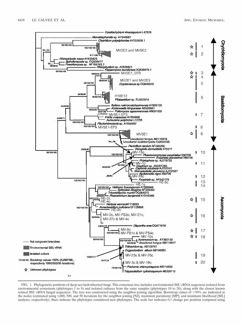
FIG. 1. Phylogenetic positions of deep-sea hydrothermal fungi. This consensus tree includes environmental SSU rRNA sequences isolated fromenvironmental ecosystems (phylotypes 1 to 9) and isolated cultures from the same samples (phylotypes 10 to 20), along with the closest knownrelated SSU rRNA fungal sequences. The tree was constructed using the neighbor-joining algorithm. Bootstrap values of �50% are indicated atthe nodes (estimated using 1,000, 500, and 50 iterations for the neighbor-joining [NJ], maximum parsimony [MP], and maximum likelihood [ML]analyses, respectively). Stars indicate the phylotypes considered new phylotypes. The scale bar indicates 0.1 change per position computed using
6418 LE CALVEZ ET AL. APPL. ENVIRON. MICROBIOL.
S4 in the supplemental material) and Basidiomycota (Fig. 1,phylotypes 3, 4, and 9).
On the one hand, the Basidiomycota phylotypes detectedbelonged to the Agaricomycotina (Fig. 1, phylotypes 3 to 8),mostly to the genus Cryptococcus, a phylotype previously de-tected in another hydrothermal area (33), and the Filobasidium(East Pacific Rise) anamorphs, which have economic, agricul-tural, and medical importance. Phylotype 9 (Fig. 1), a closerelative of uncultured fungi, was detected previously in deep-sea and hydrothermal sediments (2) and fluids (22). Moreover,homobasidiomycete yeasts belonging to the order Auriculari-ales (Fig. 1, phylotype 7) were also found. On the other hand,the Chytridiomycota phylotypes retrieved belonged to speciesthat have not been described previously (Fig. 1, phylotypes 1and 2; see Fig. S4 in the supplemental material) and, in par-ticular, to an apparently ancient evolutionary lineage in theorder Chytridiales. These sequences shared only 95.7% of sim-ilarity with their closest relative, Chytridium polysiphoniae (Fig.1). The phylogenetic position of C. polysiphoniae is uncertainbecause it is a pathogenic fungus (19) and such fungi tend toshow a long-branch attraction bias (Fig. 1). When C. polysi-phoniae was removed along with the other ambiguously placedpathogenic Chytridiomycota, the new group of species formedone of the most ancient branches of the fungal kingdom (seeFig S4 in the supplemental material). It is very probable thatthe phylogenetic position of these newly discovered fungi wasdetermined correctly because the branches in this group wereshort, a result that would not be expected if the terminalorganisms were pathogens. This result is also supported by aBayesian phylogenetic reconstruction (result not shown).
Different fungal communities were discovered when the fun-gal diversities of the Mid-Atlantic Ridge and the East PacificRise were compared; these two ridges are known to havedistinct endemic animal communities (40) (Bathymodiolusazoricus and Alvinella pompejana samples, respectively). How-ever, Agaricomycotina (Fig. 1, phylotypes 3 to 8) and Dothideo-mycetes (Fig. 1, phylotypes 14 to 16) sequences were found in
both ecosystems. Agaromycotina fungi seem to be widely dis-tributed because they were also found in deep-sea sediments(11, 23) and methane seeps (33).
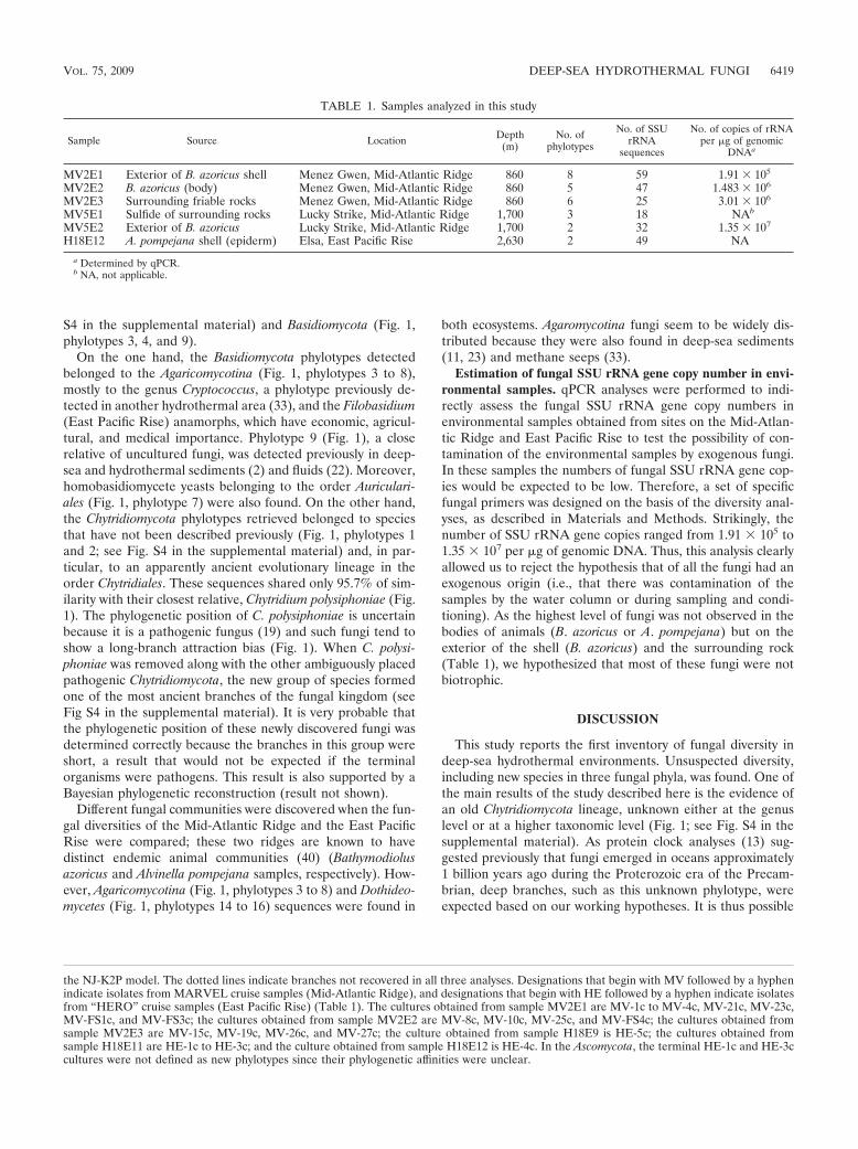
Estimation of fungal SSU rRNA gene copy number in envi-ronmental samples. qPCR analyses were performed to indi-rectly assess the fungal SSU rRNA gene copy numbers inenvironmental samples obtained from sites on the Mid-Atlan-tic Ridge and East Pacific Rise to test the possibility of con-tamination of the environmental samples by exogenous fungi.In these samples the numbers of fungal SSU rRNA gene cop-ies would be expected to be low. Therefore, a set of specificfungal primers was designed on the basis of the diversity anal-yses, as described in Materials and Methods. Strikingly, thenumber of SSU rRNA gene copies ranged from 1.91 105 to1.35 107 per �g of genomic DNA. Thus, this analysis clearlyallowed us to reject the hypothesis that of all the fungi had anexogenous origin (i.e., that there was contamination of thesamples by the water column or during sampling and condi-tioning). As the highest level of fungi was not observed in thebodies of animals (B. azoricus or A. pompejana) but on theexterior of the shell (B. azoricus) and the surrounding rock(Table 1), we hypothesized that most of these fungi were notbiotrophic.
DISCUSSION
This study reports the first inventory of fungal diversity indeep-sea hydrothermal environments. Unsuspected diversity,including new species in three fungal phyla, was found. One ofthe main results of the study described here is the evidence ofan old Chytridiomycota lineage, unknown either at the genuslevel or at a higher taxonomic level (Fig. 1; see Fig. S4 in thesupplemental material). As protein clock analyses (13) sug-gested previously that fungi emerged in oceans approximately1 billion years ago during the Proterozoic era of the Precam-brian, deep branches, such as this unknown phylotype, wereexpected based on our working hypotheses. It is thus possible
the NJ-K2P model. The dotted lines indicate branches not recovered in all three analyses. Designations that begin with MV followed by a hyphenindicate isolates from MARVEL cruise samples (Mid-Atlantic Ridge), and designations that begin with HE followed by a hyphen indicate isolatesfrom “HERO” cruise samples (East Pacific Rise) (Table 1). The cultures obtained from sample MV2E1 are MV-1c to MV-4c, MV-21c, MV-23c,MV-FS1c, and MV-FS3c; the cultures obtained from sample MV2E2 are MV-8c, MV-10c, MV-25c, and MV-FS4c; the cultures obtained fromsample MV2E3 are MV-15c, MV-19c, MV-26c, and MV-27c; the culture obtained from sample H18E9 is HE-5c; the cultures obtained fromsample H18E11 are HE-1c to HE-3c; and the culture obtained from sample H18E12 is HE-4c. In the Ascomycota, the terminal HE-1c and HE-3ccultures were not defined as new phylotypes since their phylogenetic affinities were unclear.
TABLE 1. Samples analyzed in this study
Sample Source Location Depth(m)
No. ofphylotypes
No. of SSUrRNA
sequences
No. of copies of rRNAper �g of genomic
DNAa
MV2E1 Exterior of B. azoricus shell Menez Gwen, Mid-Atlantic Ridge 860 8 59 1.91 105
MV2E2 B. azoricus (body) Menez Gwen, Mid-Atlantic Ridge 860 5 47 1.483 106
MV2E3 Surrounding friable rocks Menez Gwen, Mid-Atlantic Ridge 860 6 25 3.01 106
MV5E1 Sulfide of surrounding rocks Lucky Strike, Mid-Atlantic Ridge 1,700 3 18 NAb
MV5E2 Exterior of B. azoricus Lucky Strike, Mid-Atlantic Ridge 1,700 2 32 1.35 107
H18E12 A. pompejana shell (epiderm) Elsa, East Pacific Rise 2,630 2 49 NA
a Determined by qPCR.b NA, not applicable.
VOL. 75, 2009 DEEP-SEA HYDROTHERMAL FUNGI 6419
that the emergence and initial diversification of fungi occurredin a marine environment. This study supports this hypothesis,but the data are not conclusive. The loss of flagella and there-fore motility in fungal gametes, which likely occurred morethan once (15), does not invalidate our conclusion since thisloss could be compensated for in terms of fitness by the resis-tance and long-range dispersal of both mitotic and meioticspores in aquatic environments. If the molecular clock esti-mates are correct, this hypothesis implies that fungi diversifiedin oceans before they colonized the land during the late Silu-rian or early Devonian period (i.e., approximately 400 millionyears ago). In this study we found a variety of fungi belongingto three of the five fungal phyla, which supports our assump-tion even if the possibility of secondary colonizations from landto marine ecosystems cannot be excluded. This conclusioncontradicts the widely accepted hypothesis that the diversifica-tion of fungi occurred on land. The possible early colonizationof hydrothermal habitats and the ecological function of thedeeply branching Chytridiomycota (Fig. 1; see Fig. S4 in thesupplemental material) are being addressed in an ongoingmetagenomic analysis of the fungi living in deep-sea hydro-thermal ecosystems.
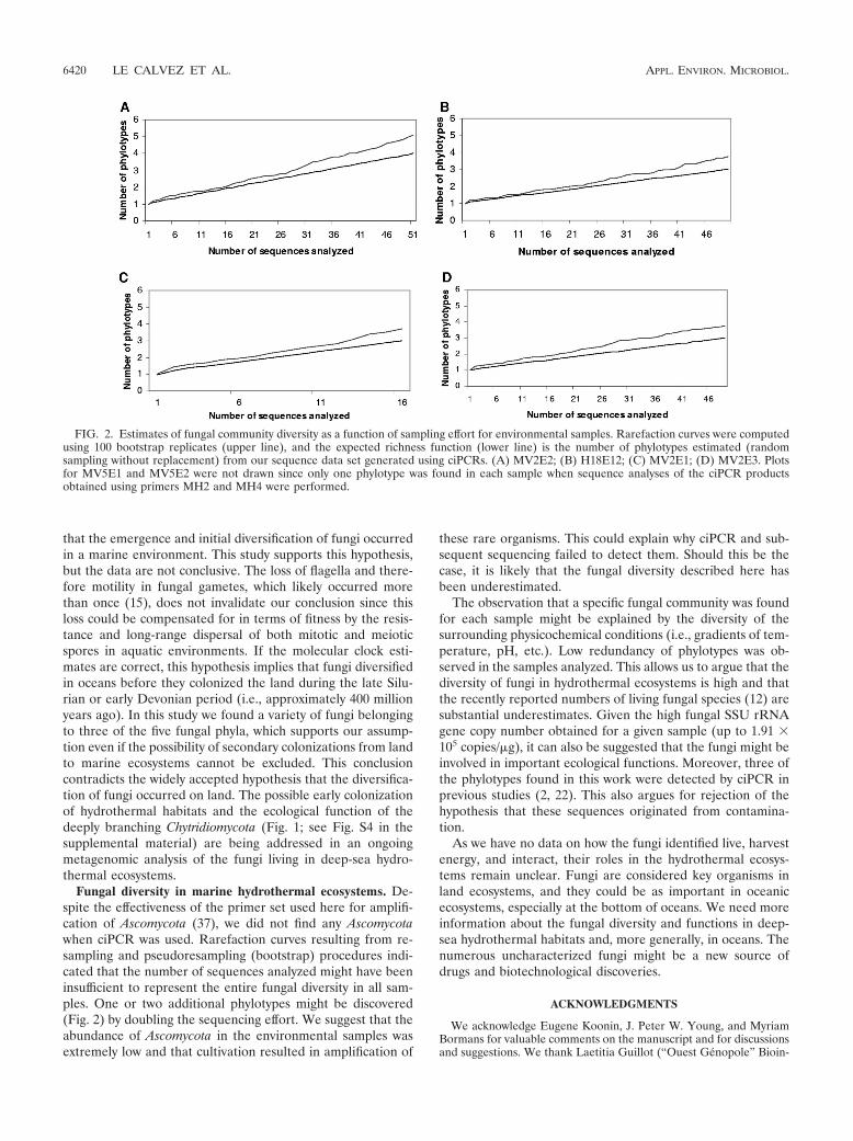
Fungal diversity in marine hydrothermal ecosystems. De-spite the effectiveness of the primer set used here for amplifi-cation of Ascomycota (37), we did not find any Ascomycotawhen ciPCR was used. Rarefaction curves resulting from re-sampling and pseudoresampling (bootstrap) procedures indi-cated that the number of sequences analyzed might have beeninsufficient to represent the entire fungal diversity in all sam-ples. One or two additional phylotypes might be discovered(Fig. 2) by doubling the sequencing effort. We suggest that theabundance of Ascomycota in the environmental samples wasextremely low and that cultivation resulted in amplification of
these rare organisms. This could explain why ciPCR and sub-sequent sequencing failed to detect them. Should this be thecase, it is likely that the fungal diversity described here hasbeen underestimated.
The observation that a specific fungal community was foundfor each sample might be explained by the diversity of thesurrounding physicochemical conditions (i.e., gradients of tem-perature, pH, etc.). Low redundancy of phylotypes was ob-served in the samples analyzed. This allows us to argue that thediversity of fungi in hydrothermal ecosystems is high and thatthe recently reported numbers of living fungal species (12) aresubstantial underestimates. Given the high fungal SSU rRNAgene copy number obtained for a given sample (up to 1.91 105 copies/�g), it can also be suggested that the fungi might beinvolved in important ecological functions. Moreover, three ofthe phylotypes found in this work were detected by ciPCR inprevious studies (2, 22). This also argues for rejection of thehypothesis that these sequences originated from contamina-tion.
As we have no data on how the fungi identified live, harvestenergy, and interact, their roles in the hydrothermal ecosys-tems remain unclear. Fungi are considered key organisms inland ecosystems, and they could be as important in oceanicecosystems, especially at the bottom of oceans. We need moreinformation about the fungal diversity and functions in deep-sea hydrothermal habitats and, more generally, in oceans. Thenumerous uncharacterized fungi might be a new source ofdrugs and biotechnological discoveries.
ACKNOWLEDGMENTS
We acknowledge Eugene Koonin, J. Peter W. Young, and MyriamBormans for valuable comments on the manuscript and for discussionsand suggestions. We thank Laetitia Guillot (“Ouest Genopole” Bioin-
FIG. 2. Estimates of fungal community diversity as a function of sampling effort for environmental samples. Rarefaction curves were computedusing 100 bootstrap replicates (upper line), and the expected richness function (lower line) is the number of phylotypes estimated (randomsampling without replacement) from our sequence data set generated using ciPCRs. (A) MV2E2; (B) H18E12; (C) MV2E1; (D) MV2E3. Plotsfor MV5E1 and MV5E2 were not drawn since only one phylotype was found in each sample when sequence analyses of the ciPCR productsobtained using primers MH2 and MH4 were performed.
6420 LE CALVEZ ET AL. APPL. ENVIRON. MICROBIOL.
formatics) for constructing the fungal database. We are also grateful tothe “Ouest Genopole” sequencing service.
This work was supported by GIS Genomique Marine and by a grantfrom the Total Corporate Foundation for Biodiversity and the Sea.
REFERENCES
1. Altschul, S. F., W. Gish, W. Miller, E. W. Myers, and D. J. Lipman. 1990.Basic local alignment search tool. J. Mol. Biol. 215:403–410.
2. Bass, D., A. Howe, N. Brown, H. Barton, M. Demidova, H. Michelle, L. Li, H.Sanders, S. C. C. Watkinson, S. Willcock, and T. A. A. Richards. 2007. Yeastforms dominate fungal diversity in the deep oceans. Proc. R. Soc. B 274:3069–3077.
3. Beatty, J. T., J. Overmann, M. T. Lince, A. K. Manske, A. S. Lang, R. E.Blankenship, C. L. Van Dover, T. A. Martinson, and E. G. Plumley. 2005. Anobligately photosynthetic bacterial anaerobe from a deep-sea hydrothermalvent. Proc. Natl. Acad. Sci. USA 102:9306–9310.
4. Berbee, M. L., B. Kloeckener, and J. W. Taylor. 1992. Yeast biology. Science257:1610–1611.
5. Boussau, B., S. Blanquart, A. Necsulea, N. Lartillot, and M. Gouy. 2008.Parallel adaptations to high temperatures in the Archaean eon. Nature456:942–945.
6. Burgaud, G., T. Le Calvez, D. Arzur, P. Vandenkoornhuyse, and G. Barbier.2009. Diversity of culturable marine filamentous fungi from deep-sea hydro-thermal vents. Environ. Microbiol. 11:1588–1600.
7. Damare, S., C. Raghukumar, and S. Raghukumar. 2006. Fungi in deep-seasediments of the Central Indian Basin. Deep-Sea Res. Part I 53:14–27.
8. Dawson, R. C., and N. R. Pace. 2002. Novel kingdom-level eukaryotic diver-sity in anoxic environments. Proc. Natl. Acad. Sci. USA 99:8324–8329.
9. Edgcomb, V. P., D. T. Kysela, A. Teske, A. de Vera Gomez, and M. L. Sogin.2002. Benthic eukaryotic diversity in the Guaymas Basin hydrothermal ventenvironment Proc. Natl. Acad. Sci. USA 99:7658–7662.
10. Fisher, C. R., K. Takai, and N. Le Bris. 2007. Hydrothermal vent ecosystems.Oceanography 20:14–23.
11. Gadanho, M., and J. P. Sampaio. 2005. Occurrence and diversity of yeasts inthe Mid-Atlantic Ridge hydrothermal fields near the Azores archipelago.Microb. Ecol. 50:408–417.
12. Hawksworth, D. L. 2001. The magnitude of fungal diversity: the 1.5 millionspecies estimate revisited. Mycol. Res. 105:1422–1432.
13. Heckman, D. S., D. M. Geiser, B. R. Eidell, R. L. Stauffer, N. L. Kardos, andS. B. Hedges. 2001. Molecular evidence for the early colonization of land byfungi and plants. Science 293:1129–1133.
14. Huber, C., and G. Wachtershauser. 1998. Peptides by activation of aminoacids with CO on (Ni,Fe)S surfaces: implications for the origin of life.Science 281:670–672.
15. James, T. Y., F. Kauff, C. L. Schoch, P. B. Matheny, V. Hofstetter, C. J. Cox,G. Celio, C. Gueidan, E. Fraker, J. Miadlikowska, H. Thorsten Lumbsch, A.Rauhut, V. Reeb, A. E. Arnold, A. Amtoft, J. E. Stajich, K. Hosaka, G. H.Sung, D. Johnson, B. O’Rourke, M. Crockett, M. Binder, J. M. Curtis, J. C.Slot, Z. Wang, A. W. Wilson, A. Schubler, J. E. Longcore, K. O’Donnell, S.Mozley-Standridge, D. Porter, P. M. Letcher, M. J. Powell, J. W. Taylor,M. M. White, G. W. Griffith, D. R. Davies, R. A. Humber, J. B. Morton, J.Sugiyama, A. Y. Rossman, J. D. Rogers, D. H. Pfister, D. Hewitt, K. Hansen,S. Hambleton, R. A. Shoemaker, J. Kohlmeyer, B. Volkmann-Kohlmeyer,R. A. Spotts, M. Serdani, P. W. Crous, K. W. Hughes, K. Matsuura, E.Langer, G. Langer, W. A. Untereiner, R. Lucking, B. Budel, D. M. Geiser, A.Aptroot, P. Diederich, I. Schmitt, M. Schultz, R. Yahr, D. S. Hibbett, F.Lutzoni, D. J. McLaughlin, J. W. Spatafora, and R. Vilgalys. 2006. Recon-structing the early evolution of Fungi using a six-gene phylogeny. Nature443:818–822.
16. Jørgensen, B. B., and A. Boetius. 2007. Feast and famine—microbial life inthe deep-sea bed. Nat. Rev. Microbiol. 5:770–781.
17. Kohlmeyer, J., and E. Kohlmeyer. 1979. Marine mycology: the higher fungi.Academic Press, New York, NY.
18. Koonin, E. V., and W. Martin. 2005. On the origin of genomes and cellswithin inorganic compartments. Trends Genet. 21:647–654.
19. Kupper, F. C., I. Maier, D. T. Muller, S. Loiseaux-De Goer, and L. Guillou.2006. Phylogenetic affinities of two eukaryotic pathogens of marine macroal-gae, Eurychasma dicksonii (Wright) Magnus and Chytridium polysiphoniaeCohn. Cryptogam. Algol. 27:165–184.
20. Liu, Y. J., M. C. Hodson, and B. D. Hall. 2006. Loss of the flagellumhappened only once in the fungal lineage: phylogenetic structure of kingdomFungi inferred from RNA polymerase II subunit genes. BMC Evol. Biol.6:74–86.
21. Lopez-García, P., H. Philippe, F. Gail, and D. Moreira. 2003. Autochtho-nous eukaryotic diversity in hydrothermal sediment and experimental micro-colonizers at the Mid-Atlantic Ridge. Proc. Natl. Acad. Sci. USA 100:697–702.
22. Lopez-García, P., A. Vereshchaka, and D. Moreira. 2007. Eukaryotic diver-sity associated with carbonates and fluid-seawater interface in Lost Cityhydrothermal field. Environ. Microbiol. 9:546–554.
23. Nagahama, T., M. Hamamoto, T. Nakase, H. Takami, and K. Horikoshi.2001. Distribution and identification of red yeasts in deep-sea environmentsaround the northwest Pacific Ocean. Antonie van Leeuwenhoek 80:101–110.
24. Pirozynski, K. A., and D. W. Malloch. 1975. The origin of land plants: amatter of mycotrophism. BioSystems 6:153–164.
25. Posada, D., and K. Crandall. 1998. Modeltest: testing the model of DNAsubstitution. Bioinformatics 14:817–818.
26. Ramirez-Llodra, E., T. M. Shank, and C. R. German. 2007. Biodiversity andbiogeography of hydrothermal vent species: thirty years of discovery andinvestigations. Oceanography 20:30–41.
27. Redecker, D., R. Kodner, and L. E. Graham. 2000. Glomalean fungi from theOrdovician. Science 289:1920–1921.
28. Robert, F., and M. Chaussidon. 2006. A paleotemperature curve for thePrecambrian oceans based on silicon isotopes in cherts. Nature 443:969–972.
29. Selosse, M. A., and F. Le Tacon. 1998. The land flora: a phototroph-funguspartnership? Trends Ecol. Evol. 13:15–20.
30. Shearer, C. A., E. Descals, B. Kohlmeyer, J. Kohlmeyer, L. Marvanova, D.Padgett, D. Porter, H. Raja, J. Schmit, H. Thorton, and H. Voglymayr. 2007.Fungal biodiversity in aquatic habitats. Biodivers. Conserv. 16:49–67.
31. Simon, L., J. Bousquet, R. C. Levesque, and M. Lalonde. 1993. Origin anddiversification of endomycorrhizal fungi and coincidence with vascular landplants. Nature 363:67–69.
32. Smit, E., P. Leeflang, B. Glandorf, J. D. van Elsas, and K. Wernars. 1999.Analysis of fungal diversity in the wheat rhizosphere by sequencing of clonedPCR-amplified genes encoding 18S rRNA and temperature gradient gelelectrophoresis. Appl. Environ. Microbiol. 65:2614–2621.
33. Takishita, K., M. Tsuchiya, J. D. Reimer, and T. Maruyama. 2006. Molec-ular evidence demonstrating the basidiomycetous fungus Cryptococcus cur-vatus is the dominant microbial eukaryote in sediment at the KuroshimaKnoll methane seep. Extremophiles 10:165–169.
34. Taylor, J. W., and M. L. Berbee. 2006. Dating divergences in the fungal treeof life: review and new analyses. Mycologia 98:838–849.
35. Thompson, J. D., T. J. Gibson, F. Plewniak, F. Jeanmougin, and D. G.Higgins. 1997. The Clustal_X Windows interface: flexible strategies for mul-tiple sequence alignment aided by quality analysis tool. Nucleic Acids Res.24:4876–4882.
36. Tivey, M. K. 2007. Generation of seafloor hydrothermal vent fluids andassociated mineral deposits. Oceanography 20:50–65.
37. Vandenkoornhuyse, P., and C. Leyval. 1998. SSU rDNA sequencing andPCR-fingerprinting reveal genetic variation within Glomus mossae. Mycolo-gia 90:791–797.
38. Vandenkoornhuyse, P., S. L. Baldauf, J. Straczek, C. Leyval, and J. P. W.Young. 2002. Extensive fungal diversity in plant roots. Science 295:2051.
39. Wachtershauser, G. 1990. Evolution of the first metabolic cycle. Proc. Natl.Acad. Sci. USA 87:200–204.
40. White, T. J., T. Bruns, S. Lee, and J. W. Taylor. 1990. Amplification anddirect sequencing of fungal ribosomal RNA genes for phylogenetics, p.315–322. In M. Innis, D. Gelfand, J. Sninsky, and T. White (ed.), PCRprotocols: a guide to methods and applications. Academic Press, NewYork, NY.
VOL. 75, 2009 DEEP-SEA HYDROTHERMAL FUNGI 6421