Serological evidence for Encephalitozoon cuniculi infection (nosemiasis) in gnotobiotic guineapigs
6
Laboratory Animals (1988) 22, 337-342 337 Serological evidence for Encephalitozoon cuniculi infection (nosemiasis) in gnotobiotic guineapigs R. BOOTI.2, F. VAN KNAPEN2, B. C. KRUIJTI & H. C. WALVOORT3 IDepartment of Animal Supply, 2Laboratory for Parasitology and Mycology and 3Laboratory for Pathology, National Institute of Public Health and Environmental Hygiene, PO Box 1,3720 BA Bilthoven, The Netherlands Summary Encephalitozoon cuniculi infection (nosemiasis) was serologically diagnosed in hysterectomy- derived gnotobiotically reared guineapigs. The probability of vertical transplacental transmission is discussed. Keywords: Guineapig; Encephalitozoon cuniculi; Nosemiasis; Vertical transmission Encephalitozoon cuniculi is an intracellular microsporidian parasite that causes mild, often subclinical disease in a wide range of mammals, including man (Wilson, 1979). The infection is commonly found in rabbits (Gannon, 1980). Infection in guineapigs and other laboratory rodent species might primarily result from cross- contamination from infected rabbits (Gannon, 1980). Reports on naturally occurring E. cuniculi infection in guineapigs are relatively scarce. Perrin (1943) reported the presence of the parasite in the brain in one of 291 guineapigs examined histologically. Also Ruge (1951) and Sebek (1969) each found one animal with lesions in the brain and kidneys respectively. Moffat and Schiefer (1973) detected typical microgranulomata containing E. cuniculi in the brain and kidneys qf 2 healthy and 2 dead guineapigs. Finally, Gannon (1980) detected E. cuniculi associated kidney lesions, but no cerebral infection in 12 guineapigs showing relatively high titres at serological screening. Based on serological surveys (Chalupsky et al., 1979; Gannon, 1980), Received 11 September 1986. Accepted 29 March 1988. E. cuniculi infections seem to be common in con- ventional guineapig colonies. In infected colonies 29-85% of the animals were considered to be seropositive. No serological evidence for E. cuni- culi infection was found in hysterectomy-derived barrier-maintained guineapig colonies (Gannon, 1980), indicating that rederivation by hyster- ectomy can be used to eliminate the infection. Transmission of the infection occurs most probably through ingestion or inhalation of spores of E. cuniculi, since spores are passed pro- fusely in the urine of infected rabbits (Cox et al., 1972; Pye & Cox, 1977; Cox et al., 1979). Trans- placental transmission of E. cuniculi has been claimed for mice (Perrin, 1943; Innes et 01., 1962), blue foxes (Mohn et al., 1974) and rabbits (Hunt et 01., 1972; Cox et al., 1977), but from studies in both naturally and experimentally infected rabbits, Owen and Gannon (1980) concluded that infections are due to horizontal contamination. Recently Wilson (1986) reported that trans- placental transmission of E. cuniculi was not detected in rabbits and mice challenged with E. cuniculi at different stages of gestatio}l. In a hysterectomy-derived gnotobiotically reared guineapig colony that was examined for the presence of (potentially) pathogenic micro- organisms we found serological evidence for E. cuniculi infection in the gnotobiotic animals. The possibility of vertical, transplacental transmission of nosemiasis in guineapigs is discussed. Materials and methods Animals Hysterectomy was performed on 69 conventional guineapigs (housed separately from rabbits)
-
Upload
independent -
Category
Documents
-
view
3 -
download
0
Transcript of Serological evidence for Encephalitozoon cuniculi infection (nosemiasis) in gnotobiotic guineapigs
Laboratory Animals (1988) 22, 337-342 337
Serological evidence for Encephalitozoon cuniculi infection(nosemiasis) in gnotobiotic guineapigsR. BOOTI.2, F. VAN KNAPEN2, B. C. KRUIJTI & H. C. WALVOORT3
IDepartment of Animal Supply, 2Laboratory for Parasitology and Mycology and 3Laboratory for Pathology,National Institute of Public Health and Environmental Hygiene, PO Box 1,3720 BA Bilthoven, The Netherlands
SummaryEncephalitozoon cuniculi infection (nosemiasis)was serologically diagnosed in hysterectomy-derived gnotobiotically reared guineapigs. Theprobability of vertical transplacental transmissionis discussed.
Keywords: Guineapig; Encephalitozoon cuniculi;Nosemiasis; Vertical transmission
Encephalitozoon cuniculi is an intracellularmicrosporidian parasite that causes mild, oftensubclinical disease in a wide range of mammals,including man (Wilson, 1979). The infection iscommonly found in rabbits (Gannon, 1980).Infection in guineapigs and other laboratoryrodent species might primarily result from cross-contamination from infected rabbits (Gannon,1980). Reports on naturally occurring E. cuniculiinfection in guineapigs are relatively scarce.Perrin (1943) reported the presence of theparasite in the brain in one of 291 guineapigsexamined histologically. Also Ruge (1951) andSebek (1969) each found one animal with lesionsin the brain and kidneys respectively. Moffat andSchiefer (1973) detected typical microgranulomatacontaining E. cuniculi in the brain and kidneysqf 2 healthy and 2 dead guineapigs. Finally,Gannon (1980) detected E. cuniculi associatedkidney lesions, but no cerebral infection in 12guineapigs showing relatively high tit res atserological screening. Based on serological surveys(Chalupsky et al., 1979; Gannon, 1980),
Received 11 September 1986. Accepted 29 March 1988.
E. cuniculi infections seem to be common in con-ventional guineapig colonies. In infected colonies29-85% of the animals were considered to beseropositive. No serological evidence for E. cuni-culi infection was found in hysterectomy-derivedbarrier-maintained guineapig colonies (Gannon,1980), indicating that rederivation by hyster-ectomy can be used to eliminate the infection.
Transmission of the infection occurs mostprobably through ingestion or inhalation ofspores of E. cuniculi, since spores are passed pro-fusely in the urine of infected rabbits (Cox et al.,1972; Pye & Cox, 1977; Cox et al., 1979). Trans-placental transmission of E. cuniculi has beenclaimed for mice (Perrin, 1943; Innes et 01., 1962),blue foxes (Mohn et al., 1974) and rabbits (Huntet 01., 1972; Cox et al., 1977), but from studiesin both naturally and experimentally infectedrabbits, Owen and Gannon (1980) concluded thatinfections are due to horizontal contamination.Recently Wilson (1986) reported that trans-placental transmission of E. cuniculi was notdetected in rabbits and mice challenged withE. cuniculi at different stages of gestatio}l. Ina hysterectomy-derived gnotobiotically rearedguineapig colony that was examined for thepresence of (potentially) pathogenic micro-organisms we found serological evidence for E.cuniculi infection in the gnotobiotic animals. Thepossibility of vertical, transplacental transmissionof nosemiasis in guineapigs is discussed.
Materials and methodsAnimalsHysterectomy was performed on 69 conventionalguineapigs (housed separately from rabbits)
338
under strict aseptic conditions as described byBoot and Walvoort (1984). The gravid uterus wasligated at the end of both horns and at thecervical ends and then removed from the animal.The entirely closed uterus was passed into anautoclaved (121 ec, 20 min) stainless steel isolatorsystem via a germicidal dunk tank containing a 2070solution of Lyorthol (active substances benzyl-chlorphenol and phenylphenol; Medica B.V., DenBosch, The Netherlands) prepared immediatelybefore use. Young animals were born within theisolator by incising the uterus and removing fetalmembranes using sterile instruments. From the9 groups of 6-9 females, 260 young animals wereborn, which were raised gnotobiotically (Boot &Walvoort, 1984) within 9 separate stainless-steelisolators. The relationship between parents andyoung animals within the groups was notrecorded. Litter size ranged between 2 and 5animals. E. cuniculi seronegative, gnotobioticCRF-mice, containing a strict anaerobic coloniz-ation resistant microflora (Van der Waaij et 01. ,1971) were housed with the guineapigs for up to3 weeks. Initially autoclaved (121 ec, 45 min)semi-synthetic Lobund-L478 diet (Tekland TestDiets, Madison, Wisconsin, USA) was provided.After 3 weeks a 2· 5 mRad ')'-irradiated com-mercial pelle ted diet (Hope Farms BV, Woerden,The Netherlands) was fed.
Forty animals died within 2 days due topremature delivery. During the first 3 weeks afterbirth 10 more animals died. On the basis of grosspathological lesions, tissues from these animalswere selected for bacteriological examination.Samples from affected organs were inoculatedonto 2 plain sheep-blood agars, a sheep-bloodagar containing nalidixic acid (40 JLg/ml) and anEndo agar. A sheep-blood agar was incubatedanaerobically and all other media were incubatedaerobically for 48 h at 37 ec, read, reincubatedfor 3 more days and then discarded since nogrowth was obtained. After the first 3 weeks,9 groups of 11-36 animals, totalling 192 youngwere successfully raised. During the study surplusmales were eliminated from the different groups,thus finally the 9 groups contained 11-15animals, totalling 115 young. The gnotobiotic
Boot, van Knapen, Kruijt & Walvoort
offspring from the 9 different groups of femalesremained separated during this study. Bacterialcontamination (apart from the deliberatelyintroduced CRF-flora) was not detected in faecalsamples from the different groups of young atthe end of this study.
SerologySera were sampled at hysterectomy from all but8 of the females. At 7 to 17 weeks after birth 6animals per group (group size 11-36 animals)were sampled. Four to 10 weeks afterwardssampling of 5 seronegative groups (group size11-15 animals) was repeated (Table 1: groups1-5; 6 animals per group). The repeat test wasperformed on sera from animals not testedbefore. Sampling was performed by cardiacpuncture under ether anaesthesia within theisolator using sterile equipment. Sera were storedat - 20 ec until examination.
Immunofluorescence test (IF)The E. cuniculi used as antigen originated froma spontaneously infected rabbit. Homogenate ofthe rabbit kidney was inoculated intraperitoneallyin cortisone treated mice. Rabbit kidney cell(RK 13) cultures were then inoculated withspores from infected mouse ascites fluid asdescribed by Waller (1975). Spores were har-vested by pouring off the medium, filtrationthrough a 10 ILm membrane filter, centrifu-gation (10 000 g for 15 min) and resuspension inphosphate buffered saline (PBS) pH 7, 2 (3washing steps). Spores were counted using ahaemocytometer under phase-contrast andadjusted to a concentration of 108 spores/ml,2· 5 ILlof the counted spores were dropped perslide, air dried, fixed in methanol and stored at- 20 ec before use.
The IF-test was performed as describedby Cox et al. (1972) using a FITC-Iabelled goatanti-guineapig conjugate (Nordic, Tilburg, TheNetherlands) diluted 1: 20. All sera werescreened at 1 : ·10 dilution in phosphate bufferedsaline (PBS) pH 7, 2. Positive sera were re-examined using doubling dilutions up to 1 : 160in PBS.
Vertical transmission of Encephalitozoon cuniculi
HistologyFive seropositive healthy young animals (Table1: groups 5-9, tit res 80-160) were at the age of20 weeks submitted for postmortem examination.Samples of brain, liver and kidney were fixed in4070 neutral buffered formaldehyde and preparedfor histopathological evaluation. Tissue sectionswere stained with haematoxylin and eosin andwith Gram's stain. Selected sections were stainedaccording to Van Gieson's method for collagenfibres (Luna, 1968, slightly modified) and withthe modified congo red method to identifyamyloid according to Pearse (1968). In addition,brain and kidney sections of one animal wereexamined for E. cuniculi parasites using aperoxidase anti peroxidase labelling method asdescribed for Toxoplasma using rabbit anti-Encephalitozoon antiserum (Conley et al., 1981).
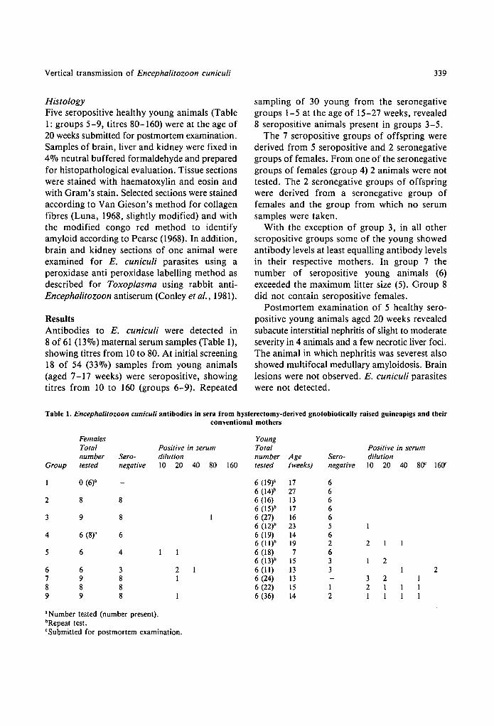
ResultsAntibodies to E. cuniculi were detected in8 of 61 (13%) maternal serum samples (Table 1),showing tit res from 10 to 80. At initial screening18 of 54 (33%) samples from young animals(aged 7-17 weeks) were seropositive, showingtitres from 10 to 160 (groups 6-9). Repeated
339
sampling of 30 young from the seronegativegroups 1-5 at the age of 15-27 weeks, revealed8 seropositive animals present in groups 3-5.
The 7 seropositive groups of offspring werederived from 5 seropositive and 2 seronegativegroups of females. From one of the seronegativegroups of females (group 4) 2 animals were nottested. The 2 seronegative groups of offspringwere derived from a seronegative group offemales and the group from which no serumsamples were taken.
With the exception of group 3, in all otherseropositive groups some of the young showedantibody levels at least equalling antibody levelsin their respective mothers. In group 7 thenumber of seropositive young animals (6)exceeded the maximum litter size (5). Group 8did not contain seropositive females.
Postmortem examination of 5 healthy sero-positive young animals aged 20 weeks revealedsubacute interstitial nephritis of slight to moderateseverity in 4 animals and a few necrotic liver foci.The animal in which nephritis was severest alsoshowed multi focal medullary amyloidosis. Brainlesions were not observed. E. cuniculi parasiteswere not detected.
Table 1. Encephalitozoon cuniculi antibodies in sera from hysterectomy-derived gnotobiotically raised guineapigs and theirconventional mothers
Females YoungTotal Positive in serum Total Positive in serumnumber Sero- dilution number Age Sero- dilution
Group tested negative 10 20 40 80 160 tested (weeks) negative 10 20 40 80' 160'
0(6)" 6 (19)" 17 66 (14)b 27 6
2 8 8 6 (16) 13 66 (1W 17 6
3 9 8 6 (27) 16 66 (12)b 23 5
4 6 (8)" 6 6 (19) 14 66 (lI)b 19 2 2
5 6 4 6 (18) 7 66 (13)b 15 3 2
6 6 3 2 6 (11) 13 3 27 9 8 1 6 (24) 13 3 28 8 8 6 (22) 15 1 2 I9 9 8 6 (36) 14 2 I I
"Number tested (number present).bRepeat test.'Submitted for postmortem examination.
340
DiscussionThe prevalence of E. cuniculi seropositive animalsin our conventional breeding colony (13070)appeared to be considerably lower than theprevalences reported by Chalupsky et al. (1979)(29-85070) and by Gannon (1980) (46-63070).Titres detected in females and their respectiveoffspring in this study were clearly lower thanobserved by Chalupsky et al. (1979) and Gannon(1980). The prevalence within guineapig coloniesmight depend on husbandry conditions. Gannon(1980) considered guineapigs housed with in-fected rabbits at greater risk of becominginfected than those housed separately. Ourconventional guineapigs were kept separatedfrom other laboratory animal species. Differencesin prevalence and titres observed might at leastpartly be due to differences in serologicalmethods employed. Both Chalupskyet al. (1979)and Gannon (1980), using an immunofluorescence(IF) test and an immunoperoxidase test re-spectively, found uniformly low titres (e.g. upto I : 32) in some colonies of guineapigs. We usedthe IF-test as described by Cox et al. (1972).Differences in antibody tit res may also be basedon the choice of the antigen and the method ofpreparation. We used an E. cuniculi strain isolatedfrom a rabbit, maintained in rabbit kidney cellculture. Chalupsky et al. (1979) used a mousestrain obtained from rabbit choroid plexus cellcultures. Gannon (1980) used a strain of unknownorigin maintained in mice (Gannon, 1978).Although Chalupsky et al. (1971) did not findimmunological differences between E. cuniculistrains from different laboratory animals, andother studies (Wilson, 1979) suggest the existenceof just one Encephalitozoon species, differencesin antigen preparation methods might inducedifferences in the expression of surface antigensand hence influence the sensitivity of the test.
The presence of E. cuniculi antibodies inhysterectomy-derived gnotobiotically reared off-spring may partly result from the transmissionof maternal antibodies (Brambell, 1970). How-ever, at 15-19 weeks after birth, some of theyoung showed tit res higher than their respectivemothers. Moreover maternal antibodies could
Boot, van Knapen, Kruijt & Walvoort
not have been present in young of group 8.Although this might suggest contamination,because of precautions taken at hysterectomy,accidental horizontal transmission of infectiousagents (including E. cunicull) from mother toyoung during this operation is very unlikely. Atfirst sight offspring in groups 3-5 appear to showa very late seroconversion (e.g. in group 3 aftermore than 16 weeks), suggesting accidentalcontamination during rearing. However, post-mortem examination of young animals whichdied during the first 3 weeks of the rearing periodand examination of faecal samples at the end ofthe study, revealed no bacteriological contamin-ations by aerobic and anaerobic cultivation (datanot shown), excluding accidental introduction ofagents afterwards. Based on the relatively smallnumber of animals tested (6 out of 18-27 ingroups 3-5) it is far more likely that seropositiveanimals were actually present within these'seronegative' groups at initial sampling. Thepresence of seropositive young animals derivedfrom seronegative mothers (group 8) might bevery well explained by the presence of oneor more females that had recently contractedthe infection and had not yet produced demon-strable antibodies at hysterectomy. The datalend full support to the vertical transplacentaltransmission of E. cuniculi infection in ourguineapigs.
The only valid report on vertical transmissionof E. cuniculi infection is the detection ofencephalitozoonosis in 3 gnotobiotic rabbits bornthrough caesarian section (Hunt et al., 1972). Allother claims must be rejected since horizontaltransmission may well have occurred duringnormal delivery, rearing by the mother (Mohnet al., 1974; Cox et al., 1977), or rearing byfoster-mothers (Perrin, 1943) that were notexamined for the absence of E. cuniculi. Inneset al. (1962) considered E. cuniculi infection tobe vertically transmitted based on the finding ofE. cuniculi parasites in a SPF-breeding colonyderived from germfree mice, but the reportremains unclear about the hygienic measurestaken to prevent horizontal contamination.Failure to demonstrate E. cuniculi infection
Vertical transmission of Encephalitozoon cuniculi
e.g. in hysterectomy-derived offspring fromexperimentally infected susceptible rabbits(Wilson, 1986) does not exclude the possibilityof transplacental transmission of the parasite.Wilson's study revealed reproductive failure in13 of 16 E. cuniculi seronegative does that werechallenged with the parasite at different stagesof gestation, but normal reproductive perfor-mance in challenged seropositive does and inuntreated animals. Apparently only tissues from3 healthy litters born from E. cuniculi challengedseronegative does, but not from aborted fetuses,were examined for the presence of the parasite.
The absence of clinical symptoms attributableto E. cuniculi infection in our study agrees withthe general view that the infection is often onlyfound at postmortem examination (Wilson,1979). The inability to detect the parasite inhistological sections in our animals is probably
ReferencesBoot R & Walvoort HC (1984) Vertical transmiSSIon
of Bacil/us piliformis infection (Tyzzer's disease) ina guineapig: case report. Laboratory Animals 18,195-199
Brambell FWR (1970)The transmission of passive immunityfrom mother to young. Frontiers of Biology, vol. 18.Amsterdam-London: North Holland
Chalupsky J, Bedrnik P & Vavra J (1971) The indirectfluorescent antibody test for Nosema cuniculi. Journal ofProtozoology 18 (Supplement), 47
Chalupsky J, Vavra J & Bedmik P (1979)Encephalitozoonosisin laboratory animals - a serologica] survey. FoliaParasitologica (Praha) 26, 1-8
Conley FK, Jenkins KA & Remington JS (1981) Toxoplasmagondii infection of the central nervous system. Use of theperoxidase-ant i-peroxidase method to demonstrate toxo-plasma in formalin fixed, paraffin embedded tissuesections. Human Pathology 12, 690-698
Cox JC, Walden NB & Nairn RC (1972) Presumptivediagnosis of Nosema cuniculi in rabbits by immuno-fluorescence. Research in Veterinary Science 13, 595-597
Cox JV, Gallichio HA, Pye D & Walden NB (1977) Applic-ation of immunofluorescence to the establishment of anEncephalitozoon cuniculi-free rabbit colony. LaboratoryAnimal Science 27, 204-209
Cox JC, Hamilton RC & Attwood HD (1979)An investigationof the route and progression of Encephalitozoon cuniculiinfection in adult rabbits. Journal of Protozoology 26,260-265
Gannon J (1978) The immunoperoxidase test diagnosis ofEncephalitozoon cuniculi in rabbits Laboratory Animals12, 125-127
341
related to the advanced stage of the lesions. Inaddition, studies in rabbits (Waller, 1977;Gannon, 1978) indicated that compared withserology, histology is an inferior method ofdiagnosing E. cuniculi infection. The interstitialnephritis observed at postmortem examinationwas considered a remnant of E. cuniculi infection,but it must be admitted that seronegativeguineapigs were not examined for kidneypathology.
We conclude that our findings providestrong evidence for the possibility of verticaltransplacental transmission of E. cuniculi inguineapigs.
AcknowledgmentsWe would like to thank G. van Amerongen,G. van der Lugt, C. Moolenbeek, H. Naringand J. L. Veenema for technical assistance.
Gannon J (1980) A survey of Encephalitozoon cuniculi inlaboratory anima] colonies in the United Kingdom.Laboratory Animals 14, 91-94
Hunt RD, King NW & Foster HL (1972)Encephalitozoonosis: evidence for vertical transmission.Journal of Infectious Diseases 126, 212-214
Innes JRM, Zeman W, Frenke] JK, Borner G & Wright R(1962) Occult endemic encephalitozoonosis of the centralnervous system of mice. Acta Neuropathologica Suppl. I,97-99
Luna LG (1968) Manual of Histological Staining Methods,3rd edn, pp. 76-77. New York: McGraw-Hili
Moffatt RE & Schiefer B (1973) Microsporidiosis (Encephal-itozoonosis) in the guineapig. Laboratory Animal Science23, 282-284
Mohn SF, Nordstoga K, Krogsrud J & Helgeboslad A (1974)Transplacental transmission of Nosema cuniculi in the bluefox (Alopex lagopus). Acta Pathologica MicrobiologicaScandinavica, B 82, 299-300
Owen DG & Gannon J (1980) Investigation into thetransplacental transmission of Encephalitozoon cuniculiin rabbits. Laboratory Animals 14, 35-38
Pearse AGE (1968) Histochemistry, vol. I, 3rd edn, p. 684.Boston: Little, Brown & Co
Perrin TL (1942) Spontaneous and experimental Enceph-alitozoon infection in laboratory animals. Archives ofPathology 36, 559-567
Pye D & Cox JC (1977) Isolation of Encephalitozoon cuniculifrom urine samples. Laboratory Animals 11, 233-234
Ruge H (195]) Encephalitozoon beim Meerschweinchen.Zentralblatt fuer Bakteriologie und Parasitenkunde(Originalreihe) 156, 543-544
342
Sebek Z (1969) The finding of the genus Nosema Nageli, 1857(Microsporidia: Nosematidae) in the kidney of a guineapig.Folia Paraistologica (Praha) 16, 165-169
Van der Waaij D, Berghuis-de Vries 1M & Lekkerkerker-van der Wees lEC (1971) Colonization resistance of thedigestive tract in conventional and antibiotic treated mice.Journal of Hygiene (Cambridge) 69, 405-411
Waller T (1975) Growth of Nosema cuniculi in establishedcell lines. Laboratory Animals 9, 61-68
Boot, van Knapen, Kruijt & Walvoort
Waller T (1977) The India-ink immunoreaction: a methodfor the rapid diagnosis of encephalitozoonosis. LaboratoryAnimals 11, 93-97
Wilson 1M (1979) The biology of Encephalitozoon cuniculi.Medical Biology 57, 84-101
Wilson 1M (1986) Can Encephalitozoon cuniculi crossthe placenta? Research in Veterinary Science 40,138