Serological, molecular and entomological surveillance demonstrates widespread circulation of West...
10
Serological, Molecular and Entomological Surveillance Demonstrates Widespread Circulation of West Nile Virus in Turkey Koray Ergunay 1 , Filiz Gunay 2 , Ozge Erisoz Kasap 2 , Kerem Oter 3 , Sepandar Gargari 4 , Taner Karaoglu 4 , Seda Tezcan 5 , Mehmet Cabalar 6 , Yakup Yildirim 7 , Gu ¨ rol Emekdas 5 , Bulent Alten 2 , Aykut Ozkul 4 * 1 Faculty of Medicine, Department of Medical Microbiology, Virology Unit, Hacettepe University, Ankara, Turkey, 2 Faculty of Sciences, Department of Biology, Division of Ecology, Hacettepe University, Ankara, Turkey, 3 Faculty of Veterinary Medicine, Department of Parasitology, Istanbul University, Istanbul, Turkey, 4 Faculty of Veterinary Medicine, Department of Virology, Ankara University, Ankara, Turkey, 5 Faculty of Medicine, Department of Medical Microbiology, Mersin University, Mersin, Turkey, 6 Faculty of Veterinary Medicine, Department of Virology, Harran University, Ankara, Turkey, 7 Faculty of Veterinary Medicine, Department of Virology, Kafkas University, Ankara, Turkey Abstract West Nile virus (WNV), a mosquito-borne flavivirus with significant impact on human and animal health, has recently demonstrated an expanded zone of activity globally. The aim of this study is to investigate the frequency and distribution of WNV infections in potential vectors and several mammal and avian species in Turkey, where previous data indicate viral circulation. The study was conducted in 15 provinces across Turkey during 2011–2013. In addition, the entomological study was extended to 4 districts of the Turkish Republic of Northern Cyprus. WNV exposure was determined in humans, horses, sheep and ducks from Mersin, Sanliurfa, Van and Kars provinces of Turkey, via the detection of neutralizing antibodies. WNV RNA was sought in human and equine samples from Mersin, Adana and Mugla provinces. Field-collected mosquitoes from 92 sites at 46 locations were characterized morphologically and evaluated for viral RNA. Neutralizing antibodies were identified in 10.5% of the 1180 samples studied and detected in all species evaluated. Viral nucleic acids were observed in 5.9% of 522 samples but only in horses. A total of 2642 mosquito specimens belonging to 15 species were captured, where Ochlerotatus caspius (52.4%), Culex pipiens sensu lato (24.2%) comprise the most frequent species. WNV RNA was detected in 4 mosquito pools (1.9%), that comprise Oc. caspius Cx. pipiens s.l. and DNA barcoding revealed the presence of Cx. quinquefasciatus and Cx. perexiguus mosquitoes in infected Culex pools. All WNV partial sequences were characterized as lineage 1 clade 1a. These findings indicate a widespread WNV activity in Turkey, in Eastern Thrace and Mediterranean- Aegean regions as well as Southeastern and Northeastern Anatolia. Citation: Ergunay K, Gunay F, Erisoz Kasap O, Oter K, Gargari S, et al. (2014) Serological, Molecular and Entomological Surveillance Demonstrates Widespread Circulation of West Nile Virus in Turkey. PLoS Negl Trop Dis 8(7): e3028. doi:10.1371/journal.pntd.0003028 Editor: Remi Charrel, Aix Marseille University, Institute of Research for Development, and EHESP School of Public Health, France Received April 8, 2014; Accepted June 6, 2014; Published July 24, 2014 Copyright: ß 2014 Ergunay et al. This is an open-access article distributed under the terms of the Creative Commons Attribution License, which permits unrestricted use, distribution, and reproduction in any medium, provided the original author and source are credited. Data Availability: The authors confirm that all data underlying the findings are fully available without restriction. All relevant data are within the paper and its Supporting Information files. All West Nile virus sequences characterized in the study have been deposited in the GenBank public repository, with the accession numbers: KJ433827, KJ433828, KJ433829, KJ433830, KJ433831, KJ433832, KJ433833, KJ433834, KJ433835, KJ433836, KJ433837, KJ433838, KJ433839 and KJ433840. All mosquito sequences characterized in the study have been deposited in the GenBank public repository, with the accession numbers: KJ012103, KJ012162, KJ012163, KJ012168, KJ012169, KJ012170 and KJ012171. Funding: The study was supported by the Turkish Council of Scientific Research (Grant number: SBAG-110S404, www.tubitak.gov.tr, primary investigator: AO) and Hacettepe University Research Fund (Grant number: 01 001 11 002, www.research.hacettepe.edu.tr, primary investigator: KE). The funders had no role in study design, data collection and analysis, decision to publish, or preparation of the manuscript. Competing Interests: The authors have declared that no competing interests exist. * Email: [email protected] Introduction West Nile virus (WNV) is a re-emerging arthropod-borne virus with a significant impact on human and animal health [1]. WNV demonstrates an extensive zone of distribution throughout Africa, the Middle East, southern Europe, western Russia, southwestern Asia, and Australia [2,3]. The global epidemiology of WNV has drastically changed during the last decades, with the introduction and spread of the virus in the American continent and increased reporting of virus activity in Europe, probably influenced by the interaction of factors such as global warming, demographic changes and modern transportation [3–5]. Since 1990s, the human disease incidence of WNV strains with probable African origin have increased in parts of Russia and southern, central and eastern Europe, with large outbreaks of notable severity occurring in Romania, Russia, Israel, Italy and Greece [3]. In the western hemisphere, WNV has spread from its initial site of detection in 1999 across North America and now circulates in Mexico, South America, and the Caribbean [6,7]. In the United States, WNV resurgence was observed in 2012 after several years of decreasing incidence [5]. Taxonomically, WNV is classified in the Japanese encephalitis serocomplex of the Flavivirus genus within Flaviviridae family, along with more than 70 viruses including important human pathogens such as dengue and yellow fever viruses [8,9]. Similar to other flaviviruses, WNV is an enveloped virus with a single- stranded, positive sense, 11-kb RNA genome that transcribes a single polyprotein, cleaved by host and viral proteases into structural and nonstructural viral proteins [10]. WNV strains PLOS Neglected Tropical Diseases | www.plosntds.org 1 July 2014 | Volume 8 | Issue 7 | e3028
-
Upload
independent -
Category
Documents
-
view
2 -
download
0
Transcript of Serological, molecular and entomological surveillance demonstrates widespread circulation of West...

Serological, Molecular and Entomological SurveillanceDemonstrates Widespread Circulation of West Nile Virusin TurkeyKoray Ergunay1, Filiz Gunay2, Ozge Erisoz Kasap2, Kerem Oter3, Sepandar Gargari4, Taner Karaoglu4,
Seda Tezcan5, Mehmet Cabalar6, Yakup Yildirim7, Gurol Emekdas5, Bulent Alten2, Aykut Ozkul4*
1 Faculty of Medicine, Department of Medical Microbiology, Virology Unit, Hacettepe University, Ankara, Turkey, 2 Faculty of Sciences, Department of Biology, Division of
Ecology, Hacettepe University, Ankara, Turkey, 3 Faculty of Veterinary Medicine, Department of Parasitology, Istanbul University, Istanbul, Turkey, 4 Faculty of Veterinary
Medicine, Department of Virology, Ankara University, Ankara, Turkey, 5 Faculty of Medicine, Department of Medical Microbiology, Mersin University, Mersin, Turkey,
6 Faculty of Veterinary Medicine, Department of Virology, Harran University, Ankara, Turkey, 7 Faculty of Veterinary Medicine, Department of Virology, Kafkas University,
Ankara, Turkey
Abstract
West Nile virus (WNV), a mosquito-borne flavivirus with significant impact on human and animal health, has recentlydemonstrated an expanded zone of activity globally. The aim of this study is to investigate the frequency and distribution ofWNV infections in potential vectors and several mammal and avian species in Turkey, where previous data indicate viralcirculation. The study was conducted in 15 provinces across Turkey during 2011–2013. In addition, the entomological studywas extended to 4 districts of the Turkish Republic of Northern Cyprus. WNV exposure was determined in humans, horses,sheep and ducks from Mersin, Sanliurfa, Van and Kars provinces of Turkey, via the detection of neutralizing antibodies. WNVRNA was sought in human and equine samples from Mersin, Adana and Mugla provinces. Field-collected mosquitoes from92 sites at 46 locations were characterized morphologically and evaluated for viral RNA. Neutralizing antibodies wereidentified in 10.5% of the 1180 samples studied and detected in all species evaluated. Viral nucleic acids were observed in5.9% of 522 samples but only in horses. A total of 2642 mosquito specimens belonging to 15 species were captured, whereOchlerotatus caspius (52.4%), Culex pipiens sensu lato (24.2%) comprise the most frequent species. WNV RNA was detected in4 mosquito pools (1.9%), that comprise Oc. caspius Cx. pipiens s.l. and DNA barcoding revealed the presence of Cx.quinquefasciatus and Cx. perexiguus mosquitoes in infected Culex pools. All WNV partial sequences were characterized aslineage 1 clade 1a. These findings indicate a widespread WNV activity in Turkey, in Eastern Thrace and Mediterranean-Aegean regions as well as Southeastern and Northeastern Anatolia.
Citation: Ergunay K, Gunay F, Erisoz Kasap O, Oter K, Gargari S, et al. (2014) Serological, Molecular and Entomological Surveillance Demonstrates WidespreadCirculation of West Nile Virus in Turkey. PLoS Negl Trop Dis 8(7): e3028. doi:10.1371/journal.pntd.0003028
Editor: Remi Charrel, Aix Marseille University, Institute of Research for Development, and EHESP School of Public Health, France
Received April 8, 2014; Accepted June 6, 2014; Published July 24, 2014
Copyright: � 2014 Ergunay et al. This is an open-access article distributed under the terms of the Creative Commons Attribution License, which permitsunrestricted use, distribution, and reproduction in any medium, provided the original author and source are credited.
Data Availability: The authors confirm that all data underlying the findings are fully available without restriction. All relevant data are within the paper and itsSupporting Information files. All West Nile virus sequences characterized in the study have been deposited in the GenBank public repository, with the accessionnumbers: KJ433827, KJ433828, KJ433829, KJ433830, KJ433831, KJ433832, KJ433833, KJ433834, KJ433835, KJ433836, KJ433837, KJ433838, KJ433839 and KJ433840.All mosquito sequences characterized in the study have been deposited in the GenBank public repository, with the accession numbers: KJ012103, KJ012162,KJ012163, KJ012168, KJ012169, KJ012170 and KJ012171.
Funding: The study was supported by the Turkish Council of Scientific Research (Grant number: SBAG-110S404, www.tubitak.gov.tr, primary investigator: AO)and Hacettepe University Research Fund (Grant number: 01 001 11 002, www.research.hacettepe.edu.tr, primary investigator: KE). The funders had no role instudy design, data collection and analysis, decision to publish, or preparation of the manuscript.
Competing Interests: The authors have declared that no competing interests exist.
* Email: [email protected]
Introduction
West Nile virus (WNV) is a re-emerging arthropod-borne virus
with a significant impact on human and animal health [1]. WNV
demonstrates an extensive zone of distribution throughout Africa, the
Middle East, southern Europe, western Russia, southwestern Asia,
and Australia [2,3]. The global epidemiology of WNV has drastically
changed during the last decades, with the introduction and spread of
the virus in the American continent and increased reporting of virus
activity in Europe, probably influenced by the interaction of factors
such as global warming, demographic changes and modern
transportation [3–5]. Since 1990s, the human disease incidence of
WNV strains with probable African origin have increased in parts of
Russia and southern, central and eastern Europe, with large
outbreaks of notable severity occurring in Romania, Russia, Israel,
Italy and Greece [3]. In the western hemisphere, WNV has spread
from its initial site of detection in 1999 across North America and
now circulates in Mexico, South America, and the Caribbean [6,7].
In the United States, WNV resurgence was observed in 2012 after
several years of decreasing incidence [5].
Taxonomically, WNV is classified in the Japanese encephalitis
serocomplex of the Flavivirus genus within Flaviviridae family,
along with more than 70 viruses including important human
pathogens such as dengue and yellow fever viruses [8,9]. Similar to
other flaviviruses, WNV is an enveloped virus with a single-
stranded, positive sense, 11-kb RNA genome that transcribes a
single polyprotein, cleaved by host and viral proteases into
structural and nonstructural viral proteins [10]. WNV strains
PLOS Neglected Tropical Diseases | www.plosntds.org 1 July 2014 | Volume 8 | Issue 7 | e3028

can be designated into at least 5 phylogenetic lineages, however,
only lineage 1 and 2 isolates have been associated with significant
outbreaks in humans [3,11]. The virus is normally maintained and
spread through a bird-mosquito cycle involving ornithophilic
mosquitoes belonging to Culex species. However, it can also be
spread to a wide range of incidental hosts including humans and
horses, via mammophilic and/or anthropophilic mosquito species
(including Aedes spp. and Ochlerotatus spp.). WNV has the
potential to cause severe illness characterized by neurological
disorders in some of these susceptible species including horses and
humans [12,13].
Turkey, located in the northeastern part of the Mediterranean
region, is considered as a potentially endemic zone for several
arthropod-borne viral infections, including WNV, due to suitable
ecological and climatic conditions [14]. A considerable body of
evidence that demonstrate the presence and activity of WNV in
Turkey have accumulated. Serological surveillance data suggested
human and animal exposure to WNV in some provinces, and an
outbreak in 2010 involving individuals as well as sporadic cases since
2009, indicate symptomatic infections [15–17]. A recent report also
revealed WNV infection in mosquitoes capable of virus transmision
to mammalian species in a region neighboring Greece, the site of a
concominant WNV outbreak in 2010 [18]. However, current
information on virus epidemiology and expansion is only prelim-
inary, with cross-sectional data originating from localized, dissipated
regions. Moreover, definitive information on WNV activity is
lacking in several regions harboring probable vector activity [14].
This study was undertaken to investigate the frequency and
distribution WNV infection in vector mosquitoes and several
mammal and avian species encompassing a large geographical area,
to better understand virus epidemiology and to predict regions with
higher risk for exposure that require established surveillance.
Methods
Study DesignThe study was conducted in 15 provinces, distributed across
Turkey, covering 21.6% of the land area (175.918/814.578 km2)
of the country and in 4 districts of the Turkish Republic of
Northern Cyprus, covering 76.6% of the land area (2.570/
3.355 km2) (Figure 1).
Ethics StatementThe study and associated protocols were designed based on
national ethical legislative rules and approved by Local Ethic
Committees of Mersin University (Nr: MULEC/01.09.10; for
human cases) and Ankara University (Nr: AULEC/201-96-346;
for animal samples). All samples were collected after written
informed consent of the individuals (blood donors), according to
the updated version of the Declaration of Helsinki (Seul, 2008), as
indicated in the aforementioned agreements. All animal samplings
were conducted based on the national regulations on the operation
and procedure of animal experiments ethics committees (Regula-
tion Nr.26220, Date:09.7.2006). Written informed consent of
animal owners were also carried out prior to sampling.
Serological Screening of WNV Exposure in AnimalsWNV exposure was evaluated in sera obtained from horses,
sheep and ducks from Sanliurfa, Van and Kars provinces as well as
human plasma samples from Mersin province, via the detection of
specific neutralizing antibodies by plaque reduction neutralization
test (PRNT). In PRNT, WNV strain NY99-4132 and Vero cells
(ATCC CCL81) were employed and the test was performed as
previously described with minor modifications [19]. Briefly,
0.1 mL diluted serum (1:10) was inactivated at 56uC for 30 min
and mixed with an equal volume of virus in Dulbecco’s modified
Eagle’s medium containing 5% fetal calf serum to produce an
estimated 100 plaque-forming units of virus per 0.2 mL. Virus-
antibody mixtures were then inoculated as 0.2 mL volumes onto
Vero cell monolayers in 12-well plates and overlaid with 3.2%
carboxymethyl cellulose in 26 Dulbecco’s modified Eagle’s
medium. Plaques were scored on 4th day following incubation
at 37uC. Sera that produced 90% neutralization of the challenge
virus (PRNT90) were considered reactive. Convescelent sera from
PCR-confimed WNV exposure in an equine and human subject
were employed as positive controls. All experiments were
performed in duplicate.
Mosquito Sampling and ProcessingField sampling of mosquitoes was performed in 11 provinces in
Turkey and at 4 provinces in the Turkish Republic of Northern
Cyprus, during May-September from 2011 to 2013 (Figure 1,Table S1). Sampling was carried out in Adana and Mersin
provinces in 2011; in Adana, Ankara, Edirne and Tekirdag
provinces in 2012; in Artvin, Bursa, Edirne, Kirklareli, Sakarya,
Samsun, Sinop, Tekirdag provinces and in Lefkosa, Girne,
Magosa and Guzelyurt provinces of Cyprus in 2013. A total of
92 sites at 46 locations in suburban environments around villages
were sampled using CDC Miniature Light Traps (John W. Hock
Company, Gainesville, FL, USA). Furthermore, mouth aspirators
were employed for collecting adult mosquitoes from inside and
outside houses and barns at each site. Light traps were placed 1–
2 meters above ground in peridomestic sites and left on site from
18:00 to 06:00 each night. Captured mosquitoes were collected
next morning, kept alive and transferred on ice. The morpholog-
ical identification of the captured mosquitoes to species level was
accomplished by using published keys [20,21]. In individual Culexspecimens, which were either damaged to prevent identification
(denoted as Culex spp.) or observed to belong in Culex pipienssensu lato complex, legs were dissected and kept in 95% ethyl
alcohol. Subsequently, all specimens were pooled according to the
collection site and species to include 1–20 individuals regardless of
Author Summary
West Nile virus (WNV), frequently transmitted to humansand other susceptible species via bites of infectedmosquitoes, is a significant global public health threat.Limited information is available on WNV epidemiology inTurkey, located in the endemic zone of the agent aroundthe Mediterranean Sea. This study was performed to revealWNV activity in potential hosts and vector mosquitoes,involving 11 provinces in Turkey and the Turkish Republicof Northern Cyprus during 2011–2013. Our findingsindicate virus exposure in humans and various animals inpreviously unexplored regions as well as a high rate ofvirus circulation in equine blood samples during themosquito season. Field-captured mosquito specimensdemonstrated the presence of major WNV vectors among15 species identified. WNV infection was detected in 1.9%of the pooled mosquito specimens. Molecular character-ization of the individual mosquitoes of the infected poolsrevealed Culex quinquefasciatus and Cx. perexiguus species,important WNV vectors where Culex quinquefasciatus notpreviously known to exist in Turkey. Partial viral genomesequences obtained from infected hosts were character-ized as lineage 1, the predominant pathogenic WNV straincirculating in Europe as well as the American Continent.
Widespread Circulation of West Nile Virus in Turkey
PLOS Neglected Tropical Diseases | www.plosntds.org 2 July 2014 | Volume 8 | Issue 7 | e3028

sex, and stored at 280uC. Mosquito pools were homogenized as
described previously and clarified by centrifugation at 4000 rpm
for 4 minutes, prior to nucleic acid purification [22].
Qualitative and Quantitative Detection of WNV NucleicAcids
Plasma samples from asymptomatic blood donors and horses
from Mersin province, horses from Adana and Mugla provinces
and mosquito pool supernatants were used for WNV RNA
detection. Each sample was subjected to nucleic acid purification
by High Pure Viral Nucleic Acid Kit (Roche Diagnostics,
Mannheim, Germany), followed by reverse transcription via
random hexamer primers using RevertAid First Strand cDNA
Synthesis Kit (Thermo Scientific, Tokyo, Japan). WNV RNA was
sought via two specific polymerase chain reaction (PCR) assays; a
nested PCR targeting E protein-coding region and an in-house
quantitative one-step real-time Reverse Transcription (rRT) PCR,
targeting 39 non-coding region in the viral genome, as described
previously [18,23]. Amplicons obtained from Vero cell culture
grown NY99-4132 strain were cloned in pTZ57R plasmid via T4
DNA Ligase (Thermo Scientific, Tokyo, Japan), quantitated
spectrophotometrically and used as standards in 10-fold dilutions
in the rRT-PCR assay, carried out using QuantiTect Probe RT-
PCR kit (Qiagen, Germany) in a Rotor-Gene 6000 instrument
(Corbett Research, Australia). The nested PCR amplicons of the
expected size of 248 basepairs were detected under ultraviolet light
after electrophoresis in 1.7% agarose gels. In positive samples,
amplicons of the nested PCR were purified using High Pure PCR
Product Purification Kit (Roche Diagnostics, Mannheim,
Germany) and subjected to nucleotide sequencing with forward
and reverse amplification primers.
DNA Barcoding in MosquitoesA 658-bp sequence of the cytochrome c oxidase I (COI) gene,
widely used for biological barcoding, was amplified in WNV-
positive mosquito pools, composed of specimens identified
morphologically as Culex spp. or as Culex pipiens sensu lato, for
further characterization [24]. Dissected legs from individual
mosquitoes, stored in 95% ethyl alcohol, were processed with
DNeasy Blood & Tissue Kit (Qiagen, Hilden, Germany) and
amplified with primers LCO1490 and HCO2198 as described
previously [24]. PCR products were cleaned up using High Pure
PCR Product Purification Kit (Roche Diagnostics, Mannheim,
Germany) and subjected to nucleotide sequencing with forward
and reverse primers.
Sequencing and Phylogenetic AnalysisNested PCR amplicons from all WNV positive mosquito pools,
viremic animals and COI amplicons from mosquito specimens
were characterized by sequencing using an ABI Prism 310 Genetic
Analyzer (Applied Biosystems, Foster City, CA, USA). The
sequences obtained were aligned and analyzed using CLC Main
Workbench v5.5 (CLCBio, Aarhus, Denmark) and subsequently,
by MEGA software v5.2 [25]. Phylogenetic trees were constructed
using the Jukes-Cantor substitution rate model with 500 bootstrap
replicates. Maximum likelihood trees were generated based on the
Unweighted Pair Group Method with Arithmetic Mean (UP-
GMA) tree.
Figure 1. Illustrative map of provinces targeted for sampling in the study. (red circle: entomological screening, blue triangle: serologicalscreening, green square: Viral RNA screening.)doi:10.1371/journal.pntd.0003028.g001
Widespread Circulation of West Nile Virus in Turkey
PLOS Neglected Tropical Diseases | www.plosntds.org 3 July 2014 | Volume 8 | Issue 7 | e3028

Results
WNV Neutralizing Antibodies in Humans and AnimalsA total of 1180 sera, obtained from horses (389, 42.6%) from
Sanliurfa and Van provinces, ducks (423, 46.2%) from Kars
province, sheep (102, 11.2%) from Sanliurfa province and
humans (266, 22.5%) from Mersin province were included in
the PRNT. Neutralizing antibodies were detected in 124
samples, and overall WNV seroprevalance was calculated as
10.5%. Due to the suboptimal storage and transport conditions,
the samples were not included in viral RNA investigations,
except for human samples from Mersin province. Seropositivity
rates according to the species and sampling locations are
provided in Table 1.
WNV Nucleic Acids in Human and Equine SamplesWNV RNA was investigated in 522 samples that comprise 266
human plasma (50.9%) from Mersin province and 256 equine
plasma (49.1%) from Mersin, Adana and Mugla provinces.
Viremia was detected in a total of 31 samples (31/522, 5.9%),
via nested and rRT-PCR assays. All samples with positive WNV
RNA originated from equines without clinical symptoms and no
viremic human sample was noted. Due to the limited amounts of
available material, WNV PRNT was not performed in these
samples. Viral RNA detection rates according to the species and
sampling locations are provided in Table 1. Viral loads
determined in positive samples were relatively low, observed
within 102 to104 copies/mL range. Amplicons of nested PCR were
sequenced and characterized in all positive samples (GenBank
accession numbers: KJ433827-KJ433836) All sequences were
grouped with WNV lineage 1 clade 1a isolates, and demonstrated
0–17% intramural divergence in the maximum composite
likelihood analyses (Figure 2, Table S2). Partial sequences with
the highest variation rates were detected in the same location, and
in different years (2011 and 2012). Comparison of the current
sequences with those obtained from horses with neurological
disease from Central Anatolia in 2011 revealed 3–17% nucleotide
divergence (Table S2). Three sequences characterized in Mugla
province were identical to patient-derived sequences from Ankara
(Central Anatolia) and Tekirdag (Eastern Thrace) provinces, as
well as mosquito-derived sequences from Eastern Thrace in 2012.
Otherwise, overall similarities of 72.29–99.60% and 84–97.6%
were observed among sequences identified previously in Turkey
and from lineage 1 strains from Hungary, Egypt and New York,
respectively (Table S2).
Species distribution and WNV detection in
mosquitoes. A total of 2642 mosquito specimens belonging to
15 species were captured in locations in Turkey and Northern
Cyprus, which comprise Ochlerotatus caspius (1385, 52.4%), Cx.pipiens s. l. (640, 24.2%), Cx. theileri (203, 7.7%), Anophelesmaculipennis s. l. (166, 6.3%), Coquillettidia richiardii (114, 4.3%),
Culex spp. (47, 1.8%), Dahliana geniculata (36, 1.4%), Cx.tritaeniorhynchus (14, 0.52%), Cx. torrentium (14, 0.52%), Cx.pusillus (7, 0.3%), An. superpictus (6, 0.2%), An. claviger (3,
0.1%), Culiseta longiareolata (3, 0.1%), Cs. annulata (2, 0.1%),
Aedes vexans (1, 0.04%) and Oc. pulcritarsis (1, 0.04%). The
number and distribution of mosquito species according to the
sampling locations are provided in Table 2.
Captured mosquitoes were distributed into 203 pools according
to the collection site and species and tested for WNV RNA. A total
of 4 pools (1.9%) were positive in nested and rRT-PCR assays,
with viral loads ranging from 104 to 107 copies/mL. WNV-
positive pools originated from Edirne and Mersin provinces and
included Oc. caspius and Cx. pipiens s.l. mosquitoes, respectively
(Table 3). Partial characterization of the amplicons revealed all
isolates to belong in WNV lineage 1 clade 1a strains (GenBank
accession numbers: KJ433837-KJ433840). Limited diversity (1–
2%) was noted among positive pools and several previous human
and mosquito-derived sequences from Central Anatolia and
Eastern Thrace were identical to those detected in pools, as well
as in horses from Mugla province, characterized in this study.
Nevertheless, 1–18% divergence was also noted between positive
pools and other sequences from Turkey and similarities of 94.8–
96.8% were observed, with WNV lineage 1 strains of the Old and
New Worlds (Figure 2, Table S2).
COI PCR and sequence analysis were performed in ethanol-
stored dissected legs from individual mosquitoes of WNV positive
pools, identified morphologically as Cx. pipiens sensu lato. In two
pools, that comprise single individuals, COI PCR did not provide
optimal amplicons for sequencing. However, in one pool,
individual mosquitoes revealed sequences of Cx. quinquefasciatusand Cx. perexiguus species, from Cx. pipiens complex (Table 3)
(GenBank accession numbers: KJ012171, KJ012162, KJ012163,
KJ012170, KJ012168, KJ012169 and KJ012103, respectively).
Table 1. WNV neutralizing antibody and nucleic acid detetion rates according to sampling location and species.
Location Species Number of samples PRNT Positive
Kars province Duck 423 42 (9.9%)
Sanliurfa province Horse 218 30 (13.8%)
Sheep 102 2 (1.9%)
Van province Horse 171 18 (10.5%)
Mersin province Human 266 32 (12.1%)
Total 1180 124 (10.5%)
RNA Positive
Adana province Horse 121 6 (4.9%)
Mersin province Horse 73 6 (8.2%)
Human 266 0 (0%)
Mugla province Horse 62 19 (30.6%)
Total 522 31 (5.9%)
doi:10.1371/journal.pntd.0003028.t001
Widespread Circulation of West Nile Virus in Turkey
PLOS Neglected Tropical Diseases | www.plosntds.org 4 July 2014 | Volume 8 | Issue 7 | e3028

Figure 2. Neighbor-joining analysis of partial West Nile virus sequences. Viruses included in the analysis are indicated with isolate nameand GenBank accession number. Japanese Encephalitis Virus (JEV) isolate GP78 is employed as an outlier. Sequences characterized in the study aremarked with an asterisk and indicated with host, location, year and number (E: equine, M: mosquito, H: human sequences).doi:10.1371/journal.pntd.0003028.g002
Widespread Circulation of West Nile Virus in Turkey
PLOS Neglected Tropical Diseases | www.plosntds.org 5 July 2014 | Volume 8 | Issue 7 | e3028

Ta
ble
2.
Dis
trib
uti
on
of
mo
squ
ito
spe
cie
sac
cord
ing
toth
esa
mp
ling
loca
tio
n.
Sp
eci
es
Nu
mb
er
of
Sa
mp
les
Ad
an
aA
nk
ara
Art
vin
Bu
rsa
Ed
irn
eM
ers
inK
irk
lare
liS
ak
ary
aS
am
sun
Sin
op
Te
kir
da
gN
.Cy
pru
sT
ota
l
Oc.
casp
ius
--
2-
11
64
-3
-1
93
19
4-
13
85
(52
.4%
)
Cx.
pip
ien
ss.
l.2
56
18
28
10
31
08
38
54
4-
38
48
64
0(2
4.2
%)
Cx.
thei
leri
--
91
72
22
--
--
--
-2
03
(7.7
%)
An
.m
acu
lipen
nis
s.l.
-2
--
9-
11
47
--
7-
16
6(6
.3%
)
Cq
.ri
chia
rdii
10
0-
--
-7
34
--
--
11
4(4
.3%
)
Cu
lex
spp
.8
--
19
20
--
--
--
-4
7(1
.8%
)
Da
.g
enic
ula
ta-
--
21
-3
3-
--
--
36
(1.4
%)
Cx.
trit
aen
iorh
ynch
us
4-
--
-1
0-
--
--
-1
4(0
.52
%)
Cx.
torr
enti
um
13
--
1-
--
--
--
-1
4(0
.52
%)
Cx.
pu
sillu
s-
--
--
7-
--
--
-7
(0.3
%)
An
.su
per
pic
tus
6-
--
--
--
--
--
6(0
.2%
)
An
.cl
avi
ger
--
--
2-
-1
--
--
3(0
.1%
)
Cs.
lon
gia
reo
lata
2-
-1
--
--
--
--
3(0
.1%
)
Cs.
an
nu
lata
--
--
--
-1
--
--
2(0
.1%
)
Ae.
vexa
ns
--
1-
--
--
--
--
1(0
.04
%)
Oc.
pu
lcri
tars
is-
--
1-
--
--
--
-1
(0.0
4%
)
To
tal
38
9(1
4.7
%)
20
(0.8
%)
40
(1.5
%)
20
6(7
.8%
)1
22
1(4
6.2
%)
13
2(5
.0%
)4
3(1
.6%
)2
38
(9.0
%)
63
(2.4
%)
3(0
.1%
)2
39
(9.1
%)
48
(1.8
%)
26
42
(10
0%
)
do
i:10
.13
71
/jo
urn
al.p
ntd
.00
03
02
8.t
00
2
Widespread Circulation of West Nile Virus in Turkey
PLOS Neglected Tropical Diseases | www.plosntds.org 6 July 2014 | Volume 8 | Issue 7 | e3028
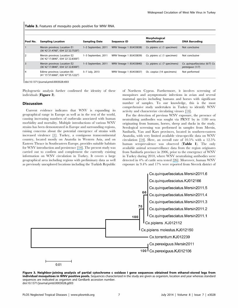
Phylogenetic analysis further confirmed the identity of these
individuals (Figure 3).
Discussion
Current evidence indicates that WNV is expanding its
geographical range in Europe as well as in the rest of the world,
causing increasing numbers of outbreaks associated with human
morbidity and mortality. Multiple introductions of various WNV
strains has been demonstrated in Europe and surrounding regions,
raising concerns about the potential emergence of strains with
increased virulence [1]. Turkey, a contiguous transcontinental
country, located mostly on Anatolia in Western Asia, and on
Eastern Thrace in Southeastern Europe, provides suitable habitats
for WNV introduction and persistence [14]. The present study was
carried out to confirm and complement the currently existing
information on WNV circulation in Turkey. It covers a large
geographical area including regions with preliminary data as well
as previously unexplored locations including the Turkish Republic
of Northern Cyprus. Furthermore, it involves screening of
mosquitoes and asymptomatic infections in avian and several
mammal species including humans and horses with significant
number of samples. To our knowledge, this is the most
comprehensive study undertaken in Turkey to identify WNV
activity and characterize circulating viruses [14].
For the detection of previous WNV exposure, the presence of
neutralizing antibodies was sought via PRNT by in 1180 sera
originating from humans, horses, sheep and ducks in the study.
Serological screening was performed in samples from Mersin,
Sanliurfa, Van and Kars provinces, located in southern-eastern
Anatolia, with very limited available virus-specific data on WNV
circulation [14]. Here, an overall rate of 10.5% with a 12.5%
human seroprevalence was observed (Table 1). The only
available animal serosurveillance data from the region originates
from Sanliurfa province in 2006, prior to the emergence of WNV
in Turkey during 2010, where WNV neutralizing antibodies were
detected in 4% of cattle sera tested [26]. Moreover, human WNV
exposure in 9.4% and 17% were reported from Siverek district of
Table 3. Features of mosquito pools positive for WNV RNA.
Pool No. Sampling Location Sampling Date Sequence IDMorphologicalIdentification DNA Barcoding
1 Mersin province; Location E1(36u42921.47600, 034u22922.75200)
1–3 September, 2011 WNV lineage 1 (KJ433838) Cx. pipiens s.l. (1 specimen) Not conclusive
2 Mersin province; Location E2(36u42917.08400, 034u22922.83000)
1–3 September, 2011 WNV lineage 1 (KJ433839) Cx. pipiens s.l. (1 specimen) Not conclusive
3 Mersin province; Location E2(36u42917.08400, 034u22922.83000)
1–3 September, 2011 WNV lineage 1 (KJ433840) Cx. pipiens s.l. (7 specimens) Cx. quinquefasciatus (6/7) Cx.perexiguus (1/7)
4 Edirne province; Location B3(41u15957.60600, 026u47955.12220)
5–7 July, 2013 WNV lineage 1 (KJ433837) Oc. caspius (14 specimens) Not performed
doi:10.1371/journal.pntd.0003028.t003
Figure 3. Neighbor-joining analysis of partial cytochrome c oxidase I gene sequences obtained from ethanol-stored legs fromindividual mosquitoes in WNV positive pools. Sequences characterized in the study are given as organism, location and year whereas standardsequences are indicated as organism and GenBank accession number.doi:10.1371/journal.pntd.0003028.g003
Widespread Circulation of West Nile Virus in Turkey
PLOS Neglected Tropical Diseases | www.plosntds.org 7 July 2014 | Volume 8 | Issue 7 | e3028

Sanliurfa province and Kiziltepe district of Mardin province,
respectively [27,28]. The current data demonstrate the circulation
of WNV not only in south-southeast Anatolia but indicates a
previously unexplored region of WNV activity in Eastern Anatolia,
neighboring Iran, Armenia and Georgia. A previous animal
serosurvey undertaken in Hatay, Adana, Antalya (Mediterranean),
Mugla, Izmir (Aegean), Bursa (Northwest Anatolia) and Ankara
provinces (Central Anatolia) had also revealed the presence of
WNV neutralizing antibodies in a variety of mammalian species,
including 2.5% of ass-mules, 4% of cattle, 37.7% of dogs, 13.5% of
horses, 1% of sheep and 20.4% of humans [26]. Moreover, human
WNV seroreactivity rates of 0.56–21.5% were reported from
Central Anatolia, Aegean and Eastern Thrace regions [14,17].
While a direct comparison among species and regions may be
misleading due to the dynamic interplay of various factors
including local climate and mosquito distribution, these results
indicate the occurence of WNV infections in a wide range of
mammals and domestic avians which can contribute to the long-
term virus survival in the absence of overt disease.
To provide further evidence for virus circulation in susceptible
mammals, WNV nucleic acids were investigated in samples
collected during the mosquito season in Mugla, Mersin and
Adana provinces of Aegean-Mediterranean Anatolia during 2011–
2012. Nested and quantitative rRT-PCR assays, that enable
sensitive detection of WNV lineages 1 and 2 were employed in
screening. Viral RNA was detected in horses in all locations with
an overall rate of 5.9% and a significantly higher rate of positivity
was observed in Mugla province (30.6%) (Table 1). No positive
human sample was noted. WNV is known to be transmitted to
equids by bridge vectors and is responsible for the majority of
equine flaviviral encephalitis worldwide [29]. Similar to that in
humans, most horses seroconvert without clinical disease after
exposure to WNV. In approximately 8% of naive horses, severe
WNV disease with neurological symptoms develop and the
incidence of disease in equids has increased significantly since
the mid-1990s, in parallel with the dispersion of lineage 1a WNV
strains [30,31]. Since 2010, WNV infections among horses were
repeatedly reported in the Mediterranean basin and other
European countries, such as in Bulgaria, Croatia, Greece, Italy,
Macedonia, Morocco, Portugal, Romania and Spain [29,32].
Equine encephalitis due to WNV lineage 1a in Turkey with a
favorable outcome has been initially described in Eskisehir
province (Central Anatolia) in 2011 [16]. Despite detection of
equine exposure via neutralizing antibodies in Bursa, Adana and
Eskisehir provinces, WNV RNA has not been characterized in
asymptomatic horses or in other mammal/avian species previously
in Turkey [16,26,33]. Horses are considered as dead-end hosts for
WNV due to the short and low magnitude viremia observed
following infected mosquito bites. A maximum serum virus titer of
103 pfu/ml and viremia duration of 6 days have been demon-
strated to occur in experimental equine infections with WNV,
which is unlikely to initiate an infection in the midgut epithelia of
the mosquito vector [29,34,35]. Since RNA levels detected via
rRT-PCR in positive equine plasma this study are low and roughly
correspond to the levels noted in experimental infections, it can be
assumed that they do not significantly contribute to the virus
lifecycle by infecting vectors. However, the RNA detection in
horses in subsequent years (2011 and 2012) reveals ongoing WNV
activity in the region, which is further supported by the findings of
the field study for vectors.
Entomological surveillance performed at 11 provinces of
Turkey and at 4 provinces of the Turkish Republic of Northern
Cyprus revealed Cx. pipiens s. l. to be the most abundant mosquito
species in Adana, Ankara, Artvin, Mersin and Samsun provinces
as well as in Cyprus (Table 2). However, specimens belonging to
15 different species were captured, with the predominance of Oc.caspius in Edirne and Sinop provinces, Cx. theileri in Bursa
province and Da. geniculata in Kirklareli province. It is known
that different mosquito species demonstrate variable competencies
to support WNV replication and transmission. Mosquitoes
belonging in Culex species, from which initial WNV isolates were
recovered, are widely accepted as the primary global transmission
vector [13]. Several Culex species have been shown to be
competent for WNV transmission in North America and Cx.pipiens s.s., Cx. perexiguus, and Cx. modestus are considered as
important vector species in Europe [36–38]. Moreover, WNV has
also been detected in other genera of mosquitoes including
Ochlerotatus, Aedes, Anopheles, Coquillettidia, Aedeomya, Manso-nia, Mimomyia, Psorophora, Culiseta and Uranoteania, which
serve as bridge vectors critical to transmission from birds to
humans and equines [38]. Cx. pipiens is known to be widespread
in the Anatolian fauna, with many other mosquito species that can
serve as bridge vectors [39]. Species distribution observed in this
study further confirmes this data and maintains the presence and
abundance of several mosquito species capable of transmitting
WNV to various avian and mammals including equines and
humans. However, since the sampling could not be carried on
during the whole mosquito season in any given sampling location,
the species frequencies and distribution reported in this study
represent cross-sectional data for each region.
Field-captured mosquitoes were pooled and screened for WNV
infection using the same approach for detecting nucleic acids in
animal plasma. Viral RNA was identified in 1.9% of 203 pools and
characterized as WNV lineage 1 (Table 3). Three of the positive
pools originated from two sampling sites of the same location in
Mersin province, and has been collected during early September,
2011. To confirm the identity of the mosquitoes in the infected
pools, DNA barcoding via COI sequence analysis was performed
in dissected legs from individual mosquitoes from each pool.
Despite the lack of succesful amplification in two individual
mosquitoes comprising two infected pools, Cx. quinquefasciatusand Cx. perexiguus mosquitoes were identified in another WNV
positive pool (Table 3, Figure 3). Detection and verification of
these specimens as Cx. quinquefasciatus comprises the first record
of the species in Turkey. Cx. quinquefasciatus, also called the
southern house mosquito, is an important WNV vector in the
southern United States as well as in Africa [12,40]. Cx. perexiguusis another highly-competent WNV vector, participating not only
in enzootic cycle of the virus but also in transmission to equines
[38,41]. This individual mosquito, confirmed as Cx. perexiguus in
DNA barcoding, is likely to be misidentified as Cx. pipiens s.l.during morphological evaluation. Identification of these species in
WNV-infected pools in Turkey is a novel finding, revealing the
activity of many competent WNV vectors in the country. Another
infected pool comprised Oc. caspius specimens collected from
Edirne province, Eastern Thrace region in 2013. Interestingly, the
initial detection of WNV sequences in vectors was accomplished in
this region during 2012, in Cx. pipiens s.s. and Oc. caspius pools
[18]. Thus, our current findings verify and demonstrate an
ongoing circulation of WNV in mosquitoes in this region. In
Turkey, other efforts to detect WNV in potential mosquito vectors
in regions with evidence for virus activity has been unsuccessful.
For example, WNV antigens or nucleic acids could not be
detected in mosquito specimens belonging in Cx. pipiens s.l., Oc.caspius, and Aedes species captured in Sanliurfa province, despite
serological evidence of virus exposure in the region [42]. Likewise,
a recent field survey undertaken in Ankara province Central
Anatolia, where symptomatic WNV cases have been demonstrat-
Widespread Circulation of West Nile Virus in Turkey
PLOS Neglected Tropical Diseases | www.plosntds.org 8 July 2014 | Volume 8 | Issue 7 | e3028

ed, Cx. pipiens s.l., An. maculipennis and An. claviger species were
collected but WNV infection in vectors could not be identified
[43]. Extensive surveillance activities are required for these
regions, to reveal and characterize WNV infection in vectors. In
Europe, several reports indicate Cx. pipiens s.l., Cx. modestus, Oc.caspius and Cx. perexiguus as the most common species associated
with WNV infections and the potential involvement of Cx.univittatus, Cx. theileri and An. maculipennis s.l. in virus
circulation [3,38]. Furthermore, in the Volgograd region of Russia
and in Israel, Cx. pipiens, Cx. modestus, Cx. perexiguus and Oc.caspius species were implicated in WNV transmission [44,45].
Accurate identification of mosquito species in a region is of
significant importance to reveal and predict WNV emergence,
since only certain species in any given area can act effectively as
primary and bridge vectors to human or equine populations.
Evidence from Italy, Spain and Greece suggests that WNV
detection in mosquitoes precedes the appearance of human or
equine cases and surveillance activities provide crucial information
on the relevant vectors and on the circulating virus strain, for
optimal diagnostic procedures and interventions for preventing
transmission [38]. Another example is provided from Turkey,
where infected mosquitoes could be detected approximately 4
weeks prior to the emergence of human cases in the Eastern
Thrace region [17,18]. Thus, monitorization of WNV activity via
vector and/or sentinel animal surveillance is required for Turkey,
especially for regions with evidence for virus exposure and
insufficient data on vectors.
WNV strains are grouped in several putative genetic lineages
[9,11]. Isolates belonging to lineage 1 are widely distributed and
highly invasive, and account for the majority of the strains
responsible for the European and the Mediterranean Basin
outbreaks [3,46]. However, lineage 2 strains, which have recently
spread from Austria and Hungary to Balkan States and Greece,
have also emerged and caused outbreaks resulting in human and
bird mortality, particularly in Greece [47]. In Italy, co-circulation
of lineage 1 and 2 strains has also been demonstrated [38]. At least
five new lineages (lineages 3–7) have been proposed for strains
isolated in central Europe, as well as in Russia and India [11]. All
WNV genomic data obtained in Turkey, including partial E gene
sequences characterized from viremic horses and mosquito pools
in this study, are grouped with WNV lineage 1 clade 1 strains
(Figure 2). Overall, maximum composite likelihood analyses
revealed similarities among regions and hosts, as well as identical
sequences observed in Mugla province in horses during 2011 to
Cx. pipiens s.l. pools in Mersin province collected during the same
year (Table S2). Moreover, current sequences displayed limited
diversity compared to previously-obtained data and sequences
identical with patient and vector-derived sequences from Ankara,
Tekirdag and Edirne provinces in 2012 were also noted, despite
geographical and temporal separation (Table S2). These data
suggest that WNV strains in circulation are generally genetically
conserved with restricted sequence diversity among strains.
Nevertheless, the origin and variability in WNV strains in Turkey
will be better elucidated upon whole genome sequencing of the
isolates, for which studies are underway by our group.
In conclusion, our findings from vectors and exposed animals
indicate a wider zone of WNV activity in Turkey than previously
anticipated, including Eastern Thrace and Mediterranean-Aegean
regions as well as Southeastern and Northeastern Anatolia.
Ongoing virus circulation with limited genomic diversity was
observed in certain regions. WNV must be considered in etiology
of human or equine febrile diseases with/without central nervous
system involvement in these regions. Zones of priority for
surveillance of WNV activity via mosquito and/or sentinel animals
must be established.
Supporting Information
Table S1 List of mosquito sampling location and sites employed
in the study.
(DOCX)
Table S2 Pairwise nucleotide diversity (above diagonal) and
genetic similarity (below diagonal) among E protein-coding
genome segment of WNVs identified in Turkey.
(DOCX)
Acknowledgments
The authors are grateful to Irfan Atmaca and Salim Calis for technical
assistance and N. Emin Guven for graphics. Preliminary findings of this
study have been presented at the 5th European Congress of Virology,
during September 11–14th, 2013 in Lyon, France and have been included
in the abstract book (Virologie, 17(2):S196).
Author Contributions
Conceived and designed the experiments: KE AO BA. Performed the
experiments: FG OEK KO SG TK ST. Analyzed the data: KE AO FG
BA. Contributed reagents/materials/analysis tools: MC YY GE. Contrib-
uted to the writing of the manuscript: KE AO FG.
References
1. Sambri V, Capobianchi M, Charrel R, Fyodorova M, Gaibani P, et al. (2013)
West Nile virus in Europe: emergence, epidemiology, diagnosis, treatment, and
prevention. Clin Microbiol Infect 19: 699–704.
2. Dauphin G, Zientara S, Zeller H, Murgue B. (2004) West Nile: worldwide
current situation in animals and humans. Comp Immunol Microbiol Infect Dis
27:343–355.
3. Calistri P, Giovannini A, Hubalek Z, Ionescu A, et al. (2010). Epidemiology of
West Nile in Europe and in the Mediterranean Basin. Open Virol J 4:29–
37.
4. Kilpatrick AM. (2011) Globalization, land use, and the invasion of West Nile
virus. Science 334:323–327.
5. Beasley DW, Barrett AD, Tesh RB. (2013) Resurgence of West Nile neurologic
disease in the United States in 2012: what happened? What needs to be done?
Antiviral Res 99:1–5.
6. Lanciotti RS, Roehrig JT, Deubel V, Smith J, Parker M, et al. (1999). Origin of
the West Nile virus responsible for an outbreak of encephalitis in the
northeastern United States. Science 286:2333–2337.
7. Komar N, Clark GG. (2006) West Nile virus activity in Latin America and the
Caribbean. Rev Panam Salud Publica 19:112–117.
8. Monath TP (1990) Flaviviruses. In: Fields BN, Knipe M, editors, Virology, 2nd
Edn, Raven Press, New York. pp.763–814.
9. Hayes EB, Sejvar JJ, Zaki SR, Lanciotti RS, Bode AV, et al. (2005) Virology,
pathology and clinical manifestations of West Nile Virus disease. Emerg Infect
Dis 11:1174–1179.
10. Chambers TJ, Hahn CS, Galler R, Rice CM. (1990) Flavivirus genome
organization, expression, and replication. Annu Rev Microbiol 44:649–688.
11. May FJ, Davis CT, Tesh RB, Barrett AD. (2011) Phylogeography of West Nile
virus. J Virol 85:2964–2974.
12. Hayes EB, Komar N, Nasci RS, Montgomery SP, O’Leary DR, et al. (2005)
Epidemiology and transmission dynamics of West Nile virus disease. Emerg
Infect Dis 11:1167–1173.
13. Colpitts TM, Conway MJ, Montgomery RR, Fikrig E. (2012) West Nile Virus:
biology, transmission, and human infection. Clin Microbiol Rev 25:635–648.
14. Ergunay K, Whitehouse C, Ozkul A. (2011) Current status of human arboviral
infections in Turkey. Vector Borne Zoonotic Dis 11:731–741.
15. Kalaycioglu H, Korukluoglu G, Ozkul A, Oncul O, Tosun S, et al. (2012)
Emergence of West Nile virus infections in humans in Turkey, 2010 to 2011.
Euro Surveill 17: pii: 20182.
16. Ozkul A, Ergunay K, Koysuren A, Alkan F, Arsava EM, et al. (2013)
Concurrent occurrence of human and equine West Nile virus infections in
Central Anatolia, Turkey: the first evidence for circulation of lineage 1 viruses.
Int J Infect Dis 17:546–551
Widespread Circulation of West Nile Virus in Turkey
PLOS Neglected Tropical Diseases | www.plosntds.org 9 July 2014 | Volume 8 | Issue 7 | e3028

17. Erdem H, Ergunay K, Yilmaz A, Naz H, Akata F, et al. (2014) Emergence and
co-infections of West Nile virus and Toscana virus in Eastern Thrace, Turkey.Clin Microbiol Infect 20:319–325.
18. Ergunay K, Gunay F, Oter K, Kasap OE, Orsten S, et al. (2013) Arboviral
surveillance of field-collected mosquitoes reveals circulation of West Nile viruslineage 1 strains in Eastern Thrace, Turkey. Vector Borne Zoonotic Dis :744–
752.19. Bunning ML, Bowen RA, Cropp CB, Sullivan KG, Davis B, et al. (2002)
Experimental infection of horses with West Nile virus. Emerg Infect Dis 8:380–
386.20. Darsie RE, Samanidou-Voyadjoglou A. (1997) Keys for the identification of the
mosquitoes of Greece. J Am Mosq Control Assoc 13:247–254.21. Schaffner E, Angel G, Geoffroy B, Hervy JP, Rhaiem A, et al. (2001) The
Mosquitoes of Europe. [CD-ROM] Paris: IRD Editions & EID Mediterrannee.22. Calzolari M, Bonilauri P, Bellini R, Caimi M, Defilippo F, et al. (2010) Arboviral
survey of mosquitoes in two northern Italian regions in 2007 and 2008. Vector
Borne Zoonotic Dis 10:875–884.23. Johnson DJ, Ostlund EN, Pedersen DD, Schmitt BJ. (2001) Detection of North
American West Nile virus in animal tissue by a reverse transcription-nestedpolymerase chain reaction assay. Emerg Infect Dis 7:739–741.
24. Folmer O, Black M, Hoeh W, Lutz R, Vrijenhoek R. (1994) DNA primers for
amplification of mitochondrial cytochrome c oxidase subunit I from diversemetazoan invertebrates. Mol Mar Biol Biotechnol 3:294–299.
25. Tamura K, Peterson D, Peterson N, Stecher G, Nei M, et al. (2011) MEGA5:Molecular evolutionary genetics analysis using maximum likelihood, evolu-
tionary distance and maximum parsimony methods. Mol Biol Evol 28:2731–2739.
26. Ozkul A, Yildirim Y, Pinar D, Akcali A, Yilmaz V, et al. (2006) Serological
evidence of West Nile Virus (WNV) in mammalian species in Turkey. EpidemiolInfect 134:826–829.
27. Ergunay K, Ozer N, Us D, Ozkul A, Simsek F, et al. (2007) Seroprevalence ofWest Nile virus and tick-borne encephalitis virus in southeastern Turkey: first
evidence for tick-borne encephalitis virus infections. Vector Borne Zoonotic Dis
7:157–161.28. Karakoc ZC, Tuzuner BM, Ergonul O, Pierro A, Di Fonzo E, et al. (2013) West
nile virus infection in the Mesopotamia region, Syria border of Turkey. VectorBorne Zoonotic Dis 13:739–743.
29. Angenvoort J, Brault AC, Bowen RA, Groschup MH. (2013) West Nile viralinfection of equids. Vet Microbiol 167:168–180.
30. Gardner IA, Wong SJ, Ferraro GL, Balasuriya UB, Hullinger PJ, et al. (2007)
Incidence and effects of West Nile virus infection in vaccinated and unvaccinatedhorses in California. Vet Res 38:109–116.
31. Nielsen CF, Reisen WK, Armijos MV, Maclachlan NJ, Scott TW. (2008) Highsubclinical West Nile virus incidence among nonvaccinated horses in northern
California associated with low vector abundance and infection. Am J Trop Med
Hyg 78: 45–52.32. Garcia-Bocanegra I, Jaen-Tellez JA, Napp S, Arenas-Montes A, Fernandez-
Morente M, et al. (2011) West Nile fever outbreak in horses and humans, Spain,2010. Emerg Infect Dis 17:2397–2399.
33. Albayrak H, Ozan E. (2010). Molecular detection of avian influenza virus but
not West Nile virus in wild birds in northern Turkey. Zoonoses Public Health
57:71–75.
34. Seino KK, Long MT, Gibbs EP, Bowen RA, Beachboard SE, et al. (2007)
Comparative efficacies of three commercially available vaccines against West
Nile Virus (WNV) in a short-duration challenge trial involving an equine WNV
encephalitis model. Clin Vaccine Immunol 14:1465–1471.
35. Minke JM, Siger L, Cupillard L, Powers B, Bakonyi T, et al. (2011) Protection
provided by a recombinant ALVAC(R)-WNV vaccine expressing the prM/E
genes of a lineage 1 strain of WNV against a virulent challenge with a lineage 2
strain. Vaccine 29: 4608–4612.
36. Sardelis MR, Turrell MJ, Dohm DJ, O’Guinn ML. (2001) Vector competence of
selected North American Culex and Coquillettidia mosquitoes for West Nile
virus. Emerg Infect Dis 7:1018–1022.
37. Balenghien T, Vazeille M, Grandadam M, Schaffner F, Zeller H, et al. (2007)
Vector competence of some French Culex and Aedes mosquitoes for West Nile
virus. Vector Borne Zoonotic Dis 8:589–595.
38. Engler O, Savini G, Papa A, Figuerola J, Groschup MH, et al. (2013) European
surveillance for West Nile virus in mosquito populations. Int J Environ Res
Public Health 10:4869–4895.
39. Ramsdale CD, Alten B, Caglar SS, Ozer N. (2001) A revised, annotated
checklist of the mosquitoes (Diptera, Culicidae) of Turkey. Eur Mosq Bull 9:18–
28.
40. Fall G, Diallo M, Loucoubar C, Faye O, Sall AA. (2014) Vector competence of
Culex neavei and Culex quinquefasciatus (Diptera: Culicidae) from Senegal for
lineages 1, 2, Koutango and a putative new lineage of West Nile virus. Am J Trop
Med Hyg. doi: 10.4269/ajtmh.13-0405
41. Munoz J1, Ruiz S, Soriguer R, Alcaide M, Viana DS, et al. (2012) Feeding
patterns of potential West Nile virus vectors in south-west Spain. PLoS One
7:e39549.
42. Ozer N, Ergunay K, Simsek F, Kaynas S, Alten B, et al. (2007) West Nile virus
studies in the Sanliurfa Province of Turkey. J Vector Ecol 32:202–206.
43. Ocal M, Orsten S, Inkaya AC, Yetim E, Acar NP, et al. (2013) Ongoing activity
of Toscana virus genotype A and West Nile virus lineage 1 strains in Turkey: a
clinical and field survey. Zoonoses Public Health. doi: 10.1111/zph.12096
44. Fyodorova MV, Savage HM, Lopatina JV, Bulgakova TA, Ivanitsky AV, et al.
(2006) Evaluation of potential West Nile virus vectors in Volgograd region, 2003
(Diptera: Culicidae): Species composition, bloodmeal host utilization, and virus
infection rates of mosquitoes. J Med Entomol 43:552–563.
45. Orshan L, Bin H, Schnur H, Kaufman A, Valinsky A, et al. (2008) Mosquito
vectors of West Nile fever in Israel. J Med Entomol 45:939–947.
46. Bakonyi T, Ivanics E, Erdelyi K, Ursu K, Ferenczi E, et al. (2006) Lineage 1 and
2 strains of encephalitic West Nile virus, central Europe. Emerg Infect Dis
12:618–623.
47. Bakonyi T, Ferenczi E, Erdelyi K, Kutasi O, Csorgo T, et al. (2013) Explosive
spread of a neuroinvasive lineage 2 West Nile virus in Central Europe, 2008/
2009. Vet Microbiol 165: 61–70.
Widespread Circulation of West Nile Virus in Turkey
PLOS Neglected Tropical Diseases | www.plosntds.org 10 July 2014 | Volume 8 | Issue 7 | e3028