
Identification of FAM46D as a novel cancer/testis antigen using EST data and serological analysis
8
Identification of FAM46D as a novel cancer/testis antigen using EST data and serological analysis ☆ Fabiana Bettoni a , Fernando Camargo Filho a , Daniela M. Grosso a , Pedro A.F. Galante a , Raphael B. Parmigiani a , Murilo V. Geraldo a , Flávio Henrique-Silva b , Sueli M. Oba-Shinjo c , Suely K.N. Marie c , Fernando A. Soares d , Helena P. Brentani d , Andrew J.G. Simpson e , Sandro J. de Souza a , Anamaria A. Camargo a, ⁎ a Laboratory of Molecular Biology and Genomics, Ludwig Institute for Cancer Research, São Paulo Branch, Hospital Alemão Oswaldo Cruz, Rua João Julião, 245, 1 andar, São Paulo, SP 01323-903, Brazil b Universidade Federal de São Carlos, Rodovia Washington Luis km 235, São Carlos, SP 13565-905, Brazil c Department of Neurology, School of Medicine, University of São Paulo, Av Dr Arnaldo, 455, room 4110, São Paulo, Brazil d Hospital A.C. Camargo, Rua Prof. Antonio Prudente, 211, São Paulo, SP 01509-010, Brazil e Ludwig Institute for Cancer Research, Memorial Sloan-Kettering Cancer Center, New York, NY 10158, USA abstract article info Article history: Received 19 February 2009 Accepted 11 June 2009 Available online xxxx Keywords: Tumor antigens Immunotherapy Gene expression profiling ESTs FAM46D Cancer/testis Antigens (CTAs) are immunogenic proteins with a restricted expression pattern in normal tissues and aberrant expression in different types of tumors being considered promising candidates for immunotherapy. We used the alignment between EST sequences and the human genome sequence to identify novel CT genes. By examining the EST tissue composition of known CT clusters we defined parameters for the selection of 1184 ESTclusters corresponding to putative CT genes. The expression pattern of 70 CT gene candidates was evaluated by RT-PCR in 21 normal tissues,17 tumor cell lines and 160 primary tumors. We were able to identify 4 CT genes expressed in different types of tumors. The presence of antibodies against the protein encoded by 1 of these 4 CT genes (FAM46D) was exclusively detected in plasma samples from cancer patients. Due to its restricted expression pattern and immunogenicity FAM46D represents a novel target for cancer immunotherapy. © 2009 Elsevier Inc. All rights reserved. Introduction Cancer/testis (CT) genes are expressed in the germ cells of testis and fetal ovary and in different histological types of tumors [1–4]. So far, more than 83 distinct CT gene families have been described, most of them are located on the X chromosome [1]. Most CT genes have been shown to encode proteins that are immunogenic (CT Antigens or CTA), eliciting both humoral and cellular immune responses in cancer patients [1]. Due to their restricted expression pattern and immunogenicity, CTAs are considered promising candidates for the development of therapeutic cancer vaccines. Advanced clinical trials with CTA vaccines are currently underway to access their efficacy in delaying or preventing recurrence of melanoma and lung cancer following surgical removal of primary tumors [5–8]. However, some characteristics shared by this group of proteins may represent challenges to the development of a universal cancer vaccine. Firstly, the expression frequency of a specific CT antigen is highly variable between different types of tumors. Second, the expression of a given CTA within a tumor is highly heterogeneous, with tumors showing single positive cells or small groups of positive cells to others with a homogeneous expression pattern. Finally, tumors frequently develop mechanisms to evade the host immune system and antigen loss is frequently observed in tumors. One way to overcome tumor heterogeneity and immune escape is to design polyvalent cancer vaccines containing epitopes derived from different CTAs. The identification of a large repertoire of CTAs that could be incorporated in polyvalent cancer vaccines is thus critical for improving current immunotherapy protocols. Most of the known CTAs were identified using immunological screening methods such as T- lymphocyte epitope cloning and SEREX (Serological analysis of cDNA expression libraries) [9–14]. However, due to their characteristic expression pattern, non-immunological techniques, relying on differ- ential mRNA expression, have also been successfully used for the characterization of novel CT genes. Methods including RDA (Repre- sentational Difference Analysis), Differential Display and cDNA micro- Genomics xxx (2009) xxx–xxx ☆ Sequence data from this article have been deposited with the DDBJ/EMBL/ GenBank Data Libraries under Accession Nos. EF537578, EF537579, EF537580, EF537581 and EF537582. ⁎ Corresponding author. Fax: +5511 3141.1325. E-mail address: [email protected] (A.A. Camargo). YGENO-08124; No. of pages: 8; 4C: 0888-7543/$ – see front matter © 2009 Elsevier Inc. All rights reserved. doi:10.1016/j.ygeno.2009.06.001 Contents lists available at ScienceDirect Genomics journal homepage: www.elsevier.com/locate/ygeno ARTICLE IN PRESS Please cite this article as: F. Bettoni, et al., Identification of FAM46D as a novel cancer/testis antigen using EST data and serological analysis, Genomics (2009), doi:10.1016/j.ygeno.2009.06.001
-
Upload
independent -
Category
Documents
-
view
1 -
download
0
Transcript of Identification of FAM46D as a novel cancer/testis antigen using EST data and serological analysis

Genomics xxx (2009) xxx–xxx
YGENO-08124; No. of pages: 8; 4C:
Contents lists available at ScienceDirect
Genomics
j ourna l homepage: www.e lsev ie r.com/ locate /ygeno
ARTICLE IN PRESS
Identification of FAM46D as a novel cancer/testis antigen using EST data andserological analysis☆
Fabiana Bettoni a, Fernando Camargo Filho a, Daniela M. Grosso a, Pedro A.F. Galante a,Raphael B. Parmigiani a, Murilo V. Geraldo a, Flávio Henrique-Silva b, Sueli M. Oba-Shinjo c,Suely K.N. Marie c, Fernando A. Soares d, Helena P. Brentani d, Andrew J.G. Simpson e,Sandro J. de Souza a, Anamaria A. Camargo a,⁎a Laboratory of Molecular Biology and Genomics, Ludwig Institute for Cancer Research, São Paulo Branch, Hospital Alemão Oswaldo Cruz, Rua João Julião, 245, 1 andar, São Paulo,SP 01323-903, Brazilb Universidade Federal de São Carlos, Rodovia Washington Luis km 235, São Carlos, SP 13565-905, Brazilc Department of Neurology, School of Medicine, University of São Paulo, Av Dr Arnaldo, 455, room 4110, São Paulo, Brazild Hospital A.C. Camargo, Rua Prof. Antonio Prudente, 211, São Paulo, SP 01509-010, Brazile Ludwig Institute for Cancer Research, Memorial Sloan-Kettering Cancer Center, New York, NY 10158, USA
☆ Sequence data from this article have been depoGenBank Data Libraries under Accession Nos. EF5EF537581 and EF537582.⁎ Corresponding author. Fax: +5511 3141.1325.
E-mail address: [email protected] (A.A. Cama
0888-7543/$ – see front matter © 2009 Elsevier Inc. Adoi:10.1016/j.ygeno.2009.06.001
Please cite this article as: F. Bettoni, et al., IGenomics (2009), doi:10.1016/j.ygeno.2009
a b s t r a c t
a r t i c l e i n f oArticle history:Received 19 February 2009Accepted 11 June 2009Available online xxxx
Keywords:Tumor antigensImmunotherapyGene expression profilingESTsFAM46D
Cancer/testis Antigens (CTAs) are immunogenic proteins with a restricted expression pattern in normaltissues and aberrant expression in different types of tumors being considered promising candidates forimmunotherapy. We used the alignment between EST sequences and the human genome sequence toidentify novel CT genes. By examining the EST tissue composition of known CT clusters we definedparameters for the selection of 1184 EST clusters corresponding to putative CT genes. The expression patternof 70 CT gene candidates was evaluated by RT-PCR in 21 normal tissues, 17 tumor cell lines and 160 primarytumors. We were able to identify 4 CT genes expressed in different types of tumors. The presence ofantibodies against the protein encoded by 1 of these 4 CT genes (FAM46D) was exclusively detected inplasma samples from cancer patients. Due to its restricted expression pattern and immunogenicity FAM46Drepresents a novel target for cancer immunotherapy.
© 2009 Elsevier Inc. All rights reserved.
Introduction
Cancer/testis (CT) genes are expressed in the germ cells of testisand fetal ovary and in different histological types of tumors [1–4].So far, more than 83 distinct CT gene families have been described,most of them are located on the X chromosome [1]. Most CT geneshave been shown to encode proteins that are immunogenic (CTAntigens or CTA), eliciting both humoral and cellular immuneresponses in cancer patients [1]. Due to their restricted expressionpattern and immunogenicity, CTAs are considered promisingcandidates for the development of therapeutic cancer vaccines.Advanced clinical trials with CTA vaccines are currently underwayto access their efficacy in delaying or preventing recurrence ofmelanoma and lung cancer following surgical removal of primarytumors [5–8].
sited with the DDBJ/EMBL/37578, EF537579, EF537580,
rgo).
ll rights reserved.
dentification of FAM46D as a.06.001
However, some characteristics shared by this group of proteinsmay represent challenges to the development of a universal cancervaccine. Firstly, the expression frequency of a specific CT antigen ishighly variable between different types of tumors. Second, theexpression of a given CTA within a tumor is highly heterogeneous,with tumors showing single positive cells or small groups of positivecells to others with a homogeneous expression pattern. Finally,tumors frequently develop mechanisms to evade the host immunesystem and antigen loss is frequently observed in tumors.
One way to overcome tumor heterogeneity and immune escape isto design polyvalent cancer vaccines containing epitopes derived fromdifferent CTAs. The identification of a large repertoire of CTAs thatcould be incorporated in polyvalent cancer vaccines is thus critical forimproving current immunotherapy protocols. Most of the known CTAswere identified using immunological screening methods such as T-lymphocyte epitope cloning and SEREX (Serological analysis of cDNAexpression libraries) [9–14]. However, due to their characteristicexpression pattern, non-immunological techniques, relying on differ-ential mRNA expression, have also been successfully used for thecharacterization of novel CT genes. Methods including RDA (Repre-sentational Difference Analysis), Differential Display and cDNA micro-
novel cancer/testis antigen using EST data and serological analysis,

2 F. Bettoni et al. / Genomics xxx (2009) xxx–xxx
ARTICLE IN PRESS
arrays have been used in the identification of CT genes expressed intestis and different tumor types, such as LAGE-1/CTAG2, CT10/MAGEC2, CTp11, SPANX-C1/SPANXA1, MMA-1A/DSCR8 and DBL/MCF2[15–19]. These CT genes were later shown to encode immunogenicproteins recognized by the immune system of cancer patients.
Computational approaches have also been used for the identificationof CT genes. In a pioneer study, Scanlan et al. [20] mined the UniGenedatabase for gene clusters composed of expressed sequence tags (ESTs)generated from normal testis and tumor-derived cDNA libraries. A totalof 1325gene clusterswere initially selected as corresponding to putativeCTgenes based on their ESTcomposition. After assessing the expressionpattern of 73 candidate clusters by RT-PCR, three novel CT genes (CT15/ADAM2, CT16/PAGE5 and CT17/LIPI) were identified.
Using a similar strategy, Chen et al. [21] analyzed MPSS (MassivelyParallel Signature Sequencing) expression data from 32 normalhuman tissues and identified 1056 genes that were predominantlyexpressed in normal testis. Further evaluation using MPSS tags fromtumor cell lines and EST data from awide variety of tumors, identified202 of these 1056 genes as candidates for encoding novel CT genesbased on their expression pattern. RT-PCR validation was carried outfor a subset of 166 intron-containing candidates using cDNAs fromnormal testis and 21 tumor cell lines. Twenty candidates werevalidated as novel CT genes based on their expression pattern.
Finally, in a recent work Hofmann et al. [22] have used an in silicoapproach that combined expression information from different plat-forms (including ESTs, MPSS and Cap-analysis of Gene Expression) toproduce a comprehensive survey of the expression profile of 153previously described CT genes. By applying this strategy in a genome-wide survey for novel CT genes they identified over 30 CT candidategenes, of which 3 (PEPP-2/RHOXF2, OTOA and AKAP4) were confirmedto be predominantly expressed in testis and tumor cell lines by RT-PCR.
In the present work, we combined in silico and experimentalevaluation of gene expression with serological analysis of cancerpatients to identify novel CTAs. Using this approach, we were able toidentify 4 CT genes frequently expressed in different types of tumors.The presence of specific antibodies recognizing the protein encodedby 1 of these 4 CTgenes (FAM46D) was exclusively detected in plasmasamples from cancer patients. Due to its restricted expression patternand immunogenicity FAM46D represents a novel target for cancerimmunotherapy. An additional CT gene candidate (PASD1) wasdescribed during the expression evaluation step of this work as anovel CTA frequently expressed in B-cell lymphomas [23]. Informationpresented in this manuscript on the high frequency of PASD1expression in different types of solid tumors and immunogenicity inpatients with cervix tumors and glioblastomas also supports PASD1 asa promising target for cancer immunotherapy.
Results
EST composition of clusters corresponding to known CTAs
ESTs can be used to identify new genes [24–28], to construct gene-based physical maps [29,30], to compare and annotate genomes ofdifferent organisms [31–33] and to study different aspects of mRNAstructure such as splicing [34–39] and polyadenylation variants [40–42].ESTs can also be used to qualitatively and quantitatively determinegene expression profiles if one considers the tissue source of the cDNAlibraries and the frequencyof ESTs corresponding to a given transcript ina cDNA library, respectively. In the present work, we have used thepublicly available human genome sequence to cluster ESTs derived froma same gene. We were then able to select clusters composed by ESTsderived from testis and tumor cDNA libraries thatwould representnovelCT genes.
To address possible limitations of our strategy, we first evaluatedthe EST tissue composition of clusters corresponding to 20 knownCTAs (MAGEA1, MAGEA3, MAGEA10, MAGEB1, CT7/MAGEC1, CT10/
Please cite this article as: F. Bettoni, et al., Identification of FAM46D as aGenomics (2009), doi:10.1016/j.ygeno.2009.06.001
MAGEC2, NY-ESO-1, SSX1, SSX2, SSX4, CT16/PAGE5, HOM-TES-85/LUZP4,BRDT/CT9, CTp11/SPANXC, OY-TES-1/ACRBP, CTAGE1, CT15/ADAM2,CSAG2, IL13RA2, HCA661/E2F-like/TFDP3). mRNA sequences corre-sponding to NY-ESO-1 and BRDT/CT9 aligned in more than onelocation in the genome, suggesting an event of gene duplication thatresulted in two identical copies [43]. Therefore the 20 CTAs wererepresented in our Transcriptome Database (see Materials andmethods) by 23 clusters all of them containing at least one EST.
CTA clusters were generally composed by a small number of ESTs.Fifty percent of the clusters contained less than 10 ESTs, confirmingthe restricted expression pattern of these CTAs. Surprisingly, 85% ofthe CTA clusters contained ESTs derived from normal tissues and only39.1% of these clusters contained ESTs derived from testis cDNAlibraries. Indeed, only 10% of the CTA clusters were composed exclu-sively by ESTs derived from testis and tumor cDNA libraries as onewould expect by the characteristic expression pattern of CT genes.Moreover, the majority of the 23 CTA clusters contained ESTs derivedfrom cDNA libraries of an unknown tissue source.
Improving the transcriptome database
The presence of ESTs derived from normal tissues in CTA clusterscan be explained, in part, by the existence of large gene families formost CTAs with high similarity between all members and by the factthat somemembers of CTA gene families have a ubiquitous expressionpattern that do not correspond to that of a characteristic CT gene. ESTscorresponding to members of these gene families align to multipleregions in the genome and are represented in different clusters,therefore explaining the presence of ESTs derived from normal tissuesin known CTA clusters. To overcome this problem, multiple align-ments were excluded from the Transcriptome Database by giving aquality score for each alignment as described in Materials andmethods and by keeping in the database only the alignments withthe highest score for each sequence.
The presence of a significant number of ESTs derived from normaltissues and unknown tissue source in CTA clusters can be alsoexplained by an erroneous description of the tissue source of cDNAlibraries submitted to GenBank. To overcome this problem we havemanually curated cDNA library descriptions and contacted the groupsresponsible for submissions when information was misleading orunavailable (Supplementary material 1). We kept in our database onlyEST sequences derived from cDNA libraries with a reliable andinformative description.
Selection of candidate clusters corresponding to novel CT genes
The EST composition of clusters corresponding to known CT geneswas also used to define parameters for computational selection ofadditional clusters corresponding to novel CT genes. After theimprovements made in the Transcriptome Database, we observedthat 20% of the CT gene clusters were composed by ESTs derived fromnormal testis and tumor cDNA libraries, 10% of the CT gene clusterswere composed only by testis ESTs and 5% were composed only bytumor ESTs. Moreover, 65% of CT gene cluster contained a smallpercentage of ESTs derived from normal tissues, probably due to thespurious expression of some of the CT genes in normal tissues as hasbeen previously reported [22].
Based on this information, we generated 3 independent lists ofcandidates: i) clusters composed only by ESTs derived from testiscDNA libraries, ii) clusters composed by ESTs derived from testis andtumor cDNA libraries and iii) clusters composed only by ESTs derivedfrom cDNA libraries of tumoral origin. Clusters composed mainly byESTs derived from testis and tumors, but containing a smallpercentage of ESTs from normal tissues, were not selected because,after analyzing clusters corresponding to known CT genes, we wereunable to establish a reliable cut-off for the presence of these ESTs.
novel cancer/testis antigen using EST data and serological analysis,
3F. Bettoni et al. / Genomics xxx (2009) xxx–xxx
ARTICLE IN PRESS
We have also restricted our analysis to clusters that were composedby at least one spliced EST that aligned non-contiguously to the genomesequence because EST data contain a significant fraction of DNAcontaminant sequences. It should be noted, however, that this was aconservative approach that might have excluded candidate clusterscomposed exclusively by 3′ unspliced ESTs. Although legitimacy of 3′ESTs can be attested by the occurrence of a bona-fide polyadenylationsignal and the presence of a poly(A) tail, we decided to undertake such aconservative approach because poly(A) tails are frequently trimmedfrom EST sequences submitted to dbEST and the identification ofpolyadenylation signals is usually hampered by the low quality of thesequences near poly(A) tails and to the low signal/background ratio inthe identification of reliable polyadenylation signals.
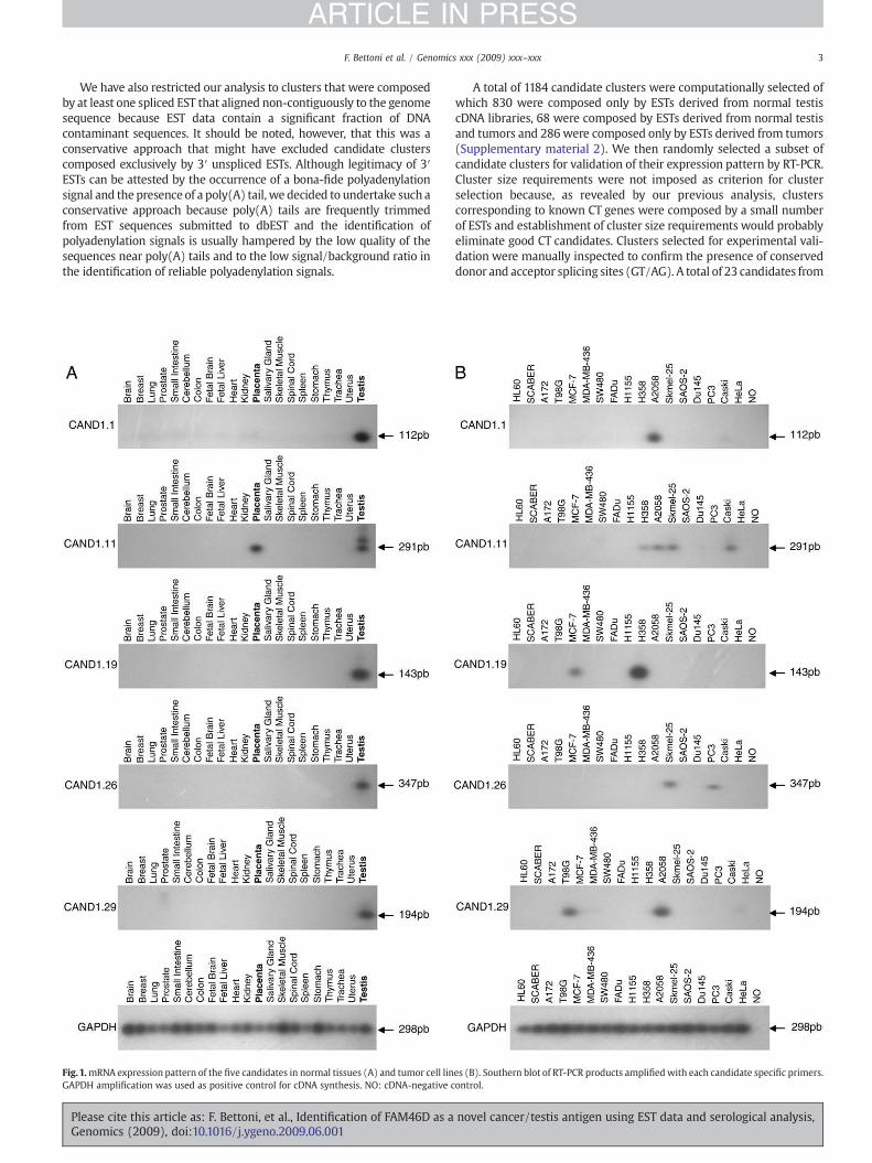
Fig.1.mRNA expression pattern of the five candidates in normal tissues (A) and tumor cell linGAPDH amplification was used as positive control for cDNA synthesis. NO: cDNA-negative c
Please cite this article as: F. Bettoni, et al., Identification of FAM46D as aGenomics (2009), doi:10.1016/j.ygeno.2009.06.001
A total of 1184 candidate clusters were computationally selected ofwhich 830 were composed only by ESTs derived from normal testiscDNA libraries, 68 were composed by ESTs derived from normal testisand tumors and 286 were composed only by ESTs derived from tumors(Supplementary material 2). We then randomly selected a subset ofcandidate clusters for validation of their expression pattern by RT-PCR.Cluster size requirements were not imposed as criterion for clusterselection because, as revealed by our previous analysis, clusterscorresponding to known CT genes were composed by a small numberof ESTs and establishment of cluster size requirements would probablyeliminate good CT candidates. Clusters selected for experimental vali-dation were manually inspected to confirm the presence of conserveddonor and acceptor splicing sites (GT/AG). A total of 23 candidates from
es (B). Southern blot of RT-PCR products amplified with each candidate specific primers.ontrol.
novel cancer/testis antigen using EST data and serological analysis,

4 F. Bettoni et al. / Genomics xxx (2009) xxx–xxx
ARTICLE IN PRESS
each of the 3 lists were randomly selected for validation and weextended our analysis to 23 additional candidates from the testis onlylist, since all candidates validated in the initial round of RT-PCRs werederived from this particular list (Supplementary material 3).
Expression analysis of candidate clusters corresponding to novelCT genes
Expression pattern of candidate clusters was determined by RT-PCR using cDNA from 21 normal tissues, 17 tumor cell lines and 160samples derived from 9 different types of tumors. RT-PCR conditionswere standardized using testis cDNA as template. Few modificationsincluding changes in the annealing temperature and MgCl2 concen-tration and addition of PCR enhancers such as betaine were intro-duced when amplification was not achieved. Amplifications wereachieved for 70 out of the 92 initially selected candidates and ampli-
Fig. 2. Expression analysis of the 5 C
Please cite this article as: F. Bettoni, et al., Identification of FAM46D as aGenomics (2009), doi:10.1016/j.ygeno.2009.06.001
fied fragments were sequenced to confirm their specificity (Supple-mentary material 3).
Six candidates (CAND1.1, CAND1.6, CAND1.19, CAND1.23,CAND1.26 and CAND1.29) were exclusively expressed in testisamong all 21 normal tissues analyzed, five candidates (CAND1.8,CAND1.17, CAND1.25, CAND1.30 and CAND1.32) were expressed intestis and normal brain and one candidate (CAND1.11) was expressedin normal testis and placenta. The expression of six of these initialcandidates (CAND1.1, CAND1.11, CAND1.17, CAND1.19, CAND1.26 andCAND1.29) with a restricted expression in normal tissues was alsodetected in at least one tumor cell line (Fig. 1).
During this expression evaluation step, CAND1.1 was described as anovel CTA (PASD1) frequently expressed in B-cell lymphomas andcapable of eliciting humoral immune response in 40% of those patients[23]. Although we cannot claim the discovery of PASD1 as a novel CTA,we decided to evaluate its expression in different types of solid tumors
T candidates in tumor samples.
novel cancer/testis antigen using EST data and serological analysis,

Table 2Frequency of anti-PASD1, -GASZ and -FAM46D antibodies in plasma samples fromcancer patients.
Tumor Antibody response (%)
PASD1 GASZ FAM46D
Cervix 1/31 (3.2) 0/31 (0.0) 0/31 (0.0)Lung 0/19 (0.0) 0/19 (0.0) 1/19 (5.2)Colon 0/18 (0.0) 0/18 (0.0) 0/18 (0.0)Glioblastoma 2/25 (8.0) 0/25 (0.0) 1/25 (4.0)Total 3/93 (3.2) 0/93 (0.0) 2/93 (2.1)
5F. Bettoni et al. / Genomics xxx (2009) xxx–xxx
ARTICLE IN PRESS
to complement the information available in the literature, and tosupport the role of PASD1 as a therapeutic target for different tumortypes other than B-cell lymphomas.
Expression analysis of PASD1 and the 4 CT candidates (CAND1.11,CAND1.19, CAND1.26 and CAND1.29) exclusively expressed in testis ortestis and placentawas then carried out in 160 samples derived from 9different histological types of tumors (Fig. 2). PASD1 was expressed in41% (65/160) of tumor samples with the highest frequency ofexpression observed in glioblastomas (70%). CAND1.11 was expressedin 65% (104/160) of tumor samples with the highest expressionfrequency in lung tumors (93%). CAND1.19 was expressed in 20% (31/155) of tumor samples with the highest expression frequency incervical tumors (50%). CAND1.26 was expressed in 24% (38/160) oftumor samples and was predominantly expressed in lung tumors(50%). CAND1.29was expressed in 18% (28/156) of the tumor samplesbeing more frequently expressed in gastric tumors (33.3%).
Candidate clusters corresponding to novel CT genes
Two (CAND1.19 and CAND1.26) out of the four CT genes arerepresented by a full-length mRNA sequence and code for a knownhuman protein (Table 1). Candidate CAND1.19 corresponds to theASZ1 or GASZ gene (Germ cell-specific ankyrin, SAM and basic leucinezipper domain containing protein 1) that maps to chromosome 7q31.2and codes for a predicted protein of 475aa containing four ankyrinrepeats and a SAM (Sterile AlphaMotif) and a bZIP domains. The GASZprotein is localized in the cytoplasm of spermatocytes and oocytes atdifferent stages of differentiation [44]. Based on its functionaldomains, GASZ may act as a signaling protein and/or transcriptionalregulator during germ cell maturation and early embryogenesis.Candidate CAND1.26 corresponds to FAM46D gene (Family withsequence similarity 46, member D) mapping to chromosome Xq21.1and encoding a 389aa protein with unknown function.
The remaining 2 CTgenes (CAND1.11 and CAND1.29)were not repre-sented by a full-length mRNA sequence at the time of this analysis andtheir partial 3′ sequenceswere extended towards their 5′ endusingRACE(Rapid Amplification of cDNA Ends). Four distinct 5′ RACE fragmentscorresponding to CAND1.11 splicing variants were obtained (GenBankaccession nos. EF537578, EF537579, EF537580, and EF537581) and asingle RACE fragment (GenBank accession no. EF537582] was generatedfor CAND1.29. After assembling a consensus sequence for CAND1.11 andCAND1.29 using the original 3′ ESTs and the RACE fragments, no openreading frames (ORF) longer than 110aa were identified.
Serological analysis of PASD1 and CTA candidates
The presence of antibodies against the recombinant his-taggedproteins corresponding to PASD1, GASZ (CAND1.19) and FAM46D(CAND1.26) genes was then evaluated by ELISA in plasma samplesfrom cancer patients. Samples from19 lung cancer patients, 31 cervicalcancer patients,18 colorectal cancer patients, 25 glioblastoma patientsand 50 healthy blood donors were used for the ELISA screening(Table 2). Antibodies against PASD1 and FAM46D were detected in3.2 and 2.1% of all plasma samples from cancer patients, respectively. Ahumoral immune response against GASZ could not be detected amongall plasma samples analyzed. Anti-PASD1 antibodies were detectedin plasma samples from patients with cervical cancer (3.2%) and
Table 1Gene name and chromosome location corresponding to each of the five CT candidates.
Candidate Reference sequence Gene name Chromosome region
CAND1.1 BI458651 PASD1 Xq28CAND1.11 AI652043 – 11p15.4CAND1.19 BG771896 GASZ 7q31.2CAND1.26 BG722950 FAM46D Xq21.1CAND1.29 AA451827 – Xq23
Please cite this article as: F. Bettoni, et al., Identification of FAM46D as aGenomics (2009), doi:10.1016/j.ygeno.2009.06.001
glioblastoma (8.0%) while anti-FAM46D antibodies were detected insamples from lung cancer (5.2%) and glioblastoma (4.0%) patients.
Discussion
CTAs are considered promising candidates for the development oftherapeutic cancer vaccines. However, continued progress in thedevelopment of such vaccines will depend on the identification of alarge repertoire of CTAs that could be incorporated in polyvalentcancer vaccines designed to overcome tumor heterogeneity andimmune escape.
In the present work, we have used the publicly available humangenome and ESTs sequences to identify novel CT genes. By examiningthe EST tissue composition of clusters corresponding to known CTantigens, we were able to detect and overcome initial limitations ofour in silico approach. We believe that three major aspects of our insilico analysis have significantly contributed to improve our candidateselection and expression validation efficiency. The first critical aspectwas the use of the human genome sequence as a scaffold for ESTclustering, since it allowed us to exclude multiple alignments relatedto the existence of large gene families among CTAs and to correctlycluster ESTs corresponding to each family member.
Manual curation of cDNA library descriptions available in GenBankwas the second important aspect of our work. Unfortunately, at thetime of this analysis there was no common structured vocabulary todescribe cDNA libraries and descriptions were generally poor andimprecise, leading to the misclassification of the libraries whenconsidering the tissue source and pathologic characteristics. Duringthe course of this work, Kelso et al. [45] developed eVOC, a controlledvocabulary for gene expression data that consists of four orthogonalcontrolled vocabularies describing the anatomical system, cell type,pathology and developmental stage for each cDNA library. Using eVOCthe authors have curated and annotated 7016 cDNA librariesrepresented in dbEST. In attempt to complement and improve ourown manual curation, eVOC annotation was integrated to ourdatabase, however no major improvements were observed in ourcandidate selection.
The analysis of EST composition of CTA clusters was the thirdimportant aspect of our analysis, since it allowed us to define broaderparameters for the selection of additional EST clusters correspondingto CT gene candidates. As opposed to other in silico analysis, we didnot limit our selection to clusters composed by ESTs derived fromnormal testis and tumors. Indeed, the selection of clusters composedexclusively of ESTs derived from normal testis was critical for ourvalidation efficiency, since all CT genes we have identified in this workwere represented in our Transcriptome Database by normal testis ESTsonly.
Using this supervised in silico analysis, we were able to select atotal of 1184 candidate clusters and the expression pattern of a subsetof these clusters was validated by RT-PCR in normal tissues and in alarge collection of tumor cell lines and tumor specimens. Ourcomputational approach was conservative and this number isprobably underestimated since we excluded clusters containing asmall percentage of ESTs from normal tissues and clusters exclusivelycomposed by bona-fide 3′ unspliced ESTs from our analysis.
novel cancer/testis antigen using EST data and serological analysis,

6 F. Bettoni et al. / Genomics xxx (2009) xxx–xxx
ARTICLE IN PRESS
Four CT genes with a restricted expression in normal tissues andbroad expression in different types of tumors were identified.Southern blot analysis of RT-PCR products was used to improve thesensitivity and specificity of our analysis. Our four CT genes arefrequently expressed in melanomas and lung tumors, which areclassified as tumors with a high CT gene expression frequency [3,22],but they are also frequently expressed in tumors considered as mode-rate and low CT expressors such as breast, prostate and colon tumors,respectively.
Two of these validated CT genes (CAND1.11 and CAND1.29) wererepresented by partial 3′ sequences andwere extended towards their 5′end using RACE. After assembling a consensus sequence for CAND1.11and CAND1.29 using the original 3′ ESTs and the RACE fragments, noopen reading frames (ORF) longer than 110aawere identified. Althoughwe cannot exclude the possibility thatwe still have partial sequences forthese transcripts, it is tempting to speculate that these CT genes mightbelong to the growing class of non-coding regulatory RNAs. To ourknowledge, these are the first descriptions of non-coding CT genesand despite the fact that they cannot be considered target for cancerimmunotherapy the role of these non-coding CT genes in the tumo-rigenic process deserves further evaluation.
While this work was in progress, Liggins et al. [23] describedPASD1 (CAND1.1) as a novel CTA eliciting humoral response inpatients with diffuse large B-cell lymphomas (DLBCL). Subsequentlystudies have demonstrated the presence of anti-PASD1 antibodies in35% (6/17) of sera from patients with acute myeloid leukaemia (AML)and 6% (1/16) of sera from patients with chronic myeloid leukaemia(CML) [46]. In addition, Guinn et al. [46] demonstrated thatmonocyte-derived dendritic cells electroporated with PASD1 mRNAcould stimulate autologous T-cells to proliferate. The PASD1 genemaps to chromosome Xq24–28. PASD1 predicted proteins have incommon a PAS (Per ARNT Sim) domain, a putative leucine zipper andnuclear localization signal, suggesting that they may act as transcrip-tion factors. Although we cannot claim the discovery of PASD1 as anovel CTA, information presented in this manuscript on the highfrequency of PASD1 expression in different types of solid tumors andimmunogenicity in patients with cervix tumors and glioblastomassupports PASD1 as a promising target for cancer immunotherapy.
Finally, we were able to detect specific antibodies against theFAM46D protein (CAND1.26) in plasma samples from patients withlung tumors and glioblastomas. As many other CTAs, FAM46D islocated on chromosome X and is a member of a large family of pro-teins with unknown function. Due to its restricted expression patternand immunogenicity FAM46D represents a novel target for cancerimmunotherapy.
To our knowledge, this is the first report that combined in silicoand experimental evaluation of gene expression with serologicalanalysis of cancer patients to identify novel CT antigens. Based on ourresults, FAM46D and PASD1 should be considered novel targets to thedevelopment of a cancer immunotherapy. Further experiments areongoing to evaluate their function and immunogenicity in othertumor types.
Materials and methods
Transcriptome database
Human ESTs available at dbEST (dbEST release 070502) and mRNAsequences from UniGene release 153 were aligned to the maskedhuman genome sequence (NCBI, build 29) by using pp-Blast [47], animplementation of MEGABLAST for a parallel cluster. The parametersused in MEGABLAST were: -f T -J F -F F -W 24. The MEGABLAST outputwas parsed and a MySQL database was loaded with alignmentscoordinates and identities. Information about the tissue origin of eachexpressed sequence was obtained after manual curation of cDNAlibrary descriptions available at dbEST (Supplementary material 1)
Please cite this article as: F. Bettoni, et al., Identification of FAM46D as aGenomics (2009), doi:10.1016/j.ygeno.2009.06.001
and was also uploaded in the database. Spurious and multiplealignments were excluded from the Transcriptome Database byusing an additional set of alignment criteria. These included aminimum identity of 93%, coverage (percentage of sequence lengthaligned) greater than 45% for EST and coverage greater than 55%for mRNAs. Sequences mapping to more than one location on thegenome were given a score for alignment quality. A higher scorewas associated with a higher identity and coverage. Only the align-ments with the highest scores were kept in the database. Clusteringof the expressed sequences was based on their genomic coordinatesas described by Galante et al. [48]. Briefly, if two sequences sharedat least partially the same gene structure (exon/intron boundaries)they were joined into the same cluster. If no exon/intron boundarywas defined, a sequence had to have at least a 100-bp overlap withanother sequence at the genome level to be added to the respectivecluster.
Cluster selection and manual inspection
By querying the Transcriptome Database, we were able to selectclusters composed by spliced ESTs derived from testis and/or tumoralcDNA libraries. Selected clusters were manually inspected to confirmthe splicing structure by checking the presence of conserved donorand acceptor splicing sites (GT/AG) and to exclude those that corres-pond to known CT antigens. Manual inspection was carried out usingthe BLAT interface provided by UCSC Genome Browser (http://genome.ucsc.edu/).
Cell lines and tumor samples
Human tumor cell lines A172 and T98G (glioblastoma); FaDu(squamous cell carcinoma); SW480 (colorectal adenocarcinoma);A2058 and SKmel-28 (malignant melanoma); DU145 and PC3(prostate carcinoma); HeLa and CasKi (cervix adenocarcinoma);MCF-7 and MDA-MB-436 (breast ductal carcinoma); HL-60 (acutepromyelocytic leukaemia); H1155 and H358 (lung carcinoma);SCaBER (transitional cell carcinoma) and SAOS-2 (osteosarcoma)were obtained from American Type Culture Collection (Manassas,VA). Tumor samples were collected from patients treated at HospitalA.C. Camargo. All samples were collected after explicit informedconsent and with local ethical committee approval.
RNA, cDNA synthesis and RT-PCR
Total RNA from cell lines and tumor samples was extracted by theconventional CsCl guanidine thiocyanate method [49]. Total RNA from21 normal human tissues (testis, lung, prostate, small intestine, breast,brain, fetal brain, cerebellum, heart, uterus, placenta, colon, fetal liver,thymus, salivary gland, spinal cord, kidney, spleen, skeletal muscle,stomach and trachea) was purchased from Clontech (Palo Alto, CA).RNA samples were checked for integrity by agarose gel electrophoresisand 2 μg were used for cDNA synthesis. Reverse transcription wasperformed with DNA-free RNA by using SUPERSCRIPT II ReverseTranscriptase (Invitrogen) and oligo(dT) (GE Healthcare). Primersused for expression analysis (Supplementary material 2) weredesigned in different exons using the Primer3 program. RT-PCRswere carried out in 25 μl containing 1 μl of first-strand cDNA, 1× TaqDNA polymerase buffer (Invitrogen), 1 mM MgCl2, 0.1 mM dNTPs (GEHealthcare), 1 U of Taq DNA polymerase (Invitrogen) and 0.3 μM ofeach primer. Amplification conditions were: initial denaturation for4 min at 94 °C followed by 40 cycles of 45 s at 94 °C, 45 s at 60 °C and1min at 72 °C, with a final extension step of 6min at 72 °C. RT-PCRwascarried out in duplicates and triplicates were used to solvediscrepancies between duplicates. RT-PCR products were analyzedon 8% silver-stained polyacrylamide gel and sequenced to confirmtheir specificity. RT-PCR products for candidates CAND1.1, CAND1.11,
novel cancer/testis antigen using EST data and serological analysis,

7F. Bettoni et al. / Genomics xxx (2009) xxx–xxx
ARTICLE IN PRESS
CAND1.19, CAND1.26 and CAND1.29 were further analyzed on agarosegels followed by Southern blot analysis.
RACE (rapid amplification of cDNA ends)
5′-RACE was performed on normal testis poly(A)+ RNA by usingthe Marathon cDNA Amplification Kit (Clontech, Palo Alto, CA).Amplifications reactions were performed in 25 μl by using 2.5 μl ofcDNA, 0.2 mM dNTPs, 0.2 μM specific primer (RACE5′/CAND704, 5′GATTTTCCAGACCTTGTCCAAGCTCC3′, RACE5′/CAND1001, 5′CACACA-GAATGCCGCTCGCTAGGAG3′), 0.2 μM adaptor primer, 1 U of Advan-tage Taq DNA polymerase. PCR conditions were: 5 cycles of 5 s at 94 °Cand 3 min at 72 °C, 5 cycles of 5 s at 94 °C, 10 s at 70 °C and 3 min at72 °C, and 25 cycles of 5 s at 94 °C, 10 s at 68 °C and 3 min at 72 °C.Nested PCR was carried out by using 5 μl of the first reaction productdiluted 50×, specific nested primer (RACE5′/CAND704N, 5′GTGGCCAACTGAGCTGCAGACTTCCC3′, RACE5′/CAND1001N, 5′GATGTTTCCTCTTAGGCCTCTTCTCC3′) and nested adaptor primer. PCRconditions differ from the first reaction only by using 20 cycles. PCRproducts were analyzed on agarose gels, cloned and sequenced asdescribed above.
Recombinant protein expression
CAND1.1 (773aa), CAND1.19 (475aa) and CAND1.26 longest ORFs(389aa) were amplified from normal testis cDNA using the followingprimers: CAND1.1F (5′- CGGAATTCATGAAGATGAGAGGGGAAAAG-3′)and CAND1.1R (5′- CCCAAGCTTTTAGCACGGCTTATTTGAGTC-3′);CAND1.19F (5′- GATATCATGGCGGCGAGCGCGCTGCGA-3′) andCAND1.19R (5′- CGGAATTCTTATTTCCTCTGGAAAGTTAG-3′);CAND1.26F (5′- CGGAATTCATGTCTGAAATCAGATTCACC-3′) andCAND1.26R (5′- CCCAAGCTTTTAACTCATACCATTTGATCC-3′). The PCRproducts corresponding to CAND1.1 and CAND1.26 were digestedwith EcoRI (New England Biolabs) and HindIII (New England Biolabs)and cloned into the expression vector pET28a (Stratagene, La Jolla,CA). The PCR product corresponding to CAND1.19 was digested withEcoRV (New England Biolabs) and EcoRI (New England Biolabs) andcloned into the expression vector pET32a (Stratagene, La Jolla, CA).After sequencing, recombinant plasmids were transformed intoEscherichia coli BL-21 Rosetta. After induction with 0.4 mM isopropylβ-D-thiogalactoside at 37 °C for 4 h, recombinant proteins fused witha 6His-tag were purified by Ni2+ affinity chromatography by usingthe NiNTA agarose resin (Invitrogen). The purified protein wasanalyzed by Western blot using an anti-His-tag monoclonal antibody(GE Healthcare) to confirm the purification efficiency.
Antibody response in cancer patients
Plasmas were obtained from patients treated at Hospital A.C.Camargo. All samples were collected after explicit informed consentandwith local ethical committee approval. In addition, plasma samplesfrom 50 healthy individuals were collected from blood donors at theHospital A.C. Camargo Blood Center. Antibodies against CAND1.1,CAND1.19 and CAND1.26 recombinant proteins were determined bydirect ELISA (Enzyme-Linked ImmunoSorbent Assay). Microplateswere coated with 50 μl of the recombinant protein (5 μg/ml) inphosphate buffered saline (PBS) pH 7.4 for 2 h at 37 °C. Wells wereblocked with 5% (w/v) skim milk in PBS, washed 3 times with PBS-Tween (0.1%) overnight at 4 °C, and incubatedwith 50 μl serumdiluted1:100 in dilution buffer (5%(w/v) skim milk in PBS) for 2 h at 37 °C.After washing, 50 μl goat anti-human IgG conjugated to horseradishperoxidase (Sigma) diluted 1:5000 in PBS was used as secondaryantibody for 1 h at 37 °C, followed by washing. Bound secondaryantibody was visualized by the HRP enzymatic reaction using OPDtablets (Sigma). Absorbance at 492 nm was read on a SpectroCountanalyzer and expressed as optical density (OD) values. All serum
Please cite this article as: F. Bettoni, et al., Identification of FAM46D as aGenomics (2009), doi:10.1016/j.ygeno.2009.06.001
samples were analyzed in triplicates and corrected for backgroundbinding to an irrelevant protein from sugar cane expressed in the sameexpression system as CTs recombinant proteins. ELISA cut-off wasestablished as the mean+2SD of healthy blood donors. Experimentswere carried out in duplicates and a serum sample was consideredpositive when reactivity was observed in two independent ELISAassays. Triplicates were used to solve discrepancies betweenduplicates.
Acknowledgments
We would like to thank Mônica C. Poli (Blood Center Hospital A.C.Camargo) for providing the plasma samples from the blood donors.We also thank Waleska K. Martins and Marilene H. Lopes whocontributedmaterials essential for the study. This work was supportedby the Ludwig Institute for Cancer Research, São Paulo, Brazil, byFundação de Amparo à Pesquisa do Estado de São Paulo Grant and byConselho Nacional de Desenvolvimento Científico e TecnológicoGrant. FB and PAFG were sponsored by fellowships from Fundaçãode Amparo a Pesquisa do Estado de São Paulo; RBPwas sponsored by afellowship from Coordenação de Aperfeiçoamento de Pessoal de NívelSuperior; MVGwas sponsored by a fellowship from Conselho Nacionalde Desenvolvimento Científico e Tecnológico.
Appendix A. Supplementary data
Supplementary data associated with this article can be found, inthe online version, at doi:10.1016/j.ygeno.2009.06.001.
References
[1] A.J. Simpson, O.L. Caballero, A. Jungbluth, Y.T. Chen, L.J. Old, Cancer/testis antigens,gametogenesis and cancer, Nat. Rev., Cancer 5 (2005) 615–625.
[2] M.J. Scanlan, A.O. Gure, A.A. Jungbluth, L.J. Old, Y.T. Chen, Cancer/testis antigens:an expanding family of targets for cancer immunotherapy, Immunol. Rev. 188(2002) 22–32.
[3] M.J. Scanlan, A.J. Simpson, L.J. Old, The cancer/testis genes: review, standardiza-tion, and commentary, Cancer Immun. 4 (2004) 1.
[4] A.J. Zendman, D.J. Ruiter, G.N. Van Muijen, Cancer/testis-associated genes: iden-tification, expression profile, and putative function, J. Cell. Physiol. 194 (2003)272–288.
[5] D. Davis, W. Chen, H. Jackson, P. Parente, M. Shackleton, W. Hopkins, Q. Chen, N.Dimopoulos, T. Luke, R. Murphy, A.M. Scott, E. Maraskovsky, G. McArthur, D.MacGregor, S. Sturrock, T.Y. Tai, S. Green, A. Cuthbertson, D. Maher, L. Miloradovic,S.V. Mitchell, G. Ritter, A.A. Jungbluth, Y.T. Chen, S. Gnjatic, E.W. Hoffman, L.J. Old,J.S. Cebon, Recombinant NY-ESO-1 protein with ISCOMATRIX adjuvant inducesbroad integrated antibody and CD4(+) and CD8(+) T cell responses in humans,Proc. Natl. Acad. Sci. U. S. A. 101 (2004) 10697–10702.
[6] M. Marchand, N. van Baren, P. Weynants, V. Brichard, B. Dreno, M.H. Tessier, E.Rankin, G. Parmiani, F. Arienti, Y. Humblet, A. Bourlond, R. Vanwijck, D. Lienard, M.Beauduin, P.Y. Dietrich, V. Russo, J. Kerger, G. Masucci, E. Jager, J. De Greve, J.Atzpodien, F. Brasseur, P.G. Coulie, P. van der Bruggen, T. Boon, Tumor regressionsobserved in patients with metastatic melanoma treated with an antigenic peptideencoded by gene MAGE-3 and presented by HLA-A1, Int. J. Cancer 80 (1999)219–230.
[7] S. Adams, D.W. O'Neill, D. Nonaka, E. Hardin, L. Chiriboga, K. Siu, C.M. Cruz, A.Angiulli, F. Angiulli, E. Ritter, R.M. Holman, R.L. Shapiro, R.S. Berman, N. Berner, Y.Shao, O. Manches, L. Pan, R.R. Venhaus, E.W. Hoffman, A. Jungbluth, S. Gnjatic, L.Old, A.C. Pavlick, N. Bhardwaj, Immunization of malignant melanoma patientswith full-length NY-ESO-1 protein using TLR7 agonist imiquimod as vaccineadjuvant, J. Immunol. 181 (2008) 776–784.
[8] P. Romero, Current state of vaccine therapies in non-small-cell lung cancer, Clin.Lung Cancer 9 Suppl. 1 (2008) S28–36.
[9] C. Traversari, P. van der Bruggen, B. Van den Eynde, P. Hainaut, C. Lemoine, N. Ohta, L.Old, T. Boon, Transfection and expression of a gene coding for a human melanomaantigen recognized by autologous cytolytic T lymphocytes, Immunogenetics 35(1992) 145–152.
[10] P. van der Bruggen, C. Traversari, P. Chomez, C. Lurquin, E. De Plaen, B. Van denEynde, A. Knuth, T. Boon, A gene encoding an antigen recognized by cytolytic Tlymphocytes on a human melanoma, Science 254 (1991) 1643–1647.
[11] P. Boel, C. Wildmann, M.L. Sensi, R. Brasseur, J.C. Renauld, P. Coulie, T. Boon, P. vander Bruggen, BAGE: a new gene encoding an antigen recognized on humanmelanomas by cytolytic T lymphocytes, Immunity 2 (1995) 167–175.
[12] B. Van den Eynde, O. Peeters, O. De Backer, B. Gaugler, S. Lucas, T. Boon, A newfamily of genes coding for an antigen recognized by autologous cytolytic Tlymphocytes on a human melanoma, J. Exp. Med. 182 (1995) 689–698.
novel cancer/testis antigen using EST data and serological analysis,

8 F. Bettoni et al. / Genomics xxx (2009) xxx–xxx
ARTICLE IN PRESS
[13] U. Sahin, O. Tureci, M. Pfreundschuh, Serological identification of human tumorantigens, Curr. Opin. Immunol. 9 (1997) 709–716.
[14] U. Sahin,O. Tureci,H. Schmitt, B. Cochlovius, T. Johannes, R. Schmits, F. Stenner, G. Luo,I. Schobert, M. Pfreundschuh, Human neoplasms elicit multiple specific immuneresponses in the autologoushost, Proc. Natl. Acad. Sci. U. S. A. 92 (1995) 11810–11813.
[15] B. Lethe, S. Lucas, L. Michaux, C. De Smet, D. Godelaine, A. Serrano, E. De Plaen, T.Boon, LAGE-1, a new gene with tumor specificity, Int. J. Cancer 76 (1998)903–908.
[16] A.O. Gure, E. Stockert, K.C. Arden, A.D. Boyer, C.S. Viars, M.J. Scanlan, L.J. Old, Y.T.Chen, CT10: a new cancer-testis (CT) antigen homologous to CT7 and the MAGEfamily, identified by representational-difference analysis, Int. J. Cancer 85 (2000)726–732.
[17] A. Westbrook, A.B. Diekman, K.L. Klotz, V. Khole, C. von Kap-Herr, W.L. Golden,R.L. Eddy, T.B. Shows, M.H. Stoler, C.Y. Lee, C.J. Flickinger, J.C. Herr, Spermatid-specific expression of the novel X-linked gene product SPAN-X localized to thenucleus of human spermatozoa, Biol. Reprod. 63 (2000) 469–481.
[18] A.J. Zendman, M. Cornelissen, U.H. Weidle, D.J. Ruiter, G.N. van Muijen, CTp11, anovel member of the family of human cancer/testis antigens, Cancer Res. 59(1999) 6223–6229.
[19] N.J. de Wit, U.H. Weidle, D.J. Ruiter, G.N. van Muijen, Expression profiling of MMA-1a and splice variant MMA-1b: new cancer/testis antigens identified in humanmelanoma, Int. J. Cancer 98 (2002) 547–553.
[20] M.J. Scanlan, C.M. Gordon, B. Williamson, S.Y. Lee, Y.T. Chen, E. Stockert, A.Jungbluth, G. Ritter, D. Jager, E. Jager, A. Knuth, L.J. Old, Identification of cancer/testis genes by database mining and mRNA expression analysis, Int. J. Cancer 98(2002) 485–492.
[21] Y.T. Chen, M.J. Scanlan, C.A. Venditti, R. Chua, G. Theiler, B.J. Stevenson, C. Iseli, A.O.Gure, T. Vasicek, R.L. Strausberg, C.V. Jongeneel, L.J. Old, A.J. Simpson, Identificationof cancer/testis-antigen genes by massively parallel signature sequencing, Proc.Natl. Acad. Sci. U. S. A. 102 (2005) 7940–7945.
[22] O. Hofmann, O.L. Caballero, B.J. Stevenson, Y.T. Chen, T. Cohen, R. Chua, C.A. Maher,S. Panji, U. Schaefer, A. Kruger, M. Lehvaslaiho, P. Carninci, Y. Hayashizaki, C.V.Jongeneel, A.J. Simpson, L.J. Old, W. Hide, Genome-wide analysis of cancer/testisgene expression, Proc. Natl. Acad. Sci. U. S. A. 105 (2008) 20422–20427.
[23] A.P. Liggins, B.A. Guinn, C.S. Hatton, K. Pulford, A.H. Banham, Serologic detection ofdiffuse large B-cell lymphoma-associated antigens, Int. J. Cancer 110 (2004)563–569.
[24] M.D. Adams, M. Dubnick, A.R. Kerlavage, R. Moreno, J.M. Kelley, T.R. Utterback, J.W.Nagle, C. Fields, J.C. Venter, Sequence identification of 2,375 human brain genes,Nature 355 (1992) 632–634.
[25] M.D. Adams, A.R. Kerlavage, C. Fields, J.C. Venter, 3,400 new expressed sequencetags identify diversity of transcripts in human brain, Nat. Genet. 4 (1993) 256–267.
[26] S. Brown, J.L. Chang, W. Sadee, P.C. Babbitt, A semiautomated approach to genediscovery through expressed sequence tag data mining: discovery of new humantransporter genes, AAPS PharmSci 5 (2003) E1.
[27] M.P. De Young, H. Damania, D. Scheurle, C. Zylberberg, R. Narayanan, Bioinfor-matics-based discovery of a novel factor with apparent specificity to colon cancer,In Vivo 16 (2002) 239–248.
[28] R.B. Parmigiani, F. Bettoni, M.D. Vibranovski, M.H. Lopes, W.K. Martins, W. Cunha,F.A. Soares, A.J. Simpson, S.J. de Souza, A.A. Camargo, Characterization of a cancer/testis (CT) antigen gene family capable of eliciting humoral response in cancerpatients, Proc. Natl. Acad. Sci. U. S. A. 103 (2006) 18066–18071.
[29] B.E. Baysal, E.M. van Schothorst, J.E. Farr, M.R. James, P. Devilee, C.W. Richard 3rd, Ahigh-resolution STS, EST, and gene-based physical map of the hereditary para-ganglioma region on chromosome 11q23, Genomics 44 (1997) 214–221.
[30] T.J. Hudson, A.M. Colbert, M.P. Reeve, J.S. Bae, M.K. Lee, R.L. Nussbaum, M.L. Budarf,B.S. Emanuel, S. Foote, Isolation and regionalmapping of 110 chromosome 22 STSs,Genomics 24 (1994) 588–592.
Please cite this article as: F. Bettoni, et al., Identification of FAM46D as aGenomics (2009), doi:10.1016/j.ygeno.2009.06.001
[31] Y. Lee, R. Sultana, G. Pertea, J. Cho, S. Karamycheva, J. Tsai, B. Parvizi, F. Cheung, V.Antonescu, J. White, I. Holt, F. Liang, J. Quackenbush, Cross-referencing eukaryoticgenomes: TIGR Orthologous Gene Alignments (TOGA), Genome Res. 12 (2002)493–502.
[32] F. Mignone, A. Anselmo, G. Donvito, G.P. Maggi, G. Grillo, G. Pesole, Genome-wideidentification of coding and non-coding conserved sequence tags in human andmouse genomes, BMC Genomics 9 (2008) 277.
[33] S. Tugendreich, D.E. Bassett Jr., V.A. McKusick, M.S. Boguski, P. Hieter, Genesconserved in yeast and humans, Hum. Mol. Genet. 3 (1994) 1509–1517.
[34] Q. Xu, C. Lee, Discovery of novel splice forms and functional analysis of cancer-specific alternative splicing in human expressed sequences, Nucleic Acids Res. 31(2003) 5635–5643.
[35] Q. Xu, B. Modrek, C. Lee, Genome-wide detection of tissue-specific alternativesplicing in the human transcriptome, Nucleic Acids Res. 30 (2002) 3754–3766.
[36] P. Bonizzoni, G. Mauri, G. Pesole, E. Picardi, Y. Pirola, R. Rizzi, Detecting alternativegene structures from spliced ESTs: a computational approach, J. Comput. Biol. 16(2009) 43–66.
[37] L. Hui, X. Zhang, X. Wu, Z. Lin, Q. Wang, Y. Li, G. Hu, Identification of alternativelyspliced mRNA variants related to cancers by genome-wide ESTs alignment,Oncogene 23 (2004) 3013–3023.
[38] Z. Kan, E.C. Rouchka, W.R. Gish, D.J. States, Gene structure prediction andalternative splicing analysis using genomically aligned ESTs, Genome Res. 11(2001) 889–900.
[39] H. Xie, W.Y. Zhu, A. Wasserman, V. Grebinskiy, A. Olson, L. Mintz, Computationalanalysis of alternative splicing using EST tissue information, Genomics 80 (2002)326–330.
[40] E. Beaudoing, D. Gautheret, Identification of alternate polyadenylation sites andanalysis of their tissue distribution using EST data, Genome Res. 11 (2001)1520–1526.
[41] C. Iseli, B.J. Stevenson, S.J. de Souza, H.B. Samaia, A.A. Camargo, K.H. Buetow, R.L.Strausberg, A.J. Simpson, P. Bucher, C.V. Jongeneel, Long-range heterogeneity atthe 3′ ends of human mRNAs, Genome Res. 12 (2002) 1068–1074.
[42] M. Kamasawa, J. Horiuchi, Identification and characterization of polyadenylationsignal (PAS) variants in human genomic sequences based on modified ESTclustering, In Silico Biol. 8 (2008) 347–361.
[43] S. Aradhya, T. Bardaro, P. Galgoczy, T. Yamagata, T. Esposito, H. Patlan, A.Ciccodicola, A. Munnich, S. Kenwrick, M. Platzer, M. D'Urso, D.L. Nelson, Multiplepathogenic and benign genomic rearrangements occur at a 35 kb duplicationinvolving the NEMO and LAGE2 genes, Hum. Mol. Genet. 10 (2001) 2557–2567.
[44] W. Yan, A. Rajkovic, M.M. Viveiros, K.H. Burns, J.J. Eppig, M.M. Matzuk,Identification of Gasz, an evolutionarily conserved gene expressed exclusively ingerm cells and encoding a proteinwith four ankyrin repeats, a sterile-alpha motif,and a basic leucine zipper, Mol. Endocrinol. 16 (2002) 1168–1184.
[45] J. Kelso, J. Visagie, G. Theiler, A. Christoffels, S. Bardien, D. Smedley, D. Otgaar, G.Greyling, C.V. Jongeneel, M.I. McCarthy, T. Hide, W. Hide, eVOC: a controlledvocabulary for unifying gene expression data, Genome Res. 13 (2003) 1222–1230.
[46] B.A. Guinn, E.A. Bland, U. Lodi, A.P. Liggins, K. Tobal, S. Petters, J.W. Wells, A.H.Banham, G.J. Mufti, Humoral detection of leukaemia-associated antigens inpresentation acute myeloid leukaemia, Biochem. Biophys. Res. Commun. 335(2005) 1293–1304.
[47] E.C. Osorio, J.E. de Souza, A.C. Zaiats, P.S. de Oliveira, S.J. de Souza, pp-Blast: a“pseudo-parallel” Blast, Braz. J. Med. Biol. Res. 36 (2003) 463–464.
[48] P.A. Galante, N.J. Sakabe, N. Kirschbaum-Slager, S.J. de Souza, Detection andevaluation of intron retention events in the human transcriptome, Rna 10 (2004)757–765.
[49] J.M. Chirgwin, A.E. Przybyla, R.J. MacDonald, W.J. Rutter, Isolation of biologicallyactive ribonucleic acid from sources enriched in ribonuclease, Biochemistry 18(1979) 5294–5299.
novel cancer/testis antigen using EST data and serological analysis,