Systems based mapping demonstrates that recovery from alkylation damage requires DNA repair, RNA...
23
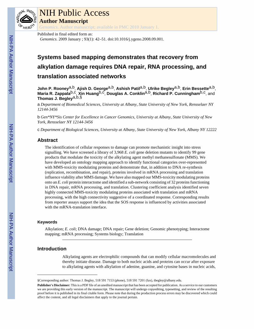
Systems based mapping demonstrates that recovery from alkylation damage requires DNA repair, RNA processing, and translation associated networks John P. Rooney a,b , Ajish D. George a,b , Ashish Patil a,b , Ulrike Begley a,b , Erin Bessette a,b , Maria R. Zappala b,c , Xin Huang b,c , Douglas A. Conklin a,b , Richard P. Cunningham b,c , and Thomas J. Begley a,b,§ a Department of Biomedical Sciences, University at Albany, State University of New York, Rensselaer NY 12144-3456 b Gen*NY*Sis Center for Excellence in Cancer Genomics, University at Albany, State University of New York, Rensselaer NY 12144-3456 c Department of Biological Sciences, University at Albany, State University of New York, Albany NY 12222 Abstract The identification of cellular responses to damage can promote mechanistic insight into stress signalling. We have screened a library of 3,968 E. coli gene deletion mutants to identify 99 gene products that modulate the toxicity of the alkylating agent methyl methanesulfonate (MMS). We have developed an ontology mapping approach to identify functional categories over-represented with MMS-toxicity modulating proteins and demonstrate that, in addition to DNA re-synthesis (replication, recombination, and repair), proteins involved in mRNA processing and translation influence viability after MMS damage. We have also mapped our MMS-toxicity modulating proteins onto an E. coli protein interactome and identified a sub-network consisting of 32 proteins functioning in DNA repair, mRNA processing, and translation. Clustering coefficient analysis identified seven highly connected MMS-toxicity modulating proteins associated with translation and mRNA processing, with the high connectivity suggestive of a coordinated response. Corresponding results from reporter assays support the idea that the SOS response is influenced by activities associated with the mRNA-translation interface. Keywords Alkylation; E. coli; DNA damage; DNA repair; Gene deletion; Genomic phenotyping; Interactome mapping; mRNA processing; Systems biology; Translation Introduction Alkylating agents are electrophilic compounds that can modify cellular macromolecules and thereby initiate disease. Damage to both nucleic acids and proteins can occur after exposure to alkylating agents with alkylation of adenine, guanine, and cytosine bases in nucleic acids, §Corresponding author: Thomas J. Begley, 518 591 7153 (phone), 518 591 7201 (fax), [email protected]. Publisher's Disclaimer: This is a PDF file of an unedited manuscript that has been accepted for publication. As a service to our customers we are providing this early version of the manuscript. The manuscript will undergo copyediting, typesetting, and review of the resulting proof before it is published in its final citable form. Please note that during the production process errors may be discovered which could affect the content, and all legal disclaimers that apply to the journal pertain. NIH Public Access Author Manuscript Genomics. Author manuscript; available in PMC 2010 January 1. Published in final edited form as: Genomics. 2009 January ; 93(1): 42–51. doi:10.1016/j.ygeno.2008.09.001. NIH-PA Author Manuscript NIH-PA Author Manuscript NIH-PA Author Manuscript
-
Upload
independent -
Category
Documents
-
view
1 -
download
0
Transcript of Systems based mapping demonstrates that recovery from alkylation damage requires DNA repair, RNA...
Systems based mapping demonstrates that recovery fromalkylation damage requires DNA repair, RNA processing, andtranslation associated networks
John P. Rooneya,b, Ajish D. Georgea,b, Ashish Patila,b, Ulrike Begleya,b, Erin Bessettea,b,Maria R. Zappalab,c, Xin Huangb,c, Douglas A. Conklina,b, Richard P. Cunninghamb,c, andThomas J. Begleya,b,§
a Department of Biomedical Sciences, University at Albany, State University of New York, Rensselaer NY12144-3456
b Gen*NY*Sis Center for Excellence in Cancer Genomics, University at Albany, State University of NewYork, Rensselaer NY 12144-3456
c Department of Biological Sciences, University at Albany, State University of New York, Albany NY 12222
AbstractThe identification of cellular responses to damage can promote mechanistic insight into stresssignalling. We have screened a library of 3,968 E. coli gene deletion mutants to identify 99 geneproducts that modulate the toxicity of the alkylating agent methyl methanesulfonate (MMS). Wehave developed an ontology mapping approach to identify functional categories over-representedwith MMS-toxicity modulating proteins and demonstrate that, in addition to DNA re-synthesis(replication, recombination, and repair), proteins involved in mRNA processing and translationinfluence viability after MMS damage. We have also mapped our MMS-toxicity modulating proteinsonto an E. coli protein interactome and identified a sub-network consisting of 32 proteins functioningin DNA repair, mRNA processing, and translation. Clustering coefficient analysis identified sevenhighly connected MMS-toxicity modulating proteins associated with translation and mRNAprocessing, with the high connectivity suggestive of a coordinated response. Corresponding resultsfrom reporter assays support the idea that the SOS response is influenced by activities associatedwith the mRNA-translation interface.
KeywordsAlkylation; E. coli; DNA damage; DNA repair; Gene deletion; Genomic phenotyping; Interactomemapping; mRNA processing; Systems biology; Translation
IntroductionAlkylating agents are electrophilic compounds that can modify cellular macromolecules andthereby initiate disease. Damage to both nucleic acids and proteins can occur after exposureto alkylating agents with alkylation of adenine, guanine, and cytosine bases in nucleic acids,
§Corresponding author: Thomas J. Begley, 518 591 7153 (phone), 518 591 7201 (fax), [email protected]'s Disclaimer: This is a PDF file of an unedited manuscript that has been accepted for publication. As a service to our customerswe are providing this early version of the manuscript. The manuscript will undergo copyediting, typesetting, and review of the resultingproof before it is published in its final citable form. Please note that during the production process errors may be discovered which couldaffect the content, and all legal disclaimers that apply to the journal pertain.
NIH Public AccessAuthor ManuscriptGenomics. Author manuscript; available in PMC 2010 January 1.
Published in final edited form as:Genomics. 2009 January ; 93(1): 42–51. doi:10.1016/j.ygeno.2008.09.001.
NIH
-PA Author Manuscript
NIH
-PA Author Manuscript
NIH
-PA Author Manuscript

and arginine, lysine, and cysteine residues in proteins as common sites of damage [1–8]. Thealkylating agent methyl methanesulfonate (MMS) has been frequently employed to mimic theeffects of both endogenous and environmental alkylating agents. MMS is an alkylating agentthat damages both nucleic acids and proteins, thereby promoting mutagenesis and cell death[8,9]. MMS has proven to be a valuable tool for characterizing cellular damage-responsemachinery [10–13].
Cellular responses to alkylation damage play an important role in preventing mutations andcell death [14–17]. A number of enzyme activities from bacteria and mammals have beenidentified that modulate the toxicity and mutagenicity of alkylating agents [18–27], and someDNA repair proteins are examples of a conserved response to alkylation damage [28–33]. Thealkylbase DNA glycosylases from E. coli (AlkA) and the mouse (Aag) are a case in point;inactivation of either renders cells sensitive to killing by alkylating agents [29]. A more recentexample is the direct repair enzyme AlkB, which was initially identified in E. coli as an activitythat modulates the toxicity of MMS [24]. AlkB repairs single and double stranded DNA andRNA lesions caused by MMS (1-methyladenine and 3-methylcytosine). Similar activities havebeen characterized in the mouse and in humans [30–32,34], and have been shown tocomplement the MMS sensitive phenotype of alkB deficient E. coli. The functionalconservation of base excision and direct repair proteins between E. coli and mammals supportsthe concept that cells use common mechanisms to repair damage caused by alkylating agents.
Mechanistic studies in E. coli have previously demonstrated that components of the adaptiveresponse and the SOS response are activated after MMS induced DNA damage [35–37].Signalling proteins that initiate the adaptive and SOS responses are Ada and RecA respectively;both proteins recognize DNA damage and initiate downstream signalling to promote repair.Different types of DNA damage are detected by Ada and RecA, and activation of each proteinwill initiate transcriptional responses that facilitate cell survival after alkylation damage.Transcriptional reprogramming and increased repair in response to alkylation damage are wellconserved themes across phylogeny [12,38–40].
We describe here a global study using a library of 3,968 unique E. coli gene deletion mutantsto identify activities that prevent cell death after treatment with the alkylating agent MMS. Weshow that at least 99 different protein activities are important for preventing MMS-inducedcell death. Functional and computational mapping of the MMS-toxicity modulating geneproducts identified protein networks specific to DNA repair, transcription, mRNA processing,and translation as being important after alkylation damage. Similarly, validation experimentsthat use newly constructed gene deletion mutants in cell killing assays and SOS-reporter assaysdemonstrate that cellular processes that promote the re-synthesis of DNA and proteins areessential for cell survival. Our results support the hypothesis that specific translational andmRNA processing activities, which are conserved from E. coli to humans, are utilized duringthe response to MMS damage.
Results and DiscussionToxicity modulating genes identified by genomic phenotyping
We used a robotic plate-based screen of E. coli gene deletion mutants to identify genes andtheir associated proteins that modulate toxicity to MMS. E. coli gene deletion mutants werefrom the Keio library [41], which was generated using a targeted homologous recombinationstrategy and which consists of 8,640 mutants, with at least two independent isolates of eachgene knockout represented in the library. The library we tested represented 3,968 E. coli genesand provided approximately 93% coverage of the genome. Mutants were grown to saturationin 96-well plates and 1 μl aliquots of a 1:10 dilution of the cell suspensions were roboticallytransferred onto agar plates containing two concentrations of MMS. Approximately 360 agar
Rooney et al. Page 2
Genomics. Author manuscript; available in PMC 2010 January 1.
NIH
-PA Author Manuscript
NIH
-PA Author Manuscript
NIH
-PA Author Manuscript

plates, with 34,560 spotted cultures, were incubated overnight at 37°C and then digitallyimaged for analysis. Images of each plate were compiled to create a data base (SupplementalFigure S1: Supplemental Table 1–2) and sensitive mutants were visually identified (Figure 1:Table 1). A virtual mutant representing at least two isolates of each gene deletion mutant inthe library was given a MMS toxicity modulating score, which is based on the behaviour ofall corresponding deletion mutants on two plates containing MMS (0.045 and 0.06% MMS).For example there were two ΔalkA mutants in the library, and a virtual mutant representingΔalkA has a compiled MMS toxicity modulating score describing the behaviour of bothΔalkA mutants on two concentrations of MMS. The MMS-toxicity modulating score is a semi-quantitative measure of the sensitivity of a virtual mutant after MMS treatment, and consistsof values from two concentrations of MMS for two independent mutants specific to each gene-product. Deletion mutants with reduced growth on MMS were given a score of 2, by ourconvention those with a color change from white to dark grey are associated with a growthdefect and were scored 1 (See Figure 1B), while those showing unaffected growth were scored0. In theory the virtual mutants most sensitive to MMS were scored as an eight (2 + 2 + 2 +2), because two corresponding isolates were sensitive to both concentrations of MMS. We useda minimum cut-off of three to identify virtual mutants that were sensitive to MMS.
In all, we identified 99 virtual mutants that scored three or greater and that were classified asMMS sensitive. We also independently constructed 96 gene deletion mutants (Rooney et al.,in press) which recapitulated some of the MMS-sensitive gene-deletion mutants. As a control,we also constructed gene deletion mutants that do not affect MMS sensitivity, as identified inour present study. We performed MMS sensitivity testing on these newly derived gene-deletionmutants (Supplemental Table S3) and determined that 90% displayed a phenotype similar tothose virtual mutants derived from the Keio library, indicating that the Keio library is of highquality and that our results are highly reproducible.
We next catalogued the 99 virtual mutants by assigning the proteins corresponding to eachcatalogued deleted gene and assembled a list of 99 MMS-toxicity modulating proteins. Weanalyzed the type of protein activities important after MMS damage, using informationsupplied by the Ecogene database [42]. As expected, a number of previously identified DNAalkylation repair and recombination proteins were identified in our set of 99 MMS-toxicitymodulating proteins, including Ada, AlkA, AlkB, and RecA. In all, we identified elevendifferent DNA repair/recombination activities that modulated the toxicity of MMS. Theseactivities represented components of direct repair (DR), base excision repair (BER), mismatchrepair (MMR) and recombinational repair (RR) (Ada, AlkA, AlkB, RecA, RecC, RuvA, RecB,RuvB, Dam, RecO, and RecN). In addition, four DNA replication proteins were identified(DnaT, PriA, DnaG, and ParC). The identification of DNA repair and DNA replication proteinsconfirms the model that the re-synthesis of damaged DNA is a priority after MMS exposure.Similar screens in budding yeast [10,11] also identified DR, BER, MMR, RR, and replicationproteins as modulators of MMS-toxicity.
In addition to DNA repair, recombination, and replication proteins, we identified activitiesspecific to transcriptional regulation, protein damage, and protein synthesis in our MMS screen.Transcriptional components important after MMS damage include members of the adaptiveand SOS response (Ada and RecA). In addition, we found that previously identified andpredicted transcriptional regulatory proteins (ArcA, Fis, CadC, UidR, and OxyR) modulatedthe toxicity of MMS. These proteins are associated with the regulation of a large (ArcA andFis) or small number (CadC, UidR, and OxyR) of downstream targets, indicating that bothglobal and specific transcriptional regulators were identified in our screen. ArcA can positivelyor negatively regulate transcription of aerobic enzymes and has been demonstrated to controlthe resistance of E. coli to specific dyes [43], presumably through regulation of envelopeproteins. In theory, cells deficient in ArcA could contain higher levels of intracellular MMS
Rooney et al. Page 3
Genomics. Author manuscript; available in PMC 2010 January 1.
NIH
-PA Author Manuscript
NIH
-PA Author Manuscript
NIH
-PA Author Manuscript
and thus be susceptible to more DNA damage. The global regulator Fis was also identified asa protein that modulated MMS-toxicity, which is a novel observation, and Fis plays roles inthe transcriptional activation of rRNA genes, site specific DNA inversion, and repression ofDNA replication [44]. The precise role of Fis after DNA damage has yet to be determined, butFis has great potential to influence DNA and protein metabolism after alkylation damage. CadCis a transcriptional activator for other cadaverin associated gene products (cadA and cadB) andis know to sense external stimuli associated with low pH and low oxygen [42], and we canspeculate that this transcriptional activator can sense environmental conditions associated withMMS in the medium. Similarly the classification of the sensor for oxidative stress OxyR [42]as MMS toxicity modulating suggests that MMS damage can alter the levels of reactive oxygenspecies inside the cell.
A third category of proteins that modulates the toxicity of MMS included those specific toprotein maintenance, protein stabilization, and translation. The Hsp70 chaperone protein DnaKwas identified in our screen, suggesting that DnaK plays a role in stabilizing MMS modifiedproteins. DnaK has also been shown to play a regulatory role in DNA replication [45], whichcould account for the MMS sensitive phenotype of the corresponding deletion mutant. Elevendifferent activities specific to translation were identified as modulating MMS sensitivity. Basicribosome machinery that includes major components of the 30S and 50S ribosome subunits(RplA, RplY, RpsO, RpsU and RpsT) were found to modulate cellular viability after MMSdamage, with a ΔrplA mutant as MMS sensitive as the DNA alkylation repair deficient mutantΔada. In addition, translational components that affect ribosome activity, including ribosomebinding (RbfA), peptide chain release (PrfB), trans-translation (SmbP), tRNA synthesis(PoxA), and tRNA modification (GidA/MnmG and YfcK/MnmC) enzymes, were classifiedas important after MMS treatment. The prominence of protein synthesis machinery in our listof MMS-toxicity modulating proteins suggests an important role for the cellular translationalapparatus in the damage response.
We have also assayed all mutants corresponding to the 99 MMS modulating proteins forsensitivity to UV irradiation (254 nm) and the oxidizing agent tert-butyl hydroperoxide (t-BuOOH) (Supplemental Table S4). We have found that 40 of these mutants are sensitive toUV, 32 mutants are sensitive to t-BuOOH), with 21 sensitive to both UV and t-BuOOH (ΔdnaT,ΔpriA, ΔrecA, ΔrecC, ΔruvC, ΔrbfA, Δfis, ΔruvA, ΔrecO, Δhfq, ΔrpsU, ΔrpsO, ΔrplA, Δrnt,ΔholC, ΔmtlA, ΔrecB, Δ rpsT, ΔyciF, ΔprfB and Δpnp). MMS, UV, and t-BuOOH are allprototypical damaging agents that have been used to study DNA repair proficiency in vivo andsensitivity to all three agents suggests corresponding mutants are defective in the DNA damageresponse. Interestingly Fis, Hfq, MtlA, Pnp, PrfB, RbfA, Rnt, RplA, RpsO, RpsT, RpsU andYciF have no known association with DNA repair and the sensitivity of the correspondingmutants to three classic DNA damaging agents suggests that these transcription, RNAprocessing, and translation associated activities have an unknown yet important role in theDNA damage response.
Functional mapping identifies responses to alkylation damageTrends observed in high throughput studies require statistical validation, and to do this wedeveloped a functional mapping algorithm using GenProtEC protein annotation information[44]. The goal was to identify functional categories which have a significant over-representation of MMS-toxicity modulating proteins. GenProtEC protein annotationinformation represents a hierarchical classification of proteins which systematically describea protein’s role and biochemical function. We downloaded all of the E. coli annotations forour analysis. We used an intermediate level of the hierarchical protein information consistingof 130 functional categories, with some proteins classified in multiple functional categories.We cross referenced the GeneProtEC annotation entries with proteins corresponding to mutants
Rooney et al. Page 4
Genomics. Author manuscript; available in PMC 2010 January 1.
NIH
-PA Author Manuscript
NIH
-PA Author Manuscript
NIH
-PA Author Manuscript
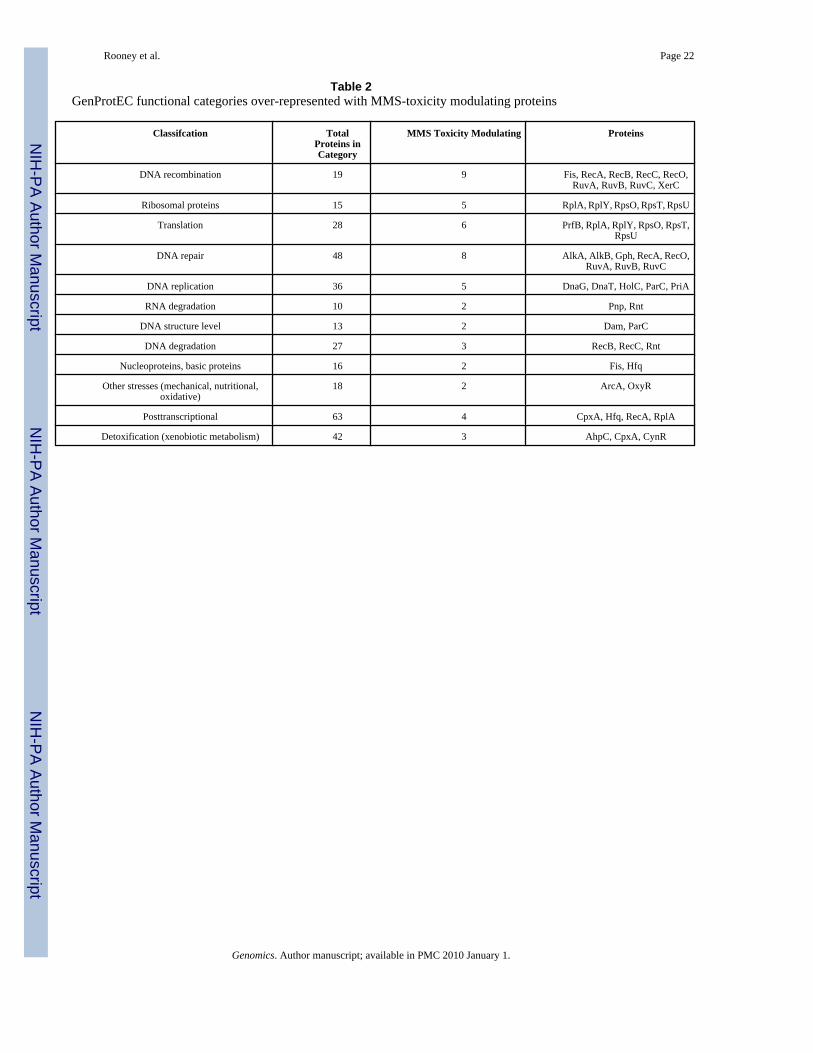
used in our screen, and identified 2,713 intersecting proteins, of which 73 modulated thetoxicity of MMS (Supplemental Table S5). Those E. coli proteins that are not annotated inGeneProtEC were not used for functional mapping. Next we determined the number of MMS-toxicity modulating proteins found in each functional category. We also performed 10,000random samplings of 73 proteins from our master list of 2,713 proteins, noted the number foundin each functional category, and determined the average number and standard deviation foreach functional category. The actual number of MMS toxicity modulating proteins, the randomsampled average, and the standard deviation for each functional category were then used todetermine whether there was significant enrichment for MMS-toxicity modulating proteins ineach functional category (Table 2). We identified 12 different functional categories that wereover-represented with MMS-toxicity modulating proteins (number in category ≥ 2, p < 0.05).All gene-deletion mutants whose corresponding proteins mapped to significantly over-represented categories were further validated using dilution based cytotoxicity assays (Figure2; Supplemental Figure S2). All were verified as being MMS sensitive.
Some of the functional categories over-represented with MMS-toxicity modulating proteinsincluded DNA recombination, DNA repair, DNA replication, DNA degradation, DNAstructure, and RNA degradation, which cumulatively demonstrate a requirement for nucleicacid metabolism and genome maintenance after MMS damage. DNA damage is a knownproduct of MMS exposure, thus these categories were expected and serve as a control tovalidate our algorithm. In addition to DNA damage, RNA has been shown to be a target forMMS. RNA processing activities that include Pnp and Rnt and the DNA/RNA demethylationenzyme AlkB were identified in our functional analysis [42]. Pnp is a component of the RNAdegradosome and will hydrolyze mRNA to remove it from the transcript pool [46], while Rntencodes a tRNA ribonuclease that plays a role in recycling uncharged adapter molecules[47]. Recently the DNA repair enzyme AlkB was shown to remove methyl groups from RNA,with methyl groups representing both damage and enzyme based modifications in tRNA. WhileΔalkB cells are sensitive to MMS, the contribution of RNA damage or RNA modifications tocell death or viability after damage is unclear. Similarly, the precise role of Pnp and Rnt afterMMS damage is unclear, as either could be used to remove damaged RNA or process RNAfor signalling purposes after damage. Nonetheless, the significant theme of RNA degradationafter MMS damage suggests that the removal and repair of RNA or RNA processing for signaltransduction purposes play an important role after DNA damage.
Xenobiotic metabolism and stress signalling are common responses to exposure and damageand the removal of damaging agents and the increased transcription or post translationalmodification of downstream proteins are recognized damage responses observed acrossphylogeny. We identified both xenobiotic metabolism (AhpC, CpxA, and CynR) and stressbased (ArcA and OxyR) regulatory proteins as being over-represented amongst the MMS-toxicity modulating proteins, which was expected and served as another validation for ouralgorithm.
One of the most prominent categories identified by functional mapping was specific to proteinsynthesis, and included ribosomal proteins and translational machinery. Six activities specificto protein synthesis were represented in our functional mapping results (PrfB, RplA, RpsO,RpsT, RpsU, RplY) and the MMS sensitivity of the corresponding mutants supports ourhypothesis that translational machinery plays an important role in recovery from damage.Clearly, protein synthesis machinery responds to transcriptional cues and is involved in thesynthesis of important toxicity modulating proteins. It is known that reactive electrophiles willdamage proteins and the replacement of damaged proteins is certainly an important cellularactivity after MMS damage [5,7]. Additionally, we can speculate that ribosomal proteins sensecellular stress or that translational regulation occurs after damage, but these roles for ribosomaland protein synthesis machinery in the damage response have yet to be proven.
Rooney et al. Page 5
Genomics. Author manuscript; available in PMC 2010 January 1.
NIH
-PA Author Manuscript
NIH
-PA Author Manuscript
NIH
-PA Author Manuscript

Interactome analysis identifies vital MMS-toxicity modulating networksSpecies-specific protein interaction information has been demonstrated to be an effective toolfor analyzing global data sets [48–54], and can assist in the identification of protein networksactivated by damage. Protein-protein interaction information can be compiled to generate aninteractome in silico, and the resulting structure is a static blueprint of potential signallingpathways and protein complexes inside the cell. We have compiled all reported protein-proteininteractions for E. coli available in the Database of Interacting Proteins (DIP) [55] andsupplemented them with a large protein-protein interaction study [56]. In all we compiled18,161 interactions between 3,467 E. coli proteins (Supplemental Table S6). The compiledinteractome can be considered a non saturated structure with regard to molecular interactions,but it is an extensive framework that can be used to identify protein networks activated byMMS damage.
The compiled interactome was mapped with MMS-toxicity modulating proteins, and filteredto show only MMS-toxicity modulating proteins and their corresponding protein-proteininteractions. Next we colored each node according to cellular function, to demonstrate thatactivities involved in DNA metabolism (repair, recombination, and replication), transcription,translation, and mRNA processing, among others, are found in the filtered interactome (Figure3, top). Further analysis of the filtered interactome identified a large connected component of32 proteins along with two two-protein modules. The 32 protein sub-network identified byinteractome filtering is connected by 37 protein-protein interactions. It should be noted thatinteractome filtering does not use statistical validation to assign p-values to sub-networks.Instead, the filtering step identifies all connected MMS-toxicity modulating proteins in theinteractome to provide a global view of how different functional activities are potentiallycoordinated.
We used clustering coefficient analysis of the mapped and filtered structure to identify highlyconnected and statistically significant protein architectures that respond to MMS treatment. Ingeneral, interactome mapping has been shown to identify biologically important architectures[49,50]. Clustering coefficient analysis of a mapped interactome has been demonstrated toidentify signatures of protein pathways and complexes responding to damage and can identifylocal areas of high connectivity in a mapped network [10,11,57]. We analyzed our 32 proteinsub-network using clustering coefficient analysis, to identify proteins whose interactingneighbors share protein-protein interactions. We identified a significantly clustered group ofseven proteins (Hfq, Pnp, RplA, RpsO, DeaD, ParC, and SmpB) centred on RplA andconnected by 13 protein-protein interactions (Figure 3, bottom). The significance (p < 10−6)of the highly clustered RplA-centred subnetwork was validated by network randomizationsand random samplings. The 50S subunit protein RplA is the focal point of a highly clusteredsubnetwork that contains components of the 30S and 50S ribosome (RpsO and RplA) and RNAprocessing activities (Hfq, Pnp, and SmpB) [42]. The 30S ribosome protein RpsT was alsoclosely associated with the RplA-centred sub-network, via interactions with Pnp and DeaD,and was added to the subnetwork based on functional overlap with other members. The RplA-centred sub-network of eight MMS-toxicity modulating proteins is suggestive of a coordinatedpathway specific to mRNA processing and protein synthesis machinery. Importantly, theseactivities have been identified again as being important after MMS exposure, albeit using adifferent mapping approach based on protein-protein interactions.
Defective SOS Responses in the RplA-Focused Sub-NetworkThe SOS response to DNA damage caused by MMS is one of the major response pathwayspromoting cell viability after DNA alkylation damage. Defects in the SOS response could beresponsible for the MMS-sensitive phenotype of gene deletion mutants specific to membersof the RplA-centred sub-network. Thus we analyzed the induction of the SOS response in each
Rooney et al. Page 6
Genomics. Author manuscript; available in PMC 2010 January 1.
NIH
-PA Author Manuscript
NIH
-PA Author Manuscript
NIH
-PA Author Manuscript

gene deletion mutant (Figure 4) specific to the RplA-centred sub-network, along with wild-type and ΔrecA control strains. We recorded a ~4-fold induction in the SOS-response in wild-type cells treated with MMS, relative to untreated wild-type, and no SOS induction in ΔrecAcells after MMS treatment, indicating our assay was working properly. Next we looked at thebasal levels of the SOS reporter in each of our cell types and determined that ΔrpsT cells hada ~4-fold induction, relative to untreated wild-type, with this induction similar to what wasobserved for wild-type cells treated with MMS. Clearly, ΔrpsT cells have a hyper active SOSresponse under normal conditions suggesting a faulty DNA damage response in these cells.Next we analyzed the MMS induced levels of the SOS response for each of the gene-deletionmutants specific to the eight proteins found in the RplA-centred subnetwork. We determinedthat six of the gene-deletion mutants have a modest (ΔdeaD, Δhfq, Δpnp) or slight (ΔrplA,ΔrpsO, ΔsmpB) decrease in their MMS induced SOS response, relative to wild-type. Based onfold-change there appears to be two groups of SOS corrupted gene deletion mutants, with thelevel of the SOS-response in ΔdeaD, Δhfq, Δpnp mutants about 63% of wild-type, while thelevel of the SOS-response in ΔrplA, ΔrpsO, ΔsmpB mutants about 80% of wild-type. Thedecreased level of the MMS-induced SOS response for ΔdeaD, Δhfq, Δpnp, ΔrplA, ΔrpsO,ΔsmpB could explain the MMS sensitive phenotype for each mutant, due to decreased DNArepair capacity. The precise roles of DeaD, Hfq, Pnp, RplA, RpsO, and SmpB in the SOSresponse are unknown. DeaD, Hfq, Pnp, and SmpB are activities associated with translationand the metabolism of RNA, while RplA and RpsO are part of the ribosome. In theory therecould be specific roles for each in damage-induced transcription or translation, withdeficiencies in each activity directly or indirectly affecting the SOS response. The finding thatthey are all highly connected suggests a coordinated role of at least six activities. It is interestingto note that Pnp is directly connected to RecA by one interaction in our filtered network, andthis network could be passing RNA or protein damage signals to RecA or influencing the levelsof SOS machinery; however these conclusions are highly speculative.
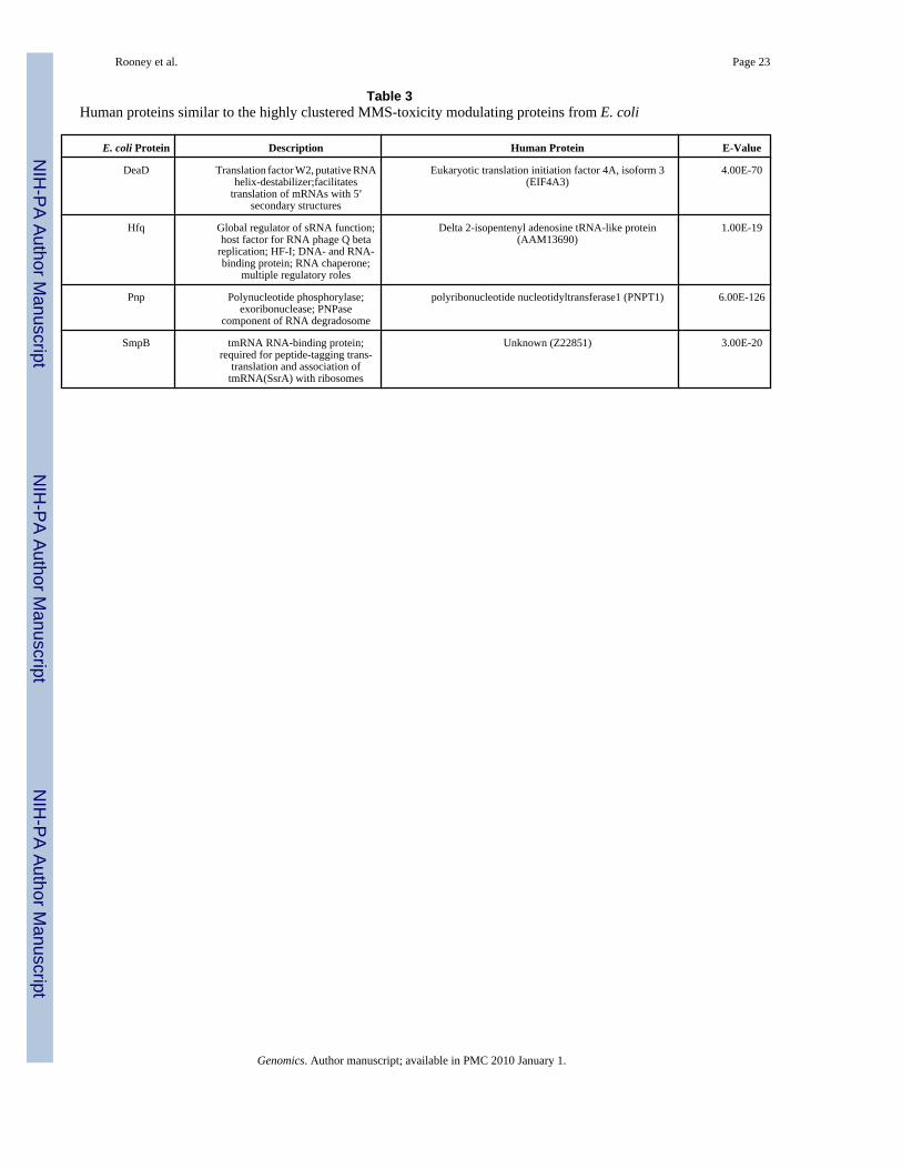
Human Counterparts of DeaD, Hfq, Pnp, and SmpBThe RplA-centred network identified in this study contains six MMS-toxicity modulatingproteins required for an optimal SOS response. One of the reasons we performed an MMSscreen was our desire to identify human proteins with the potential to modulate the toxicity ofalkylating agents. We used the Basic Local Alignment Search Tool (BLAST) to determine ifsimilar proteins were found in humans (Table 3) and identified highly similar amino acidsequences (E < 10−19) related to E. coli DeaD, Hfq, Pnp, and SmpB. Corresponding humanactivities are the translation initiation factor 4A, isoform 3 (EIF4A3), delta 2-isopentenyladenosine tRNA-like protein (AAM13690), polyribonucleotide nucleotidyltransferase 1(PNPT1), and a protein of unknown function (Z22851). Both the translation initiation factor4A, isoform 3 (EIF4A3) and delta 2-isopentenyl adenosine tRNA-like protein (AAM13690)should be associated with translation, but to date these are predicted activities based onhomology. Human polyribonucleotide nucleotidyltransferase can process mRNA to affectstability [58,59], which places this activity as a regulator of protein levels. Three of theidentified human proteins are linked to protein synthesis, suggesting that similar to DNA repairproteins, EIF4A3, PNPT1, and AAM13690 could be conserved activities associated withmodulating the toxicity of MMS. While this hypothesis and the identification of protein-proteininteractions between these human proteins are the focus of future experiments, our studyhighlights how computational and systems based studies in E. coli can be used to identifyproteins of interest for study in mammalian systems.
ConclusionsGene deletion libraries are valuable tools that can be used to asses the functional importanceof specific proteins after experimental perturbations, and in conjunction with Systems Biology
Rooney et al. Page 7
Genomics. Author manuscript; available in PMC 2010 January 1.
NIH
-PA Author Manuscript
NIH
-PA Author Manuscript
NIH
-PA Author Manuscript

based approaches can be used to identify protein pathways and protein complexes respondingto damage. We have used high-throughput screening of an E. coli gene deletion library toidentify 99 proteins that modulate the toxicity of the alkylating agent MMS. In addition, wehave used both functional and interactome mapping of identified MMS toxicity modulatingproteins to demonstrate that mRNA processing and translation specific proteins participate inthe response to macromolecular alkylation damage. Further, we have used clusteringcoefficient analysis to identify a highly connected group of activities associated with mRNAprocessing and translation, and demonstrated that the corresponding proteins influence theefficiency of the SOS response. Activities associated with protein synthesis have the potentialto play an important role in signal transduction after damage, both general and specific, andour work supports the idea that in addition to DNA repair, mRNA processing, and translationalcomponents are vital after alkylation damage. In addition, we show that systems basedapproaches coupled to homology searches can be used to identify putative alkylation resistanceproteins in humans.
Materials and MethodsE. coli mutants, high throughput screening, and validation
Luria Bertani broth (LB) (BP1426-3, Fisher Scientific, Waltham, MA) was used in both liquidand plate form to culture E. coli. The library of E. coli gene deletion mutants was acquiredfrom the Genome Analysis Project in Japan [41] and supplied in 96-well plate format. Highthroughput genomic phenotyping was performed similarly to that described for Saccharomycescerevisiae gene deletion mutants [10,11]. Briefly, 96-well plates containing the gene deletionmutants were replicated into liquid medium (LB-kanamycin) and grown for 16-hours at 37ºC.The saturated cultures were then diluted 10-fold into the same medium and 1 μl cell suspensionswere robotically (Matrix Hydra) spotted on LB-kanamycin agar plates containing increasingconcentrations of MMS (0, 0.045, and 0.060 % MMS) or tBuOH (150 and 165 μM). UVexposures were supplied using a Stratalinker (Stragene, Cedar Creek, Texas) with 254 nmbulbs, with mutants initially spotted on LB-kanamycin agar plates and then exposed to UV (6and 8 J/m2).
Inoculated plates were incubated for 16-hours at 37 ºC and the resulting plates were imagedusing an AlphaImager (Alpha Innotech Corporation, San Leandro, CA). Images of plates werecompiled into a visual database and analyzed to identify mutants with reduced growth afterMMS treatment. Reduced growth for a specific gene-deletion mutant was identified relativeto other mutants found on the 96-well plate and wild-type BW25113 cells, and was also relativeto growth of the mutant on an untreated plate. To identify MMS-toxicity modulating proteins,we linked sensitive mutants to their corresponding deleted gene and assumed the proteinencoded by the deleted gene was responsible for the observed phenotype. Scores representsemi-quantitative measures. For example, if a deletion mutant had reduced growth on a givenconcentration of MMS it was scored a two for that MMS concentration, while mutants showingonly a change in color were scored one, and all others were scored a zero. It is important tonote that our screen was designed to only identify mutants with increased sensitivity to MMS.Future screens that utilize higher MMS concentrations could be undertaken to identify mutantsthat grow better than wild-type in the presence of alkylating agents.
Newly constructed E. coli mutants were made using homology based recombination promotedby the λ-Red systems as described [60] and verified by PCR and DNA sequence analysis(Rooney et al., in press). Mutants were assayed in 96-well format as described above. Inaddition, 59 mutants from the original Keio library were further analyzed for MMS sensitivity.Each mutant was serially diluted (five 10-fold dilutions) and 5 μl of each dilution was manuallyapplied to LB plates containing increasing concentrations of MMS. Plates were incubated at37ºC for 16 hours, imaged, and analyzed. Sensitivity was determined by identifying the last
Rooney et al. Page 8
Genomics. Author manuscript; available in PMC 2010 January 1.
NIH
-PA Author Manuscript
NIH
-PA Author Manuscript
NIH
-PA Author Manuscript
dilution at which a deletion mutant grew on MMS containing plates, relative to the dilutedwild-type cells on MMS plates. Similarly, growth of both wild-type and deletion mutants onuntreated plates was used to control for the viability and growth rates of gene deletion mutantsunder normal conditions.
Functional mapping of MMS-toxicity modulating dataAll GenProtEC functional classifications specific to E. coli proteins were downloaded andcross referenced to proteins represented in our study. We choose to use an intermediate levelof functional classification because it represented a broad yet specific spectrum of cellularprocesses. First we determined the actual number of MMS-toxicity modulating proteins foundin each functional category. Next, statistics were compiled using random sampling and anormal curve approximation, as described previously [11]. Briefly, the base set of 2,713proteins represented in our data set were randomly sampled to pick N = 73 proteins. The numberof proteins specific to each functional category was determined and values were compiled forM = 10,000 iterations of N = 73 random proteins. Average values and standard deviations foreach functional category were then generated and Z-scores were compiled for each functionalcategory using the following formula:
Z-scores measure whether a functional category is over- or under-represented with MMS-toxicity modulating proteins. Corresponding p-values were determined for all over-representedcategories using a one-tailed test and normal approximation.
Protein interactome mappingE. coli protein interaction information was downloaded from the Database of InteractingProteins (DIP) [61] and combined with the large protein-protein interaction data set publishedby Mori and co-workers [56]. Protein-protein interaction information was imported intoCytoscape for network visualization and sub-network filtering. Filtering was performed byhighlighting MMS-toxicity modulating proteins and their associated protein-proteininteractions. Clustering coefficient analysis (C) was then performed as described [57] on allfiltered nodes and those nodes with C > 0 were visualized, along with correspondinginteractions, using Cytoscape [55]. Network randomizations and the significance of highlyconnected protein groups was determined as described, with some minor additions [57]. TheRplA-centred network was identified by clustering coefficient analysis of the 32 node structureidentified by interactome mapping and filtering. The average clustering coefficient of all nodesfound in the RplA-focused sub-network is CRpla-focused = 0.229, and 1000 random samplingsof 32 nodes was performed to generate an average clustering coefficient for a random 32 nodestructure (Cavg = 0.051) and standard deviation (Savg = 0.016). These values were then usedto obtain p-values using a one-tailed test and normal approximation. A second set ofrandomizations was used to further validate the significance of our findings. Theserandomizations involved taking the base 32 node structure, identified by interactome mappingand filtering, and used 1000 randomized sets of interactions to this base unit. After eachrandomization, clustering coefficient analysis was performed (Cavg = 0.019), and uponcompletion of 1000 iterations a standard deviation (Savg = 0.0046) was determined and usedto generate a significance value, as described above.
SOS-Reporter AssaysThe sulA-GFP reporter system used for the SOS studies was purchased from Open Biosystems(pMS201_sulA_GFP). We modified the plasmid by adding a chloramphenicol (CAM) resistant
Rooney et al. Page 9
Genomics. Author manuscript; available in PMC 2010 January 1.
NIH
-PA Author Manuscript
NIH
-PA Author Manuscript
NIH
-PA Author Manuscript

cassette to generate pMS201_sulA_GFP_CAM. Plasmids were transformed into mutants ofinterest and selected on LB-CAM plates. Transformants were grown to mid log phase, split intwo, and then mock or MMS treated (0.015%) for 30 minutes. Cells were then harvested bycentrifugation, washed, and suspended in phosphate buffered saline (PBS). GFP levels in30,000 cells were analyzed by fluorescent activated cell sorting (FACS) analysis using a BectonDickinson LSRII Benchtop Flow Cytometer.
BLAST AnalysisEach E. coli protein sequence was analyzed by BLAST using the tBLASTn program, whichis available from the National Center for Biotechnology Information(http://www.ncbi.nlm.nih.gov/blast/Blast.cgi) [62]. The nucleotide collection specific tohumans was used; with E-values of less than 10−1 serving as our cut-off to identify similarproteins between E. coli and humans.
Supplementary MaterialRefer to Web version on PubMed Central for supplementary material.
AcknowledgementsWork in the Begley lab is supported by the NIH (1K22ES01225101 and 1R01ES015037) and a James D. WatsonAward through NYSTAR. Work in the Cunningham lab is supported by the NIH (CRR1C06RR0154464 andGM46312).
References1. Brennand J, Margison GP. Reduction of the toxicity and mutagenicity of alkylating agents in
mammalian cells harboring the Escherichia coli alkyltransferase gene. Proceedings of the NationalAcademy of Sciences USA 1986;83(17):6292–6296.
2. Boffa LC, Bolognesi C. Methylating agents: their target amino acids in nuclear proteins. Carcinogenesis1985;6(9):1399–401. [PubMed: 4028338]
3. Saffhill R, Margison GP, O’Connor PJ. Mechanisms of carcinogenesis induced by alkylating agents.Bioch Biophys Acta 1985;823:111–145.
4. Liebler DC. Proteomic approaches to characterize protein modifications: new tools to study the effectsof environmental exposures. Environ Health Perspect 2002;110(Suppl 1):3–9. [PubMed: 11834459]
5. Shin NY, et al. Protein targets of reactive electrophiles in human liver microsomes. Chem Res Toxicol2007;20(6):859–67. [PubMed: 17480101]
6. Szapacs ME, et al. Covalent adduction of human serum albumin by 4-hydroxy-2-nonenal: kineticanalysis of competing alkylation reactions. Biochemistry 2006;45(35):10521–8. [PubMed: 16939204]
7. Dennehy MK, et al. Cytosolic and nuclear protein targets of thiol-reactive electrophiles. Chem ResToxicol 2006;19(1):20–9. [PubMed: 16411652]
8. Burgis NE, Samson LD. The Protein Degradation Response of Saccharomyces cerevisiae to ClassicalDNA-Damaging Agents. Chem Res Toxicol. 2007
9. Smith GJ, Grisham JW. Cytotoxicity of monofunctional alkylating agents. Methyl methanesulfonateand methyl-N′-nitro-N-nitrosoguanidine have different mechanisms of toxicity for 10T1/2 cells.Mutation Research 1983;111(3):405–17. [PubMed: 6646150]
10. Begley TJ, et al. Recovery Pathways in S. cerevisiae Revealed by Genomic Phenotyping andInteractome Mapping. Mol Canc Res 2002;1
11. Begley TJ, et al. Hot spots for modulating toxicity identified by genomic phenotyping and localizationmapping. Mol Cell 2004;16(1):117–25. [PubMed: 15469827]
12. Jelinsky SA, Samson LD. Global response of Saccharomyces cerevisiae to an alkylating agent. ProcNatl Acad Sci U S A 1999;96(4):1486–91. [PubMed: 9990050]
Rooney et al. Page 10
Genomics. Author manuscript; available in PMC 2010 January 1.
NIH
-PA Author Manuscript
NIH
-PA Author Manuscript
NIH
-PA Author Manuscript
13. Chang M, et al. A genome-wide screen for methyl methanesulfonate-sensitive mutants reveals genesrequired for S phase progression in the presence of DNA damage. Proc Natl Acad Sci U S A 2002;99(26):16934–9. [PubMed: 12482937]
14. Engelward BP, et al. A chemical and genetic approach together define the biological consequencesof 3-methyladenine lesions in the mammalian genome. J Biol Chem 1998;273(9):5412–8. [PubMed:9479003]
15. Chen J, Samson L. Induction of S. cerevisiae MAG 3-methyladenine DNA glycosylase transcriptlevels in response to DNA damage. Nucleic Acids Res 1991;19(23):6427–32. [PubMed: 1754379]
16. Jeggo P, et al. An adaptive response of E. coli to low levels of alkylating agent: comparison withpreviously characterised DNA repair pathways. Mol Gen Genet 1977;157(1):1–9. [PubMed: 414071]
17. Begley TJ, Samson LD. AlkB mystery solved: oxidative demethylation of N1-methyladenine andN3-methylcytosine adducts by a direct reversal mechanism. Trends Biochem Sci 2003;28(1):2–5.[PubMed: 12517444]
18. Sekiguchi M, Sanada M. Alkylation carcinogenesis in mice with altered levels of DNA repairmethyltransferase. Prog Exp Tumor Res 1999;35:25–36. [PubMed: 10377749]
19. Nakatsuru Y, et al. Characterization of O6-methylguanine-DNA methyltransferase in transgenic miceintroduced with the E. coli ada gene. Mutation Research 1991;254(3):225–30. [PubMed: 2052012]
20. Rebeck GW, et al. A second DNA methyltransferase repair enzyme in Escherichia coli. Proceedingsof the National Academy of Sciences of the United States of America 1988;85(9):3039–43. [PubMed:3283737]
21. Potter PM, et al. Characterisation and nucleotide sequence of ogt, the O6-alkylguanine-DNA-alkyltransferase gene of E. coli. Nucleic Acids Research 1987;15(22):9177–93. [PubMed: 2825131]
22. McCarthy TV, Lindahl T. Methyl phosphotriesters in alkylated DNA are repaired by the Adaregulatory protein of E. coli. Nucleic Acids Res 1985;13(8):2683–98. [PubMed: 2987862]
23. Nakabeppu Y, Kondo H, Sekiguchi M. Cloning and characterization of the alkA gene of Escherichiacoli that encodes 3-methyladenine DNA glycosylase II. Journal of Biological Chemistry 1984;259(22):13723–9. [PubMed: 6389535]
24. Kataoka H, Yamamoto Y, Sekiguchi M. A new gene (alkB) of Escherichia coli that controls sensitivityto methyl methane sulfonate. Journal of Bacteriology 1983;153(3):1301–7. [PubMed: 6337994]
25. Evensen G, Seeberg E. Adaptation to alkylation resistance involves the induction of a DNAglycosylase. Nature 1982;296(5859):773–5. [PubMed: 7040984]
26. Sobol RW, et al. Base excision repair intermediates induce p53-independent cytotoxic and genotoxicresponses. J Biol Chem 2003;278(41):39951–9. [PubMed: 12882965]
27. Sobol RW, Wilson SH. Mammalian DNA beta-polymerase in base excision repair of alkylationdamage. Prog Nucleic Acid Res Mol Biol 2001;68:57–74. [PubMed: 11554313]
28. Riazuddin S, Lindahl T. Properties of 3-methyladenine-DNA glycosylase from Escherichia coli.Biochemistry 1978;17:2110–2118. [PubMed: 352392]
29. Engelward BP, et al. Repair-deficient 3-methyladenine DNA glycosylase homozygous mutant mousecells have increased sensitivity to alkylation-induced chromosome damage and cell killing. EMBOJournal 1996;15(4):945–52. [PubMed: 8631315]
30. Duncan T, et al. Reversal of DNA alkylation damage by two human dioxygenases. Proc Natl AcadSci U S A 2002;99(26):16660–5. [PubMed: 12486230]
31. Trewick SC, et al. Oxidative demethylation by Escherichia coli AlkB directly reverts DNA basedamage. Nature 2002;419(6903):174–8. [PubMed: 12226667]
32. Falnes PO, Johansen RF, Seeberg E. AlkB-mediated oxidative demethylation reverses DNA damagein Escherichia coli. Nature 2002;419(6903):178–82. [PubMed: 12226668]
33. Aravind L, Koonin EV. The DNA-repair protein AlkB, EGL-9, and leprecan define new families of2-oxoglutarate- and iron-dependent dioxygenases. Genome Biol 2001;2(3)
34. Drablos F, et al. Alkylation damage in DNA and RNA--repair mechanisms and medical significance.DNA Repair (Amst) 2004;3(11):1389–407. [PubMed: 15380096]
35. Jeggo P, et al. The adaptive response of E. coli to low levels of alkylating agent: the role of polA inkilling adaptation. Molecular & General Genetics 1978;162(3):299–305. [PubMed: 355835]
Rooney et al. Page 11
Genomics. Author manuscript; available in PMC 2010 January 1.
NIH
-PA Author Manuscript
NIH
-PA Author Manuscript
NIH
-PA Author Manuscript
36. Samson L, Cairns J. A new pathway for DNA repair in Escherichia coli. Nature 1977;267(5608):281–3. [PubMed: 325420]
37. Courcelle J, et al. Comparative gene expression profiles following UV exposure in wild-type andSOS-deficient Escherichia coli. Genetics 2001;158(1):41–64. [PubMed: 11333217]
38. Gasch AP, et al. Genomic expression responses to DNA-damaging agents and the regulatory role ofthe yeast ATR homolog Mec1p. Mol Biol Cell 2001;12(10):2987–3003. [PubMed: 11598186]
39. Jelinsky SA, et al. Regulatory networks revealed by transcriptional profiling of damagedsaccharomyces cerevisiae cells: rpn4 links base excision repair with proteasomes [In ProcessCitation]. Mol Cell Biol 2000;20(21):8157–67. [PubMed: 11027285]
40. Workman CT, et al. A systems approach to mapping DNA damage response pathways. Science2006;312(5776):1054–9. [PubMed: 16709784]
41. Baba T, et al. Construction of Escherichia coli K-12 in-frame, single-gene knockout mutants: theKeio collection. Mol Syst Biol 2006;2:2006 0008.
42. Rudd KE. EcoGene: a genome sequence database for Escherichia coli K-12. Nucleic Acids Res2000;28(1):60–4. [PubMed: 10592181]
43. Drury LS, Buxton RS. DNA sequence analysis of the dye gene of Escherichia coli reveals amino acidhomology between the dye and OmpR proteins. J Biol Chem 1985;260(7):4236–42. [PubMed:2984198]
44. Serres MH, Goswami S, Riley M. GenProtEC: an updated and improved analysis of functions ofEscherichia coli K-12 proteins. Nucleic Acids Res 2004;32(Database issue):D300–2. [PubMed:14681418]
45. Ezaki B, et al. Involvement of DnaK protein in mini-F plasmid replication: temperature-sensitive segmutations are located in the dnaK gene. Mol Gen Genet 1989;218(2):183–9. [PubMed: 2674651]
46. Carpousis AJ, Vanzo NF, Raynal LC. mRNA degradation. A tale of poly(A) and multiproteinmachines. Trends Genet 1999;15(1):24–8. [PubMed: 10087930]
47. Zuo Y, Deutscher MP. The physiological role of RNase T can be explained by its unusual substratespecificity. J Biol Chem 2002;277(33):29654–61. [PubMed: 12050169]
48. Boulton SJ, et al. Combined functional genomic maps of the C. elegans DNA damage response.Science 2002;295(5552):127–31. [PubMed: 11778048]
49. Rual JF, et al. Towards a proteome-scale map of the human protein-protein interaction network.Nature 2005;437(7062):1173–8. [PubMed: 16189514]
50. Gunsalus KC, et al. Predictive models of molecular machines involved in Caenorhabditis elegansearly embryogenesis. Nature 2005;436(7052):861–5. [PubMed: 16094371]
51. Ideker T, et al. Integrated genomic and proteomic analyses of a systematically perturbed metabolicnetwork. Science 2001;292(5518):929–34. [PubMed: 11340206]
52. Lim J, et al. A protein-protein interaction network for human inherited ataxias and disorders ofPurkinje cell degeneration. Cell 2006;125(4):801–14. [PubMed: 16713569]
53. Goh KI, et al. The human disease network. Proc Natl Acad Sci U S A 2007;104(21):8685–90.[PubMed: 17502601]
54. Yildirim MA, et al. Drug-target network. Nat Biotechnol 2007;25(10):1119–26. [PubMed: 17921997]55. Shannon P, et al. Cytoscape: a software environment for integrated models of biomolecular interaction
networks. Genome Res 2003;13(11):2498–504. [PubMed: 14597658]56. Arifuzzaman M, et al. Large-scale identification of protein-protein interaction of Escherichia coli
K-12. Genome Res 2006;16(5):686–91. [PubMed: 16606699]57. Said MR, et al. Global network analysis of phenotypic effects: protein networks and toxicity
modulation in Saccharomyces cerevisiae. Proc Natl Acad Sci U S A 2004;101(52):18006–11.[PubMed: 15608068]
58. Leszczyniecka M, et al. Identification and cloning of human polynucleotide phosphorylase, hPNPaseold-35, in the context of terminal differentiation and cellular senescence. Proc Natl Acad Sci U S A2002;99(26):16636–41. [PubMed: 12473748]
59. Sarkar D, et al. Down-regulation of Myc as a potential target for growth arrest induced by humanpolynucleotide phosphorylase (hPNPaseold-35) in human melanoma cells. J Biol Chem 2003;278(27):24542–51. [PubMed: 12721301]
Rooney et al. Page 12
Genomics. Author manuscript; available in PMC 2010 January 1.
NIH
-PA Author Manuscript
NIH
-PA Author Manuscript
NIH
-PA Author Manuscript

60. Datsenko KA, Wanner BL. One-step inactivation of chromosomal genes in Escherichia coli K-12using PCR products. Proc Natl Acad Sci U S A 2000;97(12):6640–5. [PubMed: 10829079]
61. Xenarios I, et al. DIP, the Database of Interacting Proteins: a research tool for studying cellularnetworks of protein interactions. Nucleic Acids Res 2002;30(1):303–5. [PubMed: 11752321]
62. Altschul SF, et al. Basic local alignment search tool. J Mol Biol 1990;215(3):403–10. [PubMed:2231712]
63. Boiteux S, Huisman O, Laval J. 3-Methyladenine residues in DNA induce the SOS function sfiA inEscherichia coli. EMBO 1984;3:2569–2573.
Rooney et al. Page 13
Genomics. Author manuscript; available in PMC 2010 January 1.
NIH
-PA Author Manuscript
NIH
-PA Author Manuscript
NIH
-PA Author Manuscript
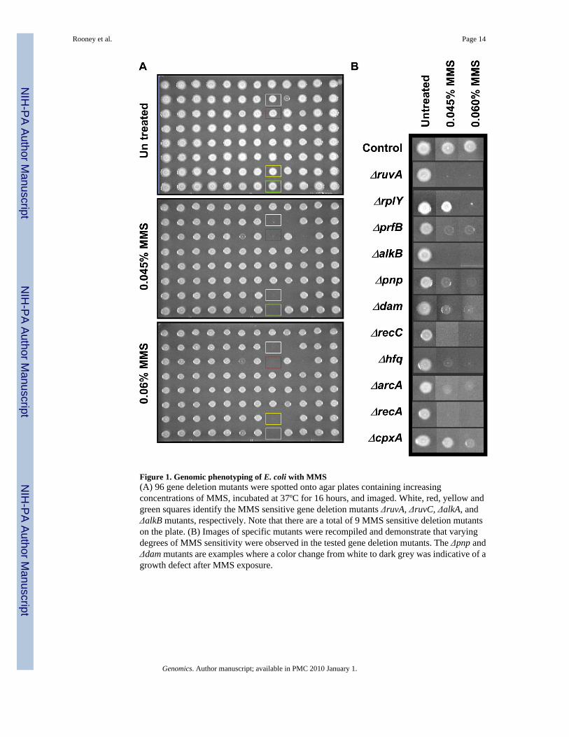
Figure 1. Genomic phenotyping of E. coli with MMS(A) 96 gene deletion mutants were spotted onto agar plates containing increasingconcentrations of MMS, incubated at 37ºC for 16 hours, and imaged. White, red, yellow andgreen squares identify the MMS sensitive gene deletion mutants ΔruvA, ΔruvC, ΔalkA, andΔalkB mutants, respectively. Note that there are a total of 9 MMS sensitive deletion mutantson the plate. (B) Images of specific mutants were recompiled and demonstrate that varyingdegrees of MMS sensitivity were observed in the tested gene deletion mutants. The Δpnp andΔdam mutants are examples where a color change from white to dark grey was indicative of agrowth defect after MMS exposure.
Rooney et al. Page 14
Genomics. Author manuscript; available in PMC 2010 January 1.
NIH
-PA Author Manuscript
NIH
-PA Author Manuscript
NIH
-PA Author Manuscript
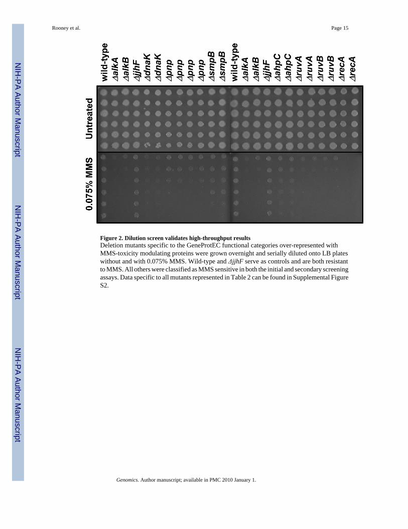
Figure 2. Dilution screen validates high-throughput resultsDeletion mutants specific to the GeneProtEC functional categories over-represented withMMS-toxicity modulating proteins were grown overnight and serially diluted onto LB plateswithout and with 0.075% MMS. Wild-type and ΔjjhF serve as controls and are both resistantto MMS. All others were classified as MMS sensitive in both the initial and secondary screeningassays. Data specific to all mutants represented in Table 2 can be found in Supplemental FigureS2.
Rooney et al. Page 15
Genomics. Author manuscript; available in PMC 2010 January 1.
NIH
-PA Author Manuscript
NIH
-PA Author Manuscript
NIH
-PA Author Manuscript

Figure 3. Functional themes and highly clustered proteins in the MMS-toxicity modulating sub-network(Top) Each of the MMS toxicity modulating proteins found in the filtered interactome wascolored according to its predominant functional theme, as defined by EcoGene or SwissProt.Red circle = DNA repair, replication, and recombination; blue circle = protein synthesis; purplecircle = transcription; yellow circle = RNA processing; orange circle = protein stabilization;grey circle = unknown; white circle = other. (Bottom) MMS-toxicity modulating proteins withclustering coefficients greater then zero were determined using MATLAB algorithms and thenvisualized using Cytoscape. This was done to identify groups of proteins that have the potentialto be part of a complex or pathway. RpsT was included due to its connectivity to the sub-
Rooney et al. Page 16
Genomics. Author manuscript; available in PMC 2010 January 1.
NIH
-PA Author Manuscript
NIH
-PA Author Manuscript
NIH
-PA Author Manuscript

network via Pnp and DeaD. Ultimately, the highly clustered sub-network centred on RplA wasidentified and contained activities involved in protein synthesis and RNA metabolism.
Rooney et al. Page 17
Genomics. Author manuscript; available in PMC 2010 January 1.
NIH
-PA Author Manuscript
NIH
-PA Author Manuscript
NIH
-PA Author Manuscript

Figure 4. SOS Reporter analysis of mutants specific to the RplA-centred sub-networkA plasmid based SOS reporter, sulA-GFP, was transformed into each cell type, transformantswere grown to mid log phase and mock (grey bars) or 0.015% MMS treated (black bars) for30 minutes. Wild-type, Δtag (hyper-SOS after MMS[63]), and ΔrecA (hypo-SOS after MMS)serve as controls. FACS analysis of 30,000 cells was then used to quantitate GFP levels, andfold change relative to untreated wild-type was plotted for each cell type. Error bars representstandard deviations between three biological replicates of 30,000 cells each.
Rooney et al. Page 18
Genomics. Author manuscript; available in PMC 2010 January 1.
NIH
-PA Author Manuscript
NIH
-PA Author Manuscript
NIH
-PA Author Manuscript
NIH
-PA Author Manuscript
NIH
-PA Author Manuscript
NIH
-PA Author Manuscript
Rooney et al. Page 19
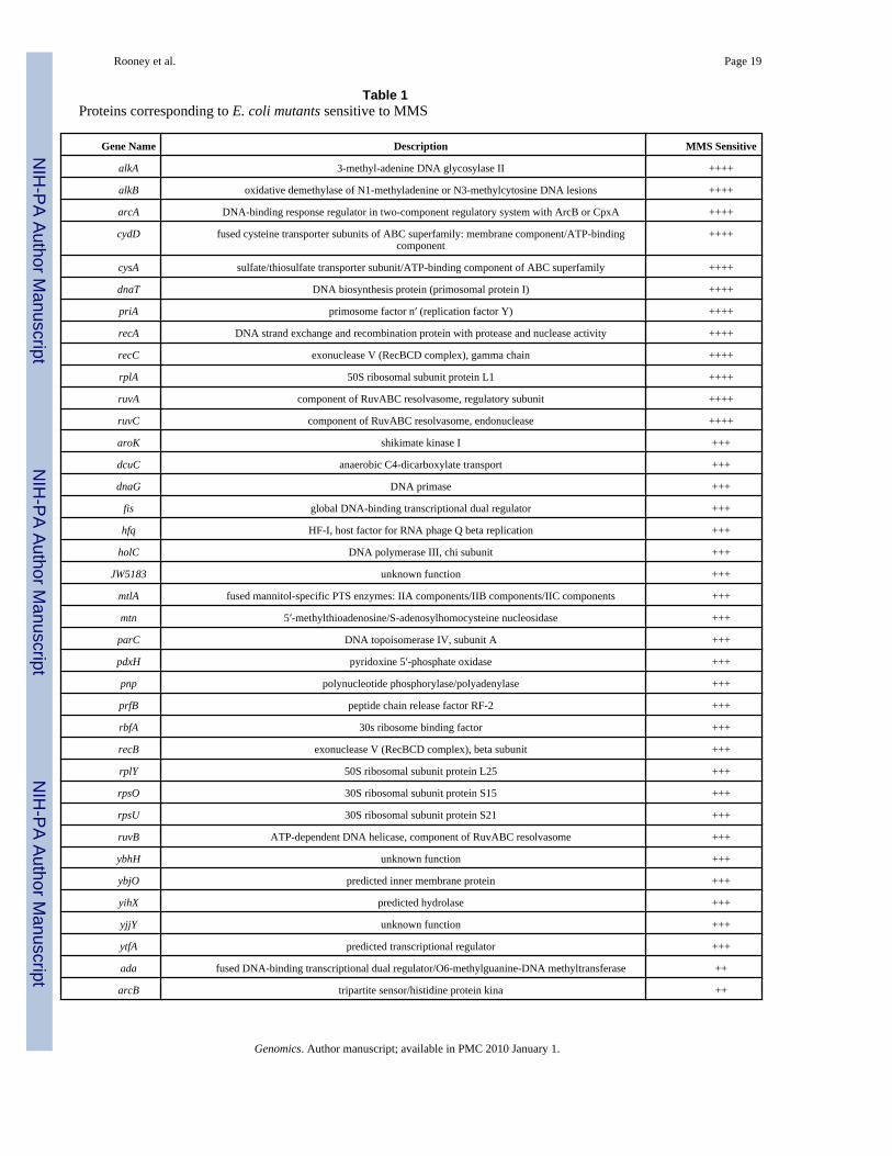
Table 1Proteins corresponding to E. coli mutants sensitive to MMS
Gene Name Description MMS Sensitive
alkA 3-methyl-adenine DNA glycosylase II ++++
alkB oxidative demethylase of N1-methyladenine or N3-methylcytosine DNA lesions ++++
arcA DNA-binding response regulator in two-component regulatory system with ArcB or CpxA ++++
cydD fused cysteine transporter subunits of ABC superfamily: membrane component/ATP-bindingcomponent
++++
cysA sulfate/thiosulfate transporter subunit/ATP-binding component of ABC superfamily ++++
dnaT DNA biosynthesis protein (primosomal protein I) ++++
priA primosome factor n′ (replication factor Y) ++++
recA DNA strand exchange and recombination protein with protease and nuclease activity ++++
recC exonuclease V (RecBCD complex), gamma chain ++++
rplA 50S ribosomal subunit protein L1 ++++
ruvA component of RuvABC resolvasome, regulatory subunit ++++
ruvC component of RuvABC resolvasome, endonuclease ++++
aroK shikimate kinase I +++
dcuC anaerobic C4-dicarboxylate transport +++
dnaG DNA primase +++
fis global DNA-binding transcriptional dual regulator +++
hfq HF-I, host factor for RNA phage Q beta replication +++
holC DNA polymerase III, chi subunit +++
JW5183 unknown function +++
mtlA fused mannitol-specific PTS enzymes: IIA components/IIB components/IIC components +++
mtn 5′-methylthioadenosine/S-adenosylhomocysteine nucleosidase +++
parC DNA topoisomerase IV, subunit A +++
pdxH pyridoxine 5′-phosphate oxidase +++
pnp polynucleotide phosphorylase/polyadenylase +++
prfB peptide chain release factor RF-2 +++
rbfA 30s ribosome binding factor +++
recB exonuclease V (RecBCD complex), beta subunit +++
rplY 50S ribosomal subunit protein L25 +++
rpsO 30S ribosomal subunit protein S15 +++
rpsU 30S ribosomal subunit protein S21 +++
ruvB ATP-dependent DNA helicase, component of RuvABC resolvasome +++
ybhH unknown function +++
ybjO predicted inner membrane protein +++
yihX predicted hydrolase +++
yjjY unknown function +++
ytfA predicted transcriptional regulator +++
ada fused DNA-binding transcriptional dual regulator/O6-methylguanine-DNA methyltransferase ++
arcB tripartite sensor/histidine protein kina ++
Genomics. Author manuscript; available in PMC 2010 January 1.

NIH
-PA Author Manuscript
NIH
-PA Author Manuscript
NIH
-PA Author Manuscript
Rooney et al. Page 20
Gene Name Description MMS Sensitive
atpF F0 sector of membrane-bound ATP synthase, subunit b ++
clpX ATPase and specificity subunit of ClpX-ClpP ATP-dependent serine protease ++
dam DNA adenine methylase ++
dnaK chaperone Hsp70, co-chaperone with DnaJ ++
hisB fused histidinol-phosphatase/imidazoleglycerol-phosphate dehydratase ++
mrsA phosphoglucosamine mutase ++
oppD oligopeptide transporter subunit/ATP-binding component of ABC superfamily ++
recO gap repair protein ++
rnt ribonuclease T (RNase T) ++
tpiA triosephosphate isomerase ++
ubiF 2-octaprenyl-3-methyl-6-methoxy-1,4-benzoquinol oxygenase ++
uidR DNA-binding transcriptional repressor ++
yaiS unknown function ++
ybgK predicted enzyme subunit ++
yciF unknown function ++
ydcS predicted spermidine/putrescine transporter subunit/periplasmic-binding component of ABCsuperfamily
++
yjiW unknown function ++
ahpC alkyl hydroperoxide reductase, C22 subunit +
ais conserved protein +
bdm osmoresponsive gene with reduced expression in biofilms, function unknown +
blr beta-lactam resistance protein +
cadC DNA-binding transcriptional activator +
cpxA sensory histidine kinase in two-component regulatory system with CpxR +
cynR transcriptional activator of cyn operon +
deaD ATP-dependent RNA helicase +
fliO flagellar biosynthesis protein +
gidA 5-methylaminomethyl-2-thiouridine modification at tRNA U34 (MnmG) +
gph phosphoglycolate phosphatase +
JW2207 unknown function +
lpcA D-sedoheptulose 7-phosphate isomerase +
marA Transcription activator of multiple antibiotic resistance +
minC cell division inhibitor +
oxyR DNA-binding transcriptional dual regulator +
potH putrescine transporter subunit: membrane component of ABC superfamily +
poxA predicted lysyl-tRNA synthetase +
pstB phosphate transporter subunit/ATP-binding component of ABC superfamily +
recN recombination and repair +
rpsT 30S ribosomal subunit protein S20 +
rzpD DLP12 prophage; predicted murein endopeptidase +
slyA global transcriptional regulator +
Genomics. Author manuscript; available in PMC 2010 January 1.

NIH
-PA Author Manuscript
NIH
-PA Author Manuscript
NIH
-PA Author Manuscript
Rooney et al. Page 21
Gene Name Description MMS Sensitive
smpB trans-translation protein +
stfR Rac prophage; predicted tail fiber protein +
tfaD pseudogene, tail fiber assembly gene +
xerC site-specific tyrosine recombinase +
yaiU predicted protein +
ybgI conserved metal-binding protein +
ybhR predicted transporter subunit: membrane component of ABC superfamily +
ycfC predicted lysogenization regulator +
ycjR unknown function +
ydaS Rac prophage +
ydaT Rac prophage; predicted protein +
yddO D-ala-D-ala transporter subunit/ATP-binding component of ABC superfamily +
yddS D-ala-D-a la transporter subunit/periplasmic-binding component of ABC superfamily +
ydeM unknown function +
yecN predicted inner membrane protein +
yfcK fused 5-methylaminomethyl-2-thiouridine-forming enzyme methyltransferase(MnmC) +
yfdQ CPS-53 (KpLE1) prophage; predicted protein +
yggA arginine transporter +
yggL unknown function +
yiiS unknown function +
yliD predicted peptide transporter subunit: membrane component of ABC superfamily +
Genomics. Author manuscript; available in PMC 2010 January 1.
NIH
-PA Author Manuscript
NIH
-PA Author Manuscript
NIH
-PA Author Manuscript
Rooney et al. Page 22
Table 2GenProtEC functional categories over-represented with MMS-toxicity modulating proteins
Classifcation TotalProteins inCategory
MMS Toxicity Modulating Proteins
DNA recombination 19 9 Fis, RecA, RecB, RecC, RecO,RuvA, RuvB, RuvC, XerC
Ribosomal proteins 15 5 RplA, RplY, RpsO, RpsT, RpsU
Translation 28 6 PrfB, RplA, RplY, RpsO, RpsT,RpsU
DNA repair 48 8 AlkA, AlkB, Gph, RecA, RecO,RuvA, RuvB, RuvC
DNA replication 36 5 DnaG, DnaT, HolC, ParC, PriA
RNA degradation 10 2 Pnp, Rnt
DNA structure level 13 2 Dam, ParC
DNA degradation 27 3 RecB, RecC, Rnt
Nucleoproteins, basic proteins 16 2 Fis, Hfq
Other stresses (mechanical, nutritional,oxidative)
18 2 ArcA, OxyR
Posttranscriptional 63 4 CpxA, Hfq, RecA, RplA
Detoxification (xenobiotic metabolism) 42 3 AhpC, CpxA, CynR
Genomics. Author manuscript; available in PMC 2010 January 1.
NIH
-PA Author Manuscript
NIH
-PA Author Manuscript
NIH
-PA Author Manuscript
Rooney et al. Page 23
Table 3Human proteins similar to the highly clustered MMS-toxicity modulating proteins from E. coli
E. coli Protein Description Human Protein E-Value
DeaD Translation factor W2, putative RNAhelix-destabilizer;facilitates
translation of mRNAs with 5′secondary structures
Eukaryotic translation initiation factor 4A, isoform 3(EIF4A3)
4.00E-70
Hfq Global regulator of sRNA function;host factor for RNA phage Q beta
replication; HF-I; DNA- and RNA-binding protein; RNA chaperone;
multiple regulatory roles
Delta 2-isopentenyl adenosine tRNA-like protein(AAM13690)
1.00E-19
Pnp Polynucleotide phosphorylase;exoribonuclease; PNPase
component of RNA degradosome
polyribonucleotide nucleotidyltransferase1 (PNPT1) 6.00E-126
SmpB tmRNA RNA-binding protein;required for peptide-tagging trans-
translation and association oftmRNA(SsrA) with ribosomes
Unknown (Z22851) 3.00E-20
Genomics. Author manuscript; available in PMC 2010 January 1.


![Activation of HydA ΔEFG Requires a Preformed [4Fe4S] Cluster](https://static.fdokumen.com/doc/165x107/63164e0f0c69af6c1c0050c7/activation-of-hyda-defg-requires-a-preformed-4fe4s-cluster.jpg)