Serological Investigation of the Collagen Degradation Profile of Patients with Chronic Obstructive...
8
Biomarker Insights 2012:7 119–126 doi: 10.4137/BMI.S9415 This article is available from http://www.la-press.com. © the author(s), publisher and licensee Libertas Academica Ltd. This is an open access article. Unrestricted non-commercial use is permitted provided the original work is properly cited. OPEN ACCESS Full open access to this and thousands of other papers at http://www.la-press.com. Biomarker Insights ORIGINAL RESEARCH Biomarker Insights 2012:7 119 Serological Investigation of the Collagen Degradation Profile of Patients with Chronic Obstructive Pulmonary Disease or Idiopathic Pulmonary Fibrosis Diana J. Leeming 1 , Jannie M. Sand 1 , Mette J. Nielsen 1 , Federica Genovese 1 , Fernando J. Martinez 2 , Cory M. Hogaboam 2 , MeiLan K Han 2 , Lloyd B. Klickstein 3 and Morten A. Karsdal 1 1 Nordic Bioscience A/S, Herlev, Denmark. 2 Division of Pulmonary and Critical Care Medicine and Department of Pathology, University of Michigan, Ann Arbor, MI, USA. 3 Novartis Institutes for Biomedical Research, Cambridge, MA, USA. Corresponding author email: [email protected] Abstract: In both chronic obstructive pulmonary disease (COPD) and idiopathic pulmonary fibrosis (IPF), abnormally high collagen remodeling occurs within the lung tissue. Matrix metalloproteinase (MMP)-degraded type I, III, IV, V and VI collagen and a disintegrin and metalloproteinase with thrombospondin motifs (ADAMTS)-degraded type III collagen were assessed in serum of patients diag- nosed with mild COPD (n = 10) or IPF (n = 30), and healthy controls (n = 15). The collagen degradation markers C1M, C3M, C5M and C6M were significantly elevated in serum of both mild COPD and IPF patients, versus controls. C3A and C4M were only elevated in patients with mild COPD, compared with controls. The most reliable indicators of mild COPD versus controls were: C1M (area under the receiver-operating characteristics (AUROC = 0.94, P , 0.0001), C3M (AUROC = 0.95, P , 0.0001), and C5M (AUROC = 0.95, P , 0.0001). The most reliable markers for the diagnosis of IPF were achieved by C1M (AUROC = 0.90, P , 0.0001) and C3M (AUROC = 0.93, P , 0.0001). Collagen degradation was highly up-regulated in patients with IPF and mild COPD, indicating that deg- radation fragments of collagens are potential markers of pulmonary diseases. Interestingly, C4M and C3A were only elevated in patients with mild COPD, indicating that these markers could be used to distinguish between the two pathologies. Keywords: collagen, extracellular matrix remodeling, biochemical marker, neoepitope, chronic obstructive pulmonary disease, idiopathic pulmonary fibrosis, matrix metalloproteinases
-
Upload
independent -
Category
Documents
-
view
1 -
download
0
Transcript of Serological Investigation of the Collagen Degradation Profile of Patients with Chronic Obstructive...
Biomarker Insights 2012:7 119–126
doi: 10.4137/BMI.S9415
This article is available from http://www.la-press.com.
© the author(s), publisher and licensee Libertas Academica Ltd.
This is an open access article. Unrestricted non-commercial use is permitted provided the original work is properly cited.
Open AccessFull open access to this and thousands of other papers at
http://www.la-press.com.
Biomarker Insights
O r I g I n A L r e S e A r c h
Biomarker Insights 2012:7 119
serological Investigation of the collagen Degradation Profile of Patients with Chronic Obstructive Pulmonary Disease or Idiopathic Pulmonary Fibrosis
Diana J. Leeming1, Jannie M. Sand1, Mette J. nielsen1, Federica genovese1, Fernando J. Martinez2, cory M. hogaboam2, MeiLan K han2, Lloyd B. Klickstein3 and Morten A. Karsdal11nordic Bioscience A/S, herlev, Denmark. 2Division of Pulmonary and critical care Medicine and Department of Pathology, University of Michigan, Ann Arbor, MI, USA. 3novartis Institutes for Biomedical research, cambridge, MA, USA.corresponding author email: [email protected]
Abstract: In both chronic obstructive pulmonary disease (COPD) and idiopathic pulmonary fibrosis (IPF), abnormally high collagen remodeling occurs within the lung tissue. Matrix metalloproteinase (MMP)-degraded type I, III, IV, V and VI collagen and a disintegrin and metalloproteinase with thrombospondin motifs (ADAMTS)-degraded type III collagen were assessed in serum of patients diag-nosed with mild COPD (n = 10) or IPF (n = 30), and healthy controls (n = 15). The collagen degradation markers C1M, C3M, C5M and C6M were significantly elevated in serum of both mild COPD and IPF patients, versus controls. C3A and C4M were only elevated in patients with mild COPD, compared with controls. The most reliable indicators of mild COPD versus controls were: C1M (area under the receiver-operating characteristics (AUROC = 0.94, P , 0.0001), C3M (AUROC = 0.95, P , 0.0001), and C5M (AUROC = 0.95, P , 0.0001). The most reliable markers for the diagnosis of IPF were achieved by C1M (AUROC = 0.90, P , 0.0001) and C3M (AUROC = 0.93, P , 0.0001). Collagen degradation was highly up-regulated in patients with IPF and mild COPD, indicating that deg-radation fragments of collagens are potential markers of pulmonary diseases. Interestingly, C4M and C3A were only elevated in patients with mild COPD, indicating that these markers could be used to distinguish between the two pathologies.
Keywords: collagen, extracellular matrix remodeling, biochemical marker, neoepitope, chronic obstructive pulmonary disease, idiopathic pulmonary fibrosis, matrix metalloproteinases

Leeming et al
120 Biomarker Insights 2012:7
IntroductionChronic obstructive pulmonary disease (COPD) is characterized by narrowing of small conducting airways and chronic changes in lung parenchyma which develop over many years.1 Idiopathic pulmo-nary fibrosis (IPF) is a progressive interstitial lung disease characterized by fibroblast proliferation and extracellular remodeling.2,3 Common to both diseases is the highly altered interaction between fibrogenesis and fibrolysis leading to functional impairment of the lungs. Fibrosis of the lungs is seen as increased deposition and abnormal distribution of extracellular matrix (ECM) components such as collagens, elastin and proteoglycans. The turnover rate of type I and III collagen in particular is changed significantly4–6 in fibrotic lungs, leading to excessive remodeling and accumulation of structural proteins.
The fibril-forming type I collagen is the most abun-dant in the lung7 and is mainly found together with type III collagen, the second most abundant collagen type. Together they provide the structural framework of the alveolar wall, pulmonary blood vessels, vis-ceral pleura and the connective tissue sheaths that surround the tracheobronchial tree.8,9 Type III colla-gen is correlated to extensibility of tissues and may contribute to elasticity, a property that is uniquely connected to this type of collagen.10 The most abun-dant non- fibrillar collagen of the lung is type IV col-lagen which is present in the basement membrane (BM) of tissue.11 It provides the blood-air barrier with tensile strength and prevents stress failure of the pul-monary capillaries under normal conditions.12 Other types of collagens, such as type V and VI, are present to a smaller extent, and are important in processes such as collagen fibril assembly and adhesion.13–15 All of these collagens are aggressively remodeled dur-ing pulmonary fibrosis,16 and the peptide fragments released systemically during their degradation may serve as potential markers of lung tissue turnover.
Lack of sensitive parameters of lung injury and lung tissue destruction makes short-term evalua-tion of lung diseases difficult. To assess impaired lung function, computed tomography analysis and biochemical measurements of ECM degradation have been described as tools.17 The pathogenesis of lung diseases such as COPD and IPF involves an inflammatory response,1–3 and the activation of macrophages partly mediates tissue turnover by the
secretion of signature proteases, including matrix metalloproteinase (MMP)-9 and -12,2,3,18,19 as well as other MMPs and a disintegrin and metalloprotei-nase with thrombospondin motifs (ADAMTSs).20 To date, the lack of sensitive parameters of lung injury and lung tissue destruction makes short-term evaluation of lung diseases difficult. Reported tools for the assessment of impaired lung function are computed tomography analysis and biochemi-cal measurements of ECM degradation.17 The most promising serological markers of fibrotic pulmo-nary diseases are desmosine and isodesmosine, two molecules involved in elastin cross-linking;17 Krebs von den Lungen 6 antigen (KL-6), a high molecu-lar weight glycoprotein expressed on the surface of alveolar epithelial cells and released as a response to injury, proliferation or stimulation;21 CC-chemokine ligand 18 (CCL18), highly expressed in the lungs and a marker of the alternative macrophage activa-tion seen in fibrotic lungs;22 and finally serum sur-factant proteins A (SP-A) and D (SP-D), C-type lectins which are only expressed in the lungs and are produced by alveolar epithelial cells, the number of which increase with the exacerbation of fibrosis.23 All of these markers, however, are in need of better validation, and there is still a lack of non-invasive markers of lung fibrosis.24,25
MMPs and ADAMTSs have been associated with collagen degradation and respiratory diseases.18–20,26 Collagen degradation fragments may be released into the circulation and potentially assessed systemi-cally as markers of collagen degradation. Such pro-tein fragments, referred to as neoepitopes or protein fingerprints,27,28 have proven to be more accurate than their unmodified intact protein of origin in detecting and quantifying certain pathophysiological processes assessed by standard technologies.29 As an example, fragments of types III, IV and VI collagen generated by MMPs have been shown to be markers of general-ized and liver fibrosis.30–33 while fragments of type II collagen degradation by MMP-9 have been demon-strated to be markers of osteoarthritis and rheumatoid arthritis.34 An assay for the assessment of type I col-lagen fragments generated by cathepsin K has already been approved by the US Food and Drug Administra-tion for monitoring bone resorption.29
The current hypothesis was that MMP-mediated fragments of types I, III, IV, V and VI collagen and

Biochemical markers for pulmonary fibrosis
Biomarker Insights 2012:7 121
fragments of ADAMTS-mediated degradation of type III collagen had diagnostic power when assessed in serum of patients with the respiratory diseases COPD or IPF.
Materials and MethodsPatient samplesSerum was collected from patients diagnosed with COPD (n = 10) or IPF (n = 30), and healthy controls (n = 15). The COPD and IPF serum samples were obtained as a part of the “lung tissue research consortium” (www.ltrcpublic.com) and were de-identified. Forced expi-ratory volume in one second (FEV1) and forced vital capacity (FVC) readings were obtained from patients and controls and also de-identified. IPF patients were divided into 3 groups according to their FVC: mild (FVC . 80%), moderate (FVC = 50%–80%) or severe (FVC , 50%). All COPD patients had a FEV1 . 80% defined as mild COPD.
eLISA procedureFasting serum samples were collected from patients and healthy controls and stored at −80 °C until assayed. Levels of the MMP-degraded types I, III, IV, V and VI collagen marker (C1M,35 C3M,31 C4M,32 C5M36 and C6M,33 respectively) and ADAMTS-4-degraded type III collagen (C3A, unpublished) marker were assessed in the collected serum samples. Briefly, each marker was run on a 96-well streptavidin plate coated with the appropriate biotinylated synthetic peptide dissolved in an optimized assay buffer and incubated for 30 minutes at 20 °C. 20 µL of peptide calibrator or sample was added to appropriate wells, followed by 100 µL of a conjugated monoclonal anti-body raised against the specific sequence of interest. This was incubated for 1 hour or overnight at 4 °C or 20 °C, depending on the individual assay. Finally, 100 µL tetramethylbenzinidine (TMB) (Kem-En-Tec cat.438OH) was added and the plate was incubated for 15 minutes at 20 °C in the dark. All the above incubation steps included shaking at 300 rpm. After each incubation step the plate was washed five times in washing buffer (20 mM Tris, 50 mM NaCl, pH 7.2). The TMB reaction was stopped by adding 100 µL of stopping solution (1% HCl) and measured at 450 nm with 650 nm as the reference. A calibration curve was plotted using a 4-parametric mathematical fit model.
Statistical analysisThe serum levels of the individual biochemical markers in healthy controls and each patient group were compared by non-parametric Mann-Whitney t-test for two-tailed observations, and are presented as box plots indicating the 5th and 95th percentiles. Area under the receiver operating characteris-tic (AUROC) was calculated for each biochemi-cal marker. All statistical analyses were performed using Graph Pad Prism software v.5 (Graph Pad S oftware, San Diego, CA). P values less than 0.05 were considered significant.
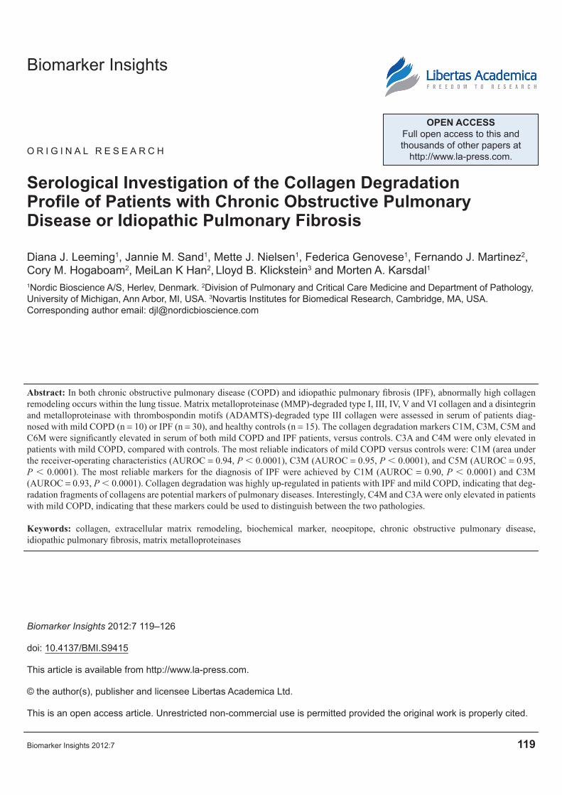
Resultsconnective tissue degradationFragments of types I, III, V and VI collagen degrada-tion by MMPs were significantly elevated in the serum of both COPD and IPF patients versus healthy con-trols, as presented in Figure 1. C1M, C3M, C3A, C4M, C5M and C6M were all significantly elevated in mild COPD compared with controls, (P , 0.05–0.01 and up to .189% for C1M). C1M, C3M, C5M and C6M were all highly elevated in all severity groups of IPF patients compared with controls, (P , 0.05–0.0001, and up to $133% for C3M). Interestingly, the C3A and C4M markers were not elevated in any of the IPF patient groups.
Diagnostic powerThe power of the individual markers to discriminate between disease and healthy controls was calculated by the AUROC and presented in Table 1. The three groups of IPF patients were combined for these calculations, since the markers were not able to differentiate severity in IPF patients. The diagnostic powers of C1M, C3M, C5M and C6M were highly significant with an AUROC . 85% (P , 0.0001). C4M and C3A presented an AUROC . 89% for COPD patients versus controls (P , 0.01), but was unable to distinguish between IPF patients and con-trols. The best discriminators between mild COPD and controls were C1M (AUROC = 0.94, P , 0.0001), C3M (AUROC = 0,95, P , 0.0001), and C5M (AUROC = 0.95, P , 0.0001). The most reliable dif-ferentiation between IPF and controls was observed using C1M (AUROC = 0.90, P , 0.0001) and C3M (AUROC = 0.93, P , 0.0001).

Leeming et al
122 Biomarker Insights 2012:7
DiscussionTo our knowledge, this is the first study investigating the serological profile of collagen turnover in fibrotic lung diseases. We tested a range of novel ECM deg-radation serum markers in patients with COPD or
IPF and in healthy controls. Interestingly, significant differences in marker levels were seen between healthy and disease-affected individuals, and also between the two lung diseases. The markers of ECM degra-dation provided diagnostic information and suggest
***
0
Contro
ls
Mild
IPF
Mod
erat
e IP
F
Sever
e IP
F
COPD
Contro
ls
Mild
IPF
Mod
erat
e IP
F
Sever
e IP
F
COPD
Contro
ls
Mild
IPF
Mod
erat
e IP
F
Sever
e IP
F
COPD
Contro
ls
Mild
IPF
Mod
erat
e IP
F
Sever
e IP
F
COPD
Contro
ls
Mild
IPF
Mod
erat
e IP
F
Sever
e IP
F
COPD
Contro
ls
Mild
IPF
Mod
erat
e IP
F
Sever
e IP
F
COPD
0
0 0
100
200
300
50
100
150
200
250
0
1000
2000
3000
4000
100
200
300
400
500
0
50
100
150
200
250
200
400
600
**
***
*****
**
*** **
***
*
**
***
***
***
***
***
***
**
C1M
(n
g/m
l)
C3M
(n
g/m
l)C
5M (
ng
/ml)
C4M
(n
g/m
l)C
6M (
ng
/ml)
C3A
(n
g/m
l)
Figure 1. Biochemical markers of collagen degradation as measured by six different ELISAs. Levels of the markers reflecting types I (C1M), III (C3M), IV (c4M), V (c5M) and VI (c6M) collagen degradation by MMPs, as well as fragments of type III collagen degraded by aggrecanase (c3A), were mea-sured in serum of patients with mild (n = 10), moderate (n = 10) and severe (n = 10) IPF, mild cOPD (n = 10), and healthy controls (n = 15).notes: Patient groups were compared with healthy controls using the non-parametric Mann-Whitney test, and results are presented as box plots. The boundaries of each box indicate the 25th and 75th percentiles, the line within the box marks the median, and the whiskers indicate the 5th and 95th percentiles. Significance levels: *P , 0.05, **P , 0.01, ***P , 0.001.

Biochemical markers for pulmonary fibrosis
Biomarker Insights 2012:7 123
that tissue degradation is highly elevated in patients with mild COPD and mild to severe IPF. C1M, C3M, C5M and C6M showed highly statistically significant power to discriminate between IPF and mild COPD patients versus healthy individuals, while C4M and C3A were only able to diagnose COPD patients. The fact that four out of six collagen degradation markers were able to detect even a mild form of IPF is inter-esting, since it is this patient group that will benefit the most from a diagnosis. These preliminary find-ings need to be validated in larger clinical settings. This study, although the first and small, has identified collagen turnover markers with the potential to sepa-rate healthy individuals from COPD and IPF patients. In future these markers may assist in the identification of patients that are fast progressers and may respond to a given intervention.
Endopeptidases play a major role in the deg-radation of ECM proteins such as collagens and proteoglycans.26,37,38 MMP-2 and -9, in particular, have been shown to be highly up-regulated in connective tissue diseases leading to fibrosis.39–41 A wide range of ADAMs/ADAMTSs are expressed in the lung42 and many have been associated with different respiratory diseases such as COPD, IPF and asthma.20 COPD has
been coupled with changes in ADAM-33, ADAM-17, and ADAMTS-4 expression,43–45 while only a weak association has been observed for a few ADAMs/ADAMTSs in IPF.46 The C3A marker showed ele-vated levels of ADAMTS-4-mediated degradation of type III collagen in COPD but not IPF patients. This finding is in agreement with previous experi-ments showing that ADAMTS-4 is up-regulated in COPD while there is no evidence of a relationship in IPF.45,46 This highlights that the differences in the pathological processes and the proteases involved in these two diseases result in distinct tissue turnover profiles.
Fibrotic lungs have an overall increased ECM turnover rate, but the normal balance between formation and degradation is changed, leading to increased deposition and increased degradation. The change in remodeling balance is not the same for different types of collagen; a key feature is the increased deposition and degradation of type I col-lagen in pulmonary fibrosis.4,5,47 This is in line with our C1M results showing significantly elevated levels in both patients with mild to severe IPF and mild COPD and thus indicating an increased deg-radation of type I collagen. Contradictory results have been published in relation to type III colla-gen deposition in pulmonary fibrosis, demonstrat-ing that both up—and down-regulation of type III collagen occurs.4–6 The serum C3M data presented here suggest an increased type III collagen degra-dation level in both IPF and mild COPD, indicating a high level of formation of type III collagen as well. Indications of a decreased content of type V collagen in fibrotic rat lungs have been presented.48 However, Parra et al demonstrated that type V col-lagen levels in biopsies from IPF patients increased with disease severity,49 supporting an increased degradation of deposited type V collagen indicated by our C5M data. Type IV and type VI collagen expression, as well as protein levels, have been reported to be elevated in lung tissue of patients with COPD50 and IPF,51 further supporting the find-ings of our biochemical marker study.
Desmosine, isodesmosine, KL-6, CCL18, SP-A and SP-D have all been extensively discussed as serological markers of pulmonary diseases.17,21–23 However, none of these markers have demon-strated optimal sensitivity and diagnostic value.
Table 1. Area under the receiver operating characteristic (AUrOc) for biochemical marker levels in healthy controls vs. IPF and cOPD.
Comparison AUROc Std. error P valuec1M IPF 0.90 0.05 ,0.0001*** cOPD 0.94 0.05 ,0.0001***c3M IPF 0.93 0.03 ,0.0001*** cOPD 0.95 0.04 ,0.0001***c4M IPF 0.59 0.09 0.36 cOPD 0.89 0.07 0.0018**c5M IPF 0.85 0.05 ,0.0001*** cOPD 0.95 0.03 ,0.0001***c6M IPF 0.86 0.05 ,0.0001*** cOPD 0.90 0.06 ,0.0001***c3A IPF 0.52 0.08 0.78 cOPD 0.82 0.11 0.0049**notes: AUrOc . 80% are highlighted in italics. Significant separation of controls and patients is indicated by asterisks. Significance levels: *P , 0.05, **P , 0.01, ***P , 0.001.

Leeming et al
124 Biomarker Insights 2012:7
Thus, more sensitive and accurate biochemical markers are needed for fibrotic lung diseases. Stratification of patients with pulmonary fibrosis is most likely not feasible using a single marker since would require a marker specific for a protein only expressed during lung fibrosis. A panel of markers reflecting different pathophysiological processes involved in pulmonary fibrosis during the two dif-ferent diseases will almost certainly be required for diagnosis, prognosis and assessment of the efficacy of interventions.
The systemic level of a biochemical marker is the sum of all tissue sites generating this one fragment, and also depends on the extent of disease- affected tissue, the aggressiveness of the disease and the protein specificity of the fragment. This was ele-gantly investigated by Meulenbelt et al52 who demon-strated that the level of a MMP-generated fragment of the signature protein of cartilage, type II collagen, was correlated to the number of affected joints in osteoarthritis. Collagen expression is not restricted to the lung tissue, but is found ubiquitously through-out the body. Thus, several co-morbidities may influ-ence the systemic level of fragments produced by MMP degradation of collagen molecules. Further investigations are needed to determine the individual contribution of different tissues to the total pool of collagen neoepitopes.
There are several limitations with the current study. The sample size was very small, and thus the findings are preliminary. The lack of informa-tion available for patients does not allow for further analysis and correlation with clinical parameters. Furthermore, this is a cross-sectional study and the prognostic value of the biomarkers could not be validated.
In conclusion, by using protein fingerprint tech-nology, we have developed assays measuring novel biochemical markers which enable us to assess pep-tides generated during degradation of collagens in pulmonary fibrosis. These markers were able to distinguish between healthy controls and patients with mild COPD and/or IPF in a small clinical pop-ulation. The collagen degradation markers demon-strated promising discriminative diagnostic power and may provide an improved tool for identification of those patients most in need of treatment, as well as for monitoring potential efficacy of interventions.
These data need to be validated in larger clinical settings.
Author ContributionsConceived and designed the experiments: MK, DJL, MH. Data analysis: DJL, JSA, MJU. Scientific writers: JSA, DJL, MK, FGE. Agree with manuscript results and conclusions: ALL. Made critical revisions and approved final version: DJL, MK. Clinical study samples retrieval: LK, FJM, CMH, MH.
FundingWe acknowledge the funding from the Danish Research Foundation (Den Danske Forskningsfond). The authors would like to acknowledge The Lung Tissue Research Consortium (LTRC) and The National Heart, Lung, and Blood Institute (NHLBI) for kindly providing the COPD and IPF lung samples.
Competing InterestsDL, MK and FG are full-time employees of Nordic Bioscience. MK is stockholder of Nordic Bioscience. FM is a board member for COPD and/or IPF for Actelion, Almirall/Forest, Nycomed/Takeda, Bayer, GSK, Ikaria, MedImmune, Merck, Pearl, Pfizer, Jannsen, Elan, Gilead, Centocor, Novartis, has con-sulted for Nycomed/Takeda, American Institute for Research, AstraZeneca, Boom Comm, Elan, HCRC, IntraMed, JK Associates, Merion, Schering, Sudler and Hennessey, Cardiomems, Janssens, spoken for Forest, Nycomed/Takeda, AstraZeneca, GSK, Sanofi, Boeheringer Ingelheim, developed educational pre-sentations for UpToDate, American College of Chest Physicians, Almirall/Forest, Bayer. DL’s institution has received a grant from Den Danske Forsknings Fond. MH is a consultant to Boehringer Ingelheim, Pfizer, GSK, Medimmune, Novartis, Grifols Thera-peutics, United Biosource Corporation, has grants/grants pending from NHLBI K23 award, Chicago Community Trust and has developed educational presentations for WebMD, National Association for
potential competing interests.
Disclosures and EthicsAs a requirement of publication author(s) have pro-vided to the publisher signed confirmation of compli-ance with legal and ethical obligations including but
Consulting Education. Other authors disclose no

Biochemical markers for pulmonary fibrosis
Biomarker Insights 2012:7 125
not limited to the following: authorship and contribu-torship, conflicts of interest, privacy and confidential-ity and (where applicable) protection of human and animal research subjects. The authors have read and confirmed their agreement with the ICMJE author-ship and conflict of interest criteria. The authors have also confirmed that this article is unique and not under consideration or published in any other publication, and that they have permission from rights holders to reproduce any copyrighted material. Any disclo-sures are made in this section. The external blind peer reviewers report no conflicts of interest.
References 1. Hogg JC, McDonough JE, Gosselink JV, Hayashi S. What drives the periph-
eral lung-remodeling process in chronic obstructive pulmonary disease? Proc Am Thorac Soc. 2009;6:668–72.
2. Selman M, King TE, Pardo A. Idiopathic pulmonary fibrosis: prevailing and evolving hypotheses about its pathogenesis and implications for therapy. Ann Intern Med. 2001;134:136–51.
3. Desai B, Mattson J, Paintal H, et al. Differential expression of monocyte/macrophage- selective markers in human idiopathic pulmonary fibrosis. Exp Lung Res. 2011;37:227–38.
4. Last JA, Siefkin AD, Reiser KM. Type I collagen content is increased in lungs of patients with adult respiratory distress syndrome. Thorax. 1983;38: 364–8.
5. Seyer JM, Hutcheson ET, Kang AH. Collagen polymorphism in idiopathic chronic pulmonary fibrosis. J Clin Invest. 1976;57:1498–507.
6. Kirk JM, Bateman ED, Haslam PL, Laurent GJ, Turner-Warwick M. Serum type III procollagen peptide concentration in cryptogenic fibrosing alveoli-tis and its clinical relevance. Thorax. 1984;39:726–32.
7. van Kuppevelt TH, Veerkamp JH, Timmermans JA. Immunoquantification of type I, III, IV and V collagen in small samples of human lung parenchyma. Int J Biochem Cell Biol. 1995;27:775–82.
8. Suki B, Ito S, Stamenovic D, Lutchen KR, Ingenito EP. Biomechanics of the lung parenchyma: critical roles of collagen and mechanical forces. J Appl Physiol. 2005;98:1892–9.
9. Dunsmore SE. Treatment of COPD: a matrix perspective. Int J Chron Obstruct Pulmon Dis. 2008;3:113–22.
10. Kadler KE, Holmes DF, Trotter JA, Chapman JA. Collagen fibril formation. Biochem J. 1996;316(Pt 1):1–11.
11. Konomi H, Hayashi T, Nakayasu K, Arima M. Localization of type V collagen and type IV collagen in human cornea, lung, and skin. Immu-nohistochemical evidence by anti-collagen antibodies characterized by immunoelectroblotting. Am J Pathol. 1984;116:417–26.
12. West JB, Mathieu-Costello O. Structure, strength, failure, and remodeling of the pulmonary blood-gas barrier. Annu Rev Physiol. 1999;61:543–72.
13. Wenstrup RJ, Florer JB, Brunskill EW, Bell SM, Chervoneva I, Birk DE. Type V collagen controls the initiation of collagen fibril assembly. J Biol Chem. 2004;279:53331–7.
14. Kuo HJ, Maslen CL, Keene DR, Glanville RW. Type VI collagen anchors endothelial basement membranes by interacting with type IV collagen. J Biol Chem. 1997;272:26522–9.
15. Engvall E, Hessle H, Klier G. Molecular assembly, secretion, and matrix deposition of type VI collagen. J Cell Biol. 1986;102:703–10.
16. Wilson MS, Wynn TA. Pulmonary fibrosis: pathogenesis, etiology and regu-lation. Mucosal Immunol. 2009;2:103–21.
17. Luisetti M, Ma S, Iadarola P, et al. Desmosine as a biomarker of elastin degra-dation in COPD: current status and future directions. Eur Respir J 2008; 32: 1146–57.
18. Joos L, He JQ, Shepherdson MB, et al. The role of matrix metalloproteinase polymorphisms in the rate of decline in lung function. Hum Mol Genet. 2002;11:569–76.
19. Marciniak SJ, Lomas DA. What can naturally occurring mutations tell us about the pathogenesis of COPD? Thorax. 2009;64:359–64.
20. Paulissen G, Rocks N, Gueders MM, et al. Role of ADAM and ADAMTS metalloproteinases in airway diseases. Respir Res. 2009;10:127.
21. Yokoyama A, Kohno N, Hamada H, et al. Circulating KL-6 predicts the outcome of rapidly progressive idiopathic pulmonary fibrosis. Am J Respir Crit Care Med. 1998;158:1680–4.
22. Prasse A, Pechkovsky DV, Toews GB, et al. CCL18 as an indicator of pul-monary fibrotic activity in idiopathic interstitial pneumonias and systemic sclerosis. Arthritis Rheum. 2007;56:1685–93.
23. Greene KE, King TE Jr, et al. Serum surfactant proteins-A and -D as bio-markers in idiopathic pulmonary fibrosis. Eur Respir J. 2002;19:439–46.
24. Thomeer M, Grutters JC, Wuyts WA, Willems S, Demedts MG. Clinical use of biomarkers of survival in pulmonary fibrosis. Respir Res. 2010; 11:89.
25. Nathan SD, Shlobin OA, Weir N, et al. Long-term course and prog-nosis of idiopathic pulmonary fibrosis in the new millennium. Chest. 2011;140:221–9.
26. Oikonomidi S, Kostikas K, Tsilioni I, Tanou K, Gourgoulianis KI, Kiropoulos TS. Matrix metalloproteinases in respiratory diseases: from patho-genesis to potential clinical implications. Curr Med Chem. 2009;16:1214–28.
27. Karsdal MA, Henriksen K, Leeming DJ, Woodworth T, Vassiliadis E, Bay-Jensen AC. Novel combinations of Post-Translational Modification (PTM) neo-epitopes provide tissue-specific biochemical markers—are they the cause or the consequence of the disease? Clin Biochem. 2010;43: 793–804.
28. Karsdal MA, Delvin E, Christiansen C. Protein fingerprints—relying on and understanding the information of serological protein measurements. Clin Biochem. 2011;44:1278–9.
29. Karsdal MA, Henriksen K, Leeming DJ, et al. Biochemical markers and the FDA Critical Path: how biomarkers may contribute to the understand-ing of pathophysiology and provide unique and necessary tools for drug development. Biomarkers. 2009;14:181–202.
30. Veidal SS, Vassiliadis E, Barascuk N, et al. Matrix metalloproteinase-9-mediated type III collagen degradation as a novel serological biochemical marker for liver fibrogenesis. Liver Int. 2010;30:1293–304.
31. Barascuk N, Veidal SS, Larsen L, et al. A novel assay for extracellular matrix remodeling associated with liver fibrosis: An enzyme-linked immu-nosorbent assay (ELISA) for a MMP-9 proteolytically revealed neo-epitope of type III collagen. Clin Biochem. 2010;43:899–904.
32. Veidal SS, Karsdal MA, Nawrocki A, et al. Assessment of proteolytic degradation of the basement membrane: A fragment of type IV collagen as a biochemical marker for liver fibrosis. Fibrogenesis Tissue Repair 2011;4:22.
33. Veidal SS, Karsdal MA, Vassiliadis E, et al. MMP Mediated Degradation of Type VI Collagen Is Highly Associated with Liver Fibrosis— Identification and Validation of a Novel Biochemical Marker Assay. PLoS ONE. 2011;6: e24753.
34. Bay-Jensen AC, Liu Q, Byrjalsen I, et al. Enzyme-linked immunosorbent assay (ELISAs) for metalloproteinase derived type II collagen neoepitope, CIIM—increased serum CIIM in subjects with severe radiographic osteoar-thritis. Clin Biochem. 2011;44:423–9.
35. Leeming DJ, He Y, Veidal S, et al. A novel marker for assessment of liver matrix remodeling: an enzyme-linked immunosorbent assay (ELISA) detecting a MMP generated type I collagen neo-epitope (C1M). Biomarkers. 2011;16:616–28.
36. Veidal SS, Larsen DV, Chen X, et al. MMP mediated type V collagen degradation (C5M) is elevated in ankylosing spondylitis. Clin Biochem. 2012.
37. Bay-Jensen AC, Hoegh-Madsen S, Dam E, et al. Which elements are involved in reversible and irreversible cartilage degradation in osteoarthritis? Rheumatol Int. 2010;30:435–42.
38. Zhen EY, Brittain IJ, Laska DA, et al. Characterization of metalloprotease cleavage products of human articular cartilage. Arthritis Rheum. 2008; 58:2420–31.
39. Chakrabarti S, Patel KD. Matrix metalloproteinase-2 (MMP-2) and MMP-9 in pulmonary pathology. Exp Lung Res. 2005;31:599–621.
40. Boeker KH, Haberkorn CI, Michels D, Flemming P, Manns MP, Lichtinghagen R. Diagnostic potential of circulating TIMP-1 and MMP-2 as markers of liver fibrosis in patients with chronic hepatitis C. Clin Chim Acta. 2002;316:71–81.

Leeming et al
126 Biomarker Insights 2012:7
41. Kirimlioglu H, Kirimlioglu V, Yilmaz S. Expression of matrix metallo-proteinases 2 and 9 in donor liver, cirrhotic liver, and acute rejection after human liver transplantation. Transplant Proc. 40:3574–7.
42. Rocks N, Paulissen G, Quesada CF, et al. Expression of a disintegrin and metalloprotease (ADAM and ADAMTS) enzymes in human non-small-cell lung carcinomas (NSCLC). Br J Cancer. 2006;94:724–30.
43. van Diemen CC, Postma DS, Vonk JM, Bruinenberg M, Schouten JP, Boezen HM. A disintegrin and metalloprotease 33 polymorphisms and lung function decline in the general population. Am J Respir Crit Care Med. 2005;172:329–33.
44. Shao MX, Nakanaga T, Nadel JA. Cigarette smoke induces MUC5 AC mucin overproduction via tumor necrosis factor-alpha-converting enzyme in human airway epithelial (NCI-H292) cells. Am J Physiol Lung Cell Mol. Physiol. 2004;287:L420–7.
45. Ezzie ME, Crawford M, Cho JH, et al. Gene expression networks in COPD: microRNA and mRNA regulation. Thorax. 2012;67:122–31.
46. Pardo A, Selman M, Kaminski N. Approaching the degradome in idiopathic pulmonary fibrosis. Int J Biochem Cell Biol. 2008;40:1141–55.
47. Bateman ED, Turner-Warwick M, mann-Grill BC. Immunohistochemi-cal study of collagen types in human foetal lung and fibrotic lung disease. Thorax. 1981;36:645–53.
48. Reiser KM, Last JA. Type V collagen. Quantitation in normal lungs and in lungs of rats with bleomycin-induced pulmonary fibrosis. J Biol Chem. 1983;258:269–75.
49. Parra ER, Teodoro WR, Velosa AP, de Oliveira CC, Yoshinari NH, Capelozzi VL. Interstitial and vascular type V collagen morphologic disorganization in usual interstitial pneumonia. J Histochem Cytochem. 2006;54:1315–25.
50. Kranenburg AR, Willems-Widyastuti A, Moori WJ, et al. Enhanced bron-chial expression of extracellular matrix proteins in chronic obstructive pul-monary disease. Am J Clin Pathol. 2006;126:725–35.
51. Specks U, Nerlich A, Colby TV, Wiest I, Timpl R. Increased expression of type VI collagen in lung fibrosis. Am J Respir Crit Care Med. 1995;151: 1956–64.
52. Meulenbelt I, Kloppenburg M, Kroon HM, et al. Clusters of biochemical markers are associated with radiographic subtypes of osteoarthritis (OA) in subject with familial OA at multiple sites. The GARP study. Osteoarthritis Cartilage. 2007;15:379–85.