Accelerated Variant of Idiopathic Pulmonary Fibrosis: Clinical Behavior and Gene Expression Pattern
11
Accelerated Variant of Idiopathic Pulmonary Fibrosis: Clinical Behavior and Gene Expression Pattern Moise ´s Selman 1 *, Guillermo Carrillo 1 , Andrea Estrada 1 , Mayra Mejia 1 , Carina Becerril 1 , Jose ´ Cisneros 1 , Miguel Gaxiola 1 , Rogelio Pe ´ rez-Padilla 1 , Carmen Navarro 1 , Thomas Richards 2 , James Dauber 2 , Talmadge E. King, Jr. 3 , Annie Pardo 4 , Naftali Kaminski 2 1 Instituto Nacional de Enfermedades Respiratorias, Mexico City, Mexico, 2 The Dorothy P. and Richard P. Simmons Center for Interstitial Lung Diseases, Pulmonary Allergy and Critical Care Medicine, University of Pittsburgh, Pittsburgh, Pennsylvania, United States of America, 3 Department of Medicine, Division of Pulmonary and Critical Care Medicine, San Francisco General Hospital, San Francisco, California, United States of America, 4 Facultad de Ciencias, Universidad Nacional Auto ´noma de Me ´xico, Mexico City, Mexico Background. Idiopathic pulmonary fibrosis (IPF) is characterized by the insidious onset of dyspnea or cough. However, a subset of patients has a short duration of symptoms with rapid progression to end-stage disease. In this study, we evaluated clinical and molecular features of ‘‘rapid’’ and ‘‘slow’’ progressors with IPF. Methods and Findings. 26 patients with ,6 months of symptoms before first presentation [rapid progressors] and 88 patients with .24 months of symptoms [slow progressors] were studied. Survival was analyzed by the Kaplan-Meyer method and proportional hazard’s model. Lung microarrays and tissue proteins were measured in a subset of patients. No differences were found in age, physiologic impairment and bronchoalveolar lavage (BAL) cellular profile. There were more males (OR = 6.5; CI:1.4-29.5; p = 0.006) and smokers (OR = 3.04; CI:1.1-8.3; p = 0.04) in the rapid progressors group. Survival from the beginning of symptoms was significantly reduced in rapid progressors (HR = 9.0; CI:4.48-18.3; p,0.0001) and there was a tendency for decreased survival from the time of diagnosis (HR = 1.5; CI:0.81-2.87; p = 0.18). We identified 437 differentially expressed genes. Lungs of rapid progressors overexpressed genes involved in morphogenesis, oxidative stress, migration/proliferation, and genes from fibroblasts/smooth muscle cells. Upregulation of two of these genes, adenosine-2B receptor and prominin-1/CD133, was validated by immunohistochemistry and were expressed by alveolar epithelial cells. BAL from rapid progressors showed a .2- fold increase of active matrix metalloproteinase-9, and induced a higher fibroblast migration compared with slow progressors and controls [238698% versus 123629% (p,0.05) and 30617% (p,0.01)]. Conclusions/Significance. A subgroup of IPF patients, predominantly smoking males, display an accelerated clinical course and have a gene expression pattern that is different from those with slower progression and longer survival. These findings highlight the variability in the progression of IPF, and may explain, in part, the difficulty in obtaining significant and reproducible results in studies of therapeutic interventions in patients with IPF. Citation: Selman M, Carrillo G, Estrada A, Mejia M, Becerril C, et al (2007) Accelerated Variant of Idiopathic Pulmonary Fibrosis: Clinical Behavior and Gene Expression Pattern. PLoS ONE 2(5): e482. doi:10.1371/journal.pone.0000482 INTRODUCTION Idiopathic pulmonary fibrosis (IPF) is a chronic fibrosing interstitial lung disease of unknown etiology characterized by progressive dyspnea, reduced lung volumes, impaired gas exchange, and the histopathologic signature of usual interstitial pneumonia (UIP). This disease, which is the most common of the idiopathic interstitial pneumonias, is unresponsive to current therapy and most patients die within 5 years after diagnosis [1– 3]. However, it is increasingly apparent that IPF patients exhibit distinct patterns of disease progression [4,5]. Most of them show a long duration of symptoms before diagnosis and then experience a slowly progressive clinical course (‘‘slow’’ progres- sors) [5]. Often, an acute clinical deterioration (‘‘acute exacer- bation’’ of IPF) precedes the terminal phase of the illness in this subgroup [5,6]. Quite distinct from these observations, some IPF patients display a more rapidly progressive clinical course with a shorter duration of symptoms before diagnosis and progression to death (‘‘rapid’’ progressors). However, a systematic charac- terization of these distinct disease progression phenotypes has not been performed. The purpose of this study was to determine whether ‘‘rapid’’ and ‘‘slow’’ progressor IPF patients could be distinguished by clinical, biological or molecular features. Better identification and understanding of these differences may provide insights into the pathogenesis and assist in the development of therapeutic interventions. METHODS Study Population This study included 114 individuals from a cohort of 167 consecutive patients with IPF evaluated at the National Institute of Respiratory Diseases, Mexico, between 1995 and 2004. The study was approved by the Ethics Committee of the National Institute of Respiratory Diseases, Me ´xico. Diagnosis of IPF was made based on established criteria and confirmed by lung biopsy in 31% of the subjects [7]. Clinical data (time of symptoms before Academic Editor: Ming You, Washington University, United States of America Received January 5, 2007; Accepted May 4, 2007; Published May 30, 2007 Copyright: ß 2007 Selman et al. This is an open-access article distributed under the terms of the Creative Commons Attribution License, which permits unrestricted use, distribution, and reproduction in any medium, provided the original author and source are credited. Funding: This work was partially supported by Universidad Nacional Auto ´ noma de Me ´xico Grant SDI.PTID.05.6. Naftali Kaminski’s work was supported by NIH grant HL073745, HL079394 and by a generous donation from the Simmons family. Funding sources were not involved in study design, performance, analysis or manuscript preparation. Competing Interests: The authors have declared that no competing interests exist. * To whom correspondence should be addressed. E-mail: mselmanl@yahoo. com.mx PLoS ONE | www.plosone.org 1 May 2007 | Issue 5 | e482
-
Upload
independent -
Category
Documents
-
view
0 -
download
0
Transcript of Accelerated Variant of Idiopathic Pulmonary Fibrosis: Clinical Behavior and Gene Expression Pattern
Accelerated Variant of Idiopathic Pulmonary Fibrosis:Clinical Behavior and Gene Expression PatternMoises Selman1*, Guillermo Carrillo1, Andrea Estrada1, Mayra Mejia1, Carina Becerril1, Jose Cisneros1, Miguel Gaxiola1, Rogelio Perez-Padilla1,Carmen Navarro1, Thomas Richards2, James Dauber2, Talmadge E. King, Jr.3, Annie Pardo4, Naftali Kaminski2
1 Instituto Nacional de Enfermedades Respiratorias, Mexico City, Mexico, 2 The Dorothy P. and Richard P. Simmons Center for Interstitial LungDiseases, Pulmonary Allergy and Critical Care Medicine, University of Pittsburgh, Pittsburgh, Pennsylvania, United States of America, 3 Department ofMedicine, Division of Pulmonary and Critical Care Medicine, San Francisco General Hospital, San Francisco, California, United States of America,4 Facultad de Ciencias, Universidad Nacional Autonoma de Mexico, Mexico City, Mexico
Background. Idiopathic pulmonary fibrosis (IPF) is characterized by the insidious onset of dyspnea or cough. However,a subset of patients has a short duration of symptoms with rapid progression to end-stage disease. In this study, we evaluatedclinical and molecular features of ‘‘rapid’’ and ‘‘slow’’ progressors with IPF. Methods and Findings. 26 patients with,6 months of symptoms before first presentation [rapid progressors] and 88 patients with .24 months of symptoms [slowprogressors] were studied. Survival was analyzed by the Kaplan-Meyer method and proportional hazard’s model. Lungmicroarrays and tissue proteins were measured in a subset of patients. No differences were found in age, physiologicimpairment and bronchoalveolar lavage (BAL) cellular profile. There were more males (OR = 6.5; CI:1.4-29.5; p = 0.006) andsmokers (OR = 3.04; CI:1.1-8.3; p = 0.04) in the rapid progressors group. Survival from the beginning of symptoms wassignificantly reduced in rapid progressors (HR = 9.0; CI:4.48-18.3; p,0.0001) and there was a tendency for decreased survivalfrom the time of diagnosis (HR = 1.5; CI:0.81-2.87; p = 0.18). We identified 437 differentially expressed genes. Lungs of rapidprogressors overexpressed genes involved in morphogenesis, oxidative stress, migration/proliferation, and genes fromfibroblasts/smooth muscle cells. Upregulation of two of these genes, adenosine-2B receptor and prominin-1/CD133, wasvalidated by immunohistochemistry and were expressed by alveolar epithelial cells. BAL from rapid progressors showed a .2-fold increase of active matrix metalloproteinase-9, and induced a higher fibroblast migration compared with slow progressorsand controls [238698% versus 123629% (p,0.05) and 30617% (p,0.01)]. Conclusions/Significance. A subgroup of IPFpatients, predominantly smoking males, display an accelerated clinical course and have a gene expression pattern that isdifferent from those with slower progression and longer survival. These findings highlight the variability in the progression ofIPF, and may explain, in part, the difficulty in obtaining significant and reproducible results in studies of therapeuticinterventions in patients with IPF.
Citation: Selman M, Carrillo G, Estrada A, Mejia M, Becerril C, et al (2007) Accelerated Variant of Idiopathic Pulmonary Fibrosis: Clinical Behavior andGene Expression Pattern. PLoS ONE 2(5): e482. doi:10.1371/journal.pone.0000482
INTRODUCTIONIdiopathic pulmonary fibrosis (IPF) is a chronic fibrosing
interstitial lung disease of unknown etiology characterized by
progressive dyspnea, reduced lung volumes, impaired gas
exchange, and the histopathologic signature of usual interstitial
pneumonia (UIP). This disease, which is the most common of the
idiopathic interstitial pneumonias, is unresponsive to current
therapy and most patients die within 5 years after diagnosis [1–
3]. However, it is increasingly apparent that IPF patients exhibit
distinct patterns of disease progression [4,5]. Most of them show
a long duration of symptoms before diagnosis and then
experience a slowly progressive clinical course (‘‘slow’’ progres-
sors) [5]. Often, an acute clinical deterioration (‘‘acute exacer-
bation’’ of IPF) precedes the terminal phase of the illness in this
subgroup [5,6]. Quite distinct from these observations, some IPF
patients display a more rapidly progressive clinical course with
a shorter duration of symptoms before diagnosis and progression
to death (‘‘rapid’’ progressors). However, a systematic charac-
terization of these distinct disease progression phenotypes has not
been performed.
The purpose of this study was to determine whether ‘‘rapid’’
and ‘‘slow’’ progressor IPF patients could be distinguished by
clinical, biological or molecular features. Better identification and
understanding of these differences may provide insights into the
pathogenesis and assist in the development of therapeutic
interventions.
METHODS
Study PopulationThis study included 114 individuals from a cohort of 167
consecutive patients with IPF evaluated at the National Institute
of Respiratory Diseases, Mexico, between 1995 and 2004. The
study was approved by the Ethics Committee of the National
Institute of Respiratory Diseases, Mexico. Diagnosis of IPF was
made based on established criteria and confirmed by lung biopsy
in 31% of the subjects [7]. Clinical data (time of symptoms before
Academic Editor: Ming You, Washington University, United States of America
Received January 5, 2007; Accepted May 4, 2007; Published May 30, 2007
Copyright: � 2007 Selman et al. This is an open-access article distributed underthe terms of the Creative Commons Attribution License, which permitsunrestricted use, distribution, and reproduction in any medium, provided theoriginal author and source are credited.
Funding: This work was partially supported by Universidad Nacional Autonomade Mexico Grant SDI.PTID.05.6. Naftali Kaminski’s work was supported by NIHgrant HL073745, HL079394 and by a generous donation from the Simmonsfamily. Funding sources were not involved in study design, performance, analysisor manuscript preparation.
Competing Interests: The authors have declared that no competing interestsexist.
* To whom correspondence should be addressed. E-mail: [email protected]
PLoS ONE | www.plosone.org 1 May 2007 | Issue 5 | e482

diagnosis, smoking status, drug treatment, clinical findings,
absence of previous environmental exposures, and collagen-
vascular disease) were extracted from case records. The duration
of illness was defined in two ways: (1) the time from the onset of the
disease, determined from the patient’s recollection of the first
appearance of dyspnea or cough throughout the day; and (2) the
time from the clinical diagnosis of IPF.
Smoking status was characterized as ‘‘never’’, ‘‘former’’
(patients who stopped smoking at least 12 month before pre-
sentation), or ‘‘current’’ (patients who were either still smoking or
stopped smoking less than a year before presentation) [8]. Smoking
index (packs/year) was also documented.
Patients were treated with several regimens: prednisone, or
prednisone plus azathioprine, or inhaled beclomethasone. Most
patients also received colchicine. There were no differences
between the type of initial treatment among both groups (data
not shown).
Control SubjectsSeven healthy volunteers (43.4611.9 years) were selected as
controls for the gelatin zymography study, and five of them were
assayed for fibroblast migration. Lungs from patients who died
from non-respiratory causes (53.766.7 years) and from patients
with chronic hypersensitivity pneumonitis (56.168.9 years) were
used as controls for immunohistochemistry.
Lung Function and Imaging StudiesPulmonary function tests, including spirometry, plethysmography,
and arterial blood gases were performed as described elsewhere
[5,9,10]. Separate comparisons of oxygen saturation (SpO2) levels
were performed at the baseline and after 6 months follow-up using
Wilcoxon rank-sum tests. A longitudinal test for group differences
at 6 months controlling for baseline oxygen saturation, was
performed using analysis of covariance (ANCOVA) to adjust for
regression toward the mean.
High resolution computed tomography (HRCT) was performed
with 1.0- or 1.5-mm-thick axial sections taken at 1-cm intervals
throughout the entire thorax and were reconstructed using a high
spatial frequency algorithm. Between 20 and 25 CT images were
acquired in each patient. HRCT scans were scored on a scale of 0-
5 for ground glass attenuation, extent of septal thickening and
honeycombing as described [10]. Also, to determine the effect of
smoking on lung architecture, the percent of emphysematous
lesions was quantified.
Bronchoalveolar lavageAs part of the diagnosis process, bronchoalveolar lavage (BAL) was
performed in 85 out of the 114 patients as described [11–13]. Cells
were stained with hematoxylin&eosin for differential cell counts.
Supernatants were frozen at 270u until use.
Histopathologic EvaluationTissue samples were obtained by open lung biopsy in 8 from 26
‘‘rapid’’ and 27 from 88 ‘‘slow’’ progressors. None of the patients
had been treated with corticosteroids or immunosuppressive drugs
at the time of biopsy. There was no mortality related to the
surgical procedure and all patients were discharge from the
hospital. One ‘‘rapid’’ progressor patient and two ‘‘slow’’
progressors showed surgical morbidity which included prolonged
air leakage ($6 days, 1 patient in each group) and hemothorax in
1 ‘‘slow’’ progressor patient. Lung samples were fixed with 10%
formaldehyde and handled routinely for light microscopy. A
pathologist, blinded to the clinical data, scored the lesions from 0–
2 (absent, mild/moderate and severe): 1) extent of honeycomb; 2)
hyperplasia of smooth muscle cells; 3) hypertensive changes; 4)
extent of fibrosis 5) extent of interstitial inflammation; 6)
hyperplasia of type 2 cells. The assessment was done as previously
described [14,15]. Hyaline membranes were evaluated as present
or absent. Fibroblastic foci (FF) were counted using hematox-
ylin&eosin and Massons trichrome. Each biopsy was viewed at
low-power magnification (x40), and the numbers of FF were
counted within all tissue specimen. The area of the tissue sample
was measured and results were expressed as FF/cm2.
ImmunohistochemistryLung tissue sections from 7 ‘‘rapid’’ progressors 8 ‘‘slow’’
progressors, 5 hypersensitivity pneumonitis, and three control
lungs were treated as previously described [12,13]. Rabbit anti-
human adenosine 2B receptor (A2BAR) (5 mg/ml) (Chemicon Int,
Tamecula CA) and mouse anti-human prominin-1/CD133
(10 mg/ml) (clone AC133; Miltenyi Biotec, Auburn, CA), were
applied and samples were incubated at 4uC overnight. A
secondary biotinylated anti-immunoglobulin followed by horse-
radish peroxidase-conjugated streptavidin (BioGenex, San Ramon
CA) was used according to manufacturer’s instructions. 3-amino-
9-ethyl-carbazole (AEC, BioGenex) in acetate buffer containing
0.05% H2O2 was used as substrate. The sections were counter-
stained with hematoxylin. The primary antibody was replaced by
non-immune serum for negative control slides.
BAL gelatin zymographyTo identify gelatinolytic activity, BAL fluid samples from 8
‘‘rapid’’ progressors, 8 ‘‘slow’’ progressors and 7 controls (1.5 mg of
protein) were analyzed in 8.5% SDS-PAGE gels containing gelatin
(1 mg/ml) and a final concentration of 0.3 mg/ml heparin as
previously described [16]. Human matrix metalloprotease (MMP)-
2 and MMP-9 zymography standards (Chemicon, CA) were used
as gelatinolytic markers.
Western Blot AnalysisBAL fluid samples were 506 concentrated by lyophilization and
solubilized in water. Aliquots containing 35 mg of protein were
mixed with 26 Laemmli buffer (V/V) and separated on 10%
SDS-polyacrylamide gels. Proteins were electroblotted onto
nitrocellulose membranes (Hybond ECL, Amersham Biosciences).
After blocking with 5% (w/v) non-fat dried milk in PBS, the
membranes were incubated overnight at 4uC with polyclonal
antibody against human adenosine A2B receptor (2 mg/ml;
Chemicon, Tamecula, CA), and monoclonal antibody against
CD-133 (1 mg/ml; ABGENT, San Diego, CA). Membranes were
subsequently washed, incubated with the corresponding secondary
antibody conjugated to horseradish peroxidase (1:20000; Jackson,
West Grove, PA) for 1 h at room temperature, and visualized with
Enhanced Chemiluminescence (ECL) detection system (Amer-
sham Biosciences, UK) using radiograph film (Hyperfilm,
Amersham Biosciences) according to the instructions of the
manufacturer. Films were digitalized and quantified using image
analysis software (ID; Eastman Kodak Company; Rochester, NY).
RNA extraction and DNA microarray hybridizationLung samples from 4 ‘‘rapid’’ and 4 ‘‘slow’’ progressor patients
were among the samples previously described by us [13].
However, the gene expression results presented in this manuscript
have not been previously published. RNA extracted from lung
tissue was used to generate labeled cRNA and hybridized to
a custom Affymetrix oligonucleotide microarray (Hu03 containing
An Accelerated Variant of IPF
PLoS ONE | www.plosone.org 2 May 2007 | Issue 5 | e482
59,619 probesets representing 29655 transcripts) that were
scanned and normalized as described [13]. The dataset is available
at http://www.dom.pitt.edu/paccm/genomics/ACC/index.htm.
Statistical analyses were performed as described [17,18]
using ScoreGenes software package (http://compbio.cs.huji.ac.il/
scoregenes/). For Data Mining and visualization, we used
Genomica (http://genomica.weizmann.ac.il/) and Spotfire De-
cision Site 8.0 (Spotfire Inc. Goteborg, Sweden). Correction for
only transcripts that had an Entrez Gene annotation was included
in the analysis.
To identify genes that best distinguish between ‘‘rapid’’ and
‘‘slow’’ progressors, we used the Threshold Number of Mis-
classification (TNoM) score [19] as well as the Student’s t-test.
TNoM score counts the number of classification errors that occur
between compared groups for each gene of the dataset. To
improve the stringency of our analysis we considered genes as
changed only if they had a t-test and a TNoM p-value ,0.05 and
a fold ratio .2 as previously described [20].
Migration assayMigration of normal human lung fibroblasts was performed using
24-well collagen-coated Boyden chambers (Chemicon Temecula,
CA) as described [11]. Fibroblasts were derived from a region
of the lung showing no histologic abnormalities of a patient
undergoing lobectomy for removal of a solitary pulmonary nodule.
Fibroblasts (36105 cells) were added to the upper chamber. The
lower chamber contained 0.3 ml of medium with 5% BSA alone
or with 50% BAL fluid. After incubation for 8 h at 37uC the non-
migrating cells on the top of the chamber were scraped and
washed. The migrating cells were determined according to
manufacturer’s instructions. Briefly, the cells were stained and
the color was eluted with 300 ml of extraction buffer and aliquots
of 150 ml were measured in an ELISA plate reader at 545 nm.
The number of cells that migrated in absence of BAL was used as
control (0% migration). All assays were performed in duplicate.
Statistical methodsData were analyzed with STATA software. In August 2005, we
obtained the vital status of each patient by reviewing the clinical
charts and using telephone or telegrams in all cases that had been
lost to follow up. We obtained the vital status on 85% of the
‘‘rapid’’ progressors and of 80% of the ‘‘slow’’ progressors. For the
remaining patients, their vital status was considered alive. Data are
summarized using mean6SD, median, and range for continuous
variables, and frequency and percentage for categorical variables.
Patient demographics were compared between ‘‘rapid’’ pro-
gressors and ‘‘slow’’ progressors, using the two-sample, rank-sum
test for continuous variables, Fisher’s exact test and a chi2 test for
categorical variables. The time elapsed between the beginning of
symptoms and the first consult either at our Institute or another
Hospital was recorded. For survival analysis, ‘‘time zero’’ was
defined in two ways: (1) from the beginning of symptoms as
reported by the patient; (2) from the ‘‘index visit’’, which was
defined as the date the patient was first seen at the National
Institute of Respiratory Diseases during the study period (1995 to
2004). Cumulative survival probabilities were estimated using the
Kaplan-Meier method. In addition, a Cox proportional hazards
regression model was used to compare survival as a function of
several variables including age, gender, clubbing, forced vital
capacity (FVC) as percentage of predicted, PaO2, SpO2 at rest
and after exercise, smoking (current and cumulative as pack-years)
and duration of symptoms before diagnosis. In all cases, two-tailed
p values ,0.05 were considered statistically significant.
RESULTSIn univariate analysis of the whole cohort (n = 167), time elapsed
between the beginning of symptoms, smoking, masculine gender,
FVC%, PaO2, SpO2 at rest and during exercise, were significant
predictors of mortality. In the multivariate Cox model, time
elapsed between the beginning of symptoms and the first consult,
smoking, masculine gender, and FVC%, remained significant.
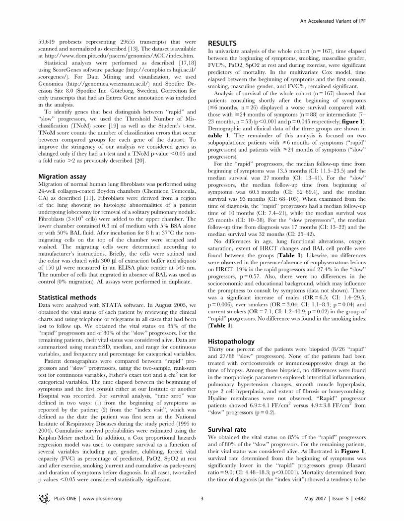
Analysis of survival of the whole cohort (n = 167) showed that
patients consulting shortly after the beginning of symptoms
(#6 months, n = 26) displayed a worse survival compared with
those with $24 months of symptoms (n = 88) or intermediate (7–
23 months, n = 53) (p,0.001 and p = 0.045 respectively; figure 1).
Demographic and clinical data of the three groups are shown in
table 1. The remainder of this analysis is focused on two
subpopulations: patients with #6 months of symptoms (‘‘rapid’’
progressors) and patients with $24 months of symptoms (‘‘slow’’
progressors).
For the ‘‘rapid’’ progressors, the median follow-up time from
beginning of symptoms was 13.5 months (CI: 11.5–23.5) and the
median survival was 27 months (CI: 13–41). For the ‘‘slow’’
progressors, the median follow-up time from beginning of
symptoms was 60.5 months (CI: 52–69.4), and the median
survival was 93 months (CI: 68–105). When examined from the
time of diagnosis, the ‘‘rapid’’ progressors had a median follow-up
time of 10 months (CI: 7.4–21), while the median survival was
25 months (CI: 10–38). For the ‘‘slow progressors’’, the median
follow-up time from diagnosis was 17 months (CI: 13–22) and the
median survival was 32 months (CI: 25–42).
No differences in age, lung functional alterations, oxygen
saturation, extent of HRCT changes and BAL cell profile were
found between the groups (Table 1). Likewise, no differences
were observed in the presence/absence of emphysematous lesions
on HRCT: 19% in the rapid progressors and 27.4% in the ‘‘slow’’
progressors, p = 0.57. Also, there were no differences in the
socioeconomic and educational background, which may influence
the promptness to consult by symptoms (data not shown). There
was a significant increase of males (OR = 6.5; CI: 1.4–29.5;
p = 0.006), ever smokers (OR = 3.04; CI: 1.1–8.3; p = 0.04) and
current smokers (OR = 7.1, CI: 1.2–40.9; p = 0.02) in the group of
‘‘rapid’’ progressors. No difference was found in the smoking index
(Table 1).
HistopathologyThirty one percent of the patients were biopsied (8/26 ‘‘rapid’’
and 27/88 ‘‘slow’’ progressors). None of the patients had been
treated with corticosteroids or immunosuppressive drugs at the
time of biopsy. Among those biopsied, no differences were found
in the morphologic parameters explored: interstitial inflammation,
pulmonary hypertension changes, smooth muscle hyperplasia,
type 2 cell hyperplasia, and extent of fibrosis or honeycombing.
Hyaline membranes were not observed. ‘‘Rapid’’ progressor
patients showed 6.964.1 FF/cm2 versus 4.963.8 FF/cm2 from
‘‘slow’’ progressors (p = 0.2).
Survival rateWe obtained the vital status on 85% of the ‘‘rapid’’ progressors
and of 80% of the ‘‘slow’’ progressors. For the remaining patients,
their vital status was considered alive. As illustrated in Figure 1,
survival rate determined from the beginning of symptoms was
significantly lower in the ‘‘rapid’’ progressors group (Hazard
ratio = 9.0; CI: 4.48–18.3; p,0.0001). Mortality determined from
the time of diagnosis (at the ‘‘index visit’’) showed a tendency to be
An Accelerated Variant of IPF
PLoS ONE | www.plosone.org 3 May 2007 | Issue 5 | e482
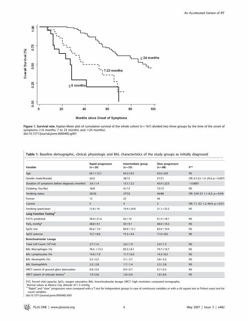
Table 1. Baseline demographic, clinical, physiologic and BAL characteristics of the study groups as initially diagnosed. . . . . . . . . . . . . . . . . . . . . . . . . . . . . . . . . . . . . . . . . . . . . . . . . . . . . . . . . . . . . . . . . . . . . . . . . . . . . . . . . . . . . . . . . . . . . . . . . . . . . . . . . . . . . . . . . . . . . . . . . . . . . . . . . . . . . . . . . . . . . . . . . .
VariableRapid progressors(n = 26)
Intermediate group(n = 53)
Slow progressors(n = 88) P**
Age 64.1612.1 64.368.5 63.668.8 NS
Gender (male/female) 24/2 38/15 57/31 OR: 6.5 (CI: 1.4–29.4; p = 0.007)
Duration of symptoms before diagnosis (months) 3.661.4 13.163.2 45.9622.0 ,0.0001
Clubbing (Yes/No) 18/8 41/12 73/15 NS
Smoking status 20/26 27/53 46/88 OR: 3.04 (CI 1.1–8.3; p = 0.04)
Former 15 23 44
Current 5 4 2 OR: 7.1 (CI: 1.2–40.9; p = 0.01)
Smoking (pack/year) 15.8616 19.4624.8 21.1633.2 NS
Lung Function Testing¥
FVC% predicted 58.4621.6 62619 61.5618.1 NS
PaO2 mmHg* 48.869.1 5069.1 48.0610.3 NS
SpO2 rest 85.667.6 84.9612.1 82.0610.4 NS
SpO2 exercise 73.768.0 75.369.4 71.068.0 NS
Bronchoalveolar Lavage
Total Cell Count (105/ml) 2.761.6 2.661.9 2.461.3 NS
BAL Macrophages (%) 78.4 613.3 83.568.7 79.7610.7 NS
BAL Lymphocytes (%) 14.667.9 11.766.9 14.368.5 NS
BAL Neutrophils (%) 3.566.2 3.163.7 3.864.3 NS
BAL Eosinophils% 3.262.8 1.761.4 2.362.8 NS
HRCT extent of ground glass attenuation 0.860.4 0.960.7 0.760.5 NS
HRCT extent of reticular lesions1 1.960.6 1.860.4 1.860.6 NS
FVC: forced vital capacity; SpO2: oxygen saturation; BAL: bronchoalveolar lavage; HRCT: high resolution computed tomography.*Normal values at Mexico City altitude: 6763 mmHg.**‘‘Rapid’’ and ‘‘slow’’ progressors were compared with a T test for independent groups in case of continuous variables or with a chi square test or Fishers exact test for
count variables.doi:10.1371/journal.pone.0000482.t001....
....
....
....
....
....
....
....
....
....
....
....
....
....
....
....
....
....
....
....
....
....
....
....
....
....
...
Figure 1. Survival rate. Kaplan-Meier plot of cumulative survival of the whole cohort (n = 167) divided into three groups by the time of the onset ofsymptoms (#6 months; 7 to 23 months; and $24 months).doi:10.1371/journal.pone.0000482.g001
An Accelerated Variant of IPF
PLoS ONE | www.plosone.org 4 May 2007 | Issue 5 | e482
increased in the ‘‘rapid’’ progressors group although it did not
reach statistical significance (HR = 1.5; CI 0.81–2.87; p = 0.18).
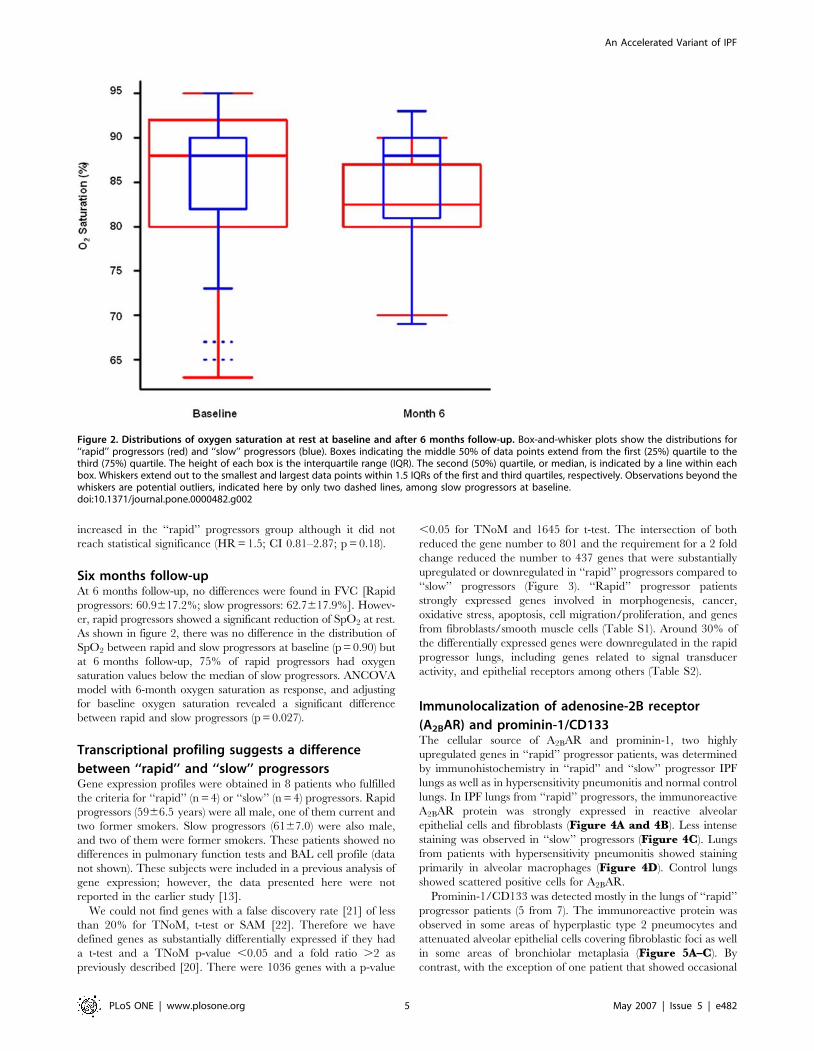
Six months follow-upAt 6 months follow-up, no differences were found in FVC [Rapid
progressors: 60.9617.2%; slow progressors: 62.7617.9%]. Howev-
er, rapid progressors showed a significant reduction of SpO2 at rest.
As shown in figure 2, there was no difference in the distribution of
SpO2 between rapid and slow progressors at baseline (p = 0.90) but
at 6 months follow-up, 75% of rapid progressors had oxygen
saturation values below the median of slow progressors. ANCOVA
model with 6-month oxygen saturation as response, and adjusting
for baseline oxygen saturation revealed a significant difference
between rapid and slow progressors (p = 0.027).
Transcriptional profiling suggests a difference
between ‘‘rapid’’ and ‘‘slow’’ progressorsGene expression profiles were obtained in 8 patients who fulfilled
the criteria for ‘‘rapid’’ (n = 4) or ‘‘slow’’ (n = 4) progressors. Rapid
progressors (5966.5 years) were all male, one of them current and
two former smokers. Slow progressors (6167.0) were also male,
and two of them were former smokers. These patients showed no
differences in pulmonary function tests and BAL cell profile (data
not shown). These subjects were included in a previous analysis of
gene expression; however, the data presented here were not
reported in the earlier study [13].
We could not find genes with a false discovery rate [21] of less
than 20% for TNoM, t-test or SAM [22]. Therefore we have
defined genes as substantially differentially expressed if they had
a t-test and a TNoM p-value ,0.05 and a fold ratio .2 as
previously described [20]. There were 1036 genes with a p-value
,0.05 for TNoM and 1645 for t-test. The intersection of both
reduced the gene number to 801 and the requirement for a 2 fold
change reduced the number to 437 genes that were substantially
upregulated or downregulated in ‘‘rapid’’ progressors compared to
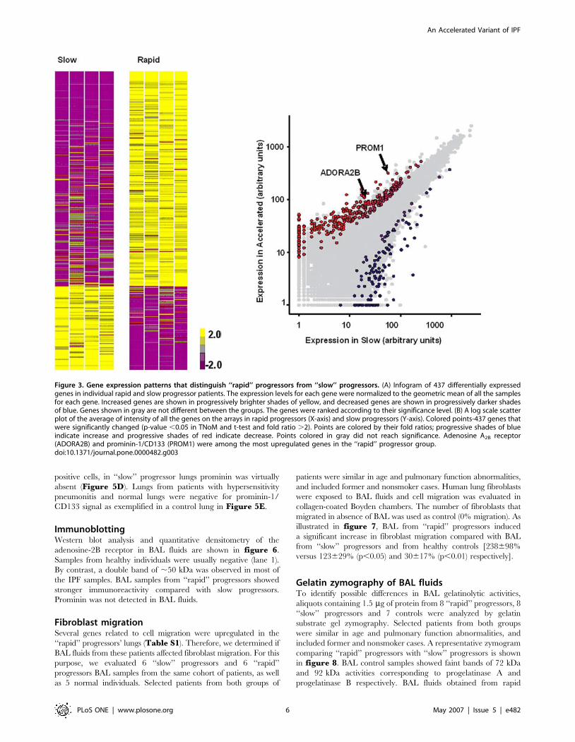
‘‘slow’’ progressors (Figure 3). ‘‘Rapid’’ progressor patients
strongly expressed genes involved in morphogenesis, cancer,
oxidative stress, apoptosis, cell migration/proliferation, and genes
from fibroblasts/smooth muscle cells (Table S1). Around 30% of
the differentially expressed genes were downregulated in the rapid
progressor lungs, including genes related to signal transducer
activity, and epithelial receptors among others (Table S2).
Immunolocalization of adenosine-2B receptor
(A2BAR) and prominin-1/CD133The cellular source of A2BAR and prominin-1, two highly
upregulated genes in ‘‘rapid’’ progressor patients, was determined
by immunohistochemistry in ‘‘rapid’’ and ‘‘slow’’ progressor IPF
lungs as well as in hypersensitivity pneumonitis and normal control
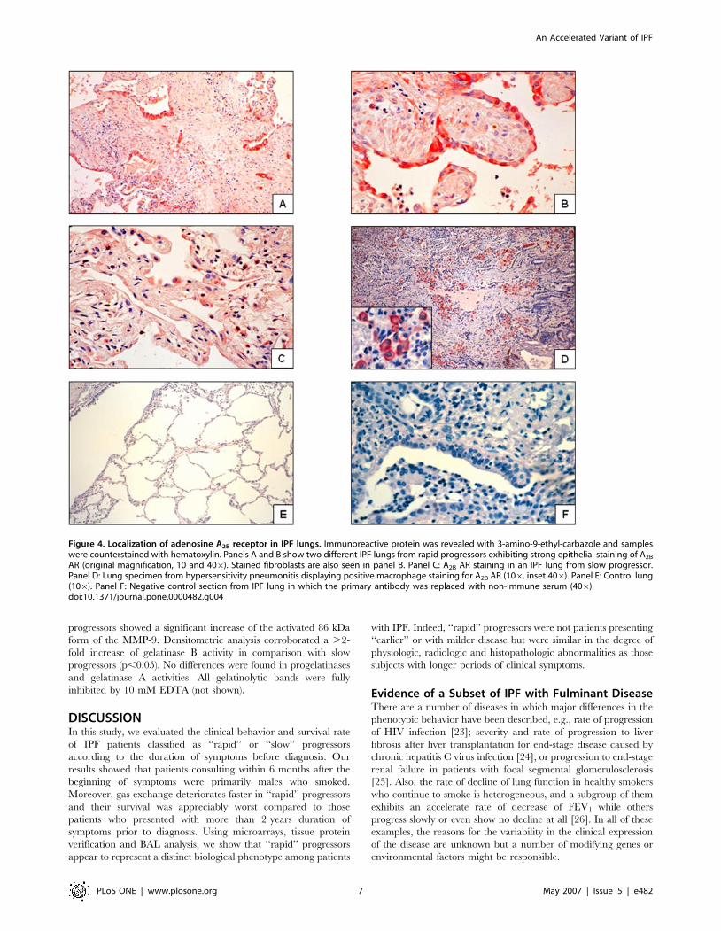
lungs. In IPF lungs from ‘‘rapid’’ progressors, the immunoreactive
A2BAR protein was strongly expressed in reactive alveolar
epithelial cells and fibroblasts (Figure 4A and 4B). Less intense
staining was observed in ‘‘slow’’ progressors (Figure 4C). Lungs
from patients with hypersensitivity pneumonitis showed staining
primarily in alveolar macrophages (Figure 4D). Control lungs
showed scattered positive cells for A2BAR.
Prominin-1/CD133 was detected mostly in the lungs of ‘‘rapid’’
progressor patients (5 from 7). The immunoreactive protein was
observed in some areas of hyperplastic type 2 pneumocytes and
attenuated alveolar epithelial cells covering fibroblastic foci as well
in some areas of bronchiolar metaplasia (Figure 5A–C). By
contrast, with the exception of one patient that showed occasional
Figure 2. Distributions of oxygen saturation at rest at baseline and after 6 months follow-up. Box-and-whisker plots show the distributions for‘‘rapid’’ progressors (red) and ‘‘slow’’ progressors (blue). Boxes indicating the middle 50% of data points extend from the first (25%) quartile to thethird (75%) quartile. The height of each box is the interquartile range (IQR). The second (50%) quartile, or median, is indicated by a line within eachbox. Whiskers extend out to the smallest and largest data points within 1.5 IQRs of the first and third quartiles, respectively. Observations beyond thewhiskers are potential outliers, indicated here by only two dashed lines, among slow progressors at baseline.doi:10.1371/journal.pone.0000482.g002
An Accelerated Variant of IPF
PLoS ONE | www.plosone.org 5 May 2007 | Issue 5 | e482
positive cells, in ‘‘slow’’ progressor lungs prominin was virtually
absent (Figure 5D). Lungs from patients with hypersensitivity
pneumonitis and normal lungs were negative for prominin-1/
CD133 signal as exemplified in a control lung in Figure 5E.
ImmunoblottingWestern blot analysis and quantitative densitometry of the
adenosine-2B receptor in BAL fluids are shown in figure 6.
Samples from healthy individuals were usually negative (lane 1).
By contrast, a double band of ,50 kDa was observed in most of
the IPF samples. BAL samples from ‘‘rapid’’ progressors showed
stronger immunoreactivity compared with slow progressors.
Prominin was not detected in BAL fluids.
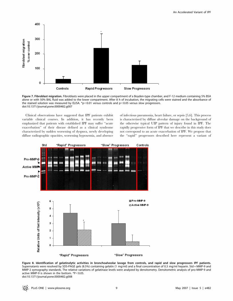
Fibroblast migrationSeveral genes related to cell migration were upregulated in the
‘‘rapid’’ progressors’ lungs (Table S1). Therefore, we determined if
BAL fluids from these patients affected fibroblast migration. For this
purpose, we evaluated 6 ‘‘slow’’ progressors and 6 ‘‘rapid’’
progressors BAL samples from the same cohort of patients, as well
as 5 normal individuals. Selected patients from both groups of
patients were similar in age and pulmonary function abnormalities,
and included former and nonsmoker cases. Human lung fibroblasts
were exposed to BAL fluids and cell migration was evaluated in
collagen-coated Boyden chambers. The number of fibroblasts that
migrated in absence of BAL was used as control (0% migration). As
illustrated in figure 7, BAL from ‘‘rapid’’ progressors induced
a significant increase in fibroblast migration compared with BAL
from ‘‘slow’’ progressors and from healthy controls [238698%
versus 123629% (p,0.05) and 30617% (p,0.01) respectively].
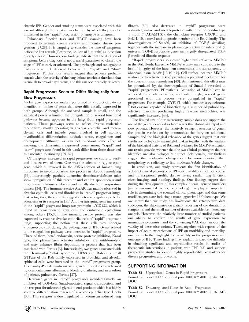
Gelatin zymography of BAL fluidsTo identify possible differences in BAL gelatinolytic activities,
aliquots containing 1.5 mg of protein from 8 ‘‘rapid’’ progressors, 8
‘‘slow’’ progressors and 7 controls were analyzed by gelatin
substrate gel zymography. Selected patients from both groups
were similar in age and pulmonary function abnormalities, and
included former and nonsmoker cases. A representative zymogram
comparing ‘‘rapid’’ progressors with ‘‘slow’’ progressors is shown
in figure 8. BAL control samples showed faint bands of 72 kDa
and 92 kDa activities corresponding to progelatinase A and
progelatinase B respectively. BAL fluids obtained from rapid
Figure 3. Gene expression patterns that distinguish ‘‘rapid’’ progressors from ‘‘slow’’ progressors. (A) Infogram of 437 differentially expressedgenes in individual rapid and slow progressor patients. The expression levels for each gene were normalized to the geometric mean of all the samplesfor each gene. Increased genes are shown in progressively brighter shades of yellow, and decreased genes are shown in progressively darker shadesof blue. Genes shown in gray are not different between the groups. The genes were ranked according to their significance level. (B) A log scale scatterplot of the average of intensity of all the genes on the arrays in rapid progressors (X-axis) and slow progressors (Y-axis). Colored points-437 genes thatwere significantly changed (p-value ,0.05 in TNoM and t-test and fold ratio .2). Points are colored by their fold ratios; progressive shades of blueindicate increase and progressive shades of red indicate decrease. Points colored in gray did not reach significance. Adenosine A2B receptor(ADORA2B) and prominin-1/CD133 (PROM1) were among the most upregulated genes in the ‘‘rapid’’ progressor group.doi:10.1371/journal.pone.0000482.g003
An Accelerated Variant of IPF
PLoS ONE | www.plosone.org 6 May 2007 | Issue 5 | e482
progressors showed a significant increase of the activated 86 kDa
form of the MMP-9. Densitometric analysis corroborated a .2-
fold increase of gelatinase B activity in comparison with slow
progressors (p,0.05). No differences were found in progelatinases
and gelatinase A activities. All gelatinolytic bands were fully
inhibited by 10 mM EDTA (not shown).
DISCUSSIONIn this study, we evaluated the clinical behavior and survival rate
of IPF patients classified as ‘‘rapid’’ or ‘‘slow’’ progressors
according to the duration of symptoms before diagnosis. Our
results showed that patients consulting within 6 months after the
beginning of symptoms were primarily males who smoked.
Moreover, gas exchange deteriorates faster in ‘‘rapid’’ progressors
and their survival was appreciably worst compared to those
patients who presented with more than 2 years duration of
symptoms prior to diagnosis. Using microarrays, tissue protein
verification and BAL analysis, we show that ‘‘rapid’’ progressors
appear to represent a distinct biological phenotype among patients
with IPF. Indeed, ‘‘rapid’’ progressors were not patients presenting
‘‘earlier’’ or with milder disease but were similar in the degree of
physiologic, radiologic and histopathologic abnormalities as those
subjects with longer periods of clinical symptoms.
Evidence of a Subset of IPF with Fulminant DiseaseThere are a number of diseases in which major differences in the
phenotypic behavior have been described, e.g., rate of progression
of HIV infection [23]; severity and rate of progression to liver
fibrosis after liver transplantation for end-stage disease caused by
chronic hepatitis C virus infection [24]; or progression to end-stage
renal failure in patients with focal segmental glomerulosclerosis
[25]. Also, the rate of decline of lung function in healthy smokers
who continue to smoke is heterogeneous, and a subgroup of them
exhibits an accelerate rate of decrease of FEV1 while others
progress slowly or even show no decline at all [26]. In all of these
examples, the reasons for the variability in the clinical expression
of the disease are unknown but a number of modifying genes or
environmental factors might be responsible.
Figure 4. Localization of adenosine A2B receptor in IPF lungs. Immunoreactive protein was revealed with 3-amino-9-ethyl-carbazole and sampleswere counterstained with hematoxylin. Panels A and B show two different IPF lungs from rapid progressors exhibiting strong epithelial staining of A2B
AR (original magnification, 10 and 406). Stained fibroblasts are also seen in panel B. Panel C: A2B AR staining in an IPF lung from slow progressor.Panel D: Lung specimen from hypersensitivity pneumonitis displaying positive macrophage staining for A2B AR (106, inset 406). Panel E: Control lung(106). Panel F: Negative control section from IPF lung in which the primary antibody was replaced with non-immune serum (406).doi:10.1371/journal.pone.0000482.g004
An Accelerated Variant of IPF
PLoS ONE | www.plosone.org 7 May 2007 | Issue 5 | e482
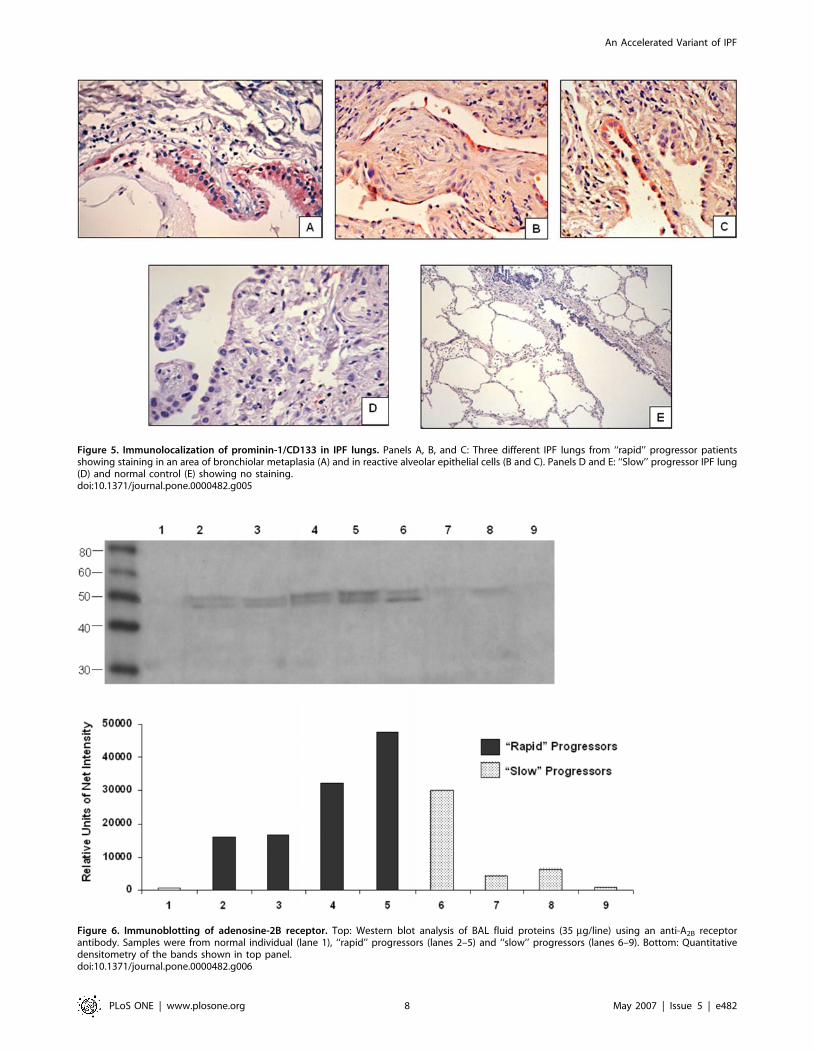
Figure 5. Immunolocalization of prominin-1/CD133 in IPF lungs. Panels A, B, and C: Three different IPF lungs from ‘‘rapid’’ progressor patientsshowing staining in an area of bronchiolar metaplasia (A) and in reactive alveolar epithelial cells (B and C). Panels D and E: ‘‘Slow’’ progressor IPF lung(D) and normal control (E) showing no staining.doi:10.1371/journal.pone.0000482.g005
Figure 6. Immunoblotting of adenosine-2B receptor. Top: Western blot analysis of BAL fluid proteins (35 mg/line) using an anti-A2B receptorantibody. Samples were from normal individual (lane 1), ‘‘rapid’’ progressors (lanes 2–5) and ‘‘slow’’ progressors (lanes 6–9). Bottom: Quantitativedensitometry of the bands shown in top panel.doi:10.1371/journal.pone.0000482.g006
An Accelerated Variant of IPF
PLoS ONE | www.plosone.org 8 May 2007 | Issue 5 | e482
Clinical observations have suggested that IPF patients exhibit
variable clinical courses. In addition, it has recently been
emphasized that patients with established IPF may suffer ‘‘acute
exacerbation’’ of their disease defined as a clinical syndrome
characterized by sudden worsening of dyspnea, newly developing
diffuse radiographic opacities, worsening hypoxemia, and absence
of infectious pneumonia, heart failure, or sepsis [5,6]. This process
is characterized by diffuse alveolar damage on the background of
the otherwise typical UIP pattern of injury found in IPF. The
rapidly progressive form of IPF that we describe in this study does
not correspond to an acute exacerbation of IPF. We propose that
the ‘‘rapid’’ progressors described here represent a variant of
Figure 7. Fibroblast migration. Fibroblasts were placed in the upper compartment of a Boyden-type chamber, and F-12 medium containing 5% BSAalone or with 50% BAL fluid was added to the lower compartment. After 8 h of incubation, the migrating cells were stained and the absorbance ofthe stained solution was measured by ELISA. *p,0.01 versus controls and p,0.05 versus slow progressors.doi:10.1371/journal.pone.0000482.g007
Figure 8. Identification of gelatinolytic activities in bronchoalveolar lavage from controls, and rapid and slow progressors IPF patients.Supernatants were resolved by SDS-PAGE gels (8.5%) containing gelatin (1 mg/ml) and a final concentration of 0.3 mg/ml heparin. Std = MMP-9 andMMP-2 zymography standards. The relative variations of gelatinase levels were analyzed by densitometry. Densitometric analysis of pro-MMP-9 andactive MMP-9 is shown in the bottom. *P,0.05.doi:10.1371/journal.pone.0000482.g008
An Accelerated Variant of IPF
PLoS ONE | www.plosone.org 9 May 2007 | Issue 5 | e482
chronic IPF. Gender and smoking status were associated with this
variant although the putative mechanism by which they may be
implicated in the ‘‘rapid’’ progression phenotype is unknown.
Pulmonary function tests and HRCT scanning have been
proposed to estimate disease severity and monitor disease pro-
gression [27,28]. It is tempting to consider the time of symptoms
before the first consult (if extreme, i.e., less of 6 months) as indication
of early disease. However, our findings indicate that the duration of
symptoms before diagnosis is not a useful parameter to classify the
stage of IPF as early or advanced. The physiologic and radiographic
features were not different between the ‘‘rapid’’ and ‘‘slow’’
progressors. Further, our results suggest that patients probably
consult when the severity of the lung lesions reaches a threshold that
is enough to provoke symptoms, and this can occur rapidly or slowly.
Rapid Progressors Seem to Differ Biologically from
Slow ProgressorsGlobal gene expression analysis performed in a subset of patients
identified a number of genes that were differentially expressed in
both groups. Although the analyzed sample is small, and the
statistical power is limited, the upregulation of several functional
pathways became apparent in the lungs from rapid progressor
patients. These pathways seem to reflect diverse molecular
mechanisms mostly operating in alveolar epithelial and mesen-
chymal cells and include genes involved in cell motility,
myofibroblast differentiation, oxidative stress, coagulation and
development. Although we can not ruled-out some effect of
smoking, the differentially expressed genes among ‘‘rapid’’ and
‘‘slow’’ progressors found in this work differ from those described
as associated to smoking [29–32].
Of the genes increased in rapid progressors we chose to verify
and localize two of them. One was the adenosine A2B receptor
gene, which is involved in the differentiation of human lung
fibroblasts to myofibroblasts-a key process in fibrotic remodeling
[33]. Interestingly, partially adenosine deaminase-deficient mice
show upregulation of this receptor and exhibit spontaneous and
progressive pulmonary fibrosis and usually die from respiratory
distress [34]. The immunoreactive A2BAR was mainly observed in
alveolar epithelial cells and fibroblasts in ‘‘rapid’’ progressor lungs.
Taken together, these results suggest a potential regulatory role for
adenosine or its receptor in IPF. Another intriguing gene increased
in the ‘‘rapid’’ progressor lungs was prominin-1/CD133, which is
found in hematopoietic stem cells and embryonic epithelium
among others [35,36]. The immunoreactive protein was also
expressed by reactive alveolar epithelial cells of ‘‘rapid’’ progressor
lungs, supporting the notion that these cells may undergo
a phenotypic shift during the pathogenesis of IPF. Genes related
to the coagulation pathway were increased in ‘‘rapid’’ progressors.
Three of them, beta3-endonexin, serine protease inhibitor, Kazal
type, and plasminogen activator inhibitor-1 are antifibrinolytic
and may enhance fibrin deposition, a process that has been
associated with fibrosis [3]. Interestingly, two genes associated with
the Hermansky-Pudlak syndrome, HPS3 and Rab38, a small
GTPase of the Rab family expressed in bronchial and alveolar
epithelial cells, were increased in the ‘‘rapid’’ progressors group.
Hermansky-Pudlak syndrome is a genetic disorder characterized
by oculocutaneous albinism, a bleeding diathesis, and in a subset
of patients, pulmonary fibrosis [37].
Decreased genes in ‘‘rapid’’ progressors included Smad6, an
inhibitor of TGF-beta Smad-mediated signal transduction, and
the receptor for advanced glycation end-products which is a highly
selective differentiation marker of alveolar epithelial type I cells
[38]. This receptor is downregulated in bleomycin induced lung
fibrosis [39]. Also decreased in ‘‘rapid’’ progressors were
a disintegrin-like and metalloprotease with thrombospondin type
1 motif, 7 (ADAMTS7), the chemokine receptor CXCR6, and
Bcl2-L-10, a novel anti-apoptotic member of the Bcl-2 family. The
downregulation of Smad6, an inhibitor of TGF-b signaling,
together with the increase in plasminogen activator inhibitor-1 (a
universal TGF-b responsive gene) may signify disregulated TGF-
b-mediated fibrotic response.
‘‘Rapid’’ progressors also showed higher levels of active MMP-9
in the BAL fluids. Excessive MMP-9 activity may contribute to the
loss of integrity of the basement membranes and subsequently to
abnormal tissue repair [13,40–42]. Cell surface-localized MMP-9
is also able to activate TGF-b providing a potential mechanism for
the aberrant tissue remodeling [43]. As mentioned, this effect may
be potentiated by the downregulation of Smad 6 revealed in
‘‘rapid’’ progressors IPF patients. Activation of MMP-9 can be
achieved by oxidative stress, and interestingly, several genes
associated with this process were upregulated in ‘‘rapid’’
progressors. For example, CYP2F1, which encodes a cytochrome
P450 enzyme capable of bioactivating a number of pulmonary-
selective toxicants producing highly reactive metabolites, was
significantly increased [44].
The limited size of our microarray sample does not support the
use of the genes identified as biomarkers that distinguish rapid and
slow patients. However, the relatively stringent selection of genes,
the protein verification by immunohistochemistry on additional
samples and the biological relevance of the genes suggest that our
results are biologically meaningful. Taken together with the analysis
of the biological activity of BAL and evidence for MMP-9 activation
our results provide evidence that the two clinical phenotypes that we
identified are also biologically distinct. Additionally, our findings
suggest that molecular changes can be more sensitive than
morphology or radiology to find moderate/subtle changes.
In conclusion, our study represents the first identification of
a distinct clinical phenotype of IPF–one that differs in clinical course
and transcriptional profile, despite having similar lung function,
chest imaging, and histology findings. Our findings suggest that
during the development of this complex disease, genetic modifiers
(and environmental factors, i.e. smoking) may play an important
role in determining the eventual clinical phenotype, and that some
modifiers genes are inducing a more aggressive IPF phenotype. We
are aware that our study has limitations: the retrospective data
collection, the dependence on patient reporting of the duration of
symptoms, and the small number of tissues available for microarray
analysis. However, the relatively large number of studied patients,
our ability to confirm the results of gene expression by
immunohistochemistry and the convincing BAL data, support the
validity of these observations. Taken together with reports of the
impact of acute exacerbations of IPF on morbidity and mortality,
our results further highlight the variability in the progression and
outcome of IPF. These findings may explain, in part, the difficulty
in obtaining significant and reproducible results in studies of
therapeutic interventions in patients with IPF [45] and support
prospective studies to identify highly reproducible biomarkers for
disease progression and outcome.
SUPPORTING INFORMATION
Table S1 Upregulated Genes in Rapid Progressors
Found at: doi:10.1371/journal.pone.0000482.s001 (0.44 MB
DOC)
Table S2 Downregulated Genes in Rapid Progressors
Found at: doi:10.1371/journal.pone.0000482.s002 (0.16 MB
DOC)
An Accelerated Variant of IPF
PLoS ONE | www.plosone.org 10 May 2007 | Issue 5 | e482
ACKNOWLEDGMENTS
Author Contributions
Conceived and designed the experiments: NK TK MS GC AE MN.
Performed the experiments: AP JC CB MG. Analyzed the data: TR TK
MS GC AE MM RP MN. Wrote the paper: NK AP TK JD MS. Other:
Patient enrollment: AE MM MN GC. HRCT evaluation and quantifica-
tion of the lesions: MM. Histological evaluation: MG. Statistical analysis:
RP TR.
REFERENCES1. Gross TJ, Hunninghake GW (2001) Idiopathic pulmonary fibrosis. N Engl J Med
345: 517–525.
2. Collard HR, Ryu JH, Douglas WW, Schwarz MI, Curran-Everett D, et al.
(2004) Combined corticosteroid and cyclophosphamide therapy does not alter
survival in idiopathic pulmonary fibrosis. Chest 125: 2169–2174.
3. Selman M, King TE, Pardo A (2001) Idiopathic pulmonary fibrosis: prevailing
and evolving hypotheses about its pathogenesis and implications for therapy.
Ann Intern Med 134: 136–151.
4. McCormack FX, King TE Jr, Bucher BL, Nielsen L, Mason RJ (1995)
Surfactant protein A predicts survival in idiopathic pulmonary fibrosis.
Am J Respir Crit Care Med 152: 751–759.
5. Martinez FJ, Safrin S, Weycker D, Starko KM, Bradford WZ, et al. (2005) The
clinical course of patients with idiopathic pulmonary fibrosis. Ann Intern Med
142: 963–967.
6. Ambrosini V, Cancellieri A, Chilosi M, Zompatori M, Trisolini R, et al. (2003)
Acute exacerbation of idiopathic pulmonary fibrosis: report of a series. Eur
Respir J 22: 821–826.
7. American Thoracic Society (2000) Idiopathic pulmonary fibrosis: diagnosis and
treatment. International consensus statement. American Thoracic Society (ATS)
and the European Respiratory Society (ERS) Am J Respir Crit Care Med 161:646–664.
8. King TE Jr, Tooze JA, Schwarz MI, Brown KR, Cherniack RM (2001)
Predicting survival in idiopathic pulmonary fibrosis: scoring system and survivalmodel. Am J Respir Crit Care Med 164: 1171–1181.
9. Selman M, Carrillo G, Salas J, Padilla RP, Perez-Chavira R, et al. (1998)
Colchicine, D-penicillamine, and prednisone in the treatment of idiopathicpulmonary fibrosis: a controlled clinical trial. Chest 114: 507–512.
10. Kazerooni EA, Martinez FJ, Flint A, Jamadar DA, Gross BH, et al. (1997) Thin-
section CT obtained at 10-mm increments versus limited three-level thin-sectionCT for idiopathic pulmonary fibrosis: correlation with pathologic scoring. AJR
Am J Roentgenol 169: 977–983.
11. Pardo A, Gibson K, Cisneros J, Richards TJ, Yang Y, et al. (2005) Upregulationand profibrotic role of osteopontin in human idiopathic pulmonary fibrosis.
PLoS Med 2: e251.
12. Selman M, Ruiz V, Cabrera S, Segura L, Ramirez R, et al. (2000) TIMP 1, 2, 3,and 4 in idiopathic pulmonary fibrosis: a prevailing nondegradative lung
microenvironment? Am J Physiol 279: L562–574.
13. Selman M, Pardo A, Barrera L, Estrada A, Watson SR, et al. (2006) Geneexpression profiles distinguish idiopathic pulmonary fibrosis from hypersensitiv-
ity pneumonitis. Am J Respir Crit Care Med 173: 188–198.
14. Perez-Padilla R, Salas J, Chapela R, Sanchez M, Carrillo G, et al. (1993)Mortality in Mexican patients with chronic pigeon breeder’s lung compared with
those with usual interstitial pneumonia. Am Rev Respir Dis 148: 49–53.
15. Hyde DM, King TE Jr, McDermott T, Waldron JA Jr, Colby TV, et al. (1992)Idiopathic pulmonary fibrosis. Quantitative assessment of lung pathology.
Comparison of a semiquantitative and a morphometric histopathologic scoringsystem. Am Rev Respir Dis 146: 1042–1047.
16. Cisneros-Lira J, Gaxiola M, Ramos C, Selman M, Pardo A (2003) Cigarette
smoke exposure potentiates bleomycin-induced lung fibrosis in guinea pigs.Am J Physiol 285: L949–956.
17. Kaminski N, Friedman N (2002) Practical approaches to analyzing results of
microarray experiments. Am J Respir Cell Mol Biol 27: 125–132.
18. Segal E, Friedman N, Kaminski N, Regev A, Koller D (2005) From signatures tomodels: understanding cancer using microarrays. Nat Genet 37 Suppl: S38–45.
19. Ben-Dor A, Bruhn L, Friedman N, Nachman I, Schummer M, et al. (2000)
Tissue classification with gene expression profiles. J Comput Biol 7: 559–83.
20. Radom-Aizik S, Hayek S, Shahar I, Rechavi G, Kaminski N, et al. (2005) Effects
of aerobic training on gene expression in skeletal muscle of elderly men. Med Sci
Sports Exerc 37: 1680–1696.
21. Benjamini Y, Hochberg Y (1995) Controlling the false discovery rate: a practical
and powerful approach to multiple testing. J Royal Stat Soc B 57: 289–300.
22. Tusher VG, Tibshirani R, Chu G (2001) Significance analysis of microarraysapplied to the ionizing radiation response. Proc Natl Acad Sci U S A. 98:
5116–5121.
23. Hogan CM, Hammer SM (2001) Host determinants in HIV infection anddisease. Part 1: cellular and humoral immune responses. Ann Intern Med 134:
761–776.
24. Marshall A, Rushbrook S, Morris LS, Scott IS, Vowler SL, et al. (2005)
Hepatocyte expression of minichromosome maintenance protein-2 predictsfibrosis progression after transplantation for chronic hepatitis C virus: A pilot
study. Liver Transpl 11: 427–433.25. Bolton WK, Abdel-Rahman E (2001) Pathogenesis of focal glomerulosclerosis.
Nephron 88: 6–13.26. He JQ, Ruan J, Connett JE, Anthonisen NR, Pare PD, et al. (2002) Antioxidant
gene polymorphisms and susceptibility to a rapid decline in lung function in
smokers. Am J Respir Crit Care Med 166: 323–328.27. Egan JJ, Martinez FJ, Wells AU, Williams T (2005) Lung function estimates in
idiopathic pulmonary fibrosis: the potential for a simple classification. Thorax60: 270–273.
28. Gay SE, Kazerooni EA, Toews GB, Lynch JP 3rd, Gross BH, et al. (1998)
Idiopathic pulmonary fibrosis: predicting response to therapy and survival.Am J Respir Crit Care Med 157: 1063–1072.
29. Heguy A, O’Connor TP, Luettich K, Worgall S, Cieciuch A, et al. (2006) Geneexpression profiling of human alveolar macrophages of phenotypically normal
smokers and nonsmokers reveals a previously unrecognized subset of genesmodulated by cigarette smoking. J Mol Med 84: 318–328.
30. Woodruff PG, Koth LL, Yang YH, Rodriguez MW, Favoreto S, et al. (2005) A
distinctive alveolar macrophage activation state induced by cigarette smoking.Am J Respir Crit Care Med 172: 1383–1392.
31. Ning W, Li CJ, Kaminski N, Feghali-Bostwick CA, Alber SM, et al. (2004)Comprehensive gene expression profiles reveal pathways related to the
pathogenesis of chronic obstructive pulmonary disease. Proc Natl Acad Sci
U S A. 101: 14895–14900.32. Harvey BG, Heguy A, Leopold PL, Carolan BJ, Ferris B, et al. (2007)
Modification of gene expression of the small airway epithelium in response tocigarette smoking. J Mol Med 85: 39–53.
33. Zhong H, Belardinelli L, Maa T, Zeng D (2005) Synergy between A2Badenosine receptors and hypoxia in activating human lung fibroblasts.
Am J Respir Cell Mol Biol 32: 2–8.
34. Chunn JL, Mohsenin A, Young HW, Lee CG, Elias JA, et al. (2006) Partiallyadenosine deaminase-deficient mice develop pulmonary fibrosis in association
with adenosine elevations. Am J Physiol Lung Cell Mol Physiol 290: L579–587.35. Kania G, Corbeil D, Fuchs J, Tarasov KV, Blyszczuk P, et al. (2005) Somatic
stem cell marker prominin-1/CD133 is expressed in embryonic stem cell-
derived progenitors. Stem Cells 23: 791–804.36. Shmelkov SV, St Clair R, Lyden D, Rafii S (2005) AC133/CD133/Prominin-1.
Int J Biochem Cell Biol 37: 715–719.37. Gahl WA, Brantly M, Troendle J, Avila NA, Padua A, et al. (2002) Effect of
pirfenidone on the pulmonary fibrosis of Hermansky-Pudlak syndrome. Mol
Genet Metab 76: 234–242.38. Demling N, Ehrhardt C, Kasper M, Laue M, Knels L, et al. (2006) Promotion of
cell adherence and spreading: a novel function of RAGE, the highly selectivedifferentiation marker of human alveolar epithelial type I cells. Cell Tissue Res
323: 475–488.39. Hanford LE, Fattman CL, Shaefer LM, Enghild JJ, Valnickova Z, et al. (2003)
Regulation of receptor for advanced glycation end products during bleomycin-
induced lung injury. Am J Respir Cell Mol Biol 29 (3 Suppl): S77–81.40. Pardo A, Ruiz V, Arreola JL, Ramirez R, Cisneros-Lira J, et al. (2003)
Bleomycin-induced pulmonary fibrosis is attenuated in gamma-glutamyltranspeptidase-deficient mice. Am J Respir Crit Care Med 167: 925–932.
41. Fukuda Y, Ishizaki M, Kudoh S, Kitaichi M, Yamanaka N (1998) Localization
of matrix metalloproteinases-1, -2, and -9 and tissue inhibitor of metallopro-teinase-2 in interstitial lung diseases. Lab Invest 78: 687–698.
42. Atkinson JJ, Senior RM (2003) Matrix metalloproteinase-9 in lung remodeling.Am J Respir Cell Mol Biol 28: 12–24.
43. Yu Q, Stamenkovic I (2000) Cell surface-localized matrix metalloproteinase-9proteolytically activates TGF-beta and promotes tumor invasion and angiogen-
esis. Genes Dev 14: 163–176.
44. Carr BA, Wan J, Hines RN, Yost GS (2003) Characterization of the human lungCYP2F1 gene and identification of a novel lung-specific binding motif. J Biol
Chem 278: 15473–15483.45. Raghu G, Brown KK, Bradford WZ, Starko K, Noble PW, et al. (2004)
Idiopathic Pulmonary Fibrosis Study Group. A placebo-controlled trial of
interferon gamma-1b in patients with idiopathic pulmonary fibrosis. N Engl JMed 350: 125–133.
An Accelerated Variant of IPF
PLoS ONE | www.plosone.org 11 May 2007 | Issue 5 | e482