Cancer-retina antigens as potential paraneoplastic antigens in melanoma-associated retinopathy
Upload
independentCategory
view
0download
0
10.1128/IAI.71.2.597-604.2003.
2003, 71(2):597. DOI:Infect. Immun. Chetan E. ChitnisSudhanshu S. Pati, S. K. Sharma, Bhabani S. Das and Rana Chattopadhyay, Amit Sharma, Vinod K. Srivastava, AntigensAntibodies against Variant SurfaceBoth Variant-Specific and Cross-Reactive
Infection ElicitsPlasmodium falciparum
http://iai.asm.org/content/71/2/597Updated information and services can be found at:
These include:
REFERENCEShttp://iai.asm.org/content/71/2/597#ref-list-1at:
This article cites 26 articles, 10 of which can be accessed free
CONTENT ALERTS more»articles cite this article),
Receive: RSS Feeds, eTOCs, free email alerts (when new
http://journals.asm.org/site/misc/reprints.xhtmlInformation about commercial reprint orders: http://journals.asm.org/site/subscriptions/To subscribe to to another ASM Journal go to:
on May 21, 2014 by guest
http://iai.asm.org/
Dow
nloaded from
on May 21, 2014 by guest
http://iai.asm.org/
Dow
nloaded from

INFECTION AND IMMUNITY, Feb. 2003, p. 597–604 Vol. 71, No. 20019-9567/03/$08.00�0 DOI: 10.1128/IAI.71.2.597–604.2003Copyright © 2003, American Society for Microbiology. All Rights Reserved.
Plasmodium falciparum Infection Elicits Both Variant-Specific andCross-Reactive Antibodies against Variant Surface Antigens
Rana Chattopadhyay,1† Amit Sharma,1 Vinod K. Srivastava,2 Sudhanshu S. Pati,2S. K. Sharma,3 Bhabani S. Das,2 and Chetan E. Chitnis1*
Malaria Research Group, International Centre for Genetic Engineering and Biotechnology (ICGEB),New Delhi,1 and Department of Biochemistry, Ispat General Hospital (IGH),2 and
Malaria Research Centre Field Station,3 Rourkela, India
Received 2 January 2002/Returned for modification 25 February 2002/Accepted 12 June 2002
Naturally acquired antibodies to Plasmodium falciparum erythrocyte membrane protein-1 (PfEMP-1), thevariant surface antigens expressed on the surface of infected erythrocytes, are thought to play a role inprotection against P. falciparum malaria. Here, we have studied the development of antibodies to PfEMP-1 inadult malaria patients living in Rourkela, India, an area with a low malaria transmission rate, and prevalenceof antibodies to PfEMP-1 in residents of San Dulakudar, India, a village in which P. falciparum malaria ishyperendemic. Convalescent-phase sera from adult malaria patients from Rourkela agglutinate homologous P.falciparum isolates as well as some heterologous isolates, suggesting that they develop partially cross-reactiveantibodies to PfEMP-1 following infection. Adult sera from San Dulakudar agglutinate diverse P. falciparumisolates, suggesting that they have antibodies with wide recognition of diverse PfEMP-1. Mixed-agglutinationassays using pairs of P. falciparum isolates confirm the presence of both variant-specific and partially cross-reactive antibodies in convalescent-phase sera from Rourkela and adult sera from San Dulakudar. Analysis ofPfEMP-1 sequences suggests a molecular basis for the observed cross-reactivity.
Following repeated exposure to Plasmodium falciparum in-fection, individuals residing in areas of malaria endemicityacquire immunity (14). As a result it is primarily nonimmunechildren in areas of endemicity who suffer from severe malariaand are at risk of death from malaria. Adults in areas ofendemicity suffer fewer clinical malaria episodes than do chil-dren and are protected from severe malaria syndromes such ascerebral malaria and severe anemia.
Naturally acquired immunity to malaria is not completelyunderstood. Antibodies directed against variant surface anti-gens referred to as P. falciparum erythrocyte membrane pro-tein-1 (PfEMP-1) that are expressed on the surface of infectederythrocytes are thought to be an important component ofnaturally acquired immunity (3, 13). Some members of thePfEMP-1 family bind to endothelial receptors and mediateadhesion of P. falciparum trophozoites and schizonts to vascu-lar endothelium, a phenomenon referred to as cytoadherence(6). Cytoadherence is implicated in a number of pathologicaloutcomes. Adhesion of P. falciparum-infected erythrocytes inbrain capillaries is implicated in cerebral malaria, and cytoad-herence in the placenta often leads to complications in preg-nancy (11, 17, 24–25).
Agglutination assays with P. falciparum trophozoites andschizonts have been used to study antibody responses toPfEMP-1 (3–4, 7–9, 12–13, 15). A large prospective study con-ducted on the eastern coast of Kenya demonstrated that, when
children suffer clinical malaria episodes, they are likelier to beinfected with P. falciparum isolates against which they lackagglutinating antibodies than with isolates against which theyhave agglutinating antibodies (3). Sera from children in areasof endemicity agglutinate a limited number of P. falciparumisolates, indicating limited recognition of PfEMP-1 variants (4,7, 9, 12). In contrast, immune-adult sera from areas of ende-micity agglutinate a wide range of P. falciparum isolates, sug-gesting that they recognize diverse PfEMP-1 variants (4, 7, 9,12). Do immune adults possess a repertoire of variant-specificantibodies directed against diverse PfEMP-1, or do they havecross-reactive antibodies directed against conserved epitopesshared by diverse PfEMP-1? Initial studies suggested that im-mune adults might have cross-reactive antibodies that aggluti-nate diverse P. falciparum isolates (12). However, later studiessuggested that antibodies directed against PfEMP-1 are pre-dominantly variant specific (8, 15).
In this study, we have used agglutination assays to studynaturally acquired antibodies to PfEMP-1 in two areas withdistinct malaria transmission patterns in Sundergarh District inthe state of Orissa in eastern India. We have studied thedevelopment of antibodies directed against PfEMP-1 in adultmalaria patients residing in the town of Rourkela, which is anarea with a low malaria transmission rate. We have also stud-ied the prevalence of antibodies directed against PfEMP-1 inchildren and adults residing in a village, San Dulakudar, lo-cated in a forest area in Sundergarh District, in which P.falciparum malaria is hyperendemic. Mixed-agglutination as-says using parasite isolate pairs stained with distinguishableDNA intercalating dyes were used to investigate the presenceof cross-reactive antibodies directed against PfEMP-1. Datafrom our field studies and comparative statistical analysis ofPfEMP-1 sequences suggest that both variant-specific as well
* Corresponding author. Mailing address: Malaria Group, Interna-tional Centre for Genetic Engineering and Biotechnology (ICGEB),P.O. Box 10504, Aruna Asaf Ali Marg, New Delhi 110067, India.Phone: 91 11 618 7695. Fax: 91 11 616 2316. E-mail: [email protected].
† Present address: Naval Medical Research Centre, Silver Spring,MD 20910-7500.
597
on May 21, 2014 by guest
http://iai.asm.org/
Dow
nloaded from
as partially cross-reactive antibodies against PfEMP-1 may beelicited during natural infection with P. falciparum.
MATERIALS AND METHODS
Study area. The study was conducted with sera collected from two areas withdistinct malaria transmission patterns in Sundergarh District, Orissa, in easternIndia. Sundergarh District lies between 21°35�N and 22°35�N latitudes and be-tween 83°32�E and 85°22�E longitudes. The area presents ideal ecological con-ditions for malaria transmission with undulating uplands intersected by forestedhills, rocky streams, paddy fields, and springs with a tropical, humid, savanna-type climate and annual rainfall between 160 and 200 cm. The monsoon rainsbegin in mid-June and last till September, the cold season occurs in Decemberand January, and the hot, dry summer extends from April to mid-June. Focalepidemiological variations are observed in malaria transmission patterns overshort geographical distances in Sundergarh District. For example, the townshipof Rourkela (population 150,000), which was constructed for the employees of alocal steel plant, has excellent urban infrastructure with around 20,000 houses ofdifferent types, roads, electricity, safe drinking water, underground sewage sys-tem, effective malaria control programs, and an excellent health care system. Asa result, malaria transmission in Rourkela is extremely low. In contrast, villagessituated in forests with perennial flowing streams surrounded by paddy fields just25 to 30 km outside Rourkela have high malaria endemicity and perennialtransmission. We have collected data over the past 3 years on the epidemiologyof malaria in one such village, San Dulakudar, which has a population of 265.Cross-sectional and longitudinal studies involving periodic mass surveys as wellas active case detection by trained village-based community workers were used tocollect data on malaria prevalence and malaria-related morbidity. A detailedreport describing the epidemiology of malaria in San Dulakudar will be pub-lished separately. Briefly, malaria is hyperendemic in San Dulakudar, with P.falciparum malaria accounting for more than 90% of malaria cases and Plasmo-dium vivax and Plasmodium malariae accounting for the rest. Malaria transmis-sion is perennial, although transmission intensity varies during the year. The hightransmission season extends from October to December following the monsoonrains, with parasite rates as high as 20 to 50% in this period. Parasite rates in thelow transmission season during the hot dry summer months of April and May arebetween 5 and 15%. Other months have intermediate levels of malaria trans-mission. Based on active case detection over a 3-year period, it is estimated thaton average there are 2.1 malaria episodes per person per year in the 1- to 5-yearage group and 0.5 malaria episodes per person per year in adults. These datasuggest that San Dulakudar residents develop natural immunity to malaria fol-lowing repeated exposure.
Parasites and sera. Adult residents of Rourkela reporting to Ispat GeneralHospital (IGH), Rourkela, and diagnosed with nonsevere P. falciparum malariabased on clinical symptoms and detection of parasites in blood smears wereenrolled in the study. Ten milliliters of blood was collected at the time ofadmission prior to initiating antimalarial drug therapy. Five milliliters of bloodwas collected in 10% citrate-phosphate-dextrose, washed, and cryopreserved inaliquots in liquid nitrogen. The remaining 5 ml was used for preparation ofacute-phase sera by standard methods. Five milliliters of blood was collectedfrom the same group of volunteers 3 to 4 weeks after recovery and used forpreparation of convalescent-phase sera. All patients had parasite-free bloodsmears at time of discharge and no malaria symptoms during the period betweendischarge and collection of convalescent-phase sera. All individuals enrolled inthe study at IGH, Rourkela, had lived in Rourkela for more than 10 years. Themajority of the volunteers had no history of malaria in the past 10 years. Oneindividual reported having malaria twice, and three individuals reported havingmalaria once in the past 10 years.
Standard blood group typing procedures were used to determine the bloodgroups of donors. Parasite isolates collected from donors with the O blood group(R1, R13, R15, R28, R30, R35, R39, and R40) were used for agglutination assayswith homologous as well as heterologous sera. Parasite isolates (R4, R7, R9,R11, R16, and R17) collected from donors with other blood groups were onlyused for agglutination assays with homologous sera.
Sera were also collected from asymptomatic adults and children (1 to 3 years)residing in San Dulakudar during periodic mass surveys. One child (DK557) was18 months old. All other children were between 24 and 36 months old. This studyreceived ethical clearance from the Institutional Review Boards of the Interna-tional Centre for Genetic Engineering and Biotechnology (ICGEB), New Delhi,India, and IGH, Rourkela. Individuals were enrolled in the study after obtaininginformed consent. In case of children, informed consent was obtained fromparents.
Agglutination assay. Cryopreserved P. falciparum isolates collected from ma-laria patients reporting to IGH, Rourkela, were thawed and cultured to thetrophozoite-schizont stages in RPMI 1640 medium (Gibco BRL-Life Technol-ogies) containing 10% heat-inactivated fetal calf serum (Gibco BRL-Life Tech-nologies). In each case, parasitemia was �1%. A hematocrit of the culture wasadjusted to �5% using RPMI 1640 containing ethidium bromide (Sigma) at aconcentration of 10 �g/ml. Agglutination assays were performed as describedpreviously (3). Briefly, 2.5 �l of each serum sample was spotted in wells ofU-bottomed 96-well microtiter plates (Costar). Ten microliters of parasite cul-ture in RPMI 1640 with ethidium bromide was added to each well. The micro-titer plate was rotated at 30 rounds per min on a vertical rotator for 60 min atroom temperature. The entire 12.5-�l reaction from each well was placed on aglass slide and covered with a coverslip. Slides were blinded and examinedmicroscopically under visual and UV light. P. falciparum laboratory isolate A4and field isolate R1 were initially used to standardize the assay and ensurereproducibility. Agglutinates were scored in 25 fields at �40 magnification. Blankboxes indicate that no agglutinates containing three or more infected erythro-cytes were seen (Fig. 1 and 2). Hatched boxes indicate that on average five or lessagglutinates containing five or more infected erythrocytes were seen per field(Fig. 1 and 2). Black boxes indicate that on average greater than five agglutinatescontaining five or more infected erythrocytes were seen per field (Fig. 1 and 2).
Mixed-agglutination assay. Mixed-agglutination assays were performed withpairs of P. falciparum isolates stained with distinguishable DNA intercalatingdyes (15). The method used for the mixed-agglutination assay was similar to thatdescribed above for a single isolate except that two isolates were used, onelabeled with ethidium bromide (10 �g/ml) and the other with acridine orange (10�g/ml). Mixed-color agglutinates are defined as agglutinates with at least threeinfected erythrocytes with at least one infected erythrocyte of each color. Blankboxes indicate that no mixed-color agglutinates were seen (Fig. 3 and 4). Thenumber of mixed-color agglutinates containing up to 20 infected erythrocyteswith at least two infected erythrocytes of each color was scored in 25 fields at �40magnification. Hatched boxes indicate that on average five or less mixed-coloragglutinates were seen per field, and black boxes indicate that on average greaterthan five mixed color agglutinates were seen per field (Fig. 3 and 4).
PCR typing of P. falciparum patient isolates. P. falciparum isolates used for theagglutination assays were typed by PCR using two polymorphic blood stageantigens, merozoite surface protein-1 (MSP-1) and merozoite surface protein-2(MSP-2), to determine if they were genotypically distinct and contain single ormultiple genotypes as described earlier (16, 27). PCR products encoding MSP-1block 2 were subtyped by nested PCR using oligonucleotide primers specific forthe K1, MAD20, and R033 subtypes of MSP-1 (21). PCR products encodingblocks 2 and 3 of MSP-2 were subtyped using oligonucleotide primers specific forFC27 and 3D7 subtypes as previously described (21).
Analysis of diversity in DBL� domain sequences of PfEMP-1 among P. falci-parum isolates. The extracellular region of PfEMP-1 contains multiple con-served, cysteine-rich domains that are referred to as Duffy binding-like (DBL)domains because they share homology with the binding domain of P. vivax Duffybinding protein (2, 5, 6, 18–20, 22). DBL domains are classified into five types, �,�, �, , and ε, based on the presence of type-specific conserved sequences (19,20). Genomic DNA isolated from P. falciparum isolates R1, R15 and R35 wasused as template in PCRs with degenerate primers based on conserved se-quences in DBL� domains of var genes as previously described (23). PCRproducts encoding the central region of DBL� domains were cloned in TAcloning vector (Invitrogen) and sequenced using an Automated ABI 310 DNASequencer (Perkin Elmer).
The var sequences identified from P. falciparum isolates R1, R15, and R35 and19 var sequences reported previously from two Kenyan P. falciparum isolates, 3and 19, referred to here as K3 and K19 (23; GenBank accession numbers fromAF221811 to AF221819 and from AF221777 to AF221786), were used for anal-ysis. Pairwise sequence comparison was performed using the SIM program withdefault values available on the server http://www.expasy.ch, which determines thepercent identity between two amino acid sequences. A matrix-based analysis wasperformed using SIM in which each sequence from a parasite isolate was com-pared to all other sequences from the same isolate or from a different isolate.This resulted in a matrix containing the percent sequence identity of eachsequence relative to every other sequence. Mean, range, and standard deviationvalues for the sequence comparison data were calculated and are shown in Table2. A multiple-sequence alignment was carried out with var sequences from R1,R15, K3, and K19 (total number of sequences 35) using the programCLUSATLW.
Nucleotide sequence accession number. Seven unique var gene sequences wereidentified from R1, and nine unique var gene sequences were identified from R15
598 CHATTOPADHYAY ET AL. INFECT. IMMUN.
on May 21, 2014 by guest
http://iai.asm.org/
Dow
nloaded from
and R35. These nucleotide sequences are available in the GenBank database andhave accession numbers from AF404238 to AF404262.
RESULTS
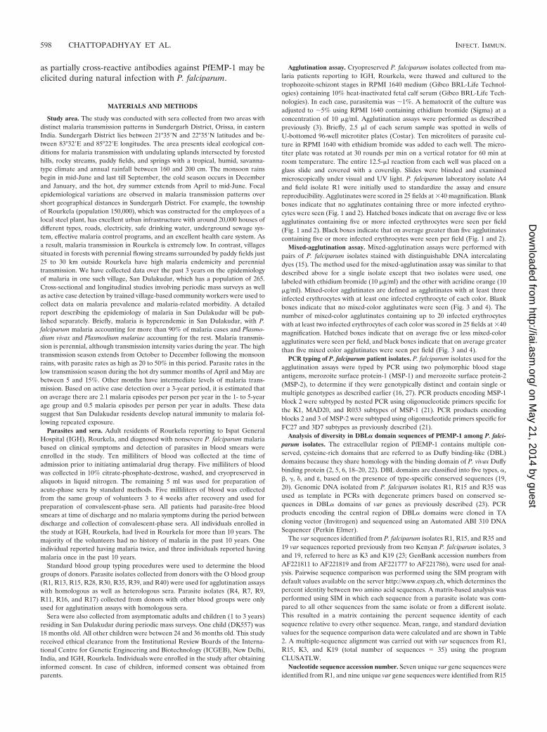
Agglutination of homologous and heterologous P. falciparumisolates by acute- and convalescent-phase sera collected fromadult malaria patients residing in Rourkela, an area with lowmalaria transmission rates. The incidence of malaria in Rour-kela is very low (see Materials and Methods). Acute- andconvalescent-phase sera collected from P. falciparum malariapatients residing in Rourkela were tested for recognition ofhomologous and heterologous P. falciparum isolates in agglu-tination assays. Most acute-phase sera (12 out of 14 acute-phase sera tested) do not agglutinate homologous P. falcipa-rum isolates (Fig. 1). All convalescent-phase sera agglutinatehomologous P. falciparum isolates, indicating that individualsseroconvert following infection and develop antibodies thatrecognize surface antigens expressed by the infecting P. falci-parum isolate (Fig. 1). Interestingly, in addition to agglutinat-ing homologous parasite isolates, a significant number of con-valescent-phase sera also agglutinate heterologous isolates(Fig. 1). In cases where both acute- and convalescent-phasesera agglutinate heterologous isolates, it is not possible toattribute agglutination by convalescent-phase sera to antibod-ies that develop in response to the recent infection. Such cases
are therefore disregarded in the following analysis. The ma-jority of convalescent-phase sera (11 out of 14 sera tested)agglutinate at least one heterologous isolate in addition to thehomologous isolate. Convalescent-phase serum from patient 1agglutinates five out of seven heterologous isolates tested, con-valescent-phase serum from patient 16 agglutinates four het-erologous isolates, and convalescent-phase serum from patient11 agglutinates 3 heterologous isolates. The observation thatconvalescent-phase sera frequently agglutinate heterologousisolates suggests that these individuals develop cross-reactiveantibodies that recognize common epitopes shared by surfaceantigens expressed by diverse P. falciparum isolates.
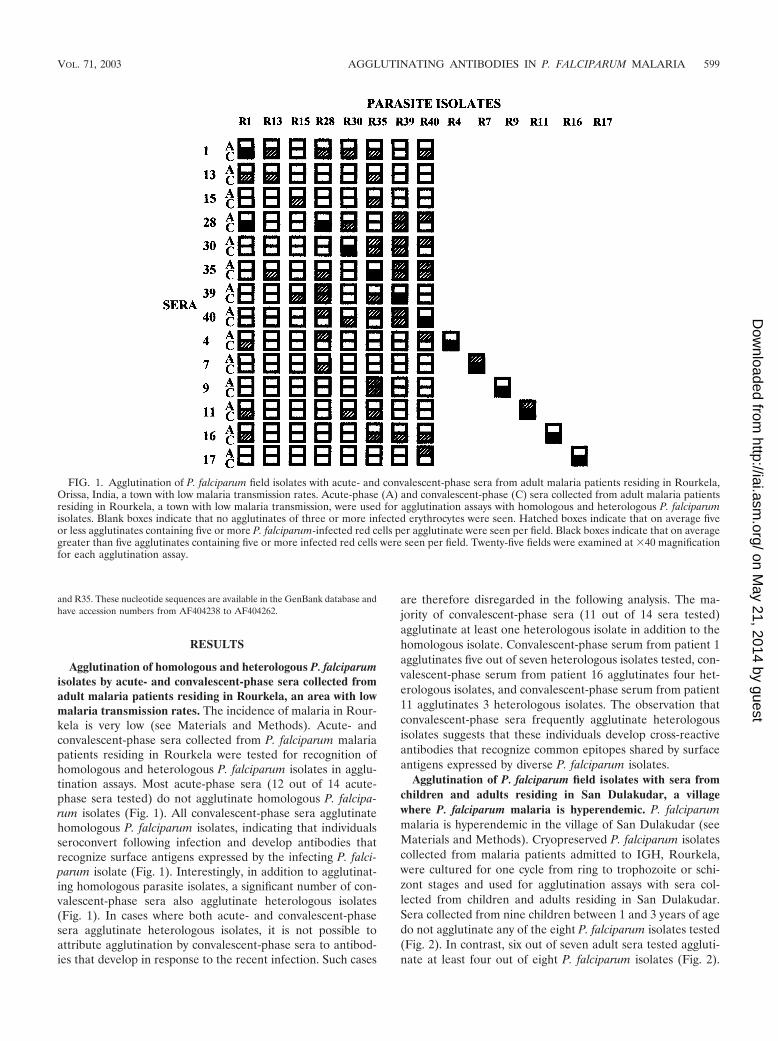
Agglutination of P. falciparum field isolates with sera fromchildren and adults residing in San Dulakudar, a villagewhere P. falciparum malaria is hyperendemic. P. falciparummalaria is hyperendemic in the village of San Dulakudar (seeMaterials and Methods). Cryopreserved P. falciparum isolatescollected from malaria patients admitted to IGH, Rourkela,were cultured for one cycle from ring to trophozoite or schi-zont stages and used for agglutination assays with sera col-lected from children and adults residing in San Dulakudar.Sera collected from nine children between 1 and 3 years of agedo not agglutinate any of the eight P. falciparum isolates tested(Fig. 2). In contrast, six out of seven adult sera tested aggluti-nate at least four out of eight P. falciparum isolates (Fig. 2).
FIG. 1. Agglutination of P. falciparum field isolates with acute- and convalescent-phase sera from adult malaria patients residing in Rourkela,Orissa, India, a town with low malaria transmission rates. Acute-phase (A) and convalescent-phase (C) sera collected from adult malaria patientsresiding in Rourkela, a town with low malaria transmission, were used for agglutination assays with homologous and heterologous P. falciparumisolates. Blank boxes indicate that no agglutinates of three or more infected erythrocytes were seen. Hatched boxes indicate that on average fiveor less agglutinates containing five or more P. falciparum-infected red cells per agglutinate were seen per field. Black boxes indicate that on averagegreater than five agglutinates containing five or more infected red cells were seen per field. Twenty-five fields were examined at �40 magnificationfor each agglutination assay.
VOL. 71, 2003 AGGLUTINATING ANTIBODIES IN P. FALCIPARUM MALARIA 599
on May 21, 2014 by guest
http://iai.asm.org/
Dow
nloaded from
These data indicate that, whereas sera from children in SanDulakudar lack antibodies directed against diverse PfEMP-1,sera from adults residing in the same village recognizePfEMP-1 variants expressed by a wide range of P. falciparumisolates. Pooled sera collected from individuals residing in ar-eas of India where malaria is not endemic do not agglutinateany of the P. falciparum isolates tested (Fig. 2). Pooled immunesera collected from adults residing in a different area of P.falciparum endemicity of Orissa (10) agglutinate all eight P.falciparum isolates tested (Fig. 2).
Mixed-agglutination assays to detect presence of cross-re-active antibodies directed against PfEMP-1 variants expressedby diverse P. falciparum isolates. Mixed-agglutination assayswere used to examine presence of cross-reactive antibodies toPfEMP-1. Since P. falciparum isolates R1 and R35 were mostcommonly recognized by heterologous convalescent-phasesera, they were used in combination with other parasite isolatesin mixed-agglutination assays. Convalescent-phase sera fromall malaria patients whose parasites were used for agglutina-tion with heterologous sera were used for mixed-agglutinationassays. Adult sera from San Dulakudar that agglutinate at leastfour out of eight isolates tested were also tested in mixed-agglutination assays.
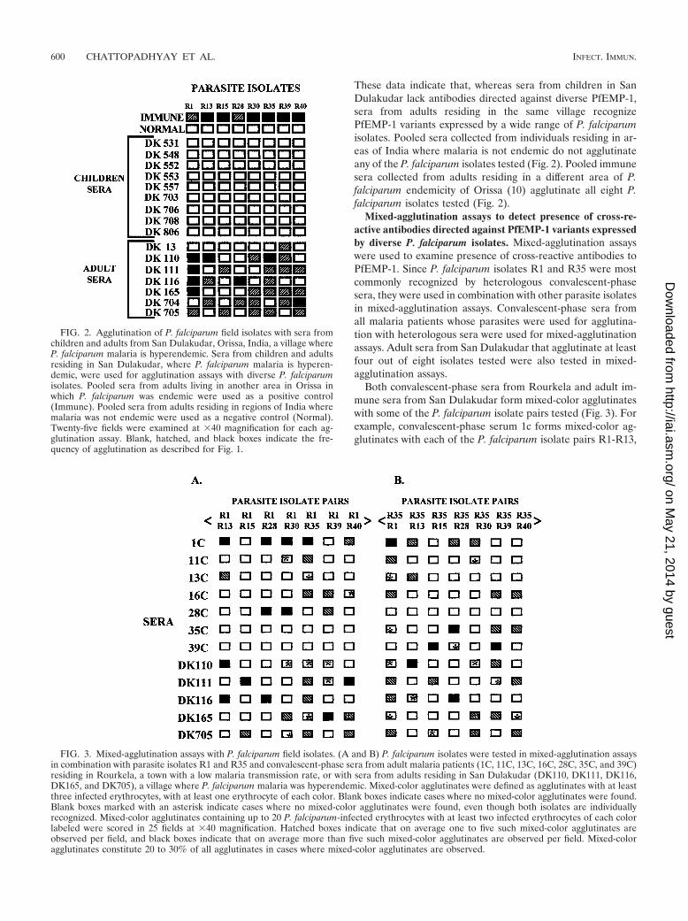
Both convalescent-phase sera from Rourkela and adult im-mune sera from San Dulakudar form mixed-color agglutinateswith some of the P. falciparum isolate pairs tested (Fig. 3). Forexample, convalescent-phase serum 1c forms mixed-color ag-glutinates with each of the P. falciparum isolate pairs R1-R13,
FIG. 2. Agglutination of P. falciparum field isolates with sera fromchildren and adults from San Dulakudar, Orissa, India, a village whereP. falciparum malaria is hyperendemic. Sera from children and adultsresiding in San Dulakudar, where P. falciparum malaria is hyperen-demic, were used for agglutination assays with diverse P. falciparumisolates. Pooled sera from adults living in another area in Orissa inwhich P. falciparum was endemic were used as a positive control(Immune). Pooled sera from adults residing in regions of India wheremalaria was not endemic were used as a negative control (Normal).Twenty-five fields were examined at �40 magnification for each ag-glutination assay. Blank, hatched, and black boxes indicate the fre-quency of agglutination as described for Fig. 1.
FIG. 3. Mixed-agglutination assays with P. falciparum field isolates. (A and B) P. falciparum isolates were tested in mixed-agglutination assaysin combination with parasite isolates R1 and R35 and convalescent-phase sera from adult malaria patients (1C, 11C, 13C, 16C, 28C, 35C, and 39C)residing in Rourkela, a town with a low malaria transmission rate, or with sera from adults residing in San Dulakudar (DK110, DK111, DK116,DK165, and DK705), a village where P. falciparum malaria was hyperendemic. Mixed-color agglutinates were defined as agglutinates with at leastthree infected erythrocytes, with at least one erythrocyte of each color. Blank boxes indicate cases where no mixed-color agglutinates were found.Blank boxes marked with an asterisk indicate cases where no mixed-color agglutinates were found, even though both isolates are individuallyrecognized. Mixed-color agglutinates containing up to 20 P. falciparum-infected erythrocytes with at least two infected erythrocytes of each colorlabeled were scored in 25 fields at �40 magnification. Hatched boxes indicate that on average one to five such mixed-color agglutinates areobserved per field, and black boxes indicate that on average more than five such mixed-color agglutinates are observed per field. Mixed-coloragglutinates constitute 20 to 30% of all agglutinates in cases where mixed-color agglutinates are observed.
600 CHATTOPADHYAY ET AL. INFECT. IMMUN.
on May 21, 2014 by guest
http://iai.asm.org/
Dow
nloaded from
R1-R28, R1-R30, R1-R35, and R1-R40, suggesting that it con-tains cross-reactive antibodies to common epitopes shared byPfEMP-1 variants expressed by P. falciparum isolate R1 andfive other P. falciparum isolates. In all cases where one of theisolates in the pair is not individually recognized, mixed-coloragglutinates are not found. Such cases serve as negative con-trols. There are a number of cases where two P. falciparumisolates are individually recognized, but mixed-color aggluti-nates are not observed (marked with asterisk in Fig. 3). In suchcases agglutination is attributed to the presence of variant-specific antibodies that react with variant epitopes that are notshared by PfEMP-1 expressed by these isolates.
PCR typing and var gene diversity in P. falciparum isolatesused for agglutination assays. PCR typing based on the poly-morphic markers MSP-1 and MSP-2 indicates that each P.falciparum isolate used in the agglutination assays is uniqueand contains a single P. falciparum genotype (Table 1).
Sequence diversity in var genes from P. falciparum isolatesused in the study was examined and compared to the diversityfound in var genes from African isolates. Degenerate oligonu-cleotide primers based on conserved DBL� sequences wereused to amplify var gene fragments encoding the central semi-conserved segment of DBL� domains of PfEMP-1 by PCRfrom three P. falciparum isolates, i.e., R1, R15, and R35 (23).R1 and R35 were selected because they were most commonlyrecognized, and R15 was selected because it was least com-monly recognized among isolates used in the agglutinationassays. Amino acid sequences encoded by each of the clonedPCR-amplified var gene fragments from R1, R15, and R35were compared to each other, and percent sequence identitywas determined for each pair (Table 2). The average “percentsequence identity values” derived from pairwise comparison of
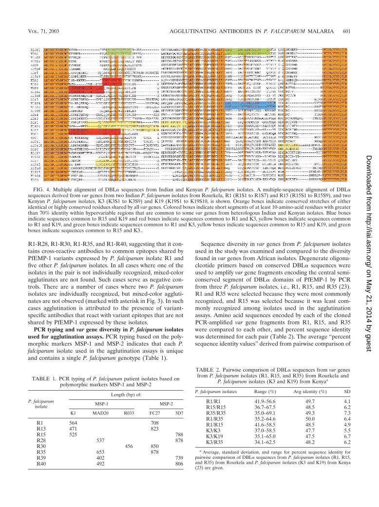
FIG. 4. Multiple alignment of DBL� sequences from Indian and Kenyan P. falciparum isolates. A multiple-sequence alignment of DBL�sequences derived from var genes from two Indian P. falciparum isolates from Rourkela, R1 (R1S1 to R1S7) and R15 (R15S1 to R15S9), and twoKenyan P. falciparum isolates, K3 (K3S1 to K3S9) and K19 (K19S1 to K19S10, is shown. Orange boxes indicate conserved stretches of eitheridentical or highly conserved residues shared by all var genes. Colored boxes indicate short segments of at least 10-amino-acid residues with greaterthan 70% identity within hypervariable regions that are common to some var genes from heterologous Indian and Kenyan isolates. Blue boxesindicate sequences common to R15 and K19 and red boxes indicate sequences common to R1 and K3, yellow boxes indicate sequences commonto R1 and K19, and green boxes indicate sequences common to R1 and K3, yellow boxes indicate sequences common to R15 and K19, and greenboxes indicate sequences common to R15 and K3..
TABLE 1. PCR typing of P. falciparum patient isolates based onpolymorphic markers MSP-1 and MSP-2
P. falciparumisolate
Length (bp) of:
MSP-1 MSP-2
K1 MAD20 R033 FC27 3D7
R1 564 708R13 471 823R15 525 788R28 537 878R30 456 850R35 653 878R39 402 739R40 492 806
TABLE 2. Pairwise comparison of DBL� sequences from var genesfrom P. falciparum isolates (R1, R15, and R35) from Rourkela and
P. falciparum isolates (K3 and K19) from Kenyaa
P. falciparum isolates Range (%) Avg identity (%) SD
R1/R1 41.9–56.6 49.7 4.1R15/R15 36.7–67.5 48.5 6.2R35/R35 35.0–69.1 49.3 7.3R1/R35 35.2–64.6 50.0 6.4R1/R15 41.6–58.5 48.5 4.9K3/K3 37.0–58.5 47.7 5.5K3/K19 35.1–65.0 47.5 6.7K3/R35 34.1–62.5 48.2 6.2
a Average, standard deviation, and range for percent sequence identity forpairwise comparison of DBL� sequences from P. falciparum isolates (R1, R15,and R35) from Rourkela and P. falciparum isolates (K3 and K19) from Kenya(23) are given.
VOL. 71, 2003 AGGLUTINATING ANTIBODIES IN P. FALCIPARUM MALARIA 601
on May 21, 2014 by guest
http://iai.asm.org/
Dow
nloaded from
var sequences from the same isolate are similar to those de-rived from pairwise comparison of var sequences from heter-ologous isolates (Table 2). Similar percent sequence identityvalues obtained upon comparison of homologous and heterol-ogous var genes suggest that var sequences from a single isolateare as similar and/or dissimilar to each other as var sequencesfrom heterologous isolates in Rourkela. Another sequencecomparison analysis was performed on var sequences reportedfrom P. falciparum isolates K3 and K19 from eastern Kenya(23). The average percent sequence identity values determinedfor pairwise comparison of var sequences from Kenyan isolatesas well as those determined from comparison of var sequencesfrom Indian isolates with Kenyan isolates are similar to aver-age percent sequence identity values found for pairwise com-parison of heterologous Indian isolates (Table 2). The level ofsequence diversity in var genes from Indian and Kenyan iso-lates is thus comparable.
A multiple-sequence alignment of DBL� sequences derivedfrom two Indian isolates from Rourkela and two Kenyan iso-lates is shown in Fig. 4. Orange boxes indicate conserved seg-ments of either identical or highly conserved residues that areshared by all var genes. Such linear segments are found at boththe amino and carboxyl termini of this region. A number ofhydrophobic residues and at least two cysteines are conservedin this region. It is noteworthy that a number of hydrophilicresidues are also found in these segments. The conserved seg-ments shared by all var sequences are interspersed with “hy-pervariable segments” that vary significantly in both sequenceand length. Within these hypervariable regions, a number ofamino acid segments of limited conservation common to varsequences from heterologous isolates (colored boxes in Fig. 4)exist. The presence of such common amino acid segments inPfEMP-1 variants from diverse P. falciparum isolates may beresponsible for the observed cross-reactivity of agglutinatingantibodies.
DISCUSSION
Individuals residing in areas of endemicity acquire naturalimmunity following repeated infection with P. falciparum (14).Antibodies directed against PfEMP-1 are thought to a play arole in naturally acquired immunity to P. falciparum malaria (3,13). Here we have studied the development of antibodies toPfEMP-1 following P. falciparum infection in adult residents ofRourkela, a town with a low malaria transmission rate, and theprevalence of antibodies to PfEMP-1 in residents of San Du-lakudar, a village where P. falciparum malaria is hyperendemic.
Our data show that acute-phase sera from adult malariapatients from Rourkela have limited recognition of P. falcipa-rum isolates. Residents of Rourkela are not exposed to a widerange of P. falciparum isolates and thus lack antibodies againstdiverse PfEMP-1. All malaria patients from Rourkela serocon-vert following P. falciparum infection to agglutinate the homol-ogous P. falciparum isolate. Interestingly, convalescent-phasesera from adult malaria patients in Rourkela also frequentlyagglutinate a limited number of heterologous P. falciparumisolates. These observations suggest that antibodies toPfEMP-1 that develop following infection recognize commonepitopes shared by at least some PfEMP-1 variants. In a fewcases convalescent-phase sera fail to agglutinate heterologous
isolates, even though the corresponding acute-phase sera ag-glutinate that isolate. Such examples have been observed be-fore and suggest that antibodies to the heterologous isolate inacute-phase serum may not be present in the correspondingconvalescent-phase serum in these cases. In the case of thehigh-transmission study, children from San Dulakudar fail toagglutinate any of the P. falciparum isolates tested, whereasadult sera agglutinate diverse P. falciparum isolates. This is inline with previous observations that children in areas of ende-micity have very limited recognition, whereas adults have widerecognition of diverse PfEMP-1 (4, 7–9, 12, 15). It is possiblethat children make more variant-specific antibodies than naïveadults upon infection with P. falciparum and therefore fail toagglutinate any heterologous parasites. This possibility couldnot be addressed in the present study since all acute- andconvalescent-phase sera were collected from naïve adults.
Mixed-agglutination assays using P. falciparum isolate pairsstained with distinguishable DNA intercalating dyes were usedto examine presence of cross-reactive antibodies to PfEMP-1.Mixed-color agglutinates were frequently found with differentP. falciparum isolate pairs tested with convalescent-phase serafrom Rourkela. The presence of mixed-color agglutinates in-dicates presence of cross-reactive antibodies that recognizecommon epitopes shared by PfEMP-1 expressed by both iso-lates. In some cases, mixed- color agglutinates were not ob-served even though both parasite isolates were individuallyrecognized. In such cases, convalescent-phase sera are likely tocontain variant-specific antibodies that recognize variantepitopes that are not shared by PfEMP-1 expressed by the twoisolates tested. Previous studies using acute- and convalescent-phase sera have also reported that convalescent-phase seraagglutinate some heterologous isolates (4, 9). However, mixed-agglutination assays were not performed and the presence ofcross-reactive antibodies was not explicitly demonstrated inthese studies (4, 9). A study using adult sera from an areawhere P. falciparum malaria is mesoendemic also suggestedthat a single P. falciparum infection might elicit cross-reactiveantibodies against diverse PfEMP-1 (8). However, these inves-tigators failed to detect cross-reactive antibodies in mixed-agglutination assays probably because they used a limited num-ber of P. falciparum isolate pairs (8). Our study is the first toexplicitly demonstrate that both variant-specific antibodies aswell as antibodies with limited cross-reactivity for diversePfEMP-1 develop following a single infection with P. falcipa-rum.
Initial studies with sera from Gambian adults reported thepresence of cross-reactive antibodies to PfEMP-1 in immunesera (12). In contrast, a later study reported that mixed-coloragglutinates were rarely observed with pooled immune serafrom Gambian adults (15). The presence of some high-titervariant-specific antibodies in pooled immune sera may haveled to the predominant formation of single-color agglutinatesin this study. Individual Gambian adult sera were also used formixed-agglutination assays in this study (15). However, onlythree pairs of P. falciparum laboratory isolates were tested andmixed-color agglutinates were observed only in a limited num-ber of cases, leading these investigators to conclude that anti-bodies to PfEMP-1 are predominantly variant specific (15). Inthe present study, we have tested individual immune sera fromadult residents of an area of hyperendemicity with 13 different
602 CHATTOPADHYAY ET AL. INFECT. IMMUN.
on May 21, 2014 by guest
http://iai.asm.org/
Dow
nloaded from
pairs of P. falciparum field isolates in mixed-agglutination as-says. Mixed-color agglutinates are observed frequently withadult immune sera from San Dulakudar. In some cases mixed-color agglutinates are not observed even though both parasiteisolates are individually recognized. We therefore concludethat adult immune sera contain both variant-specific and par-tially cross-reactive antibodies to PfEMP-1.
Are cross-reactive antibodies observed in our studies be-cause the P. falciparum isolates used have limited diversity? P.falciparum isolates used in this study were collected from ma-laria patients in Rourkela, an area with a low malaria trans-mission rate. It is possible that only a small number of P.falciparum isolates circulate and are repeatedly sampled in anarea with a low entomological inoculation rate. However, PCRtyping based on two polymorphic markers, MSP-1 and MSP-2,demonstrates that each P. falciparum isolate used in our studyis genotypically unique, ruling out this possibility.
Do P. falciparum isolates used in our study have limitedsequence diversity in var genes compared to isolates from areasof endemicity in Africa with high entomological inoculationrates? In order to address this question, we have determinedthe extent of sequence diversity in the central, semiconservedregion of DBL� domains of PfEMP-1 from three P. falciparumisolates used in our study. DBL� sequences from the Indianisolates were compared with each other and with DBL� se-quences reported from Kenyan isolates (23). The average se-quence identity derived from pairwise comparison of DBL�sequences from Indian and Kenyan isolates is comparable (Ta-ble 2). This analysis indicates that the level of conservationand/or diversity in var sequences from Indian and Kenyan P.falciparum isolates is comparable. Moreover, var sequencesfrom parasite isolates from the same geographical region areas similar and/or dissimilar to each other as they are to varsequences from parasite isolates from distant geographical re-gions.
We further performed comparative sequence analyses ofDBL� sequences from two Indian isolates and two Kenyanisolates to explore the molecular basis of cross-reactivity ob-served in our field studies. A multiple-sequence alignmentreveals the presence of highly conserved segments (grey boxes,Fig. 4). These segments contain conserved cysteines and hy-drophobic amino acid residues, suggesting that they may bestructurally important. A number of charged, potentially ex-posed amino acid residues are also present in these segments.Antibodies directed against such highly conserved segmentswould be widely cross-reactive. However, since widely cross-reactive antibodies are not observed in the field, it is likely thatthese highly conserved segments of DBL� are not immuno-genic during natural infection. The conserved segments areinterspersed with hypervariable segments that vary in bothsequence and length. These divergent segments contain mul-tiple hydrophilic residues, suggesting that they are likely to beexposed and may therefore serve as epitopes for agglutinatingantibodies. Sets of common sequences shared by diversePfEMP-1 from heterologous isolates can be found in thesedivergent segments of DBL� domains. Colored boxes illustratesuch common sequences shared by var genes from heterolo-gous Indian and Kenyan isolates (Fig. 4). Antibodies directedagainst such shared amino acid sequences may be responsiblefor the limited cross-reactivity observed with naturally ac-
quired antibodies. Similar common stretches of amino acidsequences are also found between var genes from heterologousIndian isolates (data not shown). The presence of commonsequences in var genes from geographically distant P. falcipa-rum isolates has been previously noted (26) and explains theability of adult immune sera to agglutinate parasite isolatesfrom distant locations (1).
Following infection with P. falciparum, the infected individ-ual is exposed to a series of PfEMP-1 variants as the blood-stage parasite multiplies and undergoes antigenic variation invivo. As a result, one can expect that a set of agglutinatingantibodies directed against multiple PfEMP-1 will develop.These antibodies are likely to be directed against divergentsegments of PfEMP-1 as discussed earlier. However, as oursequence analysis suggests, divergent segments contain aminoacid sequences that are shared by subsets of PfEMP-1 variants.We may therefore expect that the set of naturally acquiredantibodies elicited by a single infection should agglutinate atleast some heterologous isolates that express PfEMP-1 mole-cules, which share epitopes with the homologous isolate (col-ored boxes, Fig. 4). Indeed, agglutination assays using acute-and convalescent-phase sera from Rourkela demonstrate thedevelopment of antibodies with limited cross-reactivity.
As is well known, adults in areas of endemicity are typicallyexposed to a wide range of P. falciparum isolates and therebydevelop antibodies that together recognize a majority ofPfEMP-1 variants. We suggest that the basis of such widerecognition may not entirely be due to the acquisition of arepertoire of variant-specific antibodies. Our analysis ofPfEMP-1 sequences reveals the presence of common segmentseven in divergent regions of DBL� domains, and given thehydrophilic character of such segments, it is likely that they areboth exposed and form epitopes for antibody recognition. An-tibodies to such epitopes on PfEMP-1 are likely to be partiallycross-reactive. Sets of such naturally acquired partially cross-reactive antibodies in immune sera will have an additive effectenabling recognition of a wide range of PfEMP-1 variants.Indeed, adult sera from San Dulakudar agglutinate diverse P.falciparum isolates, and mixed-agglutination assays confirm thepresence of both variant-specific and partially cross-reactiveantibodies. We propose that the molecular basis of wide rec-ognition of PfEMP-1 variants by immune serum from areas ofendemicity lies in the prevalence of multiple sets of partiallycross-reactive antibodies that together may cover most of the“var epitope space.”
ACKNOWLEDGMENTS
This work was supported by Wellcome Trust International SeniorResearch Fellowships to C.E.C. and A.S.
We thank Shobhona Sharma for providing pooled immune sera,Virander S. Chauhan for comments on the manuscript, V. P. Sharmafor encouraging us to initiate these studies, and Sarala Subbarao forcontinued support. We also thank the staff of Malaria Research CentreField Station, Rourkela, and clinicians at the Ispat General Hospital,Rourkela, for their cooperation.
REFERENCES
1. Aguiar, J. C., G. R. Albrecht, P. Cegielski, B. M. Greenwood, J. B. Jensen, G.Lallinger, A. Martinez, I. A. McGregor, J. N. Minjas, J. Neequaye, M. E.Patarroyo, J. A. Sherwood, and R. J. Howard. 1992. Agglutination of Plas-modium falciparum-infected erythrocytes from east and west African isolatesby human sera from distant geographic regions. Am. J. Trop. Med. Hyg.47:621–632.
VOL. 71, 2003 AGGLUTINATING ANTIBODIES IN P. FALCIPARUM MALARIA 603
on May 21, 2014 by guest
http://iai.asm.org/
Dow
nloaded from
2. Baruch, D., B. Pasloske, H. Singh, B. Xiahui, X. Ma, M. Feldman, T.Taraschi, and R. J. Howard. 1995. Cloning of the Plasmodium falciparumgene encoding PfEMP1, a malarial variant antigen and cytoadherence re-ceptor on the surface of parasitised human erythrocytes. Cell 82:77–87.
3. Bull, P., B. Lowe, M. Kortok, C. Molyneux, C. Newbold, and K. Marsh. 1998.Parasite antigens on the infected red cell surface are targets for naturallyacquired immunity to malaria. Nat. Med. 4:358–360.
4. Bull, P., B. Lowe, M. Kortok, and K. Marsh. 1999. Antibody recognition ofPlasmodium falciparum erythrocyte surface antigens in Kenya: evidence forrare and prevalent variants. Infect. Immun. 67:733–739.
5. Chitnis, C. E., and L. H. Miller. 1994. Identification of the erythrocytebinding domains of Plasmodium vivax and Plasmodium knowlesi proteinsinvolved in erythrocyte invasion. J. Exp. Med. 180:497–506.
6. Chitnis, C. E., P. Sinnis, and L. H. Miller. 1999. The sporozoite, the mer-ozoite and the infected red cell: parasite ligands and host receptors, p.249–285. In M. Wahlgren and P. Perlmann (ed.), Malaria: molecular andclinical aspects. Harwood Academic Publishers, Newark, N.J.
7. Forsyth, K. P., G. Phillip, T. Smith, E. Kum, B. Southwell, and G. V. Brown.1989. Diversity of antigens expressed on the surface of erythrocytes infectedwith mature Plasmodium falciparum parasites in Papua New Guinea. Am. J.Trop. Med. Hyg. 83:464–469.
8. Giha, H. A., T. Staalsoe, D. Dodoo, I. M. Elhassan, C. Roper, G. M. H. Satti,D. E. Arnot, L. Hviid, and T. G. Theander. 1999. Overlapping antigenicrepertoires of variant antigens expressed on the surface of erythrocytesinfected by Plasmodium falciparum. Parasitology 119:7–17.
9. Iqbal, J., P. Perlmann, and K. Berzin. 1993. Serological diversity of antigensexpressed on the surface of erythrocytes infected with Plasmodium falcipa-rum. Trans. R. Soc. Trop. Med. Hyg. 87:583–588.
10. Lobo, C. A., S. K. Kar, B. Ravindran, L. Kabilan, and S. Sharma. 1994.Novel proteins of Plasmodium falciparum identified by differential immuno-screening using immune and patient sera. Infect. Immun. 62:651–656.
11. MacPherson, G., M. Warrell, N. White, W. Looareesuwan, and D. Warrell.1985. Human cerebral malaria: a quantitative ultrastructural analysis ofparasitized erythrocyte sequestration. Am. J. Pathol. 119:385–401.
12. Marsh, K., and R. J. Howard. 1986. Antigens induced on erythrocytes by P.falciparum: expression of diverse and conserved determinants. Science 231:150–153.
13. Marsh, K., L. Otoo, R. J. Hayes, D. C. Carson, and B. M. Greenwood. 1989.Antibodies to blood-stage antigens of Plasmodium falciparum in rural Gam-bia and their relation to protection against infection. Trans. R. Soc. Trop.Med. Hyg. 83:293–303.
14. Marsh, K. 1992. Malaria—a neglected disease? Parasitology 104:S53–S69.15. Newbold, C. I., R. Pinches, D. J. Roberts, and K. Marsh. 1992. Plasmodium
falciparum: the human agglutinating antibody response to the infected redcell surface is predominantly variant specific. Exp. Parasitol. 75:281–292.
16. Okoyeh, J. N., C. R. Pillai, and C. E. Chitnis. 1999. Plasmodium falciparumfield isolates commonly use erythrocyte invasion pathways that are indepen-dent of sialic acid residues of glycophorin A. Infect. Immun. 67:5784–5791.
17. Patnaik, J. K., B. S. Das, S. K. Mishra, S. Mohanty, S. K. Satpathy, and D.Mohanty. 1994. Vascular clogging, mononuclear cell margination, and en-hanced vascular permeability in the pathogenesis of human cerebral malaria.Am. J. Trop. Med. Hyg. 51:642–647.
18. Smith, J. D., C. E. Chitnis, A. G. Craig, D. J. Roberts, D. E. Hudson-Taylor,D. S. Peterson, R. Pinches, C. I. Newbold, and L. H. Miller. 1995. Switchesin expression of Plasmodium falciparum var genes correlate with changes inantigenic and cytoadherent phenotypes of infected erythrocytes. Cell 82:101–110.
19. Smith, J. D., G. Subramanian, B. Gamain, D. Baruch, and L. H. Miller.2000. Classification of adhesive domains in the Plasmodium falciparum eryth-rocyte membrane protein 1 family. Mol. Biochem. Parasitol. 110:293–310.
20. Smith, J. D., B. Gamain, D. Baruch, and S. Kyes. 2001. Decoding thelanguage of var gene and Plasmodium falciparum sequestration. TrendsParasitol. 17:538–545.
21. Snounou, G., X. Zhu, N. Siripoon, W. Jarra, S. Thaithong, K. N. Brown, andS. Viriyakosoi. 1999. Biased distribution of msp1 and msp2 allelic variants inPlasmodium falciparum populations in Thailand. Trans. R. Soc. Trop. Med.Hyg. 93:369–374.
22. Su, X., V. Heatwole, S. Wertheimer, F. Guinet, J. Herrfeldt, D. Peterson, J.Ravetch, and T. E. Wellems. 1995. The large diverse gene family var encodesproteins involved in cytoadherence and antigenic variation of Plasmodiumfalciparum infected erythrocytes. Cell 82:89–100.
23. Taylor, H., S. Kyes, and C. I. Newbold. 2000. Var gene diversity in Plasmo-dium falciparum is generated by frequent recombination events. Mol. Bio-chem. Parasitol. 110:391–397.
24. Turner, G., H. Morrison, M. Jones, T. Davis, S. Looareesuwan, I. Buley, K.Gatter, K. C. I. Newbold, S. Pukritayakamee, B. Nagachinta, N. J. White,and A. R. Berendt. 1994. An immunohistochemical study of the pathology offatal malaria: evidence for widespread endothelial activation and a potentialrole of intercellular adhesion molecule-1. Am. J. Pathol. 145:1057–1069.
25. Walter, P., Y. Garin, and P. Blot. 1982. Placental pathological changes inmalaria. Am. J. Pathol. 109:330–342.
26. Ward, C. P., G. T. Clottey, M. Dorris, D. Ji, and D. E. Arnot. 1999. Analysisof Plasmodium falciparum PfEMP-1/var genes suggests that recombinationrearranges constrained sequences. Mol. Biochem. Parasitol. 102:167–177.
27. Wooden, J., S. Kyes, and C. Sibley. 1993. PCR and strain identification inPlasmodium falciparum. Parasitol. Today 9:303–305.
Editor: S. H. E. Kaufmann
604 CHATTOPADHYAY ET AL. INFECT. IMMUN.
on May 21, 2014 by guest
http://iai.asm.org/
Dow
nloaded from
Copyright © 2022 FDOKUMEN