Cryptosporidium Infection Causes Undernutrition and, Conversely, Weanling Undernutrition Intensifies...
17
CRYPTOSPORIDIUM INFECTION CAUSES UNDERNUTRITION AND, CONVERSELY, WEANLING UNDERNUTRITION INTENSIFIES INFECTION Bruna P. Coutinho, Reinaldo B. Oriá, Carlos M. G. Vieira, Jesus Emmanuel A. D. Sevilleja, Cirle A. Warren, Jamilly G. Maciel, Meghan R. Thompson, Relana C. Pinkerton, Aldo A. M. Lima, and Richard L. Guerrant * Center for Global Health, School of Medicine, University of Virginia, MR4, Lane Road, Room 3148, P.O. Box 801379, Charlottesville, Virginia 22908 Abstract Cryptosporidium parvum is a leading pathogen in children in developing countries. To investigate whether early postnatal malnutrition leads to heavier C. parvum infections, we assessed intestinal adaptation and parasite load in suckling mice during the first 2 wk of life, analogous to the first postnatal yr in humans. Undernutrition was induced by daily C57BL6J pup separation from lactating dams. Half of the pups were separated daily, for 4 hr on day 4, 8 hr on day 5, and for 12 hr from day 6 until day 14. On day 6, each pup received an oral inoculum of 10 5 to 10 7 parasites in 10–25 μl of PBS. Littermate controls received PBS alone. Stools were assessed from days 8, 11, and 14 for oocyst counts. Mice were killed on day 14, 8 days postinoculation, at the peak of the infection. Ileal and colon segments were obtained for histology, real-time and reverse transcriptase PCR, and immunoassays. Villus and crypt lengths and cross-sectional areas were also measured. Undernourished and nourished mice infected with excysted 10 6 or 10 7 oocysts exhibited the poorest growth outcomes compared with their uninfected controls. Nourished 10 6 -infected mice had comparable weight decrements to uninfected undernourished mice. Body weight and villi were additively affected by malnutrition and cryptosporidiosis. Hyperplastic crypts and heavier inflammatory responses were found in the ilea of infected malnourished mice. Undernourished infected mice exhibited greater oocyst shedding, TNF-α and IFN-γ intestinal levels, and mRNA expression compared to nourished mice infected with either 10 5 or 10 6 oocysts. Taken together, these findings show that Cryptosporidium infection can cause undernutrition and, conversely, that weanling undernutrition intensifies infection and mucosal damage. Cryptosporidiosis, first described by Tyzzer (1907) in the gastric glands of infected mice (Tzipori and Ward, 2002), has emerged as an increasingly recognized public health threat. Cryptosporidium spp. have been identified in watery diarrhea of patients with HIV and other immunocompromised patients and in large human outbreaks of diarrhea in both developed and developing parts of the world (Harp, 2003; Ramirez et al., 2004; Houpt et al., 2005). Furthermore, the long-term impact of cryptosporidial infection has been increasingly recognized in impoverished settings around the world (Checkley et al., 1997; Guerrant, 1997; Checkley et al., 1998). The wide-range zoonotic potential, resistance to disinfectants, low infectious dose, and easy dissemination upon shedding impressively show how this © American Society of Parasitologists 2008 * To whom correspondence should be addressed. Center for Global Health, Division of Infectious Diseases and International Health, University of Virginia, Charlottesville, Virginia 22908. [email protected]. NIH Public Access Author Manuscript J Parasitol. Author manuscript; available in PMC 2011 April 5. Published in final edited form as: J Parasitol. 2008 December ; 94(6): 1225–1232. doi:10.1645/GE-1411.1. NIH-PA Author Manuscript NIH-PA Author Manuscript NIH-PA Author Manuscript
-
Upload
independent -
Category
Documents
-
view
0 -
download
0
Transcript of Cryptosporidium Infection Causes Undernutrition and, Conversely, Weanling Undernutrition Intensifies...

CRYPTOSPORIDIUM INFECTION CAUSES UNDERNUTRITIONAND, CONVERSELY, WEANLING UNDERNUTRITIONINTENSIFIES INFECTION
Bruna P. Coutinho, Reinaldo B. Oriá, Carlos M. G. Vieira, Jesus Emmanuel A. D. Sevilleja,Cirle A. Warren, Jamilly G. Maciel, Meghan R. Thompson, Relana C. Pinkerton, Aldo A. M.Lima, and Richard L. Guerrant*Center for Global Health, School of Medicine, University of Virginia, MR4, Lane Road, Room3148, P.O. Box 801379, Charlottesville, Virginia 22908
AbstractCryptosporidium parvum is a leading pathogen in children in developing countries. To investigatewhether early postnatal malnutrition leads to heavier C. parvum infections, we assessed intestinaladaptation and parasite load in suckling mice during the first 2 wk of life, analogous to the firstpostnatal yr in humans. Undernutrition was induced by daily C57BL6J pup separation fromlactating dams. Half of the pups were separated daily, for 4 hr on day 4, 8 hr on day 5, and for 12hr from day 6 until day 14. On day 6, each pup received an oral inoculum of 105 to 107 parasites in10–25 μl of PBS. Littermate controls received PBS alone. Stools were assessed from days 8, 11,and 14 for oocyst counts. Mice were killed on day 14, 8 days postinoculation, at the peak of theinfection. Ileal and colon segments were obtained for histology, real-time and reverse transcriptasePCR, and immunoassays. Villus and crypt lengths and cross-sectional areas were also measured.Undernourished and nourished mice infected with excysted 106 or 107 oocysts exhibited thepoorest growth outcomes compared with their uninfected controls. Nourished 106-infected micehad comparable weight decrements to uninfected undernourished mice. Body weight and villiwere additively affected by malnutrition and cryptosporidiosis. Hyperplastic crypts and heavierinflammatory responses were found in the ilea of infected malnourished mice. Undernourishedinfected mice exhibited greater oocyst shedding, TNF-α and IFN-γ intestinal levels, and mRNAexpression compared to nourished mice infected with either 105 or 106 oocysts. Taken together,these findings show that Cryptosporidium infection can cause undernutrition and, conversely, thatweanling undernutrition intensifies infection and mucosal damage.
Cryptosporidiosis, first described by Tyzzer (1907) in the gastric glands of infected mice(Tzipori and Ward, 2002), has emerged as an increasingly recognized public health threat.Cryptosporidium spp. have been identified in watery diarrhea of patients with HIV and otherimmunocompromised patients and in large human outbreaks of diarrhea in both developedand developing parts of the world (Harp, 2003; Ramirez et al., 2004; Houpt et al., 2005).Furthermore, the long-term impact of cryptosporidial infection has been increasinglyrecognized in impoverished settings around the world (Checkley et al., 1997; Guerrant,1997; Checkley et al., 1998). The wide-range zoonotic potential, resistance to disinfectants,low infectious dose, and easy dissemination upon shedding impressively show how this
© American Society of Parasitologists 2008*To whom correspondence should be addressed. Center for Global Health, Division of Infectious Diseases and International Health,University of Virginia, Charlottesville, Virginia 22908. [email protected].
NIH Public AccessAuthor ManuscriptJ Parasitol. Author manuscript; available in PMC 2011 April 5.
Published in final edited form as:J Parasitol. 2008 December ; 94(6): 1225–1232. doi:10.1645/GE-1411.1.
NIH
-PA Author Manuscript
NIH
-PA Author Manuscript
NIH
-PA Author Manuscript

protozoan is adapted to survive and spread in the environment (Dillingham et al., 2002;Karanis et al., 2007).
Crowded households with inadequate sanitation further aggravate the likelihood of theinfection spreading from person to person (Newman et al., 1999; Caccio and Pozio, 2006).The lack of adequate treatment or prevention for high-risk groups for this chlorine-resistantfood- and waterborne protozoan adds to the difficulties controlling cryptosporidial infections(Smith and Corcoran, 2004).
Impoverished household environments and water contamination tremendously increase therisk of exposure to waterborne pathogens and the likelihood of oocyst spread. Predispositionto adverse outcomes from repeated or prolonged bouts of diarrhea and enteric infectionsmay additionally have a strong genetic component (Guerrant et al., 2005; Oria et al., 2005,2007), especially with respect to host–parasite immune interactions.
Longitudinal cohort studies in poor shantytown areas in the developing world by our groupand others have highlighted the short- and long-term impact of cryptosporidial infections ongrowth and development of children (even without overt diarrhea), effects that remaingreatly underestimated (Checkley et al., 1997; Guerrant et al., 2005; Savioli et al., 2006;Bushen et al., 2007). In addition, young malnourished children are at greatest risk fordeveloping heavy Cryptosporidium spp. infections and likely present the worst outcomes. Incrowded household environments, low-birth-weight children were found to be at greater riskfor acquiring symptomatic Cryptosporidium spp. infection, which, in turn, highlights thevicious cycle of enteric infections and malnutrition leading to increased risk forcryptosporidial infection, malabsorption, and diarrhea during early childhood (Newman etal., 1999).
Undernutrition and enteric illnesses may disrupt the developing immune system (Pallaro etal., 2001; Cunningham-Rundles et al., 2005) against C. parvum in growing children,especially in the first 2 yr of life, the same time frame for rapid physical and cognitivedevelopment (Guerrant et al., 1999; Niehaus et al., 2002). Therefore, we have focused on aneonatal murine model of cryptosporidiosis to address the interactions of cryptosporidialinfection and malnutrition in early life.
Despite worldwide epidemiological data on DALYs (disability-adjusted life years) lost dueto enteric infections and undernutrition in early childhood, few studies have addressed theinteractions of infection and undernutrition and their effects on postnatal development inwell-defined animal models. In the current study, we have investigated the additive effectsof Cryptosporidium sp. infection with undernutrition in the suckling period in mice, whichwas found in our model to severely impair normal growth. Furthermore, we find thatmalnourished mice develop substantially heavier infections and greater mucosal damagethan nourished mice given the same inoculum.
MATERIALS AND METHODSUndernutrition protocol
The protocol described herein is in accordance with the Institutional Animal Care and UseCommittee (IACUC) policies of the University of Virginia. Both pregnant and nonpregnantinbred mice (C57BL6J) were purchased from Charles River Laboratories, Inc. (Wilmington,Massachusetts). The pregnant mice were monitored daily before delivery under pathogen-free conditions. Standard chow diet and water were given ad libitum to the dams. Afterbirth, litter size was adjusted to 6–8 pups. Their first day of life was recorded as day 1.Analogous in some ways to weanling undernutrition in impoverished children, the pups
Coutinho et al. Page 2
J Parasitol. Author manuscript; available in PMC 2011 April 5.
NIH
-PA Author Manuscript
NIH
-PA Author Manuscript
NIH
-PA Author Manuscript
were separated from their lactating dams for increasing intervals starting on day 4 of life.Half of the pups from each litter were separated, starting with 4 hr on day 4, 8 hr on day 5,and 12 hr on day 6 until day 14, according to protocol adapted from Calikoglu et al. (2001).Separated pups were kept in an incubator box under controlled temperature (28 ± 2 C).Nourished controls stayed with their mothers. Litters were kept undisturbed in the first 4postnatal days to reduce stress and to assure that all pups received initial colostrum. Bodyweight was recorded daily until mice were killed. A thermal pad was used to warm the pupsduring daily measurements (28 ± 2 C). Care was taken to keep the same level of animalhandling for all groups.
Cryptosporidium parvum infectionOn day 6, 1 hr prior to parasite inoculation, pups were separated from their lactating dams toempty their stomachs. Each pup received an oral inoculum of 105, 106, or 107 excysted C.parvum oocysts in 10–25 μl of PBS (pH 7.2). Oocysts were obtained from experimentallyinfected calves (Iowa isolate; Waterborne, Inc., New Orleans, Louisiana). Littermatecontrols received PBS orally at the same time.
Because excystation rates varied from 20 to 40%, experimental groups were paired toinclude nourished controls (n = 11) and infected groups given identical inocula at the sametime with either 105 (n = 14), 106 (n = 16), or 107 (n = 6) oocysts. Malnourished uninfectedcontrols, (n = 10), and infected mice were given either 105 (n = 14), 106 (n = 16), or 107 (n =13) oocysts. Due to high mortality rates observed in mice inoculated with the 25 μl ofvolume required for the 107 oocysts, data from the 107 dose are only presented for fecal andtissue parasite quantification.
Follow-upBody weight was measured daily, and stools were collected by gentle stroking of theabdomen. Animals were killed on day 14, 8 days post-inoculum (PI), at the peak of theinfection. Animals were anesthetized with sodium pentobarbital, 8 mg/100 g i.p., followedby cervical dislocation. After opening of the abdominal cavity, approximately 1 cm of eachmiddle ileal and colon segments (proximal to the ileocecal valve) were removed withoutwashing, then weighed, divided, and prepared either for histology (24 hr in 10% zincformalin) or real-time and reverse-transcriptase PCR (frozen immediately in liquid nitrogenand transferred to −80 C). On the day mice were killed, stools were collected beforetermination or after cervical dislocation, when mice would pass some feces immediatelyafter death. Intestinal sections that had the least amount of fecal material were collected forhistology, real-time PCR, and reverse-transcriptase PCR (they were not washed with PBS; 1cm of tissue was excised). For the mice that had a lot of stool, their intestines were milked toremove feces. Results of C. parvum parasites by real-time PCR were expressed in log countsper gram of stool or tissue, respectively.
Morphology analysesDigital micrographs of intestinal histology were taken with the use of a high-resolutionmicroscope (BH-2, Olympus, Tokyo, Japan), with Photoshop CS software. To address themucosal villus surface and crypt hyperplastic response, villus height and crypt depth andarea were measured with Image J software, at low magnification (×100), from ileumsamples at day 14 (8 days PI), following calibration as described elsewhere (Carneiro-Filhoet al., 2004). Twenty longitudinal sections of crypts and villi were measured per ileum (n =4, per group). To perform direct visualization of oocysts, and to confirm infectivity, highermagnifications were used (×400).
Coutinho et al. Page 3
J Parasitol. Author manuscript; available in PMC 2011 April 5.
NIH
-PA Author Manuscript
NIH
-PA Author Manuscript
NIH
-PA Author Manuscript

Fecal detection of oocyst sheddingA direct immunofluorescence detection procedure was used to confirm infection, accordingto the manufacturer's instructions (MeriFluor® Cryptosporidium/Giardia; MeridianBioscience, Inc., Cincinnati, Ohio), with the use of FITC labeled anti-C. parvum monoclonalantibodies. Stools were collected in a preweighed tube and diluted to 1 mg/25 μl in PBS.Oocysts were detected with the aid of a fluorescence microscope adapted with an image-capturing platform (12-megapixel digital camera) under ×40 magnification. Oocysts wereidentified by their green color and characteristic size (Bushen et al., 2007).
Real-time PCR protocolsFive microliters of DNA sample was added to 20 μl of master mix (consisting of 12.5 μl ofSYBR Green Supermix (BioRad, Hercules, California), 1.0 μl of forward primer, 1.0 ofreverse primer, and 5.5 μl of DNAse-, RNAse-free water) to make a total amplificationreaction volume of 25 μl. Forward and reverse primers, with the following sequences, 5′-CTGCGAATGGCTCATTATAACA-3′ and 5′-AGGCCAATACCCTACCGTCT-3′,respectively, were used targeting the gene coding for 18s rRNA. Amplification consisted of15 min at 95 C followed by 40 cycles of 15 sec at 95 C, 30 sec at 60 C, and 30 sec at 70 C.Amplification and detection were performed with the use of the iCycler real-time detectionsystem (iCycler IQ, Biorad). Fluorescence was measured during the annealing step of eachcycle. Cycle numbers of each run were compared to a standard curve of known C. parvumDNA and transformed into oocyst count per gram of stool sample.
Reverse-transcriptase PCR protocolsBriefly, ileal fragments were immediately immersed and frozen in liquid nitrogen and storedat −80 C until immediately before analyses. Samples were thawed and resuspended in adenaturating solution (4 M guanidinium thiocyanate, 25 mM sodium citrate, pH 7, 0.5%sarcosyl) and extracted with the use of RNeasy mini columns, according to manufacturer'sinstructions (RNeasy mini kit, Qiagen, Inc., Valencia, California). Total RNA wasquantified and checked for purity (A260:280 ratio) by standard spectrophotometry(Biophotometer, Eppendorf, Hamburg, Germany). The first strand of cDNA was synthesizedfrom 2 μg of total RNA with the use of oligo(dT)12–18 as primers in the presence ofMoloney murine leukemia virus reverse transcriptase (Superscript™ Reverse Transcriptase,Invitrogen, Carlsbad, California), for 1 hr at 37 C. PCR was carried out with 5 μl of the RTproduct (50 μl final reaction volume) and with 5 μl of reverse and forward murine primers(Invitrogen) for IFN-γ, TNF-α, and IL-10. The following reverse and forward murineprimers (Eckmann et al., 1996) were used:
IL-10: 5′-GTGAAGACTTTCTTTCAAACAAAG-3′
5′-CTGCTCCACTGCCTTGCTCTTATT-3′ (274 bp);
IFN-γ: 5′-TGAACGCTACACACTGCATCTTGG-3′
5′-CGACTCCTTTTCCGCTTCCTGAG-3′ (460 bp);
TFN-α: 5′-ATGAGCACAGAAAGCATGATC-3′
5′-TACAGGCTTGTCACTCGAATT-3′ (276 bp); and
β-Actin: 5′-GTGGGCCGCTCTAGGCACCAA-3′
5′-CTCTTTGATGTCACGCACGATTTC-3′ (540 bp).
Amplicons were generated in the thermal cycler (MyCycler, Biorad) conditions. Thetemperature profile of the amplification consisted of 35 cycles of 15 sec each fordenaturation at 95 C, annealing at 55 C and extension at 72 C. Negative controls were run
Coutinho et al. Page 4
J Parasitol. Author manuscript; available in PMC 2011 April 5.
NIH
-PA Author Manuscript
NIH
-PA Author Manuscript
NIH
-PA Author Manuscript
without RNA. PCR products were separated onto 2% agarose gel and visualized by ethidiumbromide staining and photographed digitally with the use of a gel documentation system(Alpha Imager, Alpha Innotech Corp., San Leandro, California). The housekeeping gene, β-actin, was used as an internal control. Sample band densities were ratio-normalized with theuse of β-actin band intensities, obtained within the same PCR reaction, with the aid of NIHImage J software (NIH, Bethesda, Maryland).
ImmunoassaysIleal segments were obtained and frozen in liquid nitrogen for TNF-α and IFN-γimmunoassays. PBS containing 0.05% Tween 20 detergent was added to the tissue and leftfor 10 min on ice before tissue homogenization. Ileal samples were then centrifuged for 5min, and supernatants were collected for cytokine analyses. Mouse IFN-γ and TNF-αconcentrations were measured from ileal homogenates by ELISA kits, with the use ofQuantikine-colorimetric sandwich assay (R&D Systems, Inc., Minneapolis, Minnesota),following the manufacturer's protocols.
Statistical analysesThe data analyses and graphs were done with SPSS 14.0 and GraphPad 4.01 for Windows.All statistical analyses were done from raw data with the use of either 1-way ANOVA orStudent's t-tests where applicable. A P-value <0.05 was considered significant. Data arerepresented as mean ± standard error.
RESULTSEffects of undernutrition and Cryptosporidium parvum infection
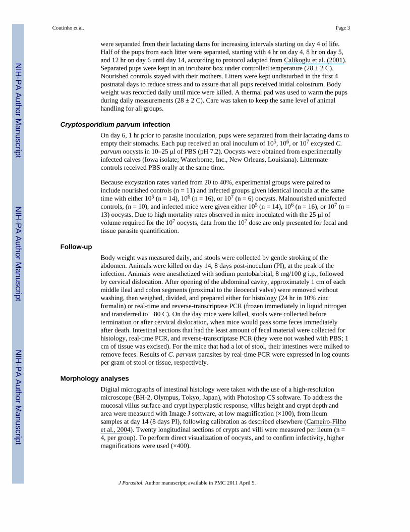
Malnourished and nourished mice infected with 106 oocysts had impaired weight gaincompared with their noninfected controls or with nourished mice infected with 105 oocysts(Fig. 1A, B). Nourished mice infected with 106 oocysts showed 41% less weight gaincompared with uninfected controls (P < 0.001) at day 14, 8 days postinfection (PI).Interestingly, nourished 106-infected mice have comparable weight decrements touninfected malnourished mice at the same endpoint. Undernourished mice given 106 oocystshad an additional 48% decrement in weight gain at day 14, compared to the average weightgain seen in the uninfected, undernourished controls, thus reaching a full 68% reductioncompared with uninfected nourished animals (P < 0.001). Therefore, body weight wasadditively affected by undernutrition and cryptosporidiosis, the latter in an inoculum-load–dependent fashion. Although some animals given 107 oocysts died with diarrhea within 1wk of infection, the few surviving animals provided samples to quantify infection, as shownin Table I.
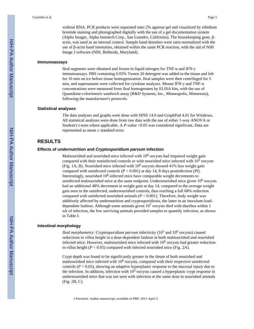
Intestinal morphologyIleal morphometry: Cryptosporidium parvum infectivity (105 and 106 oocysts) causedreductions in villus height in a dose-dependent fashion in both malnourished and nourishedinfected mice. However, malnourished mice infected with 106 oocysts had greater reductionin villus height (P < 0.05) compared with infected nourished mice (Fig. 2A).
Crypt depth was found to be significantly greater in the ileum of both nourished andmalnourished mice infected with 106 oocysts, compared with their respective uninfectedcontrols (P < 0.05), showing an adaptive hyperplastic response to the mucosal injury due tothe infection. In addition, infection with 105 oocysts caused a hyperplastic crypt response inundernourished mice that was not seen with infection at the same dose in nourished animals(Fig. 2B, C).
Coutinho et al. Page 5
J Parasitol. Author manuscript; available in PMC 2011 April 5.
NIH
-PA Author Manuscript
NIH
-PA Author Manuscript
NIH
-PA Author Manuscript
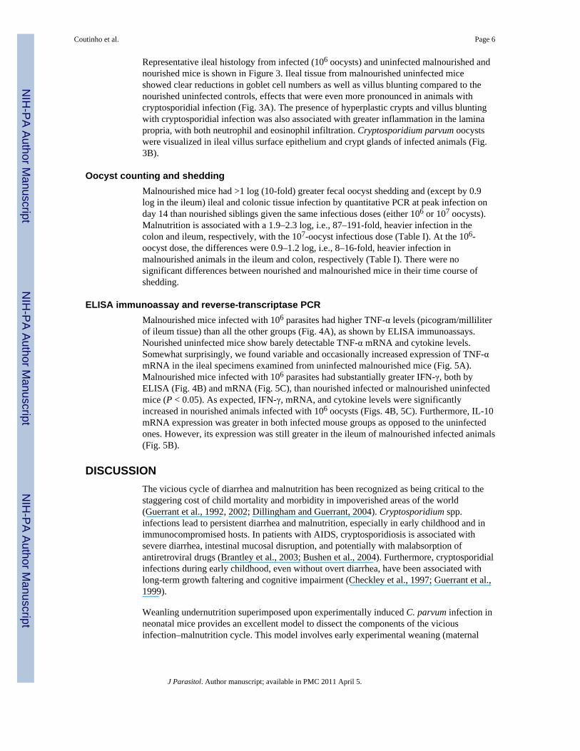
Representative ileal histology from infected (106 oocysts) and uninfected malnourished andnourished mice is shown in Figure 3. Ileal tissue from malnourished uninfected miceshowed clear reductions in goblet cell numbers as well as villus blunting compared to thenourished uninfected controls, effects that were even more pronounced in animals withcryptosporidial infection (Fig. 3A). The presence of hyperplastic crypts and villus bluntingwith cryptosporidial infection was also associated with greater inflammation in the laminapropria, with both neutrophil and eosinophil infiltration. Cryptosporidium parvum oocystswere visualized in ileal villus surface epithelium and crypt glands of infected animals (Fig.3B).
Oocyst counting and sheddingMalnourished mice had >1 log (10-fold) greater fecal oocyst shedding and (except by 0.9log in the ileum) ileal and colonic tissue infection by quantitative PCR at peak infection onday 14 than nourished siblings given the same infectious doses (either 106 or 107 oocysts).Malnutrition is associated with a 1.9–2.3 log, i.e., 87–191-fold, heavier infection in thecolon and ileum, respectively, with the 107-oocyst infectious dose (Table I). At the 106-oocyst dose, the differences were 0.9–1.2 log, i.e., 8–16-fold, heavier infection inmalnourished animals in the ileum and colon, respectively (Table I). There were nosignificant differences between nourished and malnourished mice in their time course ofshedding.
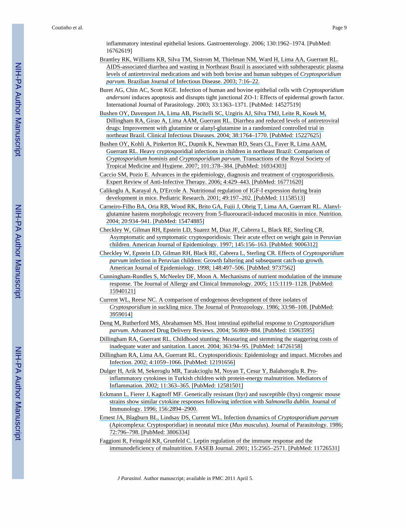
ELISA immunoassay and reverse-transcriptase PCRMalnourished mice infected with 106 parasites had higher TNF-α levels (picogram/milliliterof ileum tissue) than all the other groups (Fig. 4A), as shown by ELISA immunoassays.Nourished uninfected mice show barely detectable TNF-α mRNA and cytokine levels.Somewhat surprisingly, we found variable and occasionally increased expression of TNF-αmRNA in the ileal specimens examined from uninfected malnourished mice (Fig. 5A).Malnourished mice infected with 106 parasites had substantially greater IFN-γ, both byELISA (Fig. 4B) and mRNA (Fig. 5C), than nourished infected or malnourished uninfectedmice (P < 0.05). As expected, IFN-γ, mRNA, and cytokine levels were significantlyincreased in nourished animals infected with 106 oocysts (Figs. 4B, 5C). Furthermore, IL-10mRNA expression was greater in both infected mouse groups as opposed to the uninfectedones. However, its expression was still greater in the ileum of malnourished infected animals(Fig. 5B).
DISCUSSIONThe vicious cycle of diarrhea and malnutrition has been recognized as being critical to thestaggering cost of child mortality and morbidity in impoverished areas of the world(Guerrant et al., 1992, 2002; Dillingham and Guerrant, 2004). Cryptosporidium spp.infections lead to persistent diarrhea and malnutrition, especially in early childhood and inimmunocompromised hosts. In patients with AIDS, cryptosporidiosis is associated withsevere diarrhea, intestinal mucosal disruption, and potentially with malabsorption ofantiretroviral drugs (Brantley et al., 2003; Bushen et al., 2004). Furthermore, cryptosporidialinfections during early childhood, even without overt diarrhea, have been associated withlong-term growth faltering and cognitive impairment (Checkley et al., 1997; Guerrant et al.,1999).
Weanling undernutrition superimposed upon experimentally induced C. parvum infection inneonatal mice provides an excellent model to dissect the components of the viciousinfection–malnutrition cycle. This model involves early experimental weaning (maternal
Coutinho et al. Page 6
J Parasitol. Author manuscript; available in PMC 2011 April 5.
NIH
-PA Author Manuscript
NIH
-PA Author Manuscript
NIH
-PA Author Manuscript

separation), much like the early weaning with consequent heavy diarrhea burdens andmalnutrition that occurs in Brazilian shantytown children (Lima et al., 1992).
Novel real-time PCR methods have enabled us to quantify fecal, as well as tissue, parasites,as early as 2 days PI, in both nourished and malnourished experimentally infected mice. Wefound peak parasite numbers 8 days PI in mice (at 14 days old), a time similar to that foundby Ernest et al. (1986) in Swiss Webster mice.
In contrast to Sasahara et al. (2003), we found that C. parvum–infected mice, regardless ofnutritional status, showed significant decrements in weight gain as early as 2 days PI. Thisdiscrepancy is likely because of the inoculum tested. Sasahara et al. (2003) infected sucklingmice with only 104 oocysts, compared to our infections, which used 105 to 107 oocysts.Even with lighter doses of oocysts, however, they still observed severe ileal villus blunting.
Undernutrition and C. parvum infection were synergistic in disrupting the normalarchitecture of the ileum. The distal ileum was consistently found to be the predominant siteof C. parvum colonization and multiplication in suckling mice in our studies, as in those ofothers (Current and Reese, 1986). Blunted villi and deeper crypts in the ileum wereconsistently associated with the heavier C. parvum burden in the undernourished mice. Theaddition of undernutrition to C. parvum infection in suckling mice approximated the effectsof an additional log of oocyst infection in nourished mice. Even with the 105 parasiteinoculum, the addition of undernutrition increases the effect of deepening mucosal crypts,compared with nourished mice infected with the same oocyst inoculum, suggestingincreased cell turnover in response to mucosal epithelial disruption.
In addition, our morphological findings show increased mucosal inflammatory changes andleukocyte recruitment in the ileum of C. parvum–infected mice, particularly the onessubjected to undernutrition. High levels of IFN-γ and TNF-α (as we found in the ileal tissuesfrom undernourished infected mice), have been recently implicated in intestinal barrierdysfunction and enterocyte apoptosis (Begue et al., 2006; Wang et al., 2006), and correlatewith the degree of inflammatory response seen with heavy infection (Buret et al., 2003).Interestingly, severe undernutrition is also reported to elevate proinflammatory cytokines inchildren (Dulger et al., 2002).
IFN-γ is a well-recognized factor in the Th-1 cell-mediated response against C. parvuminfections. Increased expression of IFN-γ has been found following C. parvum challenge(Deng et al., 2004). Furthermore, in the absence of IFN-γ, TNF-α expression in the infectedintestinal mucosa is significantly decreased. TNF-α has been shown to reduce oocystshedding, via NF-κp upregulation (Lacroix et al., 2001; Lean et al., 2002). Hence, theincreased IFN-γ and TNF-α seen with heavier cryptosporidial infections in our malnourishedmice may either be blunted or rendered less effective by yet unknown mechanisms.
Interestingly, although micronutrient and vitamin deficiencies, such as zinc or vitamin Adepletion (Prasad, 2000; Wieringa et al., 2004), are believed to impair adaptive and innateimmune responses, malnourished infected mice did not show immune cytokine suppression;conversely, these animals showed a strong proinflammatory response, which might haveaccentuated the mucosal damage due to the malnutrition challenge. However, the effects ofspecific micronutrients on immunity following enteric infections are not well understood(Hughes and Kelly, 2006) and were not addressed in these studies.
Nutritional impairments may reduce immune-cell mass and indirectly affect immunefunction, particularly T helper cells (Fraker et al., 2000). Lymphopenia and thymic atrophy,which can be found in zinc deficiency, are now known to be due to loss of precursor T andB cells in the bone marrow (Thurnham, 1997). Additionally, T and B cell reductions may be
Coutinho et al. Page 7
J Parasitol. Author manuscript; available in PMC 2011 April 5.
NIH
-PA Author Manuscript
NIH
-PA Author Manuscript
NIH
-PA Author Manuscript

aggravated by premature weaning, due to reduced maternal-milk–derived lymphocytesbeing transferred to the pups (Flo et al., 1994). Deprivation of critical nutrients,lymphocytes, and maternal antibodies provided by full breast feeding might explain whyhigher intestinal levels of IFN-γ and TNF-α in malnourished infected mice were notsufficient to reduce the intensity of cryptosporidial infections.
Alcantara et al. (2003) found increased stool TNF-α and lactoferrin (a marker of neutrophilicinflammation) levels in Brazilian children with cryptosporidiosis. Kirkpatrick et al. (2002)also found that cryptosporidiosis stimulates intestinal inflammation in malnourished Haitianchildren, with predominant Th2 cytokine response and increased IL-10, but not IFN-γ. Thissuggests a counterregulatory role of IL-10 in blocking the Th1-mediated overresponse seenin malnourished and heavy infected ilea. However, IL-10 expression might also beconsidered a feature of ongoing immune responses against infectious pathogens (Mocellin etal., 2004). In our animal model, we also see increased IL-10 mRNA, especially in theinfected ileum in weanling malnourished animals. However, this occurred without absolutesuppression of Th-1-mediated responses, as seen by higher levels of IFN-γ secretion(detected by ELISA) and also RNA transcription (detected by reverse-transcriptase PCR). Itremains possible that these higher levels of IFN-γ represent relative suppression of levelsthat might otherwise have been even higher with the heavier intensity of infection.Undernutrition and poor weight gain (low fat mass) may also impair immune responses byreducing leptin levels. Leptin typically polarizes Th cells toward a Th1 phenotype (Faggioniet al., 2001).
In summary, undernutrition along with cryptosporidiosis causes mucosal disruption, reducedabsorptive surface, and increased proinflammatory cytokine responses, leading to growthimpairment. Furthermore, the intensity of the inflammatory response and mucosal disruptionparallel the burden of C. parvum infection.
Neonatal mice provide a model to demonstrate both the effect of cryptosporidial infection ongrowth impairment and the effect of weanling undernutrition on increased susceptibility tointensified infection. This model allows the dissection of the components of infection andmalnutrition in the “vicious cycle” that now enables further study of mechanisms andpotential interventions to help break this cycle that is so devastating to children's health anddevelopment.
AcknowledgmentsThis research was supported by NIH Cooperative Agreement U54 AI57168 for the Mid-Atlantic Regional Centerfor Excellence (MARCE) funded by the National Institute of Allergy and Infectious Diseases, and by FogartyInternational Center grant TW006713-01. Bruna P. Coutinho was supported in part by the Brazilian CAPES agencyand the Fund for the Improvement of Postsecondary Education (FIPSE) from the United States–Brazil HigherEducation Consortium Program and, with Dr. Carlos Vieira, by the Fogarty International Center Global InfectiousDiseases Research Training (GIDRT) program at NIH, grant D43 TW006578. Dr. Jesus Emmanuel Sevilleja wassupported by the Ellison Medical Foundation, grant ID-T-0019-03, and the Pfizer Initiative in International Healthat the University of Virginia.
LITERATURE CITEDAlcantara CS, Yang CH, Steiner TS, Barrett LJ, Lima AAM, Chappell CL, Okhuysen PC, White AC
JR. Guerrant RL. Interleukin-8, tumor necrosis factor-alpha, and lactoferrin in immunocompetenthosts with experimental and Brazilian children with acquired cryptosporidiosis. The AmericanJournal of Tropical Medicine and Hygiene. 2003; 68:325–328. [PubMed: 12685639]
Begue B, Wajant H, Bambou JC, Dubuquoy L, Siegmund D, Beaulieu JF, Canioni D, Berrebi D,Brousse N, Desreumaux P, et al. Implication of TNF-related apoptosis-inducing ligand in
Coutinho et al. Page 8
J Parasitol. Author manuscript; available in PMC 2011 April 5.
NIH
-PA Author Manuscript
NIH
-PA Author Manuscript
NIH
-PA Author Manuscript
inflammatory intestinal epithelial lesions. Gastroenterology. 2006; 130:1962–1974. [PubMed:16762619]
Brantley RK, Williams KR, Silva TM, Sistrom M, Thielman NM, Ward H, Lima AA, Guerrant RL.AIDS-associated diarrhea and wasting in Northeast Brazil is associated with subtherapeutic plasmalevels of antiretroviral medications and with both bovine and human subtypes of Cryptosporidiumparvum. Brazilian Journal of Infectious Disease. 2003; 7:16–22.
Buret AG, Chin AC, Scott KGE. Infection of human and bovine epithelial cells with Cryptosporidiumandersoni induces apoptosis and disrupts tight junctional ZO-1: Effects of epidermal growth factor.International Journal of Parasitology. 2003; 33:1363–1371. [PubMed: 14527519]
Bushen OY, Davenport JA, Lima AB, Piscitelli SC, Uzgiris AJ, Silva TMJ, Leite R, Kosek M,Dillingham RA, Girao A, Lima AAM, Guerrant RL. Diarrhea and reduced levels of antiretroviraldrugs: Improvement with glutamine or alanyl-glutamine in a randomized controlled trial innortheast Brazil. Clinical Infectious Diseases. 2004; 38:1764–1770. [PubMed: 15227625]
Bushen OY, Kohli A, Pinkerton RC, Dupnik K, Newman RD, Sears CL, Fayer R, Lima AAM,Guerrant RL. Heavy cryptosporidial infections in children in northeast Brazil: Comparison ofCryptosporidium hominis and Cryptosporidium parvum. Transactions of the Royal Society ofTropical Medicine and Hygiene. 2007; 101:378–384. [PubMed: 16934303]
Caccio SM, Pozio E. Advances in the epidemiology, diagnosis and treatment of cryptosporidiosis.Expert Review of Anti-Infective Therapy. 2006; 4:429–443. [PubMed: 16771620]
Calikoglu A, Karayal A, D'Ercole A. Nutritional regulation of IGF-I expression during braindevelopment in mice. Pediatric Research. 2001; 49:197–202. [PubMed: 11158513]
Carneiro-Filho BA, Oria RB, Wood RK, Brito GA, Fujii J, Obrig T, Lima AA, Guerrant RL. Alanyl-glutamine hastens morphologic recovery from 5-fluorouracil-induced mucositis in mice. Nutrition.2004; 20:934–941. [PubMed: 15474885]
Checkley W, Gilman RH, Epstein LD, Suarez M, Diaz JF, Cabrera L, Black RE, Sterling CR.Asymptomatic and symptomatic cryptosporidiosis: Their acute effect on weight gain in Peruvianchildren. American Journal of Epidemiology. 1997; 145:156–163. [PubMed: 9006312]
Checkley W, Epstein LD, Gilman RH, Black RE, Cabrera L, Sterling CR. Effects of Cryptosporidiumparvum infection in Peruvian children: Growth faltering and subsequent catch-up growth.American Journal of Epidemiology. 1998; 148:497–506. [PubMed: 9737562]
Cunningham-Rundles S, McNeeley DF, Moon A. Mechanisms of nutrient modulation of the immuneresponse. The Journal of Allergy and Clinical Immunology. 2005; 115:1119–1128. [PubMed:15940121]
Current WL, Reese NC. A comparison of endogenous development of three isolates ofCryptosporidium in suckling mice. The Journal of Protozoology. 1986; 33:98–108. [PubMed:3959014]
Deng M, Rutherford MS, Abrahamsen MS. Host intestinal epithelial response to Cryptosporidiumparvum. Advanced Drug Delivery Reviews. 2004; 56:869–884. [PubMed: 15063595]
Dillingham RA, Guerrant RL. Childhood stunting: Measuring and stemming the staggering costs ofinadequate water and sanitation. Lancet. 2004; 363:94–95. [PubMed: 14726158]
Dillingham RA, Lima AA, Guerrant RL. Cryptosporidiosis: Epidemiology and impact. Microbes andInfection. 2002; 4:1059–1066. [PubMed: 12191656]
Dulger H, Arik M, Sekeroglu MR, Tarakcioglu M, Noyan T, Cesur Y, Balahoroglu R. Pro-inflammatory cytokines in Turkish children with protein-energy malnutrition. Mediators ofInflammation. 2002; 11:363–365. [PubMed: 12581501]
Eckmann L, Fierer J, Kagnoff MF. Genetically resistant (Ityr) and susceptible (Itys) congenic mousestrains show similar cytokine responses following infection with Salmonella dublin. Journal ofImmunology. 1996; 156:2894–2900.
Ernest JA, Blagburn BL, Lindsay DS, Current WL. Infection dynamics of Cryptosporidium parvum(Apicomplexa: Cryptosporidiae) in neonatal mice (Mus musculus). Journal of Parasitology. 1986;72:796–798. [PubMed: 3806334]
Faggioni R, Feingold KR, Grunfeld C. Leptin regulation of the immune response and theimmunodeficiency of malnutrition. FASEB Journal. 2001; 15:2565–2571. [PubMed: 11726531]
Coutinho et al. Page 9
J Parasitol. Author manuscript; available in PMC 2011 April 5.
NIH
-PA Author Manuscript
NIH
-PA Author Manuscript
NIH
-PA Author Manuscript

Flo J, Elias F, Massouh E, Roux ME. Impairment of B and T cell maturation in gut associatedlymphoid tissues due to malnutrition during lactation. Developmental and ComparativeImmunology. 1994; 18:543–555. [PubMed: 7768319]
Fraker PJ, King LE, Laakko T, Vollmer TL. The dynamic link between the integrity of the immunesystem and zinc status. Journal of Nutrition. 2000; 130:1399S–1406S. [PubMed: 10801951]
Guerrant DI, Moore SR, Lima AA, Patrick PD, Schorling JB, Guerrant RL. Association of earlychildhood diarrhea and cryptosporidiosis with impaired physical fitness and cognitive functionfour—seven years later in a poor urban community in northeast Brazil. The American Journal ofTropical Medicine and Hygiene. 1999; 61:707–713. [PubMed: 10586898]
Guerrant RL. Cryptosporidiosis: An emerging, highly infectious threat. Emerging Infectious Diseases.1997; 3:51–57. [PubMed: 9126444]
Guerrant RL, Kosek M, Moore S, Lorntz B, Brantley R, Lima AA. Magnitude and impact of diarrhealdiseases. Archives of Medical Research. 2002; 33:351–355. [PubMed: 12234524]
Guerrant RL, Oria R, Bushen OY, Patrick PD, Huopt E, Lima AA. Global impact of diarrheal diseasesthat are sampled by travelers: The rest of the hippopotamus. Clinical Infectious Diseases. 2005;41(Suppl. 8):S524–S530. [PubMed: 16267713]
Guerrant RL, Schorling JB, Mcauliffe JF, de SOUZA MA. Diarrhea as a cause and effect ofmalnutrition: Diarrhea prevents catch-up growth and malnutrition increases diarrhea frequency andduration. The American Journal of Tropical Medicine and Hygiene. 1992; 47(Suppl.):28–35.[PubMed: 1632474]
Harp JA. Cryptosporidium and host resistance: Historical perspective and some novel approaches.Animal Health Research Reviews. 2003; 4:53–62. [PubMed: 12885209]
Houpt ER, Bushen OY, Sam NE, Kohli A, Asgharpour A, Ng CT, Calfee DP, Guerrant RL, Maro V,Ole-Nguyaine S, Shao JF. Short report: Asymptomatic Cryptosporidium hominis infection amonghuman immunodeficiency virus-infected patients in Tanzania. The American Journal of TropicalMedicine and Hygiene. 2005; 73:520–522. [PubMed: 16172475]
Hughes S, Kelly P. Interactions of malnutrition and immune impairment, with specific reference toimmunity against parasites. Parasite Immunology. 2006; 28:577–588. [PubMed: 17042929]
Karanis P, Kourenti C, Smith H. Waterborne transmission of protozoan parasites: A worldwide reviewof outbreaks and lessons learnt. Journal of Water and Health. 2007; 5:1–38. [PubMed: 17402277]
Kirkpatrick BD, Daniels MM, Jean SS, Pape JW, Karp C, Littenberg B, Fitzgerald DW, LedermanHM, Nataro JP, Sears CL. Cryptosporidiosis stimulates an inflammatory intestinal response inmalnourished Haitian children. The Journal of Infectious Diseases. 2002; 186:94–101. [PubMed:12089667]
Lacroix S, Mancassola R, Naciri M, Laurent F. Cryptosporidium parvum-specific mucosal immuneresponse in C57BL/6 neonatal and gamma interferon-deficient mice: Role of tumor necrosis factoralpha in protection. Infection and Immunity. 2001; 69:1635–1642. [PubMed: 11179338]
Lean IS, McDonald V, Pollok RC. The role of cytokines in the pathogenesis of Cryptosporidiuminfection. Current Opinion in Infectious Diseases. 2002; 15:229–234. [PubMed: 12015455]
Lima AA, Fang G, Schorling JB, de Albuquerque L, Mc-Auliffe JF, Mota S, Leite R, Guerrant RL.Persistent diarrhea in Northeast Brazil: Etiologies and interactions with malnutrition. ActaPaediatrica. 1992; 81(Suppl. 381):S39–S44.
Mocellin S, Marincola F, Rossi CR, Nitti D, Lise M. The multifaceted relationship between IL-10 andadaptive immunity: Putting together the pieces of the puzzle. Cytokine & Growth Factor Reviews.2004; 15:61–76. [PubMed: 14746814]
Newman RD, Sears CL, Moore SR, Nataro JP, Wuhib T, Agnew DA, Guerrant RL, Lima AA.Longitudinal study of Cryptosporidium infection in children in northeastern Brazil. The Journal ofInfectious Diseases. 1999; 180:167–175. [PubMed: 10353875]
Niehaus MD, Moore SR, Patrick PD, Derr LL, Lorntz B, Lima AA, Guerrant RL. Early childhooddiarrhea is associated with diminished cognitive function 4 to 7 years later in children in anortheast Brazilian shantytown. The American Journal of Tropical Medicine and Hygiene. 2002;66:590–593. [PubMed: 12201596]
Coutinho et al. Page 10
J Parasitol. Author manuscript; available in PMC 2011 April 5.
NIH
-PA Author Manuscript
NIH
-PA Author Manuscript
NIH
-PA Author Manuscript

Oria RB, Patrick PD, Blackman JA, Lima AA, Guerrant RL. Role of apolipoprotein E4 in protectingchildren against early childhood diarrhea outcomes and implications for later development.Medical Hypotheses. 2007; 68:1099–10107. [PubMed: 17098371]
Oria RB, Patrick PD, Zhang H, Lorntz B, Castro-Costa CM, Brito GA, Barrett LJ, Lima AA, GuerrantRL. APOE4 protects the cognitive development in children with heavy diarrhea burdens innortheast Brazil. Pediatric Research. 2005; 57:310–316. [PubMed: 15611352]
Pallaro AN, Roux ME, Slobodianik NH. Nutrition disorders and immunologic parameters: Study ofthe thymus in growing rats. Nutrition. 2001; 17:724–728. [PubMed: 11527659]
Prasad AS. Effects of zinc deficiency on Th1 and Th2 cytokine shifts. The Journal of InfectiousDiseases. 2000; 182(Suppl. 1):S62–S68. [PubMed: 10944485]
Ramirez NE, Ward LA, Sreevatsan S. A review of the biology and epidemiology of cryptosporidiosisin humans and animals. Microbes and Infection. 2004; 6:773–785. [PubMed: 15207825]
Sasahara T, Maruyama H, Aoki M, Kikuno R, Sekiguchi T, Takahashi A, Satoh Y, Kitasato H,Takayama Y, Inoue M. Apoptosis of intestinal crypt epithelium after Cryptosporidium parvuminfection. Journal of Infection and Chemotherapy. 2003; 9:278–281. [PubMed: 14513402]
Savioli L, Smith H, Thompson A. Giardia and Cryptosporidium join the “Neglected DiseasesInitiative.”. Trends in Parasitology. 2006; 22:203–208. [PubMed: 16545611]
Smith HV, Corcoran GD. New drugs and treatment for cryptosporidiosis. Current Opinion inInfectious Diseases. 2004; 17:557–564. [PubMed: 15640710]
Thurnham DI. Micronutrients and immune function: Some recent developments. Journal of ClinicalPathology. 1997; 50:887–891. [PubMed: 9462235]
Tyzzer EE. A sporozoan found in the peptic glands of the common mouse. Proceedings of the Societyfor Experimental Biology and Medicine. 1907; 5:12–13.
Tzipori S, Ward H. Cryptosporidiosis: Biology, pathogenesis and disease. Microbes and Infection.2002; 4:1047–1058. [PubMed: 12191655]
Wang F, Schwarz BT, Graham WV, Wang Y, Su L, Clayburgh DR, Abraham C, Turner JR. IFN-gamma-induced TNFR2 expression is required for TNF-dependent intestinal epithelial barrierdysfunction. Gastroenterology. 2006; 131:1153–1163. [PubMed: 17030185]
Wieringa FT, Dijkhuizen MA, West CE, van der Ven-Jongekrijg J, Muhilal, van der Meer JWM.Reduced production of immunoregulatory cytokines in vitamin A- and zinc-deficient Indonesianinfants. European Journal of Clinical Nutrition. 2004; 58:1498–1504. [PubMed: 15162133]
Coutinho et al. Page 11
J Parasitol. Author manuscript; available in PMC 2011 April 5.
NIH
-PA Author Manuscript
NIH
-PA Author Manuscript
NIH
-PA Author Manuscript

Figure 1.Body weight gain (% initial weight) from the experimental groups starting the day of C.parvum inoculation at day 6 until day 14 (8 days PI). (A) Nourished controls (NC, n = 11)are compared with nourished infected (NI) groups given 105, n = 14; or 106 oocysts, n = 16.(B) Malnourished uninfected controls (MC), n = 10, and malnourished infected mice (MI)treated with 106, n = 16; or 105, n = 14, oocysts. Statistical analyses were done from rawdata with the use of ANOVA. The significance level was set at P < 0.05. The results areshown as mean ± SEM.
Coutinho et al. Page 12
J Parasitol. Author manuscript; available in PMC 2011 April 5.
NIH
-PA Author Manuscript
NIH
-PA Author Manuscript
NIH
-PA Author Manuscript

Figure 2.Morphometric analyses of villus height (A), crypt depth (B), and (C) crypt area from C.parvum-infected and uninfected mice (n = 4 for each group). Statistical analyses were donefrom raw data with the use of ANOVA. The significance level was set at P < 0.05. C =uninfected control, 105 = inoculum with 105 oocysts; 106 = inoculum with 106 oocysts.
Coutinho et al. Page 13
J Parasitol. Author manuscript; available in PMC 2011 April 5.
NIH
-PA Author Manuscript
NIH
-PA Author Manuscript
NIH
-PA Author Manuscript
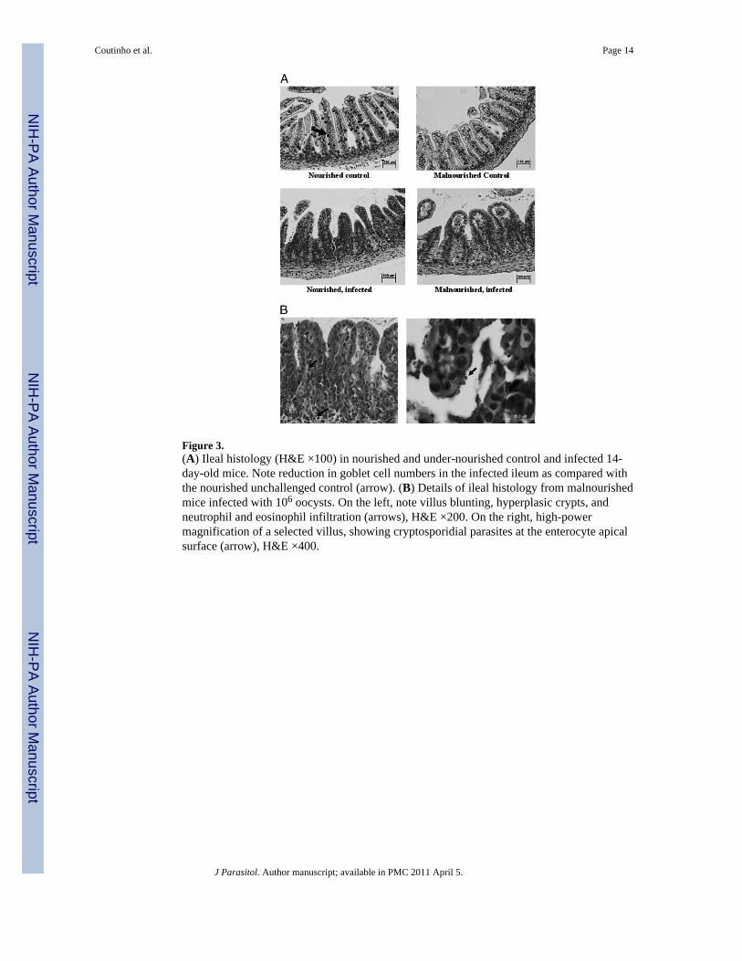
Figure 3.(A) Ileal histology (H&E ×100) in nourished and under-nourished control and infected 14-day-old mice. Note reduction in goblet cell numbers in the infected ileum as compared withthe nourished unchallenged control (arrow). (B) Details of ileal histology from malnourishedmice infected with 106 oocysts. On the left, note villus blunting, hyperplasic crypts, andneutrophil and eosinophil infiltration (arrows), H&E ×200. On the right, high-powermagnification of a selected villus, showing cryptosporidial parasites at the enterocyte apicalsurface (arrow), H&E ×400.
Coutinho et al. Page 14
J Parasitol. Author manuscript; available in PMC 2011 April 5.
NIH
-PA Author Manuscript
NIH
-PA Author Manuscript
NIH
-PA Author Manuscript

Figure 4.Tumor necrosis factor-alpha (TNF-α) (A) and interferon-gamma (IFN-γ) (B) concentrationsfrom frozen ileal segments. Mouse IFN-γ and TNF-α concentrations were measured in ilealhomogenates by ELISA. Statistical analyses were done with raw data (Student's t-test). Theresults are shown as mean ±SEM. (C) Noninfected control (n = 5, for both nourished andundernourished mice); 106 = inoculum with 106 oocysts (n = 6, for both nourished andundernourished mice). An asterisk indicates P < 0.05 versus respective control.
Coutinho et al. Page 15
J Parasitol. Author manuscript; available in PMC 2011 April 5.
NIH
-PA Author Manuscript
NIH
-PA Author Manuscript
NIH
-PA Author Manuscript

Figure 5.RT-PCR analysis on (A) tumor necrosis factor-alpha (TNF-α) and (B) interleukin-10 (IL-10)mRNA expression in the distal ileum (n = 4 for each group) from day 14 (8 days PI) ininfected (1 × 10⁶ C. parvum oocysts) and noninfected control mice, according to nutritionalstatus. (C) Interferon-gamma (IFN-γ) mRNA expression in the distal ileum in infected (1 ×10⁶ C. parvum oocysts) and noninfected control at day 14, according to nutritional status.The results are shown as mean ± SEM; *P < 0.05.
Coutinho et al. Page 16
J Parasitol. Author manuscript; available in PMC 2011 April 5.
NIH
-PA Author Manuscript
NIH
-PA Author Manuscript
NIH
-PA Author Manuscript

NIH
-PA Author Manuscript
NIH
-PA Author Manuscript
NIH
-PA Author Manuscript
Coutinho et al. Page 17
Tabl
e I
Log
coun
ts o
f C. p
arvu
m p
aras
ites i
n th
e ile
um, c
olon
, and
stoo
ls, a
s det
erm
ined
by
quan
titat
ive
real
-tim
e PC
R (q
RT-
PCR
).*
Sour
ceD
ay o
f life
Ooc
yst d
ose
Nou
rish
edN
Und
erno
uris
hed
NP
valu
e
Ileum
tiss
ue h
omog
enat
e14
106
3.23
± 0
.23
64.
13 ±
0.4
47
0.10
7
107
2.74
± 0
.13
35.
02 ±
0.1
16
<0.0
01
Col
on ti
ssue
hom
ogen
ate
1410
64.
13 ±
0.1
811
5.29
± 0
.44
110.
024
107
2.85
± 0
.11
34.
79 ±
0.2
57
0.00
1
Stoo
l dilu
tion
1410
63.
53 ±
0.2
614
4.55
± 0
.28
120.
012
* Res
ults
are
show
n in
a lo
g sc
ale
as m
ean
± SE
M. D
ata
wer
e an
alyz
ed w
ith th
e us
e of
the
Stud
ent's
t te
st. C
rypt
ospo
ridi
um p
arvu
m in
fect
ion
in th
e ile
um, c
olon
, and
shed
ding
in st
ools
was
det
erm
ined
by
qRT-
PCR
. qR
T-PC
R d
ata
wer
e ex
pres
sed
per g
ram
of s
tool
or t
issu
e.
J Parasitol. Author manuscript; available in PMC 2011 April 5.