Pemphigus Vulgaris Autoantibody Profiling by Proteomic Technique
10
Pemphigus Vulgaris Autoantibody Profiling by Proteomic Technique Mina Kalantari-Dehaghi 1 *, Grant J. Anhalt 2 , Michael J. Camilleri 3 , Alex I. Chernyavsky 1 , Sookhee Chun 4 , Philip L. Felgner 4 , Algis Jasinskas 4 , Kristin M. Leiferman 5 , Li Liang 4 , Steve Marchenko 1 , Rie Nakajima- Sasaki 4 , Mark R. Pittelkow 3 , John J. Zone 5 , Sergei A. Grando 1,6,7 * 1 Department of Dermatology, University of California Irvine, Irvine, California, United States of America, 2 Department of Dermatology, Johns Hopkins University, Baltimore, Maryland, United States of America, 3 Department of Dermatology, Mayo Clinic, Rochester, Minnesota, United States of America, 4 Department of Medicine, University of California Irvine, Irvine, California, United States of America, 5 Department of Dermatology, University of Utah, Salt Lake City, Utah, United States of America, 6 Department of Biological Chemistry, University of California Irvine, Irvine, California, United States of America, 7 Institute for Immunology, University of California Irvine, Irvine, California, United States of America Abstract Pemphigus vulgaris (PV) is a mucocutaneous blistering disease characterized by IgG autoantibodies against the stratified squamous epithelium. Current understanding of PV pathophysiology does not explain the mechanism of acantholysis in patients lacking desmoglein antibodies, which justifies a search for novel targets of pemphigus autoimmunity. We tested 264 pemphigus and 138 normal control sera on the multiplexed protein array platform containing 701 human genes encompassing many known keratinocyte cell-surface molecules and members of protein families targeted by organ-non- specific PV antibodies. The top 10 antigens recognized by the majority of test patients’ sera were proteins encoded by the DSC1, DSC3, ATP2C1, PKP3, CHRM3, COL21A1, ANXA8L1, CD88 and CHRNE genes. The most common combinations of target antigens included at least one of the adhesion molecules DSC1, DSC3 or PKP3 and/or the acetylcholine receptor CHRM3 or CHRNE with or without the MHC class II antigen DRA. To identify the PV antibodies most specific to the disease process, we sorted the data based on the ratio of patient to control frequencies of antigen recognition. The frequency of antigen recognition by patients that exceeded that of control by 10 and more times were the molecules encoded by the CD33, GP1BA, CHRND, SLC36A4, CD1B, CD32, CDH8, CDH9, PMP22 and HLA-E genes as well as mitochondrial proteins encoded by the NDUFS1, CYB5B, SOD2, PDHA1 and FH genes. The highest specificity to PV showed combinations of autoantibodies to the calcium pump encoded by ATP2C1 with C5a receptor plus DSC1 or DSC3 or HLA-DRA. The results identified new targets of pemphigus autoimmunity. Novel autoantibody signatures may help explain individual variations in disease severity and treatment response, and serve as sensitive and specific biomarkers for new diagnostic assays in PV patients. Citation: Kalantari-Dehaghi M, Anhalt GJ, Camilleri MJ, Chernyavsky AI, Chun S, et al. (2013) Pemphigus Vulgaris Autoantibody Profiling by Proteomic Technique. PLoS ONE 8(3): e57587. doi:10.1371/journal.pone.0057587 Editor: Andrzej T. Slominski, University of Tennessee, United States of America Received January 4, 2013; Accepted January 23, 2013; Published March 7, 2013 Copyright: ß 2013 Kalantari-Dehaghi et al. This is an open-access article distributed under the terms of the Creative Commons Attribution License, which permits unrestricted use, distribution, and reproduction in any medium, provided the original author and source are credited. Funding: The authors have no support or funding to report. Competing Interests: The authors have declared that no competing interests exist. * E-mail: [email protected] (SAG); [email protected] (MKD) Introduction Pemphigus vulgaris (PV) is a mucocutaneous blistering disease characterized by IgG autoantibodies against stratified squamous epithelium. PV antibodies demonstrate epithelial cell-surface staining by indirect immunofluorescence (IIF), and, because this staining appears between cells, initially the antibodies were described as ‘‘intercellular’’ antibodies [1,2]. Although the in- cidence of PV is only 1 to 16 per million population per year [3,4], this disease represents a significant burden to health care professionals, and the health care system. Systemic administration of glucocorticosteroid hormones is essential to establish control of disease during the acute stage [5]. While glucocorticosteroid treatment is life saving, it may cause severe side effects, including death [6,7]. The development of non-steroidal treatment has been hampered by a lack of clear understanding of the mechanisms leading to keratinocyte detachment in PV. During the last decade, the studies of autoimmune responses in PV have been supplemented and, to some extent, replaced by analyzing the levels of antibodies to desmoglein (Dsg) 3 by enzyme linked immunosorbent assay (ELISA) representing a hallmark and a diagnostic criterion of PV [8]. However, Dsg 3 antibody levels do not always correspond to the presence of cell-surface antibodies by IIF or correlate with disease activity [9,10,11] or predict relapse of the disease [12]. Furthermore, anti-Dsg antibodies can be absent in the active stage of disease but present in PV patients during remission [13,14,15,16,17,18], patients with unrelated medical conditions, and healthy subjects, including relatives of PV patients [17,19,20,21,22,23,24,25,26]. For example, 16 PV patients positive for cell-surface antibodies by IIF had normal Dsg 3 antibody levels [27]. Identification of proteins targeted by autoantibodies in PV is a subject of intense research. The first evidence that keratinocyte antigens other than Dsg 1 and Dsg 3 are pathophysiologically relevant was provided by experiments showing the ability to PLOS ONE | www.plosone.org 1 March 2013 | Volume 8 | Issue 3 | e57587
-
Upload
independent -
Category
Documents
-
view
7 -
download
0
Transcript of Pemphigus Vulgaris Autoantibody Profiling by Proteomic Technique

Pemphigus Vulgaris Autoantibody Profiling byProteomic TechniqueMina Kalantari-Dehaghi1*, Grant J. Anhalt2, Michael J. Camilleri3, Alex I. Chernyavsky1, Sookhee Chun4,
Philip L. Felgner4, Algis Jasinskas4, Kristin M. Leiferman5, Li Liang4, Steve Marchenko1, Rie Nakajima-
Sasaki4, Mark R. Pittelkow3, John J. Zone5, Sergei A. Grando1,6,7*
1Department of Dermatology, University of California Irvine, Irvine, California, United States of America, 2Department of Dermatology, Johns Hopkins University,
Baltimore, Maryland, United States of America, 3Department of Dermatology, Mayo Clinic, Rochester, Minnesota, United States of America, 4Department of Medicine,
University of California Irvine, Irvine, California, United States of America, 5Department of Dermatology, University of Utah, Salt Lake City, Utah, United States of America,
6Department of Biological Chemistry, University of California Irvine, Irvine, California, United States of America, 7 Institute for Immunology, University of California Irvine,
Irvine, California, United States of America
Abstract
Pemphigus vulgaris (PV) is a mucocutaneous blistering disease characterized by IgG autoantibodies against the stratifiedsquamous epithelium. Current understanding of PV pathophysiology does not explain the mechanism of acantholysis inpatients lacking desmoglein antibodies, which justifies a search for novel targets of pemphigus autoimmunity. We tested264 pemphigus and 138 normal control sera on the multiplexed protein array platform containing 701 human genesencompassing many known keratinocyte cell-surface molecules and members of protein families targeted by organ-non-specific PV antibodies. The top 10 antigens recognized by the majority of test patients’ sera were proteins encoded by theDSC1, DSC3, ATP2C1, PKP3, CHRM3, COL21A1, ANXA8L1, CD88 and CHRNE genes. The most common combinations oftarget antigens included at least one of the adhesion molecules DSC1, DSC3 or PKP3 and/or the acetylcholine receptorCHRM3 or CHRNE with or without the MHC class II antigen DRA. To identify the PV antibodies most specific to the diseaseprocess, we sorted the data based on the ratio of patient to control frequencies of antigen recognition. The frequency ofantigen recognition by patients that exceeded that of control by 10 and more times were the molecules encoded by theCD33, GP1BA, CHRND, SLC36A4, CD1B, CD32, CDH8, CDH9, PMP22 and HLA-E genes as well as mitochondrial proteinsencoded by the NDUFS1, CYB5B, SOD2, PDHA1 and FH genes. The highest specificity to PV showed combinations ofautoantibodies to the calcium pump encoded by ATP2C1 with C5a receptor plus DSC1 or DSC3 or HLA-DRA. The resultsidentified new targets of pemphigus autoimmunity. Novel autoantibody signatures may help explain individual variations indisease severity and treatment response, and serve as sensitive and specific biomarkers for new diagnostic assays in PVpatients.
Citation: Kalantari-Dehaghi M, Anhalt GJ, Camilleri MJ, Chernyavsky AI, Chun S, et al. (2013) Pemphigus Vulgaris Autoantibody Profiling by ProteomicTechnique. PLoS ONE 8(3): e57587. doi:10.1371/journal.pone.0057587
Editor: Andrzej T. Slominski, University of Tennessee, United States of America
Received January 4, 2013; Accepted January 23, 2013; Published March 7, 2013
Copyright: � 2013 Kalantari-Dehaghi et al. This is an open-access article distributed under the terms of the Creative Commons Attribution License, whichpermits unrestricted use, distribution, and reproduction in any medium, provided the original author and source are credited.
Funding: The authors have no support or funding to report.
Competing Interests: The authors have declared that no competing interests exist.
* E-mail: [email protected] (SAG); [email protected] (MKD)
Introduction
Pemphigus vulgaris (PV) is a mucocutaneous blistering disease
characterized by IgG autoantibodies against stratified squamous
epithelium. PV antibodies demonstrate epithelial cell-surface
staining by indirect immunofluorescence (IIF), and, because this
staining appears between cells, initially the antibodies were
described as ‘‘intercellular’’ antibodies [1,2]. Although the in-
cidence of PV is only 1 to 16 per million population per year [3,4],
this disease represents a significant burden to health care
professionals, and the health care system. Systemic administration
of glucocorticosteroid hormones is essential to establish control of
disease during the acute stage [5]. While glucocorticosteroid
treatment is life saving, it may cause severe side effects, including
death [6,7]. The development of non-steroidal treatment has been
hampered by a lack of clear understanding of the mechanisms
leading to keratinocyte detachment in PV.
During the last decade, the studies of autoimmune responses in
PV have been supplemented and, to some extent, replaced by
analyzing the levels of antibodies to desmoglein (Dsg) 3 by enzyme
linked immunosorbent assay (ELISA) representing a hallmark and
a diagnostic criterion of PV [8]. However, Dsg 3 antibody levels
do not always correspond to the presence of cell-surface antibodies
by IIF or correlate with disease activity [9,10,11] or predict relapse
of the disease [12]. Furthermore, anti-Dsg antibodies can be
absent in the active stage of disease but present in PV patients
during remission [13,14,15,16,17,18], patients with unrelated
medical conditions, and healthy subjects, including relatives of
PV patients [17,19,20,21,22,23,24,25,26]. For example, 16 PV
patients positive for cell-surface antibodies by IIF had normal Dsg
3 antibody levels [27].
Identification of proteins targeted by autoantibodies in PV is
a subject of intense research. The first evidence that keratinocyte
antigens other than Dsg 1 and Dsg 3 are pathophysiologically
relevant was provided by experiments showing the ability to
PLOS ONE | www.plosone.org 1 March 2013 | Volume 8 | Issue 3 | e57587
induce suprabasal acantholysis and gross skin blisters in Dsg32/2
neonatal mice by passive transfer of PV antibodies [28]. In this
model, murine epidermis lacks Dsg 3 and the passively transferred
PV IgG lacks Dsg 1 antibody. Hence, the injected PV antibodies
cause blisters by targeting non-Dsg 1 and Dsg 3 keratinocyte
antigens. Current understanding, however, does not adequately
explain the mechanism of acantholysis in patients lacking Dsg 1
and 3 antibodies. Furthermore, results of a recent study indicate
that autoreactivity in PV relies on somatic mutations generated in
response to an antigen unrelated to Dsg 3 [29]. Taken together,
these facts justify a search for novel targets of pemphigus
autoimmunity.
In general, autoimmune diseases are characterized by the
presence of multiple types of autoantibodies mediating a co-
ordinated immunological attack against a fraction of the tissue
proteome. For example, 116 autoantibodies were described in
patients with systemic lupus erythematous [30]. The number of
targeted self- antigens varies dramatically from patient to patient.
Therefore, multiplex analysis of autoantibody responses against
a spectrum of candidate antigens represents a powerful screening
tool to delineate biomarker signatures in autoimmunity, allowing
elucidation of the overall autoimmune process rather than
individual components [31]. The availability of multiplex tech-
nologies has made possible the simultaneous detection of several
different autoantibodies overcoming some of the limitations of
conventional methods [32]. For instance, antigen arrays proved to
be 4- to 8-fold more sensitive than conventional ELISA analyses
for detection of autoantibodies specific for some autoantigens [33].
Thus, autoantibody profiling may serve purposes including
classification of individual patients and subsets of patients based
on their ‘‘autoantibody fingerprint,’’ examination of epitope
spreading and antibody isotype usage, discovery and character-
ization of candidate autoantigens, and tailoring antigen-specific
therapy [34,35].
Historically, studies of autoimmune responses had been
conducted by analyzing the presence and/or concentration of
single antibodies in biological fluids using conventional immu-
noassays, such as ELISA, radioimmunoassay, immunoblot, and
others. More recently, antigen microarrays have been constructed
and validated for over a dozen autoimmune diseases, including
connective-tissue diseases, primary biliary cirrhosis, experimental
autoimmune encephalomyelitis, multiple sclerosis, rheumatoid
arthritis, diabetes, Crohn’s disease and sclerosing cholangitis
[36]. Previous reports demonstrated feasibility of the protein
microarray as a laboratory tool allowing parallel analysis against
hundreds of different antigens in minimal serum quantities (less
than 2 mL). We pioneered the use of multiplexed protein array
platforms to evaluate PV autoantibody profiles [37]. In our
previous study, the sera from acute PV patients and healthy
donors were probed using the microarray containing self-antigens
characteristic of the organ-non-specific autoimmune disorders,
such as rheumatoid arthritis, lupus erythematosus, scleroderma,
diabetes and some other autoimmune disorders [37]. The results
identified the presence of several non-organ specific antibodies,
but the relatively small sample size did not allow determination of
their prevalence in PV patients. Most recently, some of these new
autoantibodies were validated in an independent proteomic study
[38], indicating reliability of identifying novel disease-specific
autoantibodies in PV through multiplexed parallel testing.
To elucidate the immunopathological mechanisms underlying
keratinocyte detachment in PV, we designed a multiplexed protein
array platform encompassing most of known keratinocyte cell-
surface molecules as well as members of protein families targeted
by organ-non-specific PV antibodies. In the present study, we
utilized this specialized microarray to test a large number of PV
patient and control sera to define the proteome targeted by PV
autoimmunity. As predicted by the multiple hit hypothesis [39], an
in-depth analysis of PV sera revealed new self-antigens and
identified specific patterns of differentially reacting autoantibodies.
Such novel autoantibody signatures may help explain individual
variations of disease severity and response to treatment in PV
patients, and serve as sensitive and specific biomarkers for new
diagnostic assays.
Materials and Methods
Test SeraWe tested 264 PV patient and 138 normal serum specimens.
Patient specimens were selected based on IIF results showing the
presence of cell-surface antibodies staining stratified squamous
epithelial substrate (human skin and/or monkey esophagus
purchased from California National Primate Research Center,
Davis, CA; http://www.cnprc.ucdavis.edu/) in the characteristic
pemphigus pattern at a serum titer of 1:40 concentration and
higher. The Dsg3 antibody level was measured using the
MESACUP Dsg3 ELISA test system (MBL International Corp.,
Nagoya, Japan). Patient specimens were de-identified prior to
testing. As controls, we used sera collected from healthy donors at
University of California Irvine and normal human sera purchased
from Bioreclamation, Inc. (Westbury, NY). This research has been
approved by Institutional Review Boards (IRB) at University of
California Irvine. Participants provided their written informed
consent on the IRB-apprroved consent forms to participate in this
study.
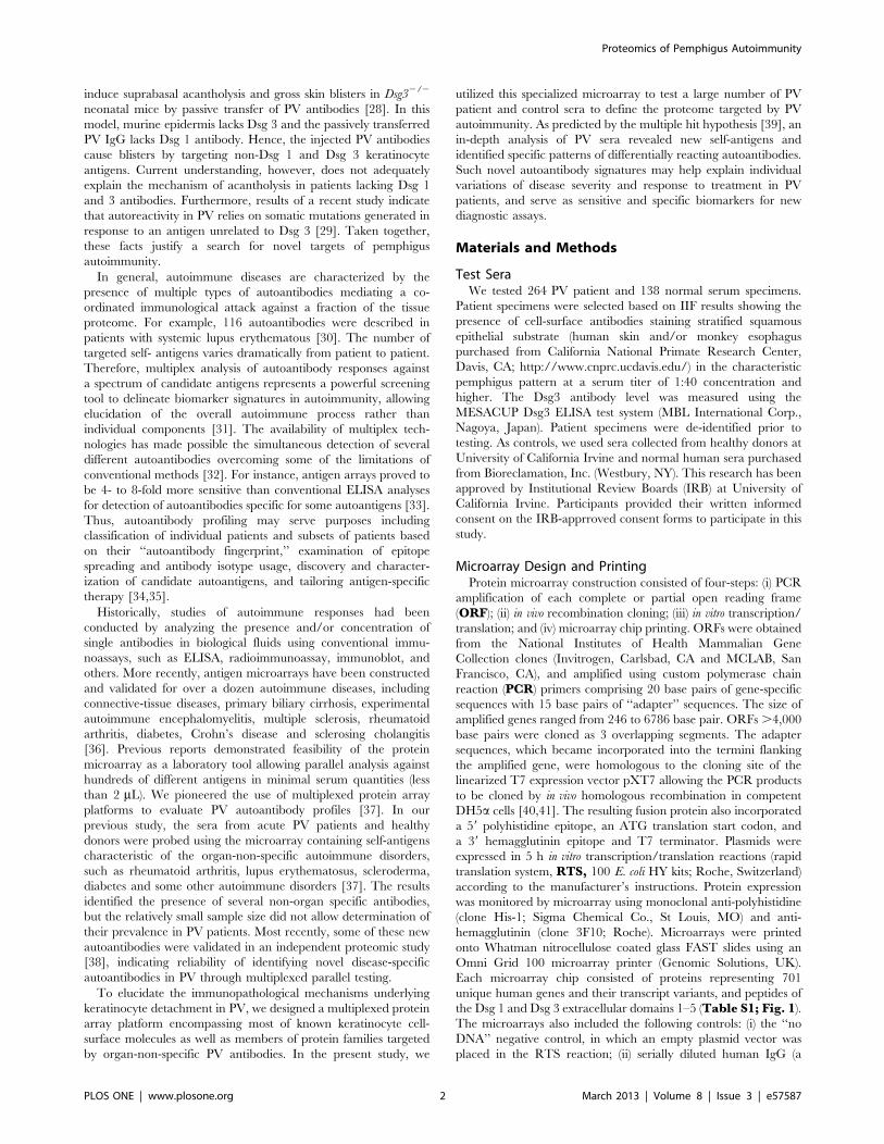
Microarray Design and PrintingProtein microarray construction consisted of four-steps: (i) PCR
amplification of each complete or partial open reading frame
(ORF); (ii) in vivo recombination cloning; (iii) in vitro transcription/
translation; and (iv) microarray chip printing. ORFs were obtained
from the National Institutes of Health Mammalian Gene
Collection clones (Invitrogen, Carlsbad, CA and MCLAB, San
Francisco, CA), and amplified using custom polymerase chain
reaction (PCR) primers comprising 20 base pairs of gene-specific
sequences with 15 base pairs of ‘‘adapter’’ sequences. The size of
amplified genes ranged from 246 to 6786 base pair. ORFs .4,000
base pairs were cloned as 3 overlapping segments. The adapter
sequences, which became incorporated into the termini flanking
the amplified gene, were homologous to the cloning site of the
linearized T7 expression vector pXT7 allowing the PCR products
to be cloned by in vivo homologous recombination in competent
DH5a cells [40,41]. The resulting fusion protein also incorporated
a 59 polyhistidine epitope, an ATG translation start codon, and
a 39 hemagglutinin epitope and T7 terminator. Plasmids were
expressed in 5 h in vitro transcription/translation reactions (rapid
translation system, RTS, 100 E. coli HY kits; Roche, Switzerland)
according to the manufacturer’s instructions. Protein expression
was monitored by microarray using monoclonal anti-polyhistidine
(clone His-1; Sigma Chemical Co., St Louis, MO) and anti-
hemagglutinin (clone 3F10; Roche). Microarrays were printed
onto Whatman nitrocellulose coated glass FAST slides using an
Omni Grid 100 microarray printer (Genomic Solutions, UK).
Each microarray chip consisted of proteins representing 701
unique human genes and their transcript variants, and peptides of
the Dsg 1 and Dsg 3 extracellular domains 1–5 (Table S1; Fig. 1).The microarrays also included the following controls: (i) the ‘‘no
DNA’’ negative control, in which an empty plasmid vector was
placed in the RTS reaction; (ii) serially diluted human IgG (a
Proteomics of Pemphigus Autoimmunity
PLOS ONE | www.plosone.org 2 March 2013 | Volume 8 | Issue 3 | e57587

positive control); and (iii) serially diluted EBNA-1 (another positive
control, given the high prevalence of latent Epstein Barr viral
infection).
Antibody Assays and Reading of MicroarraysPrior to array staining, experimental and control serum samples
were diluted to 1/25 in Whatman’s Protein Array Blocking Buffer
containing Escherichia coli lysate at a final concentration of 30% and
incubated at room temperature for 1 h with constant mixing. The
arrays were rehydrated in the blocking buffer for 30 min that was
replaced with preabsorbed test sera and incubated overnight at
4uC with constant agitation. The slides were washed five times in
10 mM Tris (pH 8.0)-150 mM NaCl containing 0.05% Tween 20
buffer, and bound antibodies were detected after 1 h incubation in
Biotin-SP conjugated affinity purified human goat secondary
antibody to IgG Fc (Fc-c fragment specific) (Jackson Immunor-
esearch, West Grove, PA) diluted 1/200 in blocking buffer. The
slides were washed three times, and bound antibodies detected by
1 h incubation with StreptAvidin conjugated with the dye PBXL-3
as tertiary antibody (streptavidin-conjugated SureLightH P-3)
(Columbia Biosciences Corporation, Columbia, MD) diluted 1/
200 in blocking buffer. After being washed three times, the slides
were air-dried under brief centrifugation and stored at 18uC in
a desiccator. The arrays were examined with a Perkin Elmer
ScanArray Express HT apparatus at a wavelength of 670 nm and
intensities were quantified using ProScanArray Express software
(Perkin Elmer, Waltham, MA). All signal intensities were corrected
for spot-specific background.
Data AnalysisSignal intensity was obtained from ScanArray and background
signal was corrected by subtracting ‘‘no DNA’’ negative control
plus 2 standard deviations (SD) from the signals in each antigen.
This standardized signal was used to determine positives in
response to antigen on array. The positives were defined as the
signal intensity higher than the average signal intensity of control
group plus 2 SD. A two-tailed unpaired Student’s t-test was used
to verify the significance of differences between the groups, and p
values less than 0.05 were considered significant. The microarray’s
ability to detect disease-related antibodies was evaluated based on
the ratio of positive patient vs. control sera. JMP Software (SAS
Institute Inc., Cary, NC) was used to perform the principal
component analysis as detailed elsewhere [42].
Results
Self-antigens Targeted by Pemphigus AntibodiesAnalyses of the data revealed positive reactions of both PV
patient and control sera with a large number of peptides included
in the microarray (Table S1). The specificity was determined by
the frequency of antigen recognition by PV sera. To identify the
PV antibodies most specific to the disease process, we re-sorted the
data based on the ratio of patient to control frequencies of antigen
recognition in the microarray. The unsupervised principal
component analysis demonstrated that the top 30 antigens
recognized by PV antibodies with the highest sensitivity or top
15 antigens recognized with the highest specificity were clearly
distinct from controls (Fig. 2). These results indicated that PV
features a unique serological profile that distinguishes pemphigus
autoimmunity from normal immune response.
The sensitivity analysis (Fig. 3) demonstrated that the majority
of patients’ sera targeted the DR a chain of the class II major
histocompatibility complex (MHC) encoded by the human
leukocyte antigen (HLA)-DRA gene (45% PV patients), desmo-
collin 1 and desmocollin 3 (DSC1 and DSC3, respectively; 44%
each), ATPase, Ca++ transporting, type 2C, member 1 (ATP2C1;
43%), plakophilin 3 (PKP3; 43%), M3 subtype of muscarinic
acetylcholine receptor (AChR) (CHRM3; 42%), collagen a1, typeXXI, (COL21A1; 42%), annexin A8-like 1 molecule (ANXA8L1;
42%), complement component 5a receptor 1 (CD88; 42%) and esubunit of nicotinic AChR (CHRNE; 41%).
The specificity analysis (Fig. 4) demonstrated that self-antigens
recognized by PV antibodies with the frequency that exceeded
that of control by 10 and more times included the cell-surface
molecules sialic acid-binding immunoglobulin-like lectin 3 (CD33;
ratio = 27.7) and glycoprotein Iba (GP1BA; 27.7), d subunit of
nicotinic AChR (CHRND; 17.6), proton-coupled amino acid
transporter 4 (SLC36A4; 17.3), the antigen-presenting protein
CD1B (13.1), Fc-fragment of IgG (CD32; 12.5), cadherins 8
(CDH8; 11.3) and 9 (CDH9; 11.5), peripheral myelin protein 22
(PMP22; 11.0), the MHC class I molecule E (HLA-E; 10.8) and
the mitochondrial proteins NADH-ubiquinone oxidoreductase
(NDUFS1; 16.2), cytochrome b5 outer mitochondrial membrane
isoform precursor (CYB5B; 13.1), superoxide dismutase (SOD2;
Figure 1. Composition of the multiplexed protein array platform.doi:10.1371/journal.pone.0057587.g001
Proteomics of Pemphigus Autoimmunity
PLOS ONE | www.plosone.org 3 March 2013 | Volume 8 | Issue 3 | e57587

10.6), a subunit of pyruvate dehydrogenase E1 component
(PDHA1; 10.3) and fumarate hydratase (FH; 10.1).
Non-Dsg 3 Antibodies Produced by the Dsg 3 Antibody-positive PV PatientsAlthough sera from all 264 patients demonstrated a pemphigus
cell-surface staining pattern by IIF, only 183 (69%) of them
recognized at least one Dsg 3 peptide in the microarray (TableS1). This was not surprising, because Amagai’s group reported
that only 60 to 75 percent of PV patients develop autoantibodies
recognizing recombinant Dsg 3 proteins produced in a bacterial
expression system [43]. In our study, the Dsg 3 antibody-negative
patient sera likely were from PV patients who did not have
detectable levels of Dsg 3 antibody, which is not uncommon [16],
and/or pemphigus foliaceus (PF) patients, since both PV and PF
antibodies demonstrate an indistinguishable IIF staining pattern
[21,44,45]. Therefore, to extrapolate our results specifically to the
immunopathology of PV, we separately determined antigen
reaction patterns of the Dsg 3 antibody-containing patient sera
(Table S2). The antigen reactivity pattern of the Dsg 3 antibody-
Figure 2. Principal component analysis of top antigens. Unsupervised principal component analysis of the signal intensity for samples from PVpatients and healthy controls revealed that these two groups could be segregated on the basis of top 30 antigens with the highest sensitivity (A) and15 antigens with the highest specificity (B) for PV.doi:10.1371/journal.pone.0057587.g002
Figure 3. Sensitivity of the microarray in detecting disease-related autoantibodies. Sorted by percent of positive samples in the group ofantigens recognized by 10 percent and more of PV patients. Inset: 25 top antigens.doi:10.1371/journal.pone.0057587.g003
Proteomics of Pemphigus Autoimmunity
PLOS ONE | www.plosone.org 4 March 2013 | Volume 8 | Issue 3 | e57587

positive sera was similar to that of the entire collection of patients’
sera (Table S1). Most importantly, the top 10 most frequently
recognized self-antigens were identical, but displayed a slightly
different order within the group (Table 1). Therefore, the
microarray results of the entire serum collection were represen-
tative of PV autoimmunity.
Combinations of most Common Individual AntigensTargeted by Antibodies in PV PatientsThe top 10 most commonly recognized antigens (Table 1) were
subjected to an association analysis. We combined two and more
from the top 10 most commonly recognized antigens and
calculated the number of patient and control sera that contained
antibodies against those combined antigens, then computed the
ratio of patient over control sera positivity. The most common
combinations of target antigens included at least one of the
adhesion molecules DSC1, DSC3 and PKP3 and/or the AChR
CHRM3 or CHRNE with or without the MHC class II antigen
DRA (Fig. 5A). The highest specificity to PV showed combina-
tions of antibodies to the calcium pump encoded by ATP2C1 with
CD88 (C5a receptor) plus DSC1 or DSC3 or HLA-DRA
(Fig. 5B).
Discussion
In this study, we used sera from a large cohort of pemphigus
patients to identify novel targets for PV antibodies. We applied
a protein microarray approach to characterize the autoantibody
response profile associated with the disease process. This approach
has been employed broadly to identify differentially reactive,
serodiagnostic antigens against infectious agents, and is now also
being used in autoimmune diseases. In the past, our group has
extensively characterized disease-associated antibody profiles in
large cohorts of healthy and infected individuals (e.g., [42,46]), and
also initiated the proteomic analysis of pemphigus autoimmunity
[37]. The pilot study of PV sera demonstrated that the reactivities
of novel PV antibodies correlate closely with the Dsg 3 antibody
levels [37]. In the present study, we used polypeptides representing
Figure 4. Specificity of the microarray in revealing disease-specific PV antibodies. Sorted by ratio of positive patient/control samples inthe group of antigens recognized by two and more time frequently by PV patient than control sera. Inset: 25 top antigens.doi:10.1371/journal.pone.0057587.g004
Table 1. Top 10 antigens most frequently recognized by PV antibodies.
Symbol Name % of all patient samples% of Dsg3-antibodypositive samples
HLA-DRA major histocompatibility complex, class II, DR a chain 45 53
DSC3 desmocollin 3 isoform Dsc3a preproprotein 44 54
DSC1 desmocollin 1 isoform Dsc1a preproprotein 44 54
ATP2C1 ATPase, Ca++ transporting, type 2C, member 1 43 50
PKP3 plakophilin 3 43 53
CHRM3 cholinergic muscarinic receptor type 3 42 51
COL21A1 collagen, type XXI, a1 42 50
ANXA8L1 annexin A8-like 1 42 49
CD88 complement component 5a receptor 1 42 50
CHRNE nicotinic acetylcholine receptor e subunit 41 50
doi:10.1371/journal.pone.0057587.t001
Proteomics of Pemphigus Autoimmunity
PLOS ONE | www.plosone.org 5 March 2013 | Volume 8 | Issue 3 | e57587

Proteomics of Pemphigus Autoimmunity
PLOS ONE | www.plosone.org 6 March 2013 | Volume 8 | Issue 3 | e57587

different regions of Dsg 3 because, at variance with early studies
[47,48], it has been recently demonstrated that the Dsg 3
autoantibody response in PV is polyclonal [29]. We also tested
autoantibody reactivities with other proteins implicated in PV
pathogenesis, such as AChRs [39].
We sought to reconcile the knowledge about important role of
Dsg 3 antibody in PV with the evidence that Dsg 3 is not essential
for keratinocyte adhesion and Dsg 3 antibody is not requisite for
acantholysis (reviewed in [49]). The results revealed new targets of
pemphigus autoimmunity, and demonstrated variability of the
autoantibody profile among different PV patients. We identified
both the most common and the most specific autoantibodies
directed against members of the cell adhesion molecule and the
AChR families, which are known targets in pemphigus, as well as
a number of new organ-specific and non-specific targets from
previously unsuspected protein families. Based on these data, it is
apparent that the immunopathology of PV is complex and
variable, inducing alterations in vital keratinocyte functions due to
a simultaneous hits by an array of autoantibodies of different
antigenic specificities. Targeting of non-Dsg molecules potentially
exacerbates the pathogenic effects of Dsg 3 antibody. Our new PV
autoantibody database should provide a roadmap to navigate
pemphigus research toward new disease pathways and treatment
approaches.
An important role of the conformational epitope of self-antigens
in PV has been vividly demonstrated in the studies of the
recombinant Dsg 3 species that were raised in different expression
systems and had various posttranslational modifications
[43,50,51,52]. An inherent drawback of the high-throughput
approach applied here is that not all proteins on the array likely
are folded in the same way as in human cells, and/or that they are
not post-translationally modified in the same way. Consequently,
they may not display all possible antigenic epitopes. This potential
limitation, however, did not prevent us from reaching the main
objective of our study, which was identification of new pathogenic
PV autoantibodies that may work together with Dsg 3 antibodies
to disrupt keratinocyte adhesion. Hundreds of autoantigens were
found to be significantly more reactive with PV than control sera.
Importantly, the top antigenic targets significantly separated PV
patients from healthy individuals, demonstrating strong sensitivity
and specificity. This observation indicated that the two approaches
to microarray data analysis can be used together. The proteomic
approach, therefore, serves as a starting point to ‘‘rule in,’’ but not
necessarily ‘‘rule out,’’ individual self-antigens or their combina-
tions that contribute to the autoimmune response.
The results indicate that acantholysis in PV likely derives from
simultaneous inactivation of several physiological pathways
maintaining keratinocyte adhesion, as predicted by the ‘‘multiple
hit’’ hypothesis of pemphigus pathophysiology [39]. In addition to
Dsg 3, other keratinocyte adhesion molecules are presumably
destroyed or inactivated by PV antibodies. Notably, previous
results showing that chimeric proteins containing the extracellular
epitope of Dsg 1 or Dsg 3 combined with the Fc portion of human
IgG absorbed out all disease-causing pemphigus IgGs [50,53,54]
should be interpreted with caution, because both this and previous
[37] proteomics studies demonstrated that PV patients produce
autoantibodies against Fc-IgG.
Our results are consistent with previous reports that desmo-
collins [55,56] and classical cadherins [57,58] also are targeted by
pemphigus antibodies. The significance of the autoantibody
targeting DSC3 in PV is underscored by the evidence that: (i)
the DSC3 loss of function mouse model exhibits phenotypic
similarity to PV [59], (ii) adsorption with DSC3 can eliminate
acantholytic activity of PV IgG [60], and (iii) DSC3 monoclonal
antibody causes intraepidermal blistering in in vitro model of
human skin and loss of cell-cell adhesion in keratinocyte cultures
[61]. Indeed, production of certain autoantibodies against cell
adhesion molecules and structural proteins, such as the in-
tracellular desmosomal plaque protein, plakophilin 3, and the
collagen a1, type XXI, may be secondary to acantholysis, as
discussed in detail elsewhere [62].
The high percentage of patients’ sera reacting with AChRs of
the muscarinic and/or the nicotinic classes is in keeping with
previous reports that downstream signaling from these receptors
regulates keratinocyte cell-cell adhesion through physiological
control of phosphorylation/dephosphorylation of desmosomal and
classical cadherins [63]. Blocking AChRs expressed on keratino-
cytes leads to disassembly of desmosomal and adherence junctions
due to phosphorylation of key adhesion molecules. Our early
reports that PV and PF patients develop antibodies to keratinocyte
AChRs [28,64] and that cholinomimetic drugs can ameliorate
pemphigus [65] have been recently corroborated by new clinical
and laboratory data. AChR antibodies in PV patients correlate
with disease extent at the time of diagnosis and during follow-up
[66], and, in a patient with bipolar disorder, cholinolytic drugs
worsen PF [67].
The high reactivity of PV sera with the annexin A8-like
molecule was not surprising either, because it had been reported
that PV patients develop antibodies to different annexins [68].
Probing of keratinocyte lgt11 cDNA library with the PV IgG
eluted from a 75 kD band that stained epidermis in a pemphigus-
like cell-surface pattern and caused acantholysis in keratinocyte
monolayers revealed a novel type of AChRs, termed pemphaxin
(a.k.a. annexin 9) [69]. Annexin A8 is specifically expressed in
adult stratified epithelia [70], where it may participate in the
organization of certain actin associated membrane domains and
regulate late endosome organization and function [71]. By analogy
to annexins 1, 2, 3 and 9 that act as non-professional AChRs
[69,72], the annexin A8-like molecule also may mediate acetyl-
choline signaling and, thus, be involved in regulation of
keratinocyte shape and adhesion. This possibility needs further
investigation.
The microarray results from this study demonstrate that
mitochondrial antibodies are highly specific to the PV sera. The
presence of antibodies against mitochondrial proteins in PV and
PF patients had been reported [73,74]. Mitochondrial antibodies
are apparently pathogenic because their absorption abolishes the
ability of PV IgG to cause acantholysis both in vitro and in vivo [74].
Based on the known functions of targeted proteins, the following
mitochondrial pathways may be subject to dysfunction: tri-
carboxylic acid cycle, oxidative phosphorylation, O2 respiration,
and production/inactivation of reactive oxygen species (ROS).The mitochondrial dysfunction in PV has been directly or
indirectly suggested by an increase of lipid peroxidation, reflecting
an increased production of ROS [75,76], and the peroxidant-
antioxidant balance, measuring oxidative stress [77], and activa-
tion of the mitochondria-dependent intrinsic apoptotoc pathway in
keratinocytes exposed to PV IgG [74,78,79]. At this point,
however, it remains unclear how PV mitochondrial antibodies
enter keratinocytes. A cell-surface protein may act as a surrogate
Figure 5. Combinations of top 10 most common individual antigens targeted by PV antibodies. A, Top 25 combinations sorted bypercent of positive samples. B, Top 25 antigen sorted by the patient/control ratio.doi:10.1371/journal.pone.0057587.g005
Proteomics of Pemphigus Autoimmunity
PLOS ONE | www.plosone.org 7 March 2013 | Volume 8 | Issue 3 | e57587

for the nominal mitochondrial antigen. For example, since
annexins can relocate to the cytosol reaching mitochondria [80],
mitochondrial antibodies may enter keratinocytes bound to
annexins. Additionally or alternatively, instead of binding to an
antigen on the plasma membrane, an antibody may be in-
ternalized in a complex with Fc receptors expressed on
keratinocytes [81,82].
Of particular interest is a discovery in PV of autoimmunity
against members of the PMP (peripheral myelin protein)-22/gas3
family termed PMP-22 and PERP (p53 apoptosis effector related
to PMP-22). The high specificity of PMP22 antibody demonstrat-
ed in this study confirms our previous microarray results [37]. A
relatively high prevalence of autoantibody against the structurally
related PERP (31% positive patients, 5% controls), a tetraspan
membrane protein originally identified as an apoptosis-associated
target of the p53 tumor suppressor, was not surprising either. The
relevance of anti-PERP autoimmunity to the pathophysiology of
PV is underscored by the fact that PERP knockout mice display
a phenotypic similarity to PV [83]. Perp2/2 mice die within the
first week of life as a result of severe adhesion defects and blistering
of the skin and oral mucosa. Further, they exhibit highly abnormal
desmosomes by electron microscopy. Dissolution of desmosomes
and PV-like intraepidermal split in Perp2/2 mice may result from
aberrant inside-out signaling along the altered cell death pathways,
because PERP is involved in the extrinsic apoptotic pathway,
playing a ‘‘death receptor’’ role [84].
An interesting and unexpected discovery from this study is the
PV autoantibody targeting the human secretory pathway Ca2+/
Mn2+-ATPase, or hSPCA1, encoded by the ATP2C1 gene located
on chromosome 3. One copy of this gene is mutated in patients
with Hailey–Hailey disease (a.k.a. familial benign chronic pem-
phigus) exhibiting the clinical-and-pathological features resem-
bling very closely PV [85,86]. Both Hailey-Hailey disease
keratinocytes [87] and normal keratinocytes treated with PV
antibodies [88] exhibit altered intracellular calcium metabolism
that can lead to abnormal cell-cell adhesion [89].
This microarray study also revealed previously unrecognized
autoantibodies to a number of proteins known to subserve various
vital functions in cell types other than keratinocytes. The findings
suggest that these cells contribute to the pathophysiology of PV
and/or that the targeted proteins also function in keratinocytes.
The highest specificities to PV demonstrated autoantibodies to the
cell-surface molecules, sialic acid-binding immunoglobulin-like
lectin 3 and glycoprotein Iba, both of which are known to be
involved in cell adhesion [90,91].
The complement component 5a receptor 1 (a.k.a. C5a receptor)
is a G protein-coupled transmembrane protein [92]. Although
contribution of the complement system to the pathophysiology of
pemphigus is controversial [93,94], clinical evidence suggests that
it plays a pathogenic role [95]. Hence, if autoantibodies against
C5a receptor inhibit the complement cascade, they may play
a protective role. Alternatively, complement activation by anti-
C5a receptor may facilitate acantholysis.
Autoantibodies against the antigen-presenting protein CD1B,
and MHC class I and II molecules may interfere with normal
functioning of the skin immune system [38]. They may also
directly affect keratinocyte shape and adhesion. Incubation of skin
explant culture with HLA-A, -B and -C alloantibodies has been
reported to produce keratinocyte detachment and structural
disorganization similar to that found in cultures treated with PV
antibodies [96].
Another protein targeted by disease-specific PV antibodies,
proton-coupled amino acid transporter 4, coded by the gene
SLC36A4, is a widely distributed member of the solute carrier
family. It is a high-affinity transporter for proline and tryptophan
[97]. In the past, we have reported that PV antibody targets a novel
antigen similar to the taurine transporter that controls cellular size
and water content [98], but we have much to learn about
transporter proteins in keratinocyte biology and pemphigus
pathophysiology.
We believe that a simultaneous and, perhaps, synchronized
inactivation of the physiological pathways regulating and mediat-
ing keratinocyte adhesive function is required to disrupt the most
important phylogenetic function of tegumental cells, such as
integrity of epidermal barrier. To further explore the ‘‘polypatho-
genic’’ nature of pemphigus autoimmunity, we looked for the most
common combinations of individual autoantibodies associated
with Dsg 3 autoimmunity in PV. The results demonstrated that
synergy may stem from functional cooperation of antibodies to
distinct proteins mediating the same biologic function, such as
heterophilic trans-interactions of desmogleins and desmocollins
within the desmosome [99]. Simultaneous blockade of both
desmosomal protein partners would distort cell-cell attachment in
epidermis more efficiently, compared to interference with cis-
interactions of single-type desmosomal proteins suggested by the
‘‘monopathogenic’’ theory of pemphigus pathophysiology [8].
Individual variations within the constellations of pathogenic
antibodies targeting molecules that mediate and regulate kerati-
nocyte adhesion likely determine the magnitude of the ‘‘multiple
hit’’ attack required to disrupt the integrity of epidermis in
a particular PV patient and explain the clinical and immuno-
pathological variability of PV.
In conclusion, considerable progress in elucidation of the
immunopathology of pemphigus can be achieved through the
use of proteomic technology enabling a large-scale characteriza-
tion of immune responses against self-antigens that may be
involved in development and progression of the disease process in
PV. Thus, autoantibody profiling using antigen arrays is well-
positioned to become an anchor technology for the development
of multiplex autoantibody-based biomarker assays for use in
management of pemphigus patients.
Supporting Information
Table S1 Reactivities of patient and control sera on protein
micoarrays.
(PDF)
Table S2 Reactivities of Dsg3-positive patients’ sera on protein
micoarrays.
(PDF)
Acknowledgments
The late J-C Bystryn, M.D., contributed to this study by providing 50
patient serum specimens that were stored at Beutner Laboratories (3580
Harlem Road, Buffalo, NY 1421) and subsequently shipped to Dr.
Grando’s laboratory.
Author Contributions
Conceived and designed the experiments: SG. Performed the experiments:
MKD SC. Analyzed the data: MKD SC. Contributed reagents/materials/
analysis tools: GA MC MP KL JZ AC PF AJ LL SM RNS. Wrote the
paper: SG KL PF.
Proteomics of Pemphigus Autoimmunity
PLOS ONE | www.plosone.org 8 March 2013 | Volume 8 | Issue 3 | e57587

References
1. Beutner EH, Lever WF, Witebsky E, Jordon R, Chertock B (1965)
Autoantibodies in Pemphigus Vulgaris: Response to an Intercellular Substanceof Epidermis. JAMA 192: 682–688.
2. Anderson HJ, Newcomer VD, Landau JW, Rosenthal LH (1970) Pemphigus
and other diseases. Results of indirect intercellular immunofluorescence. Arch
Dermatol 101: 538–546.3. Pisanti S, Sharav Y, Kaufman E, Posner LN (1974) Pemphigus vulgaris:
incidence in Jews of different ethnic groups, according to age, sex, and initial
lesion. Oral Surg Oral Med Oral Pathol 38: 382–387.
4. Chams-Davatchi C, Valikhani M, Daneshpazhooh M, Esmaili N, Balighi K, etal. (2005) Pemphigus: analysis of 1209 cases. International Journal of
Dermatology 44: 470–476.
5. Carson PJ, Hameed A, Ahmed AR (1996) Influence of treatment on the clinicalcourse of pemphigus vulgaris. Journal of the American Academy of Dermatology
34: 645–652.
6. Ahmed AR, Moy R (1982) Death in pemphigus. Journal of the AmericanAcademy of Dermatology 7: 221–228.
7. Rosenberg FR, Sanders S, Nelson CT (1976) Pemphigus: a 20-year review of
107 patients treated with corticosteroids. Archives of Dermatology 112: 962–970.
8. Amagai M, Stanley JR (2012) Desmoglein as a target in skin disease and beyond.
J Invest Dermatol 132: 776–784.
9. Lambert LL, Spriet E, Vandewiele A, Naeyaert J (2006) Desmoglein 1 and
3 IgG auto-antibody titers do not correlate with pemphigus disease activity ina prospective study. J Invest Dermatol 126 (Suppl. 1): 11 (Abstract 65).
10. Abasq C, Mouquet H, Gilbert D, Tron F, Grassi V, et al. (2009) ELISA testing
of anti-desmoglein 1 and 3 antibodies in the management of pemphigus. ArchDermatol 145: 529–535.
11. Kamiya K, Aoyama Y, Shirafuji Y, Hamada T, Morizane S, et al. (2012)
Detection of antibodies against the non-calcium-dependent epitopes ofdesmoglein 3 in pemphigus vulgaris and their pathogenic significance. British
Journal of Dermatology 167: 252–261.
12. Akman A, Uzun S, Alpsoy E (2010) Immunopathologic features of pemphigus inthe east Mediterranean region of Turkey: a prospective study. Skinmed 8: 12–
16.
13. Arin MJ, Engert A, Krieg T, Hunzelmann N (2005) Anti-CD20 monoclonal
antibody (rituximab) in the treatment of pemphigus. Br J Dermatol 153: 620–625.
14. Kwon EJ, Yamagami J, Nishikawa T, Amagai M (2008) Anti-desmoglein IgG
autoantibodies in patients with pemphigus in remission. J Eur Acad DermatolVenereol 22: 1070–1075.
15. Sharma VK, Prasad HR, Khandpur S, Kumar A (2006) Evaluation of
desmoglein enzyme-linked immunosorbent assay (ELISA) in Indian patients withpemphigus vulgaris. Int J Dermatol 45: 518–522.
16. Belloni-Fortina A, Faggion D, Pigozzi B, Peserico A, Bordignon M, et al. (2009)
Detection of autoantibodies against recombinant desmoglein 1 and 3 moleculesin patients with pemphigus vulgaris: correlation with disease extent at the time of
diagnosis and during follow-up. Clin Dev Immunol 2009: 187864.
17. Khandpur S, Sharma VK, Sharma A, Pathria G, Satyam A (2010) Comparison
of enzyme-linked immunosorbent assay test with immunoblot assay in thediagnosis of pemphigus in Indian patients. Indian J Dermatol Venereol Leprol
76: 27–32.
18. Horvath B, Huizinga J, Pas HH, Mulder AB, Jonkman MF (2012) Low-doserituximab is effective in pemphigus. British Journal of Dermatology 166: 405–
412.
19. Brandsen R, Frusic-Zlotkin M, Lyubimov H, Yunes F, Michel B, et al. (1997)Circulating pemphigus IgG in families of patients with pemphigus: comparison
of indirect immunofluorescence, direct immunofluorescence, and immunoblot-ting. Journal of the American Academy of Dermatology 36: 44–52.
20. Kricheli D, David M, Frusic-Zlotkin M, Goldsmith D, Rabinov M, et al. (2000)
The distribution of pemphigus vulgaris-IgG subclasses and their reactivity with
desmoglein 3 and 1 in pemphigus patients and their first-degree relatives.Br J Dermatol 143: 337–342.
21. Torzecka JD, Narbutt J, Sysa-Jedrzejowska A, Waszczykowska E, Lukamowicz
J, et al. (2003) Detection of pemphigus autoantibodies by IIF and ELISA tests inpatients with pemphigus vulgaris and foliaceus and in healthy relatives. Med Sci
Monit 9: CR528–533.
22. Torzecka JD, Wozniak K, Kowalewski C, Waszczykowska E, Sysa-JedrzejowskaA, et al. (2007) Circulating pemphigus autoantibodies in healthy relatives of
pemphigus patients: coincidental phenomenon with a risk of disease de-
velopment? Archives for Dermatological Research Archiv fur DermatologischeForschung 299: 239–243.
23. Hilario-Vargas J, Dasher DA, Li N, Aoki V, Hans-Filho G, et al. (2006)
Prevalence of anti-desmoglein-3 antibodies in endemic regions of Fogo Selvagemin Brazil. J Invest Dermatol 126: 2044–2048.
24. Yoshimura T, Seishima M, Nakashima K, Yasuhara Y, Adachi S, et al. (2001)
Increased antibody levels to desmogleins 1 and 3 after administration ofcarbamazepine. Clin Exp Dermatol 26: 441–445.
25. Gallo R, Massone C, Parodi A, Guarrera M (2002) Allergic contact dermatitis
from thiurams with pemphigus-like autoantibodies. Contact Dermatitis 46: 364–365.
26. Cozzani E, Rosa GM, Drosera M, Intra C, Barsotti A, et al. (2010) ACE
inhibitors can induce circulating antibodies directed to antigens of the superficialepidermal cells. Archives for Dermatological Research Archiv fur Dermatolo-
gische Forschung.
27. Zagorodniuk I, Weltfriend S, Shtruminger L, Sprecher E, Kogan O, et al. (2005)A comparison of anti-desmoglein antibodies and indirect immunofluorescence in
the serodiagnosis of pemphigus vulgaris. Int J Dermatol 44: 541–544.
28. Nguyen VT, Lee TX, Ndoye A, Shultz LD, Pittelkow MR, et al. (1998) The
pathophysiological significance of non-desmoglein targets of pemphigusautoimmunity. Pemphigus vulgaris and foliaceus patients develop antibodies
against keratinocyte cholinergic receptors. Archives of Dermatology 134: 971–980.
29. Di Zenzo G (2012) Pemphigus autoantibodies generated through somatic
mutations target the desmoglein-3 cis-interface. J Clin Invest doi:101172/JCI64413.
30. Sherer Y, Gorstein A, Fritzler MJ, Shoenfeld Y (2004) Autoantibody explosion
in systemic lupus erythematosus: more than 100 different antibodies found inSLE patients. Semin Arthritis Rheum 34: 501–537.
31. Hueber W, Robinson WH (2006) Proteomic biomarkers for autoimmune
disease. Proteomics 6: 4100–4105.
32. Tozzoli R (2008) The diagnostic role of autoantibodies in the prediction oforgan-specific autoimmune diseases. Clin Chem Lab Med 46: 577–587.
33. Robinson WH, DiGennaro C, Hueber W, Haab BB, Kamachi M, et al. (2002)
Autoantigen microarrays for multiplex characterization of autoantibody
responses. Nat Med 8: 295–301.34. Hueber W, Utz PJ, Steinman L, Robinson WH (2002) Autoantibody profiling
for the study and treatment of autoimmune disease. Arthritis Res 4: 290–295.
35. Plebani M, Pittoni M, Celadin M, Bernardi D, Mion MM (2009) Recentadvances in diagnostic technologies for autoimmune diseases. Autoimmun Rev
8: 238–243.
36. Fathman CG, Soares L, Chan SM, Utz PJ (2005) An array of possibilities for the
study of autoimmunity. Nature 435: 605–611.37. Kalantari-Dehaghi M, Molina DM, Farhadieh M, John Morrow W, Liang X, et
al. (2011) New targets of pemphigus vulgaris antibodies identified by protein
array technology. Experimental Dermatology 20: 154–156.
38. Sinha AA (2012) Constructing immunoprofiles to deconstruct disease complexityin pemphigus. Autoimmunity 45: 36–43.
39. Grando SA (2000) Autoimmunity to keratinocyte acetylcholine receptors in
pemphigus. Dermatology (Basel) 201: 290–295.
40. Davies DH, Liang X, Hernandez JE, Randall A, Hirst S, et al. (2005) Profilingthe humoral immune response to infection by using proteome microarrays: high-
throughput vaccine and diagnostic antigen discovery. Proc Natl Acad Sci U S A102: 547–552.
41. Luevano M, Bernard HU, Barrera-Saldana HA, Trevino V, Garcia-Carranca
A, et al. (2010) High-throughput profiling of the humoral immune responsesagainst thirteen human papillomavirus types by proteome microarrays.
Virology.
42. Liang L, Tan X, Juarez S, Villaverde H, Pablo J, et al. (2011) Systems biologyapproach predicts antibody signature associated with Brucella melitensis
infection in humans. J Proteome Res 10: 4813–4824.
43. Amagai M, Karpati S, Prussick R, Klaus-Kovtun V, Stanley JR (1992)
Autoantibodies against the amino-terminal cadherin-like binding domain ofpemphigus vulgaris antigen are pathogenic. Journal of Clinical Investigation 90:
919–926.
44. Judd KP, Mescon H (1979) Comparison of different epithelial substrates usefulfor indirect immunofluorescence testing of sera from patients with active
pemphigus. Journal of Investigative Dermatology 72: 314–316.
45. Jiao D, Bystryn JC (1997) Sensitivity of indirect immunofluorescence, substratespecificity, and immunoblotting in the diagnosis of pemphigus. J Am Acad
Dermatol 37: 211–216.
46. Vigil A, Davies DH, Felgner PL (2010) Defining the humoral immune responseto infectious agents using high-density protein microarrays. Future Microbiology
5: 241–251.
47. Payne AS, Ishii K, Kacir S, Lin C, Li H, et al. (2005) Genetic and functionalcharacterization of human pemphigus vulgaris monoclonal autoantibodies
isolated by phage display. J Clin Invest 115: 888–899.
48. Yamagami J, Takahashi H, Ota T, Amagai M (2008) Genetic characterizationof human Dsg3-specific B cells isolated by flow cytometry from the peripheral
blood of patients with pemphigus vulgaris. J Dermatol Sci 52: 98–107.
49. Grando SA (2012) Pemphigus autoimmunity: hypotheses and realities.
Autoimmunity 45: 7–35.50. Amagai M, Hashimoto T, Shimizu N, Nishikawa T (1994) Absorption of
pathogenic autoantibodies by the extracellular domain of pemphigus vulgaris
antigen (Dsg3) produced by baculovirus. Journal of Clinical Investigation 94:59–67.
51. Amagai M, Ishii K, Hashimoto T, Gamou S, Shimizu N, et al. (1995)
Conformational epitopes of pemphigus antigens (Dsg1 and Dsg3) are calciumdependent and glycosylation independent. Journal of Investigative Dermatology
105: 243–247.
52. Amagai M, Ishii K, Takayanagi A, Nishikawa T, Shimizu N (1996) Transport toendoplasmic reticulum by signal peptide, but not proteolytic processing, is
Proteomics of Pemphigus Autoimmunity
PLOS ONE | www.plosone.org 9 March 2013 | Volume 8 | Issue 3 | e57587

required for formation of conformational epitopes of pemphigus vulgaris antigen
(Dsg3). Journal of Investigative Dermatology 107: 539–542.
53. Amagai M, Hashimoto T, Green KJ, Shimizu N, Nishikawa T (1995) Antigen-specific immunoadsorption of pathogenic autoantibodies in pemphigus foliaceus.
Journal of Investigative Dermatology 104: 895–901.
54. Amagai M, Nishikawa T, Nousari HC, Anhalt GJ, Hashimoto T (1998)
Antibodies against desmoglein 3 (pemphigus vulgaris antigen) are present in serafrom patients with paraneoplastic pemphigus and cause acantholysis in vivo in
neonatal mice. Journal of Clinical Investigation 102: 775–782.
55. Dmochowski M, Hashimoto T, Garrod DR, Nishikawa T (1993) Desmocollins Iand II are recognized by certain sera from patients with various types of
pemphigus particularly Brazilian pemphigus foliaceus. Journal of InvestigativeDermatology 100: 380–384.
56. Dmochowski M, Hashimoto T, Chidgey MAJ, Yue KKM, Wilkinson RW, et al.
(1995) Demonstration of antibodies to bovine desmocollin isoforms in certain
pemphigus sera. British Journal of Dermatology 133: 519–525.
57. Evangelista F, Dasher DA, Diaz LA, Prisayanh PS, Li N (2008) E-cadherin is anadditional immunological target for pemphigus autoantibodies. J Invest
Dermatol 128: 1710–1718.
58. Flores G, Culton DA, Prisayanh P, Qaqish BF, James K, et al. (2012) IgGAutoantibody Response against Keratinocyte Cadherins in Endemic Pemphigus
Foliaceus (Fogo Selvagem). J Invest Dermatol.
59. Chen J, Den Z, Koch PJ (2008) Loss of desmocollin 3 in mice leads to epidermalblistering. Journal of Cell Science 121: 2844–2849.
60. Mao X, Nagler AR, Farber SA, Choi EJ, Jackson LH, et al. (2010)
Autoimmunity to desmocollin 3 in pemphigus vulgaris. American Journal of
Pathology 177: 2724–2730.
61. Spindler V, Heupel WM, Efthymiadis A, Schmidt E, Eming R, et al. (2009)Desmocollin 3-mediated binding is crucial for keratinocyte cohesion and is
impaired in pemphigus. Journal of Biological Chemistry 284: 30556–30564.
62. Amagai M, Ahmed AR, Kitajima Y, Bystryn JC, Milner Y, et al. (2006) Aredesmoglein autoantibodies essential for the immunopathogenesis of pemphigus
vulgaris, or just ‘witnesses of disease’? Exp Dermatol 15: 815–831.
63. Chernyavsky AI, Arredondo J, Piser T, Karlsson E, Grando SA (2008)Differential coupling of M1 muscarinic and a7 nicotinic receptors to inhibition of
pemphigus acantholysis. J Biol Chem 283: 3401–3408.
64. Nguyen VT, Ndoye A, Grando SA (2000) Novel human a9 acetylcholine
receptor regulating keratinocyte adhesion is targeted by pemphigus vulgarisautoimmunity. Am J Pathol 157: 1377–1391.
65. Nguyen VT, Arredondo J, Chernyavsky AI, Pittelkow MR, Kitajima Y, et al.
(2004) Pemphigus vulgaris acantholysis ameliorated by cholinergic agonists.Arch Dermatol 140: 327–334.
66. Tirado-Sanchez A, Vazquez-Gonzalez D, Ponce-Olivera RM, Lopez-Lozano
HE (2012) Acetylcholine receptor antibodies in patients with pemphigusvulgaris: correlation with disease extent at the time of diagnosis and during
follow-up. Dermatol Online J 18: 14.
67. Botelho-Nogueira L, Quarantini L, Miranda-Scippa A (2011) Use of
anticholinergic drugs and worsening of pemphigus foliaceus in a patient withbipolar disorder. Rev Bras Psiquiatr 33: 412–413.
68. Bastian BC, Nuss B, Romisch J, Kraus M, Brocker EB (1994) Autoantibodies to
annexins: a diagnostic marker for cutaneous disorders? Journal of Dermatolog-ical Science 8: 194–202.
69. Nguyen VT, Ndoye A, Grando SA (2000) Pemphigus vulgaris antibody identifies
pemphaxin–a novel keratinocyte annexin-like molecule binding acetylcholine.J Biol Chem 275: 29466–29476.
70. Runkel F, Michels M, Franken S, Franz T (2006) Specific expression of annexin
A8 in adult murine stratified epithelia. J Mol Histol 37: 353–359.
71. Goebeler V, Poeter M, Zeuschner D, Gerke V, Rescher U (2008) Annexin A8
regulates late endosome organization and function. Molecular Biology of theCell 19: 5267–5278.
72. Zimmerman UJ, Hennigan BB, Liu L, Campbell CH, Fisher AB (1995)
Annexins I, II and III are specific choline binding proteins. Biochemistry andMolecular Biology International 35: 307–315.
73. Geoghegan W, Jordon R (1992) Anti-mitochondrial IgG1 subclass antibodies in
pemphigus foliaceus. Journal of Investigative Dermatology 98: 589, Abstr.#225.
74. Marchenko S, Chernyavsky AI, Arredondo J, Gindi V, Grando SA (2010)Antimitochondrial autoantibodies in pemphigus vulgaris: a missing link in
disease pathophysiology. J Biol Chem 285: 3695–3704.
75. Yesilova Y, Ucmak D, Selek S, Dertlioglu SB, Sula B, et al. (2012) Oxidative
stress index may play a key role in patients with pemphigus vulgaris. J Eur AcadDermatol Venereol.
76. Naziroglu M, Kokcam I, Simsek H, Karakilcik AZ (2003) Lipid peroxidation
and antioxidants in plasma and red blood cells from patients with pemphigusvulgaris. J Basic Clin Physiol Pharmacol 14: 31–42.
77. Yazdanpanah MJ, Ghayour-Mobarhan M, Taji A, Javidi Z, Pezeshkpoor F, etal. (2011) Serum zinc and copper status in Iranian patients with pemphigus
vulgaris. International Journal of Dermatology 50: 1343–1346.
78. Arredondo J, Chernyavsky AI, Karaouni A, Grando SA (2005) Novelmechanisms of target cell death and survival and of therapeutic action of IVIg
in pemphigus. Am J Pathol 167: 1531–1544.79. Gil MP, Modol T, Espana A, Lopez-Zabalza MJ (2012) Inhibition of FAK
prevents blister formation in the neonatal mouse model of pemphigus vulgaris.Experimental Dermatology 21: 254–259.
80. Rainteau D, Mansuelle P, Rochat H, Weinman S (1995) Characterization and
ultrastructural localization of annexin VI from mitochondria. FEBS Lett 360:80–84.
81. Tigalonowa M, Bjerke JR, Livden JK, Matre R (1990) The distribution of Fcgamma RI, Fc gamma RII and Fc gamma R III on Langerhans’ cells and
keratinocytes in normal skin. Acta Derm Venereol 70: 385–390.
82. Lim PL, Zouali M (2006) Pathogenic autoantibodies: emerging insights intotissue injury. Immunol Lett 103: 17–26.
83. Ihrie RA, Marques MR, Nguyen BT, Horner JS, Papazoglu C, et al. (2005) Perpis a p63-regulated gene essential for epithelial integrity. Cell 120: 843–856.
84. Davies L, Gray D, Spiller D, White MR, Damato B, et al. (2009) P53 apoptosismediator PERP: localization, function and caspase activation in uveal
melanoma. J Cell Mol Med 13: 1995–2007.
85. Hu Z, Bonifas JM, Beech J, Bench G, Shigihara T, et al. (2000) Mutations inATP2C1, encoding a calcium pump, cause Hailey-Hailey disease. Nat Genet 24:
61–65.86. Sudbrak R, Brown J, Dobson-Stone C, Carter S, Ramser J, et al. (2000) Hailey-
Hailey disease is caused by mutations in ATP2C1 encoding a novel Ca(2+)pump. Hum Mol Genet 9: 1131–1140.
87. Behne MJ, Tu CL, Aronchik I, Epstein E, Bench G, et al. (2003) Human
keratinocyte ATP2C1 localizes to the Golgi and controls Golgi Ca2+ stores.J Invest Dermatol 121: 688–694.
88. Seishima M, Esaki C, Osada K, Mori S, Hashimoto T, et al. (1995) PemphigusIgG, but not bullous pemphigoid IgG, causes a transient increase in intracellular
calcium and inositol 1,4,5-triphosphate in DJM-1 cells, a squamous cell
carcinoma line. Journal of Investigative Dermatology 104: 33–37.89. Kitajima Y (2002) Mechanisms of desmosome assembly and disassembly. Clin
Exp Dermatol 27: 684–690.90. Ware J, Russell S, Ruggeri ZM (1997) Cloning of the murine platelet
glycoprotein Ibalpha gene highlighting species-specific platelet adhesion. Blood
Cells Mol Dis 23: 292–301.91. Taylor VC, Buckley CD, Douglas M, Cody AJ, Simmons DL, et al. (1999) The
myeloid-specific sialic acid-binding receptor, CD33, associates with the protein-tyrosine phosphatases, SHP-1 and SHP-2. Journal of Biological Chemistry 274:
11505–11512.92. Gerard C, Gerard NP (1994) C5A anaphylatoxin and its seven transmembrane-
segment receptor. Annu Rev Immunol 12: 775–808.
93. Jordon RE, Kawana S, Fritz KA (1985) Immunopathologic mechanisms inpemphigus and bullous pemphigoid. J Invest Dermatol 85: 72s–78s.
94. Lessey E, Li N, Diaz L, Liu Z (2008) Complement and cutaneous autoimmuneblistering diseases. Immunol Res 41: 223–232.
95. Kawana S, Geoghegan WD, Jordon RE, Nishiyama S (1989) Deposition of the
membrane attack complex of complement in pemphigus vulgaris and pemphigusfoliaceus skin. J Invest Dermatol 92: 588–592.
96. Vermeer BJ, Kardaun SH, Koerten HK, Claas FH, Ploem JS (1985) Model fordetection of membrane-associated antigens on epithelial cells (HLA-A, -B, -C
alloantigens and pemphigus antigens). Br J Dermatol 113 Suppl 28: 118–123.
97. Thwaites DT, Anderson CM (2011) The SLC36 family of proton-coupledamino acid transporters and their potential role in drug transport. British Journal
of Pharmacology 164: 1802–1816.98. Grando SA (2006) Pemphigus in the XXI Century: New life to an old story.
Autoimmunity 39: 521–530.99. Desai BV, Harmon RM, Green KJ (2009) Desmosomes at a glance. J Cell Sci
122: 4401–4407.
Proteomics of Pemphigus Autoimmunity
PLOS ONE | www.plosone.org 10 March 2013 | Volume 8 | Issue 3 | e57587