
O "selvagem" entre dois tempos: a escrita etnográfica de Couto de Magalhães
Upload
independentCategory
view
0download
0
IntroductionFogo selvagem (FS) is the endemic form of pemphigusfoliaceus that has been reported in certain regions ofBrazil since the beginning of this century (1). An endem-ic form of pemphigus foliaceus has also been describedin certain parts of Colombia (2, 3) and Tunisia (4, 5). Theclinical, histological, and immunological features of FSare indistinguishable from those of the nonendemicform of pemphigus foliaceus, which occurs sporadicallyin other parts of the world (6, 7). The distinctive featuresof FS, however, are its unique epidemiology, the high fre-quency of cases among young adults and children, andthe common occurrence of familial cases (8). FS isstrongly associated with certain HLA-DRB1 alleles, suchas DRB1*0102, *0404, *1402, and *1406. Individuals car-rying these alleles have a relative risk as high as 14. There-fore, it has been hypothesized that these HLA-DRB1 alle-les confer susceptibility to the development of FS (9, 10).
Although the epidemiological data strongly suggest anenvironmental etiology for FS, the agent(s) precipitatingthe disease remain unknown. Recently, we characterizeda human settlement with a high prevalence and incidenceof FS (11). This new FS focus area is located on the Limao
Verde Reservation in the State of Mato Grosso do Sul,Brazil, and is populated primarily by native Amerindiansbelonging to the Terena tribe. The incidence of FS on thisreservation is 3%. A recent study by our group (12)revealed a correlation between the geographic distribu-tion of the sites where patients live and the distributionsof certain species of simuliids (black flies). Work is inprogress to define the possible etiologic role of blood-feeding insects in FS in this unique human settlement.
FS is an autoimmune disease that specifically tar-gets the epidermis, sparing mucosal and other epithe-lial tissues. The typical skin lesions are superficialvesicles that rupture and leave large areas of denudedskin (13). These intraepidermal vesicles are formed asa result of a process of epidermal cell detachmentknown as acantholysis (14). The majority of thesepatients possess anti–desmoglein-1 (Dsg1) IgGautoantibodies that are predominantly of the IgG4subclass (15). Passive transfer of FS anti-Dsg1 IgGinto neonatal mice reproduces the key features of thehuman disease in the epidermis of these experimentalanimals (16, 17). Dsg1 is a desmosomal glycoproteinthat belongs to the cadherin family of cell adhesion
The Journal of Clinical Investigation | January 2000 | Volume 105 | Number 2 207
Desmoglein-1–specific T lymphocytes from patients withendemic pemphigus foliaceus (fogo selvagem)
Mong-Shang Lin,1 Chang-Ling Fu,1 Valeria Aoki,2 Gunter Hans-Filho,3
Evandro A. Rivitti,2 Jose R. Moraes,4 Maria E. Moraes,4 Ana Maria Lazaro,5
George J. Giudice,1,6 Peter Stastny,5 and Luis A. Diaz1,7
1Department of Dermatology, Medical College of Wisconsin, Milwaukee, Wisconsin 53226, USA2Departamento de Dermatologia, Universidade de São Paulo, São Paulo CEP 04532-082, Brazil3Departamento de Dermatologia, Universidade Federal do Mato Grosso do Sul, Campo Grande, Mato Grosso do Sul 79013, Brazil
4Laboratório de Imunogenética, INCA/HSE Rio de Janeiro-RJ, Brazil5Department of Internal Medicine, Southwestern Medical Center, University of Texas, Dallas, Texas 75235, USA6Department of Biochemistry, Medical College of Wisconsin, Milwaukee, Wisconsin 53226, USA7The Veterans Administration Medical Center, Milwaukee, Wisconsin 53226, USA
Address correspondence to: Mong-Shang Lin, MACC Fund Research Center Room 4004, Department of Dermatology,Medical College of Wisconsin, 8701 Watertown Plank Road, Milwaukee, Wisconsin 53226, USA. Phone: (414) 456-8183; Fax: (414) 456-6518; E-mail: [email protected].
Received for publication August 6, 1999, and accepted in revised form November 30, 1999.
Fogo selvagem (FS), the endemic form of pemphigus foliaceus, is a cutaneous autoimmune diseasecharacterized by subcorneal blistering of the epidermis and the production of autoantibodies againstthe desmosomal antigen desmoglein-1 (Dsg1). Previously, we showed that mice injected with autoan-tibodies from FS patients develop a skin disease that reproduces the clinical, histological, andimmunological features of FS, indicating that autoantibodies play an essential role in the develop-ment of this disease. The purpose of this study was to characterize the autoimmune T-cell responseassociated with FS. We provide here the first evidence, to our knowledge, that the great majority ofFS patients have circulating T lymphocytes that specifically proliferate in response to the extracellu-lar domain of Dsg1. Long-term T cells developed from these patients also responded to Dsg1, andthis antigen-specific response was shown to be restricted to HLA-DR molecules. These Dsg1-reactiveFS T cells exhibited a CD4-positive memory T-cell phenotype and produced a T helper 2–like cytokineprofile. These findings represent the initial steps in defining the role of T cells in FS autoimmunity.
J. Clin. Invest. 105:207–213 (2000).

molecules (18, 19). The epitope(s) recognized by path-ogenic FS autoantibodies are Ca2+ dependent and arelocalized to the ectodomain of Dsg1 (20–23). The pre-cise nature of these epitopes and the molecular mech-anisms of acantholysis remain unknown.
The diagnostic and pathologic relevance of anti-Dsg1autoantibodies in FS has been well established (24).However, very little is known about the role that T cellsplay in the onset and progression of the disease. In gen-eral, antibody production by B cells requires collabora-tion of T helper cells in the T cell–dependent antibodyresponses (25–28). Because FS is an autoimmune dis-ease mediated by autoantibodies, it is postulated that Tlymphocytes also participate in the pathogenesis of thisdisease in the stage leading to the production of patho-genic autoantibodies. T cells have been implicated inother autoimmune diseases, such as multiple sclerosis(29), myasthenia gravis (30), Graves’ disease (31), herpesgestationis (32), and pemphigus vulgaris (33). Disease-specific T-cell lines and clones have been isolated fromthese patients. These T cells recognize self antigens andmay play an important role in the initiation and theprogression of the respective autoimmune diseases.
The purpose of this study was to identify and char-acterize the immune response of T lymphocytes fromFS patients living in the Limao Verde Reservation (12).We have demonstrated for the first time that T cellsfrom the majority of these FS patients proliferate whenstimulated with recombinant Dsg1 (rDsg1). T-cell linesand clones derived from 5 patients were shown toexpress a CD4-positive memory T-cell phenotype and aT helper 2–like (Th2-like) cytokine profile. Moreover,the response of these T cells to Dsg1 was HLA-DRrestricted. This information will be useful in dissectingthe cellular events leading to production of pathogen-ic anti-Dsg1 autoantibodies in FS patients.
MethodsFS patients and controls. Peripheral blood was obtained from15 well-characterized FS patients (9 males and 6 females).These patients were followed at the Terena Indian LimaoVerde Reservation and have been described in a previouspublication (12). The heparinized blood samples weretransported in less than 24 hours to our laboratories at theMedical College of Wisconsin following guidelines post-ed by the Centers for Disease Control, Atlanta, Georgia.The FS patients included in this study showed positivehistological and immunofluorescent findings of FS, andblood from all patients demonstrated Dsg1 recognitionby immunoprecipitation (23). Further information aboutthese patients, such as age, sex, and indirect IF titers, isshown in Table 1. The indirect IF titers of the patients test-ed ranged from less than 1:20 to 1:640; this may representvarious disease stages of FS (Table 1). Patients with othercutaneous autoimmune diseases, such as bullous pem-phigoid (n = 4), systemic lupus erythematosus (n = 1), andpsoriasis (n = 2) were included along with normal volun-teers from the USA (n = 7) and normal individuals (n = 8)from the Limao Verde Reservation as controls.
Preparation of recombinant Dsg1, Dsg3, and control BP180fusion proteins. To evaluate the response of T cells andautoantibodies of FS patients to Dsg1, the entire extra-cellular domain of human Dsg1 was expressed in abaculovirus expression system as described (34) (Fig-ure 1). Briefly, the appropriate Dsg1 cDNA segmentswere isolated by PCR amplification from a human ker-atinocyte cDNA library. The construct contains the 5′end of the Dsg1 coding region, including the flankingKozak sequence. To facilitate purification of rDsg1 bynickel column chromatography, a sequence of 6 histi-dine codons was engineered immediately downstreamof the Dsg1 sequence. The insert was subcloned intothe pVL1393 vector (Invitrogen Corp., Carlsbad, Cali-fornia, USA), and recombinant baculoviruses weregenerated by homologous recombination using stan-dard protocol provided by the manufacturer. Subse-quently, the rDsg1 was purified by nickel columnchromatography according to the procedure providedby the manufacturer (QIAGEN Inc., Chatsworth, Cal-ifornia, USA). A similar procedure was used to con-struct and produce rDsg3 (23).
The BP180 fusion protein that covers the NC16Aregion was used in this study (32). To generate thisfusion protein, the human BP180 cDNA segmentencoding the NC16A region was PCR amplified andthen subcloned into the bacterial expression vectorpGEX2T (Pharmacia Biotech Inc., Piscataway, New Jer-sey, USA) as described (35). These recombinant pro-teins containing glutathione S-transferase (GST) wereexpressed in Escherichia coli strain DH5α and purifiedby glutathione-agarose affinity chromatography (36).The purified fusion proteins were dialyzed againstPBS, concentrated by ultrafiltration, and filter steril-ized. The protein concentration was determined byBradford protein assays (Bio-Rad Laboratories Inc.,Hercules, California, USA).
208 The Journal of Clinical Investigation | January 2000 | Volume 105 | Number 2
Table 1HLA typing, antibody titers, and T-cell response to rDsg1 of FS patients
Patients MHC II IIF titer Sex Age T-cell responseHLA-DRB1 HLA-DQB1 to rDsg1
(SI)A
FS4 1402/1602 0301/0301 1:20 M 45 17.8FS5 1406/1602 0301/0301 1:40 F 23 3.1FS6 1406/1602 0301/0301 1:20 M 23 7.8FS7 0404/0404 0302/0302 1:640 M 33 12.3FS8 0404/1404 0301/0302 1:320 M 41 13.2FS12 1406/1602 0301/0301 1:40 M 31 3.7FS14 0404/1602 0301/0302 1:40 F 17 3.4FS15 0404/1602 0301/0302 1:20 M 40 4.2FS16 0404/1402 0301/0302 (–) F 39 1.5FS20 0404/1602 0301/0301 (–) M 65 4.0FS21 1402/1602 0301/0301 1:40 F 56 2.1FS28 1402/1602 0301/0301 (–) M 65 3.1FS29 1402/1602 0301/0301 1:80 F 42 20.3FS31 ND ND 1:20 M 43 3.7FS32 1402/1602 ND 1:40 F 20 3.2
AT-cell proliferative response of FS patients to rDsg1 was determined by[3H]thymidine incorporation assays as described in Methods. Results areexpressed as SI. ND, not done.

MHC II expression of FS patients. HLA-DR and HLA-DQtyping of FS patients was conducted in the Immuno-genetic Laboratory at the Fundação Pro-Sangue, SãoPaulo, Brazil, and the Department of Internal Medi-cine, Southwestern Medical Center, University of Texas,using PCR sequence–specific primers as described (10,37). The HLA typing result of these FS patients isshown in Table 1.
Purification of PBMC and T-cell proliferation assays. PBMCwere isolated by Ficoll-Hypaque (Pharmacia BiotechInc.) density gradient separation (32). T cells were thenpurified by E rosetting using sheep red blood cells (Col-orado Serum Co., Denver, Colorado, USA) treated with2-aminoethylisothiouronium bromide (Sigma Chemi-cal Co., St. Louis, Missouri, USA) (38). The purity of theisolated T cells was greater than 95%, as determined byFACS analysis (not shown). The purified T cells werewashed with medium 3 times, and then resuspended inRPMI-1640 medium supplemented with 10% humanAB serum (NABI, Miami, Florida, USA) for the T-cellproliferation assays.
T-cell responses to rDsg1, rDsg3, and GST fusion pro-teins were determined by proliferation assays asdescribed (32). Briefly, 105 T cells/mL were culturedwith 105 cells/mL of irradiated autologous PBMC asantigen-presenting cells in 96-well U-bottom plates for7 days. Phytohemagglutinin (0.25 µg/mL; Sigma Chem-ical Co.) or IL-2 (10 U/mL; Collaborative Research Inc.,Bedford, Massachusetts, USA) served as positive controlin all T-cell proliferation experiments. Cells in individ-ual wells were pulsed with 1 µCi of [3H]thymidine (ICNPharmaceuticals Inc., Costa Mesa, California, USA) dur-ing the last 18 hours of incubation, and were then har-vested using an automated cell harvester (InotechBiosystems International Inc., Lansing, Michigan, USA).Proliferation of T cells was determined by measuring[3H]thymidine uptake on a beta counter (Wallac Inc.,Gaithersburg, Maryland, USA). Data were presented asstimulation index (SI): cpm of cells treated with anti-gen/cpm of cells treated with no antigen or GST at thesame concentration. An SI greater than or equal to 3was considered a positive response.
Epstein-Barr virus–transformed B-cell lines and antibodies.Epstein-Barr virus cell lines from FS patients weredeveloped as described (39). These cell lines were cul-tured in medium supplemented with 10% FBS(HyClone Laboratories, Salt Lake City, Utah, USA) andwere used as antigen-presenting cells in the mainte-nance of T-cell clones and T-cell proliferation assays.
Anti-CD3, -CD4, -CD8, CD19, –HLA-DR (clone B-F1),and -CD45RA Ig, and fluorescein-labeled goat F(ab′)2
anti-mouse Ig were purchased from BioSource Interna-tional (Camarillo, California, USA). Anti–HLA-DQ,anti–HLA-DP, and anti-TCRα/β antibodies wereobtained from Becton Dickinson ImmunocytometrySystems (San Jose, California, USA). Anti-CD45RO waspurchased from Immunotech (Westbrook, Maine, USA).Negative control mouse antibodies IgG2a and IgG1 wereobtained from DAKO A/S (Glostrup, Denmark).
Development and characterization of rDsg1-specific T-celllines and clones. The Dsg1-specific T-cell lines and cloneswere developed from 5 FS patients using rDsg1, fol-lowing the in vitro repeat stimulation protocoldescribed previously (33). The cell surface expression ofCD3, CD4, CD8, CD19, CD45RA, CD45RO, andTCRα/β on Dsg1-specific T cells was examined by flowcytometric analysis using a FACScan flow cytometer(Becton Dickinson Immunocytometry Systems) andspecific monoclonal antibodies. Mouse IgG was usedas a negative control. Fluorescein-conjugated F(ab′)2
goat anti-mouse Ig was used as the secondary antibody.The antigen specificity of derived T-cell lines and
clones was examined by culturing T cells with irradiat-ed autologous PBMC or Epstein-Barr virus–trans-formed B cells (5 × 10⁴ cells/mL of each) pulsed with 20µg/mL of antigens for 5 days at 37°C in 96-well plates(Fisher Scientific Co., Pittsburgh, Pennsylvania, USA)at a final volume of 200 µL. T cells in each well werepulsed with 1 µCi of [3H]thymidine during the last 18hours of incubation. An SI greater than or equal to 3was considered a positive response.
Cytokine profile analysis of Dsg1-specific T cells. For cytokineprofile analysis, 106 T cells per well were cultured in a 24-well plate in the presence of 10 ng/mL of phorbol myris-tate acetate (Sigma Chemical Co.) and 100 ng/mL ofanti-CD3 antibodies (40). Stimulated cells were collect-ed and washed before total RNA was extracted usingTRIzol reagent (GIBCO BRL, Grand Island, New York,USA). Subsequently, first-strand cDNAs was synthesizedusing the reverse transcription kit SuperScript (GIBCO
The Journal of Clinical Investigation | January 2000 | Volume 105 | Number 2 209
Figure 1The structure of desmoglein-1. Dsg1 contains 5 major cadherin-likedomains on its extracellular portion. The black strips are the Ca2+
binding sites. The recombinant Dsg1 used in this study comprises theentire ectodomain of the molecule and is represented by the hori-zontal bar. There is a 6-histidine tail immediately downstream of therDsg1, represented by circles.
Table 2Number of T-cell clones derived from FS patients and their responseto rDsg1
Patients No. of T-cell Range of proliferative Average proliferativeclones derived response to Dsg1A response to Dsg1
FS5 10 3.0–43.0 11.92FS6 4 3.1–4.9 4.1FS12 39 3.0–26.8 5.31FS14 1 3.5 3.5FS31 4 3.2–5.4 4.95
AThe proliferative response of FS T-cell clones to rDsg1 was determined asdescribed in Methods. Results are expressed as SI.
BRL), following the manufacturer’s protocol. The mes-sages of cytokines expressed by rDsg1-specific T lym-phocytes were amplified by PCR using specific sets ofprimers (41) and Taq polymerase (GIBCO BRL). ThePCR products were separated by a 1.5% agarose gel andwere revealed with ethidium bromide staining under UVlight. The sequences of the cytokine PCR primer set were:β-actin: 5′-TCCTGTGGCATC-CACGAAACT-3′ and 5′-GAAG-CATTTGCGG-TGGACGAT-3′; IL-2: 5′-AA-CTCCTGTCTTG-CATTGCA-3′ and 5′-GTGTTGAGATGATGCT-TTGAC-3′; IL-4: 5′-CAACTTT-GCTGGACTTCAACA-3′ and 5′-TCCAACG-TACTCTGGTT-GG-3′; IL-5: 5′-AGGATGCTTCTG-CATTTGAG-3′ and CTATTA-TCCATCTCGGTGTTC-3′; IL-6: 5′-AACTCCTTCTCCATAAGC-G-3′ and 5′-TGGACTGC-AGGAACTCCTT-3′; TGF-β: 5′-AACATGATCGTGCGCTCCT-GCA-AGTGCAGC-3′ and 5′-AAGGAATAGTGCAGACAG-GCAGGA-3′; and IFN-γ: 5′-ATGAAATAT-ACAATTATATC-3′and 5′-TTACTGGGATGCTCT-TCGACCTCGAAACAGCAT-3′.The expected sizes of the PCR products were: β-actin:314 bp; IL-2: 441 bp; IL-4: 345 bp; IL-5: 394 bp; IL-6: 610bp; TGF-β: 200 bp; and IFN-γ: 501 bp. The presence ofIL-2, IL-4, IL-5, IL-6, and IFN-γ in the supernatant ofstimulated T cells was verified by commercial humancytokine ELISA kits (Cytimmune Sciences Inc., CollegePark, Maryland, USA).
ResultsFS patients express limited MHC II alleles. As reported inTable 1, almost all FS patients who were included in thisstudy expressed at least 1 of the FS-associated HLA-DRB1alleles: DRB1*0404, *1402, or *1602. Interestingly, all ofthese patients also expressed either the HLA-DQB1*0301or the *0302 allele.
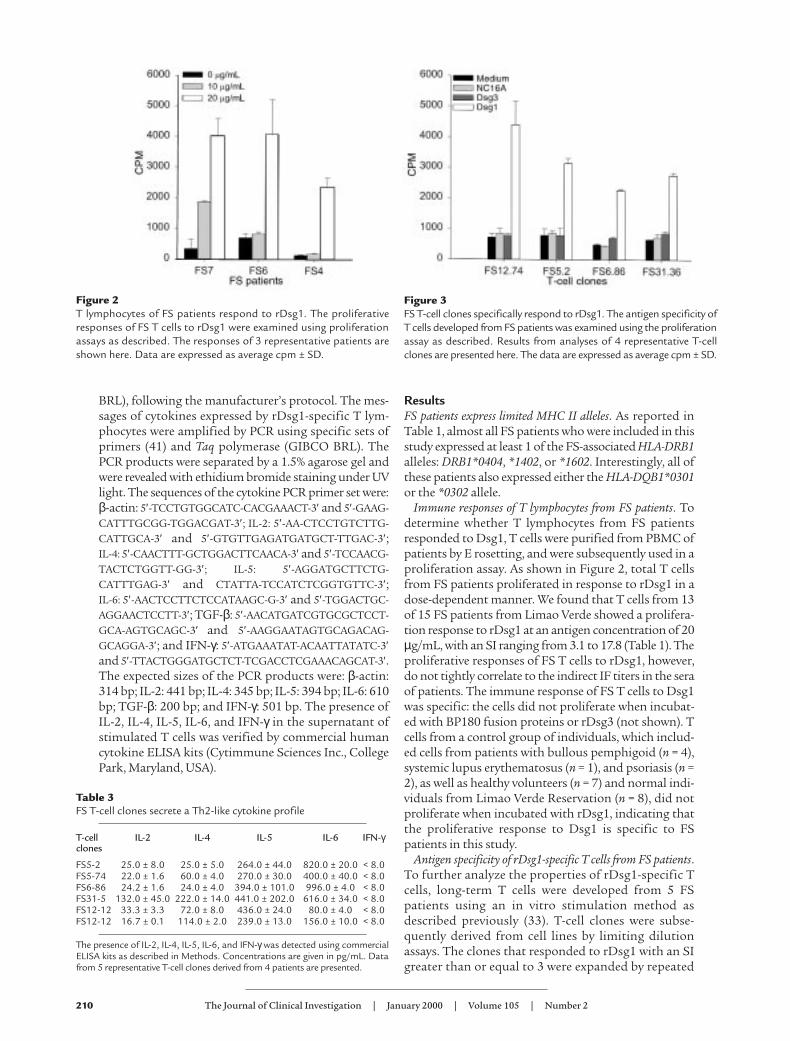
Immune responses of T lymphocytes from FS patients. Todetermine whether T lymphocytes from FS patientsresponded to Dsg1, T cells were purified from PBMC ofpatients by E rosetting, and were subsequently used in aproliferation assay. As shown in Figure 2, total T cellsfrom FS patients proliferated in response to rDsg1 in adose-dependent manner. We found that T cells from 13of 15 FS patients from Limao Verde showed a prolifera-tion response to rDsg1 at an antigen concentration of 20µg/mL, with an SI ranging from 3.1 to 17.8 (Table 1). Theproliferative responses of FS T cells to rDsg1, however,do not tightly correlate to the indirect IF titers in the seraof patients. The immune response of FS T cells to Dsg1was specific: the cells did not proliferate when incubat-ed with BP180 fusion proteins or rDsg3 (not shown). Tcells from a control group of individuals, which includ-ed cells from patients with bullous pemphigoid (n = 4),systemic lupus erythematosus (n = 1), and psoriasis (n =2), as well as healthy volunteers (n = 7) and normal indi-viduals from Limao Verde Reservation (n = 8), did notproliferate when incubated with rDsg1, indicating thatthe proliferative response to Dsg1 is specific to FSpatients in this study.
Antigen specificity of rDsg1-specific T cells from FS patients.To further analyze the properties of rDsg1-specific Tcells, long-term T cells were developed from 5 FSpatients using an in vitro stimulation method asdescribed previously (33). T-cell clones were subse-quently derived from cell lines by limiting dilutionassays. The clones that responded to rDsg1 with an SIgreater than or equal to 3 were expanded by repeated
210 The Journal of Clinical Investigation | January 2000 | Volume 105 | Number 2
Figure 2T lymphocytes of FS patients respond to rDsg1. The proliferativeresponses of FS T cells to rDsg1 were examined using proliferationassays as described. The responses of 3 representative patients areshown here. Data are expressed as average cpm ± SD.
Table 3FS T-cell clones secrete a Th2-like cytokine profile
T-cell IL-2 IL-4 IL-5 IL-6 IFN-γclones
FS5-2 25.0 ± 8.0 25.0 ± 5.0 264.0 ± 44.0 820.0 ± 20.0 < 8.0FS5-74 22.0 ± 1.6 60.0 ± 4.0 270.0 ± 30.0 400.0 ± 40.0 < 8.0FS6-86 24.2 ± 1.6 24.0 ± 4.0 394.0 ± 101.0 996.0 ± 4.0 < 8.0FS31-5 132.0 ± 45.0 222.0 ± 14.0 441.0 ± 202.0 616.0 ± 34.0 < 8.0FS12-12 33.3 ± 3.3 72.0 ± 8.0 436.0 ± 24.0 80.0 ± 4.0 < 8.0FS12-12 16.7 ± 0.1 114.0 ± 2.0 239.0 ± 13.0 156.0 ± 10.0 < 8.0
The presence of IL-2, IL-4, IL-5, IL-6, and IFN-γ was detected using commercialELISA kits as described in Methods. Concentrations are given in pg/mL. Datafrom 5 representative T-cell clones derived from 4 patients are presented.
Figure 3FS T-cell clones specifically respond to rDsg1. The antigen specificity ofT cells developed from FS patients was examined using the proliferationassay as described. Results from analyses of 4 representative T-cellclones are presented here. The data are expressed as average cpm ± SD.

stimulation in vitro. In total, we selected 58 T-cellclones for further property characterization. The num-ber of clones derived from each patient and their aver-age responses to rDsg1 are presented in Table 2. Theantigen specificity of these T cells to rDsg1 was con-firmed by T-cell proliferation assays. As shown in Fig-ure 3, representative FS T-cell clones strongly prolifer-ate in response to rDsg1, but not in response to theBP180 fusion protein or rDsg3, indicating that theseFS T-cell clones respond specifically to rDsg1.
Immune response of FS T-cell clones to rDsg1 is restricted toHLA-DR molecules. To investigate MHC II restriction ofthe response of FS T cells to Dsg1, anti–HLA-DR,anti–HLA-DQ, and anti–HLA-DP antibodies were usedin the proliferation assays as described previously (32).Briefly, T cells were cultured with rDsg1-pulsed antigen-presenting cells in the presence of anti–MHC II anti-bodies or mouse isotype control antibodies for 5 days.Cell proliferation was subsequently determined by the[3H]thymidine incorporation assay. As demonstrated inFigure 4, only the anti–HLA-DR, and not the anti–HLA-DQ or anti–HLA-DP antibodies, inhibited the prolifera-tion responses of FS T cells to rDsg1. This result clearlydemonstrates that the response of FS T cells to Dsg1requires HLA-DR molecules on antigen-presenting cells.
Cell surface phenotype and cytokine profile of Dsg1-specificT cells. Flow cytometric analysis was used to determinethe cell-surface phenotype of rDsg1-specific T-cellclones developed from the FS patients. We found thatDsg1-specific T-cell clones expressed CD3, CD4,CD45RO, and TCRα/β, but were negative for CD8,CD45RA, and the B-cell marker CD19 (not shown),indicating that they are CD4-positive memory T cells.
The cytokine profiles of the T cells from FS patients
were determined as follows. T cells were activated byanti-CD3 antibodies in the presence of phorbol myris-tate acetate for 30 hours. The expression of IL-2, IL-4,IL-5, IL-6, TGF-β, and IFN-γ mRNA messages in theseT cells was then examined by RT-PCR as described pre-viously (41). As shown in Figure 5, a representativerDsg1-specific T-cell clone derived from an FS patientproduced IL-2, IL-4, IL-5, and IL-6, but not IFN-γ orTGF-β. These results were confirmed by humancytokine–specific ELISA kits (Table 3). Although theIL-2 mRNA and IL-2 can be detected in these activatedT cells, the message for this cytokine is weaker whencompared with that of IL-4. Because the IFN-γmessageis not detectable in these cell clones, it is suggested thatFS-specific T cells express a Th2-type cytokine profile.
DiscussionThe data presented in this paper clearly demonstratethat T cells from FS patients specifically proliferatewhen incubated with the extracellular domain ofDsg1. This study thus provides the first evidence thatT lymphocytes from FS patients recognize the sameautoantigen targeted by autoantibodies. T cells from13 of 15 FS patients responded to rDsg1. Interesting-ly, the 2 patients whose T cells did not proliferate inresponse to Dsg1 were undergoing steroid therapyand were in partial remission at the time their lym-phocytes were isolated. The proliferation of FS T cellsto rDsg1 was antigen specific: they did not respondwhen incubated with other epidermal antigens suchas BP180. T cells from other control groups, such aspatients with BP, lupus, and psoriasis, and normalindividuals from both the USA and Limao Verde,Brazil, remained unresponsive to rDsg1.
To further characterize the properties of Dsg1-responsive T cells from FS patients, cell clones weredeveloped from 5 patients. These T-cell clones specifi-cally responded to Dsg1, but not to other epidermalantigens such as Dsg3 and BP180. From the phenotype
The Journal of Clinical Investigation | January 2000 | Volume 105 | Number 2 211
Figure 4The response of FS T-cell clones to rDsg1 is restricted to the expres-sion of HLA-DR molecules. To examine the MHC II restriction of theFS T-cell response to Dsg1, anti–HLA-DR, anti–HLA-DQ, andanti–HLA-DP, monoclonal antibodies (and mouse isotype controlantibodies) were incorporated into the proliferation assays asdescribed. The results using 2 T-cell clones, FS12.74 and FS5.2, in 1of the representative experiments are shown here. The data areexpressed as average cpm ± SD.
Figure 5FS T cells express a Th2-like cytokine profile. To examine the cytokinemRNA messages produced by FS T cells, total RNA was isolated fromthe cells and subjected to RT-PCR analysis. After production of first-strand cDNAs using reverse transcriptase, the mRNAs of cytokines wereamplified using specific primer sets as described (40). The RT-PCR prod-ucts were separated on a 1.5% agarose gel and viewed using ethidiumbromide staining. Lane M, molecular weight marker; lane 1, IL-2; lane 2,IL-4; lane 3, IL-5; lane 4, IL-6; lane 5, IFN-γ; lane 6, TGF-β; lane 7, β-actin.

analysis, it was revealed that these T cells express CD3,CD4, CD45RO, and TCRα/β, but not CD8, CD19, orCD45RA, suggesting that they are CD4-positive mem-ory T cells, a cell population shown to regulate the pro-duction of antibodies (33). This result further empha-sizes the possibility that the Dsg1-reactive T cellsparticipate in the autoimmune response of FS.
It is also known that the isotype of antibodies producedby a given B cell is dependent on the type of helper T lym-phocytes that it encounters during the T-B cell interac-tion (42). For example, T cells that secrete Th1 cytokinesare capable of stimulating B cells to produce IgG1, where-as Th2-type cytokines induce B cells to secrete IgG4 (43,44). Because IgG4 is the predominant isotype of the anti-Dsg1 autoantibodies of FS, it is thought that T cells ofthe Th2 helper cell lineage may be relevant in this autoim-mune disease. Our study supports this hypothesis,because T-cell clones derived from the study’s FS patientsproduce IL-4, IL-5, and IL-6, but not IFN-γ, suggestingthat they secrete a Th2-like cytokine profile. Thecytokines expressed by autoimmune FS T cells may pro-mote the production of anti-Dsg1 autoantibodies of theIgG4 subclass in these patients. However, all of the T-cellclones analyzed in this study also express IL-2 mRNA andIL-2. Whether IL-2 plays a role in modulating the anti-Dsg1 production in these FS patients is not clear.
We have previously demonstrated that certain HLA-DRB1 alleles, such as DRB1*0404, *1402, *1406, and*1602 are prevalent in FS patients (10). As shown inTable 1, all of the FS patients recruited from the LimaoVerde Reservation express at least 1 of the disease-asso-ciated alleles. Although these patients also express lim-ited HLA-DQ alleles, such as DQB1*0301 and *0302,these 2 alleles are not significantly associated with FS (9,10). In this study, we demonstrate that the proliferativeresponse of FS T-cell clones was blocked by anti–HLA-DR antibodies, but not by anti–HLA-DQ or anti–HLA-DP antibodies (Figure 4), further indicating that theHLA-DR molecules play an important role in the Dsg1-specific immune reaction of FS T cells.
Previous investigations on rheumatoid arthritis–asso-ciated DRB1 alleles have shown that several groups ofamino acid residues on the DRβ chain may regulate thebinding of antigenic peptides (45). Among these keyclusters, DRβ67–71 at the P4 position seems to play anessential role in the selection of the sequence of thebinding peptides. Other positions, such as the anchorresidues P1 and P6, also appear to be relevant in deter-mining the affinity for sequences of peptides presentedby DR molecules (46). Based on this information, it hasbeen possible to predict Dsg3 antigenic peptides thatare recognized by T cells from pemphigus vulgarispatients (47), and certain viral or bacterial peptides thatstimulate myelin basic protein–specific T cells (48). InFS, the DRβ hypervariable domain from amino acids 67to 74 of the alleles DRB1*0404, *1402, *1406, and *1602share the same sequence (LLEQRRAA). Hence, it is pos-sible that in FS these MHC II molecules may be involvedin presenting the same Dsg1 peptides to autoimmune
T cells. This possibility is currently under investigation.In conclusion, our findings provide strong evidence
that FS patients develop autoimmune T cells thatspecifically respond to the ectodomain of Dsg1, andthis antigen-specific response requires the expressionof HLA-DR. These Dsg1-reactive FS T cells exhibited aCD4-positive memory T-cell phenotype and produceda Th2-like cytokine profile. These T cells may stimulateautoimmune B cells in the production of pathogenicanti-Dsg1 autoantibodies. Further characterization ofdetailed Dsg1 epitopes that stimulate these T cells mayhelp to elucidate the pathogenic mechanisms underly-ing the development of this disease.
AcknowledgmentsWe acknowledge with gratitude the support of theCooperative Group on Fogo Selvagem Research. Thiswork was supported in part by US Public Health Ser-vice Grants RO1-HL47145, UO1-AI34621 (P. Stastny),R01-AR40410 (G.J. Giudice), R37-AR30281, and RO1-AR32599 (L.A. Diaz), a Merit Award from the VeteransAdministration Central Office (L.A. Diaz), a Derma-tology Foundation Career Development Award, and aDermatology Foundation Research grant (M.S. Lin).
1. Diaz, L.A., et al. 1989. Endemic pemphigus foliaceus (fogo selvagem). II.Current and historic epidemiologic studies. J. Invest. Dermatol. 92:4–12.
2. Martins-Castro, R., Proenco, N., and deSalles-Gomez, L.F. 1974. On theassociation of some dermatoses with South American pemphigus foli-aceus. Int. J. Dermatol. 13:271–275.
3. Robledo, M.A., Prada, S., Jaramillo, D., and Leon, W. 1988. South Amer-ican pemphigus foliaceus: study of an epidemic in El Barge and Nechi,Colombia 1982 to 1986. Br. J. Dermatol. 118:737–744.
4. Bastuji-Garin, S, et al. 1995. Comparative epidemiology of pemphigusin Tunisia and France: unusual incidence of pemphigus foliaceus inyoung Tunisian women. J. Invest. Dermatol. 104:302–305.
5. Morini, J.P., et al. 1995. Pemphigus foliaceus in young women. Anendemic focus in the sousse area of Tunisia. Arch. Dermatol. 129: 69–73.
6. Diaz, L.A., et al. 1989. Endemic pemphigus foliaceus (fogo selvagem). I.Clinical features and immunopathology. J. Am. Acad. Dermatol. 20:657–669.
7. Stanley, J.R., Klaus-Kovtun, V., Sampaio, S.A.P. 1986. Antigenic speci-ficity of fogo selvagem autoantibodies is similar to North Americanpemphigus foliaceus and distinct from pemphigus vulgaris. J. Invest. Der-matol. 87:197–201.
8. Sequiquera, H.L., et al. 1988. Serologic abnormalities in patients withendemic pemphigus foliaceus (fogo selvagem), their relatives, and nor-mal donors from endemic and non-endemic areas of Brazil. J. Invest. Der-matol. 91:189–191.
9. Cerna, M., et al. 1993. Genetic markers for susceptibility to endemicBrazilian pemphigus foliaceus (fogo selvagem) in Xavante Indians. Tis-sue Antigens. 42:138–140.
10. Moraes, M.E., et al. 1997. An epitope in the third hypervariable region ofthe DRB1 gene is involved in the susceptibility to endemic pemphigusfoliaceus (fogo selvagem) in three different Brazilian populations. TissueAntigens. 49:35–40.
11. Hans-Filho, G., et al. 1996. An active focus of high prevalence of fogo sel-vagem on an Amerindian reservation in Brazil. J. Invest. Dermatol. 107: 68–75.
12. Eaton, D.P., et al. 1998. Comparison of black fly species (Diptera: Simuli-idae) on an Amerindian reservation with a high prevalence of fogo sel-vagem to neighboring disease-free sites in the State of Mato Grosso doSul, Brazil. The Cooperative Group on Fogo Selvagem Research. 1998. J.Med. Entomol. 35:120–131.
13. Lin, M.S., Mascaro, J.M., Jr., Liu, Z., España, A., and Diaz, L.A. 1997. Thedesmosome and hemidesmosome in cutaneous autoimmunity. Clin.Exp. Immunol. 107:9–15.
14. Lever, W.F. 1953. Pemphigus. Medicine. 32:1–123.15. Rock, B., et al. 1989. The pathogenic effect of IgG4 autoantibodies in
endemic pemphigus foliaceus (fogo selvagem). N. Engl. J. Med.320:1463–1469.
16. Roscoe, J.T., et al. 1985. Brazilian pemphigus foliaceus autoantibodiesare pathogenic to BALB/c mice by passive transfer. J. Invest. Dermatol.
212 The Journal of Clinical Investigation | January 2000 | Volume 105 | Number 2

85:538–541.17. Rock, B., Labib, R.S., and Diaz, L.A. 1990. Monovalent Fab′
immunoglobulin fragments from endemic pemphigus foliaceus autoan-tibodies reproduce the human disease in neonatal Balb/c mice. J. Clin. Invest.85:296–299.
18. Wheeler, G.N., et al. 1991. Desmosomal glycoprotein DGI, a componentof intercellular desmosome junctions, is related to the cadherin familyof cell adhesion molecules. Proc. Natl. Acad. Sci. USA. 88:4796–4800.
19. Buxton, R.S., et al. 1993. Nomenclature of the desmosomal cadherin. J.Cell. Biol. 121:481–483.
20. Olague-Alcala, M., and Diaz, L.A. 1993. The epitopes on bovine pem-phigus foliaceus antigen are calcium-dependent and located on the pep-tide backbone of this glycoprotein. Chronica Dermatologica. 2:189–209.
21. Emery, D.J., et al. 1995. Pemphigus foliaceus and pemphigus vulgarisautoantibodies react with the extracellular domain of desmoglein-1. J.Invest. Dermatol. 104:323–328.
22. Amagai, M., Tsunoda, K., Zillikens, D., Nagai, T., and Nishikawa, T. 1999.The clinical phenotype of pemphigus is defined by the anti-desmogleinautoantibody profile. J. Am. Acad. Dermatol. 40:167–170.
23. Ding, X., Diaz, L.A., Fairley, J.A., Giudice, G.J., and Liu, Z. 1999. The anti-desmoglein 1 autoantibodies in pemphigus vulgaris sera are pathogen-ic. J. Invest. Dermatol. 112:739–743.
24. Lin, M.S., Liu, Z., Drolet, B.A., and Diaz, L.A. 1998. Cutaneous autoim-mune diseases. In The autoimmune diseases. 3rd edition. N.R. Rose and I.R.Mackay, editors. Academic Press. San Diego, CA. 545–570.
25. Coffman, R.L., et al. 1988. The role of helper T cell products in mouse Bcell differentiation and isotype regulation. Immunol. Rev. 102:5–28.
26. Barlett, W.C., et al. 1989. Cognate interactions between helper T cells andB cells. II. Dissection of cognate help by using class II-restricted, antigen-specific, IL-2-dependent helper T cell clone. J. Immunol. 143:1745–1754.
27. Hamano, T., et al. 1992. Direct interaction between an antigen-specificB cell clones and an MHC class II-reactive T helper cell clone. J. Leukoc.Biol. 52:89–96.
28. Stevens, T.L., et al. 1988. Regulation of antibody isotype secretion by sub-sets of antigen-specific helper T cells. Nature. 334:255–258.
29. Allegretta, M., Nicklas, J.A., Siram, S., and Albertini, R.J. 1990. T cellsresponsive to myelin basic protein in patients with multiple sclerosis. Sci-ence. 247:718–721.
30. Melms, A., et al. 1989. Autoimmune T lymphocytes in myasthenia gravis.Determination of target epitopes using T cell lines and recombinantproducts of the mouse nicotinic acetylcholine receptor gene. J. Clin.Invest. 83:785–790.
31. Mullins, R.J., et al. 1995. Identification of thyroid stimulating hormonereceptor-specific T cells in Graves’ disease thyroid using autoantigen-transfected Epstein-Barr virus-transformed B cells lines. J. Clin. Invest.96:30–37.
32. Lin, M.S., Gharia, M.A., Swartz, S.J., Diaz, L.A., and Giudice, G.J. 1999.Identification and characterization of epitopes recognized by T lym-phocytes and autoantibodies from patients with herpes gestationis. J.Immunol. 162:4991–4997.
33. Lin, M.S., et al. 1997. Development and characterization of desmoglein-3 specific T cells from patients with pemphigus vulgaris. J. Clin. Invest.99:31–40.
34. Amagai, M., Hashimoto, T., Green, K.J., Shimizu, N., and Nishikawa, T.1995. Antigen-specific immunoadsorption of pathogenic autoantibod-ies in pemphigus foliaceus. J. Invest. Dermatol. 104:895–901.
35. Liu, Z., et al. 1995. Molecular mapping of a pathogenically relevantBP180 epitope associated with experimentally induced murine bullouspemphigoid. J. Immunol. 155:5449–5454.
36. Liu, Z., Diaz, L.A., Haas, L., and Giudice, G.J. 1992. cDNA cloning of anovel human ubiquitin carrier protein. An antigenic domain specifical-ly recognized by endemic pemphigus foliaceus autoantibodies is encod-ed in a secondary reading frame of this human epidermal transcript. J.Biol. Chem. 267:15829–15835.
37. Lazaro, A., et al. Evolution of HLA class I compared to HLA class II poly-morphism in Terena, a South American Indian tribe. Hum. Immunol. Inpress.
38. Indiveri, F., Huddlestone, J., Pellegrino, M.A., and Ferrone, S. 1980. Iso-lation of human T lymphocytes: comparison between wool filtrationand rosetting with neuraminidase (VCN) and 2-aminoethylisothiouro-nium bromide (AET)-treated sheep red blood cells. J. Immunol. Methods.34:107–115.
39. Ohlin, M., Danielsson, L., Carlsson, R., and Borrebaeck, A.K. 1989. Theeffect of leucyl-leucine methyl ester on proliferation and Ig secretion ofEBV-transformed human B lymphocytes. Immunology. 66:485–490.
40. Del Prete, G., et al. 1995. Preferential expression of CD30 by humanCD4+ T cells producing Th2-type cytokines. FASEB J. 9: 81–86.
41. Brenner, C.A., et al. 1989. Message amplification phenotyping (MAP-Ping): a technique to simultaneously measure multiple mRNAs fromsmall numbers of cells. Biotechniques. 7:1096–1101.
42. Lin, M.S., and Chen, Y.W. 1993. B cell differentiation. II. Isotype poten-tial of a single B cells. Cell. Immunol. 150:343–352.
43. Armitage, R.J., MacDuff, B.M., Spriggs, M.K., and Fanslow, W.C. 1993.Human B cell proliferation and Ig secretion induced by recombinantCD40 ligand are modulated by soluble cytokines. J. Immunol.150:3671–3680.
44. Romagnani, S. 1992. Human Th1 and Th2 subsets: regulation of differ-entiation and role in proliferation and immunopathology. Int. Arch. Aller-gy Appl. Immunol. 98:279–285.
45. Hammer, J., et al. 1995. Peptide binding to HLA-DR4 molecules: corre-lation with rheumatoid arthritis association. J. Exp. Med. 181:1847–1855.
46. Hammer, J., et al. 1993. Promiscuous and allele-specific anchors in HLA-DR binding peptides. Cell. 74:197–203.
47. Wucherpfennig, K.W., and Strominger, J.L. 1995. Molecular mimicry inT cell-mediated autoimmunity: viral peptides activate human T cellclones specific for myelin basic protein. Cell. 80:695–705.
48. Wucherpfennig, K.W., and Strominger, J.L. 1995. Selective binding of selfpeptides to disease-associated major histocompatibility complex (MHC)molecules: a mechanism for MHC-linked susceptibility to humanautoimmune diseases. J. Exp. Med. 181:1597–1601.
The Journal of Clinical Investigation | January 2000 | Volume 105 | Number 2 213
Copyright © 2022 FDOKUMEN