


Bacter Ology Including Pathogenic Protozoa - Forgotten Books
315
-
Upload
khangminh22 -
Category
Documents
-
view
0 -
download
0
Transcript of Bacter Ology Including Pathogenic Protozoa - Forgotten Books


Hughes
Practice of M edicine
ELEVENTH EDITION REV ISED
GIV ING THE SYNONYM S, DEFINITIONS, CAUSES, SYMPTOMS PATHOLOGY,
DIAGNOSIS,PROGNOSIS, AND TREATMENT or EACH DISEASE.
By DANIEL E . HUGHES, M .D .,La te Chief Res ident Phys ic ian,
Phi ladelphia Hosp ita l ; formerly Demonstrator Of Cl inica l Medic ine,Jefferson Medica l Co llege . Edited by R. J. E . S( OT1
‘
,
M .D Formerly Attending Physic ian to the Demi l t D ispensary, NewYork ; Editor of Gould and Pyle
’
s Cyclopedia of Medic ine and
63 Il lustra tfions . 1 2mo . wim+ 785 Pages . Cloth, postpaid.
The Treatment is spec ia l ly full . 406
va luable prescr iptions have been included .
This popularity is due to the fact tha t the book gives practica ldiscussions Of diseases as briefly as is consistent w ith
°
the subject,theories be ing el iminated .
”—Journa l American Medi ca l Associatp'
on .
This book is intended as a work ing manua l in which the
phys ician may find that which he nee ds for a rap id restudy Of any
disease w ithout having to go over the controversia l matter usual lyfound in
'
treatises On practice . For case reference and rap idstudy, there is no bette r boo -Medica l Council .
The work is thoroughly modern and is particularly va luablefor its discussion of diagnosis and treatment
,an immense number
of excellent prescr iptions be ing incorporated under the latter head .
It is, we bel ieve, unique in including sections on menta l diseases and
diseases of the skin.—New York Medical Journa l .

L I AM L L QU L L L LU l L 0 I v a p u i p c u
D ictionaryThird Edition
,Revised and Enlarged by R . J E . SCOTT, M .A .
,
M .D.,Fe llow Of New York Academy Of Med ic ine, etc . Many
thousands Of new medica l words a re included in this edit ion. The
work conta ins over terms,962 pages and we ighs only lbs .
Bound in Handsome F lex ible Cloth, Marbled Edges, Round Corners,w ith Thumb Index,
Mom s Human AnatomyA Comp lete Systematic Treatise
6th Edition Revised and Largely Rewritten. W ith 1 164 I llustrations . Cloth
,
CONTRIBUTORS—Char les R. Bardeen, A .B .,M .D .,
Professor of
Anatomy, Univers ity Of W isconsin, formerly A ssoc iate ProfessorJohns Hopkins ; E l iot R . Clark, A.B .,
M .D .,Professor of Anatomy,
University of M issour i , formerly A ssoc iate in Anatomy, JohnsHopkins Univers ity ; A lbert C . Eyc leshymer
,Ph .D .,
M .D .,Pro fessor
of Anatomy, Univers ity of I llinois ; J . F . Gudernatsch, Ph.D.,As
sistant Professor Of Anatomy, Corne ll Univers ity Medica l Co llege,NewYork ; Irving Hardesty, A .B .
,Ph.D .
,Professor
'
of Anatomy,Tulane Univers ity Of Louisiana ; C . M . Jackson
,M
,S .
, M .D .,the
Editor,Pro fessor of Anatomy, University of M innesota ; Dean D .
Lew is,M .D .
,Assoc iate Pro fessor of Surgery
~ in the Rush Medica lCo llege, Chicago, I lls . ; Richard E . Scammon
,Ph .D .
,Assistant PrO~
fe ssor of Anatomy, University of M innesota ; J. Parsons Schaefi‘
er,
Ph .D ., M .D .,Professor of Anatomy, Jefferson Medica l Co llege
,Phi la
delphia ; H .
‘
D . Senior," M .B .,
Professor Of Anatomy, Univers ity and Be llevue Hosp ita l Medica l Col lege, New York ; G . E ll iotSmith, M .A .
,M .D .,
Pro fessor of Anatomy, University of London ; Charles R . Stockard, Ph .D .
,D .Sc .,
Professor of
Anatomy, Cornel l University Medica l Col lege, New York ; R . J. Terry,A .B .
,M .D .
,Pro fessor of Anatomy, Washington Univers ity, St. Lou is,
A very important feature of any textbook of anatomy is tha tof the i llustrations. They should be very
‘
we l l executed and c lear tog ive the student a proper notion of the part under discussion. In
the new Morri s,the i llustrations have been careful lgr selected
prepared . Many of them are printed in colors. This edition in
eludes many new p ictures redrawn to supp lant the older ones. The
draw ings are a distinguishing feature of Morris’s Anatomy.
M icrobiology and M icroan'
alyses ofFoods
131 I l lustrations. 8vo . Cloth, By ALBERT SCHNEIDER,M .D., Former ly Micr oana lyst, U. S . Bur eau of Chemistry.


BLAKISTON’
S COMPENBSThe Best Series of Manuals for the Use of Students
Pr lc e o f e a c h, C lo th, ne t
QThese Compends are based on the most popular text-books and the lectur es o fprominent professors , and a re kept constantly r ev ised , so that they may thorough lyrepres ent the p resent state Of the subjects upon which they trea t.
t§=The authors hav e had large exper ience as Quiz-Masters and a tta ches o fcolleges , and are well a cqua inted with the wants of students .
(QTThey are arrang ed dn the.
most approv ed form , thorough and con c ise ,‘
c onta in ing over 900 fine illustrations , inserted wher ev er they c ould be used to
a dv antage .
WCan be used by students of any college .
hQThey contain information nowhere e lse co llected in such a condensed , pra ctica l
s ap e .
Illustrated Circular Free
POTTER ’S ANATOMY. E ighth R evised and En larged Edition . IncludingVisce ra l Anatomy . Can b e used with e ithe r M orr is ’ or Gray’s Anatomy .
1 39 Illustrations and 1 6 Plates of Nerves and Arter ies , w ith Explanatory Tables ,etc .
BRUBAKER . PHYSIOLOGY. Fifte enth Edition , w ith 26 Illustrations . En largeda nd Rev ised .
LANDIS . OBSTETRICS . Nin th Edition . Rev ised a nd Edited by WM . H .
WE LLS , M . D Late Assoc iate Profe ssor Of Obstetr ics , Jefferson M edical C o llege ,Ph i la de lphia . 8 0 Illustrations .
POTTER . MATERIA MEDICA, THERAPEUTICS AND PRESCRIPTIONWRITING . Eighth R ev ised Edition .
WELLS . GYNECOLOGY. Fourth Edition . With 1 53 Illustrations .
GOULD and PYLE . DISEASES OF THE EYE AND REFRACTION Inc ludingTrea tment and Operations and a Section on Loca l Therapeutics . V
V
l th Formulasa nd 109 Illustrations , severa l of which are in colors . Fourth Editio n .
LIPSHUTZ. COMPEND OF SURGERY. 1 85 Illustra tions .
This v o lume r epla ces the C ompend of Surgery former ly wr l tten by the la teOrv i lle Horwitz , M . D .
LEFFMANN. CHEMISTRY, Ir‘iorganic and O rgan ic . S ixth Edition . Inc ludingUr ina lysis , An ima l C hem istry , C hem istry of M ilk , B lood , Tissues , the Secr etl ons ,etc .
STEWART . PHARMACY. Ninth Edition . Based upon Pro f . R emington ’
s
Text-book Of Pharma cy .
ST. CLAIR . MEDICAL LATIN. S e cond Edition .
SCHAMBERG . DISEASES OF THE SKIN. S ixth Edition . R ev ised and
Enlarged . 1 19 Illustrations .
PITFIELD .
’
BACTERIOLOGY. Fourth Edition . 82 Illustra tions .
HIRSCH . GENITO-URINARY AND VENEREAL DISEASES , AND SYPHILIS .
Th ird Edition . With 59 Illustrations .

A C OMPEND
BACTER OLOGYINC LUDING
PATHOGENIC PROTOZOA
BY
ROBERT L. PITFIELD , M. D .
PATHOLOGIST TO THE GERMANTOWN HOSPITAL ; LATE DEMONSTRATOR OF
BACTERIOLOGY AT THE MEDICO-CHIRURGICAL COLLEGE , PHILADELPHIA ; V ISITING PHYSICIAN TO ST. TIMOTHY'
S HOS
PITAL AND CHESTNUT HILL HOSPITAL , PHILA.
FOURTH EDITIONWITH 4 PLATES AND 82 OTHER
ILLUSTRATIONS
PHILAD ELPH IA
P . BLAKISTON’
S SON C O .
1 0 1 2 WALNUT S TREET

COPYRIGHT,1 9 2 z
P R IN T E D IN U. 5 . Ni
This little book was designed by the writer to serve the needsof the medical student p reparing for examination, and for the
practitioner ofmedicine who desires to acquaint himself with the
p rincip le facts of the rap idly growing science of bacteriology. Aneffort has been made to reduce the subjec t ma tter to as concretea form as possible .
While the literature of the subject of immunity is as vast almostas the rest of bacteriology
, yet it is hoped tha t the chap ter in thisbook on immunity gives in outline the essential accep ted teachingson the subject .M inute details of cultures and technic are not given . They
must be sought for in books on descrip tive bacteriology .
The author has dr awn very freely from many standard textbooks . Many illustrations are from K olle Wassermann ’sAtlas
,Park andWilliams
,Williams
,McFarland
,Tyson ’s Prac
tice and Abbo tt .The writer ’s best thanks are tendered to D r . Herbert Fox of
the University of Pennsylvania (Pepper Labora tory) to whomentire credit is due for the chap ters on fil terable viruses ; the re
arrangement oi the chap ters, and the new ma tter that has beenadded throughout the book .
ROBERT L . PITFIELD .
48 835 3


TABLE OF CONTENTS
CHAPTER I
THE CLASSIFICATION,MORPHOLOGY, AND THE BIOLOGY or BACTERIA .
CHAPTER II
RODUCTS or BACTERIAL ENERGY . .
CHAPTER III
INFECTION
CHAPTER IV
IMMUNITY
CHAPTER V
STUDY OF BACTERIA
CHAPTER VI
BACTERIOLOGICAL LABORATORY TECHNICCHAPTER VII
ANTISEPTICS AND DISINFECTANTS .
CHAPTER VI IIBACTERIA
CHAPTER IX
ANIMAL PARASITES
CHAPTER XTHE FILTERABLE
CHAPTER XI
BACTERIOLOGY OFWATER , SOIL, AIR AND MILK
INDEX


COMPEND OF BACTERIOLOGY
CHAPTER I
THE CLASSIFICATION,MORPHOLOGY , AND THE
BIOLOGY OF BACTERIA
BACTERIA (fission fungi or schizomycetes) may be defin edvery minute unicellular vegetable organisms
, almost alwaysof chlorophyll , and generally unbranched, tha t reproduceIves asexually by means of direc t division or fission , spores ,idia . They are allied closely on the one hand to the higher
ngi,such as the moulds
,and on the o ther to the algze . Many
forms in one phase of development closely resemble members ofother group s, and it has always been difficul t to classify them.
Various bo tanica l classifications have been emp loyed by differentbacteriologists . The following one is based somewhat uponMigula
’s, and that adop ted by Lehmann andNeumarm
,which was
comp iled from the systems ofFliigge, Fischer , L'
ojfier, andMigula .
CLASSIFICATION.
—Bacteria may be conveniently dividedinto six families
,according to their morphology or Shape .
I. COCCACElE .—Spherica1 or Spheroidal bacteria. Globular
in free state but usually seen with one axis slightly larger .
They do not have parall el sides like the bacilli . To mul
tip ly , the cell divides into halves, quarters, or eighths
, eachof which grow again into p erfect spheres . Endosporesand flagella are very rare (Lehmann and Neumann) . Ifmobile they are ca lled Planococcus or Planosarcina .

BACTERIA
(a) s.—Cells that divide in one direction only
and grow in chains .
(b) Micrococcus.—Cells that divide in two directions
, or
irregularly ; with this group staphylococcus may be
classed . Also tetrads,which form into fours by division
in two directions .
(0) Sarcina.—Cell s that divide in three directions so that
bale-like packages, or blocks of eight are formed. Atleast one variety (Sarcina agilis) is motile
,having fla
gella . Pla tes of cocci,one thick in the p lane, are called
merismopedia .
II. BACTERIACEZE.—ROD bacteria are straight or slightly
curved . Each cell is from two to six times as long as broad .
D ivision takes p lace in one direction only, and at right anglesto the long axis . Spores may be produced or may not.
They may have flagella , or may not.
(a) Bacterium.—Neumann—Have no endospores . M igula
—no flagella .
(b) Bacillus.—Neumann —Have endospores, and often grow
in long threads . M igula—Flagella present at any partof cell, peritrichic in arrangement .
(6) Pseudomonas.—Have endospores very rarely . Flagella
only at ends .
SPIRILLACEZE.
—Spiral bacteria . Unicellular,more or less
elongated. Twisted more or less like a corkscrew. Cellsare sometimes united in short chains . Generally verymo tile . Spores are known in two varieties only .
(a) Spirosoma .-Rigidly bent . No flagella .
(b) Vibrio or Microspira.—Cell s that are rigidly bent like
a Comma, and have always one, occasionally two polarflagella .
(c) Spirillum.—Are long and sp iral, like a corkscrew
, are
rigid, and have a bunch of polar flagella .

CLASSIFICATION
(d) Spirochaeta.—Cells with long flexible sp iral threads,
without flagella . Some move by means of an undulatingmembrane . These have been thought to belong to thebacteria but those thatmove by an undulatingmembraneshould be .classified with the protozoa .
IV . MYCOBACTERIACEE .—Cells as short or long fil aments
,
, Which are often cylindrical, clava te (club-shap ed) , cuneateor irregular in outline
,and display true or false branching .
Spores are not formed,but gonidia are . They have no fia
gella,and division takes p lace at right angles to the long
axis . There is no surrounding sheath as in the next family (V) .
(a) Mycobacterium .
—Cells are short cylindrical rods,some
times wedge-like,bent
,or Y-shap ed : long andfilamentous .
They exhibit true branching, and perhap s produce coccoidelements and gonidia, but no flagella . The Corynebac
teriam Of Lehmann and Neumann belongs to this group .
Many are acid-fast.
(b) Streptothrix or Actinomyces (ray fungus) are longmycelial threads, that radiate in indian-club, or loop-likeforms
,with true branching and delica te sheaths
,devoid of
g onidia and flagella . Growth coherent,mould-like and
dry. Often powdery on the surface in culture media,
frequently emitting a musty odor . A few species areweakly acid-fast .
V. CHLAMYDOBACTERIACEZE.—Sheathed bacteria. Cells
are charac terized by an envelop ing shea th about branchedand unbranched threads . D ivision takes p lace at rightangles to the long axis of the cells .
(a) Cladothrix are distinguished by false dicho tomousbranching . Multip lication is affected by separation of
Whole branches, and by swarm spores or motile gonidia
having flagella .

BACTERIA
(b) Crenothrix.—Filaments are fixed to a nutrient base .
Are usually thinner at the base than at the apex, formedof unbranched threads that divide in three directions ofspace , and p roduce in the end two kinds of gonidia
,
probably Of bisexual nature .
(0) Phragmidiothrix.—Cells are first united into unbranched
threads by means of delica te shea ths,branching threads
are then formed . D ivision takes p lace in three directionsof space , p roducing sarcina-like group s of gonidia
,
which,when free
,are spherical .
(d) Thiothr ix.—Are unbranched cells
,sheathed
,without
flagella,divided only in one direction,and contain sulphur
granules .
VI. BEGGIATOACEE .—Cells united to form threads that are not
sheathed : have scarcely visible sep ta ; divide in one direction ,andmo til e only by an undula ting membrane
,not by flagella
(a) Beggiatoa .—Cells containing sulphur granules .
Bacteria may furthermore be classified according to the ir biological characteristics, which may be wonderfully different . The ultimate differentiation of one sp ecies from ano ther dep ends not onlyon themorphology, which may be p recisely similar , but on its biological behavior in culture media and in the tissues of animals uhder identical conditions . Again , different individuals of a givensp ecies may vary extraordinarily one from another in form and
size, yet the chemical behavior is invariably the same. Hence it
is only by Observation of the development of bacteria in culturemedia
, and the reactions produced in it, and in the bodies of ex
periment animals, tha t we can identify them positively from othersof a foreign sp ecies. NO bacteriologist is able by a simp le microscopical examination ofa given bacterium,
to identify it absolutelyat all times .The higher group s of fungi may be classified conveniently asfoll ows
CLASSIEICATION
BLASTOMYCETES-YEASTS .—Budding fungi . Character
istic lies in p redominant round or ellip tical unit ; some few formmycelia ; division by endospores or budding ; important in fermentation and in disease . D ivided intoSaccharomyces .
—Endospores and budding, fermenters . No
mycelia .
Monilia .—Budding . NO spores—m ycelia— fermenters .
Oidia’
.-Budding . NO spores—mycelia—non-fermenters .
Coccidioides .—Spores . —No budding—mycelia —non-fermen
ters .HYPHOMYCETES-MOULDS .
—Mycelium-forming fungus ;division by spores , branching, budding, or intercalary division ;some bisexual . Divided into :Phycomycete s.
—Mucorinae—s ometimes bisexual— division bygrouped spores or segmentation of mycelium. Not important
pathologically. Example—Mucor .Mycomycete s.
—Asexual forms dividing by Spores in a sac
or by end organs—sexual forms dividing by specially developedcells . Mycelia predominate . Examp le —Asp ergillus .These are
-
the principal group s ofyeasts which can be reasonablywell classified . Thereare o thers
,M icrosporon,Trichophyton ,
and
Sporo thrix , that have a decided pathogenic importance but forwhich -
a systematic position is not easy to give . They belong
probably between the two above classes in tha t mycelial growthwith lateral budding and spore formation are their characters .Bacteria that are globular in form are called cocci .
Cocci that divide in one direction of space and grow in chainsare called streptococci (Fig. I) .
Cocci that divide irregularly and form pairs of fours , or irregulargroups , are called micrococci. Those of this class tha t form pairsare frequently called diplococci .
1 When they form fours by division in two directions
,they are called tetrads. But when they
1 This word is frequently used as if it were a biological term indicating some
species identity , e .g.,D ip lococcus pneumoniae . There is no biological
group called D ip lococc i and the term should be used in a descrip tive sense .
The cause of pneumonia is now called Streptococcus pneumonia;.

BACTERIA
divide irregularly and form masses resembling bunches Of grapes,they are spoken
\
Oi a staphylococci (Fig.
Cocci that divide in three directions are called sarcince. One
single coccus,by division in three directions
,
FIG . I.
—Large and very large streptococci . (Kolle andWassermann .)
or more, each of which becomes globular and equal in size to the
parent.Motilemicrococci are those that divide in two directions of space
and have flagella . They are known as planococci.
FIG. 2 .
—Staphylococci . Strep tococci . Dip lococci . Tedrads. Sarcinae .
(Williams )
M icrococci that divide in three directions,and are motile, are
called planosarcince (Fig.
Bacteria tha t resemble straight rods are called bacilli . These’
i
may be short and thick, or long and thread-like ; are never curved,but may be slightly bent.

BACTERIA
commonly met with in this form in clinical examinations, and in
cultures . Hence,we frequently hear of the bacillus of tubercu
losis, and not the Mycobacterium tuberculosis.
Among the higher bacteria,the differentiation of those belong
ing to the sheathed group , or Chlamydobacteriacea , is difficul t, as itdep ends largely upon the formation Of the false branching and
the gonidia . When bacteria exhibit many,or various forms
,
in the same culture,as does the typhoid bacillus, we
5 .
—Spirochaeta of relapsing fever. (Kolle andWassermann. )
them as pleomorphic, or as showing p leomorphism. To eluci
date : Man is p leomorphic, because among adult individualssome are tall or short
,fat or thin .
Irevolution or Degeneration Forms .
—When the best or op timum conditions for bacterial life (see page I8) are not found,bacteria p resent appearances quite different from those of the
young, active or perfect adult typ e . These changes are calledinvolutionary if temporary , or degenerative if p ermanent.For examp le : the diphtheria bacillus under good conditions forlife is a straight or slightly bent rod staining in a granular manner .

CLASSIFICATION 9
living under unsuitable conditions it becomes quite short,and stains solidly . Again
,bacilli that are accustomed to appear
as short elements may grow to long threads without dividing,or swell into unrecognizable form. Branching is sometimesseen in rods and Sp irals , a condition due in certain cases to
o thers naturally among the higher bacteria .
bacteria, we use the thousandth part of a milli
‘called the micromillimeter,or micron
,as the unit . The
letter p. is the symbol for this unit . A micron is about
0 of an inch, yet a bacterium I,u. long, and p. in width,
large in comparison to some things that scientists measure ,3 the thickness of oil films
,soap bubbles , or light-wave
in which the unit is a micromicron, and is symbolizedThe shortest light-wave lengths are about 40 0 up , or
it, while chromatic threads in cells of bacteria are often 1 0 0 up.
width . Then again there are many things smaller than thesereads . The thinnest part of a bursting soap bubble is but
thickness . There are certain infectious agents that aresubmicroscop ic ; that is, invisible even by the aid of Siedentopf
’s
ultraviolet microscope , which shows Objects smaller by half alight-wave length up) .The structure of the bacterial cell is very simple , consisting of
a delicate poorly sta ining limiting membrane or wall enclosinga mass of substance with strong aflinity for basic dyes likemethylene blue . Just what part of the bac terial interior iscytop lasm and what is nucleus is not definitely known . SomeObservers believe that all that is stained is chroma tin , or nuclearmatter diffusely distributed through the bacterial cell , whileothers think that a delicate cytoplasm exists under the wall andthat it is overshadowed by relatively grea t p roportional bulkof the nucleus .In the course of the rod we often see metachromatic bodies ,
called the Babes-Ernst granules,and unstained Spaces called
vacuoles, both of which are common to many bacteria . They

IO BACTERIA
may be ingested substances but some are lipoidal or carbohy
drate in nature . These bodies are demonstrated by stainingwith basic dyes and may be Of importance in determining themycobacteria . It is thought that they p lay a rOle in reproduc
tion (Fig.
The food of the bacterium passes through the cell wall byosmosIS. The cell wall of certain organisms, for examp le
FIG. 6 .
—Zooglea formation. (Leuconostoc .) (Kolle andWassermann.)
pneumococcus, undergoes a change whereby a mucilaginous orgelatinous capsule is formed outside the cell wall . Its use isnotknown . The cell wall is generally the first portion of the cellto be attacked by certain Specific substances (ferment) foundin the blood of immunized animals
,called bacteriolysins and
agglutinins. Where great masses of bacteria are clumped inexcessive mucilaginous material we Sp eak of this condition as
zoOglea (Fig.
We sometimes find,as a p rolongation of the cell wall , filament
ous organs of locomotion known as flagella . Bacteria withoutflagella are sometimes called gymnobacteria , those possessing

CLASSIFICATION II
them,trichobacteria but these terms are falling
.
in to disusebecause the latter is now—a-days app lied to higher group s that growin hair like forms . However the following may be described :When they have one flagellum we call themmonotrichous bacteria ,and amphitrichous when there are two flagella , one at each pole
(Fig. When the cell is surrounded by flagella,it is known as
a peritrichous bacterium,and loplio
trichons when the flagella are ar
ranged in tufts of two or more .
These are simp le adjectives and not
FIG. 7 .
—Sp irillum undula with polar FIG. 8 .
—Bacillus proteus vul
flagella . (Kolle and Wassermann.) garis,
showing peritrichous dagella . (Kolle and Wassermann.)
now used as terms of classifica tion . The tetanus bacil lus is anexamp le of a peritrichous organism,
while the bacill us of green
pus is called monotrichous, because of its single flagellum .
Flagella are not p seudopods, but distinc t organs of locomotion .
In certain bac teria of the Beggiatoa , locomo tion is aecom
plished by a peculiar amoeboid motion, or by an undula tingmembrane . On looking at bacteria known to have no powers
I2 BACTERIA
of voluntary mo tion, they are seen to osc illate , tremble or moveslightly . Susp ensions of india-ink in water are ‘seen to do the
same thing, as are other inanimate suspensions . This molecularmovement is known as the Brownian mo tion . By ordinarysta ining methods
,and in preparations of living bacteria known
to be flagellated, these organs of locomo tion canno t be seen,
as a rule . Occasionally, however, one may be seen under eithercondition . Generally
,strong solutions of aniline dyes
,to which
powerful mordants have been added,are necessary to stain the
capsule of bacteria and the a ttached flagella . The motion of
bacteria varies from a simple rotatory, on one axis,to a swing
lng, shaking, boring or serp entine action . The location of the'
flagella has some influence upon the motion they impart ,
Flagella may be broken Off from the cell body by agitation,
but when separated may still be clumped by agglutinating sera .
Flagella may have other functions than locomotion . It is
possible that they serve as organs for the absorp tion of nourishment from the surrounding media . The presence of very longor very numerous flagella does not necessarily p resage veryactive motion . At times
,under certain conditions
,an organism
ordinarily motile and flagellated will appear immobile andnon
flagellated (Lelunann and Z iferler) , but this Is rare . Certainflagella have in their continuity little round granules, or bodies ,which apparently have nothing to do with the functions of
locomotion but may have something to do with the nutritionof the cell . The test of motility of a bacterium is to see it progress by itself completely across the field of the microscope .
REPRODUCTION.
—~The process of direct cell division isthe commonest way by which bacteria multiply ; hence comesthe name of fission fungi . The ways of reproduction of the
bacteria high in the scale are by direc t division , branching, andby means of spores, and by o ther granules called gonidia . The
spores appearing in the lower bacteria , bacilli for example, arenot reproduction forms but sta tes of high resistance.

SPORULATION 1 3
The p rocess of direct or binary division is very simp le , andmay be a matter of twenty minutes
,or as long as six hours .
D ivision is almost always across the cell in the direction of the
short axis,though it may in some bacteria be in a direction
parallel to the long axis, but this is uncommon .
By means of the hanging-drop or the block-culture method,on an inverted cover-glass the process may be observed easily.
The phenomena of division begin by an elongation of the cell,soon followed by a constriction of p inching in of the cell on bo thSides
, at an equatorial point . The process begins to be apparentin the cell wall and extends Inward .
D ivision may occur in one,two , or three directions, or p lanes .
By cell division bacteria multip ly by geometrical p rogression .
One cell at the end Of a period becomes two , and at the end of a
second period these two become four ; at the end of another
p eriod these four become eight ; after twenty-four hours theymay number many millions .It is well that the food supply soon gives out and that the
products of bac terial metabolism,such as acids and ferments
,
inhibit their growth , By this rap id bac terial. multiplication,carcasses of animals are disintegrated and the higher nitrogenouscompounds are reduced to s imp le gases that are quickly dissi
pated in the air .
SPORULATION.
—Sporulation is of two kinds : the first andmost important for hygiene is that in to which some pathogenicbacteria go when they meet unfavorable conditions and it affords
protection against all but the most vigorous disinfection ; thesecond kind is a spec ialized function of the higher bac teria and
moulds by which reproduction occurs (vegeta tive) . In the
latter case it is not impossible that some sexual Specializationoccurs . The first mentioned are called Endospores.
Vegetative Sporulation corresponds to the flowering of the
higher plants, and is observed under the most favorable vital
conditions . Endospores are p roduced under stress of circum

I4 BACTERIA
stances, when certain agencies or conditions,such as absence Of
food, drying, and heat, threaten the extinguishment of the organism. Spores are bright, shining, oval, or round bodies, which do
‘
fFIG. 9 .
—The formation of FIG. Io .
—Spores and their location in bacSpores. (After Fischer from terial cells. (After Frost andM cCampbell .)Frost and M ccampbell . )
not take aniline dyes readily,and which
,when they are stained,
retain the color more tenac iously than the adult cells . Theyresist heat
,Often withstanding a tempera ture of I50
°
C . dry heatfor an hour . Steam under pressure at a temperature of 1 50
°
C .
will Invariably kill them after a Short exposure .
0 0 0FIG. II.
—Spore germination. a , direct conversion of a spore into a bacilluswithout the shedding of a spore-wall (B . leptosporus) ; b, polar germination ofBact. anthracis,
‘
c, equatorial germination of B . subtilis,‘
d, same Of B . mega
terium,‘
e,same with “ horse-shoe ” presentation. (After Novy . )
Spores are situated either in the ends of the adult organism
(polar) or in the middle (equatorial) , and the spore is discharged
(sporulation) either from the end or through the side .

1 6 BACTERIA
found in bacteria Of high thermal death-point, are called byHeuppe arthrospores. It is believed tha t they are withoutsignificance . Arthr ospores are common among the micrococciand may be associated with cap sul e formation and cell enlargement . The whole cell may stain more in tensely . They are alsoto be sought among the Strep tothrix genus .Spores resist chemicals for a long p eriod, andwithstand drying,
even in lime p laster, for years . It is believed that the thickcap sul e enables them to resist these deleterious agents .
FIG. I3.
—Pest bacilli Showing cap sules. (Kolle andwassermann.)
Sporulation is more apt to occur under poor nutritive conditions .The anthrax bacill us thrives at I3
°
C . but canno t sporulatebelow 1 8
°
C . Anthrax spores have been known to resist thegermicidal action of a 5 p ercent carbolic
'
acid solution for fortydays .Capsule s—Certain well-known pathogenic bacteria havethick well-marked capsules . The pneumococcus, pneu
'
mobacillus,and Bacillus aerogenes capsulatus, are well-known examples ofsuch cap sulated organisms . The cap sule is not always constant .It often disappears when the organism is grown in cul turemedia (Figs . 1 2 and I3) .

THE CHEMICAL COMPOSITION OF BACTERIA 1 7
The higher bacteria are those from the Mycobacteriacea up to
the yeasts and moulds . They are higher than the Bacteriacecebecause they tend to form truly or falsely branching fil amentsand specialized segments, gonidia , which may behave as sex
organs . Few of them are pathogenic, excep t in the generaMycobacterium and Strep tothr ix . To the former belongs thediphtheria and tubercle bacill us
,both of which are said to have
branching involution forms,while to the la tter belong the organ
isms Oi actinomycosis and Madura foot . The Chlamydo
bacteriacece andBeggiatoa are Saprophytes . These require specialtechnique for the laboratory culture .
The Yeasts or Blastomycetes or budding fungi are next in order .They consist of sharply and doubly outlined
,refractive
,oval
bodies which may grow out into short sta lks ca lledmycelia . Theygrow well in the laboratory and may produce p igments . Theyare much larger than the bacteria (1 0—25 it long) . They multip ly by budding with a separation and removed growth of the
young form. They may produce a loca l or general infection inman
,Blastomycosis . They are used in beer making . The
commonest genus is Saccharomyces .The Moulds or Hyphomycetes represent the next highest group
of the p lant alga . They are charac teriz ed by a greater p rominence
‘
of the mycelium over simp le segments or bodies . Theyare widespread in nature and many are pathogenic . Theymultiply by segmentation of the mycelia into gonidia or by thedevelopment of Special spore masses called sporangia . Fur
ther refinements of the spores into sexual elements is known .
They are chiefly of interest to the physician on account of theskin diseases that they occasion .
THE CHEMICAL COMPOSITION OF BACTERIA
Bodies of bacteria conta in water,salts
,certain albumins
,and
bodies that may be extracted with ether . Among the latter arelecithin, cholesterin, and triolein . In acid-fast organisms, fatty
2

I8 BACTERIA
acids and wax have been found. In o thers,xanthin bases
,cellu
lose, starch, chitin, iron salts, and sulphur grains have been discovered . The essential p ro tein Oi the cell body is highly nitrog
enons and is usually combined with some carbohydrate as a
glyconucleO-
protein . The salts in the ash are mostly composedof various phosphates . Intracellular toxins
"
In combinationwith the cytop lasm are found in certain group s of bacteria ,
e.g.
,B . typhosus.
BIOLOGICAL CONDITIONS
Bacteria are arbitrarily classes as parasites, or sa'
prophytes .
They may be so dependent upon the tissues of the infectedorganism as to be a stric t parasite and incapable of growth underany other condition (Mycobact. leprce) , or they may be capableof life on artifical culture media (tubercle bacillus) , or of life inthe body
,on culture media containing organic matter (influenz a
bacillus) , or in the so il (B . tetani) .
Saprophytes are bacteria capable of living upon dead organicmatter
,in soil
,in
'
water , in air ; they are not parasitic and do not
resist the defenses of the living body .
Certain biological conditions are essential for the growth of
bacteria : water,oxygen
,carbon
,nitrogen , and salts are neces
sary . For certain parasitic bacteria , highly complex substancesare indispensable : meat albumins, p ep tones, milk, egg albumin,blood serum
, and sugars are the ingredients of various culturemedia .
The chemical reaction of such media is important : it shouldeither
’
be faintly acid or faintly alkaline . The greatest numberof water bacteria grow in media that are Slightly acid, whilediphtheria produces its strongest toxins and grows best inalkaline media . Salt-free media is required for a number of
pathogenic bacteria , e.g.,the gonococcus , B . lepra .
All bacteria require for their growth either free oxygen, as inair
,or combined oxygen
,as in albumin, water, etc . Those that
BIOLOGICAL CONDITIONS 1 9
only grow when deprived of free oxygen are known as obligate
anaerobes, while those that require the presence Of oxygen are
call ed obligate aerobes . Those tha t grow under either conditionsare namedfacultatioe anaerobes. Free oxygen is needed for sporeformation by cer ta in bacteria . Anaerobes obtain oxygen as
they need it by breaking up their foodstuffs .Nutriment is most important for the growth of bacteria
,
nitrogenous compounds (albumins) particularly being required .
Simp le aquatic forms of bacteria can live and grow in distilledwater . The addition of the various sugars is of advantage in
tivation of many bacteria,and glycerine for the growth
of some members of the Mycobacteriaceaz. Blood serum or wholeblood is required by some pathogenic organisms . The foodstufls
must be in a form tha t can diffuse through the cell wall .The temperature of the medium in which various bacteriagrow is most important. Bacterial growth is possible between0°
C . and 70°
C .,some varieties thrive at the one extreme
, and
others at the other .Psychrophilic bac teria , are those tha t grow at I5
°
C,with a
maximum of 30°
C . and a minimum of 0°C . Water bacteria of
the polar seas belong to this group .
Mesophilic grow best at 37°
C .
—the temperature of the bodyand thrive from 1 0
°
C . (minimum) to 45°
C . (maximum) . All
pathogenic bacteria belong to this group .
Thermophil ic (min . temp . 40°
C . ,max. 60 are most
prolific at 50—55
°
C . To this class belong bacteria of the so il .All of this class are Spore-bearing .
Darkness favors bacterial growth .
Association of different kinds of bacteria is of some importancetheir growth and welfare andwhen thus associated , they some
benefit each other . Such combination is call ed symbiosis.
the condition when one or more of a mixture of
fi ers by the presence of others , e.g.,the destruction
acid bacilli .

2 0 BACTERIA
Certain anaerobic bacteria grow in the presence of oxygen ifo ther '
particular varieties of aerobic bac teria are present .Attenuated tetanus bacilli become virulent if cultivated with
'
Bac terium vulgae . Again , comp licated chemical changes, as thedecomposition of nitrites with the evolution Of nitrogen cannotbe accomp lished by certain bacteria severally
,but jo intly
,this
is quickly brought about .Pfeiffer has shown that certain chemical substances (foods ,
albumins,
a ttract bacteria (positive chemotaxis) , whileo ther substances
, as turpentine, rep el them (negative chemotaxis) .Oxygen rep els anaerobes and is particularly attractive to aerobes .
FREE AGENTS PREJUDICIAL TO THE LIFE OFBACTERIA
High temperatures are surely germic idal : 60 °C . coagulatesmycopro tein ofbacteria andother common albumins . The degreeof temperature at which bacteria are killed is called the thermaldeath-point. Most vegeta tive forms die after a Short exposure at60
°C .
,though some require a higher temperature , e.g.
,tubercle
bacillus .Spores resist boiling , often for hours .
l
spore-bearing bacill ifrom the so il often survive a temperature of 1 1 5
°
C . moist hea t
(steam) , from thir ty to sixty minutes . Bacteria resist dry hea tof 1 75
°C . from five to ten minutes .
Cold inhibits bacteria ; destroys some ; but is not a safe germicidal agent
,as typho id bacilli have been isolated from me lted ice
in wh ich they had been frozen for months .Ravenel exposed bacteria to the extreme cold of liquid air
and found that typhoid bacilli survived an exposureof sixty minutes ; diphtheria , thir ty minutes, and anthrax spores,three hours ; during this exposure , however , many were destroyed .
Light is inimical to the life of bac teria , direc t sunl ight being themost germicidal
,as it destroys some, reduces the virulence of

AGENTS PREJUDICIAL TO BACTERIAL LIFE 2 1
others , or interferes with the chromogenic p roperties . Typhoid,cholera, diphtheria , andmany o ther organisms are kill ed after anhour or two ’s exposure to bright sunlight . The ultraviolet oractinic rays are the efi cient ones . If free oxygen is excluded
,the
germicidal action is very materially reduced . Sunlight actingon culture media (free oxygen and wa ter be ing p resent) producesafter ten minutes, peroxide of hydrogen . This action of light onbac teria has been extensively used , notably by Hansen, as a
therapeutic measure for the cure of bacterial skin diseases,espe
cially lupus . D iffuse sunlight,electric light
,Roentgen-rays
, con
«t inuous and alternating currents of electricity, are also more or
less germicidal . Antisep tics, such as metallic salts,formalin
,
carbolic acid,cresol
,mineral acids
,and essential o ils
,are powerful
germicides ; some even in high dilution .
According to K och,absolute alcohol , glycerine, distilled wa ter,
and concentrated sodium chl oride solution do not affect anthr axspores, even after acting on them for months . Halogen elements
(iodine , bromine , chlorine) are the most powerful germicides .Free acids and alkalies must be very strong to act as disin
fectants. Excessive amounts of sugar, salt, glycerine , and the
pyroligneous acids act,
as destroyers , or inhibitors to bacterialgrowth in foodstuffs .Metals act as lethal agents in the presence of light andwa ter, byforming metallic peroxides , which either destroy the vitality of
bacteria or hinder their growth . Silver , zinc , cadmium,bismuth ,
and Copper , have this action . Consequently silver wire and foil,
are used in surgery because of their antisep tic action . Metallicfil lings in teeth prevent the growth of bacteria tha t cause caries .Certain cells in the bodies of animals (leucocytes) and some elecuts of the blood serum
,being bactericidal
,are a powerful means
of internal defense against infec tion .
If the wa ter of the cytoplasm of bacterial cells is dried out, thevitality of the organism suffers . The length of time required fordrying varies
,anthrax spores resisting the p rocess for over ten

2 2 BACTERIA
years . Ancientmethods ofpreserving foods from putrefying , andwhich are still in vogue, depend upon the emp loyment Of some ofthese agents
,which are prejudicial to bacterial l ife . Meats are
salted, p ickled, dried, or smoked . Fruits are dried
, p ickled, orimmersed in strong saccharine solution
,in order to preserve them
from decay,in every instance , the absence ofmoisture , the excess
of salt,sugar
,or vinegar, or the pyroligneous acid from the smok
ing, prevents bacterial growth, and consequently, decay of the
foodstuff. The products of bacterial growths often inhibit, or
destroy,the cell s that made them,
as well as other bacteria .
B . pyocyaneus and S . cholera , have this property of secretingautolytic ferments .

24 PRODUCTS OF BACTERIAL ENERGY
From the bodies of ground yeast cells a soluble ferment,
Zymase, has been expressed, which causes alcoholic fermentationof cane and grape sugars . This fact proves that fermentationis not necessarily a vital process . The fermenta tions of bacterialenzymes may give acids
,and also aldehydes
,ketones
,C0 2,
CO,H
, N, NH3, marsh gas andH28 . The carbohydrate Sp litting
powers are used in determinative bacteriology .
Fermentation and putrefac tion are bacterial enzymic processesof indispensible importance to life . Bacteria reduce excrementi
tious matters to their elements and then others build up theseelements into conditions favorable for p lants . This processaffects the cycle of utility of carbon , sulphur and particularlynitrogen in the air and soil . Some soil bacteria can fix nitrogenfrom the air for the use of p lants . Because of the importance ofthese p rocesses, culture of approp riate bacteria may be spreadupon exhausted soil . These are chiefly nitrifying bacteria .
Manure contains the denitrifying organisms . Bacterial fermentations p roduce the flavor of tobacco , Op ium andbutter .Enzyme Production by Bacteria—These p roducts are difficult
to define because few have been obtained in an entirely purestate . They may be described as soluble , but non-dialyzable
products , p recip itable by salts of heavy metals or by alcohol ,destroyed by 70
° but resisting drying and decomposition .
They are restrained by excess Of alkali , of acid, and by an accummulation of their own products . Ferments of great varietyand power are formed by the zymogens
,as proteolytic, which dis~
solve proteids, such as casein ; tryptic, gelatine liquefying ; diastase,which converts starch into sugar ; invertase, which changes canesugar into grape sugar ; ferments that curde the casein of milk ;and it may well be that the activity Of pathogenic bacteria in thebody is due to ferments of some kind . The hemolytic action Of
the golden staphylococcus or the tetanus bacillus is thought, bysome, to be of enzymic nature .
Organizedferments (bacteria, yeasts) differ from the unorganized

SAPROGENS AND PATHOGENS 2 5
(pep sin ,diastase) . The latter exercise solely a hydrolytic
action ” (Fischer) , causing the molecules of insoluble compoundsto take up water and to separa te into less complex molecules of adifferent constitution , which are soluble in water . The organizedones act differently . Highly comp lex molecules are Sp lit up , andnumerous substances of a to tally different character are formedwith the evolution of gases and by
-products (Fischer) . The
reason for this is, p erhap s , to be found in the supposition that
the bacteria abstract oxygen for their own use , and thus cause theatoms to unite into an entirely different substance . Accordingto the above-named investigator
,it is not possible to exp ress
such chemical changes by a simp le equation . Experiments haveshown that B . typhosus and pyocyaneus are able to Sp lit up Oliveoil or fat
,and produce glycerine and fatty acids
,thus making
them accessible to fermentation (Fischer) . The action Of the
buttermilk organisms,while usually very comp lex, may be
rep resented by the following
C 1 2H220 1 1 H20 C 6H 120 6 C 6H 120 6
lactose ga lactose dextroseC sHmOe 2C3H60 3
ga lactose lactic ac id
Saprogens produce putrefaction which is the chemical transformation of albuminous bodies with the evolution of nitrogen
,
and of alkaloidal substances,known as ptomaines . Aromatic
elements are also produced , such as indol, phenol , kresol, etc .
It is therefore obvious that fermentation and putrefac tion are
separate p rocesses, the former an ac tion upon carbohydrates,
the latter a Sp litting up Of p ro teins . If has been found that whenorganisms can attack bo th substances
,the Sligars and starches
are fir st broken up ; this is what is meant when it is stated tha tcarbohydrates have a “ sparing ac tion
” upon proteins.
Pathogens—If the tissues are recep tive to bacteria , and if thelatter, in any way, injure the tissues, then the invading organismis called pathogenic . Theoretically the tissues of the body are
2 6 PRODUCTS OF BACTERIAL ENERGY
sterile,but as a matter of fact, isolated pathogenic bacteria
such as colon and diphtheria bacilli, strep tococci, and pneumococci
,have been found in the tissues and cavities of the body
in the absence of pathological evidence of their p resence .
Sixteen hours after death the blood and tissues teem withbacteria that have wandered in from the intestines . It has beenshown that bacteria
,even non-motile ones
,can migrate through
the body during the agonal period .
Bacteria may cause disease in the following ways : (a) mechanically, a clump of bacteria may plug a cap ill ary ; (b) simp ly overwhelm the tissues and absorb the oxygen (anthr ax) ; (c) they maycause new growths (tubercle) ; or false membranes to form in thelarynx causing suffocation (diphtheria) ; (d) ulceration of heartvalves causing cardiac insufliciency ; (e) thrombosis in the veinsand arteries ; (f) pus formation ; (g) by generating toxins thatcause anamias
,or degeneration of important elements of the
nervous system, parenchymatous organs and the walls of the
blood-vessels .The tissues of certain animals are recep tive for particular
bacteria,and the latter are therefore pathogenic to that animal .
B . of swine p lague is pathogenic to swine,'
but not to man . B .
typhosus is pathogenic for man , but not to Swine .
As emphasized above, the activities of bacteria are due to the
enzymes they produce . In the course of their life, bodies, calledtoxins
,are formed that have the power of p roducing illness in
higher p lants and animals . These bodies are similar to the
enzymes . Both are produced in minute quantities . Their exactchemistry is not known, and pure toxins , at least, have probablynever been isolated . We test for them by animal exp erimentswhile the presence of enzymes may be Observed upon artificialculture media . Toxins of bacteria are not the only ones formed .
Castor bean produces a body classed among the toxins as doesthe rattlesnake in its venom . These bodies differ from p tomaines,also poisons, by being less resistant to heat, in causing a p eculiar

TOXINS 2 7
reaction and by refusing isolation . The toxins are not
essential to the life ofpathogenic bacteria and some of the usuallyvirulent organisms may grow without toxin development . Toxin
produc tions may be lost and regained . The real objec t of thetoxins is not known , as it is not thought that bacteria gain any
producing disease . They are separate from the otherbacterial products . Toxins may be divided into thosee secreted through the bacterial cell wall and diflusee median in which organisms are growing
,the extra
soluble toxins,and those which remain within the
are only liberated upon the ir death and disClosely related to the second class
rial proteins or plasmins. These dofrom the structures since bacteria which producea toxic mass if thoroughly washed
,ground and
and Characters. Soluble or Extracellular Toxins .
amp les are those of the tetanus and diphtheria bacill i .S caused by these germs
,bacteria do not enter the body
the general manifestations are due to absorbed solubleSuch toxins are soluble in water ; they are rendered inert
and some chemicals . They dialyze verynot crystallizable . They may be precip itated with
albumen fraction of the medium. They may be precip itateddr ied
,in which state they keep much longer than when in
tion, and then are more resistant to heat . Curiously enoughtoxinsmaybe destroyed by p roteolytic enzymes . Some toxinscomplex ; the tetanus toxin for examp le, contains two elements,a dissolving power on red blood cells
,the other a stimulator
motor system. They are Specific for each organism.
otoxins.
—These are exemp lified by the po isons of the tyand p lague organisms . We know little of their chemistry
material and Similar toare le ss rigidly specific

28 PRODUCTS or BACTERIAL ENERGY
than the extracellular po isons . They are probably quite comp lexin activity as they give rise to various anti-po isons when in theanimal body . These po isons are resistant to heating at 80
°C . and
keep under artifical conditions much longer than soluble toxins.
The toxic bacterial proteins are best exemp lified by tuberculin .
This is complex mixture of the proximal princip les of the tuberclebac illus and is probably albuminose in character . These substances are almost as spec ific for their own germs as the toxins andmuch more so than the endotoxins . They are capable of producing a reaction in animals similar to that which might be producedby the organisms themselves . For examp le tuberculin , whollyfree from tubercle bacill i
,will produce a reddening of the skin or a
rise of temperature if injected into a tuberculous individual .dead tubercle bac illarymass if p laced beneath the skin ofa hguinea p ig will set up a local limited miliary tubercle .
reactions from mallein and luetin injection are
p roteins . The proteins are usually thermostable, that is no
destroyed at IOO°C ; this is also called coctostabile .
In p ractice it may not be so simp le to separate bacteria tha
produce the various poisonous elements as the above descripwould indicate . Toxins are all in a sense sp ecific, that is thare for the most par t selective in action‘
,and are harmless
swallowed . The diphtheria toxin is absorbed from a raw inflam
surface under cover of an exudate composed offibrin andbaThe tetanus toxin is absorbed from its seal ofmanufacturedep ths of a punctured wound . The endotoxin of typhoidhas no pathogenic effec t if swallowed or rubbed in skmembrane . If it be injected under the skin in thebacteria it will call forth reactions on the part of the bto those exp ressed when living typhoid germs are
Toxins areagain relative in their affinities . Tetafor man and horses while rats and birds ause this exp ression of sp ecificity for detecertain germs . We m

TOXINS
recep tivity on the part both of the microbe and the injectedrimd .
Other characters of toxins are that they act in dilute suSp en
Jns,are destroyed by heat, and p roduce , when injected in small
>ses into animals,a specific anti-substance .

CHAPTER III
INFECTION
Infection means the successful invasion of the tissues of thebody by either animal (pro tozoa , vermes) or vegetable (bacteriaand moulds) organisms with the evidences of the ir action . To
successfully infect the body, bacteria must enter the t'
of sufficient number, find the tissues recep tive, and continueto multiply .
The skin,mucous membranes, and the various cavities of the
bodyconnected with the outside air , teem with countless bacteriaat all times
,many of which are pathogenic , yet there is no infec
tion,because the tissues are not invaded . Again
,there can be no
doubt that highly pathogenic bacteria enter the tissues of healthy
pe0p1e at times,in small numbers
,and yet nodisease is produced ,
because of their scarcity, or by reason of the tissues not beingrecep tive . Infection imp lies not only invasion of the body
,but
injury . to the tissue . Certa in bacteria may invade a body, and
yet create no harm. These bacteria may enter dead or dyingbody tissues , and secrete poisonous substances (toxins) whichmay be absorbed , and produce pathologic symp toms known as
Saprwmia . Clots of blood in the parturient uterus, and gan
grenous limbs may be invaded by strict saprophytes incapable of
life in living tissues, and yet cause much harm by the absorp tionof their p roducts .Infestation is when organisms, even pathogenic, are p resent in
a p lace without exciting a reaction ; the term is best used howeverto irnply the presence and action of animal parasites . Mattercarrying pathogenic germs is called infective.
30

32 INFECTION
B . of Typhoid. B . of Dysentery.
Spirillurn Cholerce. Meningococcus.
Pneumococcus (Pneumonia) .Spirochceta of RelapsingFever and of Syphilis
There are several other organisms that are considered to bethe cause of specific disease, but they do not fulfill the postulates .Among these are :
The Protozoa ofMalarial Fever
Amoeba Dysenterice .
While the specifications outl ined by K och as indicating the
etiological role of an organism were sufficient for the p eriod at
which they were laid down , advances in immunology have addedso much information about antigens and antibodies that it isbut right today to expect that a virus should behave as an
antigen by calling forth certain immunity reaction under spontaneous and exp erimental conditions . Such as exp ectation isfulfilled in p ractically all cases , and indeed has been, even ina few instances where all K och ’s postulates could not be com
pleted, typhoid fever being a notable example .
In rheumatic fever,measles , mump s , yellow fever , chicken
pox, rabies, and dengue, the specific cause has, thus far, eludeddiscovery . In the case of measles , hog Cholera
,and some of
the erup tive diseases, it has been found that the cause of thesediseases resides in the blood, and if the serum of the latter iscarefully filtered through a Berkefeld filter
,it is still capable of
producing the disease in suscep tible animals . Careful microscop ic search fails to show any bodies in the serum that might beconsidered the agents of infection , and it is thought that theseorganismsare submicroscop ic (see chap ter on Filterable Viruses) .
If the invading organism is a pure saprophyteforces for internal defense immediately act upon andBacteria are disposed of in diverse

ATTENUATION OF BACTERIA 33
channels they are carried to the various mucous surfacesbody
,intestinal and bronchial . The liver
,according to
destroys at once bacteria absorbed from the intestines .typhoid fever, the typhoid bac illi are often found inthe organisms escap ing from the blood or from the
foci in the kidney . Pa thogenic bacteria are dischargedbody in feces, pus, sputum,
and in scales in the desg skin diseases .ccessfully inoculate a guinea pig with tuberculosis, thebacilli should be injected beneath the skin .
been said that successful invasion demands a sufficienty true that the number admitted
character of disease to arise , as is indica tedservation of Cheyne .
with the staphylococcus aureus , it was foundd to cause an abscess ; and
e needed to cause death . The internal powers of defensele to cope with or limit the action of a few million to a
loca lity,but could not withstand the injection of over
caused the animal’s death .
ee attributes which make successful the invasiongerms into the body : virulence , toxicity and
These factors shade into each other sometimesre of course capable of varying proportionsIn order to combat successfully the pri
ody, a virus requires virulence , the degreewith this is accomp lished being due to the amount
poison the invader can elabora te to keep the safety devicesthe economy from conquering . The physical damage donea ttr ibutable to the pathogenicity of the germ . It is like a
man has strength,and staying powers and does
e to his adversary .
lanation of virulence assumes that bacteria havemore of these a germ has, the

34 INFECTION
moreof the natural defenses It can anchor and remove from the
field .
Their virulence can be lessened by cultivation at a highertemperature than the body, 4 2 .5
° by drying ; the exposureto light ; the action of chemicals ; compressed oxygen ; and by
passing the organism through the bodies Of non-suscep tibleanimals . The attenuation or weakening of the pathogenic powersof bacteria is useful for the production of various vaccines whichare valuable in preventive medicine .
By growing the anthrax bacillus ata high temperature, 42 . 5°C .
,
i t becomes so avirulent that it is incapable of destroying sheep or
rabbits . It is then used as a vaccine to prevent infection withvirulent bacilli . By exposing the Sp inal cords of animals deadfrom hydrophobia to the action of drying for various p eriods ,Pasteur was able to attenuate the virus
,so tha t it would not
produce hydrophobia , but on the contrary,it,by rep eated
inoculation,caused immunity . The inoculation of monkeys
(which are non-suscep tible) with hydrophobia virus attenuatesit . The growth of the small-pox organism in the cow
,causing
cow-pox, so reduces the virulence of the germ that it is incapable
of producing small-pox in man ,but only vaccinia ; infection with
this gives immunity against small-pox . The flesh of animalsthat have died from quarter
-evil is SO changed by heat and desiccation that if it is injected into suscep tible animals, they do no t
succumb but are vaccinated against infection with the virulentorganismWhen we Sp eak of attenuation of virulence we usually refer to
the effects on exp erimental animals and specify what attenuationis meant when they are to be used as vaccine . A very interesting
pathogenic, yet attenuated, form of strep tococcus is to be met
in subacute endocarditis . These organisms produce Serious oreven fatal valvulitis , and yet have no effect upon lower animals .They are extremely hard to remove from the body . Theyhave accustomed themselves to residence in the body, have estab

AVENUES OF INFECTION 35
balance of poise between their offenses and the bodilyand practically cannot be rap idly dislodged . These are
or fast strains . Such strains may be seen under otheruch as the typhoid bac ill us in the gall-bladder . Theseusually are found at p laces remo te from intimate con
of the body,the leucocytes and blood serum
in the cases cited.
The malignancy of bac teria may be heightened in various waysBy passing them rep eatedly through the bodies of suscep tibles ; (2) by cultivation in culture media in collodion sacsin the abdominal cavities of animals ; (3) by injectionsh o ther injurious substances
,such as lactic acid
, and the
products of foreign bacteria . Cultures ofpneumococcide so virulent by the first means that only one pneu
cus is capable of setting up a fatal sep ticaemia in a rabbit .ecting a ttenuated diphtheria bac ill i with strep tococci into a
the virulence of the bacilli can be raised, asmixed infectionadds to the virulence of an organism. Malignant strep toc infection added to virulent diphtheria infection , grea tlyases the severity of the disease . The transference of infecagents from one
'
p erson to ano ther during an ep idemicthe virulent ac tion of the organism by reason of the
sage from individual to individual .Mixed infec tions are those in which more than one kind of
virus is active . It is Of course possible that two kinds may
originate a disease, but it is usual for one germ to initiate a processand another to be sup erimposed upon it, usually intensifying thelesions . The active ulcerative inflammation in tuberculous lungsis usually due to be secondary effect of strep tococci .The secondary strep tococcic infection in small-pox and in
phthisis comp licates the primary infection and frequently causesdeath of the individual affected .
The avenue of infection and the tissues infec ted alter the typ e ofthe disease exceedingly . Strep tococci invading the tonsils cause

36 INFECTION
tonsil litis , but the same organisms entering the skin cause erysipelas of phlegmons; or if the uterus is infec ted after the bir th of a
child the disease is still different andmore serious . If the tuberclebacill i enter the Skin they produce lupus ; if swallowed they causeulceration of the bowels, and subsequently invade the p eritoneum ;if inhaled, tuberculosis of the air passages , phthisis , or tuberculouslaryngitis may follow. If cholera Sp irilla be injected into a veinof a guinea pig, it may develop choleraic sep ticaemia ; if they areinjected into the peritoneal cavity, a choleraic inflammation of the
peritoneum is produced, and not a sep tice mia . Pneumococci ifinjected into a vein cause a rap id sep ticaemia , or they may giverise to abscesses anywhere in the body . Like strep tococci, theymay be the cause of inflammation in any tissue, particularlyserous membranes
,and Show different clinical entities, according
to the organs involved, and'
the morbid anatomy and physiology
produced . The fa tality of a bacterial infection varies with theavenue of
"
inoculation : it is safer to have a skin in fection than ameningeal, or endocardial one
,not only from the likelihood of
rap id toxin absorp tion , but from purely mechanical damage,as
p ressure and interference with vital functions by inflammatory
products such as fibrin , tubercles , serum and pus.
How Bacteria Are B rought to the BodyZ—Air-borne infection
may occur by the direct transference of the bare organisms,a very
rare occurrence,or by dust or by drop lets of fluid usually sputum.
Organisms settle on objects of our environment when leaving theSick and can be stirred up with the dust . This is important fordiphtheria , the acute exanthemata and tuberculosis althoughthe
’
most dangerous source for the last is the coughing con
sump tive . The transmission ofpertussis andpneumonia is almostsurely always a droplet convection .
Water-bOrne infection,including typhoid, chOlera and dysen
tery occurs by the contamination of water coursesWith the discharges of the respective diseases .
‘Milk-borne diseases, tuberculosis, diphtheria, ep idemic sore

SOURCES OF INFECTION 3
some others,occur because p ersons or animals
‘sufferingisease have handled or supp lied the milk . This directtion also applies to food like meat and oysters (typhoidisoning) .
diseases are chiefly those arising by direct implantainto the body . Of course the ground may be soiled
infectious diseases and contamination of
disease include those acting as intermediatequito in malaria) ; asmechanical conveyances
in the former case like transmissionfrom a sick man or animal to a well one
,in the latter
ring the germs to food consumed by a healthy being ;as a passive host for the germ as
.
is the case in theon of plague bacill i by the rat flea .
transmission of infective matter is the most importantas it is an axiom that a person suffering with a
ost capable of transmitting it . This occurs by directby the carrier state and by passing the contagium to the
rs.—After recovery from certa in diseases , notably ty
cholera , convalescents may carrygerms with no outward evidences
are called carriers”and are of the highest
tance in hygiene . The reasons for this condition are several .germs may be removed from the bodily defenses or themay be immune to them ; again they may be fixed or fas tWherever they are they may escape and infect anotherAfter typhoid fever bacill i remain in the gall-passagesdder ; after cholera in the deep mucous membranes anddiphtheria the cryp ts of the tonsils or the nasopharynx maythem. Vaccination or operation may be needed to removePersons never known to have had enteric fever haveknown to harbor bacill i In their gall-bladder . One typhoid

38 INFECTION
carrier, Typho id Mary a cook,is known to have infected 2 6
p ersons. Such p ersons because of their apparent innocencemight be called “ hidden carriers.
” They have been found transmitting dysentery and poliomyelitis as Well as the above typhoidfever, and, j udging from the continued existence of the exanthemata in c ities
,it may be that we Shall find such persons harboring
the virus of varicella,mump s and p ertussis .
Local Immunity toInfection .
—There is evidentlymore resistanceOffered by the liver against invasion than by the p eritoneum. Itis not likely that a man
‘
would contract typhoid through Skininfec tion , nor is it p robable that he would contract tetanus byswallowing tetanus bacilli
,but the reverse of these conditions
certainly p roduces infection .
Infection may be.
caused fromwithout the body,or from within .
Lockjaw,sep sis , hydrophobia , or anthrax may follow injuries from
rusty nails,sp linters , weapons, unsterile fingers , or instruments .
Personal intercourse,bites
,kisses
,sexual intercourse
,association
with persons suffering from exanthematous or contagious diseasesmay transmit disease .
Winslow has found colon bacilli upon 9 percent Of the hands he‘
examined . Tubercle bacilli have been found on the hands of thnon-tuberculous . Some organisms , notablythe sme
pyocyaneus bacilli and cocci resembling the white pus former ,be said to be normal inhabitants Of the skin .
The bite ‘s of insects that are intermediate hosts of infectioagents (plague bacilli , malarial organisms , etc . ) are sourcesinfec tion from without
,as is also the ingestion of infected food
water .Infection from within may be caused by the migration of
teria from the Skin inwards, or from any ofthemucousmembr
on which,and in which many pathogenic bacteria at a
be found .
Bacteria from the mouth, stomach intestines andmay invade the tissues and the blood under cer

40 INFECTION
ante-delivery sep sis and cause peritonitis in thechild . Recurrentfever has been transmitted from mo ther to foetus
,and the Sp ecific
Sp irillum has been detected in the latter’s blood .
A case has been recorded in which a woman suffering from
pneumonia gave birth to a child , which died thirty-six hours afterward , and autopsy revealed a consolidation of the lower left lung
,
andmicroscopic examination discovered pneumococci . A hydro
phobic cow was delivered ofa calf that developed rabies three daysafter birth .
McFarland divides microbic infection in three headsPhlogistic .
-Characteriz'
ed by restricted growth and localirrita tion .
Toxic.
—Characterized by restricted growth and toxin dissemination .
Septic.
-Characterized by unrestricted growth in the blood andlymph . In the three group s , the damage is done ultimately, bymetabolic products acting on the tissues . If the p roduct be notsoluble the harm done is purely local , as in the formation of tubercles by the toxin Of the tubercle bacilli .If the growth be restricted
,as in tetanus and diphtheria , the
toxin being soluble and diffusible,harm is done to tissues remote
from the infected area .
Anthrax and strep tococci and other ‘
pus organisms by rap idincrease in the blood eventually infect all the tissues .Combinations of these forms of infection may be atfirst confined
to some particular area ; the pneumococcus , which is generallyrestricted to the lungs at the outset
,may ul timately infect the
blood,causing sep ticmmia and localized lesions in more or less
remote parts , such as the veins Of the leg, or inflammation of themeninges . In a topographical sense infection may be local , focaland general . Local disease is limited in extent and at most givesonly trifling general manifestations by absorp tion Of p roducts Ofinflammation
,a boil . When an infection becomes well established
in a small locali ty but without active general evidences, it may
SOURCES OF INFECTION 4 1
s end out a few organisms or small quantities Of po ison whicha ttack o ther parts . Thus from a root abscess , germs mayk into the blood stream and settle in the kidneys or joint
this is focal infection and is usually subacute or
chr onic in character . General inffection is self exp lanatory .
Bacteria may become accustomed to the fluids of the body by asimilar process and:may elaborate free recep tors or their own
pro tection , i.e.,anti-bacteriolysins (Welch ’s theory) .
In the aged,and in chronic disease of the liver and kidneys ,
the comp lement existing in the blood may become reduced in
q uantity, and the individual may succumb to an infection , whichordinarily would
'
be mild .
Soluble products of bacterial activi ty which are alkaloidalasic) , crystalline in character , andmostly poisonous . are known
ptomaines , or putrefaction alkaloids . They are highly com
structure,and are difficult to isola te .
processes are due to the toxic products of bacring their growth in the tissues , substances mentionedut now deserving a more detailed study from the stand
point of the diseased tissue .
Bacterial endotoxins. are poisonous substances liberated onlyupon the dea th and disintegration Of the germ cells . They are
modera tely active in attacking wandering and spec ial tissuecells ; they are more resistant to hea t and ferments than toxins .
The identity of these toxins has given rise to considerable disputeand there will be given the two p rinciple ideas concerning them .
The Older theory considered them integral parts ofthe cell, peculiarto each virus and calling forth sp ecific responses because of the
individuality of each toxin . Recent work has shown that thechemical and immunological response to bacterial injection issimilar to that Obtained by the use of serum,
or egg white or
animal cells . For this and more minute reasons i t is believed bysome that there is in every protein (bacterium,
cell,serum) a
toxic part with a common construction . Another and peculiar

4 2 INFECTION
moiety for each is non-toxic but represents the part which callsforth specific antibodies . The characteristic infectiousphenomenaof each disease are therefore due to the non-toxic part of itsmolecular construction . It has been shown that proteincleavage p roducts can cause fever if injected into animals .Cholera and typhoid organisms do not p roduce soluble toxins
in the body,but when they are disintegrated therein
,soluble
poisons (intracellular) are liberated .
Bacterioprote in or plasmins are albuminous bodies producedby bacteria
,are not altered by heat , and produce fever and in
flammation . The best examp les of these are mallein a p roductobtained from old cultures of glanders bacilli , and the original
,
or Old tuberculin Of Koch .
Toxins or toxalbumins are soluble,non-crsytallizable, non
dialyzable bacterial p roducts which are removable by filtrationfrom the bacteria
,and which are thermolabile .
These various poisons produce many of the clinical pathologicalentities and symp toms , known to physicians . Their highlycomp lex molecular structure enables a group of atoms in the
tozdc molecule to unite with a certain other group of atoms inthe protoplasmic molecule Of a body cell . The latter is eitherkilled outright , or else is stimulated to p roduce other free group sof combining atoms (lateral chains) which may unite with o thertoxic group s .Various kinds Of cells are a ttacked in infective p rocesses .
Leucocytes may be degenerated , forming pus ; red blood cells maybe dissolved
,causing anaemia ; important nerves may be degener
ated ;ormuscle fibers of the heart may undergo fatty degenerationand die . Again
,mechanically important serous cavities may be
filled with serum,interfering with normal functions Of the en
veloped organs . The heart orifices may be closed partially or
emboli may form,or false membranes block the air passages, and
a hundred other pathological changes may be wrought by thesetoxins .

TOXINS OR TOXALBUMINS 43
toxins are easily decomposed by sunlight, air, and heat .e alcohol separates the active p rincip les from the bouillonit grows . Ammonium sulphate also p recip itates the
s from cul tures of tetanus and diphtheria bacilli , from whichmay be collected , dried and powdered , and in this statehe kep t much longer without deteriorating
'
into inert subes. Small quanti ties of bile and pancreatic juice destroy
e toxic p roperties of diphtheria and te tanus toxin .
Since the toxins canno t be isolated in a chemically pure form,
their exact composition canno t be known ,excep t by studying
their effects upon animals and animal tissues. Hence,when anti
toxin,added to toxin in a test-tube is injected into an animal
,
i t is rightly assumed that the toxin ised
,and both are chemically bound ; yet if fresh toxin is
d to the mixture,it is no longer neutral .
the toxin of the pyocyaneus and the anti-toxin be mixed SO
they neutralize each other, and if the mixture is heated , thedisappears , and the mixture becomes toxic again .
union is a chemical one,may be inferred from the fac t
more rap id in,
concentrated solution than in weak , anduch quicker when warmed than when cold
,and it follows the
Of multiples , I part toxin neutralizing 1 part Of anti-toxin ,IO parts Of toxin neutralizing IO parts of anti-toxin . Allis in accord with chemical laws . Toxins sometimes degenerinto what Ehrlich has called toxoids
,substances that bind
te with) anti-toxin just as effectively as toxins,while they
not poisonous, yet may stimulate healthy cells to secretexins if they are injected into the body Of exp eriment
More is known about the toxins ofdiphtheria and tetanus bacillithan Of any o ther . D iphtheria toxin has numerous componentsubstances , one of which is the toxin that causes the acute phenomena of diphtheria intoxica tion . Another
, toxon ,causes
cachexia and paralysis some time after infection .

INFECTION
Tetanus toxin is composed of two substances : tetanospasminand tetanolysin . The first unites chemically with the motor elements of the nervous system
, producing degeneration and causingtremendous contractions of the muscles governed by the nervesinvolved . The second has the prop erty of dissolving tissue, suchas blood cells .Tetanus toxin travels from the infected site to the cord by
way Of the nerves ; i t is exceedingly poisonous ; a single p rick of
the finger with a needle moistened with toxin,has induced tetanic
symp toms .If tetanus toxin of known strength is mixed in a test-tube with
fresh brain substance of a guinea pig, the toxin is no longer toxicfor guinea p igs . This Shows that there is a chemical union of
the toxin and the cells of the brain . Cells of other organs haveno such effect. This exp lains Sp ecific action of tetanus uponnervous tissue .
Aggressins .
—If tubercle bacilli are injected into the abdominalcavity of a guinea pig, tuberculosis is produced . If the exudate
p roduced in the peritoneum,consisting Of fluid and cells , be ,
sterilized and injected into another guinea pig, together withsome virulent tubercle bacilli
,the animal will succumb in twenty
four hours . If the exudate alone be injected no effect will follow ;if bacilli alone are injected
,a tuberculous peritonitis will be
produced in a few weeks . It is the exudate plus bacilli thatdoes the harm. The exudate is
,in this instance
,the aggressin .
Bail , who originated the doctrine Of aggressins, believes that abacteriolysin is p roduced , which , acting on the bacilli , liberatesan
’
endotox1n,which paralyzes the polynuclear leucocytes ,
inhibiting their action as phagocytes .By heating the exudate to OO
°
G. the aggressins are increased inactivity
,and it has been found that small amounts are relatively
stronger than larger ones .This phenomenon has been exp lained by Bail in this way. He
assumes that there are two substances in the exudate ,“
one is

AGGRESSINS
thermolabile , which prevents rap id death, the other is thermostabile and this is favorable to rapid death .
Bail assumes that a tuberculous cavity in an animal containsa great amount of the aggressin , which p revents chemo taxis ofthe polynuclear leucocytes, but not of the mononuclears orlymphocytes .In the p eritoneal cavity into which tubercle bacilli without
aggressins, have been injected , an ac tive phagocytosis at once isbegun by the polynuclears, and the injected bacilli are in a greatmeasure destroyed
,and those left develop more Slowly, producing
a tuberculosis in normal course of time . It is possible to im
munize animals against this aggressin producing an anti-aggressin
,which substance will not only neutralize the aggressin
but also stimulate the leucocytes to phagocytosis .This aggressin theory has been applied
~to o ther infections withlike results
,no tably in pneumococcus, typhoid, dysentery , and
plague infection .

CHAPTER IV
By immunity is understood the inherent power of a living bodyto successfully withstand the invasion Of infective agents
,e .g.
,
bacteria,or such deleterious and toxic substances as toxins
,drugs
,
comp lex poisonous albumins , snake venom, foreign blood sera , etc .
The following tables will , perhap s , be help ful in the study of thesubject .
NaturalRacral immumty
Anti-toxicAnti-bacterial
It is a well known fact that one a ttack of an infectious diseasegenerally protects an individual against a subsequent attack . Ithas also been known for centuries that the human system,
by firsttaking very Small doses , and gradually increasing them, can be so
accustomed to poison , that large , and otherwise deadly quantitiesmay be taken at one time with impunity . Among the poisonoussubstances to which men can accustom themselves are : tobacco
,
morphia,‘
arsenic,and alcohol . Animals treated in a like manner
also become immun ized to powerful toxins, snake venom,etc .
Natural Immunity.
-The hog is immune to snake venom ; thechicken to tetanus . Man is immune to hog, or chicken cholera .
The negro is not SO suscep tible to yellow fever as is the white .
Animals cannot be infected with scarlet fever, malaria , and
measles . Young adults are more suscep tible to typhoid feverthan are elderly ones . Infants are exceedingly prone, to sufferfrom milk infection whil e Older children are not. Again , one
46

48 IMMUNITY
Amoeba and certain o ther unicellular vegetable organismsbelonging to the myxomycetes possessing amoeboid p roperties andhaving the faculty of throwing out p seudopodia or pro top lasmicarms
,acquire their food by envelop ing smaller organisms and
other nutritious matter which they absorb . Certain intracellularferments
,which they possess, digest fibrin and gelatine , and con
vert starch into sugar . These cell s protect themselves againstinimical microorganisms by envelop ing and digesting them .
FIG. I4 .
—Phagocytosis. Gonococci in leucocytes in pus from gonorrhoea .
(Kolle and“Wassermann.)
They are attracted by food andmoisture (calledpositive chemotaxis) and repelled by strong solution of salt
, poisons, etc . (negativechemotaxis) .Higher in the animal scale among the multicellular organisms,
the cells of the intestines have the p roperty of absorbing and di
gesting food . These fixed cells are called sessile phagocytes.
Still higher in the scale (man) certain digesting cell s are presentin the digestive tract , which are incapable of absorbing food .
They,however , secrete ferments which digest gelatine and fibrin ,
and convert starch into sugar . But other cells of the animalbody
,the leucocy tes , large mononuclears andcertainfixed tissues
cells,have the p roperty of engulfing foreign bodies like bacteria

PHAGOCYTOSIS 49
Of digesting them ; i t is to these that Metchnikoff ascribed
p rotective power of phagocytosis . Cells of a p rotectingacter in man are either microphages or macrophages . The
hages are the polynuclear leucocytes , which are concerned
p ro tection of the organism against acute infections, thea Of which they take up and devour . The macrophagesof the large lymphocytes , the endothelial cells
,and some
tissue cells,which take up foreign bodies . Both of
contain ferments ; microcytase being found in theand macrocytase in the macrophages . The latter
onnective tissue cells through their particular ferments,
active in immunizing against tuberculosis . These cellsvarious functions in the body . When the tissues arewith bacteria
,the blood shows an increase in the number
microphages, which have been ca lled the“ hygienic
Summoned to repel invasion , they leave the lymphor that of the blood . All the phenomena of leucocytic
in inflammation is a manifestation of positiveDuring practically all the infections , the peripheral
contains an excess of leucocytes over the normal amountbic millimeter In excep tional infections, typhoidinfluenza
,measles
,and tubercul osis
,there is no such
cytosis . In malaria (not a bacterial infection)
nikofl has described a p rocess in which the phagocyteswhat be
’
calls phagolysis . The ferment, cytase, is dis
and acts extracellularly,digesting the cell body and
erment which will act against the invaders . Metchnikoffclaims that bo th phagocytosis and phagolysis, either
combination,are responsible for na tural or ac
f acquired immunity,it is supposed that the leuco
educated . Regarding the toxins against whichimmunized by gradually increased doses
,it is held

50 IMMUNITY
by him that the educated leucocytes neutralize the poison btheir secretions .In the case of anthrax infectiqn ,
animals infected wicultures of this organism quickly succumb , withoutany leucocytosis (negative chemotaxis) .If the animal has been p reviously immunized with atten
culture the injection of a virulent culture is followed by an enormous outpouring of leucocytes at the site (positive chemotaxis) ,while if the site of the inoculation in the non-immune animal isexamined
,only a few leucocytes, and some clear serum will be
found .
Toxins,if injected
,cause a negative chemotaxis . If tetanus
spores are injected into an animal,together with some toxin
, the
animal rap idly succumbs to tetanus, without evincing any leucocytosis . If the spores are washed free from toxin, and injected,active leucocytosis occurs and the animal survives .A mixed infection Of a highly virulent culture
,and a non
virulent one , Often hastens the action of the virulent one . Itis supposed that the non-virulent bacteria engage the leucocytes
,
SO that these cells cannot cope with the virulent ones .Phagocytosis thus p lays an important part in a protectiverOle
in natural immunity,but no satisfactory-theory has yet been
Offered in exp lanation of the p rotective process in acquiredimmunity
,at least against toxins and o ther soluble and unorgan
ized poisons .In order to meet the criticisms arISIng after Ehrlich
’s theories .Metchnikofl added to his theory by sta ting that comp lementand'
anti-body are enzymic substances derived fromphagocytes .The cellulo-humoral theory claims the attention of most bac
teriologists, as the p robable explanation of the phenomena of
immunity ; It is certain that cells, either sessile or mobile, andfluids
,are important means of internal defense . In order tha t
this theory may be comprehended, certain well-known p ropertieson formal and artificially immunized serum must be understood .

NATURAL IMMUNE BODIES SI
Alexins .
—It has been found by numerous Observers , tha t normal blood serum i s germicidal for many bacteria , and pecu
liarly active substance that is contained in the serum, was calledby Buchner Alexin . This dissolves bacteria and destroys them .
It also destroys the red blood cell s of o ther animals . The alexina dog ’s serum dissolves the red cells of a rabbit ; it is therefore
It also is thermolabile, that is, its p rop erties are
by heat (5 It is identical with the complement
and the cytase ofMetchnikoff.
plement, as it will hereafter be called , takes, as alreadyve part in bacteriolysis , or bacteria-dissolving, andor blood-dissolving, it is p resent in normal nonR . Pfe iffer found that if some serum from a guineaagainst cholera sp irilla is injec ted into the perif a healthy non-immune guinea pig, with somethat the latter are agglutinated , and ul timM ely
e bacteriolysis (Pfeiffer’s reaction) .
serum alone in a test-tube , with the cholera sp irillahave this action
,but if some normal guinea-
pig serumto the mixture
,an immedia te solution takes p lace ,
that the presence of bo th the normal serum containingnd the immune serum
,containing the immune
or amboceptor , are necessary to comp lete the solution of
acteria . If the complement is heated above 55°
C . for an
solution does not take place , even if the immune serum isafter heating the mixture
,it may be reactivated by
some fresh unheated complement. The comp lement isthermolabile
,i .e.
,destroyed by heat .
The immune serum is not affected by heat,and is therefore
called thermostabile .
These various reactions may be exp ressed concretely thus
Bacteria immune body no solution .
Bacteria comp lement no solution .

52 IMMUNITY
Bacteria+immune body comp lement solution (Pfeiffer’
reaction) .Bacteria immune body comp lement (hea ted) no
tion .
Bacteria immune body (heated) comp lement soluti
The Same phenomena have been observed in‘
the bloodanimals immunized against the red blood corpuscles ofanimal of foreign species .If a rabbit is immunized with the blood of a dog by
and increasing doses,the serum of that rabbit will become
lytic to the corpuscles of the dog’s blood if they are mixed ,
vided some normal rabbit ’s blood comp lement is added tomixture .
Dog’s erythrocytes immune rabbit serum no solutioDog’s erythrocytes immune rabbit serum comp lementsolution .
Dog’s erythrocytes immune rabbit serum complement ,heated no solution .
The immune body acts as a p reparer of the corpuscles , or
bacteria, SO that the complement can act upon the cells .
‘
reaction is very like the action of pep sin on fibrin . Hydrochlori
acidmust be present .
(I) Pep sin fibrin no solution or lysis .
(2 ) Hcl fibrin no solution or lysis .
(3) PepsIn HCl fibrin solution or lysis .The HCl corresponds to the immune body .
In the case of haemolysis , or bacteriolysis the action of the imune body is specific . The immune body of cholera Spirillanot prepare, orfix typhoid bacilli , so that they can bethe complement . Nor will the immune body ofdog ’s eryp repare those of a pig, so that the complement may act
A loose chemical union takes place between the bac

AGGLUTININS 5
and the bacteria . The same chemical union occurs betweene red cells and the immune body in haemolysis, but not betweene cells and the complement .Ehrlich holds that there are many comp lements
,each one dif
rent from the o ther,and that their action is specific for the
fferent kinds of bacteria or cells with which an animal may beBordet and Buchner
,on the other hand
,maintain
that there is but one comp lement .The solution of any cells by immune bodies , or anti-bodies , asthey have been called
,is known as cytolysis . And cytolysins
may be p roduced by making anti-bodies of nerve cell s , leucocytes,ep ithelial cells , liver cells , as well as blood cells , by immunizing
animal against these different cells with rep eated injec tionsthe cells or emulsions Of them.
Agglutinins are peculiar bodies which have the p roperty of
causing certain cells to agglutinate . One Of the earliest manifestations of immunity Of a certain serum to bacteria
,or to blood
cells,is this peculiar action Of the serum causing either the bac
teria or blood cells to clump together in masses . Part OfPfeiffer ’sreaction is the agglutination of the cholera Sp irilla in clump sbefore they are dissolved by the complement and immune body .
Recent studies accord to agglutinins one of the most important p laces in the defense against disea se . Their action isbelieved to facilitate that of lysins and Op sonins , help ing thelatter by fixing a group of bacteria SO that they may be thebetter p repared for phagocytosis .If the serum of a typhoid fever patient is mixed , even in highdilutions with some typho id bacilli , the latter are clump ed in
79
isolated group s . Clinically this is known as theWidal reaction ,and is the most reliable single Sign of typhoid fever .These agglutinins may be produced artificially by injectinglarge and increasing doses Of bacteria into animals . After a time
,
in the Serum of the rabbit,there develop s a peculiar body which
agglutinates typhoid bacilli , if they are brought in contac t with

54 IMMUNITY
i t . Sera can be rendered SO highly agglutinative as to pro
this reaction even if diluted times or more .
If an animal is immunized against sp ermatozoa,or the
blood cells Of a foreign species , its serum becomes agglutinativeto these cells .Precipitins .
—If a rabbit,or any other animal in fact
,i s immun
iz ed by rep eated injections of foreign p ro tein (blood , bacterialculture
,etc .) p eculiar bodies develop in its blood serum call ed
p recip itins, and these can be demonstrated by adding to the
serum of the immunized animal in a test-tube a minute portionof the material against which the animal was immunized . AS
soon as the immunized serum and the specific substance are
mixed,a p recip itate forms . This is another phenomenon of
immunity,and is ofmore than theoretical importance in medicine .
The reac tion is strictly Specific ; thus , if the serum of a goat isinjected into a rabbit repeatedly the rabbit
’s blood will form a
p recipitate with normal goa t’s serum if the two are mixed in
a test-tube . Old dried blood , semi—putrid blood , blood on whi tewash
,Or rusty steel
,even in minute quantities
,if dissolved in salt
solution,may be used to p roduce this reaction . In medico-legal
matters , this test is of use for the identification of human blood .
By some , the phenomenon of agglutination is supposed to be dueto the formation of a p recip itin , in the meshes ofwhich bacteria orblood cells are caught and agglutinated
,and that agglutination is
but a modification of the formation of p recip itins .Anti-toxin forma tion is also another phenomenon of immunity .
If an animal,such as a horse
,receives numerous increasing doses
of a given toxin,say that Of tetanus , it , in a short time
,becomes so
accustomed to the poison , that it canwiths tand the administrationOf immense doses . (If these large doses had been given at fir st,they would “have p roved fatal .) If the horse is then bled, and itsserum injected into rabbits or guinea p igs , they may receiveshortly after
,at one dose
,enough toxin to kill ten such animals .
The horse serum thus pro tected these animals against the toxin ,as i t was anti-dotal , or in other words anti~ toxic . A chemical
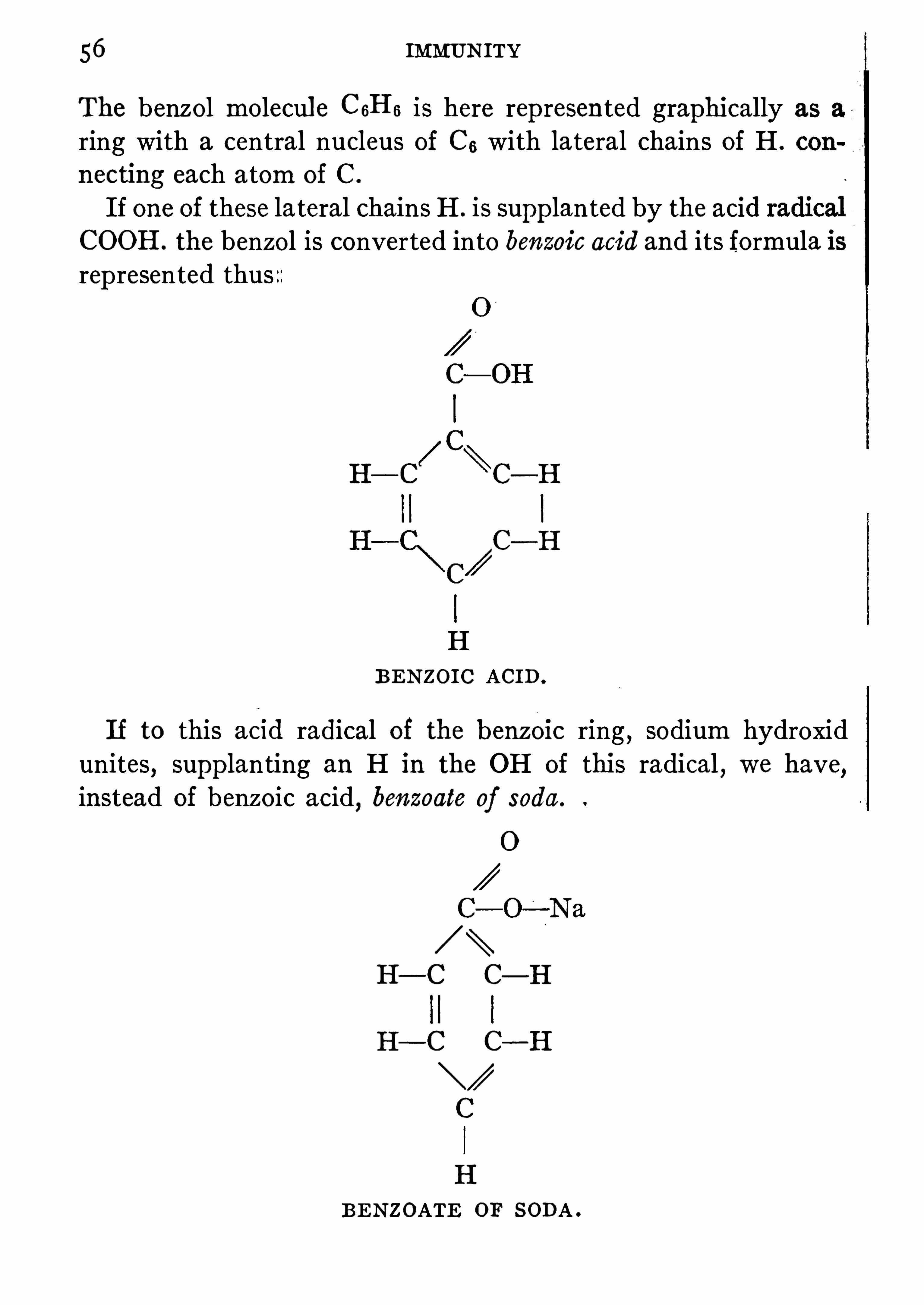
56 IMMUNITY
The benzol molecul e CGHG is here represented graphicallyring with a central nucleus of C5 with lateral chains of H .
necting each atom Of C .
If one of these lateral chains H . is supp lanted by the aciCOOH . the benzol is converted into benzoic acid and its formularep resented thus
C—H
H
BENZOIC ACID .
If to this ac1d radical Of the benzo1c ring , sodium hydroxidunites
,supp lanting an H in the OH of this radical , we have,
instead of benzoic acid,benzoate of soda .
0
H—C C—H
H—C C—H
C
H
BENZOATE OF SODA .

RECEPTORS 57
t is thought that as the soda is brought in”
contac t with the
ntral nucleus of the benzol ring, SO foodstufls uni te with thentral body of the cell molecule in the organism and nourish it .In the case of toxin
,the two lateral chains of its molecul e are
tophores and toxophores. The haptophores seize the
ins of the cell and the toxophores poison it .conceived that cells were nourished by their lateral
a central nucleus with many lateral chainstling all over it . Complex albumins
,food
the case may be) unite with it . This meansn of a part of a cell with all or part of a group Of
rtaimbody cells are only capable of uniting withIt is known that the toxin of tetanus has a chem
for the nervous system and for its neural elementsliver or Sp leen cells . The poisons of snake venomble of uniting with any cells of the pig ; therefore , this
S immune to snake venom.
Now,as these toxins unite with the cells by means of the recep
rS, the cell is stimulated to p roduce an excessive number of theseceptors, which are cast OH and become free . Nature is very
and whenever'
any Of the tissues of the body have beened
,or there is a deficiency
,an enormous excess of reparative
is p roduced . Weigert first called attention to this phenomewhich has been called Weigert’s overproduction theory. SO
the haptophores of the toxin molecule combine with thetors of the cell
,the latter are incapable of any further union
useless‘
to the cell . Accordingly a great number of freeS are generated
,and floating in the blood , engage the
portion of the toxin . Thus the toxophore is neud rendered innocuous before it can reach the cell .
These free overp roduced recep tors constitute the anti-toxin .
This is the essence of Ehrlich’s theory (Fig.
Through thep rocess of time and oxygenation the toxophorousgroup in the toxin becomes innocuous , and only the hap tophorous

58 IMMUNITY
group remains active ; nevertheless the hap tophorous group isto combine with the recep tors and to stimula te the cellgenera ting free recep tors . This a ttenua ted toxin isEhrlich toxoid. The recep tors have been compared torod
,which if p laced within a building would
,if S
disaster,while the same rod p laced outside Of the building
,
means Of p ro tection to the structure against lightning .
FIG.
—a, receptor on cell ; b, toxin molecule ; c, haptophorous portion 0
the molecule ; d, toxophorous portion ; e, receptor. (Willi ams )
theory can be app lied to the production of o ther anti-bodiesIf blood cell
,bacterial cell
,or any animal fluid possessing
phore is capable Of combining with Side chains (recep torcells of the immunized
,just as a key fits a lock , then the
stimul ated to produce excessive numbers Of recep tors , andconstitute the anti
,or immune body . It is possible to pr
from rennet , egg-albumin
,cow’s milk
,and from many
albuminous substances, immune bodies by injectinstances into animals (Figs . 1 6,

IMMUNE BODIES 59
FIG. 1 6.
—EHRLICH’S LATERAL-CHAIN THEORY. Cell with num
receptors of various kinds and to which are united the toxinmolecule . Note the free receptors.
FIG. 1 7.
—EHRLICH’SLATERAL-CHAINTHEORY. In one figure the
free receptors (anti-bodies) are united with the toxin molecule, the attachedrecep tors have no haptophores united to cell.

IMMUNITY
List of immune,
bodies and their anti-bodies (Ricketts)Antigens or Products ofImmunizmg ImmunizationSubstances
Ferments
Precipitogenous
Substances
Agglutinogenous Agglutinins
Substances Hemolysins
Opsinogenous Opsinins Bacteriolysins
Substances Special cytotox
Cytotoxin Produc Cytotoxins ins Such as
ing Substances Spermotoxin
Nephrotoxin
Hepatotoxin ,etc.
SynonymsAmboceptor .
Immunk'
o’
rper .
Zwischenk'
o’
rper .
Intermediary body.
Fixateur .
Preparateur .
Copula .
Desmon .
Consisting oftwo bodies
Complement
Amboceptor
Substance sensibilisatrice.
It is well -known that rennet coagulates milk , but if some of theserum ofan animal immunized against rennet is added to the milk,the latter canno t be coagulated because the anti-rennin combineswith the rennet and renders it inert .
IMMUNE BODIES 6 1
duction of bacteriolysins is explained by Ehrlich’s lat
hypothesis . Immunization against bacteria which dooxins is easily secured by rep eated injectiong bacteria into the organism. It is not easy,assive immunity , as in the case of diphtheria ,
of the serum of the immunized animal . The
alone present in the serum generally and someement must be added to effect bacteriolysis . The serumsaid in the solution Of bacteria are known as anti-bacterialS,which
,though not anti-toxic , may Check invasions and
recovery by destroying bacteria . It is possible to effectcorpore bacteriolysis in the case of typhoid fever if the
and complement are injected in suflicient amounts
p roportions . AS yet the results are not satisfactory from a
cal standpoint .study Of Fig. 1 8 will Show clearly the exac t combinations ofous substances engaged in the immunity p rocess . Some Of
terms must be defined .
ntigen , the body , bacterium,red blood cell
,etc .
,used for
the production Of thermostabile anti-bodies,which
then the substances formed against antigens ; inciting-antigen .
Toxins,ferments
,see above .
Toxophore, the poison-carrying frac tion Of the antigen .
Haptophore, the binding frac tion of antigen or anti-body .
Complement, alexin the normal thermolabile anti-body sub
serum.
Zymophore, toxophore for agglutinins and precip itins .Cytophile fraction
‘ is that part Of anti-body which combineswith cell
,while complementophile fraction joins with complement .
Immune body, the thermostabile anti-body against bacterial orother cells .By immunizing with comp lement or anti-body we Obtain
respectively anti-comp lement and anti-immune body which

IMMUNITY
64 IMMUNITY
istration . The Skin erup tions, joint pain s and edemasickness are also evidences of this condition . It is said t
persons who suffer after anti-toxin are suscep tible to the emanations from horses and the physician Should make inquiries In thisdirection when contemplating the injection Of all sera .
In experimentally induced hyp ersuscep tibility the reaction isSp ecific . The condition is transmissible from mother to foetusand it can be transferred from adult to adul t passively by injectingthe blood of a sensitive animal into a normal one . The first doseis called the sensitizing one
,the second the intoxicating . The
incubation period of the sensitization varies with the nature of the
p rotein ; for horse serum it is from eight to twelve days,for bac
terial p roteins from five to eight days . The sensitive p eriod maylast for several years . In searching for the cause Of this reait was found that there are (1 ) a spastic distention of the
monary alveoli probably both of central and local nature, (2 )
scattered hemorrhages in the organs and (3) hemorrhages withulcerations in the gastricmucosa . There have beenmany theoriesfor this phenomenon
,but those ofVaughan ,Friedberger andWolff
Eisner may be condensed andcompounded aboutbody is unp repared to care for parenterally (o therintestinal tract) introduced protein andmust (1or enzyme to care for it . This enzyme or an
and carefully disposes of the foreignare Slowly absorbed and removed . Iduction theory this anti-substance is in large quao ther introduction of p ro tein occurs
,and goes to
avidity so that it rap idly breaks the protein up intowhich cannot suddenly be cared for by the body .
thought by some workers tha t the body protein of the
question is attacked and split , liberating toxic fractions,injected p ro tein woul d not be adequate in amount to ac
poisoning . T hese p ro tein toxins attack nervous and pmatous tissues . Another very p lausible theory would

ANAPHYLAXIS 6
first injection directly sensitizes important cells which are(1 when an intoxicating dose arrives .
characters be tween serum Shock and immedi
shock was thought to Shed some light on the subter is believed to be due to protein degradation
due to trauma or low blood p ressure , which have a
effect upon unstriped muscle or upon the vasomotor
een shown that an anti-anaphylac tic sta te can be pro
by repeated small injections Of p ro tein at intervals too0 allow incubation of an intoxicating dose . Use is madeknowledge in the case of p ersons who need anti-serum butsensitivi ty to it . Repea ted small but increasing quan
put into or under the skin or into the vein until the11 receive without reaction the full dose . This is called
used these facts to elaborate a theory of infec
believes that bacteria circulating in the body stimulateS,combine with them and that when comp lement acts
this union toxic substances are set free .
exp laining all infectious diseases on this basis one assumessometime in life a p erson has been sensitized by bacteria or
p roteins so that he is recep tive for a virulent germ whenas overcome the p rimary external bodily defenses . It is also
be considered the modern exp lanation of diathesis .McK ail divides anaphylaxis as follows
Natural Anaphylaxis, depending upon
(a) Sp ecies Of animal , for examp le cholera in man,anthrax
in cattle,glanders in horses .
(6) Age—diphtheria in children,erysip elas in the elderly .
(c) Individual—to white Of egg, or blood serum,even by in
gestion (“one man ’s meat is ano ther man ’s poison

66 IMMUNITY
AcquiredAnaphylaxis, depending upon
(a) An attack Of disease , erysipelas , diphtheria .
(b) The injection of dead cells, tuberculin .
(c) Injection of nitrogenous matter,blood serum and egg
white .
D irect practical app lication Of theoretical Speculation abouthyp ersensitivity may be made in exp laining , treating andpreventing certain states in man . Mention has already been made of the
possibility of intoxicating p ersons suscep tible to the presence ofhorses by the injecting Of horse serum. The only treatment forsuch a
“Schock ” is an injection of ep inephrin or atrop in .
Serum Sickness is exp lained as a digestion offoreign serum stillthe body as such when sufficient ferment has accumulateddigest it
,a p eroid of three to twelve days . Whether
rect or not,symp toms can be mademilder or preventedb
the doses by some hours . A state of hypersensitivitymay also exist to pollen Of p lants , certain foods and drugs , (1of feathers and hair or dandruff from cats and dogs . “
The
dences Of this state take the form of asthma,gastrointestinal
turbances,and Skin erup tions of which urticaria and eczema
the commonest . Detection of this hypersensitivity mayjective at times but usually it has to be established by tmeans . It SO happens that allergic p ersons have either aanti-body reaction or the local sessile recep tors in the cucells are stimulated
,for the app lication of the responsible
to an abraded area on the Skin will p roduce at the point ared areola . In the cases Of infection of a bacterial oture this reaction usually requires twenty-four hours toa delay suggesting that anti-bodies are involved . In the caseserum or pollen allergie , the reaction is almost immediate , suggeing local cellul ar p reparation . This is the basis of Skin tesexcep t with tuberculin and the Schick test .allergie poisonings is __best accomplish

ANAPHYLAXIS 6
the offending material . Treatment and p rophylaxis take the
form of injecting solutions of the responsible protein underthe Skin . For example ragweed is the most common causeOf hay fever . Solutions Of its pollen , made in standard dilutions of 1 to 1
—50 0 are injected in rising quantities
,
beginning with the weakest . Treatment should be given duringthe winter and Sp ring SO that as high a degree of disensiti
zation as possible will be accomplished . Little can be expectedin the hay fever season .
1 9 .
—Illustrating the conception of deviation of comp lement. a,
Amboceptor; 6, antigen ; k, complement. (MacNeal .)
Complement Fixation .—Hemolysis occurs when the serum of a
hbit immunized against washed Sheep’s red blood cells is mixed
esh washed Sheep’s corpuscles in the presence of comp le
If,however
,complement be absorbed in any way,
a soluOi the coloring matter of the red cells will not occur in thisure . Comp lement will combine with anti-body in the p res
ence of antigen . This fact has been taken advantage of in determining both the nature Of antigen and the p resence of anti-body .
Its most important prac tical use is in syphilis , to the diagnosis ofwhich Wassermann applied it , and the Wassermann test is for the
presence of syphilitic anti-body in the blood serum of syphilitics .

68 IMMUNITY
This test is positive from the initial lesions all during life unlessthe patient has been successfully treated . Indeed the parasyphilitic states also give it . The p rincip les of the test are alsoused for determining the presence of tuberculous
,leprous, - ty
phoid and other anti-bodies .The materials necessary in the Wassermann test are as follo
1 Syphilitic antigen , extrac t from the syphili tic liver ofin alcohol , ether or water ; lipoids like lecithin or ext
guinea p ig’s heart will act as antigen .
2 a . Serum from a known case of syphilis and containingfore syphilitic anti-body .
2b. K nown non-syphilitic serum without anti-body .
3 . The suspected Serum .
4 . Fresh serum from a guinea pig,'
rich in comp lement .
5. Serum from a rabbit tha t has been immunized againstwashed red cells from a sheep ; called ambocep tor .6 . Fresh Sheep’s red blood cell s , washed in saline andmade into
5 p ercent sup ension .
The solutions are all standardized SO that only sufficient of eachis added to comp lete the absorp tion or p roduce the hemolysis .The serum known to be syphilitic and the
'
suspected serum are
heated to 56°C . for thirty minutes to destroy the native and in
herent comp lement . The rabbit anti-Sheep cell serum is alsoheated to this degree .
The hemolytic series , i .e.,Sheep
’s cells,rabbit’s anti-sheep
’
s
cells serum and complement are standardized to find out whatquantities will exactly comp lete hemolysis . These quantitiesare the units . It is necessary to control tests to find out whatquantity of the antigen and known syphilitic anti-body will uniteto bind the determined quantity of comp lement . The tests are
performed in small tubes so as to have a long column offluid easierto Observe. Tubes are set as follows

WASSERMANN REACTION 6
A . 1 un it # 1 1 un it #2a I un it #4 . 1 un it 7515 1 un it 196 NO hemo ly sis .
B . I un it # 1 I unit 343 1 unit £4 . I un it #5 1 un it #6if #3 be Syph il itic . no hemo lysis .
i f #3 be Nou-Syph il itic hemo ly sis .
C . I unit #I 1 un it #2b 1 un it #4 . 1 un it #5 1 un it £6 Hemoly sis.
I un it fza 1 un it #4 . I un it #5 I un it at6 Hemolysis .
1 un it #2b 1 un it #4 . I un it #5 I un it #6 Hemoly sis .
I unit #3 1 un it 1544 . 1 un it #5 I un it #6 Hemolysis.
1 un it #4 . I un it #5 1 un it 7546 Hemolysis.
1 un it # 1 . 1 un it #5 1 un it #6 NO hemolysis .
I unit £2 a . 1 un it #5 1 un it #6 NO hemolysis .
K . I un it #2b . I un it is I un it #6 NO hemolysis .
L . I un it £3 . 1 un it #5 1 un it #6 NO hemo lysis.
The tubes receive first the solutions on the left and are p lacedin the 37
°C . incubator for two hours to allow union of their various
parts , particularly the comp lement with o thers . They then re
ceive the solutions on the right, are p laced in the incubator forhalf an hour and in the ice-box overnight
,when they are ex
amined for a solution of the red coloring matter . If it occurs, the
column is perfectly clear red with some residue of extracted cells .
If no hemolysis has occurred,the red cells form a layer at the bot
tom,and the column is clear and colorless .
A and B are the tests of syphilitic sera while the remaining areto find out if the other solutions affec t the resul ts ofA andB . Ofcourse tube G represents Simp ly the complete hemolytic system.
The extra tests are to exclude the possibility Of interference on
the part of any Single member with the comp lement NO. 4 . The
character of the test is found in tube A where syphilitic antigen and
serum have bound orfixed the complement so that it cannotunitewiththe rabbit serum and sheep
’s corpuscles to hemolyze the latter . This
is a positive test. A negative test is when hemolysis occurs , since no
anti-body is present to unite with complement in the presence ofantigen .
Complement Deviation- This is a condition arising when there
is too much ambocep tor and too little comp lement . The freeambocep tors adsorb comp lement and there is none left for cellneeds or renewed demands . It is to be distinguished from com
plement fixa tion . The terms are not interchangeable .
70 IMMUNITY
ANTI-TOKINS, VACCINES, AND TOKINS
The following is Wassermann ’s list of anti-toxins :
Anti-toxins for bacterial toxins :Diphtheria
Tetanus
Botulism
Pyocyaneus
Symptomatic Anthrax
Anti-leucocidin,an anti-toxin against the leucolytic poinson
staphylococcus
Anti-toxins for the blood dissolving toxins of certain bacteria .
Anti-toxin for animal toxins :Anti-venenefor snake venom
Anti—toxin for spider poisonAnti-toxin for scorpion poisonAnti-toxins for certain poisons in fish, eel, salamander, turtle, and wasp
sera .
Anti-toxins for plant poisonsAnti—ricin for castor-oil poisonAnti—abrin for jequerity bean poisonAnti-robin for locust bean poisonAnti-croton for crotin-oil bean poison
Anti-pollen for pollen of plants that produce hay-fever .
There may be added to this list a number ofanti-sera developedto a p ractical value in recent years whose activity is dependentnot On anti-toxin content
,but upon their ability to agglutinate
,
p recip ita te , and op sonify the ir respective microorganisms . Thesesera are made against infections with :
Pneumococcus
Meningitis coccus
Streptococcus
Dysentery (bacillary)Staphylococcus
Typhoidfever

72 IMMUNITY
glanders . Being very suscep tible to infection with tetanusundergoing treatment
,a p rophylactic injection of tetanus
toxin is given each animal . McFarland found that therate from tetanus
,in a large stable
, was grea tly reducedusing tetanus anti-toxin as a p rophylactic measure .
Immunization is star ted by injecting into a previouhealthy borse a mixture of toxin and anti-toxin in whichformer is not quite neutralized by the la tter . These quantitiesdetermined by guinea-pigtests . Such a mixture is safer for thehorse and begins the immuni ty reaction more p romp tly. Afew doses like this are given after which pure toxin is used .
This is followed by a rise of temp era ture , local reaction and
systemic disturbance . After waiting for all reactions to disapp earinjections are continued by Slow increases
,until
,after a
weeks or months,
c .c . of toxin are injected at one time
(enough to have killed a dozen horses that had not received thesmaller doses p reviously) . The injection of the toxin is followedby an immediate fall in the anti-toxic power of the serum ,
onlyto be followed by a quick rise . The horse will not p roduce antitoxin indefinitely. After the animal has been immunized sufhciently ,
his blood is drawn from the jugular vein , and after theclot has formed the serum is drawn Off and
.
‘stored .
Anti-toxins are found to lie in the p seudoglobulin fraction of
the serum. Even though an anti-serum be strong , a large quantity woul d have to be injected to Obtain high unit value . The
globul ins of the whole blood are now p recip ita ted by 30 p ercentammonium sul phate and the pseudoglobulins are dissolved bynormal salt solution . This concentrates high unit values intosmall bulk . When removed from the horse , serum may contain
30 0—80 0 units per cubic centimeter . After concentration a value
of 3 units may be Obtained .
McFarland found tha t a horse was capable ofp roducing enoughanti-toxin to p rotect 80 6 other horses against doses of toxin , eachone Ofwhich was equivalent to the total amount of toxin that the

ANTI-TOXINS 73
immunized horse received . Thus there is evidently a tremendousoverp roduction of anti-toxin far above the needs of the animal .The various component parts of the toxin stimul ate the cells of
the horse to p roduce the recep tors , or anti-toxin . The toxoids,
themselves not poisonous, have the p roperty of stimulating the
production of anti-toxin . We measure the anti-toxic powers ofthe anti-toxin with units arbitrarily devised . An anti-toxic unit
is that amount of horse serum just necessary to protect a 2 50-gram
guinea pig against 1 0 0 times the minimum lethal dose of toxin .
TO standardize anti-toxin , we must employ animals,into the
“ bodies of which toxins and anti-toxins are injected . If a certainamount of anti-toxin is necessary to pro tect a guinea pig againsttimes the minimum fatal dose of toxin per 1 0 0 grams Ofguineaweight
,then we know tha t the anti-toxin contains so many
ts . The minimum lethal dose of toxin is the smal lest quantitywill kill a guinea pig of 2 50 grams in four days .
standard anti-toxin is kep t by governments to be used as a1 0 1 of the p roducts of biological chemists .
this standard anti-toxin a toxin of unknown strength isby means of guinea p igs . The toxin un
i
t thus found isto determine the anti-toxic unit of anti-toxins of un
rum is p reserved by the addition of 5 percent ofr phenol . It remains practically unchanged in strengthor more .
ot only ofvalue as a curative agent,neutralizing the toxins
formed,but is valuable as an immunizing one against in
Persons exposed to diphtheria and giving a positivetest Shoul d be given an immunizing dose of to
If injected early in a case Of diphtheria , i t is much moreto do good
,than if used later . Some desperate cases have
ed units and have recovered . The following is the

74 IMMUNITY
SINGLE DOSE ONLYInfant, 1 0 to 30 pounds (under 2 years)
Moderate Severe Malignant
Child, 30 to 90 pounds (under 1 5 years)
l
Adults, 90 pounds and over
M ethod of AdministrationIntramuc lar
or
Intravenousand
pg Intramuscularor
Subcutaneous
Tetanus Anti-toxin .—Tetanus anti-toxin is p roduced in aman
ner Similar to that of diphtheria anti-toxin . As the horse is exceedingly sensitive to tetanus toxin , before the immunizingis begun , the toxin is attenuated by heat or iodine .
The anti-toxin is standardized,as in diphtheria ,
by tes
potency ,
against the toxin . A guinea pig of 50 0 gramsused, and test toxin is emp loyed of such strength that .0 1 cc .
kill~ the guinea pig in about four days . The United Statesoff
-Ltetanus anti-toxin is now the least quantity of anti-tetserum necessary to save the life of a 350
-gram guinea pininety-Six hours against the Official test dose of standard toxinfurnished by the Hygienic Laboratory of the Public HealthService .
The toxinof the tetanus bacillus has such an affinity for nervoustissue that
,once i t becomes attached , separation is difficult , or
even impossible if union has existed for some time . More thanthis the toxin has a greater affinity for nervous tissue than for its

ANTI-TOXINS 7
own anti-toxin . If an animal be injected intravenously withanti-toxin and intracerebrally with toxin it will die of te tanus .This exp lains why the use Of anti-toxin in the treatment Of thediesase must be vigorous , early and direc t . Dosages of 1 0units are necessary before symp toms have become well established. Subcutaneous methods are too slow and indirec t . Antitoxin should be g iven into the Sp inal canal , into the vein and
around the wound . Prophylactically anti-serum should be givenin every case of penetra ting , lacerated wound especially if soiledwith earth
,street dirt
,rust or gun waddings . Tetanus antitoxin
is more efi cient as a p rophylactic than as a remedy .
Streptococcus Anti serum.—The three p rincipal group s of
c,non-hemolytic and viridans , have differ
and each kind produce s i t own anti-bodies .however are never in grea t amount and this seemsthe rather feeble antigenic power of the cocci them
selves . The only useful anti-sera are those p repared against alarge number Of strains of each of the varieties by rep ea tedinjections over a long period . Their antigenic fractions seem to
be hemolytic,leucocytolytic and neurolytic . The anti-bodies
formed in horse ’s serum seem to decrease in value after removalfrom the body . They are chiefly agglutinative and anti-toxic .
For clinical use the serum should be as fresh as possible and usedintravenously in doses of 50 to 2 0 0 cc . It has been emp loyedin endocarditis
,osteomyelitic and puerperal sep ticemia with some
p romise .
The Anti-pneumococcus serum is p repared in the same way .
Horses are immunized by the injection of first autolysates then3 living cultures
,and the horse ’s blood
,after a period Of treatment
by cultures,is drawn off
, preserved with tri-cresol . It is used inthe crude form of serum as the anti—bacterial powers are not easilyconcentra ted
,It is standardized so that . 2 c c . Shall p rotect a
mouse against times the amount Of a culture of pneumococci tha t woul d kill a control mouse . It has been found that

76 IMMUNITY
there are in this country four group s of pneumococci in a se
ical sense,a discovery confirmed by therapeutic results .
possible to p repare cura tive anti-sera against two Of the
while the other two fail to call forth useful anti-bodies . In any
given case of pneumonia the typ e of infecting organi sm 13 determined by isolating it from the sputum and performing the agglutination test with the individual sera ofTyp es I and II ,whereuponshould one Of these react the app rop riate anti-serum can be in
jected. The organisms are Obtained for the agglutination testby injecting the sputum into a mouse ’s peritoneum,
killing theanimal after six to eight hours and using the rich growth of cocciin the peritoneal fluid as the bacterial suspension . With Typ e Ithe therapeutic results are very p romising ; with II helpful at times .Typ e III is the Pneumococcus mucosus yielding no useful antiserum
,while Type IV is a heterogeneous group possessing no
serological uniformity and unable to call forth any valuable antibody in the injected horse . The serum is given intravenously indose of 50
—1 0 0 c .c . and repeated when the temperature rises
again,sometimes every eight hours .
Meningitis Anti-serum.
-Ep idemic memngitis, caused by the
M icrococcus meningitidis intracellularis or meningococcus,can
be treated by anti-serum. The cocci owe‘
their power to endotoxins and pus formation , exerted chiefly in the coverings of thecentral nervous system . They are p resent in the Sp inal fluid
,
only being found in the blood early in an ordinary attack or
in highly sep ticemic cases . Anti-serum is made by injectinghorses with first dead then living bacteria until its serum Shallhave acquired high agglutinative
,Op sonic and bactericidal
power . The horse ’s serum is separated , p reserved as usual , andused by injecting into the arachnoid Space by lumbar or cranial
puncture .
“
This route is selected to bring the anti-serum intoclose relation with the cocci Since by the subcutaneous or intravenous routes insufficient anti-bodies pass from the blood to themeningeal fluid . It Should be given also into the blood stream

VACcINATION 7
in order to p revent sep ticemia . Its effect is to increase phagocytosis and cause bacteriolysis . NO satisfactory standard has beendevised . Dosage varies but a safe guide is to injec t under themeninges 75 p ercent as much as the fluid removed at lumbar
puncture . Intravenous dosage Should be 2 0—50 c .c . The antiserum p robably has no p rophylactic value .
Anti-plague Serum.-Yersin
,a French bacteriologist, treated
horses with living cultures Ofp lague bacilli , and after a long p eriodof immunization used a serum which either effec tually vaccinatedan individual against the p lague , or greatly modified the diseaseafter i t had once begun .
The action of the serum is bactericidal , as well as anti-toxic .
The dose varies with the stage of the disease ; 2 0 c .c . is an effective
p rophylactic dose , while from 2 0 to 30 0 c .c . have been used Oftenas curative doses .
VACCINATION
By the use Of attenuated , or killed microorganisms , i t is possibleto effectively vaccinate men and~animals against many diseases
,
no tably,small-pox, hydrophobia , p lague , cholera , typhoid fever,
anthrax and quarter-evil .Any of the bacterial p roducts used as prophylactics are some
times call ed vaccines,the word being borrowed from small-pox
vaccine . It is better to use the word bacterin for the purpose ,even when they are given p rophylactically . Bacterin is emp loyedfor the dead bacterial masses used therap eutically . Sensitized
bacterins are vaccines that have been exposed to the action of the
respective anti-sera before their use . It is the purpose of this
procedure to prepare the organisms by combining them with thehomologous anti-bodies SO that when injected they have only tocombine with comp lement to begin their immunizing effec t .Theoretically this is correct but the p ractical value has not beenfully confirmed .

78 IMMUNITY
Vaccination Against Small -pox
Jenner ’s Observations in 1 794 established the etiological relationship between human variola and cow-
pox . Human small
pox virus passed through a cow will lose its power to producetyp ical variola but will produce in man a loca l condition calledvaccinia which
,upon recovery , leaves behind immunity to
variola .
By the term vaccina tion , in its strict sense , we mean the app lication Of attenuated small-pox virus , weakened by passage through
rkine, to human beings and infec ting them with the modified
disease . The disease is localized at first at the site Of inoculation ,and a bleb or vesicle forms . AS a rul e the disease does not becomegeneralized . It creates
,in the vaccinated individual , an active
immunity against small-pox. The toxins diffused through the
blood-stream stimul ate the cells of the body into forming eitheranti-toxic or o ther anti-bodies . These various substances , as
yet unknown , remain for a long period within the body of the
vaccinated p erson and may p ro tect i t for years against invasionand infection with the cytorcyteS in virulent form . A person whohas variola cannot be vaccinated
,subsequently be is immunized
against vaccinia by this attack ofvariola,just
'
as he can_be immunized against variola by vaccinia infection .
Since Jenner first discovered that cow-
pox introduced into the
bodyp revented small-pox, it has been the world-wide custom to
use either the dried virus or liquid glycerinized virus from the cow
or human beings in the p rocess of vaccination . It has been found'
that,
human virus generally used was likely in rare instances totransmit syphilis , SO it is now the universal custom to use cow
virus . This virus is coll ected from fresh vesicles in calves oryoung heifers
,as clean as possible , as it is used as seed to inoculate
the animals and the operation is done under strict antisep tic
p recaution . After a week the virus is collected under Similarantisep tic precautions by scrap ing the base of the vesicle with a

80 IMMUNITY
virus on aga
l /
r the surface growth is washed Offwith sterile water
(8 c .C . and_
18 part of this IS used as a dose . As the virus rap idly
attenuates it must be reactivated by passing it through guinea
p igs from time to time .
The first injection is given in the flank,and the second fo llows
in five days . Accordingly as the symp toms are severe , so will theresulting p rotection be strong . Haffkine has given injec
tions without an accident . The following results were obtainedby Haffkine who worked in India for the British Government :
DeathsPopulation
The immunity conferred by this mode of vaccination is notcomp lete until ten days after treatment . It is possible to vac
'
cinate with these relatively virulent bacteria because they are
given under the Skin,a p lace where life of the vibrios soon ceases .
During an attack of cholera the vibrios donot enter the bloodbut remain in the deep layers of the intestinal mucosa .
Vaccination AgainstTyphoid
By the injection Of sterilized cultures Of typhoid bacilli , i t is
possible to create an immunity Of a moderate kind against entericfever . The method was p erfected by Wright , and his modeof p rocedure is to secure a culture of typhoid both virul ent andable to call ~ forth a large amount of anti-body in the injected
p erson , which is tested on guinea p igs , and the minimum lethaldose for a 1 0 0 -gram guinea pig is used as the dose for man . Thi sdose varies from 5 c .c . to c .c . of a culture sterilized by heat

ANTI-TYPHOID VACCINATION 8
at 60°C .
,and p reserved with lysol . After the injection there is
often redness and pain at the Site of inoculation,some fever and
lymphangitis . The method at p resent in use in the UnitedStates is to emp loy a twenty-four hour agar Slant or bouillonculture
,killed by heating as above ; these are susp ended in
saline with trikresol or phenol and counted in a hemocytometer .In order to control the toxicity of this Suspension a mouse is in
jected subcutaneously with a billion ; i t should live at least fivedays . The doses are 50 0 million , million and millioneight to ten days apart .The results of Col . F. F. Russell
,U . S. A.
,a man who has had
much experience, since he was in Charge Of the army vaccinations,are interesting and instructive . He says
I.
“ Anti-typhoid vaccinations in healthy persons is a harmless
procedure .
2 . It confers almost absolute immunity agains t infection .
3. It is the p rincipal cause of the immunity Of our troop sagainst typhoid in the recent Texas maneuvers .
4 . The duration of the immunity is not yet determined, butis assuredly two and One-half years and probably,
longer .
5. Only in excep tional cases does its adm inistration cause an
appreciable degree of personal discomfort .6 . Itapparently protects against the chronic bacill us carriers,
and is at p resent the only means by which a person can be pro
tected against typhoid under all conditions .
7 . Al l persons whose p rofession or duty involves contact withthe sick shoul d be immunized .
8 . The general vaccination of an entire community is feasibleand could be done without interfering with general sanitaryimprovements and Should be urged wherever the typhoid rateis high .
”
The wisdom of these conclusions has been abundantly p rovenin our armv during the WorldWar when p ractically no typhoidfever occurred in vaccinated men . It might be added that no
6

82 IMMUNITY
immunizing procedure is p erfect and this one does not justifythe dr inking Of water known to be polluted.
Vaccination against all the typhoid fevers , due to the typho idbacillus
,the paratyphoid bacillus A and B
,can be accomplished
by combining these organisms in one vaccine . The form now In
common use,as emp loyed by our armed forces , is one suspension
in each cubic centimeter Of which are contained mill iontyphoid bac illus , 750 million of paratyphoid A and 750 millionof paratyphoid B . Three doses
, 5 c .c .
,1 c .c . and 1 c .c are given
ten days apart. This makes a total Of mill ion organisms
injected .
Vaccination Against Pneumococcus Infections
Experiences in South Africa by Lister and in the AmericanArmy by Cecil seem to hold out encouragement for the prophylaxis of pneumonia by vaccines of the respective coccus . The
typ es of pneumococci prevalent in a district must be determinedand used in the suspension . The p reparations employed byCecil contained the three fixed American types and were givenin doses of mill ion four times at six to eight day intervals .Under war conditions some advantage was Observed but availabledata are .too few to form a general evaluation . Experimentation and human exp erience indicate the harmlessness of this
procedure and use is recommended. The action of the vaccineseems to be stimulation of op sonins and Of bacteriolysins .
Vaccination Against Diphtheria
According to the experiments OfBehr ing, Theobal d Smith andW. H . Park it is perfectly feasible and without danger to immunize children against diphtheria . As already mentioned immunization Of horses is begun with nearly neutral toxin-anti-toxinmixtures . For immunization Of children only such are used .
Solutions Of the toxin and anti-toxin are made to contain 1 unit
V ACCINATION AGAINST ANTHRAX 8
e latter and 60 percent and 80 percent of a unit Of the former .or three injections are given , the first or the first and second
strength , the las t having the higher value of toxin .
ion occurs and not infrequently a general oneaise . Immunity requires a week to start and about abe fully developed . It is believed to last at least
four years . Immunization need only be given to those who havea positive Schick test .
Vaccination Against Plague
Haflkine,in India
,has vacc inated natives and o thers aga inst
plague by somewha t the same methods emp loyed in anti-choleravaccination . The B . pestis is cultiva ted in flasks of bouillon ; asit grows
,the stalactite-like scum on top is Shaken from time to
time to the bottom Of the flask . After growing for Six weeks inthe bouillon
,the c ulture is killed at 70
°C . for three hours . It is
then used as vacc ine, 3 c .c . is the usual dose for man,2 c .c . for
woman,
. and childr en still less . After the inocii lation ,heat and
redness appear at the site of inoculation,and the patient feels ill
and has some fever . Haffkine holds that immunity aga inst the
p lague is complete in twenty-four hours after vacc ination . Hisresults are at times really very good .
The Indian Plague Commission reported that the measure wasvaluable as a means of preventing infection ; while it was not anabsolutely cer tain ‘means
, yet it sensibly diminished the deathrate . The immunity lasts about a month . Such vaccines arenot to be used after attack has started .
Vaccination AgainstAnthrax
Of all forms Of vaccination against disease with attenuatedbacteria this is the most successful . Its use is confined to domestic animals, Sheep , cattle, and horses
,and has reduced the
mortality in the country where it is used from 1 0 percent to 5

84 IMMUNITY
percent . The method requires the employment of two vac
cines made Of a ttenuated anthrax bacilli . NO . I is a culture Of
bacilli attenuated by growing them at a high temp era ture , 4 2 5°
C,
in a current of air for twenty-four days . NO . 2 is grown at the
same temperature for -only twe lve days . The first vaccine is
used to immunize the animal against the second,which causes a
marked local reaction , and which is the real immunization agentagainst infection with virulent anthrax bacilli . The injectionsare given about one week apar t . Many Sta te Governments aswell as the Federal Government Of the United States supp lythe vaccine gratis to stock raisers and o thers .
Vaccination AgainstBlack-leg orQuarter-evilQuarter-evil, or Rauschbrand, is due to a Sp ecific bacillus .Vaccination against this disease may be accomp lished by inoculating with a powder consisting of dr ied muscle from the affected
par t of infected animal . There are two vaccines, NO . 1, andNO .
2 . The first is p repared by heating (and thus attenuating) thebacilli up to 1 0 3
°
C . The second is prepared by raISIng the tem
perature up to 93°
C . These vacc ines are given at a Short timeapart, and the immunity is effective . The method is valuableto stockmen .
Vaccination Against Tuberculosis
There is at present no safe and satisfactory p rophylactic meas
ure for the production of increased resistance in man to tubercu
losis. In cattle the repea ted injec tion ofbacilli of the human typehas been found capable of ra ising the animal’s resisting power toa sort Of immunity . The therapeutic use of the various tuberculines has onthe o ther hand, met with better success, and it seems
that for human medicine at least that they have won a p lace in'
the treatment of surgical tuberculosis . They may be also'
Of
value in pulmonary disease .

THE TUBERCULINS 8
The Tuberculins
The toxin Of the tubercle bac ill i (Old tuberculin) is prepared bygrowing the organism for a long p eriod in glycerinized veal broth ,after which the flasks are steamed in a sterilizer for an hour ormore
,and then the bacilli are filtered out through porcelain filters .
The filtrate is reduced by boiling to one-tenth of its bulk,and to
this 5 p ercent of carbolic acid is added as a preservative . Ifthis toxin
,even in minute doses
,is injected under the Skin of a
tuberculous animal,it acts as a powerful poison . In a few hours,
it causes a rap id rise Ofbody temperature , accompanied by nauseaand
, perhap s, vomiting . About the localized foci Of tuberculosis,
a vigorous reaction occurs . Around indolent Old sores and otherlesions there is a tendency to heal by the casting Off Of necrosedtissues
,and the infiltra tion of the perituberculous area with leu
cocytes. In lupus (tuberculosis of the Skin) redness and heatoccur about thelesion . This febrile phenomenon following theinjection of tuberculin into tuberculous animals is a val uablediagnostic feature toward the recognition of tuberculosis inanimals and in man . In 90 percent of cases the reaction istrustworthy .
Its use in man has been much questioned,as i t is thought by
some todisseminate the disease from original and confined foci .This , however , has been denied . Many able clinicians use it andrecommend it (OSler, Trudeau, Musser) .K och’s new, or T .R . tuberculin was
,like the Old
,designed by
him as a therapeutic agent for the cure of tuberculosis . It ismade by pulverizing the bodies of living tubercle bacilli and dissolving the residuum in an indifferent fluid
,centrifuging this and
collecting the sediment which is Tuberculin Rest , T .R . The
solution above this sediment containing soluble substances fromthe bacillary bodies is Tuberculin Obers
,T .O. It produces a
more intense reaction than the Old tubercul in . Like the Old, itis used in the treatment of lung
,bone
,laryngeal
,and Skin tuber

86 IMMUNITY
culosis. It certainly causes a local reaction about tuberculousfoci
,and no doubt aids in the building up of healthy tissue .
The dose of tuberculin for testing purposes Shoul d be .5—2 . mg.
for a child , 2 .
—6 . mg. for a young or weak person , and 5.
—1 0 . mg.
for a larger person . It is well to give the highest dose that it isbelieved the patient will stand in order to get a p romp t anddefinite result thus avoiding the necessity of a repetition . Re e
p eating the injection of such amounts is not without danger asi t might light up a latent lesion . Any focus that is at the bottomof a clinical condition requiring such a test
,will give a positive
reaction with the quantities mentioned . For therapeutic pur
poses one begins with an injection Of .0 0 0 0 0 0 1 gram or smallerand increases S lowly according to the patient
’s condition . Tuber ;
cul in Should only be administered by exp erts .
Mal le in
Mallein is a p reparation made from the toxin of the glandersbacilli
,and is p repared p recisely as the Old tuberculin . By in
creasing the virul ence of the glanders bacilli , by passage througha series of guinea pIgS, a highly virulent bacillus is obtained . Itis then grown in glycerinized bouillon for a month at 37
°C . The
resul ting fluid is sterilized by heat and filtered through a Pasteurfilter.
‘
The filtrate is evaporated to one-tenth its quantitywhen intended for conjunctival use or left in its natural statewhen for subcutaneous use . A small amount of carbolic acidis added in order to preserve it .In a horse with glanders
,the injection ofmallein is followed by
a large painful swelling at the injection site . With this thereis a rise Of temperature , which is the diagnostic reaction tha tindicates infection with glanders . In this respec t the reactionis like tuberculin . In healthy horses no rise Of temp eraturefollows the injection
,and the resulting swelling more quickly sub
sides . A convenient test is the introduction Of a drop of concen
trated mallein into the conjunctival sac . Positive reaction is

88 IMMUNITY
Berlin the weakest injection used (the first) is made from a cordthat has dried but eight days , and the course is much quicker .It was at first thought tha t short drying might carry over toomuch v irus but in order to treat certain serious head bites
,cords
of 3 and 4 days drying were tried not only without damage butwith p romising results . Now in threatening cases treatmentmay be begun with 3 day cords, then 2 day cords . The effectof this mode of inoculation is to p roduce in the bitten individuala very rap id active immuni ty, quicker in its action than the
infection . The treatment is solely p rophylactic and in no way
curative . If symp toms of rabies have set in, the treatment
is of no avail . In rabies the incubation p eriod is about six weeks,SO that there is p lenty of time to immunize the patient by injection with a ttenuated virus .Since the immunizing process is always begun after the bite of a
rabid,or supposedly rabid dog, it differs from o ther vaccinations
,
which are resorted to before infection .
Results Of_Treatment. —Among those bitten by rabid animals
the total mortality before the introduction of vaccination was notless than 1 0 percent . Among the same class of patients in ,
the
Pasteur institutes,the death rate of all cases
,early and late
,has
been reduced to a fraction of 1 percent . Those cases in whichthe bites are on the head, are always more serious
,and the mortal
i ty is higher . Like tetanus the virus travels,i t is supposed, from
the Site of injury to the central nervous system by way of the
nerves . If the bite was on the toe,i t would take
‘
longer for
infection to reach the brain , than if i t was on the upper lip . Thisis a very plausible exp lanation of the varying incubation
.periodsIn both tetanus and hydrophobia .
C oley’s Fluid in the Treatment ofTumors
This method of treatment is in no wise a p rophylactic one , butstrictly a curative one . It consists in the injection of the toxinsof strep tococci , in the hope that they wil l cause a Shr inking, or dis
OPSONINS AND OPSONIC INDEX 89
appearance of malignant sarcomata . An attack of erysip elas (i thas long been Observed) occurrIng In a patient with some malignant disease
,has the effect of causing a disapp earance , or retro
gression ,of the tumors . Artificial infection with strep tococci
was then practiced with the idea that it might produce the Same
But this was found to be dangerous . Coley p reparedof strep tococci by allowing them to grow with the B . Pro
idgiosus . The mixture after a long period ofincubationwas steriliz ed by heat
,and the fluid thus obtained was injected into the
tissues . Virulent strains of strep tococci are used and the dosedead culture is about half a drop given under strict anti
p recautions. The best results are Obtained in Sp indlercoma
, .and the poorest in the melanotic variety . The
thod by no means Should be employed where the tumor canbe removed by operation . It canno t supp lant th e knife, and
only. in inoperable cases or as a supp lementary treatment whereother forms of treatment are employed , should it be used .
Opsonins and Opsonic Index
Peculiar substances in blood serum have been called by Wright and Douglass opsonins (Greek: prepare food for) . If fresh blood ismixed with an emulsion of some bacteria and then incubated for half an hour, it will then befound thatmany of the bacteria are within the polymorphonuclear leucocytes.
If the serum is washed away from the leucocytes before adding bacteria , noneof the latter will be found within the leucocytes. This proves that the serumhas some influence on phagocytosis. In order to Show that this efiect is
.
on
the bacteria rather than on the leucocytes, the bacterial suspension may be
treated with some serum for half an hour and then washed free from thisserum by means of a salt solution in a centri fuge, and then mixed with someserum-free leucocytes ; then it w ill be found that phagocytosis occurs as before .
The bacteria have been “sensitized .
” According to Wright this action isor peptoniz ing .
then depends upon the action of serum upon bacteria ,w ith in the body , first by the action of the serum, and thenS . It is thermostabile .
ive action of phagocytosis may be estimated by Leishman’sblood and an emulsion ofbacteria in salt solution in equal

90 IMMUNITY
quantities, and allowed them to stand for thirty minutes in the incubator.After this the mixture was stained and the average number of bacteria perleucocyte was Obtained . The result was known as the phagocytic index.
Wright has devised the following technique . Young cultures, a few hoursold
,are employed . These are scraped off agar tubes and mixed with salt
solution. After this has sedimented, the supernatant fluid is Separated from
the bacterial masses by a centrifuge ; is p ipetted off, and preserved .
Washed leucocytes are obtained by collecting 2 c .c . of blood in 30 . c .c . of
salt solution containing 1 percent c itrate of soda to prevent blood coagulation. The serum and c itrate of soda '
are separated from corpuscles bywashing twice in a centrifuge . The upper layer of the sediment is rich inwashed leucocytes, and is used in the experiments.
To Obtain the Opsonic index , blood serum from various cases is collected .
In the case Of staphylococcus infection—say furuncle—the blood serum is
drawn from the patient and, w ith equal portions of an emulsion of staphylojcocci (young cul ture) , and a suspension of washed corpuscles, is thoroughlymixed in a p ipette , which after the ends are sealed
,is placed in an incubator
for fifteen minutes. A drop of the mixture is then Spread upon a Slide ; fixed,and stained with Jenner’s stain. The number Of staphylococci in 50 polynuclear leucocytes is determined and divided by 50 to obtain the average .
At the same time that this experiment is being p erformed , some normalserum Should be used in another experiment ; an emulsion of staphylococciand washed leucocytes being used as above . After pursuing the same steps
in this experiment as in the first,the average number of staphylococci per
leucocyte is determined .
To obtain the opsonic index, it is necessary to know the ratio of staphylococci in the leucocytes treated w ith furuncular serum and w ith normal serum .
If the normal serum leucocytes contained 1 0 staphylococci , and the furuncular serum contained 1 5, the index would be I .5 .
In the case of tubercle bacilli,the latter must be heated to IOO
°
C . to killthem, otherw ise they will be agglutinated by the serum,
and a homogeneousemulsion w ill not be obtained . After heating , the clumps must be broken upby grinding the masses in an agate mortar
,adding a little salt solution from
time to time until the mass is thoroughly broken up . The bacilli must then,after phagocytosis, be stained by carbol fuchsin and decolorized w ith acidalcohol . If the leucocytes are left too long in contact w ith the organismsthey may become so engorged as to prevent counting , the number increasingfrom percent after five minutes to percent in two hours.
Highly immunized anti-bacterial serums have much greater opsonic powersthan have normal ones, anti-streptococcus and anti-pneumococcus serabeing especially pwerful toward streptococci and pneumococci . It is pos
9 2 IMMUNITY
determined that persons giving a negative reaction need not receiving doses of anti-toxin . About 80 percent of adults and 30 to 4
of children are immune . A negative reaction seems to indicate that the bloodcontains % 0 c .c . more unit of anti-toxin per cubic centimeter.
Isoagglutinin and Isohemolysin
There are frequently in the blood ofanimals,lytic andagglutina
tive anti-bodies for the red cell s of o ther members Of the same
Species . By this is meant that if the blood ofa man be mixed withthat Of another the cells of one of them may be clump ed or dissolved . This is of considerable importance to the surgeon whowishes to transfuse blood
,for were he to use a donor whose blood
was unl ike that of the recip ient the la tter would have a seriouschill, indica tions ofblood destruction and shock, andhe might die .
These two substances are p robably indep endent in action butare so commonly found acting in harmony that they may be con
sidered inter-dependent . Landste iner andJansky divided personsinto four group s according to their agglutinin and agglutinogen .
Moss made the same Observation but classified them differently.
JANSKY’S GROUPINGSerum
Cells PercentI
II
III 4
I\7 —kThis means that the cells ofTyp e I are not agglutinated by anyof the serums
,whereas the cells of Type IV are clumped by
serums but the ir own .
Agglutination is accomp lished according to Landsteinagglutinins a and b which are both contained in serumsall type of corpuscles , whereas both are absent in serumsno cells . Agglutinable bodies
,represented by a andb , are
to be p resent in the resp ective cells which can be clumpein the others . Group I therefore contains agglutininsbut does not contain agglutinable bodies a and b .

ISOAGGLUTININ AND ISOHEMOLYSIN 93
d be exp lained on the same basis . While Jansky ’s me thods
p riori ty in time of publica tion the American surgeon hasbe grea t war become a ccustomed to the Moss group ing
which is as follows :
Ce llsI
II
III
IV
t will be seen that this is merely the reverse of the first one andat Typ e IV has agglutinins but no agglutinable bodies .Agglutinins and agglutinable bodies may be absorbed from a
iven blood by saturation with their respective antigen .
In transfusion,homologous bloods should be mixed, but group s
or IV respectively may be considered as universa l donors SinceShows it
(safe to use a blood which agglutinates the
cells . It is however,incorrec t to use a blood whose
uscles are clumped by the recip ient’s serum . The agglutina
titer of human blood is not very high,1—1 0 to 1
42 5, and as
the entering serum is diluted the agglutina tion titer would be
Serum
nts on the independence of the two anti-bodiest usually agglutina tion p recedes hemolysis but
not be so , in a small percentage Of cases . For practicalagglutination tests are suffic ient indications of the com
ibility of bloods . Tests for compatibility take the form‘
Of
direct mixture of the blood of recip ient and of donor and (2 )testing Of donor ’s blood by standard serums .
1 . Patient’s blood A—Serum separated from the clot . B
cells from defibrinated blood washed in saline and resusp ended insaline . Donor ’s blood a -serum separated from the clot , b—cellsfrom defibrinated blood washed in saline andresuspended in saline .
Test A—4 parts b 1 p t.
a —4 parts B 1 p t.
Incubate at 37°for one hour .

94 IMMUNITY
If bloods be compatable no change will occur in the blood cells .If donor ’s corpuscles be agglutinated by recip ient
’s serum therewill be clump ing in the first tube ; if the reverse , a change willoccur in the second tube . This method while often used is moretrouble and no more reliable than the following
2 . It is necessary to have for the group determinations , knownsera of the various typ es , but for practical purp oses types II andIII will reveal any type corpuscles susp ended in them . The bloodto be tested is caught from a prick in the finger into a few drop s ofsodium citra te solution . One drop of this cell susp ension is mixedwith one drop of typ e II and of typ e III sera separa tely on a Slide .
After a p eriod of five to thirty minutes clump ing may occur .If it occur in bo th
,the blood is by the Moss scale group I ; if in
neither it is group IV ; if clump ing occur in serum III and not inserum II
,the blood is group II ; if clump ing occur in group II
and not in group III the blood is group III .

96 STUDY OF BACTERIA
so swiftly that the eye can hardly follow them,or they may merely
roll or wiggle across the field Slowly . D irec t division,if pro
ceeding under the best conditions, requires but fifteen to for tyminutes . It is best Observed in a warm stage or when working ina room kep t at a temperature of 35
°C . Sporulation occurs differ
ently in different Sp ecies . In some it will be found soon after theculture has been removed from the incubator
,While in others
several hours are required . Sporulation, it must be rememberedis a resistant stage when unfavorable conditions are met.
The Gruber-Widal reaction is thus studied . A drop of the
serum andbullion culture,mixed in p roper proportions , is dropp ed
on a cover-Slip , which is then placed, drop downward, over thecavity of the Slide (hanging drop, Fig. 2 0 ) (see Agglutination) ;
FIG. 2 0 .
-Hanging drop , over hollow ground slide . (Williams )
Staining bac teria is a matter tha t is easily accomp lished , andvery many staining solutions andmethods have been invented forthis purpose .
The Simplest procedure is to take a drop Of_puS, blood or culture ,and spread it upon a very clean Slide with a sterilized p latinumneedle . The ma tter must be sp read thinly and evenly . After thewater has evaporated and the preparation has become dry withoutthe use i heat, it must be fixed . T0 do this various agents areused . The objec t Of the fixing is to coagulate the protop lasm of
the cell s,and to fasten all the smeared matter fast to the glass
,
so that the staining fluid andwater will not wash them off. Thisis accomp lished, for bacteria usually, by holding the smeared Slidein thefap ex of a bunsen flame until quitewarm to the hand . Grea tcare must be used not to char the film . Exp erience is neededto fix Sl ide smears correctly . The beginner would do well touse cover-Slip s . If a cover-Slip is used it must be passed throughthe flame three times rap idly . After fixing and thorough cooling,

STAINING BACTERIA 97
the s ta ining fluid is poured on ,and after remaining a fewminutes
is poured off and the slide is washed,dried by blotting paper ,
and examined . If a cover-slip has been used a drop of balsam is
put upon a clean slide and the cover , smeared with stainedbacteria , is inverted on the balsam . Upon the stained bacteriathemselves (if a cove r-glass has not been used) or upon the coverSlip a drop of cedar oil may be p laced, and the p repara tionexamined with a one-twelfth Objective . This is one Of the
simp lest staining procedures p ractised in bacteriology . Othermore comp licated methods will now be described .
Besides heat, absolute alcohol, methyl alcohol,or formalin may
be used as fixatives. Some stains are made up with methyl0 1
,and instead of fixing by heat, the stain is merely dropp ed
the dried film,and the bacteria are fixed and stained by the
e solution at the same time, water be ing added for differentiaat the end.
Aniline dyes are almost entirely used as stains in bacteriologynd these are divided into two classes, the basic and acid stains
,
ccording as their staining properties dep end upon the basic or
Cid part of the molecule . Basic dyes stain nuclear tissues of cell sbacteria . The acid are used as contrast stains and do not
but tissues in which they may be imbedded .
11 basic stains are methyl violet, and gentian violet,
methyl blue, andmethylene blue,thionin blue
,Bis
fuchsin , and saffranin . These are used for stainingbacteria under different conditions . The most usefulethylene blue
,since it is difficult to overstain with it
,and
easily applied . It has been found that certain physical(1 chemical conditions are necessary for successful sta ining with
aniline dyes . Alcoholic solution of dyes entirely devoid of waterdo not stain ; absolute alcohol does not decolorize bacteria afterstaining with aniline colors
,while diluted alcohol decolorizes
readily . The more comp letely a dye is dissolved, the weaker is itsstainingpower . A dyestuff unites
, as a whole,with the bacterial

98 STUDY OF BACTERIA
plasm,forming, as it were, a double salt between the two . Cer
tain substances, alkalies , carbolic acid , iron and COpper sulphate,tann icacid, alum,
and aniline oil,are added to a solution ofaniline
dyes,and they act as mordants
,or fix atives
,making the dye bite
into the protop lasm of the bacterial cells . Spores , capsules, andflagella , are hard to stain, and sp ecial heavily mordanted stains areused to demonstrate them . Chemical reac tion occurring in thecell protop lasm is of great value in differentiating bacteria . The
presence of granules in bacterial cells is often only Shown bythe use Of Special stains, which deep ly color them. Bacteria ofthetubercle group are call ed
“acid-fast
,because
,after being stained
,
it is difficult to decolorize them with acid solutions . These bac .
teria are
"
hard to stain and resist decolorizing agents after theyare stained .
1 . LOfller’s alkaline methylene blue solution consists of
Saturated alcoholic solution ofmethylene blue 30 c .c .
solution caustic soda solution in water 1 0 0 c.c .
Mix.
This is the most useful Of all the staining mixtures employed .
2 . Zeihl’s solution carbol-fuchsin consists of
Fuchsin 1 gram.
Carbo lic acid crystals 5 grams.
D issolved in 1 0 0 c .c . ofwater,to which is added 1 0 c .c . of absolute alcohol .
This can also be made by taking a 5 p ercent solution of carbolicacid in water and adding suflicient saturated solution of fuchsin inwa ter until a bronze scum persists upon the top . This is used forstaining tubercle bacill i in sputum and sections . It must beheated when used for rap id staining . Tubercle bacilli can be
stained in cold solution, if immersed over night in it .
3. Fuchs in solution .
Saturated alcoholic solution Of basic fuchsinWater

IOO STUDY OF BACTERIA
now used . If the bacteria on examination rema in a dark violetblue they are then said to sta in by Gram’s method
,or are Gram
positive . If they are decolorized they take the contrast stain andare sa id not to stain by this method
,and are Gram-negative .
Many bac teria stain in this way, andmany do not. Importantbacteria often may be differentiated in this manner .Examples of Gram
’s stain are as follows :Gram-
positive —Bact. aerogenus capsulatus, Bact. anthracis,
Bact. diphtherice, B . tetani,Bact. tuberculosis
, Streptococcus
pneumonice, Staph. pyogenes, Strep . pyogenes . Gram-negativeB . coli
,B . dysenterica, Bact. influenzx ,
Bact. mallei,Bact. pestis,
B . pyocyaneus, B . typhosus, Diplococcus intracellularis menin
gitidis, Micr . catarrhalis,Micr . gonorrhaece, Spirillum cholerce.
Thionin Blue , or Carbol Thion in
This is a useful stain, prepared thus
Thionin blue .
Carbolic acid .
WaterFilter. Good for staining bacteria in tissues .
Spe cial Stains
Wright’s Stain .—This not only stains
,but fixes . It has a wide
range of usefulness in a bacteriological laboratory for the stainingof blood
, pus, malaria] parasites, trypanosomes,as well as manv
bacteria,and is prepared as follows
5 percent solution Of sodium bicarbonate 1 0 0 c .c .
Methylene blue 1 gram.
M ix and heat in sterilizer one hour at IOO°C . Cool, filter, thenmix%0 per
cent yellowish eosin in water until the mixture loses its blue color and becomes
purp lish . Ofthe eosin solution add 50 0 c .c . to each 1 0 0 c .c . of the methyleneblue mixture . M ix and collect the abundant precip itate which immediatelyforms on a filter. Dry this and dissolve in methyl alcohol in the proportionof 1 gram of powder to 60 0 c .c . of the alcohol . This is the staining fluid .
Keep well stoppered . Fresh alcohol may be added for that which evaporates.

SPECIAL STAINS ‘ I
This comp lex stain represents aft
’
y
’
r
pie
°
ofiw liic1i
man ’s, and Romanowsky
’
s are members. T0 use this stain,a
blood or pus film is Spread and air-dried . The stain is then runon the slip , or slide, for one minute . After this time slowly dropdistilled water in‘ quantity Similar to that of stain used . This iswhen the true staining takes p lace . After three minutes wash indistilled water, dry and mount . Nuclei, malarial parasites, try
panosomes, and bacteria are stained blue ; red cells are sta ined
p inkish-orange, while the granules Of the leucocytes are stained
p ink, lilac , or blue, depending upon their character .
Giemsa’s Stain
This stain is used for demonstrating the organism of syphilis,trypanosomata, granules and the like, and is p repared as follows
Azur IIEosin 3 grams .
Azur II. 8 grams.
Glycerine C . P 2 50 c .c .
2 50 c .c .
Bacteria are Often covered with cap sules that are difficult tosta in, and special methods have been devised to demonstratethem.
Air-dry the specimen.
Harden and fix in absolute methyl alcohol .D ilute stain with distilled water, using one drop of stain to each cubiccentimeter of water.Cover preparation with dilute stain fifteen minutes to three hours(longer period for sp irochaetes) .Wash in running water.Blot and mount.
Capsule Staining
Welch’s Method.
1 . Cover-glass preparations are made in the usual manner, and over thefilm after fix ing
,glacial acetic acid is poured .
2 . Without washing off the acid, aniline water gentian violet is poured on .

1 0 2 STUDY -OF BACTERIA
Change t ’hé lstain‘ fear. or : five. times to remove the acid . Stain fourminutes, and wash with 2 percent NaCl solution, not water. . Thisdemonstrates the capsule very well .
His’s Method.
“ A .
I. Make cover-glass preparation mixing specimen with blood serum. Fix
in flame .
Stain for a few seconds with a half concentrated water solution ofgentianviolet.Wash in weak potassium carbonate solution for a few minutes.
Dry and mount.
N
Dry and fix.
Heat and pour on the following stain, steaming thirty seconds(a) Saturated alcoholic solution of gentian violet(b ) Water .
4 . Wash in a 20 percent solution cupric sulphate .
5 . Dry andmount.
3 .
4 .
B .
2 .
3 .
Spore Staining
Spores resist stains , and when stained are hard to decolorize .
1 . Dry and fix in the usual way .
2 . Flood cover-glass with hot carbol-fuchsin ; heat until it steams ; repeatthis once or tw ice . This stains bacteria and spores .
Wash in water.4 . Decolorize with
Alcohol1 percent acetic acid
5 . Wash .
6 . Counterstain w ith methylene blue .
7 . Wash, dry and mount.
00
By this method,which is a simple and satisfactory one
,the
spores are stained a brilliantfi
red,while the body of the bacilli
are stained blue .
Flagella Sta iningTO a beginner flagella staining is difficult ; there have been many
well-known methods devised . The Simpler are as effective as themore comp licated but do not always make as p retty p reparations .

1 0 4 STUDY OF BACTERIA
Pitfield’s Method .
This is the Simp lest sta in and the easiest to use,but does no t
give the good results that the previous one does. But one solutionis needed
,this is made in two parts and mixed .
A . Tannic acidWater
B . Saturated watery solution alum (Old)Saturated alcoholic solution gentian violetM ix.
A heavy precip itate is formed by this process which is useful in the staining . The stain is almost a saturated solution of alum and tannic acid, andwhen it becomes supersaturated by evaporation andheat, staining takes place .
After this the process is very Simple . The cover—Slip is carefully flooded w iththe stain and warmed for a minute over the flame of a bunsen burner, turnedvery low , until steam arises . Not too much stain should be run over thecover-slip . After steaming occurs, the stain should remain for a minute , thenthe preparation is washed, dried andmounted . It w ill be found that the beststained flagella are on those bacteria nearest to the edges where the evaporation has been most intense . If the preparation is not equally stained,Weigert’s aniline gentian violet can be run on for a minute to deepen the color.
Lofil er’s Method.
This is the original flagella stain and is a very good one .
It is made as follows
A . Mordant2 0 percent watery solution tannic acidSat. solution ferrous sulphateFuchsin sat. alcoholic solutionMix.
B . StainCarbol-fuchsin.
Proceed as in the previous methods.
The most important step s in flagella sta ining are to clean thecover-slip s thoroughly, to mix the culture with water and have noculture media with it
,to fix gently
,and not to overheat the stain .
Even in expert practised hands it is not always easy to demonstrate flagella readily .

STAINING DIPHTHERIA BACILLI 1 0 5
Neisser’s method of sta in ing the diphtheria bacillus .stains are needed (Fig. 2 1 )
2 1 .
—B . D iphtheria stained by Neisser’s method . (Williams )
A . Methylene blue .
95 percent alcoholWater .Mix and add
Glacial acetic acidB . VesuvinD istilled water
The staining step s are as follows
1 . Prepare film,fix and dry .
2 . Pour on “ A ” for thirty seconds.
3 . Wash well in water.4 . Dry and pour on “
B for thirty seconds.
5 . Wash, dry and mount.
The protoplasm of the bacilli will be stained brown , and thecharacteristic (diagnostic) chromatin po ints will be stained a
deep blue black .

1 0 6 STUDY OF BACTERIA
Tubercle BacillusStain
1 . Spread the Sputum, pus or culture , over the surface of the cover-slipAllow the preparation to thoroughly dry .
2 . Fix in flame and cool .3 . Pour carbol-fuchsin over the slide and heat with steaming for five min
utes. Young bacilli in tubercles and other fluids are very difficult to stainin this way . The preparation containing them Should he stood in cold carbolfuchsin for twenty-four hours. This method stains everything on the Slide“
.
4 . Wash in water.5 . Decolorize the preparation with a 25 percent solution of sulphuric acid
in water until the red color is lost. Repeat this once or tw ice .
6 . Wash and counterstain w ith Loffler ’s methylene blue .
7 . Dry and mount.
Gabbet’s solution
,methylene blue 2 grams
,HgsOr 25 c .c .
,
water 75 c .c .,is a very useful
,convenient decolorizer and counter
stain for Sputum.
In such a p reparation , if tubercle or other acid-fast bac illi are
p resent, the bacilli will be colored a bril liant red, while the puscells
,ep ithelial cells , and other bacteria will be stained blue
The microscop e dark field illumination enables one to .see
flagella and cap sules . This illumination is obtained by blockingout the central portion of the Abbe condenser in the substage of
the microscope . Light is admitted only from the Sides and objec tsin the field at the point of crossing of the rays reflect these fromtheir sides . India ink may be used as a background for bacteriatha t sta in poorly and have low refractive index .
Protozoa are stained by Wright ’s or Giemsa’s method in one ofits various forms . Sp irochetes, particularly that of syphilis ,may be stained by Giemsa
’s method but Show up more clearlyin the following technic of Stern .
1 . Dry film in incubator for several hours .2 . Immerse in 1 0 percent aqueous Silver nitrate in diffuse
daylight,six hours to three days
,depending on thickness of smear
and need for haste .

CHAPTER VI
BACTERIOLOGICAL LABORATORY TECHNIC
In Order to study bacteria by other methods than the simpleexamination of their morphology by means Of sta ins
,and by the
hang-drop , or block method,they must be cultivated either in
the bodies of exp eriment animals,or in culture media artificially
prepared . The latter method is the most widely used in laboratories . It is necessary, in order to study bacteria , that the mediashall not contain any extraneous bacteria to begin with
, and
tha t they shall be cultivated under such conditions that suchbacteria cannot reach the media at any time . To accomplish allthis
,the culture media must be kep t in glass vessels , such as test
tubes and flasks that have been sterilized . And,since all animal
and vegetable substances , not actually alive,are teeming with
a multitude of bacteria, these substances must be sterilized too ,in order that the media shall be free from any living organisms .Glassware is cleaned by boiling with soap suds or'
powder or ifvery dir ty by immersion in saturated watery
'
Solution of bichromate of potash p lus an equal part of sulphuric acid . This lattermust be very carefully washed away in running water .Glassware , such as p ipettes, Petri dishes, flasks and test-tubes
,
are sterilized best by dry heat in hot-air sterilizers . The apparatus is subjected to a temperature of 1 50
°
C . for one hour,or until
the cotton plugs are slightly brown . The glassware Should be
put in wire baskets and the test-tubes Should be kep t erect .Petri dishes are best steril ized in a wrapp ing of paper . Flasksand test-tubes' are always p lugged with raw cotton
,which pre
vents the ingress of‘ bacteria,
‘
while air can reach the mediathrough it freely .

STERILIZATION IO
Steriliza tion of culture media is accomp lished in steam steriliz ers of two patterns ; of these, the autoclave, using steam under
p ressure, is the most satisfactory and is most generally used at
present.The baskets conta ining the culture media are p laced in the
autoclave after suflicient water has been put in it . The instru
FIG . 2 2 .
-Autoclave .
ment must never be allowed to run without wa ter . The lid isscrewed down and the flame started ; free flowing steam shouldescap e from the valve before the la tter is Shut . When the
pressure has risen to 1 a tmosphere (1 5 pounds) or 1 20°
C . and
held there for twenty minutes,all bacteria are destroyed
, and
the media can be safely assumed to be'
Sterilized.If media
conta ining sugar or gelatine are to be sterilized, the temp era ture
should‘
not run above 1 1 0°
C since, if this is done the gelatine will

IIO BACTERIOLOGICAL LABORATORY TECHNIC
not solidify when cold, the sugar is caramelized and the mediablackened .
Potato tubes are harder to steriliz e at times,and it is safer
to repeat the operation in twenty-four hours .Fractional method of sterilization
,or Tyndallization ,
is aecom
p lished by heating the media to IOO°
C . on three successive days
FIG. 23.
—Arnold sterilizer.
in a Koch or Arnold sterilizer . By heating cul ture media to thistemperature, all the vegetative, or adul t, forms are killed, whilethe spores are not affected ; after the first sterilization, at roomtemperature, the Spores vegetate and become adult bacteria,when on the second sterilization they are non-resistant to IOO
°
C .
and are kill ed . Spores remaining after this develop in to adultforms again and are kill ed on the third day, at the third sterilization . This fractional sterilization is emp loyed under many

1 1 2 BACTERIOLOGICAL LABORATORY TECHNIC
jacket,and
‘
the interior air Chamber,where the cultures are kep t,
is thus heated to the required temp erature . The amount Of hea tis automatically regulated by a thermo-regulator
,which dimin
ishes the gas supply if the temperature runs too high, or increasesit if it runs too low . The Roux regulator is the Simp lest andmosteflicient one .
FIG. 25 .
-Blood serum coagulating apparatus.
A serum coagulating apparatus is needed in laboratories inorder to coagulate the tubes of blood serum (Fig.
Serum tubes are coagulated in it at a temp er'
ature ofabout 70°C .
They are then sterilized by heating them either by the fractionalmethod or in the autoclave .
The separation Of bac teria from the bouillon in which theygrow for the p reparation of toxins requires the use of a bacteriaor germ p roof fil ter , the best typ e of which is the Chamberland orPasteur unglazed porcela in filter . These filters are of varyinggrades offineness, and are so made as to be easily sterilized . The
common pathogenic bacteria cannot pass through the pores ofthe ordinary:filter , but toxic agents are known to pass through thefinest filters
,though they cannot be discovered, as they are
submicroscop ic .
To Opera te the porcelain fil ter it must fit into the neck of a

NUTRIENT MEDIA
vessel very tightly,so that a vacuum may be mainta ined in the
la tter bymeans Of an air p i’
imp .
Collodion sacs are sometimes used in animal exp eriments .Bouill on cultures are p laced within the
sacs,which are then inserted in the ah
domen Of an animal and left there . The
sac is made of collodion because it isnon-absorbent and allows the bac terialjuices and p roducts to osmose outwardand be absorbed by the animal
,while the
animal fluids percolate into the sac .
There are several very ingenious ways ofmaking these sacs
,but the de tails are too
elaborate to be described here .
BOUILLON
Bouillon or broth is the most useful ofall the nutrient media
,Since it is not only
used as a liquid medium,but by the addi
tion of gelatine, or agar, it is converted
into solid media . FIG . 2 6 —Kitas.
atoThere are two methods of making filtfff for filtermg tOXmS
bOLl lllOIl
M ethod 1 .
Take 50 0 grams of lean beef free from all fat, chop it fine andcover with c .c . of water
,shake and p lace on the ice over
night . Then squeeze the fluid out of the met by means of aCloth
,and supp ly enough water to make a litre . Inoculate this
meat juice with a fluid culture of the colon bacillus for the pur
pose of fermenting the meat sugar . For this purpose the inoculated juice is allowed to stand at room temperature overnight .Bring to a boil and add
1 0 grams ofWitte’s peptone .
5 grams common salt.

II4 BACTERIOLOGICAL LABORATORY TECHNIC
Weight the saucepan and contents and hea t to 60 °C . Supp lythe water lost by evaporation . Neutraliz e either by addingsuflicient sodium hydra te, 1 0 percent solution, until red litmus
paper is colored a faint blue, or else titrate 1 0 c .c . of the mixturewith a decinormal solution of sodium hydrate
,using phenol
phthale in as an indicator, and after finding how much of a normalsolution is required to neutralize 990 c .c . c .c .
— 1 0 c .c .
used for titration) this normal solution is added . The mixturethus neutralized is then .boiled for five minutes and the weightrestored . After bo iling
,from .5 percent to p ercent normal
hydrochloric acid solution is added and the acidity thus p roducedi s spoken of as 5 p ercent or 1 .5 p ercent as the case may be .
Upon‘ boil ing
,the albumins are coagulated by heat, and the
phosphates are thrown down . The acid re-dissolves the latter .The former must be removed by fil tration . The filtrate is a clearstraw-colored fluid of an acid reaction which Should not become
cloudy upon boiling . This is then run into flasks or test-tubesand sterilized .
The second method is much more convenient, and is preparedby adding 3 grams ofLiebig
’s beef extract to a litre of water
,and
adding the pep tone and salt, as in the p revious method,and p ro
ceeding as before . To fil ter the bouillon , the filter paper mustbe folded many times
,and the funnel must be carefully Cleaned .
Newer methods for the titration ofmedia have been constructedon a
‘
physiCO-chemical basis
,attemp t being made to estimate
exact reaction in terms of ionic dissociation and hydrogen concemtration . D istilled water contains ionizable hydrogen accordingto the following formula 1 1 0 7 log. 7, a mathematicalstatement abbreviated for convenience to or the symbol ofstrict neutrality and called the Ph or hydrogen concentration .
As the ionization of hydrogen increases , acidity becomes grea terand the numerical factor drop s to or lower ; as the hydrogenvalue decreases the figure rises . Substances in solution , such as
Salts or organic matter act as buffers or agents which attemp t to

1 1 6 BACTERIOLOGICAL LABORATORY TECHNIC
Titrate with N/20 NaOH or N/20 HCl to match tint of. standard tube corresponding to the Ph desired . Calculate the amountof normal alkali or acid to be added to the -medium to give the
proper reaction .
If the natural color of the medium makes it difficult to matchwith the standard solutions, a tube Of 2 0 p ercent solution of themedium in watermay be p laced back of the standard solution .
The best generally useful reaction is a figure suitable formost pathogenic organisms .
GELATINE
To make ge latine , bouill on is made to which gelatine is addedin order to render it solid . The following step s are taken
(a) Take a litre ofwater in a saucepan and add Chopped beef or'
,
beef extract as in bouillon . After standing overnightsqueeze the beef and extract the juice .
(6) Add 1 percent pep tone, 5 percent salt, 1 0 percent to 1 5
p ercent best gelatine and weigh .
(c) Heat until ingredients are all dissolved .
(d) Neutralize, gelatine is highly acid and requires much alkali .
(e) Boil five minutes and restore weight, boil till albumincoagulates .
(f) Cool to 60°C . and add an egg well beaten up in wa ter .
(g) Boil Slowly till all the egg is coagulated . This Clears the'
medium of fine particles tha t are not removed by filtra tion .
Add .5 percent normal hydrochl oric acid .
(h) Filter through absorbent cotton on a funnel previously wetwith boiling water .
(i) Tube and sterilize in autoclave for fifteen minutes at 1 1 0°C .
Litmus,or lacmoid, or neutral red may be added to the
gelatine as an indicator .
AGAR-AGAR II
To make agar
(a) Take 2 0 grams of powdered or chopped agar .
(6) Add to 50 0 c .c . of water, place? in a can in autoclave and
heat to 1 20°
C . Then cool .
(c) Add this to 50 0 c .c . of bouillon of double strength,making
c . c .
(d) Neutralize .
(e) Cool t o 60°C .
(f) Add the egg to the mixture, stir .
(g) Boil till egg is coagulated thoroughly .
(h) Titrate and adjust to desired acidity as given under bouillon
,and while boiling hot
,fil ter through absorbent cotton
wet with boiling water .
(i) Run into tubes . Sterilize . Slope the tubes for twelvehours and store in dark p lace .
To make glycerine agar add 5 p ercent of glycerine to the agarbefore neutralizing . T0 make agar for tubercle bacill i, vealbouillon may be employed, and glycerine must be added .
Carefully skimmed milk,to which litmus tincture has been
added , is run into tubes and sterilized . This is a valuable culturemedium . It is also a reagent .
Potato Tubes
1 . Wash some large potatoes and with a Ravenel potato cutter ,cut out semi-cylinders ofpotato . Immerse in running water overnight, in order to prevent them from turning black . It is wellto wash these bits of potato with 1 bichloride ofmercurySix hours and running water over night . Some laboratories soaktheir slices in sodium carbonate solution . It is desirable to know

1 1 8 BACTERIOLOGICAL LABORATORY TECHNIC
the reaction of the medium and each batch Should be tested,then
marked whether fain tly or strongly acid or alkaline .
Thrust absorbent cotton to the bottom of the tube and wet
with distilled wa ter ; p lace the potato upon the
cotton, then p lug the tube and sterilize in autoclave twice . The tubes should be seal ed .
PEPTONE SOLUTION—DunhamTake Peptone 1 0 grams.
Salt .Water c .c.
Mix. Boil . Fil ter and store in tubes and sterilize.
This is used to demonstrate the production of
indol . Reaction Shoul d be neutral .
SUGAR MEDIA
One of the most important par ts ofdeterminativebacteriology is the discovery Of the different fermentative powers upon carbohydrates of otherwisesimilar germs . Monosaccharides (dextrose and
galactose) , disaccharides (lactose, saccharose) ,alcohols (glycerine mannite)
'
and some starches
tiff'
irffifltfigé (dextrin , inulin) are in common use daily in the
tu b e (Wil labora tory to show the enzyme action of varioushams)
Sp ecies, indicated by acidity and gas p roduction .
One p ercent solutions of these carbohydrates are made in neutralbro th or agar . The best method is to p repare a 2 0 percentsolution of the material, sterilize it in the steam sterilizer and addfrom this to the stock medium sufficient to make the required
p ercentage . This avoids rep eated heating . The media shouldbe neutral . Addition of litmus tincture Or Andrade ’s indicator
(1 0 0 c .c . of 5 p ercent watery acid fuchsin decolorized by the
addition of 1 6 c .c . N/I NOOH) will supp ly an index for acid
production .

1 20 BACTERIOLOGICAL LABORATORY TECHNIC
SPECIAL MEDIA
Endo medium for theTyphoid series . Add 1 p ercent lactose to
p lain neutral agar and 1 percent of the following solution : 1 0 c .c .
of 1 0 percent watery sodium sulphite and I c .c . of saturatedalcoholic solution Of fuchsin
,heated in theArnold twenty minutes .
This Should be mixed freshly each time since the medium is
p rop erly colorless but becomes a pale p ink on standing,being
then useless . It is poured into p lates and allowed to cool andharden thoroughly before use .
Russe ll’s M edium.
—Plain neutral agar receives 1 p ercentlactose
,. 1 p ercent glucose and an indicator
,either litmus or
Andrade . It is used as Slants with a deep butt .The fir st medium is to distinguish typho id colonies from colon .
bacillus ; pale bluish round colonies versus irregular red ones .The Russell medium gives with typhoid , a colorless surfacegrowth and acid in the butt ; paratyphoids give bubbles offermen
tation in the butt ; colon makes distinc t red slant growth and
much gas.
Eggs are emp loyed as culture media . The yolks and whites ofa number of eggs are shaken together in a flask and then strainedthrough a towel to remove the froth . The mixture is then runinto tubes and coagulated and sterilized like
'
blood serum . On
this mixture the tubercle bacillus grows very well .These are the common culture media used in laboratories . For
a more technical descrip tion of the manufacture of these and
o ther media,the student is referred to books devoted to labora
tory technique .
Litmus tincture is made by adding a large handful of litmuscubes to a p int ofwater and bo iling down to one-fourth its volume .
This is then filtered through paper and stored after sterilization.
The Study of the Growth of Bacteria—Cultures
Bacteria growing in group s on culture media are spoken of as
colonies . Aerobic bacteria may be made to grow on culture
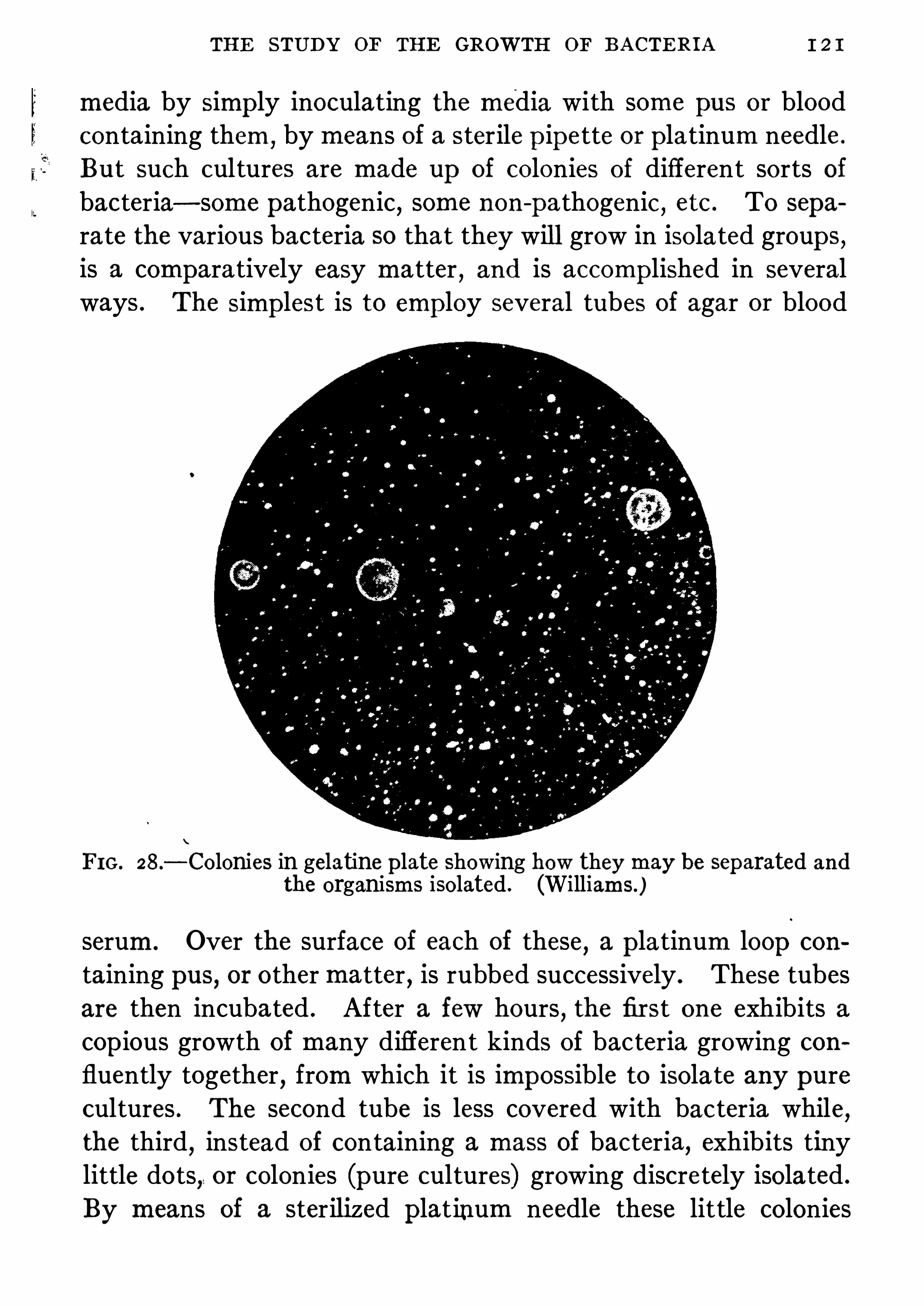
THE STUDY OF THE GROWTH OF BACTERIA
media by Simp ly inoculating the media with some pus or bloodcontaining them,
by means of a sterile p ipe tte or platinum needle .
But such cultures are made up of colonies of different sorts ofbacteria—some pathogenic, some non-
pa thogenic , etc . To separa te the various bacteria 80 that they will grow in isola ted group s,is a compara tively easy matter
,and is accomplished in several
ways . The simplest is to emp loy several tubes of agar or blood
FIG . 28 .
—Colonies in gelatine plate showing how they may be separated andthe organisms isolated . (Williams )
serum. Over the surface of each of these,a p latinum loop con
taining pus, or other matter , is rubbed successively . These tubesare then incubated . After a few hours , the first one exhibits acop ious growth of many different kinds of bac teria growing confluently together
,from which it is impossible to isolate any pure
cultures . The second tube is less covered with bac teria while,the third
,instead of containing a mass of bacteria, exhibits tiny
little dots,or colonies (pure cultures) growing discretely isolated .
By means of a sterilized p latinum needle these little colonies

I2 2 BACTERIOLOGICAL LABORATORY TECHNIC
may be fished out and transp lanted to fresh culture tubes , andafter a few hours’ growth they become pure cultures . An Old
method emp loyed in many laboratories, in breweries and originated by Pasteur was wha t is known as the dilution method .
FIG. 29 .
-Series of stab cultures in gelatine,showing modes of growth of
different Species of bacteria . (Abbott.)
Numerous flasks are inoculated by matter containing bacteriavery highly 3diluted in bouillon and by means of a sterile p ip ettedrop s of this highly attenuated mixture are dropped into flasksOf sterilized bouillon or wort . Most of the flasks will Show amixed growth but a few will Show only one kind of organism.

1 24 BACTERIOLOGICAL LABORATORY TECHNIC
position and the co tton p lug is removed, and held by its outsideend between the fingers of the left hand
,care being taken to p re
vent the tubal part of the p lug touching anything and being contaminated. The p latinum loop is then slowly and carefullyintroduced into the median
,and stirred around so that the tube
wall s are not touched . The needl e is again sterilized and tubenumber two is held in the palm of the left hand parallel tothe first one and its p lug is removed also ; then with a carefullysterilized needle
,three loop s of the inoculated gela tine are re
moved from number one and mixed with number two tube .
FIG. 3 1 .
—M ethod of inoculating culture media . (Williams )
The needle is then again carefully sterilized in the flame,the plug
of number one is carefully rep laced and another tube,number
three,is held in the palm of the left hand and its plug is carefully
removed and held as the p revious ones were . With the sterilizedloop three loopfuls of the gelatine from number two are carefullyintroduced into number three and the needle is then sterilizedandput aside . The p etri dishes Should now be laid on a cold levelSlab,and the contents of the tubes run into the different dishes .Tube number one is taken first ; the lip of the tube is wiped withthe cotton plug and then held in the flame to destroy all bacteriaclinging to it . The lid of a petri dish is carefully and partiallylifted and the contents of the tube rap idly and evenly pouredover the bottom of the plate, and the lid quickly replaced .

THE STUDY OF THE GROWTH OF BACTERIA
FIG. 3 2 .
—D ilution method ofmaking cultures. 1 , IS first tube containinggreat number of colonies ; 2
, contains less number ; 3, relatively few .
(Williams )

1 26 BACTERIOLOGICAL LABORATORY TECHNIC
This p rocedure is followed with the other tubes,andthen the
p lates or dishes are put in a cool dark p lace, and the tubesare putinto a solution of bichloride ofmercury
,or into boilingWater .
The p lates Should be examined from time to time . After severaldays a p erfect Cloud of round colonies are seen in number one ; alarge number inNo . 2 and a much fewer number , say fifty, inNo . 3 .
It is an easy matter then to p ick out a colony tha t is surroundedby a bluish-green halo and transfer it to a tube of agar or bouillon.
In the case of pus it is more than probable that the colony is thatof the pyocyaneus bacillus , and tha t it contains nothing but thesebacilli . It must be studied in a dozen ways, before it is certainthat i t is this bacillus, but the preceding method is a necessary
primary step to secure this organism in pure culture and may
be taken as a pattern for all p late methods .Agar p lates are usually used since they have this advantagethey do not melt at 37
°C . incubator tempera ture . When agar is
used it must be melted at IOO°
C . and cooled below 48°C . and
above 43°C . Above 48
°C . bacteria may be kil led . Below 43
°
C .
the agar begins to harden,so this method must be p erformedquickly ; the p lates Should be slightly warmed, the culture pour edon and the agar hardened, they must be inverted in the incubator ,since the water of condensation forming in the lids Of the p latesoften falls and washes one colony into another . When gela tine
p lates are made, they must be kep t at 2o°—2 5
°C . It is often of
advantage to cool the p lates by means of ice,before they are
filled .
The so-called Stroke p lates are extremely useful for hosp italbacteriology . The agar is softened, poured into plates, allowed toharden and the material to be examined is smeared upon the firmsurface by a fla ttened platinum rod
,a Spatula .
”The separate
colonies develop along the lines of Sp read and can be isolated toindividual tubes as given above .
Roll Culture.
—Instead of pouring out the contents of the inoculated tubes the gelatine may be made to harden on the walls of

1 2 8 BACTERIOLOGICAL LABORATORY TECHNIC
The proper ty of converting sugar into acids and gases is beststudied in fermentation tubes .Into sterile fermentation tubes bouillon containing sugar is run
,
these are p lugged and sterilized . They may be inoculated withbacteria and if gas production occurs it is quickly manifested inthe closed arm . The component gases may be studied and the
various properties determined . This gas ratio is of use in identi
FIG . 33.
—Fermentation tube .
fying various bac teria and differentiating them . The closed armof the tube being shut Off from free air by the amount of bouillonin the Open arm is p ractically an anaerobic tube and is emp loyedfor this purpose . Bacteria that grow only in the closed arm are
considered anaerobes. By inoculating a gelatine tube with bacteria while it is melted and then letting it solidify
, previouslyshaking the tube vigorously, gas formation will be speedilymani
FERMENTATION METHODS
tested by the presence of bubbles . Acids are detected in culturesby the employment of various indica tors in the culture media .
Li tmus, lacmoid,andrade , and neutral red are used for this pur
pose . By titrating bouillon of p revious .known acidity with a
decinormal soda solution , the amount of acid produced by different bacteria can be estimated .
Various sugars are fermented by bac teria, and lactic, acetic ,
and butyric acids are p roduced . Indol is also produced by manybacteria (colon bacillus, Cholera bacillus) , and its presence in culture is an important means of identifying different bacteria . The
organism to be studied must be grown in culture media known tobe free from indol . For this purpose, all mea t extrac ts must beexcluded and a Simp le solution of pep tone and salt, run into tubes
have grown in this mediaseveral days the indol p roduced , if it is produced, is detec tedadding a few drop s of pure sulphuric acid .
~ If a red colorroso-indol) isnot produced , a few drops of sodium nitrite solu
to 1 0 0 c .c . Of water) must be added, and if a p inkor does app ear
‘
it may be safely assumed that indol
nia is detected in culture by suspending a p iece Of pap erNessler ’s reagent above a bouillon culture of a given orIf a yellow to brown color “ is produced ammonia is
s are detected by growing the organism in a solution,
of a
gram potassium nitrate , 1 0 grams pep tone inc .c . ofwater) .Incubate for a week and then add 1 c .c . each of the followingsolutions
(a) Sulphuric acidAcetic acid .
Boil, filter, and add 1 80 c .c . of dilute acetic acid .
9

1 30 BACTERIOLOGICAL LABORATORY TECHNIC
If nitrites are p resent a p ink color is produced by these reagents .Enzymes may be detected by noting whether gelatineis liquefied,or milk curd digested . Both these actions are evidences of the
presence of enzymes .Bacteria growing exclusively in the absence of oxygen are
known as anaerobes ; to cultivate these successively various formsOf apparatus are necessary .
FIG. 34 .
—A streak made in agar by a needl e inoculated with aerobicand then covered at one Spot with cover-glass. The aerobic organisms willnot grow in the anaerobic conditions under the glass. (Williams )
The following methods are pursued in ordinary laboratorymanipulations :I. Exclusion of oxygen .
2 . Exhaustion of oxygen by means of an air-pump .
3. Absorption of oxygen by means of Chemicals that absorboxygen from the air . A mixture of pyrogallic acid and sodiumhydrate absorbs oxygen rap idly, leaving nitrogen only in thechamber.

I32 BACTERIOLOGICAL LABORATORY TECHNIC
inoculated with the organism he wishes to grow . The ends ofthe tube are then sealed in a bunsen flame and there being no air
,
anaerobic conditions are fulfilled,and organisms grow . After
colonies app ear the tube is broken at a file-mark near the colonyand tubes inoculated therefrom.
Under other methods large Novy jars are used for the recep tionof p etri dishes and test-tubes . From these jars the air is withdrawn
,and hydrogen allowed to flow into it.
~
A solution of
pyrogallic and sodium hydrate is p laced in the bottom of the jar
to absorb any remaining oxygen . There are many other ingeniousmechanical ways Of growing bacteria under anaerobic conditionsand the student is referred to works devoted entirely to technique .
Animal Experiments
To determine the pathogenicity of bacteria ; to measure the
strength of toxins and anti-toxins, to standardize anti-toxins, andto recover bacteria in pure culture , it is Often imp erative thatsmall laboratory animals be used . Guinea p igs, rabbits, and
mice are oftenest emp loyed . Strong young animals are the best .Culture toxins and pathological material are introduced into theirbodies in various ways . A favorite one is to
'
shave the abdomen,
scour it with soap andwater, and then bichloride ofmercury
,and
finally sterile water . With a pair of sterile scissors a small holeis cut in the abdominal parieties and through it a loop containinga drop of culture is run into the p eritoneal cavity, or under theskin . With ordinary fluid material, a syringe may be used toinject i t directly through the abdominal wall . The animal iscarefully weighed, and it is watched from day to day . If it diesan autospy is made on it .Other methods consist in injec ting fluid culture into the veins
of the ear,or into the p eritoneum ,
by means of sterile hypodermic syringe . The autop sy should be made carefully, thea nimalShoul d be thoroughly wet with a solution Ofbichloride ofmercury,

HISTOL'
OGICAL METHODS 1 33
then it should be stretched over a pan ,especially devised for the
purpose, or nailed to a board . The skin over the abdomen and
thorax must'
then be Shaved and steriliz ed with a solution Of
bichloride ofmercury. The walls Should then be seared in a linefrom the throat to the pubes with a hot knife
,and through this
line a cut should be made opening up the thoracic and abdominalcavities .By means of a hot knife Spo ts must be seared on the various
organs,andwith another sterile knife cuts should be made into the
organs,then through these cuts sterile p latinum needles are
thrust, and then culture media are inoculated with them . Some
times it is necessary to remove bits of tissue from various organsand p lace them in culture media . In the recovery of the tuberclebacillus from animals this p rocedure is necessary . Great caremust be taken in making the culture and all tubes Should becarefully stored . It is of grea t importance to make smears oncover-slip s as well as cultures
,from the heart cavities
,liver
,
kidneys, peritoneal cavity, etc .
,and stain them directly with
Gram stain . It is sometimes necessary to injec t cil ltures,or
bits of nerve tissue from a rabies case into the brain . To do
this, remove under stric t asep tic p recautions , a button of bonefrom the skull by means ofa trephine . It Should not be forgottenthat animals inoculated and killed or dying after infection maypresent dangerous material to the laboratory personnel . Afteran autop sy, a strong disinfectant should be generously sp readover all parts of the animal and autop sy tray .
Histological Methods
Sections of tissues from infec ted animals are Often examined andstained by app ropriate methods . To demonstra te bacteria , thetissues should be hardened in alcohol or formaldehyde solution
(4 p ercent) , and imbedded in celloidin , then cut into sections andmounted in the following different ways

1 34 BACTERIOLOGICAL LABORATORY TECHNIC
I . Liimer’s Method.
(a) Float section in water.(b) Remove with section lifter to Lofll er’s methylene blue fromthirty minutes.
(c) Decolorize in 1 percent solution of acetic acid for ten seconds.
(d) Dehydrate in absolute alcohol for a few minutes.
(6)
(f) Mount in balsam.
II . Weigert’s Method.
(a) Place in lithium carmine five minutes.
(b) Then in acid al cohol fifteen seconds.
(c) Wash in water.(d) Transfer to slide and dry with blotting paper.(e) Apply Ehrlich’s gentian violet for three minutes.
(f) Blot and place in Gram’s solution for two minutes .
(g) Wash and dehydrate in aniline oil .(h) Wash with xylol .(i) Dry, mount in balsam and examine .
In Loffler’s method all the tissues
,especially the nuclei and the
bacteria,ap pear blue .
In Weigert’s method,if the bacteria stain by Gram’s method
,
the tissues appear p ink, the bacteria a deep blue-black . Tuberclebacilli are to be stained in tissues, best fixed in formaldehyde solution
,by hea ting with carbol-fuchsin as given on page 1 0 6 ; the sec
tion is decolorized by 3 5 percent hydrochloric acid and Clearedby passing it through 95 percent alcohol, absolute alcohol andfinally xylol . It is then mounted in balsam.
Parafline embedding methods may be emp loyed, but for theseand o ther methods of staining the student is referred to workssolely devoted to technique . The staining methods are the
same for parafline and in experienced hands give better results .
1 36 ANTISEPTICS AND DISINFECTANTS
Ano ther method is to make bouillon cultures ofan organism and
add to each a cer tain percentage of the solution of the antisep tic,and abstrac t every few minutes after the addition of the chemicalone loopful of the mixture and inocula te fresh media .
Pieces of thread sterilized,and then put in fluid cultures may
be used in exp eriments ; they are dipp ed into solutions of Chemicalsfor varying lengths of time and then p laced in culture mediaand growth noted .
It will be found in the case ofmost antisep tics in dilute solutionthat an interval of time must elap se before the organisms arekilled . This is determined by observing the cultures made fromthe mixture . After five minutes
,growth may occur
,but after.
one hour,all may be dead, or i tmay take two or three hours .
The student should refer to works on hygiene for standardmethods of controlling disinfectants
,for examp le the Hygienic
Laboratory and the Rideal-Walker methods .The most valuable chemical disinfectants are those that kill in
highly dilute solution in a short time .
Bichloride ofmercury is a highly eflicient germicide in waterysolutions ; if, however, albuminous matter is present its action isinhibited very much .
CHEMICAL DISINFECTANTS
Mercury Salts—Bichloride ofmercury in highly dilute solution
is a very valuable antisep tic . It dissolves in 1 6 parts of tep idwater . It requires an acid reaction for most favorable actionand the tablets now on the market are made up with some acidhaving no effect upon the mercury salt . In 1
—1 0 0 water solution
this salt will kill anthrax spores in twenty minutes . In blood,the anthrax
“
bacillus is killed by a 1 solution in a fewminutes .In bouill on the same organism is kill ed in a dilution of 1in water
,1 all in the same interval of time . The p res
ence of the albumins in the blood or bouill on,no doubt acts as a

CHEMICAL pISINFECTANTS
protecting envelop e about the bodies of the bacteria, being therefore unreliable for disinfecting Sputum and pus. It is also moreuseful and powerful when it is acidulated with a 5. percent ofHCl
,or when it is mixed with common salt or ammonium chloride .
In_
culture 1 solution p revents the growth Of most
pathogenic bacteria . Biniodide of mercury 15 said by some ob.
servers to be more powerful than the bichloride . It is certa inlyless likely to be interfered with by albuminsSulphate of copper in water is a powerful germicide . It is more
potent in watery solution than in bouillon . It has a remarkableaffinity for alga and for moulds . The author found tha t ifmoulds are put into alkaline solution of Copper sulphate and
heated,the copper enters into Chemical union with the protoplasm
of the mycelia,hyphae, and the spores ; 1 of COpper sul
phate in wa ter destroys the typhoid bacilli . Even nascent copp erkills the typhoid bacilli , so tha t Copper foil in drinking water hasthe power, after a few hours
’contact
,of destroying bacteria in
the water .The silver salts are useful in medicine as disinfectants
,especially
on mucous surfaces . T he nitrate of silver is one o f the most valuable Of all p repara tions ; it is about a fourth as eflicient as
bichloride of mercury and is not nearly so toxic . Some of the
albumina tes of Silver are useful because of their non-irri ta tingaction .
Acids,especially the mineral ones, are valuable disinfectants in
not too dilute solutions . They act chiefly as inhibitors of growthrather than destroyers ofbacterial cells . In the healthy stomach
,
hydrochloric acid acts as a normal disinfectant,and in disease
,
where it is absent,it must be added in order to p revent decom
position of food . Boric acid is useful in medicine on mucousmembranes .The halogens, iodine, bromine and chlorine
,are active agents
for the destruction of bacteria . The Cheapest of these is chlorine .
It acts best in contact with moisture,Since i t decomposes the,

1 38 ANTISEPTICS AND DISINFECTANTS
molecule of water combining with the hydrogen to form free Hc land setting free oxygen .
Dry Chlorine gas (45 percent) failed to kill dry anthrax sporesin One hour
,but when moisture was introduced 4 p ercent chlorine
killed the spores .“Chloride of lime,
” chlorinated lime,in 1 p ercent solution kills
most bacteria in one to five minutes . Iodine p reparations likechlorine ones are very powerful . They are of great use in medicine ; ordinary tincture of iodine painted over infected areas actsas a powerful germicidal agent . It is too exp ensive to use in
house disinfection and it is exceedingly destructive to all metallicObjects . A 5 percent solution in 50 p ercent alcohol acts as a
sp lendid disinfectant for intrauterine injection in puerperal sep sis .It is now said that 1 0 p ercent iodine tincture in 70 p ercent alcoholis the most eflicacious, p ractical, medical disinfectant .
‘
Manyclaim it to have the highest p enetrating powers .Dakin
’s solution is a mixture of Chlorinated lime and sodium
carbonate in wa ter and sodium bicarbonate, with an alkalinity of
.45 percent. This mixture when in the presence of organicmatter decomposes with the formation of hypochl orous ac idwhich may further Change into Chlorimido—(NCl) to whichChanges
‘
the antisep tic action is due . The solution is used forinfectedWounds
,being introduced by methods Of infil tration and
drainage . It has an irritant effect upon the skin which tissuemust be protected. The germ-killing power of this solution isvery high . Its use was followed in the great European war, by amarke d reduction in Sp reading and fatal infections from laceratedwounds .Chloramin is another p roduct of chl orinated lime, depending
on‘
chlorin for its action . Being less toxic it may be app lied totissues or even mucous membranes in 2 p ercent solution but it isirritating and surrounding surfaces must be protected .
Dichloramin T. still a Chl orine disinfectant, is less irritating thantheforegoing but being unstable, must be suspended in an Oil like

1 40 r ANTISEPTICS AND DISINFECTANTS
bacteria after twelve hours . It is best to keep the room closedfor twenty-four hours . It may be thrown into the room in manyways ; by generators which decompose the vapor of wood alcohol,when they reach hot p latinum sponges, salt, or hot copp er ; byvaporizing a solution by means of heat ; by adding permangana teof potash to a solution of formaline ; by Sp raying a concentratedsolution over bedding, floors
,and walls
,then Closing the apart
ment . It is very much more active in warm air than in cold, andwhen the air is moist . It has been known to destroy anthraxSpores wrapped up in paper and p laced under blankets . All ofthe pathogenic bacteria are killed by it
, the Staphylococcusaureus and anthrax Spores being more resistant than anythingelse . It will not kill moulds unl ess highly concentrated . As dilu te watery and alcoholic solutions decompose they Should onlybe used when freshly made .
Sulphur Dioxide Gas—An old and ra ther unreliable form of
disinfectant . It does not kill anthrax spores very readily, as
it requires an exposure of twenty-four hours to a 40 p ercent vaporin a room. It isgenerated by burning sulphur in a room tightlyclosed
,and it is much more efficient if weter is vaporized in
the room. It is not very p enetrating, is poisonous to breathe,speedily bleaches fabrics, and attacks metal
'
objects. It is muchsup erior to formaline as an agent for the destruction of insects
,
esp ecially mosquitoes,also to kill rats infected with p lague bacilli .
Lima—Ordinary quick lime,or whitewash
,is highly germicidal .
It is esp ecially efficacious in disinfecting feces from typhoid cases .Typhoid bacilli are killed after one hour
’s exposure to a 2 0 p ercentmixture .
Potassium permanganate in 3 p ercent solution is said by Kochto kill anthrax spores in twenty-four hours . It is not so eflicient
a germicidal ‘ agent as supposed .
Turpentine and essential oils are eflicient germicides in con
centration . Common mustard rubbed in the hands is said tomake them Sterile .
'

CHEMICAL DISINFECTANTS 1 4 1
Alcohol . -Ninety-five p ercent and absolute alcohols are not
antisep tic for the anthrax spores, since they will live for manyhours in contact with absolute alcohol . In general it is unreliable .
Seventy p ercent alcohol is the most efli cient strength .
Zinc chloride in concentration is a powerful germicide . A 2 p er
cent solution will kil l the ordinary pyogenic bacteria in two hours .Sputum,
urine and dejecta are best disinfected by heat . Chemicals often are inert because ‘they cannot penetra te the albuminous masses of the sputum or feces . Long contact with carbolicacid acidulated with HCl is very efficient . Concentrated forma
line and solutions of chloride of lime may be used, also a heavymush of lime in water .Boiling or heating instruments and dressings by highmoist heat
,
as in an autoclave,is the most reliable method of rendering them
sterile . The exposure of dressings to 1 50°C . for one hour
,or
boiling instruments,thoroughly Cleaned mechanically
,for twenty
to thirty minutes makes them certainly sterile .
D isinfection of the skin is a difficult undertaking from a
bacteriological standpoint . In the deep layers of the skin, and inthe sweat glands andhair follicles, bacteria Often exist
,even after
the most thorough and prolonged disinfection . The app licationof soap and water with a stiff brush is by all means the mostvaluable part of the process , since with the removal of the dirtmost of the bacteria are removed . Thorough scrubbing with soapand sterile water
,followed by scrubbing with a 1 bichloride
solution , Cleansing the nails with a sterile brush,and prolonged
immersion in bichloride or permanganate of po tash solution ,complete the process . Modern methods
,even after all this
preparation , require the use of rubber gloves that have beensterilized by boiling . The faultiest part of the p reparation for anasep tic Op eration from a bac teriological standpoint, has alwaysbeen considered to be the sterilization of the hands
,and if these
can be covered by rubber gloves that are sterile,the fault can be
surely eliminated .

1 42 ANTISEPTICS AND DISINFECTANTS
Antiseptic Values (after Park) .The figures refer to the relative antisep tic powers of various
agents for fluids containing organic matter .
Aluminium acetateAmmonium chlorideBoric acidCalcium ChlorideCalcium hypochlorite
Copper sulphateFerrous sulphateFormal dehyde, 40 percentHydrogen peroxideMercuric iodide
mnine sulphate

1 44 BACTERIA
one tha t is faintly acid and contains animal juices like serum.
They must be transp lanted frequently . On gelatine they growscantily without liquefaction
,the growth consists of discre te li ttle
masses, while on agar they appear translucent colonies of verysmall grayish granula . In bouillon cultures some varieties eithercloud the medium uniformly
,or else sedimentate in the form of
li ttle balls, the supernatant fluid remaining Clear . It fermentssome simp le sugars but does not form gas. Inmilk the growth ismore luxuriant, and becoming acid, may be coagulated in twentyfour hours . On potato the growth is invisible and scanty
,or
absent . On blood agar plates colonies appear as tiny gray
points with a zone of hemolysis about them. Sugar mediaof the Simp ler carbohydra tes, Show acidifica tion.
Vital Resistance—Thermal death-point is 54°
C . in five minutes . Virulence in dried albuminous matter (pus) is retained formonths . If kep t on ice , vitality and virulence are retained formonths also .
Chemical Activities—Lactic acid and sulphuretted hydrogen are
produced, also ferments which have e the p roperty of dissolvingfibrin under anaerobic conditions . They are also capable of dis
solving red blood corpuscles, either in culture media or in thebody and about cultures on blood agar p la tes. there is a Clear haloof hemolysis, streptocolysin . They p roduce a strong solubletoxin, which can be filtered from the bouillon and precip ita tedwith alcohol . This causes necrosis, anaemia and death .
Habitat—In sewage, dwellings, dust, on the healthy humanbody
,and in the cavities of the resp iratory tract, vagina, rectum,
and in the fa ces . It is the cause ofmany diseases, i.e.,erysipelas,
puerperal fever, meningi tis, pneumonia, endocarditis, peritonitis,
tonsillitis, osteomyelitis, and the diarrhoea of children .
In general septicwmia strep tococcus is found in the blood, and
p lays an impor tant rOle in secondary infection , causing an aggravation Of the original infection, and often death . It is especiallyac tive in phthisis, scarlatina, small -pox, and diphtheria, in which

STREPTOCOCCUS PYOGENES 1 45
. diseases i t is Often the cause of death . Many Of the symp tomsof phthisis are due to the toxins of the strep tococcus ; cavity formation and hectic fever for examp le . Its virulence can be in
tensified by passing it through a series of animals,until
,finally
,
cu . mm. ki lls in one day all the mice injected with thisdose . The toxin contains a peculiar haemolytic substance, which ,as before remarked, dissolves red cells of the blood, hence theanaemia in sep ticmmia and in suppuration . The toxin of the
strep tococcus , if injected under the Skin,causes redness like ery
sipelas . Coley ’s fluid containing this toxin is used to trea tsarcomata , since infection with the strep tococcus has been knownto cause a disappearance Of these tumors . Prac tically all animalsare suscep tible to
'
the strep tococcus .Agglutinations.
—The serum from an animal injected with streptococci
,or immunized agains t it, will agglutinate strep tococci .
Anti-toxic sera have been prepared by injec ting horses withhighly virulent living culture of strep tococci . The serum pro tectsto a limited degree, and has some curative properties . Culturesof cocci from human sources have been found to p roduce the
best toxins ; there are , however, many strains .The foregoing descrip tion represents the p rincipal characters of
the most important member of a large group of closely relatedstrep tococci . D istinctions in the group are based upon the
solution of red blood cells,the fermenta tion of lactose
,ofmaltose,
of salicin and the coagulation of milk. The rela tive value of
each of the members is not yet settled but investigations haveenabled laboratory workers to elaborate certain techniqueswhich may be expected to clear up the matter . The varietiesnow recognized are Str . hemolyticus, epidemicus, anginosus,fecalis
,salivarius
,equinus
,andmitis . Some of these organisms
produce no hemolysis ; they come under the term non-hemolyticus
and are Of importance in certain resp iratory and allied diseases .There is a group of organisms
,a sort of connecting link with
pneumococci, which p roduce green colonies on blood agar and10

1 46 BACTERIA
are called Str . viridans . These are to be distinguished fromtrue pneumococci by the formation of a tiny rim of bloodclearing in blood p lates and by their insolubility in 'bile . Theseand the group of non-hemolytic strep tococci are held responsiblefor some cases of arthritis
,endocarditis
,Sinusitis
,nephritis, etc
PNEUMOCOCCUS
Streptococcus pneumonia commonly known as the pneumococcus , or Diplococcus lanceolatus (Fig. (For types see
pageMorphology and Stains—This organism is usually found in thetissues and sputum,
in the form of lance-shaped cocci, surroundedby a cap sule . Is almost always associated in pairs, though sometimes in Chains of five or six members . In albuminous fluids
, or
blood serum,and in milk
,the organism exhibits a well-defined
cap sule ; in bouillon and other media, i t loses the cap sule and thelanceolate shap e, and often appears spherical, in pairs , or Chains .It is not mo tile
,has no flagella OI spores, is easily stained by all
the basic aniline dyes, and keep s the color by Gram’s method .
Under certain conditions i t strongly resembles the strep tococcus
pyogenes, and may be differentiated therefrom by growing it onagar smeared with blood . The strep tococcus causes ahaemolysisof the corpuscles, while the pneumococcus does not and the
colonies are greenish .
Oxygen Relations—It is a facul ta tive aerobe .
Grows rap idly, but never luxuriantly at 37 .5°
C . ; at 2 2°
C . muchmore S lowly, often not at all . Grows better in the p resence of
serum or haemoglobin .
Vital Resistance—Easily killed at a temperature of 52°
C . ex
posed for ten minutes . D irect sunlight also kills i t in twelvehours . While it quickly dies on ordinary culture media, i t maylive in dried sputum or pus exposed to diffuse light anddesiccation ,for four months .

1 48 BACTERIA
will not grow . It ferments some of the sugars,the most impor
tant and characteristic being inulin . No gas is formed . Pneumococcic are soluble in bile or a solution ofits salts
,a distinguishing
determinative character .Habitat—Outside the human body it has not been found
,but
is normally p resent in the mouth of about 30 percent of all peop le .
In apparent health cultures from the throat or the sputum in
into animals often causes pneumococcic sep ticamia becauseType IV (see page 76) may be frequently found in them. The
so-called “
fixed types I, II, and III occur in the throat during
pneumonia but disapp ear shortly after recovery . It also may be
found on the conjunctiva and nose in health .
Chemical Activities.—No soluble toxin has been discovered .
The toxic properties are due to an endo-toxin . This organism isa pyogenic one
,and causes dense fibrinous exudates on serous
membranes . All tissues of the body may be attacked . Some
strains of pneumococci are more neurotoxic than others .In rabbits an intravenous injection of pneumococci will cause
a sep ticaemia with at times areas of lobular pneumonia . The
only successful rep roduction of pneumonia in the lower animalsis accomplished by tracheal insufliation of pure virul ent cultures .In human infection the organisms are forcibly inhaled into thedeepest recesses of the lungs . Pneumonia may be haematogenousin origin also .
Besides pneumonia, any serous membrane may be attacked and
p leuritis, peritonitis, p ericarditis , or meningitis may be caused.
Abscesses anywhere may be due to the pneumococcus . Mucousmembranes of the throat often are affected ; middle ear abscessesalso may be caused by this organism. Pneumococcic sep ticaemiasare common .
During pneumonia , pneumococci may be recovered from the
blood before the crisis by means of blood cultures ; ro c .c . of bloodabstracted from veins is mixed with soo c .c . of milk or bouillonand
'
incubated. In twenty-four hours pneumococci, if present,

coccus or MENINGITIS 1 4
grow luxuriantly . Just before the crisis the organisms will notgrow.
Immunity and Susceptibility.—The suscep tibility ofman varies
greatly . Exposure to , cold and hardship s of various kinds p redis
pose to pneumonia . One a ttack does not prevent another . Ithas been observed that normal leucocytes only become phagocytictoward the pneumococcus when lying in anti-pneumococci serum.
It has even been noticed tha t these organisms grow better in theanti-serum,
rather than in the normal serum . Animals have beenimmunized by injecting cultures and toxin . The immune serumthus produced pro tects small animals against infection, and stimulates phagocytosis . It has been used therapeutically in man forthe cure of pneumonia with hopeful results . Olea te of soda aidsin bacteriolysis of pneumococci by sera , if added to the variousvarieties of immune sera (see page Most mammals
,but few
if any birds are suscep tible to the pneumococci ; mice , beingvery easily infected, are used for isolation purposes .Agglutination of pneumococci is caused by the blood of infectedindividuals
,even diluted at r
—6o . Immune serum also has thesame action .
Opsonins increase during the course of pneumonia and are at
their height at or j ust after crisis ..
Pneumococcus mucosus , also called Typ e III is distinguishedby its large size
,long chains
,cap sule, more generous growth
in large moist colonies of not such a distinct green color and itsability to produce a serious form of pneumonia .
COCCUS OF MENINGITIS
Micrococcus Meningitidis.
Diplococcus intracellularis meningitidis.
Meningococcus (Fig.
This organism is the cause ofep idemic cerebro-sp inalmeningitis .Morphology and Stains.
—Resembles the gonococcus closely,

r50 BACTERIA
because it grows in biscuit-shaped pairs ; is nearly always within
pus cells, and like the gonococcus it is decolorized by Gram’s
stain . It has no spores or flagella ; is not motile ; grows in shortchains at times
,and on ordinary media best at 37
°C .
FIG. 38 .
—Meningococcus in spinal fluid . (From Hiss and Zinsser’s Bacteriology, Copyright by D . Appleton Co .)
Relation to Oxygen .—It can be cultiva ted from the meninges
at first best under conditions of low oxygen tension, as in a Novy
jar or in an atmosphere of carbon dioxide and moisture ; oncegrowing in the laboratory it is more luxuriant in aerobic tubes .Vital Resistance .
—It is killed after ten-minutes ’ exposure to

I52 BACTERIA
tions and absence of active pathogenic properties . It will appearon nearly all p la tes made from sputum and throat cul tures
,in
crowded city life of temperate zones . It seems not to be able toincite an infection but to continue or aggravate one already underway .
STAPHYLOCOCCUS PYOGENES AUREUS
Staphylococcus Pyogenes Aureus (Fig.
Micrococcus Pyogenes.
Staphylococcus pyogenes aureus,albus
,and t itreus are known
Commonly as staphylococcus, or grape coccus . They difler onlyin color production on artificial media .
FIG. 39 .
—Staphylococcus aureus. (Williams )
The Micrococcus pyogenes aureus only is here described ;other varieties have similar but much feebler pathogenic powers .Morphology and Stains .
—Round cocci, often growing inbunches like grapes . Individual cocci dividing in two p lanes .They stain very well with all basic dyes, and are not decolorized
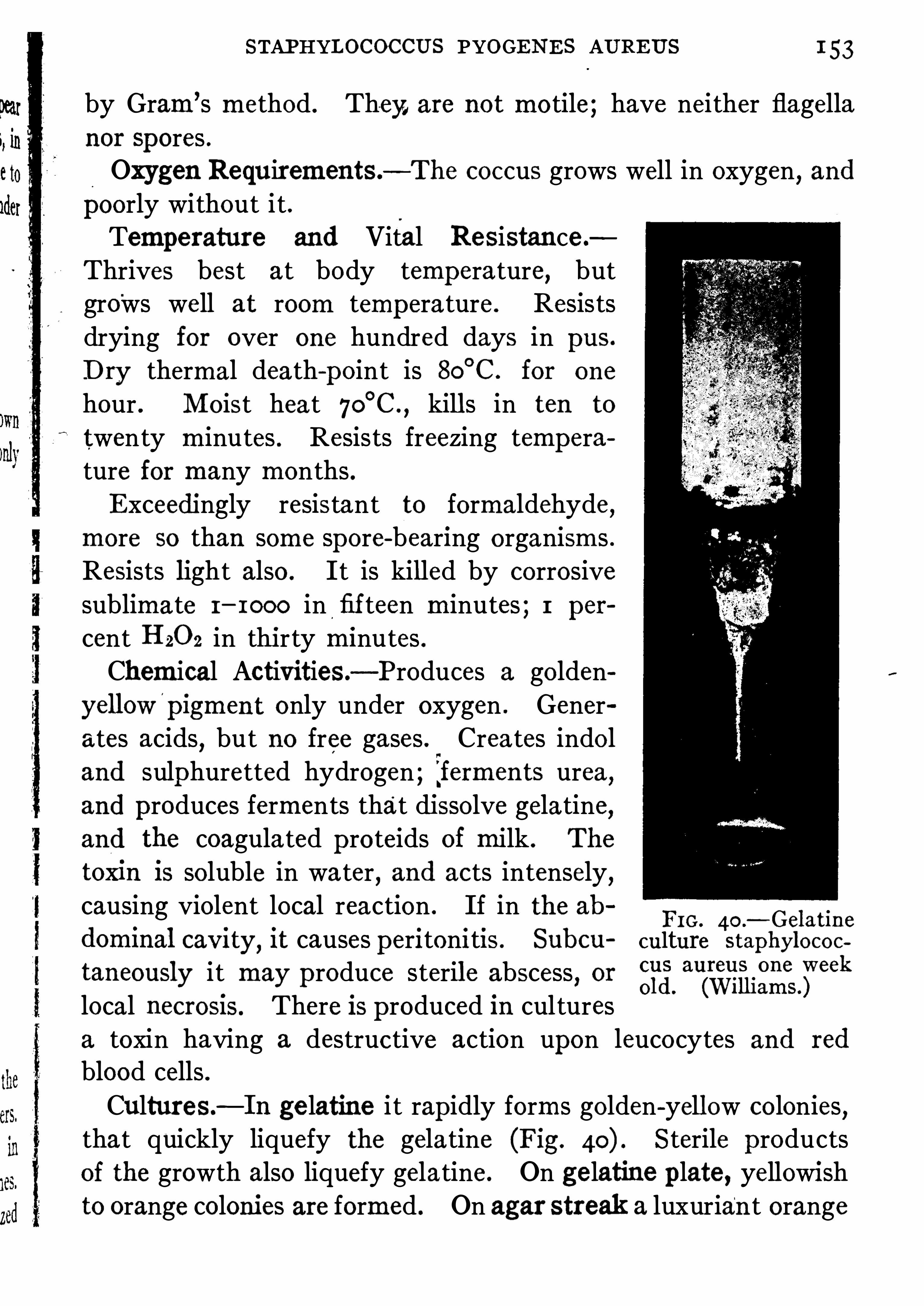
STAPHYLOCOCCUS PYOGENES AUREUS 1 53
by Gram’s method . They, are not motile ; have neither flagellanor Spores .Oxygen Requirements.
—The coccus grows well in oxygen,and
poorly without it .Temperature and Vital Resistance .
Thrives best at body temperature, butgro
'
ws well at room temp erature . Resistsdrying for over one hundred days in pus.
Dry thermal death-point is 80°
C . for one
hour . Moist heat 70°C .
,kill s in ten to
twenty minutes . Resists freezing temperature for many months .Exceedingly resistant to formaldehyde ,
more so than some spore-bearing organisms .Resists light also . It is killed by corrosivesublimate I
—IOOO in fifteen minutes ; 1 per
cent Hzoz in thirty minutes .Chemical Activities—Produces a golden
yellow'
pigment only under oxygen . Generates acids
,but no free gases . Crea tes indol
and sulphuretted hydrogen ; iferments urea ,and produces ferments that dissolve gelatine,and the coagula ted p roteids of milk . The
tox m Is soluble in water,and acts intensely,
causing violent local reaction . If in the abFIO . 4o .
—Gelat1nedom1nal cav1ty, 1t causes peri toni tis . Subcu culture staphylococ
taneously it may p roduce steril e abscess,or 3
1
3local necrosm. There Is p roduced In culturesa toxin having a destructive action upon leucocytes and red
blood cells .Cultures—In ge latine i t rap idly forms golden-yellow colonies
,
that quickly liquefy the gela tine (Fig. Sterile productsof the growth also liquefy gelatine . On gelatine plate , yellowishto orange colonies are formed . On agar streak a luxuriant orange
I54 BACTERIA
growth develops . In bouillon there is a marked even cloudiness,with a fine pellicle on surface ; moderate sediment, which uponshaking is broken up . Milk is rendered acid and curdles verysoon
,the curd being digested finally .
Potato cultures are dry,whitish then yellow
,and finally deep
orange .
Habitat—Widely distributed ; found in dirty water , sewage, air,dust of str
‘
eets and houses ; also upon the skin ; normally present inthemouth
,nose
,rectum
,anterior urethra
,vagina
,andexternal ears .
Pathogenesis.—In man it is the cause of carbuncles , abscesses,
osteomyelitis,sep ticaemia , puerperal infection , and any inflamma
tion of the serous membranes . It causes acne and boils ; can ,and
does attack any tissue of the body . Endocarditis is a very graveaffection that is caused by this organism . It al so p lays an
important role in secondary infection , causing necrosis of previously infec ted tissues (tubercles) and is ac tive in small-pox and
diphtheria . Experimental endocarditis has been produced inanimals by injecting it into the veins . By passage throughanimals i t is rendered highly virulent . In young , diabetic andanaemic subjects
,i ts action is often rap idly fatal . Its pathogenic
action is often wide and disastrous . By growing it under an ae
robic conditions its virulence may be intensified, and the ac
tivity with which it liquefies gelatine is an index of itsmalignancy .
In man acne,boils
,and carbuncles have followed the rubbing of
culture into the skin .
Imm1mity.—Careful injections may result in the immunization
of the lower animals . An anti-serum with op sonic , agglutinative ,lytic
'
and anti-toxic properties has been produced and, if used
fresh,seems to have a slight beneficial effect upon staphylococcus
sep ticemia . Too little is known for definite sta tements . Bac
terins made from this germ have been used with excellent resul tsin all but the very aggravated and fulminating affections causedby it . Bacterin treatment of acne and furunculosis has established itself as most efficacious .

I56 BACTERIA
This point is most important in differentiating it from o therdip lococci, excep t the meningococcus . A dip lococcus is said
'
to
exist normally in some urethras that resembles the gonococcus,
but is Gram-positive .
Oxygen Requirements—Ir is a facultative anaerobe .
Vital Conditions .—It is cultivated with difficulty in culture
media . Grows best at about 37°C . As i t dies quickly in usual
culture media , a special one must be emp loyed ; that containing ascitic or hydrocele fluid
,blood or urine is best . It does not with
stand high temperature , drying, or light, very long, and is veryeasily killed in culture by silver salts . In tissues of the urethra itmay live many months .Cultures—Ou agar , containing ascites fluid
,it grows very spar
ingly . The colonies are exceedingly delicate, and gray, turning
to yellowish,and are scarcely above the culture media . It will not
grow in gelatine , milk, or ordinary bouillon, but in one made of
nutrose,serum , beef-extract, and pep tone .
Habitat—Never found outside the human organism,excep t on
linen,towels
,instruments
,etc . It is in all senses a strict parasite .
Bacterial Activities.—Apparently does not produce a soluble
toxin,but an endo-toxin (gonotoxin) , which is highly resistant to
heat.Pathogenic Virulence .
—This organism does not infect any of
the lower animals . The gonotoxin,” if injec ted into small ani
mals, produces a doughy infil trated area ,which undergoes necrosis .
It has been found that fil trates ofold cultures (sterile) , if p laced onurethral mucous membranes
,can produce suppuration . In man
,
the organism causes a distressing disease (gonorrhoea) , whichmaybecome a dangerous one, ending even in death . It may produceviolent inflammation of the urethra vagina , uterus fallop ian tubes ,and the peritoneum. It frequently affects the conjunctivae, andsometimes causes a pan-Ophthalmia , which destroys the sight . Itmay be a cause of p lastic arthritis gonorrhoeal rheumatism,
endocarditis
, p leuritis . In fact, anyserousmembranesmaybe infected,
MICROCOCCUS TETRAGENUS I57
and very serious results follow . Cystitis caused by the gonococcusis sometimes followed by infection
"
of the kidneys . In the urethra,the cocci may burrow deep beneath the ep ithelial cell s, and set upa metap lasia , or abscess formation . The purulent exudate is richin phagocytes gorged with cocci, often as many as 40 being foundwithin a cell .Immunity.
—One infection does not confer immunity againstfurther infection . There is no reliable means of producing artificial immunity. However
,gonococcus bacterins are of some value
for chronic gonorrhoea . Torrey has been able to obta in fromits ananti-serum of therapeutic value in gonorrhoeal arthritis .
MICROCOCCUS TETRAGENUS
Micrococcus Tetragenus.
Morphology and Stains.—Round or oval cocci ; found in pa irs ;
more commonly in fours differing in size . In culture this form of
growth is ap t to vary, and not to be characteristic . In sections ofhuman or animal tissues
,tetrads only are found that are always
surrounded by a cap sule which is stained easily by eosin . The
cocci are stained by Gram’s method . It is not motile,and does
not form Spores .Oxygen Requirements.
—It grows very well in the presence of
oxygen,and poorly without it .
Cultures.- Grows well on all common culture media . On gela
tine plates its growth is characterized by small white colonies,
elevated,with sharp outlines . It does not liquefy the gelatine .
On agar it grows even more luxuriantly than on gala tine . Inbouillon i t thrives well
,deposi ting a heavy precip itate . In milk
it causes acidity but no coagulation . On potato i t also grows,leaving a silvery streak where the inoculating needle was drawn .
Chemical Activities—It produces acid in sugar bouillon, butdoes not form gas, indol, or HZS.
Habitat. —Has never been found outside the human body ; is

r58 BACTERIA
normally present in the saliva, Sputum of tuberculous subjects, inthe cavities of phthisical lungs,and in abscesses.Pathogenesis.
—While causing a fatal sep ticaemia in mice, andabscesses in rabbits
,i t is not ofmuch moment from a pathological
standpoint, though it p lays an important role in secondary infection in phthisis and bronchiectasis .
COCCUS OF MALTA FEVER
Micrococcus Melitensis.
BacteriumMeliteusis .
Bacillus ofMalta Fever .
Coccus ofMalta Fever .
An organism belonging somewhere . between the Coccacae and
Bacteriacae. It is small,oval-shap ed, and of about .5ju diameter ,
occurring in culture singly,in pairs , or in chains . In the latter
form, the organism elongates and resembles,more strongly
,bacilli .
It is non-mo tile and i t has no spores . Stain faintly with the common basic dyes
,but not by Gram’s method . It has been found in
the blood during life , and by splenic puncture .
Cultures—On ge latine its growth is slow'
; without liquefaction .
On agar the growth, at 37
°C .
,is more rap id . The colonies are
p early white, becoming yellow . In bouillon it produces turbidity ,with a flocculent deposit . No p ellicle is formed . On potato an
invisible growth occurs . Milk is not coagulated, nor are acids orgases produced .
Pathogenesis—It causes in man,Malta fever . Rabbits,
guinea p igs, and mice are not suscep tible to inoculation, but thedisease can be p roduced in monkeys .Agglutination .
-The serum from an individual suffering fromMalta fever agglutinates the bacilli , even in dilutions as high asr—roo .

1 60 BACTERIA
Pathogene sis.—Catarrhal symp toms follow the smearing of a
culture. upon the nasal mucosa of monkeys . Pure cultures,injected into the p eritoneum ofguinea p igs cause fa tal peritonitis .This bacillus was isolated by Pfeifler during the influenza ep idemicof 1 889 and by him believed to be the cause of the disease . Be
tween tha t time and the pandemic of 1 9 1 8 it has been found inacute resp iratory infections, chronic bronchitis, smusitis, o titis andmeningitis
,all a ttacks being characterized by great depression ;
Convic tion has never been obtained that it was the principal causeof acute disease excep t for meningitis
,but the bronchitis of tuber
culosis has been ascribed to it on many occasions . The pandemiccases of 1 9 1 8 showed a high percentage of positive findings in thesputum and lungs at autop sy . There was however no definite increase in agglutinins
,lysins or comp lementfix ing anti-bodies so that
many have doubted its e tiological relation in ep idemic influenza . Itwas present in a large percentage of cases and certainly aggravated
pneumonitis and sinusitis in association with strep tococci . Its'
effect seems due to an endotoxin , possibly also to some exotoxin,
having a definite aflinity for the nervous svstem . It circulatesin the blood rarely, principally in the meningitic form and inearly stages of the intense general cases .Influenzal meningitis is more frequent
'
than formerly or
at least is more often diagnosed . It can be rep roduced inmonkeys .Byimmuniz ing a goat with influenza bacilli Wollstein obtained
a serum which has a pronouncedly favorable effect upon the
experimental disease in monkeys and promises some therap eutic
powerfor human beings . Its most important effect is to stimu
la te phagocytosis in the cerebro-sp inal fluid .
A short immunity remains after a spontaneous attack in man
but a ttempts at p roduction of immunity by vaccines have beendisappointing . By the use of a mixed vaccine of influenzabacilli
,strep tococci and pneumococci, the chance of pneumonic
comp lications seems reduced,but influenza may not be prevented .

INFLUENZA BACILLUS 1 6
Therapeutic use of vaccines is useful only to increase leucocyteswhich are characteristically low during the disease .
Bordet-Gengou Bacillus of Whooping Cough.—This is a very
minute ovoid rod lying separately, varying from .8—1 .5u long andbeing 3a wide . No Spores , nomotility or flagella . Stains poorly,best at ends ; Gram-negative . It may be cultivated from expec
toration early in the disease upon media containing glycerine,
po tato, blood and agar . Aerobe, and grows best at 37°C . There
is an endo-toxin . Infective for monkeys . The discoverers claimthis to be the cause of pertussis, because i t will act as an antigenand fix complement away from the hemolytic series .Its relation to pertussis has been explained by the report that it
is found lying between the cilia of the resp iratory ep ithelium,an
embarrassment of the movements of which causes the coughingattack . The only bacteriological diagnosis is by growing the
microbes on the medium given above,and by the comp lement
fixation test . Vaccines are said to have a p rophylactic value ,and some relief of paroxysms certainly seems to follow their use .
The last two microorganisms are types of the so-called hemo
globin0philic bac teria because of the requirement of bloodcoloring matter in laboratory culture media . Other forms havebeen found in trachoma and in the sp inal fluid .
Conjunctivitis.—There are two Specific germs for conjunc tivitis
separate from the gonococcus . They are the bacillus of K och
Weeks and that ofMorax and Axenfeld .
Koch-Weeks Bacil lus.—The organism of p ink eye . This is
a minute,1 .5aX. 2p non-mo tile
,Gram-negative
,Sporeless, poorly
staining rod,very like the influenza bacillus . It is aerobic and
non-liquefying . It grows as minute, p early, glistening, discrete
colonies only upon ag ar of 5 percent strength plus serum .
The Bacillus ofMorax andAxenfeld.—A non-motile , sporeless
diplo-rod; negative to Gram stain . Grows only in the presence ofserum or blood and liquefies the former . It is larger than theKoch-Weeks bacillus
,measuring up to 2p .
1 1

1 62 BACTERIA
PLAGUE BACILLUSBacterium Pestis.
Plague Bacillus (Fig .
Morphology and Stains.—Shor t p lump rods with rounded ends,
containing no spores and non-motil e . It is said by some that acap sule is formed . Organisms from exudates
, or blood, exhibitcharacteristically peculiar polar staining . They are often found
FIG. 42 .—Pest Bacilli from sp leen of rat. (Koll e andWassermann.)
within the leucocytes . In bouillon the organism grows in longchains ; is stained with all the common basic dyes, but is notcolored by Gram’s method in cultures . It exhibits a great varietyof involution forms when grown in salty culture media
percent salt) .Relation to Oxygen .
—Strict aerobe , the growth is stopped bythe exclusion of oxygen .
Vital Requirements—Grows well at 2 2°C .
,but best at 35
°C ;
is killed after a short exposure to 5 stands drying fromfour to eight days
,and dies in water after a week . In the buried
bodies ofman and animals it lives from twenty-two to thirty-eightdays . Withstands freezing for months, but does not stand lightor chemicals very long .

1 64 BACTERIA
In animal s, p lague occurs in rats . It is supposed that sometrop ical soil bacill i infect rats , and becoming accustomed to therodent’s body,
’
are eventually transmi tted to man . The bacillimay be transmitted from rat to rat in India by the rat fleaswhich also can bite man . The organisms remain in the flea forsome time . R ats are also infected from dead rats . In ep idemictimes the soil becomes infected and p ersons going barefoot maybe infected .
FIG . 44 .
—B . Pestis in pus of bubo . .(Jackson)
Pathogenesis—Highly pathogenic for man . IS the causethe bubonic or Oriental plague ; bacilli gain entrance by waythe skin
,causing localized foci of infection from which buboes
develop , followed by pest-sep sis and death . The lungs may bethe
‘
original Site of invasion,and p lague pneumonia (worst form
of the disease) may result . The typ ical bacilli .can be found inthe sputum of the patient thus affected but not in quietly exp iredair . The mortality from this p lague is from 50 percent to 80
percent .Almost all domestic animals—rats , mice, gu1nea p1gs, rabbits
and squirrels are suscep tible ; horses and swine are very suscep
tible ; cows and dogs less so . Rats seem to be affected with a

PLAGUE BACILLUS 1 6
chronic form of the malady, and by inhabiting ship s and warehouses ih foreign countries, spread the disease . Post mortems
on infected animals reveal haemorrhagic petechia and serousinfil tration into serous cavities . D eath is generally due to a
profound toxaemia and exhaustion .
The virulence of the organism can be raised by passing itthrough a series of animals .Serum from infected animals agglutinates p la gue bacilli .
FIG. 45.
—Pest bacil lus involution forms(pr
o
duce
dby grow ing on 3 percent
salt agar. (Kolle an Wassermann.)
The diagnosis of the p lague bacilli is made by rubbing the sus
pected culture upon the freshly Shaven Skin of a guinea pig; ifthe animal develop s buboes and dies , and polar staining bacilliare found, it is p robable that the organism is the p lague bacillus .Further, if curious involution forms develop an heavily saltedagar (3 percent) the diagnosis is confirmed (Fig.
Immunity.—It is possible to immunize against the disease .
Kitasato and Yersin produced an anti-toxic serum,which has,
not only a prophylactic , but a curative action .
'
By the use of
kill ed culture Haffkine vaccinated manypeop le against the plaguevery successfully (see page 8

1 66 BACTERIA
MUCOSUS CAPSULATUS GROUPThere IS a large group of organisms of modera te pathogenic
powers and importance called variously, Bacterium aerogenes,Bacterium mucosus or Aerogenes mucosus group of which theFriedliinder bacillus is the most important . They all have a
luxuriant growth on media ; are negative to Gram stain ; fermentmost of the carbohydrates ; are non-motile and most of themShow a cap sule when in the animal body .
Perkins divides them as follows :
I . Bacterium aerogenes typ e ferments all carbohydrates with
gas.
II . Bacterium pneumoniae group ferment all carbohydrates butlactose
,with gas.
III. Bacterium lactis aerogenes group ferment all carbohydratesexcep t saccharose, with gas.
These organisms are important members of the intestinal flora .
Bacterium Pneum'
oniae.
Friedldnder’s Pneumonia Bacillus.
Morphology and Stains .—Short p lump rods with rounded ends,
surrounded by a thick gelatinous cap sule In ammal fluids,and
when grown in milk ; is not mo tile, and hasno Spores ; does notstain byGram
’s method, but ea sily by the common basic dyes.Oxygen Requirements—Grows in and without oxygen
,upon
all cul ture media .
Chemical Activities.
‘ —Produces abundant acids,C0 2 and H,
gas, alcohol, indol, ferment andH28 .
Habitat—Has been found in so il ; sometimes in healthy saliva .
CultureMedia .
—Grows luxuriantly on all culturemedia .
On gelatine it grows in roundish elevated colonies that areyellowish-white with a slimy lustre, and never liquefies the gelatine. In agar irmultip lies even more abundantly with amoistergrowth . The border of streak cultures is smooth '
and wavy ,and
the water of condensa tion is cloudy. In bouillon the growth is

1 68 BACTERIA
TYPHOID BACILLUS
Bacterium Typhi . Eberth.
Bacillus Typhosus
Typhoid Bacillus (Fig.
A most important pathogenic organism which causes typhoidfever .Morphology and Stains.
—Generally short p lump rods'
I to 311
long,and .6 to .8y. broad . Forms long threads
,
in cultures,
especiallyon po ta toes. Polar metachromaticbodies are sometimes seen as are unstainedareas when alkaline methylene blue is used .
The rod is flagellated (peritrichous) ; con
tains no spores ; exhibits p leomorphic and
involution forms ; is actively mo tile, and
0
stains with all the basic aniline dyes,but
b£3 ggég’
ggpgggl not by Gram’s method .
ella . (K o l l e and Vital Resistance .—The thermal death
Wassermann ’ )point is 60
°
C .,ten to fifteen minutes . Re
mains alive in ice for three months ; even the temperature of
liquid air does not destroy it at once . In distilled water it livesfor months
,but if o ther sap rophytic bacteria are associated
with it, however, i t quickly dies . Does not resist drying or
chemicals,excep t carbolic acid, towards which it exhibits a
tolerance . Sunlight kill s it in an hour .Habitat—It never exists in na ture
,excep t where water or soil
has been contamina ted by faeces or urine . It may multip ly in
potable waters, in milk, and the juices of oysters .Chemical Activities.
—Does not p roduce p roteolytic enzymes ;forms s , but will not ferment the sugars with gas formation .
D oes not yield indol or nitrites . Produces levorotatory lacticacid . Its toxin is all contained within the bacterial cell (endotoxins) and is not water-soluble . This toxin
'
is manifested byinjecting washed and killed bacilli into animals, or by freezing the

TYPHOID BACILLUS 1 6
bac illi with liquid air,and then crushing them . This injected
into guinea p igs causes diarrhoea , mydriasis and death .
Oxygen Requirements .—It is a facultative aerobe .
Cultural Characteristics.—It grows upon all media at the tem
perature of the body, 37
°
C . and more Slowly at 2 0 °C . On gelatine plate it p roduces atfirst small colonies
,yellowish andpunctate ,
which become whitish,delica tely no tched and ridged (Fig.
In gelatine stab culture it grows in a thread-like granular
FIG . 4 7 .
—Seventy-
'
two hour old culture of typhoid bacillus on gelatine .
(Kolle andWassermann. )
line,without p roducing gas.
‘
In neither case is the gelatine liquefied. On agar plates the colonies are not so characteristic , beinground
,grayish-white
,and Shining . In milk i t grows well, not
coagula ting it even after boiling, and only a very little acid is
p roduced . On acid potato the growth is characterized by itsinvisibility
,and this fac t is used to differentia te it from o ther
kindred bacteria . The growth is detected only by scratchingwith a needle . In bouillon it grows uniformly
, p roducing verylittle acid
, and no gas. In special media (Hiss’s semi- solid media)
thread-like colonies are p roduced, which are characteristic . OnElsner
’
s potato media it p roduces small granular, glistening

1 70 BACTERIA
points . It also grows characteristically in Endo and the D rigalskiand Conradi media . In sugar media no gas is formed but thereis acidification in dextrose, galactose,mannit,maltose , xylose andlevulose . The addition of sterile bile to culture medium
,1 0—50
percent, increases the chance of isolating the germ in blood, fa cesor urine cultures ; i t acts by inhibiting o ther organisms and bysupp lying salts favorable to the typhoid bacillus .Invasion of Body.
-This organism generally invades the bodyby way of the alimentary tract, in food and water . Flies mayinfect milk and o ther foods . Oysters may become infected and
cause disease . Personal contac t,by hand to hand for examp le,
is a very potent method of transmission .
Pathogenesis.—It is certainly the cause of typhoid fever .
During the attack the germs circulate in the blood-stream duringthe en tire fastigium but are obtained with ease only in the firsttwo weeks . They are constantly in the fa ces for varying periodseven after clinical recovery and may be frequently found in theurine . Rose Spots also contain them. Also found in the sp leenand gall-bladder. It p roduces well-marked histological change sin the lymphoid structures, particularly in
.
Peyer’s patches ,solitary follicles, and o ther lymph-glands . There is, accordingto Mallory, a massive endothelial p roliferation in the lymphglands . This causes occlusion of the lymph-vessels, and is followed by necrosis (ulceration) oi the Peyer
’s patches . The ih
tense phagocytic action of the fixed lymphatic cells in the glandsis manifest toward the red blood cells
,which are devoured in
great n umbers. The toxin causes degeneration of o ther organs ,
particularly in the liver . Bacilli are found in the Sp leen and
blood . The roser colored spots are found to be full of them .
The disease is certainly not a merely localized infection of the
lymph structures, but is a bacteriamia . There is often a mixedinfection in which strep tococcus pyogenes in the blood p lays anactive role . In the necrosis of bone and in subphrenic abscessthe typhoid bacilli may act as a pus former. Commonly i t

1 72 BACTERIA
disease is considered typhoid fever . The test may be either witha hanging drop and examined microsc0p ically, or macroscop icallyby adding a drop of diluted serum to fresh bouillon cul ture of
typhoid bacilli, when, if the case is typhoid, large clump s of thebacilli will form and drop to the bottom of the tube . Animalsimmunized against typhoid exhibit this reaction to a high degree .
Serum diluted with parts of water has caused the reactionin less than one hour ’s time . This reaction with a known cultureof typhoid bacilli is used clinically to identify serumfrom a doubtful case of typhoid, and establish a diagnosis . On the o ther hand,a known serum p repared artificially by immunizing rabbits withbacilli is used to identify typhoid bacilli when found in water,or elsewhere . There are two stagesto the reaction ; immediatelyafter mixing the serum and culture, the bacilli will be seen tobecome less motile
,and then still . After this they begin to
huddle together into clump s . In comp lete reaction they remainimmobile and tightlymassed . In some cases bacteriolysis occurs
,
andmany of the bacteria are dissolved in the serum . The foetusof a woman suffering from typhoid contains a gglutinins in itsblood . The milk
,tears
,and other body fluids from an individial
with typhoid, agglutinate typhoid bacilli . Serum to perform the
test may be obtained by puncturing the Skin,or by blistering it
and drawing off the serum,or else by abstracting blood from a
vein,with a hypodermic syringe .
Agglutinin app ears during typhoid, generally after the fifth day,and p ersists for some time (several years ?) after convalescence , orif the actual agglutinin titer be not high in an immune p erson, itrises rap idly Should typhoid bacilli gain entrance to the body .
In p ersons vaccinated against the . germ,the agglutinin titer
remains partly high for at least six months but when the amountShall have returned to normal , it has the power to revive rap idlyShould infection threaten . Agglutinins are in a measure an
index of resistance , or at least their facility of action is our
guarantee of protection .
PARATYPHOID BACILLUS 1 73
Paratyphoid Bacillus.
—A pa thogenic organism p roducing all
the clinica l symp toms of typhoid, only in milder form (at times)has been discovered . It differs from the true bacillus because i tferments dextrose andmaltose p roducing gas and acid, and is notagglutinated by the serum from a true typhoidal infection .
There are several closely rela ted varieties differing in growth uponlitmus milk and in fermenting several sugars but in o ther resp ectsthey resemble the typhoid bacillus, and seem to
'
occupy a positionbetween it and the colon bacillus . Paratyphoid endotoxin resists60
°
C . from thirty to sixtyminutes and in the case of the organismsofmea t poisoning, paratyphoid beta, paracolon and the G
‘
artnerbacillus
,a short exposure to the boiling point does not seem to
destroy the toxin .
It is generally taught today that the foregoing organisms
p roduce infections of similar clinical characters in that they are
contracted in the same manner,have comparable pa thology
and immunity reactions and are amenable to the same pro
phylactic measures ; they are designated the typhoid fevers .”
The'
Paracolons are organisms like the paratyphoids , but somewhat closer to the colon bacillus (for examp le, see pageThis term is best app lied to organisms of the mea t poisoninggroup , as the Gartner bacillus, the hog cholera bacillus
,so tha t
the varieties which cause typhoid fever in man can be recognizedunder the term paratyphoid .
Blood cultures are often emp loyed in large hosp itals for‘
the
diagnosis of typhoid fever . During the first week of the a ttackbacilli may be recovered from the blood by withdrawing 1 0 c .c .
of blood from a vein andmixing it with 50 0 c .c . of bouillon . The
large amount of blood is necessary,because the bacilli are few in
number,and the bactericidal ac tion of the serum outside the
body is powerful until mixed with the bouillon, after which thebacilli are able to withstand it . The bacilli may be easily isolatedfrom the blood by adding the la tter to some bile and then incubating it. From the bile
,cultures are made in agar or in bouillon .

1 74 BACTERIA
COLON BACILLUSBacterium Coli.Bacillus coli or Bacillus coli communis.
Colon Bacillus.
While not strictly a pathogenic organism,it plays such an im
portant part in secondary infection, and resembles so closely thetyphoid bacill us, that i t will be described here .
Morphology and Stains.—Is not so motile as typhoid ; has not
so many flagella ; and is devoid of spores . It exhibits p leomor
phism; may grow in chains ; and possesses vacuoles and polar
FIG. 49 .
—Colon bacillus showing flagella . (Kolle andWassermann.)
bodies at times . Is readily stained by all the common basicstains, but not by Gram
’s method“
.
Oxygen Requirements.—It grows especially well in oxygen ;
without oxygen its growth is not so good .
Temperature requirements, and vital resistance . It grows wellat room and incubator temperature . Its thermal death-point isabout 62 °C . ; light and heat are destructive to it, and its resiStance to antisep tics is somewhat less than tha t of typhoid bacillus .

1 76 BACTERIA
it p lays rather a subordinate pathogenic role, but it has beenfound the causal agent of some cases of suppura tive appendicitis,p eritonitis, and cystitis . It may a ttack the lungs and meningesof feeble children
,and cause dea th by setting up a pneumonia or
meningitis . During the agonal p eriod in wasting diseases it maycause terminal infection andhasten death . Co lon bacilli encystedin the liver and kidney have been found by Adami in cirrhosis ofthese organs
, and i t is believed by him to be -
partly the cause of
these diseases ; chronic infections of the rectum are due to thisorganism.
Agglutination .—Animals immunized against colon bacilli by
repeated injections, exhibit agglutinins in their blood .
The differentiation of the typhoid from the colon bacillus (IS
largely accomp lished by noting the chemical reactions of bothorganisms in culture media . The chief differences are :
I . The typhoid bacillus has more flagella than the colon,and is
“
much more motile .
2 . On gelatine,
culture p lates , the typhoid colonies developmore Slowly than the colon , and are much more delicate and trans
parent . If litmus is p resent the colon colonies are red, the
typhoid bluish .
3 . In media containing dextrose , or lactose,gas is produced bythe colon
,but not by typhoid .
4 . In p ep tone solution the colon produces indol , while the
typhoid does not.
5. M ilk is coagulated by the colon,but not by the typhoid .
6 . On potatoes colon growsmuchmore luxuriantly than typhoid .
7 . Typhoid reddens neutral red; colon changes it to brightyellow.
8 . The most important test is the agglutinative one .,Typhoid
is clumped by anti-typhoid sera , highly diluted , while the colonis not.
No anti-sera of value have been found for colon bacillus infection
,but bacterins have been used with much benefit .

GARTNER ’S BACILLUS 1 7
GARTNER’S BACILLUS
Bacillus Enteritidis.
Bacillus ofGartner .
The cause of one form of meat poisoning, and closely allied tothe paratyphoid bacillus in its morphological characteristics . Itgives a classical p icture of the typ e
“
paracolon .
”
Morphology and Stains .—This organism is a short plump
ovoid ; is motile ; has about eight flagell a ; does not form spores ;and stains well with all the common aniline dyes
,but not with
Gram ’s method .
Vital Resistance .—It is a facultative anaerobe . It is destroyed
by means outlined for the colon bacillus when in culture . Inmeat it must be subjected to prolongedh eating.
Cultures.—Grows on all the common culture media . In
bouillon thrives well , producing gas in media containing dextrose .
It ferments without gas production la cigse , galactose, maltose,and cane sugar . Does not produce indol , which distinguishesit fromthe colon bacillus , to which it is closely allied . In milkit reduces litmus and coagulates the casein in .a few days . On
potato it grows well, producing a yellowish shining layer . Onge latine i t multip lies without liquefying the medium . Superficial colonies In p lates are pale and gray
,deep colonies yellow
and spherical .Chemical Activities.
—Acid, gas and a powerful heat—resisting
toxin which is soluble, are found . Infected meat contains thistoxin
,which is not destroyed by cooking .
Pathogenesis.—It is pathogenic for man
,horses
,cattle
,and
laboratory animals . Neither the bacilli nor the toxin they elabora te are destroyed by moderate heat . Flesh l s infected beforedeath
,after which
,both the bacilli and toxin increase . M ischief
follows the partaking (usually In the fo1m of sausages,etc .) of
this meat,causing
,in men
,violent nausea and diarrhoea
,Skin
erup tion , and In severe cases, pneumonia , nephritis, collap se and
1 2

1 78 BACTERIA
dea th . Mortality is from 2 percent to 1 5 percent. The postmortem findings are not Specific . There may be evidence of an
enteritis with swollen lymph follicles, and an enlarged Spleen .
Agglutination —The blood of infected individuals may agglu
tinate bacilli . A dilution of such blood with 8,0 0 0 parts ofwater
has p roduced the reaction .
No anti-serum or bacterin treatment is as yet possible .
DYSENTERY BACILLUSBacterium Dysenteria .
Dysentery Bacillus.
The cause of one form of trop ical dysentery . The group to
which this belongs comprises many closely related varieties someof which are thought to be the cause of infant diarrhoea in thiscountry . There are numerous varieties of this organism
,the
differentiation of which depend upon their chemical activi ties,
fermentation of various carbohydrates being the most important,and agglutinative properties with different sera . The tropical form of dysentery is due to the type orginally described byShiga ; this type is uncommon in temperate zones, the Flexnervariety being much more common . The Shiga variety is muchthe more virulent .Morphology and Stains—The organism is , in many respects,similiar to the typhoid bacillus, but is plump er . It is non-motile,has no spores, and exhibits p leomorphism. It stains well withthe common aniline dyes
,but not by Gram’s method .
Vital Properties.—It is killed .by I percent carbolic solution
in thirty minutes . Lives for twelve to seventeen days whendried . Direct sunlight kills i t in thirty minutes . Its thermaldeathipoint is 58
°
C . in thirtyminutes . It is a facultative aerobe ;grows at ordinary temperature, but better at 37
°
C .
Cultures.—Grows on all the common culturemedia, but more
slowly than the colon bacilli . Gelatine cultures resemble typhoid .
The growth in this media (which it does not liquefy) produces no

1 80 BACTERIA
disorders favor infection . D eath may be due to toxamiaor ex
haustion . AS a causal agent in the produc tion of summer diarrhoeas of children
,the dysentery bacillus p lays a part . it has
been isola ted from the stools of infants,with this disease
,and
their sera have been found to agglutinate the bacilli . Nevertheless i t is known that other bacteria (strep tococci, etc .) causethis disease , and Weaver found that “
clinically twenty-four ofour ninety-seven cases of ileocoli tis in which dysentery bacilliwere discovered did not differ from cases in which dysenterybacilli were not found .
Immunity.—The sera from convalescents from dysentery Show
a strong bactericidal action . Anti-bodies are develop ed by infection and by artificial inoculation with kill ed cultures . Kruseobtained a serum from horses which strongly protected a guinea
pig against a lethal injection of bacilli. The pro tective propertyof the serum is due to its bactericidal action . Here the amboceptors act, but only in the presence of a comp lement . It is
possible that a small amount of anti-toxin is present since there issome reason to think that a modicum of free toxins is producedby the bacilli .Vaccination—Shiga tried to induce (I) passive and (2) activeimmunity in many individuals by injecting both anti-toxic serumand bacteria into them. This was not followed by a lowerednumber of infections
,but by a lowered mortali ty . A serum may
be produced by injecting horses with several dysentery strains,call ed a polyvalent anti-serum. This has good therapeutic effectsbut does not immunize prophylactically .
Agglutination .—The serum from a pa tient suffering from either
dysentery or summer diarrhoea,will
,after about a week’s illness
,
agglutinate bacilli . This p roperty is not always present, and
its absence -does not exclude the possibility of infection . In
performing the reaction, both Shiga’s and Flexner ’s type of
organism Should be used . These types probably bear the samerela tion to each other that typhoid and paratyphoid do .

PYOCYANEUS BACILLUS
PYOCYANEUS BACILLUS
Bacillus Pyocyaneus (Fig.
Bacillus of Blue Pus . Also called Pseudomonas pyocyanea .
An organism of minor importance as a pathogenic agent, tha tis often met with in groin or axilla .
Morphology and Stains.—Slender rods, often growing into
thread - like forms . Exhibits p leomorphism . Some times IS
rounded cocci-like,is motile
,has a polar flagellum
,and
FIG . 50 .
—Bacillus pyocyaneus . A (Kolle and Wassermann. )
spores . Stains with all the basic aniline dyes,but not w ith
Gram’s method .
Oxygen Requirements.—Usually a strict aerobe .
Cultures—Grows on all the common culturemedia luxuriantly ,at room and incubator temperatures . It elaborates two pigments, a water—soluble greenish bacteriofluorescein ,
and a chloroform soluble p igment
,a beautiful blue in color
,called pyocyanin .
On gelatine plates i t produces yellowish-white to greenish , yellowcolonies which liquefy the ge latine , causing crater-like excavationsabout the colonies . Gelatine stab cultures rap idly liquefy along

1 82 BACTERIA
the line of inoculation,coloring the gelatine greenish-blue
, and a
white crumbly deposit forms in the bottom of the stab . On agar
plates it produces yellowish-white colonies , surrounded by a zoneof bluish-green fluorescence . It grows luxuriantly . In agar
tubes i t _multip lies rap idly, Spreading over the medium,with
wavy thickened edges . The agar quickly turns a dark greenishblue
,and in old cultures the growth changes from yellow to
greenish-blue .
In bouillon it is very dense and yellowish-green ; a pellicle formson the surface
,and a sediment l s deposited . In old bouillon cul
tures the bacilli undergo autolysis and disappear . In milk the
growth 1S luxuriant,thecasein is coagulated, and the clot is ulti
mately digested . The reaction is alkaline . On potato it varies inluxuriance
,often being Slightly elevated
,yellowish,turning to green .
The variance in ‘ growth is due to the kind ofpotato used . Dryingkills the organism speedily ; four hours in sunlight also destroys it .Chemical Activities—No ‘
gas is generated. Besides the pigments (already specified) ammoma IS produced, also a peculiarenzyme called pyocyanase by Emmerich and Lowe
,which not
only digests gelatine and milk curd,but its own and o ther bac
terial cells as well . Old cultures are poisonous ; a hamolysin is
p roduced—an endo-toxin,and a soluble toxin . The last-named
toxin stands a temperature of IOO°C . Against the endoAtoxin
and the soluble toxin it is possible to prepare an anti-serum. Thismay pro tect laboratory animals .Pathogenesis—Has been found a sole cause ofmeningitis andvegetative endocarditis ln man ; is a pyogenic organism ; can causesupp liration anywhere In the body ; produces blue pus; is pathogenic to guinea p igs ; and its virulence can be raised by passingit through a series of animals .Agglutination—The serum of infected and immunized animalsboth in moderate dilution causes agglutination of bacilli . It is
possible to use bacterins of this germ. Bactericidal substancesdevelop by the use of kill ed cultures .

1 84 BACTERIA
spores and grow in long threads, these threads form long spirallytwisted masses
,like locks of wavy hair . No flagella are formed
,
and the organism is not motile . In old cultures,bizarre involu
tion forms are found . It stains well withall the common basic dyes and by Gram ’
s
method .
Oxygen Requirements—Is a facultativeanaerobe, but grows much better in the
p resence of oxygen . If oxygen is excluded,
no liquefaction occurs .FIG. 5 1 .
—Auth Temperature .—Grows between 1 4
°C . and
H
flgg’
i
‘
é‘
ii 45°C . ; best at 37
°C . Spores are formed
,if
D iagnosis. ) oxygen is p resent, between 1 5°
C .
Sporula tion is more rap id at 37°C . Spores
withstand,
high temperature (dry) for a long time,I0 0
°
C . for one
hour . The bacillus itself is killed at 70°
C .
,moist heat
,in one
FIG. 52 .
—Anthrax bacilli growing in a chain and exhibiting spores. (KolleandWassermann.)
minute . The thermal death-point may be put down for theorganism
,at I0 0
°
C . steam,for five minutes .

ANTHRAX BACILLUS 1 8
Vital Resistance .4—Highly resistant to chemicals, light and dry
ing . Spores resist 5 p ercent carbolic solution for days (Esmarch) ,but 1
—1 0 0 0 corrosive sublima te for only a few hours . They also
resist formaldehyde and sulphur for a long time, and withstand
light . A 2 percent fresh solution'
of H20 2 kills Spores in threehours. Three and one-half hourS’ exposure to bright sunlightkilled the spores if oxygen was not excluded (D ieudonne) (Fig.
Sporulation'
Phenomena .—At 1 2
°C . spores are formed if oxy
genis p resent . The most favorable tempera ture for Sporulationis that of the body Spores are never found in the bodiesof living or dead animals if they remain unop ened, and oxygenis excluded . If bacilli are cultiva ted at 4 2
°C . for a long time and
frequently reinoculated , on fresh media,the ability to form Spores
is lost even if grown again at 30°C . (Phisalix) . If cultiva ted
upon media containing carbolic acid and hydrochloric acid,the
abili ty to sporulate may be lost .Chemical Activities.
—Acetic acid is formed, as is s . Liquefying, milk coagulating, and milk digesting enzymes are formed .
Toxins have not been isolated, but may be produced .
Habitat—Only found where infected animals,
. hides,and hair
have been . Fields,hay, bristles , hides, manure, etc .
,have been
found to contain bacilli . D rinking water may be po lluted by tanneries and the bodies of dead animals . M eadows and fields may
“
be contaminated for years . From the buried bodies of infectedanimals anthrax spores may be brought to the top of the soil ~byearth-worms .Cultures.
—Grows exceedingly well on all culture media in theair . On gelatine i t grows in whitish round colonies, rapidly sinking into the gelatine
,due to the liquefaction . The liquid medium
nterior of the colony is crumbly . When magniSeem to be made up of tangled waving bundl es
,
especially about the p eriphery . In gelatine stab

1 8 BACTERIA
appears, lateral hair-like outgrowths are seen in the gelatine . Atthe bottom of the crater a white crumbly mass is formed
,but no
p ellicle . On agar plates, small whi tish colonies develop whichare elevated and round . Whenmagnified, wavy hair-like growthsappear on the edge
,caused by many twisted parallel chains of
bacilli (Fig.
In agar stab, the growth is more luxuriant near the top ; lateralfil amentous branches are seen along the stab line . In agar
streak the colonies are abundant, thick and fatty ; have tanglededges
,and the water of condensation is cloudy . In bouillon, it
FIG. 53 .
—Anthrax bac illi . Cover—glass has been pressed on a colonythen fixed and stained . (Kolle andWassermann.)
forms homogeneous flocculi,which precip itate , leaving the bouillonclear . A fragile pellicle is formed . Inmilk , it multiplies rap idly,the proteids are coagulated, generally rendered acid, and later thecoagulum is dissolved . Potato cultures are likewise luxuriant .The growth is elevated
,dull in lustre, and the outline is wavy.
Pathogenesis—The anthrax bacillus increases so rap idly, andso luxuriantly
,that it has been supposed to cause death merely
by mechanically overwhelming the animal : absorbing nutrimentand oxygen
,and blocking cap illaries . Its action is certainly not

1 88 BACTERIA
TETANUS BACILLUSBacillus Tetani.Tetanus Bacillus (Fig.
Lockjaw Bacillus.
First seen by Nicolaier, and isolated in pure culture byK itasato .
Morophology and Stains .
—Rod-Shap ed . Varying from inlength
,to very long threads of 2 0 to Sometimes grow in
FIG . 54 .
—Tetanus bacilli showing end spores. andWassermann.)
chains ; frequently appear like short drum-sticks with a spore atoneend
, which is ei ther round or oval . At times,the bacilli in chains
sporulate . The organism is motile ; possesses numerous flagella
(from 50 to 1 0 0 ) peritrichously arranged ; stains well with all thecommon basic aniline dyes, and retain s the color in Gram’smethod (Fig.
Oxygen Requirements— Strictly anaerobic when freshly isolated from earth or wounds , but , after long cul tivation on culturemedia
,it becomes more tolerant to small amounts of oxygen .
Teniperature .—Grows best at 37
°C . Below 1 4
°
C . not at all .
Vital Resistance .
—Spores resist 80°C . for an hour . This fact

TETANUS BACILLUS 1 8
enabled K itasato to kill all o ther organisms , excep t their spores , in
pus. Six days’ exposure to direct sunl ight is needed to kill thespores . The thermal death-point is best considered as I0 0
°
C .
for one hour . They are killed in two hours by 5 percent phenol
5 p ercent HCl and in thirty minutes by 1—1 0 0 0 Hn
5 percent HCl .Chemical Activities.
—Ferments sugar ; produces gas, indol,alkali
,and 11 28 . which gives to the cul ture an odor of burnt garlic
or onion ;marsh gas, C0 2, and nitrogen are p roduced . Gelatine isliquefied. The most important p roduct of growth is the highly
poisonous comp lex toxin , which is made up of tetanolysin,and
tetanospasmin ; the latter has a great aflinity for nerve tissues .This toxin is soluble in water
,and can be separated from i t by
means of ammonia sulphate .
Habitat—Is found in garden soil,hay ,
manure,and dust .
Has been found in cobwebs,on weapons , in cartridges, and in the
fa ces ofman and ofanimals . It has been isolated from bronchi ina case of rheumatic tetanus in which there was no lesion in thebody (Carbon andPerrorS) . In disease it is found in the infectedwound
,generally in a deep ly punctured one , which is usually puru
lent and contains butfew bacilli . Puerp eral tetanus, and tetanusof the new-born , are but varieties of the disease , dep endent uponthe site of infection whether of the p lacenta or umbilical cord .
Tetanus sometimes occurs Spontaneously , without a Signof injuryanywhere . Sheep and goats are suscep tible to infection , so,
are
guinea p igs and rabbits . Horses are peculiarly suscep tible . Soil ,0 1 manure
,getting into wounds
,is often a cause of tetanus . Cow
dung poul tices , mud dressings, or cobweb applications to st0p
hamorrhages, have also caused the disease . Te tanus followingvaccination may be due to infected virus , the latter becoming infected from the fa ces of the vaccine-p roducing cows but morecommonly is due to dirt ge tting into vaccina tion wounds .Cultures.
-This organism is difficult to grow,and always
requires an atmosphere of hydrogen .

1 90 BACTER IA
On gelatine plates, the colonies app ear first as minute whitespecks, which slowly liquefy the medium. As i t grows, hair-likethreads branch out into the medium
,and the colony resembles
the p eriphery of a chestnut burr ; later , the white appearancechanges to yellow . In ge latine stab the growth is
,at firSt
,
whitish along the line of the needle,eventually the gelatine
becomes liquid, and a bubble of gas, partly filled with whitishcloudy liquid gelatine , appears . On agar plates the colonies areragged
,and are surrounded by delica te out-spreading filaments .
In deep stab culture , down in the agar and remote from the top ,
a sp reading tree-like form appears , with Sp ike-like growths in theagar . Blood serum is sometimes liquefied . Bouillon is uniformlyclouded
, gas is genera ted if sugar is present , and toxin is produced .
M ilk is , generally, not coagulated .
All cultures of tetanus must be grown under an atmosphere of
hydrogen in media,from
_which all free
oxygen has been driven by boiling, or else
abstracted by a mixture of pyrogall ic acidand sodium hydrate . It is possible to cultivate the organism under mica covering, or
parafline poured upon freshly boiled media .
If sterile glass tubing is filled with agar or
FIG. 55_ Tetanus gelatine
,and inoculated with tetanus bacilli
,
bacilli showing peri then sealed,colonies will develop ,
as perfect
j]?21
1
1
13$§e£i anaerobic conditions are thus obtained .
mann.) Often the organism grows best in the p resence of Saprophytic ones . Strongly patho
gemc organisms do not grow well in culture media,while com
paratively non-virulent ones grow very well .Pathogenesis .
-Tetanus may follow any wound , no matter howinsignificant, though deep ly punctured ones
,caused by
‘
nail s orsp linters , are more often followed by tetanus infection , especiallyif the puncture is sealed by blood clots or pus, and so creating ananaerobic condition necessary for growth . If the wound is on the

1 92 BACTERIA
from opisthotonos . The seizures may be constant or tonic; or convulsive and violent, then they are designated as clonic.
Immun ity.—Metchnikoff claims that the only natural immu
nity possessed by man against tetanus resides in his leucocytic
powers of defense . Suscep tibility of the na tural recep tors of thenerve cells for the toxin , and the degree of affini ty, constitutes thecause of intoxication , its degree , and ultimate result . Aflinity for
the recep tors of other less vital organs , on the part of the toxin,establishes a means of na tural defense . Acquired immunity isdependent upon the formation of anti-toxin . The anti-toxin
,
formed by suscep tible animals injec ted with tetanus toxin , ischiefly useful and valuable as a prophylactic measure . An epi
demic of puerp eral tetanus in a lying-in hosp ital was checked byits use . Sprinkling dry powdered anti-toxic serum on wounds infectedwith tetanus bacilli, or toxin, prevented infection (CalmetteandM cFarland) . The anti-toxin may be injected either into thesubstance of the brain in cases of well developed tetanus, or intothe cerebrosp inal fluid
,in the hope of neutralizing the toxin not
already in firm combination with the nervous elements . Largenerves near the infecting wound may be injected with anti-toxinin the hope of binding the toxin
'
already in.
combination with thenerve cells (see pageFemale mice immunized against te tanus toxm,
transmit a greatamount -of immunity to their offsp ring . The milk ofan immunized mouse also causes a passive immunity in o ther young that aresuckled by her .
BACILLUS OF MALIGNANT (EDEMA
Bacillus (Edematis Maligni .Vibrion soptioué.
Bacillus ofMalignant (Edema .
Morphology and Stains.—Thickish rods
,resembling tetanus
and symp tomatic anthrax bacilli, with a tendency to grow in long
BACILLUS or MALIGNANT (EDEMA 1 93
threads . It is actively mo tile,and is possessed of numerous peri
trichous flagella . Spores are found which may be either equatorially or polarly situated . This organism is readily stained by theordinary methods
,but not by Gram’s .
Chemical Activities.—M ilk is coagulated
,digested
,and the
reaction is ampho teric . Abundant alkali is formed at times ;albumin is decomposed, forming fatty acids, leucin , an oil , and an
offensive odor . COzN. and marsh gas, are also formed .
Habitat—It is found in soil, dust, manure and dirty water andis widely distributed .
Cultures.-This organism is a stric t anaerobic
,and grows well
in most culture media,at incubator or room temp era ture . On
ge latine plates colonies develop on the surface (under hydrogen)in tiny Shining white bodies, which upon magnification are foundto be filled with a grayish-white substance composed of meltedgelatine
,and long tangled filaments . The edges of the colonies
are fringed . In gelatine stab cultures (made, in liquid gela tine ,which , after inoculation, is rap idly solidified in ice water) a
globular area of liquefaction occurred . If sugar is added,active
fermentation takes p lace , with the p roduction of large amounts ofoffensive gas. It grows well on agar, in bouillon , and in milk.
Pathogenesis .—Is pathogenic for man
,horses
,Sheep , dogs, rab
bits,calves
, p igs , goats, rats , mice , and guinea p igs . Cattle are
said to be immune . When bacilli are app lied to a scratched surface
,infection is not likely to occur
,as free oxygen seems to inhibit
the growth ; if, however , the wound is deep ,rap id infection follows,
young domestic,and laboratory animals dying within forty-eight
hours . The bacillus produces a modera te quantity of toxin and
has an antagonistic action on leucocytes . In man,the clinical
manifesta tion of infec tion with this organism is known as malignant oedema . Infection has followed penetrating wounds of the
body,by dirty tools
,nails
,Sp linters, bullets , etc . The disease
is often quickly fatal . It produces, frequently, rap id moistgangrene .
1 3

1 94 BACTERIA
‘
The organisms and spores are not so very resistant as any
strong germicide or a temperature of 1 0 0°acting a few minutes
will kill them .
SYMPTOMATIC ANTHRAX BACILLUS
Bacillus Chauvoei.Bacillus of Symptomatic Anthrax
“
.
Rauschbrand Bacillus (Figs . 56 and
FIG . 56.—Rauschbrand bacilli showing Spores. (Kolle Wassermann.)
The cause of symp tomatic anthrax, black-leg, or quarter-evil,in ca ttle.
Morphology andStains.—This is a large organism, 5a in width,
and‘
3 to 5p in length . It has rounded ends, and grows in pairs,but not in strings or chains . It is motile
,and has many peritri
chous flagella . When stained for spores, these bodies may befound distending the organism in the middle or at the end, and thebacillus assumes a drum-stick
,or Sp indle shape . Often chromo
philic granules are p resent ; involution forms also appear, and are
of enormous size . This organism stains wi th all the common

1 96 BACTERIA
Pathogenesis.
—Young cattle, Six months to four years old
,
Sheep , goa ts, rats, mice , and more esp ecially guinea p igs, are suS
cep tible to it . Swine are immune,while dogs
,ca ts
,birds
,and
rabbits are not suscep tible . Man is immune . It causes in ani
mals p eculiar group s of emphysema tous crep itating pustul es, followed by emaciation and dea th . These areas contain dark fluid
,
p robably broken-down blood . In guinea p igs inoculation is followed by death within thirty-six hours . The Site of inoculationis found to be oedematous
,and contains bloody fluid . The bacilli
are mostly found at the site of the inoculation,but la ter in the
blood in every part of the body . The virulence of this organismin culture media is soon lost . The addition of lactic acid to thecultures increases their virulence .
Immunity —It is possible to decrease the virulence of thisorganism
,and to use the weakened bacteria as a vaccine against
infection . To a ttenuate this bacillus, p rolonged exposure to heat,
or to hea t and drying together is necessary . Inoculation withbacilli treated in this way is followed by a mild local reaction
,
which affords comp lete immunity“
against infection with virulentbacilli . It has been found by K itt that the muscles of an infectedanimal
,if subjected to a high temp erature—85
°
C . to 90°C.
afforded comp lete p ro tec tion to the animal inoculated with them .
It is best to use a weaker vaccine muscle that has been heated toI0 0
°
C . for two hours,in order to p ro tect against the ac tive
vaccine . Before heating,the mea t is ground . When used as an
injection , it is crushed and mixed in a mortar with sterile water .Guillod and Simon found tha t this means of p reventa tive inoculation reduced the dea th ra te in unp ro tected animals from 2 0 per
cent to 5 p ercent . If this bacillus,and the p rodigiosus bacillus
are injected into na turally immune animals,dea th will often
result .There is a soluble toxin, anti-toxin aga inst which app ears in
immunized animals . The toxin may be used for p rophylaxis .One attack confers immunity .
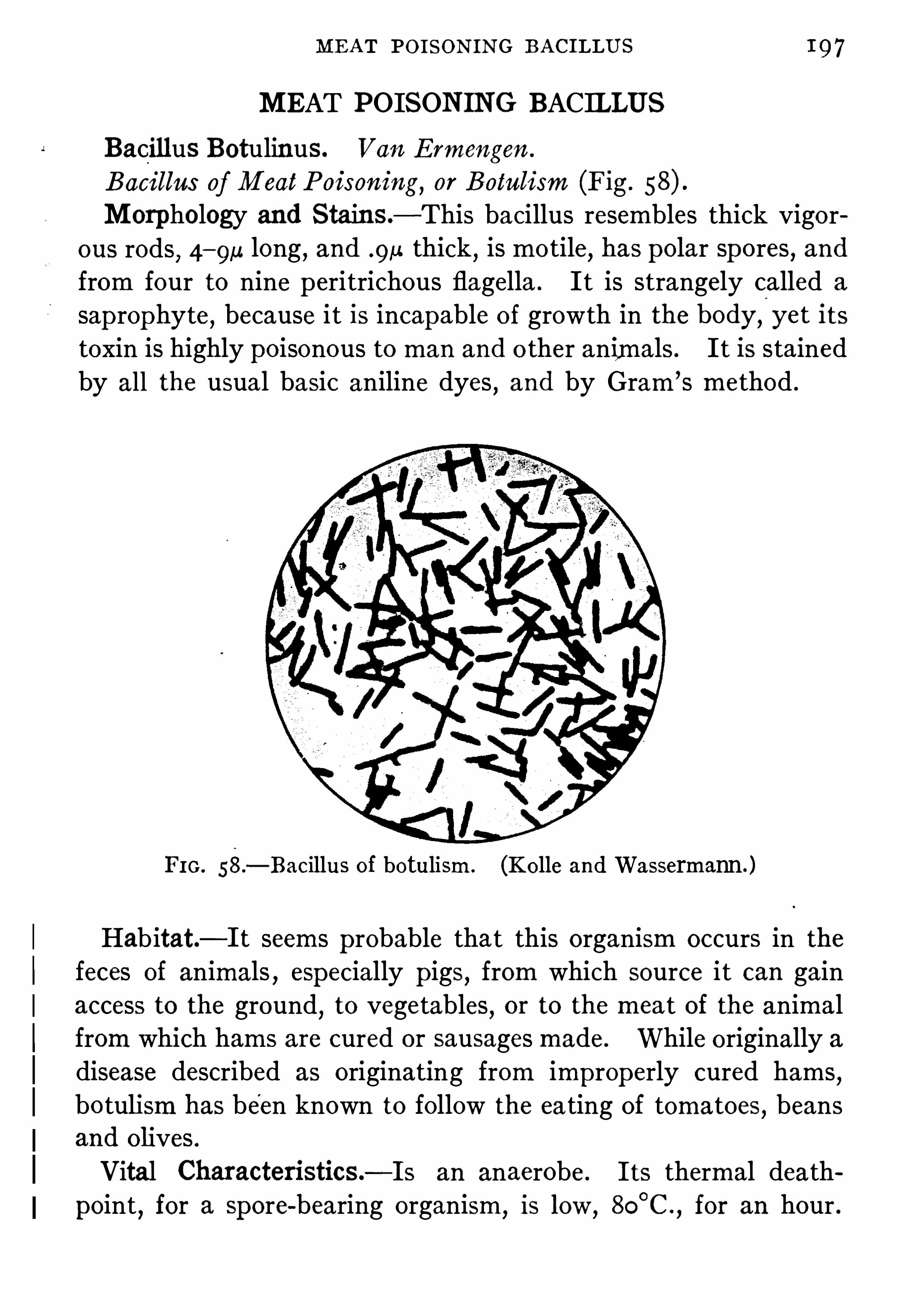
MEAT POISONING BACILLUS
MEAT POISONING BACILLUS
Bacillus Botulinus. V an Ermengen .
Bacillus ofM eatPoisoning, or Botulism (Fig.
Morphology and Stains.
-This bacillus resembles thick vigorous rods
, 4—9u long, and 9 51 thick, is motile
,has polar spores , and
from four to nine p eritrichous flagella . It is strangely called a
sap rophyte , because i t is incapable of growth in the body,l
yet its
toxin is highly poisonous to man and o ther animals . It is stainedby all the usual basic aniline dyes
,and by Gram’s method .
FIG. 58.
—Bacillus of botulism . (Kolle andWassermann. )
Habitat—It seems probable that this organism occurs in thefeces of animals , especially p igs, from which source it can gainaccess to the ground
,to vegetables
,or to the mea t of the animal
from which hams are cured or Sausages made . While originally adisease described as originating from improperly cured hams,bo tulism has been known to follow the ea ting of tomatoes, beansand olives .Vital CharacteristicS .
—Is an anaerobe . Its thermal death
point, for a spore-bearing organism,is low
,8C
°
C .
,for an hour .

1 98 BACTERIA
Grows only in media that are alkaline,and is capable of growth
at from 1 8°
C . to 35°
C ., though best below 2 5
°
C . ; 1 0 percentof chloride of soda checks growth .
Chemical Activities.—It can produce, at room temperature ,
a water-soluble toxin sufficiently stable to withstand drying of
meat if not exposed to sunlight, and not destroyed by the gastricjuice . It is destroyed by thorough cooking of meat . M ilk isnot coagulated
,grap e sugar is fermented
,and a foul
,sour odor
is p roduced in a culture . It liquefies gelatine . There are two
varieties of the germ,A and B
,differing in the quality of the
toxin p roduced, both having the same physical and pathogenic
properties but develop ing different anti-toxins .Cultures.
—On gelatine plate , that contains sugar, colonies areproduced tha t are coarse and prickly in app earance . The liquefaction oi the gelatine is slow . Bouillon is rendered turbid . The
cultures resemble tetanus andmalignant oedema .
Pathogenesis—Its pathogenic action is marked, but only byits toxin
,which has a decided aflinity for nervous tissue . The
toxin is absorbed'
from the intestinal tract unchanged by the
gastric juice . In this it differs from“
the toxin of diphtheria and
tetanus . If the toxin is mixed with the emulsified nerve tissue,i t becomes neutralized. In fatal cases of infection
,the gan
glionic nerve cells are degenerated . Man is very suscep tible,while cats and dogs are more or less non-suscep tible . If bacilliare inoculated into animals
,they do not proliferate . A men
ingitic disease of horses and limberneck of fowl are believed tobe due to this intoxication . Animals that recover are foundto have develop ed strong anti-toxin in the blood serum .
Immunity.—An artificially prepared anti-toxin has been found
to be active,and is ofuse in treating cases ofpoisoning with meat .
The correctly typ ed anti-toxin Should be used or that made byartificial immunization with both varieties of bacilli . The latteri s now preferred Since no rap id distinguishing clinical test oftypes is available .

2 0 0 BACTERIA
FIG. 60 .
-B . aerogenes capsulatus, agar culture showinggas formation. (Williams )
by all the basic dyes,and holds the
stain in Gram’
s method . Starnrng
Shows tha t i t possesses a cap sule .
Habitat—The soil,the intestines,
and,some times
,the skin of man .
Vital Characteristics—Vital resistance is low
,the thermal death
point being 58°
C . with ten minutes ’
exposure , while spores require ten
minutes’ boiling for killing . Itgrows best at
‘ body temp erature .
Has lived for one hundred days onculture media in the incubator . Itis an anaerobe .
Chemical Activities—Produces
gas ; does not usually liquefy gelatine
,but curdl es milk (Fig.
Cultures—Grows best in neutralor alkaline media
,p roducing abundant gas . Colonies app ear grayishor brownish-white
,and are often
surrounded by'
projections whichare feathery or hair-like . On agarstrict anaerobic conditions are
necessary for growth, gas bubblesapp ear in the media
,and the agar
may be forced out of the tube instab cultures . In bouillon i t growsunder anaerobic conditions . The
growth is rap id , bouillon is clouded,and a fro th app ears on the surface .
After a few days the medium be
comes clear,owing to the sedi
mentation of the bacilli . Growth

GASEOUS (EDEMA BACILLUS 2 0 I
occurs best in sugar bouill on, which becomes '
strongly a cid. Inmilk the growth is rap id and luxuriant ; the p roteids are coagu
lated . Anaerobic conditions must be observed . On potato i tgrows well, producing bubbles in the wa ter which may coverthe potato in the tube . The growth app ears thin , moist
,and
grayish-white .
Pathogenesis.—The pathogenic prop er ties of this organism are
limited . It is not able to endure the oxygen of the circulatingblood . Grows best in old clots
,and in the uterus . It p roduces
gas rap idly in some cases of abortion and in peritonitis in man ,
which is quickly followed by death . It causes gaseous phlegmons in guinea p igs , and injection are usually fatal to birds.
In man infection has followed wounds , and delivery of the childin puerperal cases . It produces in fatal cases the conditionknown as frothy organs Schaumorgane . Itmay be isolatedfrom . infected matter , fa ces, etc .
,by injec ting the latter into a
rabbit’s vein and then killing the animal . The carcass is then
p laced in an incubator and an enormous growth of the organisms
follows ; anaerobic conditions favorable to groyvth are obtainedin the body so that gas distention of the tissues results ; from the
la tter pure cultures are easily obtained .
Vincents Angina is due to an anaerobic organism of two stages ,as Bacillus fusiformis and Spirochceta vincenti . The bacillus is afusiform irregularly staining pointed rod
, 3—1 2u long by .3
—.8,u
wide . Under cultivation it grows out into forms such a s are seenWith it in smears from the diseased throa t, tha t is, long, wavy,uniformly stained
,flexible
, po inted ended Sp irals . The bacillusforms endospores chiefly at the end. Obligate anaerobe, requiring serum
, asci tic fluid or glycerine . Colonies delicate and
whitish . Gas in glucose media . Litmus milk o nly decolorized .
Gives a foetid odor on all cultures . No sp ecific immunity reac
tions known .
These same Sp irals have been found in -abundance in manycases of
l
ulcerative stomati tis,no tably the variety which became

2 0 2 BACTERIA
well known during the late war . Their connection with thisso-called
“
Trench mouth” is not so generally accep ted as 13 the
case with ulcerative angina but their discovery should suggestremedies, salvarsan , Silver nitra te
,which have been useful in
Vincents Angina .
CHOLERA BACILLUS
Vibrio Cholera . Koch .
Spirillum Cholera (Fig.
Cholera Bacillus.
Comma Bacillus .
Morphology and Stains—Curved or bent rods,the ends not
lying in the same p lane . This bending varies greatly. Under
FIG. 6 1 .
—Cholera Sp irilla . (Kolle andWassermann.)
certain conditions of growth such as the p resence of alcohol, orinsuflicient albumin or oxygen in cul ture media , long Sp iral chainsare formed . It is mo tile
,has one terminal flagellum
,and like
other members of this family, has no spores . It stains well withthe common dyes but not by Gram’s method .
’
D ilute fuchsin

2 0 4 BACTERIA
becomes more and more granular,and finally the whole medium
i s liquefied . In gelatine tube stab culture, the growth, at first, isnot characteristic ; but, after a few hours , a semi-spherical depression app ears , which extends downward
,and resembles a large
bubble of gas. AS liquefaction p rogresses, the whole line of punCture disapp ears , and the excava tion looks cylindrical . This areabecomes Cloudy . On agar plates the colonies are elevated, roundandwhite
,with moist lustre . D eep colonies areWhetstone Shap e .
Old agar colonies become yellowish-brown . Coagulated bloodserum is rap idly liquefied at 37
°
C . Milk, at times , is coagulated .
No curdling ferment is formed ; the acid p roduced is thought to besufficient . On potato the growth is Slow
,or not at all
,if the
medium is acid . If the po tato is rendered alkaline, growth occurs,with a moist lustre
,slightly elevated ; white at first, later becoming
brown . On acid fruits i t will not grow . In bouillon, after sixteenhours
,a diffuse cloudiness occurs
,with the formation of a stiff
pellicle, which in some cultures becomes wrinkled . In peptone ,abundant growth takes p lace , with the p roduction of indol andnitrites . If a fewd1 0p s of H280 4 are added
,a beautiful red
app ears if nitri tes are p resent . This is the cholera red” reaction .
If the color does not at once app ear, nitrites must be added . Onbloodmedia certain strains p roduce distinct
'
hemolysis (El Tor) .
Pathogene sis.—Cholera Sp irilla are pathogenic for man, but
only under exp erimental conditions, for lower animals for whichguineap igs may be taken as an examp le . If the stomach of the
la tter is rendered alkaline with bicarbona te of soda , and a bouillonculture introduced
,choleraic symp toms will follow and the animal
will die . If cholera sp irilla are injected into the peritoneum,the
animal will quickly succumb to a general cholera p eritonitis .Young rabbits are equally suscep tible . When cholera Sp irilla inculture havebeen swallowed by man (labora tory workers) , eitherby design or accident, the disease has followed , sometimes withfatal results . The toxin of this organism is intracellular (anendo-toxin) . Old cultures become pathogenic through a bac

CHOLERA BACILLUS 2 0
teriolytic action, by which the cells are dissolved, and the toxinlibera ted . Filtrates from young cultures are non-toxic . Ifbouillon cul tures are killed by chloroform,
and then injected intoanimals
,toxic ac tion follows . In cholera the pathogenic process
ismostly confined to the intestines . Toxic absorp tions, due to thelibera tion of toxic products by the bacteriolytic action of serum
,
follow later . There is a desquamation of the ep ithelium of the
bowel,and ep ithelial flakes found in the wa tery discharges resem
ble rice grains . Peyer ’s patches may become slightly swollenand reddened
,and later
,there may be diphtheritic necrosis above
the iliocecal valve,and often a parenchymatous nephritis . The
vibrios do not enter the blood .
D iagnosis.—Bacteriological diagnosis of cholera is accomplished
by examining the alvine'
discharges . A mucous flake is mixedwith some p ep tone solution , this is incubated, and the Spirill a ,if p resent, rap idly grow on the surface ; after a few hours, p latesare poured from this surface growth, and from the p la tes liquefying colonies are p icked out
,and bouillon cultures made . These
are tested by serum,from horses artificially immunized by inject
ing cholera sp irilla into them. If the organism under examination
(after serum mixed with to parts of water is added)agglutinates
,it is considered to be the cholera Sp irill um. Both
in early and fatal cases,the agglutinating reac tion is not available
,
since it takes some time for the agglutinins to form in the blood .
Under the chap ter on immunity an account of the Pfeifler reactionis given , also one on vaccina tion against cholera infection, bymeans of killed cultures
,under the chap ter on vaccines .
Vibrios Allied to the Cholera Vibrio
Several o ther vibrios have been discovered that resemble thecholera vibrio . These are mostly found in po table wa ter, andthough in many respects identical with the cholera vibrio , theydifler in essential points , i .e .
, pathogenicity, and in their agglutinability with specific sera . The most important of these organisms

2 0 6 BACTERIA
are : Vibrio Metchnikomi ; Vibrio proteus; Vibrio tyrogenum; and
Vibrio schuylkilliensis . There are no important pathogenic members of this group excep t the cholera vibrio .
GLANDERS BACILLUS
Bacterium Malle i.Bacillus Mallei .
Glanders Bacillus .
Morphology and Stains.—Slende
'
r rods 2 to 3a. in length con
taining no true Spores , but Shining chromatophilic bodies (BabesErnst granules) . In old culture
,long club-like threads appear ,
which exhibit true branching . This organism is not motile,and
has no flagella . It is stained with difliculty by ordinary methods,and not at all by Gram’s method .
Vital Activities.—It is a facultative aerobe
,growing feebly in
the absence of air,and best at 37
°
C .,in glycerine agar . Resists
drying but feebly . Its thermal death-point is 55°
C .,ten minutes’
exposure .
Chemical Activities.—Produces a brown p igment on potato,
also mallein,and a little indol in old bouillon cultures . It forms
no gas.
Cultures.—On ge latine it produces small punctiform colonies
that are’
white, and become,after a time
,surrounded by a distinct
halo . The colonies are often very delicate and ragged . The gelatine is not liquefied . On agar the growth is best if glycerine is
present, but is not characteristic . Bouillon cultures cause an
abundant sediment,above which the medium is clear . Milk is
coagulated. On potato the growth is characteristic . The coloris, at first, yellowish-white like honey, becoming, finally, reddishbrown . The potato is much darkened .
Pathogenesis.—This organism is pathogenic for horses and
man ; 50 p ercent ofmen succumb after infection . Horses, asses,cats
,dogs
,sheep , and goats are suscep tible in the ordermentioned .
Cattle and birds are immune . In horses the disease is known as

2 0 8 BACTERIA
Morphology and Stains .—Long, bent, or curved bacilli of
irregular contour, frequently clubbed or fil iform at one or bothends ;which contain chromatophilic granules, and often exhibit truebranching ; have no spores or flagella
, andare notmo tile .Accord
ing toWesbrook, stained bacilli are of three typ es ; (I) granular(containing the Babes-Ernst granules) ; (2 ) barred like a strip edstocking ; or 3) solid, staining uniformly throughout . The p leo- A
morphic diflerences of various bacilli are most characteristic,and
of diagnostic importance . This organism stains with all the basicA
FIG. 6 2—Forms of B . diphtheria in cultures '
on Lofll er’s serum. A
,
Characten stic clubbed and irregular shap es with irregular.
staining of thecell contents. X 1 1 0 0 . B
,Irregular shapes w1th even starning . X 1 0 0 0 .
(After Park and Williams.)
dyes,no tably by Loflier
’
s blue, or Neisser’s sp ecial granule stain .
It is also stained by Gram’s method . The length of the organismdifl
'
ers'
much,according to the reaction of the medium in which it
grows . Alkaline media favor long forms, and acid the reverse .
Its length is from 1 .5“ to It does not form chains . Bizarre,
or involution shap es p redominate in old cultures (Fig.
Culture and'
Temperature Requirements—It grows best atbody temp erature, and on glycerine agar , or in Liifiler
’s blood
serummixture of alkaline reaction .
Vital Characteristics .
—It resists drying for a long time, and has
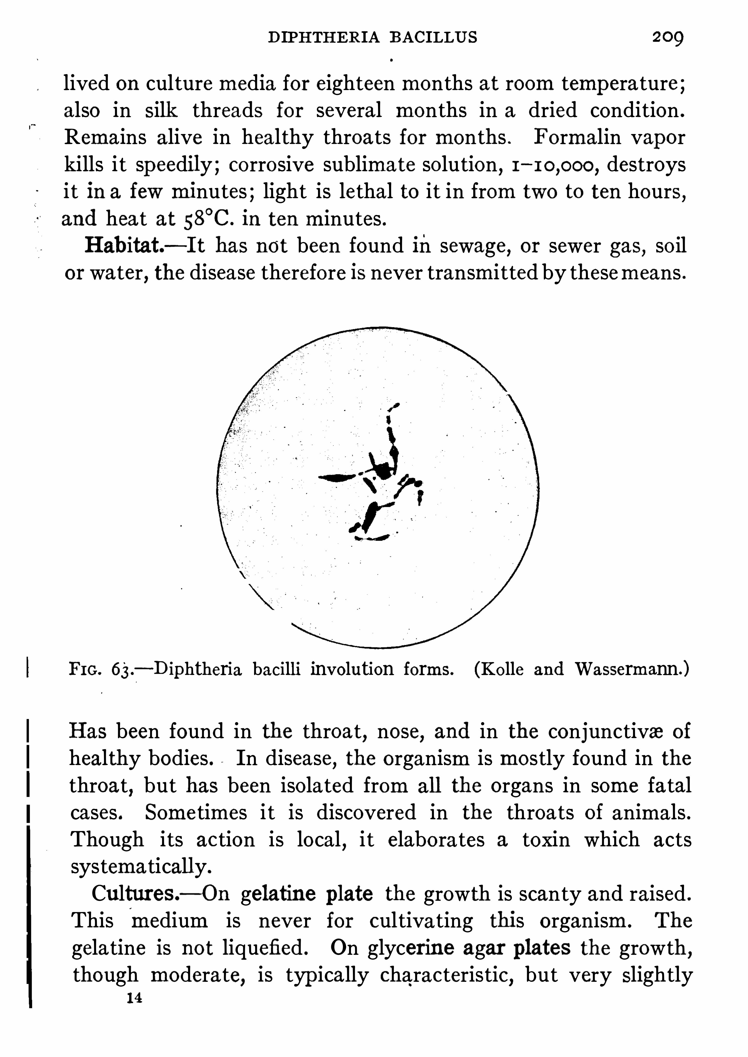
DIPHTHERIA BACILLUS
lived on culture media for eighteen months at room temp erature ;also in Silk threads for several months in a dried condition .
Remains alive in healthy throats for months . Formalin vaporkills i t sp eedily ; corrosive sublimate solution, 1 destroysit in a few minutes ; light is lethal to it in from two to ten hours,and hea t at 58
°
C . in ten minutes .Habitat—It has not been found insewage, or sewer gas, soil
or water, the disease therefore is never transmitted by thesemeans .
FIG. 63.
—D iphtheria bacilli involution forms. (Kolle and Wassermann.)
Has been found in the throat,nose
,and in the conjunctiva of
healthy bodies . In disease, the organism is mostly found in thethroat
,but has been isolated from all the organs in some fa tal
cases . Some times i t is discovered in the throats of animals .Though its ac tion is local
,i t elabora tes a toxin which acts
systematically.
Cultures.—On gelatine plate the growth is scanty and raised .
This(
medium is never for cultivating this organism . The
gelatine is not liquefied . On glycerine agar plates the growth,though modera te
,is typ ically characteristic
,but very Slightly
14

BACTERIA
raised above the medium,and is of duller lustre . Old colonies
become yellowish-brown , the center ofwhich, under a magnifica
tion of sixty diameters, app ears darker, and with ravelled edges .
On L6ffler’s blood serum mixture,the organism grows rap idly
and well . This and ascites-glycerine-agar culture media are
the best for it. Bouillon made from fresh meat is an excellentmedium for its growth . The bouillon
,which must be alkaline
and freshly made, becomes first cloudy ; then a fine p recip itatesettles
,and over the surface a delicate pellicle forms . The
reaction of the culture p resents three typ es : A,is acid in the
beginning,and becomes p rogressively more acid . B
,is alkaline
from the start, and p rogressively more alkaline ; this is the mosttoxic growth . C ,
acid at the start,becoming alkaline finally .
’
The growth is not so luxuriant as in B ,nor is there as much
toxin p roduced . In milk, the growth is luxuriant,without
coagulation . The reaction is amphoteric, but in old cultures i tbecomes alkaline . On potato, rendered alkaline
,it will grow
,
but not characteristically.
Chemical Activities—No gas IS formed,or any curdling or
gelatine dissolving ferment . Acids are evolved from sugars ;even the sugar found in meat is converted into lactic acid . Inthe manufacture of toxin this muscle sugar must be removed . Asoluble
‘
toxalbumin is created, both in the body and in culture,which is intensely poisonous . See chap ter on bacterial p roducts .
Pathogenesis—D iphtheria in man means generally an infec
tion of the mucous membrane of the upper resp iratory tract, withthe formation of false membranes . The latter may cause deathby Suffocation . Infection may occur in the skin, vagina , or p re
puce . The toxin not only causes a local necrosis, with the formation of an exudate, consisting of fibrin and leucocytes, but alsograve systemic action, with marked degeneration of importantnerves and nerve centers, and also of the parenchyma of the
kidneys,liver
,and heart, paralysis following . In certain struc
tures fragmentation of the nuclei of the cells is noted . Guinea

2 I2 BACTERIA
ery. AS a p rophylac tic , from 60 0 to units should be used .
As many as units have been injected in a single patient .N0 case is too trivial, or too far advanced in which to use it . The
serum is anti-toxic , and not bactericidal . Wassermann has pre
pared a serum that is bactericidal, and is designed to destroy the
Pseudo-diphtheria bacill i,whichmorphologically and culturallyresemble the true bacilli
,have been described . They are not
pathogenic , in the sense of p roducing exu'
da tive diphtheria, andare believed to be attenuated diphtheria bacilli by many observ
ers. The diagnosis of diphtheria by culture is an importantmeasure . It depends upon the rap id growth of the bacilli uponLtiffler
’s blood serum. Of all the various organisms found in the
throats of pa tients with diphtheria , the diphtheria bacilli outstrip ,
them in rap idity of growth . After eight to twelve hours,the
serum inoculated with the smear from the false membrane iscovered with fine granular colonies of pure diphtheria bacilli .After twenty-four
,0 1 more hours
,the other organisms present
overgrow the diphtheria colonies . A sterile swab of co tton , or astick, is rubbed over the false membrane
,or throat
,and theno ver
the serum ; the latter is incubated, and the culture examined aftereight or twelve hours
,by staining with Lo'ffl er ’s blue . If curved,
clubbed,irregul arly stained bacilli are found
,esp ecially if they
contain dark polar granules, and are generally uneven in Size andbizarre
,i t may be safely considered that they are diphtheria
bacilli . Gram’s stain may be needed to confirm the diagnosisoccasionally, or i t may be necessary to inoculate guinea p igs .
This may be done by inoculating the whole throat culture and
plating out from the local inflamma tion,or the organisms may be
isolated directly by p lating and then injec ted into animals .It is well
-
to check up the test by immunizing one animal with2 0 0 units of anti-toxin before the culture is given , using anguinea pig unp ro tec ted . This serves as a control upon
production . Virulent bacilli will kill a guinea

PSEUDO-DIPHTHERIA BACILLUS 2 I3
in three days if I c .c . of forty—eight has serum bro th culture begiven subcutaneously .
Certain of the p seudo-diphtheria bacilli, or diphtheroids”as
they are called,seem to have the power of increasing or continuing
inflammation after it has been started by other germs . They arefrequently found in sinusitis
, prosta titis, bronchi tis , etc . In a fewreported cases they seem to have been the only microorganismalcause of disease but their pathogenic powers are usually not great .
Corynebacterium Pseudo-diphtheriticum.
Pseudo-diphtheria Bacillus (Hoffmann) .Morphology and Stains .
—This bacillus resembles the diphtheria bacillus . The rods, however, are Shorter and thicker ;
it stains like the true bacillus, but not by Neisser ’s
—On glycerine agar the growth becomes diffuse,
spreading from the line of inocula tion in a grayish-yellowish
pasty expanse . It grows well on gelatine . In bouillon it formsa denser and more luxuriant growth than the bacillus .Habitat—It is found in healthy throats and conjunctiva .
Pathogenesis—It is non-
pathogenic for guinea p igs (see above) .Diagnosis.
—It can be differentiated from the true bacillus by ’
1 . Being non-
pathogenic .2 . Not exhibiting polar granules with Neisser ’s stain .
3 . Not p roducing acids in certain carbohydra te media .
Bacillus xerosis is a p seudo-diphtheria organism found on the
ormal conjunctiva . It is not thought to possess any virulence .
TUBERCLE BACILLUS
Mycobacterium Tuberculosis .
Bacillus tuberculosis (Fig.
Tubercle bacillus .

2 1 4 BACTERIA
Morphology and Stains.-Slender rods
,generally unbranched
,
1—5,u long, and 411. thick, usually slightly bent ; are non-motile
,
and have no Spores or flagella . In old cultures,and sometimes
in Sputum,branching forms are seen
,and
,rarely
,some that are
Club-Shap e . On acid potato , thread forms are found . In the
continuity of most of the bacilli, unstained spaces are seen ; ino thers dense deep red granules are foundby fuchsin . AS this bacillus is diflicult tostain
,Special methods have been devised to
demonstrate it,
as the Sheathing cap sulerenders it extremely unsuscep tible to the
ordinary methods of staining . The cause ofthis resistance is supposed to be a fatty or
PEG,
64—T‘1befde waxy substance in the cap sule which is more
bac1111 In sputum ;stained w ith fuchsin than p robable, because of the fact that stamsand methylene blufi that are fat selective, such as Sudan III,(Greene’s M ed1calD iagnosis.) color 1t very well . Borhng hot carbol-fuchsm
gives it the best stain . It keep s the colorin Sp ite of the action of strong solutions of mineral acids inwater, or dilute alcohol . So when tissues, or secretions, are
stained with hot carbol-fuchsin for a short time,or cold carbol
fuchsin for a long time, and then treated with a 2 5 p ercent
solution of HNO3, or H280 4, in water, everything is dep rived of
the red color,excep t the tubercle bacilli . All such organisms
tha t are acid p roof, are called “acid-fast . There are many
o ther bacilli that have this p roperty. Aniline water and gentianviolet solution also stain it . Gram’s method dyes the organismviolet . Sometimes very young bacill i do not stain at all .
Vital Requirements.—This bacillus thrives best at 37 .5
°
C .
It grows Slowly,is a stric t parasite, and an obligate aerobe . In
cultures it'
dies quickly in sunlight,and in diffuse daylight
dies in a few days . It resists drying and light in sputummonths . Its thermal death-point (moist) is SO
°
C . futes ; can resist 60
°
C . for one hour,but su

2 1 6 BACTERIA
darker . This medium IS never liquefiedby the culture . On glycerine agarmade of veal broth containing 6 percent oi glycerme
, the organism growswell after isolation from the tiSSIies,
often luxuriantly (Fig. A wrinkledfilm covers the surface of the agar
,
from which it is removed with ease .
On bouill on , made of veal and glycer
inized,it develop s rapidly, covering
the medium with a dense whitewrinkled p ellicle , which, though thick,is friable . After a time it falls to the
bo ttom of the flask . It grows well on
glycerinized po tato also,
and milkagar . On egg-albumins mixed together ,sterilized and coagulated
,this bacillus
also develop s well .Pathogenesis.
—The discovery of the
tubercle bacillus,its methods of culti
vation and differential staining,i
maybe ranked with the greatest ofmedicaldiscoveries . This organism causes inman and cattle
,chiefly
,the disease
called tuberculosis . It rarelythe carnivora , but has been fsuch animals when confined .
are often infected ; cats and dogstimes
,but sheep ,
goats , and
seldom. It is easy to'
inoculate
p igs or

TUBERCLE BACILLUS 2 1 7
The disease is widesp read , but is much more common wherehuman beings '
are huddled toge ther in d ark , badly ventilatedrooms and Shop s . In tissues , the characteristic lesion is a tubercle . This is a globular mass, about the Size of a very small sho t
,
and grayish pearly white . M icrosc0 pically, in the centre of thetubercle, are found several large multinuclear cells, called giantcells
,
“
which often contain thirty or more nuclei, and a number of
tubercle bacilli , the nuclei often being si tuated at one pole , while
FIG . 66 .-Tubercle bacilli showing involution forms. (Kolle and
Wassermann.)
bacilli are at—the other . About the giant cells ep ithelioids
.
are group ed, and about these small round cells are massed ingreat numbers . No new blood-vessel formation is ever foundin the ep ithelial cell layers , or among the giant cells . Owingto insufficient blood-supp ly the centre of the tubercle frequentlyundergoes caseous degeneration . If the lesion heals , the caseous ,centres become calcareous
,and the p eriphery changes into
connective tissue . If the tubercles coalesce , great masses of
caseous tissue form . If the latter becomes infected with other

2 1 8 BACTERIA
pathogenic bacteria (strep tococci and pneumococci) rap id softening occurs, with cavity formation, etc . Tubercles may developin any organ or tissue of the body. The lungs
,intestines
,
peritoneum,glands
,larynx
,Sp leen, and bones become infected .
The liver and pancreas seem to resist invasion more than o therorgans . Bacilli are rarely found in the blood in tuberculousdiseases . They may, however, be found in the urine, in kidneyor bladder tuberculosis; M ilk from tuberculous cows, with,infected udders
,often contains bacilli
,and is certainly a means
of transmi tting the disease . Cerebro-sp inal fluid, in tuberculousmeningitis contains the bacill i . Bacill i may p enetrate mucousmembranes
,and not causes any local lesions , but infect distant
organs . Tuberculosis may be Sp read in the body in four ways .Sputum may be swallowed and infect the intestines,by continuity ,by the lymph-stream,
or by the blood ; this may cause intestinalul ceration and invasion of the peritoneum . If the bacilli reachthe blood-stream
,the disease produced is generally acute miliary
in type . This is manifested by the formation of fine graytubercles . In tuberculosis of the lungs it is more than p robablefollowed skin inoculation
,either by accidental or intentional
trauma that the bacilli are inhaled . Local tuberculTuberculous mothers may have tuberculosis of theand fathers , having tuberculous testes, discharge bsemen . Placental transmission of the bacilli from mooccurs rarely.
Types ofTubercle Bacilli. -It has been considered probablemany observers that there are two types of bacilli , a human anbovine typ e . Theobald Smith was the first to advan
theory . Koch has announced that the two types weredifferent
,and that the human was incapable of infecting
and the bovine was not pathogenic for man. In view of the
that cattle are frequently tuberculous,and the bacilli are 0
found in the milk,it is important to know if the bovine type
develop in man . Ravenel has Shown that it

2 20 BACTERIA
o ther acid-fasts several method are now emp loyed . The materialto be examined may be stained in the ordinary manner and
'
thendecolorized by Papp enheim solution or a sa turated solution of
methylene blue in absolute alcohol . Preparations Should be driedthoroughly before using such solutions . For
“enriching ” in
organisms,the bulk of material
,e .g.
,sputum,
is susp ended in 1 5
p ercent antiformin (the p roprietary name for a mixture of Javellewa ter and caustic soda) , allowed to stand in the incuba tor for awhile and the suspension centrifuged . In the sediment manymore bacilli will be found than in the same bulk of the raw Specimen . This antiformin seems to dissolve mucus
,tissue and all
bacteria excep t tubercle bacilli . The method can be used to
p rocure cultures .
Even with this method organisms escap e de tection in some
certainly tuberculous lesions . This is said to be due to non-acidfast
,but Gram-staining granules . They are said to be found by
a-modified Gram-Weigert staining,according to Much . Such
sp ecimens Should always be injec ted into guinea p igs forcorroboration .
Immunity.—It is possible to immunize ca ttle against virulent
bovine tubercle bacilli by inoculating thernp reviously with ature of human tubercle bacilli that have been grown fortime on culture media
,and thus attenuated . T
if injected into a p erson with chronic tuberculosistissues to a slightly grea ter resistance to the discanti-tuberculous sera are not of a p ronounced or ce
p eutie value . By immuniz ing horses , Maragliano
serum that he claims is effective . The milk fromca ttle is used as a die t in tuberculous pa tients by him .
ous tuberculins,some containing endo-toxins
,or p
so lution,are capable of stimulating the forma tion
the Sera of man and animals . Blood from infe
also contains these bodies .to be of

BACILLUS ,OF LEPROSY 2 2 I
fixa tion gives some information and is growing in favor as assistance in obscure clinical cases .
BACILLUS OFLEPROSY
Mycobacterium Lepra . Hansen .
Lepra Bacillus.
The original descrip tion is of a po inted, curved, acid fast rodoccurring in group s within lep ra cells . In recent years severaldiflerent organisms have been isola ted onmedia containing tryp to
phan . They have a few fea tures in common : frankly acidfast or decolorized with difliculty. Gram positive , non-motile
,
non-Spore forming, staining shows beading or barring, polar
bodies,all capable of p leomorphism. They have been group ed
i nto 4 , (1 ) acid fast bacilli varying from cocco id to filamentousShap es, not easily isolated but growing well after once accustomedto media ; growth yellow or orange ; 2 ) acid fast, non-chromogenic
,
plump bipolar rods, short and long, growing with great sparsityon labora tory media ; (3) diphthero id bacilli sta ining solidly, orbeaded
,growing best at 37
°
C . in a yellow-white manner on agarand with a p ellicle on
' bro th ; (4) anaerobic bacilli ofmore solidstaining character and growing feebly as a dry band on media .
Very marked variations in luxuriance and color p roduction are
no ted on different media .
To cultiva te the lep rosy organisms bits of tissue are stripp edoff and allowed to digest with tryp sin on blood serum or agar
p lates . When the tissue has softened and the bacilli multip lied ,transfers are made to serum glycerine media or tho se containing
It is best alkaline in reac tion .
s .
-The bacilli are seen in enormous numbers inewhere in diseased tissues and have been found
in the blood . The lep ra cells are large and vacuolated,and literally
crammed full to bursting with bacilli . In general the lep rous
Norm—Tube rcle bacill i causing av ian and fish tuberculosis , and other acid fast ba cill iexist, but not be ing p athogemc for man . are not described here .

2 2 2 BACTERIA
lesion resembles a tubercle,as it consists of giant cells, ep ithelial ,
and round cells .Immunity.
—There is very little accurate knowledge as to
immunity against this organism ; of late bacterins have been triedwith some success i t is claimed .
STREPTOTHRIX (Eppinger) 0R NOCARDIA
The genus of truly branchingmycelium-forming higher bacteria
(see page such as the a ctinomyces, belonging to the groupbetween bacteria p rop er and the moulds, called Trichomycetes .K ruse has described nineteen different members of the strep to
some pathogenic to man and animals .
FIG. 67.
—Streptothrix candida . (Kolle andWassermann.)
A number of cases of strep tothrix (Streptothrix Hominis) infection in man have been reported . The disease
,in general, resem
bles phthisis . In the pus, Sputum,and stained sections of these
cases,strep tothricial threads have been found (Figs . 67 and
Morphology and Stains—Threads are thick and short, orand Slender
,depending upon themedium on which they grow.
bouillon the threads are thin and long,on blood serum, short

2 24 BACTERIA
ical bacilli are not found, i t is well to look for the streptothr lx bystaining with Gram’s stain .
Actinomyces Bovis.
Morphology and Stains—This organism is called the ray fungusbecause of the stellate arrangement of its threads in the colonies
FIG. 68 .
-Actinomyces bovis. (Williams )
found in tissues . It is of a more complex structure than theteria hitherto described . There are three elements found in ecolony : (I) long thread which may be branched
(2 ) threads that are clubbed, which may,or may 11
(3) spore-like bodies conta ined within the threadarise by breaking u

RAY FUNGUS 2 2
threads radially situated . Through the p eriphery and extendingbeyond are other unclubbed threads, while sca ttered throughoutthe colony and beyond it
,and in the threads, may be seen many
Spore-like bodies . The threads a nd spores s tain by Gram’s
method while the clubs do not. Basic stains also color all theelements . The Spores do not stain like bacterial endo-Spores .Vital Requirements—It is a facultative anaerobe, and growsbest in the absence ofair
,at 37
°
C . Resists drying for a long time,
andits thermal death-point is 80°C .
"
after fifteen minutes’.exposure .
FIG. 69 .
—Actinomyces. (Williams )
Chemical Activities—Slowly liquefies gelatine, does not curdleproduces a mouldy odor . No gas or acids are formed
,
develop ed .
Habitat—It has been found in straw and hay, but never in a
healthy body .
Cultures—On gelatine plates it produces yellowish-gray colonies that are very small . These grow into the gelatine
,slowly
liquefying it . The colonies are very tough and fibrous . In agartubes it grows
'
very slowly,the first growth being like dewdrop s ;
enlarge,turning yellow,
and finally brown . The cul

2 2 6 BACTERIA
ture grows down into the agar, and the medium darkens . Oldcultures /fare dark and crumbly looking, adhere firmly to the agar ,and have a downy dust-like covering. On blood serum the colonies appear as dewdrop s , which later become brownish, then,yellowish-orange
,or brick-red. In bouillon the growth is at
bottom in ball-like masses that cohere firmly . Clubs do not formin this medium. The supernatan t bouillon is clear, with no surface growth . In milk it p roduces no chemical change . On
potato i t grows ln knot-e colonies .Pathogenesis—Causes in ca ttle the disease known as lumpy
jaw .
”The fungus reaches the jaw from the teeth and gums
,the
latter first being injured by sharp spines in the food . In man,the
internal organs,lungs
,intestines
, and, rarely, the braiinfected . The liver often is abscessed . In both cattleuniversal actinomycosis sometimes occurs . The lesionsare rather massive at times ; the n idus is often S
enormous numbers of polynuclear leucocytes, whi
p lay a defensive role in the tissues . The disease isca ttle and to man . It is hard to inoculate '
lab
with the disease, though Wright succeeded inuseful immunity reactions seem to Vaccines haveused intreatment with encouraging results but no cures . Po
Slum Iodide internally is always indicated .
ACTINOMYCES MADURA
ActinomycesMadura .
Streptothrix Madura,Vincent.
Morphology and Stains—A non-motile,non-flagellated organ
ism said to have spores Its growth resembles that ofActinomycesbovis. It consists of long threads that are clubbed. These stainby «all the basic aniline dyes and by Gram’s method . There arethree recognized forms of this organism
,white
,black and red,
which have been found in various cases but the interrelation of

2 28 BACTERIA
BLASTOMYCOSIS
OIDIOMYCOSIS
Oidium Albicans. Thrush,Soor .
—This organism resemblesbo th a yeast and a mould
,because it exhibits characteristics that
are common to bo th of these forms . It exhibits budding yeastcells and budding mycelia . The yeast cell is 614 long and 1 “ wide,but the cells vary very much in length and width .
It stains well in tissues and cultures by Gram’smethod,and by
the ordinary basic stains . It may be cultivated on
FIG. 7 1 .—Thrush fungus. (Kolle andWassermann. )
bloo d serum, agar, po tato , etc .
,and it is rather indifferent to
reaction of the media . It grows best if sugars are p resent . Ihowever, very suscep tible to such antisep tics as phenol,acid
,sublimate, etc .
Pathogenesis—Causes in man a condition known as o
myco sis,and in young children a very troublesome s toma
which, if the child is weak and illy nourished,may resultIt may cause metasta tic abscesses

OIDIOMYCOSIS 2 2 9
neys,or nodules in the lungs . This organism may penetra te
mucous tissues, and fil l the lumen of vessels (Virchow) .Tropical sp ruce is held by some as due to a near relative
of the o idium,namely Monilia psilosis . The organism is to be
found all along the alimentary tract . It possesses many culturalcharacters like oidium. Vaccines are said to be of practical usein therapeutics .
FIG. 7 2 .
-Doubly contoured organisms found in oidiomycosis (blastomycosis) . (From Buschko after Hyde andMontgomery . )
Sporothricosis is a subacute or chronic infection usually of the
but at times involving the internal organs , caused by theothrix Schencki. The organisms grow as delica te myceliavery numerous spores , 2—4 X 3
—6p in Size . They grow bestcid media at body temp erature as white fluffy massesater become brown . The disease in man takes the formtumefac tions under the skin which may ulcera te leaving

230 BACTERIA
indolen t ulcers . Fever, prostration and emaciation follow. Ratsare suscep tible . AI1 agglutinin app ears ; this may assist indiagnosis .Saccharomycetes are some times pathogenic .
’
They are budding fungi, multiplying by Sp li tting ofl the bud when conditionsare favorable for active growth but capable of intracellular sporulation, ascospores, when under adverse conditions . They usuallyhave a rather resistant cap sule, sometimes double . Saccharo
myces busse or hominis is capable“
of settingUp in man a cutane0q and subcutaneous ulcerative and infil trative or even suppurativje lesion which may last for a long time ; involvement of internalorgans can occur . Transmission to animals is difficul t. The
organisms are from 3 to 30 th round or ellip tical, rarely formingmycelia in the tissues . They stain ’ well but not by Gram’smethod . They are best seen by mixing the pus with a causticsolution . They grow under aerobic conditions at 37
°C . upon acid
sefum,dextrose or maltose agar aS ‘
white p laques which laterbecome wrinkled and velvety . Theyare easily killed .
Coccidiosis Gr oidiomycosis is an infection very similar to theforegoing but the cafrsative organism, Coccidioides immitis, differsfrom Sac.
’
homin is in showrng Intracellular sporulation and no
budding.
MOULDS OR HYPHOMYCETES
These are the next higher order of p lant alga and consistcells which can elongate to threads, dividing by intracsporulation or by the development of reproductive organsin Some varieties are bisexual in character . They are
distributed in nature living mostly as saprophytes . D
due to these forms are practically confined to the skinextremely
'
rare cases of dissemination are on record.
Ringworm of all kinds is due to the mould Trichophyton ei
of the Species megalosporon or microsporon . The spores offormer are 7—8p , of the latter 2—3p . They grow readily as

CHAPTER IX
ANIMAL PARASITES
Whil e numerous diseases are caused by vege table parasitessuch as bacteria and moulds
,there are others in which the etio
logical role is p layed by ,minute microscop ic organisms of theanimal kingdom. There are also infectious diseases that are
supposedly caused by animal parasites, and yet the exac t knowledge that they are the cause is lacking . Not all of the pathogensof the animal kingdom will fulfil K och ’s postulates but their numher is increasing . Within the past few years it has been found
possible to cultiva te Trypanosomata , Spirocha ta ,amoeba
,and
hemosporidia with comp letion ofK och’s postulates in the firsIn general
,i t may be said ofanimal parasites, particularly
belonging to the p rotozoa , tha t an intermedia te host,
suctorial insect,is necessary for the transmission of the
to man or animal . This is called alternate generationvery characteristic feature .
The p rotozoa, as parasites in man,are the cause of sev
known diseases,namely : dysentery, malaria
,sleep
and coccidiosis . In hydrophobia , scarlet fever,tain p eculiar bodies are constantly found that resemble p robut Since i t is not known whether they are animal bodies atthey canno t be classed as p rotozoa, and are discussed un
Chlamydozoa in the next chap ter .
PROTOZOA
The p rotozoa of importance as disease p roducers are to be foin the classes
,orders and families given as follows
23 2
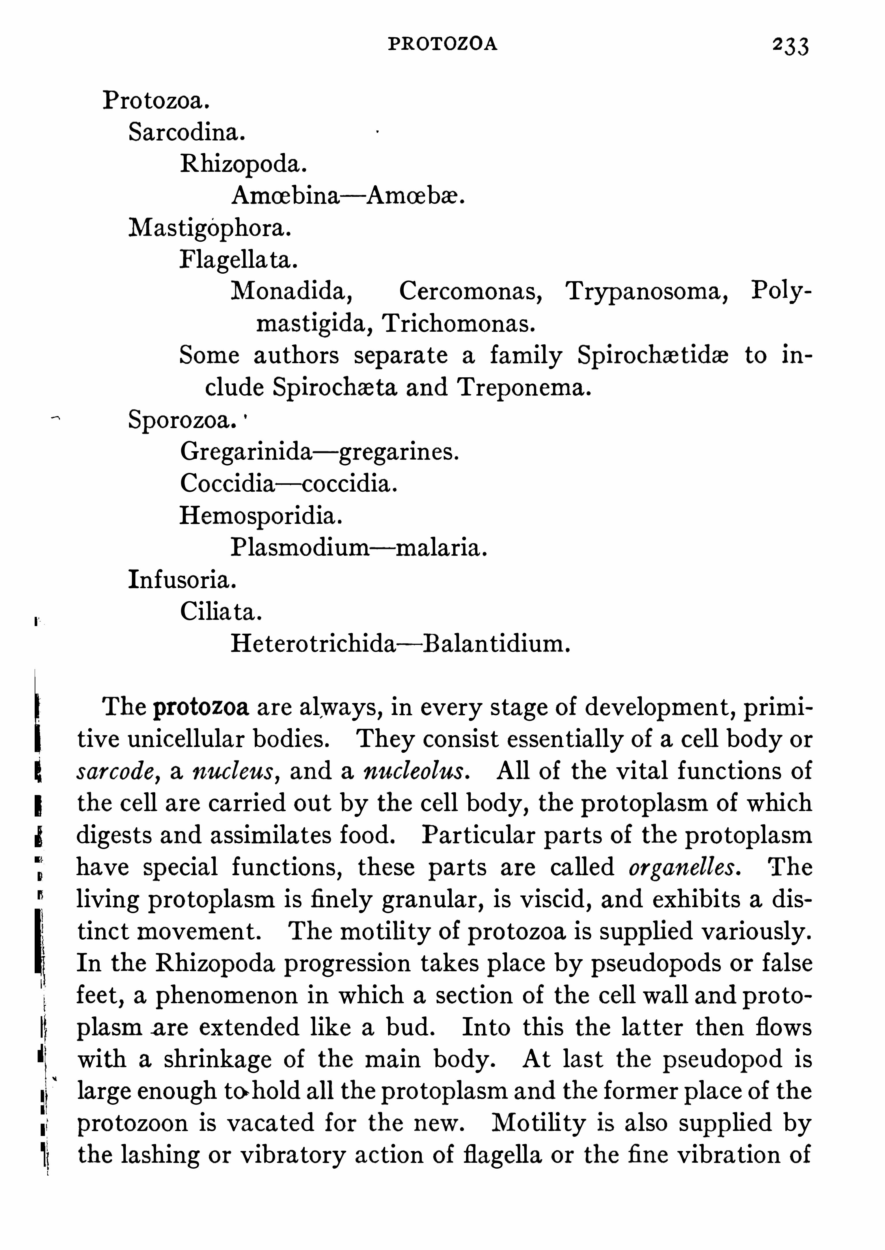
PROTOZOA 233
Pro tozoa .
Sarcodina .
Rhizopoda .
Amoebina—Amoeba .
Mastigophora .
Flagella ta .
Monadida,
Cercomonas,Tryp anosoma
,
mastigida, Trichomonas .Some authors separate a family Sp irocha tida to in
elude Sp irocha ta and Treponema .
Sporozoa .
’
Gregarinida—gregarines .Coccidia—coccidia .
Hemosporidia .
Plasmodium—malaria .
Infusoria .
Ciliata .
Hetero trichida —Balantidium.
The protozoa are always, in every stage of development, p rimi
tive unicellular bodies . They consist essentially of a cell body orsarcode
,a nucleus
,and a nucleolus . All of the vital functions of
the cell are carried out by the cell body,the p rotop lasm of which
digests and assimilates food . Particular parts of the p rotop lasmhave Special functions , these parts are ca lled organelles . The
living p rotop lasm is finely granular, is viscid, and exhibits a distinc t movement . The mo tili ty of p ro tozoa is supp lied variously .
In the Rhizopoda p rogression takes p lace by p seudopods or falsefeet
,a phenomenon in which a section of the cell wall and p ro to
p lasm a re extended like a bud . Into this the latter then flowswith a shrinkage of the main body . At last the p seudopod islarge enough to bold all the p rotop lasm and the former p lace of the
pro tozoon is vacated for the new . Mo tili ty is also supp lied bythe lashing or vibratory action of flagella or the fine vibra tion of

234 ANIMAL PARASITES
circumferential cilia . In o thers a sp ecial muscular segmentof the body may exist . The suctorial tubes act also for mo tionat times . In most p rotozoa two layers can be seen— the ectosaro
,
and endosarc . The ectosarc originates the movement,is concerned
in the ingestion and excretion of food, and the resp iration . The
endosarc,which circulates Slowly
,is mainly for digestive purp oses .
In it are ferments,crystals
,food particles (seen in the food vacu
oles) , oil globules, gas, and p igment granules .Flagella and suctorial tubes— in pro tozoa that have them
belong to the ectosarc . Skeletal tissues,Shells
,etc .
,also belong
to this layer .The food consists of bacteria
,smaller animals
,alga
,and animal
waste .
Propagation is effec ted by direct cell division , beginning in thenucleus
,by cell budding or by a comp licated course of sporula tion
which may be sexual or asexualf Sometimes division,or budding
,
occurs rap idly without the segments separating, leading to the
formation of p rotozoal colonies, or swarm spores .In the case of the malarial p lasmodia , asexual development
,
(schizogony) takes p lace in man’s blood
,while the sexual develop
ment (sporogony) takes p lace in the mosquito . Pro tozoa a re
found in salt and fresh water,in damp p laces, and in animals as
parasites .Since the zoological classification has been given and may be
used for reference to larger works, the various pathogenic p ro tozoa are given separately without direct reference to their systematic classification .
There are but two Rhizopods that are parasitic and pathogenicto man . The only one of these of any import is the Amoeba .
AM CEBA DYSENTERIZE OR ENTAMCEBA
HISTOLYTICA
This is a p ear-shap ed roundish body from .0 0 8 to . 0 5 mm.
diameter . The ectosarc is easily discernible in the p seud

ANIMAL PARASITES
killed at low temp eratures, and resist up to 60
0°
C . when in theencysted state . Quinine, permanganate ofpo tash, weak acid andsilver nitrate are quickly fa tal to vegetative forms . Emetin isa useful drug since i t kills all but encysted forms after a veryshort exposure if direct .Entamoeba tetragena , formerly classified as a separate variety,
is now generally believed to be but one stage in the developmentof But. histolytica .
In stools (from dysenteric cases) over a day old,amoeba are
not often found,as they undergo a rap id disintegration outside
the body .
Amoeba are cultivated upon stiff agar p referably with defibrimated blood and in company with bacteria . If a colony can be
obtained free of bacteria,development will continue on agar
FIG . 73.
—Entama ba tetragona . The same living individual drawn briefintervals while moving . (From Doflein after Hartmann.)
smeared with organ extracts . The addi tion of deadculture media seems favorable to their development .is not known . The free amoeba in the colon arewhen encysted are more resistant .ten minutes in strength of 1—2 50 0 .
Endamoeba buccalis i

FLAGELLATA 237
tee th and in inflamed gums ; it may have some effect in con
tinuing a gingivitis .
The flagellata derive their name from the fact tha t all are possessed
, at some time in their existence,of flagella
,which are not
only organs of locomo tion,but serve to app rehend food .
The p rincipal members of this class of interest from a pathological viewp oint, are the trypanosomes . Trypanosoma gam
FIG. 74f—Trypanosome in rats’ blood . (Williams )
biense, transmitted by the tsetse-flyGlossina palpalis, pathogenicfor man (see page 2 The Trypanosoma brucei, which causesthe tsetse-fly disease (nagana) in horses and ca ttle, is transmittedto cattle by the bite of the tsetse-fly, Glossina morsitans. It canbe grown on blood agar (Novy) .
Trypanosoma evansi causes surra, a disease ofhorses in CentralAsia transmitted by a fly of the genus Stomoxys .

238 ANIMAL PARASITES
Trypanosoma equiperdum causes a sexual disease in stallionsand mares called dourine ; this is akin to syphilis in man .
Trypanosoma lewisi of ra ts is transmitted from animal toanimal by means of fleas .Trypanosoma noctua .
-A parasite of the little owl, which isintroduced into the bird through the bite of the mosquito Culex
Several o ther forms have been no ted but these are p erhapsthe most important and serve as examp les for this family of
p rotozoa .
Trypanosomes are elongated fusiform bodies pointed at bothends
, p rovided by a fin fold, or undula ting membrane,running
along the dorsal edge and forming frill-like folds which terminatein a whip -like extremity or flagellum.
A large nucleus is always seen, also a centrosome,a small chro
matic mass— likewise called a blepharop last—near one pole .
The flagellum is at the anterior extremity ; the blunt pointedend is the posterior extremity . Cell division begins in the blepharop last, the cell dividing longitudinally, the n ucleus, flagellum
,
and the p rotop lasm dividing last . D ividing trypanosomes fre
quently appear in clump s with the ends .united, resembling a
wheel .The trypanosomes exist in two hosts—one a suctorial insect
and have a sexual and an asexual existence (alternate generation) .In an infected owl the organism has been observed clinging fastto the red cells
,absorbing nutriment during the day, while at
night it swims about freely in the p lasma .
In owl’s blood the trypanosome assumes asexual forms,call ed
macrogametes. These macrogametes p enetrate the erythrocytes,accumulating the remnants of the red cells in theThe nucleus of the trypanosome
‘h
may be seen in ththe p rotop lasm . The microgametocytes arise fromforms and when mature , give rise to eight microgametes .

240 ANIMAL PARASITES
mixtures such as Leishman ’s,Romanowsky ’s
,etc .
,the nucleus
,
centrosome and flagella,staining deep est . Thus far the T . gam
biense has not been cultiva ted in artificial media .
Novy has succeeded in growing the T . leW1su and T . brucei onagar mixed with defibrinated rabbit ’s blood . These are the firstanimal parasites to be cultivated artificially .
Trypanosomiasis of South America is not unlike sleep ing-sickness ofAfrica . It is caused by Tr . cruzi
,a parasite of eight spores
develop ing in organs,serum or red cells . It is transmitted by
Conorrhinus megistus, a large insect .In Dum Dum fever or Kala Azar , a disease characterized by
wasting,anemia , fever and sp lenomegaly occurring in India
,
curious bodies,called L eishmann-Donovan bodies
,have been
found . These resemble the malarial p lasmodia roughly, and ifcultivated on blood agar elongated herp etomas-like bodies without undulating membrane s will develop . They are to be foundin the juice obtained by sp lenic puncture lying within cells, especially endo thelium and large lymphocytes ; on rare occa
sions they have been met in the blood . The transmiss1on 15
not certainly known but may be by the bed-bug or by fleas “Trichomonas vaginalis and intestinalis
,are flagellates which
are apparently able to set up some inflammatory irrita tion in the
places indicated by their sp ecial names .
TREPONEMA PALLIDUM (Schaudinn)
(Spirochaeta Pallida .)Treponema Pall idum.
—There has been some discussion as to
the p roper classification but now this organism is usually p lacedamong the Flagellata, genus Treponema . It does not posundulating membrane
,is flagell ated, is of stiff and regular sh
and multip lies by longitudinal division .
Morphology and Stains.—This organism is extremely deli
in structure,from 4 to 1 4“ in length and about .3p in width ;

TREPONEMA PALLIDUM 24
from 3 to 1 2 turns or bends, and its ends are delicately po inted .
Its curves form a large arc of a small circle ; the Sp. refringens
curves form a small arc,frequently irregular
,of a larger circle .
It multip lies by both transverse and longitudinal division . Asthis organism is stained with difliculty it requires a sp ecial one,tha t of Giemsa yielding the best results . Aniline gentian viole t
,
Romanowsky ’s,and Leishman’s stains also color it . It may be
stained in tissues by silver and pyrogallic acid methods .Habitat—It has not been found in tissues of normal persons, orthose ill with carcinoma
,tuberculosis
,etc .
,but only in the tissues
of individuals suffering with syphilis . It is a strict parasite .
Vita lity.—The organism is readily destroyed by the ordinary
disinfectants and dies.after a few minutes’ exposure to 50
°
C .
The Treponema pallidum has now fulfilled the postulates of
K och . It can be cultiva ted from .human lesions (with some
difficulty to be sure) , it can be implanted in animals (monkeysand rabbits) and there rep roduce syphilitic lesions ; and i t can bere-cultivated from them . In these experimental diseases i t reta ins the p roper morphology . According to Noguchi there are
two types, a slender and a stout, which breed true to these characters and correspond to slight pathogenic variations . Noguchisucceeded in cultivating the Tr . pall . in pure culture by usingthe juice from human or monkey’s lesions or from the syphili ticorchitis of rabbits . This he grows in serum water or serum agarto which has been added fresh tissue of rabbit . The organismgrows as fine fibrils in arborescent colonies . These can be selected
pure by cutting the tube and the agar column . Motion is ofscrew and serp entine character . No odor or Spores are p roduced .
This organismmust be imagined and remembered as a corkscrewand not a waving line . The Gram stain is negative .
From the cultures of this organism a toxic extract can be obtained which
,when rubbed into the skin of a syphilitic in the
late stages,gives a typ ical skin reaction , luetin and the luetin
reaction .
1 6

242 ANIMAL PARASITES
The Spirochceta refringens, which has been also cultivated byNoguchi and thought by him to be a Treponema also , grows without fresh animal tissue in a short time and p roduces no odor.Pathogenesis.
—It has been found in chancre,condylomata
,and
mucous patches in the early stages of syphilis ; also in the blood ,blister-fluids
,sp leen , bone marrow,
liver,thymus gland
,and
FIG. 76 .
—The Sp irochaeta refringens is the larger andmore darklyorganism,
while the lightly stained and more delicate parasite is the Sp irochaeta pallida (Treponema pallidum) . From a chancre stained with Wright’sblood stain. (Hirsch—by Rosenberger. )
lymphatic glands, and in the brain and cord of taboparetics .
Associated with this organism,in nearly every case
,is a coarse
looking larger Sp irochaeta (Treponema) , which stains deep er , andhas been called the Spirocha ta (Treponema) refringensIn a series of experiments
,M etchnikoff and Roux caused abor
tion of the chancre following inoculation of syphilitic virus on theeyelid of a chimpanzee , by calomel inunction carried out less thanone hour after the infection ; a solution of sublima te has not thesame prophylactic property .

244 ANIMAL PARASITES
motility,and do not form spores . There is a single terminal
flagellum. They are stained with reasonable ease by p lychrome
methods,especially Giemsa
,but not by Gram’s method . They
measure from 1 0 to 40 14 in length and about In in breadth .
Co ils vary from 6 to 2 0 . The American typ e is smaller than therest .
FIG. 77 .
—Spirilla of relapsing fever from blood of a man . (Kolle and
Wassermann.)
Transmission - The known tick which transmits these organisms becomes infective in one week after biting a patient andremains so all its life ; its young are also infective . The types ofdisease vary but little . In all these is a relasping
‘
fever with
periods ofapyrexia in between . During the fever the spirochwtesare swimming free in the blood and disappear in the afebrileinterval .Cultivation .
—They are cul tivated in the manner given forTrep. pallidum by Noguchi , by adding citrated, therefore defibrinated, blood to serum or ascitic-fluid-fresh-tissue-agar . Theybreed true to type . They remain alive several days underfavorable artificial conditions but canno t be cultivated after

SPOROZOA 245
they have left the body a few hours without being on suitableculture media .
The periods of fever last from five to seven days,when a crisis
occurs . After an apyrexial p eriod the fever'
recurs. The Sp irochaetae are found in great numbers in every microscop ical field .
In the apyrexial period the sp leen becomes engorged and the
leucocytes devour the parasites . Monkeys with excised spleensare more suscep tible to infection than others .Immunity.
—The blood from rats tha t have been immunizedby repeated injections of blood from sp irochetal ra ts , if injectedinto o ther rats
,is capable of conferring an immunity on them by
causing sp irochaetes to disapp ear from their blood . One attackseems to confer immunity to the sp ecial form causing it but
probably not to the o thers .
SPOROZOA
The most important of this family are the malarial parasites
(which belong to the order Haemosporidia) , and the Coccidia .
In general the sporozoa are unicellular organisms that lead a
parasite existence in the tissues,especially cells
,of higher ani
mals . They ingest liquid food, have no cilia in the adult stage,
and flagella are possessed only by the males . There may be oneor more nuclei . Propagation is eflected by spores, but buddingand division do occur
,though rarely . Alternate generation
takes p lace frequently .
Hamosporidia of Mam—The most important disease causedin human beings by the haemosporidia is malaria , or ague
,and
excep ting the deserts, mountains, and arctic regions, this diseaseis very widely distributed .
Three different parasites p roducing different clinical entitiesare known . According to the time
,frequency
,and order of the

246 ANIMAL PARASITES
outbreak of chills and fever,various clinical names have been
given to the manifestation of the disease . Mannaberg has
arranged the following scheme to show the different forms of
outbreaks . The numbers app ly to the paroxysms . Eachdevelopmental cycle IS numbered alike :
I I I I I I I . Simple quotidian fever .
I o r o I o I Simple tertian fever .
I o o I o o I o o I. Simple quartan fever .
I 2 I 2 I 2 I 2 . Double tertian fever . (Two infections .)I 2 3 I 2 2 3 . Triple quartan fever . (Three infections .)I 2 o I 2 o I 2 0 . Double quartan fever (Two infections .)
The figures refer to days on which paroxysms of fever occur .’
The 0 rep resents the afebrile day .
PLASMODIUM MALARIZE (Laveran)
This is the quartan parasite , and p roduces in man ,in ca ses of
one infection, paroxysms of fever every fourth day .
It app ears in the blood , after a paroxysm,as a small nonfp lg
mented body on the bodies of the red blood cells . It has feebleamoeboid motion ; slowly p enetrates the cor
'
puscle , and specks ofmelanin appear in its p ro toplasmi Forty-eight hours after thea ttack the parasite measures from one-half to two-thirds theSize of the red cell . Sixty hours after the paroxys 'twelve
before the next—the parasite comp letely fills the red cell,leav
ing,
only a narrow rim,which later on disapp ears . Six hours
before the next paroxysm,shizogony begins. The grains of
melanin are arranged like the spokes of a wheel , and then , leavingthe radii
,crowd above the centre (the rest of the cell being pig
mentless) gradually dividing into 8 or 1 2 p ear-shaped bodies, ormerozoites. These separate from each other and individuallyattack a fresh red cell
,and this a ttack brings another paroxysm
of fever seventy-two hours after the p revious one . The grains

248 ANIMAL PARASITES
PLASMODIUM FALCIPARUMThe p lasmodium of aestivo-autumnal fever
,or pernicious ma
larial fever,also called trop ical . The outbreaks of this occur ir
regularly . The disease produced by them is very much moremalignant and is harder to cure . The young spore app ears inthe corpuscle as a small hyaline body
,smaller than the o ther
forms andmuch more active . The size and shap e of the red cellsare li ttle if any altered but they become granular and polychromatOphilic . The p igment is very finely granular and the bodyfrequently p resents the Signet-ring app earance . There may bemore than one parasite to a red cell . The cycle of development
(schizogony) is twenty-four to forty-eight hours . The p lasmodium in its schizogony divided into 8 to 24merozoitand are arranged in a spore-like form . The extraiesmay resemble a crescent or sickle ; this form istic of azstivo-autumnal fever . There are two
crescents,one delicate
, the male, and one largerfemale . They are very resistant to quinine and pp eriod in the blood . Plasmodia undergoing schizfound in the brain cap illaries aftcerebral symp toms in such cases . This form ca
from the others by the irregular and p ern1c10usduced;
‘by its great resistance to quinine ; the fewer numbmerozoites ; the finely granular app earance of the p igmentrelatively small size of the young intracorpuscular body ;by the ring shape of some of the young forms .Often
,in blood from malarial cases
, p igmented leucocytes aseen
,and ghost
,or shadow
,red corpuscles from which the haemo
globin has been dissolved are often met with . Spherical extracorpuscular bodies become flagell ated (gametes) in freshly drawnblood . The parasite may be studied in freshand by staining dried films by methylene bluenowsky
’s,or Jenner ’s methods . They are much more frequent
in the pyrexial period, and when quinine has not been given .
5
PLASMODIUM FALCIPARUM 249
The various p lasmodia are transmitted to man invariably bythe anopheles mosquito , in the bodies of which they undergo a
different (sexual) existence . It has been positively demonstratedthat the Various p lasmodia undergo an alteration of generationsand require two different hosts for their development, i .e.
,mos
quito,man .
The asexual development,or schizogony
,takes place in the
blood ofman, the sporogony, or sexual development,in the body
of the anopheles mosquitoes,the bite of which sets up an infec
tion In man, since the sporozoites of the various plasmodia are
developed in the salivary glands of these mosquitoes . In theact of biting, the sporozoites reach the erythrocytes where theybecome the intracorpuscular hyaline bodies beginning again theirasexual cycle of development in the blood .
That the mosquito is the intermediate host of the malarial parasite and that the infection in man follows bites by infected mos
quitoes has been abundantly p roven . The mosquitoes that actin this way are the various Anopheles; the Anophelesmaculipennisbeing the offender most frequently. The freshly formed schizontsin the blood of an infected man are conveyed into the in testinesof the mosquito . Here sexual reproduction of the parasite begins .The male elements
,flagellar microgametes penetrate the female
elements,macrogametes (cellular) , and after a time there appear
intra-cellular fusiform bodies,ob
'
kinets . These bore into the
intestinal walls of the mosquito and there remain . After a time
they are converted into round bodies,or oo
’
cysts . The nucleusof the oo
'
cysts divides rap idly and o ther daughter nuclei are formedand new cells called sporoblasts . After about eight days theseform the sporozoites . The number of sporozoites in each oticystvaries from-hundreds to many thousands (often Theseoocysts burst and the sporozoites in the circulation find theirway to the salivary glands of the mosquito . When a mosquitobites a human being they are introduced into the blood wherethey are quickly transformed into the intracellul ar hyaline bodies
DESCRIPTION OF FIG . 78
Life history of malaria parasite, Plasmodium. I, Sporozoite, introducedby mosquito into human blood, the sporozoite becomes a schizont ; 2 , youngschizont ; 3, young schizont in a
,
red blood corpuscle ; 4 , full-grown schizont ;5, nuclear division; 6, spores, ormerozoites, from a single mother-cell ; 7, youngmacrogamete (female) , from a merozo ite , and situated in a red blood cor
puscle ; 7a, young microgametoblast (male) ; 8, full-grown macrogamete ; 80 ,full-grown microgametoblast; 9 , mature macrogamete ; 9a , mature microgametoblast; 9b, resting cell, bearing six flagellate microgametes (male ) ;1 0
, fertilization of a macrogamete by a motile microgamete ; the macrogametenext becomes an ookinete ; 1 1 , ookinete, or wandering cell, which penetratesinto the wall of the stomach of the mosquito ; 1 2 , ookinete in the outer regionof the wall of the stomach, i.e. ,
next to the body cavity ; 1 3, young oocyst,derived from the ookinete ; 1 4, oocyst, containing sporoblasts, which developinto sporozoites ; 1 5, older oocyst ; 1 6, mature oocysts, containing sporozoites ;1 7, transverse section of salivary gland of an Anopheles mosquito, showingsporozoites of the malaria parasite in the gland cells surrounding the centralcanal.
1 -6 illustrate schizogony (asexual production of spores) ; 7—1 6, sporogony(sexual production of spores) .(FOLSOM—After GRASSI andLEUCKART, by permission ofD r. Carl Chun .)

252 ANIMAL PARASITES
and begin their o asexna l—sporogony. in the blood . Each developmental cycle causing a febrile p
’
aroxysm either every day or alternate days
,or on every fourth day ,
etc .,depending on the character
of the organisms and the number of infections . To p revent
FIG. 79 .
—Coccidium hominis,‘ from intestine of rabbit : 1 , a degenerate e
thehal cell containing two'
coccidia ; 2 , free coccidium from intestinal3, cocc idium with four spores and residual substances ; 4, an isolated spore ;spore show ing the two falciform bodies—X I1 40 . (From Railliet, in TysoPractice . )
spread of malaria,mosquitoes must be prevented from reaching
individuals infected with malaria and those not infected . Screensaccomp lish this best . The larva of the mosquito develop s instagnant wa ter . To p revent the development of these youngmosquitoes oil should be poured on the water
,thus cutting off
the air and means of resp iration .

COCCIDIUM 2 53
Bass,of New Orleans , claims to have successfully cultiva ted
malarial p lasmodia of the species vivax andfalciparum by the useof human blood . He has also succeeded when using Locke ’s fluidminus calcium chloride p lus ascitic fluid . One-half percent dextrose is usually added . The blood is drawn , so tha t it can bedefibrinated
,into small flat-bottom tubes . These are incuba ted
at 40°
C . The column of fluids is 1—2 inches high , the clear serumlayer being inch at least . The parasites grow in the upperlayer of the cellular sediment . Undiluted serum and leucocytesare lytic for p lasmodia . For renewed cultures
'
these must beremoved but uninjured red cell s must be added . Only the asexualdivision has been observed . Leucocytes phagocyte all free
parasites under artificial conditions .
COCCIDIUM
Coccidinmhominis is ano ther member of the sporozoa that occasionally infects man . Coccidia are infectious also for horses
,
goats,oxen
,-sheep , p igs , guinea p igs , weasels and rabbits . The
organism is essentially a cell parasite inhabiting-
the cells of the
gastro-intestinal tract by p reference , chiefly the liver and intestinal mucous membranes . They lead a sexual and asexualexistence like the malarial parasites (alterna te generation) . The
young sickle-shap ed nucleated sporozoite p enetrates an ep ithelialcell
,where it gradually develop s , ultimately dividing into numer
ous sporozoites . This is the asexual stage of development
(schizogony) , the sexual stage being called sporogony .
The sporozoites are differentiated into the two sex elements .These are large granular app earing cells ; the male being smaller ,divides into numerous flagellatedmicrogamates that p enetrate thefemale granular cells
,macrogametes
,and fertilize them . These
fertilized macrogametes,or zygotes
,form cap sules and become
oocysts which divide into numerous sporoblasts, changing intosickle-shaped sporozoites up on liberation .

2 54 ANIMAL PARASITES
The coccidia are easily demonstrable in tissue and in fa ces .They p roduce in man occasionally a
"
fatal disease infecting theliver and intestines . Cattle some times die from haemorrhagic
FIG. 80 .
—Development of coccidium cuniculi : a, b, 6 , young coccidia inthelial cells of gall duct ; d, e, f, fully grown encysted coccidia ; g, h, i,showing development of sthe two falciform bodiesspore compressed so as to separate the two 5 rozoites, o, a sporofalciform body with y, itsnucleus. (From Railligt after Balbiani—in TysonPractice .)
dysentery due to one of the coccidia . The disease is transmitteby the ingestion of food contaminated by faeces containingsporozoites .Acid fuchsin stains the sporozoa .

2 56 THE FILTERABLE V IRUSES
Hydrophobia.—This disease has long been considered to be an
infectious one,but the causal parasitic agent has never been dis
covered . It is commonly found in dogs,cats
,wolves
,rabbits
,etc .
,
but other domestic animals,andman may become infected .
i
It isa disease of the central nervous system
,highly infectious
,always
following a bite or other injury in which the skin is broken,and
in which lesion the virus may be deposited . Infection may becaused by injecting emulsified infec ted nerve tissue (brain) intosuscep tible animals (rabbits or monkeys) . The disease is alwaysfatal after it is well established . Well-marked histological lesionsof the central nerve tissues
, p articularly the large ganglia, havebeen found byV an Gehutchen and Nelis
,and Ravenel and Mc
Carthy. If emulsified brain tissue from an animal that has died ofhydrophobia is fil tered through a
“
germ-pr
capable of setting up the disease ininto it . By long centrifugation of emulsified infected brain tissue,the sup ernatant fluid loses its power of reproducing the disease oninjection . Virus may also be found in mammary and lacrymalsecretions
, pancreas, cerebro-Sp inal fluid and'
aqueous humor .The organism is toxic in character
,since fil trates sometimes fail
to p roduce transmissible disease, but emaciation , paralysis,death .are caused by their injection into rabbits, the tissueswhich , in turn, are not infectious .
The unknown organisms are ra ther resistant to'
agents that aregermicidal . They are destroyed in fifty minutes by a 5 p ercentcarbolic solution, and in three hours by a 1 corrosiv
mate solution . D irect sunl ight kills them quickly,as do
emanations . The latter have been used" as curativewith reputed success . A temp era ture from 52
°
to 58°C
half hour destroys them,but they resist extreme cold of.
for many weeks . Pasteur found that desiccationated the virus . Chlorine kills i t quickly
,while glycerine d
The virus may be increased in virulence by passing thevirus ” of dogs through a series of rabbits .

HYDROPHOB IA 2 57
from three weeks to six days, but beyond
Pasteur to name it virusfixe (fixed virus) .
FIG . 8 1 .
—Section through the cornu ammonis of brain of a rabid dog ;(1 by the method of Lentz . F ive Negri bodies of different sizes are
enclosed within the ganglion cells. The smallest contains only threegranules. (After Lentz, Centralbl . f. Baht , 1 90 7, Abt. I , Vol .XLIV,
ri bodies,intracellular bodies discovered by Negri , are
the ganglionic cells of rabid animals . These bodies stainand are from 1 to in size, beinggenerally about 5p .

2 58 THE FILTERABLE V IRUSES
They are found particularly in the cornu ofAmmon ; in Purkinje’s
cell s in the cerebellum ; and in the larger cells of the cortex of thecerebrum. These may be the cause of the disease
,but there are
several objections to this hypo thesis . Their distribution does notcorrespond to the parts of the nervous system that are mostintensely affected in hydrophobia ,
i .e .,medulla and pons . In the
latter locality these bodies are rarely encountered . They are notfound invariably in animals dead from rabies
,and are considered
to be too large to pass through a Berkefeld filter ; this latter viewmay not be a correct one . The finding of these bodies has beenconsidered by Negri to be good grounds for considering the case
to be hydrophobia . The rap id diagnosis of the disease in animalscan only be effected by killing them and examining the nervoustissues
,or inoculating other animals with them . Hi
three marked changes may be no ted : (1 ) The finding of thebodies . (2 ) The finding of the degeneration of the cellslarger ganglia with the p roliferation of the endothelial cellsthe ganglionic spaces (V an Gehutchen andNelis) . (3) Thing of certain tubercles in the medulla
,which are called
tubercles,though these are not wholly characteristic
,as they
found in other diseases . Hydrophobia is transmitted fromSite of the wound to the central nervous tissues by the nerves ,the incubation p eriod varies with the distance of the woundthe central nervous system ; the majority of cases occur bettwenty to sixty days after a bite .
Immunity against infection and the development of the diafter the recep tion of an infected wound, may be accomp lishedPasteur ’s method (see chap ter on Vaccine) .
Yellow Fever
Tha t this disease is caused by a parasite there can be no doubIt is highly infectious and largely confined to the trop icalof the western hemisphere and in parts of Africa .
diseases established as due to p ro tozoa , this one is

THE FILTERAB LE V IRUSES
p roduced by the bite of a mosquito confers immunity againstsubsequent infection . (7) The period of infection is usuallythree days but may be from two to six days . (8) A house or shipmay be said to be infected with yellow fever only when there are
p resent mosquitoes capable of conveying the parasite of the
disease . (9) The Sp read of yellow fever may be prevented bydestroying the aedes and p reventing egress and ingress of theinsec ts from yellow fever patients to the non-immune . (1 0 ) No
insect, other than the aedes,has been found to be concerned in
the spread of yellow fever .Noguchi reports the discovery of a tiny
‘
Sp irochaete, 4—9p longand . 2p wide, in the blood ofyellow fever patients . The organismmay be transferred to guinea p igs in which it p roduces lesionscomparable to those ofhuman yellow fever . Itmay be cul tivatedin anaerobic serum water tubes and it will pass through a fil ter .During an attack
,either human or artificial in a guinea pig,
protective anti-bodies are formed . Sp irals are only found in theblood in the fir st few days of an attack . They do not stain welland can best be seen by aid of the dark field microscop e . It isclaimed that vaccination with these sp irals p roduces some resistance to yellow fever .Yellow fever is a trop ical or subtrop ical
.
disease, because theaedes is confined to these regions, and the disease is foundin low moist localities rather than those that are drier and higher,from the fact that the mosquito inhabits the former and not the
latter . Yellow fever dies out after the first sharp frost, becausethe
,aedes are then either killed or undergo hibernation .
conclusive exp eriments by Reed and Carrol,by Guiter
by the French Commission have p roved that the aedes isdoubt the cause of the sp read of the disease . No ithan the activity acquired one, is known .
Small -pox and Vaccin ia.—These two diseases must be
ered to be but two clinical activities of one unknownmicrotirganism.

SMALL POX 261
Certa in protozoonoid bodies have been Seen by numerous observers
,no tably by V anderLoefl , L . Peifl
'
er,and Guarnieri . The
latter gave the name Cytoryctes vacciniaa s . var iola . In thedeep layers of the ep ithelial cells of the pustules of vaccinia and
small-pox, in the exp erimental lesions on the cornea: of rabbits ,and in the protoplasm of the cells
,'
these bodies are found . Theyare about the size of a micrococcus and exhibit amoeboid movements in hanging-drop preparations . They are p erfectly characteristic of the lesion produced in vaccinia and are not-found inother diseased conditions .
0 In variola many different changes occur in the app earances ofthese cytoryc tes
,suggesting developmental cycles .
In variola they are often intranuclear,while in vaccine they are
never found within the nuclei .The cycle of development is suggestive of the development
of many of the protozoa . Stages of development exhibitingfusiform amoeboid shapes can be seen , and p seudopodia can be
detected in the process of developmental stages suggestive of
gametoc'
ytes ; the union of the gametes and the ultimate formation of the zygo te can also be discerned.
After the tenth day these bodies canno t be very well discernedthe tissues .There is reason to think that the parasites circulate in the bloodvariola . The contagion in variola is thought to be by inhala
It is certain that the disease can be p roduced by inoculawith virus from a case of small-pox. The contagion exists
the scales,pus cells, and excre tions ofpatients ill with small-pox.
If the virus of small-pox is introduced into a monkey,and then
to a cow the disease p roduced is not variola, but vacciniankman) . The hypothetical organism above described, cytobecomes attenuated in the cow
,so that i t is incapable of
are suscep tible of inoculation withtion) . Virus may be tested by

262 THE FILTERABLE V IRUSES
rubbing over the Shaven bellies of rabbits,setting up minute
vesicles and finally crusts (Calmette) .The two Viruses
,that of variola and that of vaccinia
,are now
thought to be identical . In a diluted condition it is fil terable .
It resists drying for weeks and glycerine eight to ten months . Itis destroyed at 57
°
C . in fifteen minutes and easily by most disinfectants . Passive immunization has not been achieved . No
guchi has succeeded in growing the virus in the testes of rabbits ;one of his objects in so doing was to afford a sterile cultivation ofV irus which might be used for preparing vaccine .
Scar let Fever
Mallory in 1 90 3 found certain bodies in the skin of scarlet fevercases . These bodies
,he assumed , were protozoan in character
and were the etiological cause of the disease . He named themCyclasterion Scarlatinale . They have been found rather con
stantly in the skin of scarlet fever cases,also in the skin in cases
of measles and in anti-toxin rashes . Dohl e has also described,
an inclusion body within the polynuclear leucocytes . By severalobservers these bodies have been considered to be artef
degeneration p roducts in the ep ithelial cells'
.
The‘
virus of scarlatina is now considered to be fil tetransmissible to monkeys . Mallory has lately found dipbacilli in great numbers in the pharynx in scarlatina and
they may be its cause . Virulent strep tococci , usually oftic quality
, are so frequently encounteredand in complications during scarlet fever that manupon them as the cause ; this cannot be proven as the
cannot be successfully transmitted to lower animals by use oforganisms .Dengue Fever.
—This is an acute infectious disease 0
trop ics, characterized by fever, skin erup tions, rheumatoidan afebrile remission and a febrile end
,due to a fil terable

THE FILTERABLE V IRUSES
glycerine , drying and autolysis”
. It is destroyed at 50°
C . in one
half hour . The virus can be cultiva ted by growing bits of brainand cord from a case dead'
of the disease in unheated ascitic fluidto which bits of sterile tissue have been added ; the growth isanaerobic . It app ears as tiny globoid bodies, singly , 111 pairs orshort chains .
‘ Appropriate staining will demonstrate them alsoin the nervous tissues .Active artificial immunity and some passive immunity have
been obtained but these are not of therap eutic value . One
attackp robably confers immunity . In the therapeutics of thedisease i t is prac ticable to inject into the sp inal column , the bloodserum of human cases that have recovered from the disease ; thisindica tes that Virus-neutralizing bodies are formed during anattack .
Foot andMouth Disease .
—An acute infectious disease of cat
tle,characterized by a vesicular erup tion in the mouth and around
the crown of the hoof . It may be transmi tted to man by the
use ofmilk from infected cows . It is also directly communicable .
It has not been cul tivated ; It is fil terable ; it is said to be dueto the Cytorrycetes. It is destroyed at 50
°
C . in ten minutes ,easily by freezing and ordinary disinfectants . One
no lasting immunity but the blood is said to contain animmediately after the attack
,which will be p rotective
animals .Trachoma .
—An infectious inflammation of the conju
with‘
the p roduction of minute but visible nodules on the
Sides of the lids . By some it is said to be due to a forminfluenza bacillus , by o thers to an invisible virus . It is dcommunicable , filterable and transmissible to monkeys . Inot been cul tivated .
M easles.
—An acute erup tive fever due to a fil terable V ir
which is found in the blood,buccal and nasal secretions . It
transmissible to monkeys by inoculations of patient’s blood,
before the Koplik spots appear . It persists in the blood

MUMPS 2 6
after the appearance of the erup tion . It resists drying and freezing . It is destroyed at 55
°C
,in fifteen minutes ; it has not been
cultivated . Immunity follows an a ttack but no passive immunityhas been reported .
Mumps.-This disease seems also to be due to a filterable virus,
resident within the parotid gland, capable of transmission to
monkeys. Many organisms have been described but none is
probably sp ecific .
It must be said of bo th the hypothetical organisms of variolaand scarlatina
,that if they are the cause of these two diseases
they differ from all o ther known p ro tozoan parasites , becausethe latter require an intermediate host for the transmission of
the parasite from individual to individual while these certainlydo not.
Rocky Mountain Fever is not due to a filterable V irus but itsagent resides in the blood . It is a disease of the Rocky Moun tainStates characterized by general pains ; macular erup tion and constitutional symp toms of infection , i t is transmi tted bymeans of thetick , Dermacentor venustus . The female tick obtains the agentby blood sucking and. can transmit i t to her young . The virusis destroyed by heating and drying . A minute coccoid body hasbeen found in the tick but i t has not been cultivated . One
artificial attack in the guinea pig leaves immunity , as does thespontaneous disease in man .
Trench Fever IS a disease which became known in the GreatWar characterized by fever
, pains in the legs , back and head , withmarked dizziness, due to a resistant fil terable virus, in all p robability transmitted by lice ; the virus is found in the blood . The
entrance is effected either by the bite of a louse or its feces may bescratched into tiny wounds on the skin . The Virus seems to beexcre ted in the urine and sputum . It is not killed at 80 °C . The
louse is not infective for seven days after biting a patient andthen remains infective for three weeks . No immunity follows an

266 lTIIE FILTERABLE V IRUSEs
Encephalitis lethargica is a disease alleged to be due to a
Virus . It appears sometimes in ep idemic form and was observedafter the last pandemic of influenza ; some persons believe the twoare related . Some students of the subject report successful transmission to monkeys.
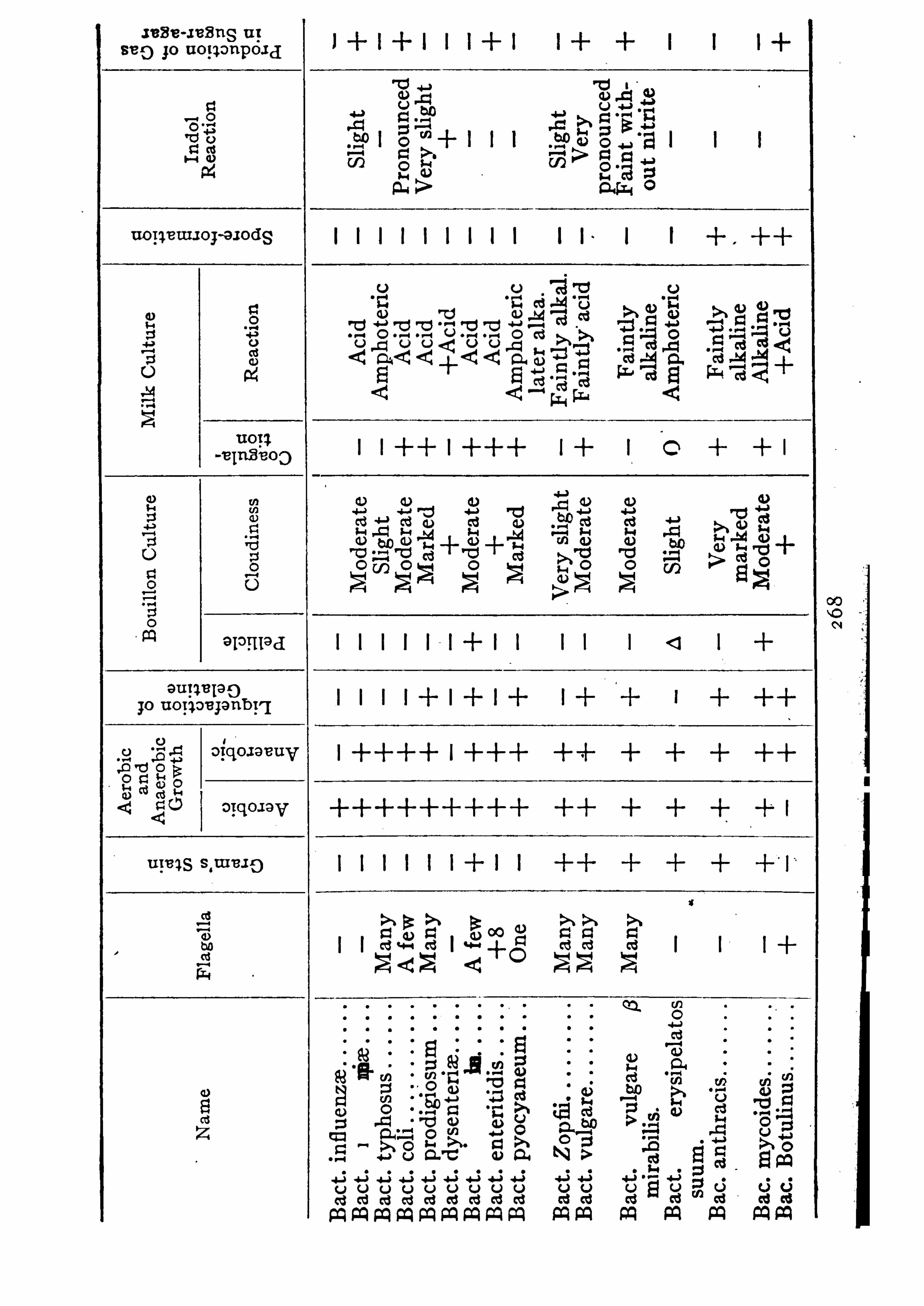
Jean-1 138 119 u ;
8 8 0 go uop onpoxd
uoIieUIIor-axodg
°l°Hl9 d
eugqe jsg);o uop oe janb rq
a reas 3311 9 1 5)
photeric
Ac
i
Ac
id
Acid
+Acid
Acid
Ac
id
Amphoteric
later
alka
.
Fa
intly
alkal
.
1+ 0
l l I l l
ra g » g
l l fifi fi l fi i g $ 5<2
0 2 2
Joa
n
.
mm
v
fitoa
ao
.
a
oe
m
.
8900
30
5.
aoe
m
.
Nt
ficomxv
.
uomm
Ezmomwmvo
a5.0
mm
.
a
oe
m
.
a
o
e
m
8
80
85
23.
uoa
m
.
wmao
smcm
.
uue
m

1
u
wfioaaa
fis
2
8
32
5
as?bass
Ewao
bm
Es
SE
E
unac
fim
32Gb
E
u
<
8820
0
2
830
32
880
v0
2
8830
0
2
8880
2”
0
38
30
0
2
EA
SE
b0
>
e l°ll lad
I+ I I I.
l O
A0
55
25
0
3h.
80
300
.
0
a
o
mama/H
mce
z
Sue
z
5
8
2
han
g
58
2
ES
E
5309
m00
>
80a
30
<
00
33
.
yownoo
h
z
.
mmmofl
zo
3885
50
0300
»
20
800"
4
8
20
5080
3523
£3
398
3.
30
5
30
0
03
85
damp
.
9
0090
5
20
0
.
uoa
a
gbo
u
.
um
am
.
mz0a
0
a

DESCRIPTION OF PLATE I
Malarial Parasites
1 . Two tertian parasites about thirty—six hours old, within blood cor
puscles magnified .
3
2 Tertian parasite about thirty -six hours old; stained by Romanowsky'
s
method The black granule in the parasite is not pigment but chromatin.
Next to it and to the left is a large lymphocyte , and under it the black spotis a blood p late .
3 . Tertian parasite division form nearby is a polynuclear leucocyte .
4 . Quartan parasite ,ribbon form.
5 . Quartan parasite . undergoing div ision.
6 . Trop ical fever parasite (aestivo-autumnal) . In one blood corpusclemay be seen a smaller
,medium , and large trop ical fever-ring parasite .
7 . Trop ical fever parasite . Gametes half-moon spherical form. Smearfrom bone marrow8 . Tropical
'
fever parasite which is preparing for division heaped up in theblood cap illaries ofthe brain.
Asexual Forms
9 . Smaller tertian ring about twelve hours old.
1 0 . Tertian parasite about thirty-six hours old, so-call ed amoeboid form.
1 1 . Tertian parasite still showing ring form, forty-two hours old.
1 2 . Tertian parasite , two hours before febrile attack . The p igment isbeginning to arrange itself in streaks or lines.
1 3 . Tertian parasite further advanced in division. Pigment collected inlarge quantities.
1 4 . Further advanced in the division (tertian parasite) .

DESCRIPTION OF PLATE II
Malar ial Parasites
1 5 . Complete division of the parasite . Typ ical mulberry form.
1 6 . To the left is the comp leted division form, an almost developed gametewhich is to be recognized by its dispersed p igment.
1 7 . A tertian ring parasite , small size broken up .
1 8 . Three-fold infection with'
tertian parasite. The oval black granulesare the chromatin granules.
1 9 . To the left, tertian parasite w ith large , sharply demarked , and deeplycolored chromatin granules. To the right
,tertian parasite . Both thirty
six hours old. Both probably gametes .
2 0 . Tertian parasite thirty—six hours old, ring form .
2 1 . Tertian parasite w ith beginning chromatin division, with eight chromatin segments.
2 2 . Tertian parasite chromatin division farther advanced with twelvechromatin granules, in part triangular in form.
23 . Comp leted division figure of a tertian parasite . Twenty-two chromatin granules.
24 . The young tertian parasites separating themselves from each other.The p igment remains behind In the middl e .
25. Quartan ring parasite , which is hard to difl erentiate from large tropicalring or small tertian ring .
26. Quartan ring lengthening itself.Small quartan ribbon form.
28 . The quartan ribbon increases in w idth . The dark places consist almostentirely of p igment.


DESCRIPTION OF PLATE III
Malaria l Parasite
30 , 3 1 . The quartan ribbon increases in width . The dark placesconsist almost entirely of p igment .3 2 . Beginning division of the quartan parasite and the black Spot in the
middle is the collected p igment .33. Quartan ring .
34 . Double infection w ith quartan parasites.
35 . Wide quartan band . The fine black stipp ling in the upper half of theparasite is p igment.36 . Beginning division of the quartan parasite . The chromatin (black
fleck) is Sp lit into four parts.
37 . D ivision advanced, quartan parasites.
38 . Typ ical divrsron figure of the quartan parasite .
39 . Finished division of the quartan parasite . Ten young parasites, pigment in the middle .
40 . Young parasites separated from one another : .
4 1 . Small and medium trop ical ring, the latter in a transition stage to a
large trop ical ring .
4 2 . Small, medium and large trop ical ring, together in one corpuscle .

DESCRIPTION or PLATE IV
43 . To the left a young (spore) trop ical parasite. To the right a mediumand large trop ical parasite .
44 . An almost fully developed trop ical parasite . The black granules are
pigment heaps.
‘
45 . Young parasites separated from one another. Broken up divisionforms twenty-one new parasites.
46 . To the left a red corpuscle with basophilic, karyochromatophilicgranules. Prototype ofmalarial parasite . On the right a red blood corpusclewith remains of nuc leus.
Sexual Forms or Gametes
4 7 . An earlier quartan gamete (macrogametocyte in sphere form) , female .
48 . An earlier quartan gamete (microgametocyte) , male .
49 . Tertian gamete,male form (microgametocyte) .
50 . Tertian gamete , female (macrogamete) .5 1 . Tertian gamete (macrogametocyte still within a red blood corpuscle.52 . M icrogamete tertian w ithin a red blood corpuscle .
53 . Trop ical fever. (ZEstivo autumnal) gamete , half moon (crescent) stilllying In a red blood corpuscle . In the middle IS the p igment. The concaveside of the crescent is spanned by the border of the red blood corpuscle .
54 . Gamete , trop ical fever parasite .
55. Gamete of trop ical fever parasite heavily p igmented .
56 . Gamete of the trop ical fever parasite (flagellated form) , microgametocyte sending out microgametes (flagella or spermatozoon) .

PLATE IV
CHAPTER XI
BACTERIOLOGY OFWATER, AIR, SOIL , AND MILK '
Bacteriological examination of water is of importance for thedetermination of the p resence of pathogenic bacteria , and for the
enumeration of the total number of all bacteria contained therein,the latter being considered an index of the purity of the water .Several well-known pathogenic bacteria have been found in
water ; among these are the typhoid , anthrax , cholera , p lague , andcolon bacilli
,also the pus cocci . Since the tetanus bacillus is a
normal inhabitant of the cultivated soil and manure,it is not at
'
all uncommon to find it,at times
,in muddy waters .
Bacteriological examinations of water are,in ,a measure , very
disappointing , because it is difficult, and at times impossible todetermine the p resence of the typhoid bacillus , even when it iscertain that it is p resent ; having been added to water to be ex
amined it is even then difficult to isolate .
The fact that the colon bacillus is always found in water contaminated by fa ces is a great help in the recognition of pollutedwater . In the case of typhoid contaminationthe typhoid bacillusmay elude detec tion
,but the colon bacillus is easily found ; we
may then assume tha t,since it is impossible for typhoid bacilli
to reach water without the colon bacilli that water having no
colon bacilli is also free from typhoid bacilli . Also water havingcolon bacilli in great numbers is contaminated with fa ces , and
perhaps typhoid fa ces . The detection of the colon bacillus istherefore of p rime importance in the examination of drinking wa ter .
_Its detection is simp le . Water must be collected
in sterile bottles,using every p recaution against accidental
contamination . Fermentation tubes are emp loyed , containingbouillon with 1 p ercent of lactose . Into a series of these tubes ,
2 78

BACTERIOLOGICAL EXAM NATIONS
To Count Bacteria inWaterThe samp le must be collected in a sterile bottle, and the p lates
‘
poured immediately,since bacteria multip ly enormously after a
few hours .Take % 0 c .c . or c .c . or 1 c .c . of the water in sterile p ip ettes
andmix with a tube ofmelted gelatine or agar, pour quickly into
cool sterile p etri dishes and p lace in a cool dry p lace . TheAmerican Public Health Association also recommends the use of 1
p ercent agar plates grown both at room and body temperature .
The counts for the two are averaged . After forty-eight hourscount the colonies and the result (after multip lication where %0
or c .c . of water was used) will be the number of bac teria p ercubic centimeter . It may be necessary to dilute the water fiveor ten times before pouring p lates . A glass p la te ruled intosquares
,known as aWolflhiigel p late, should be used for counting .
The number of bacteria in potable waters varies in many ways,
according to the amount of pollution , or albuminous matter inthe water
,while dep th, and the swiftness with which it flows are
conditions tha t modify bacterial contents . '
The water in a
reservoir becomes almost free from bacteria during the first ‘
ten
days . The number of bacteria diminishes~ ,1 0 p ercent p er day
for the first five or eight days,due no doubt to gravita tion of
the bacteria to the bottom,also in part to the action of light,
which p lays an important role in the destruc tion of the bacteriaof water supp lies .In general
,water containing less than 1 0 0 bacteria p er cubic
centimeter is considered to be from a deep source, and uncon
taminated by drainage . Deep artesian wells often contain butfrom 5 to 1 5 bacteria p er cubic centimeter
,water from rivers
often contain or depending somewhat upon the
season of the year . Rains cause an augmentation of the bac
terial content . Summer causes a diminution .
In identifying a certain water supp ly as the cause of an epi

TO COUNT BACTERIA IN WATER 2 8
demic of typhoid, the number‘
of bac teria is of grea t value inloca ting the p lace of infection .
The efliciency of filters in” large municipal Wa ter supp lies is
known only by the bac terial content of the effluent . In goodsand and mechanical (alum) fil ters, the reduction in the numberof bacteria is often over 95 percent (sometimes 99 p ercent) .Plate cultures should be made daily from every fil ter in order todetermine how each fil ter is p erforming . Sand fil ters should notfil ter more than gallons per acre a day . They should beat least 1 metre thick ; the upper }5 inch of the sand p erforms over
90 percent of the fil tra tion, due to a certain zooglea,or growth
of bacteria . Cracks,or imperfections in the fil ter beds are
quickly detected by the rap id increase of the number of the bacteria in the effluent . It ‘ is supposed that not only are bacteriafiltered by the sand but tha t destructive changes occur in thefilter which greatly dimin ish the number of bacteria . A fil termust be used for a few days before i t becomes efficient or “ ripe .
After a time it becomes inefficient and it must then be scraped,finally the sand must be removed and washed .
A sand filter is a highly efficient means of water purification .
It often converts a foul dir ty water into a bright,clean
,whole
some water of low bacterial content .Mechanical filters depend for their efliciency upon the addition
of aluminum sulphate to the water . This is decomposed by thecarbonates and aluminum hydroxide is
, p roduced, which is a
white jelly-like flocculent p recip itate, which mechanically en
tangles bacteria and removes them from the water . Mechanicalfilters
,as a rul e
,are highly eflicient. Domestic fil ters
,even the
Pasteur,are often unreliable .
In time of ep idemics of cholera and typhoid even fil tered watershould be boiled before use
,as it was found by exp eriments in the
M edico-Chirurgica] Laboratories that typho id bacilli live longerin filtered water than in bouillon ; they may even live thr eemonths .

2 82 BACTERIOLOGICAL EXAMINATIONS
The fewer the number of other bacteria the longer will typhoidlive . They can live many days in ordinary river water .Ice may contain great numbers of bacteria ; it is well known
that freezing does not destroy pathogenic bacteria , such as the
typhoid bacillus . Prudden found typhoid bacilli in ice after onehundred days
,although the number was greatly reduced over that
p laced in the ice originally. Many are squeezed outby contractionof the water . The greatest danger from ice is in dirty handling .
Disposal of sewage is a bacteriological process in many cases ;either the sewage may be treated in sand filters or it may be runout on land where over gallons may be disposed of on an
acre of land a day . As far as possible nature should be imitatedin every way and the breaking up ofmasses ofmatter in sewagemay be accomp lished in the sep tic-tank process in which activeoxidization of the matter is accomp lished by bacteria . It appearsfrom the observa tions ofmany sanitarians that both aerobic andanaerobic bacteria are necessary to finally reduce sewage to theelementary gases and pure water .In the interior of closed tanks and in the dep ths of sand filters
anaerobic conditions prevail . On beds of coke,and on the sur
face of sand filters,aerobic conditions obtain . The efl
cluent froma sep tic-tank sewage-disposal p lant is very often pure water fromboth chemical and bacteriological standpoints , due to the chemical action of the bacteria .
Bacteriology of the Air
That the lower layers of the earth’s atmosphere contain manybacteria is well known . The aif over the sea and over mountainranges is freer from bacteria than the air over arable lands andlarge cities .When air ‘
is still and confined, all bacteria , according to Tyndall ,gravitate to the ground
,and the air above becomes quite sterile .
The atmosphere of sick rooms, hosp itals , public conveyances,theatres
,etc .
,contains many bacteria and often pathogenic ones .

BACTERIOLOGICAL EXAMINATIONS
bles in the p resence of gas under p ressure ; air currents may of
course carry germs so freed a much longer distance .
Bacteriology of the Soil
At least two forms of pathogenic bacteria are habitually foundin the soil . The tetanus bacillus
,it is well known
,exists in garden
earth , manure , and top soil generally . D irt getting into woundsis the most frequent cause of tetanus . D rinking water laden withsoil has been known to have in it tetanus bacilli
,and if used in
an unsterilized condition in wounds or when a comparatively feebleantisep tic, such as creolin , has been added , it may cause tetanus .The gaseous edema group ,
the bacilli of malignant edema,
symp tomatic and parasitic anthrax are frequently found in so il .The highly tilled soil of the ba ttlefields in France was heavilyladen with the first
,hence the great incidence of infection after
wounds in the late war . Strep tococci and colon bacilli , too , havebeen found in garden soil . Typhoid bacilli may contaminate soil ,but do not multip ly in it . In sandy soil bacteria pergram have been found
,in garden soil 1
,50 0 ,0 0 0 bacteria p er gram,
and in sewage-polluted soil bacteria per gram havebeen determined . The first few inches of ordinary soil containmost of the bacteria
,after a dep th of 2 metres no bacteria
are found and the earth is sterile .
Soil may be collected in sterile sharp -pointed iron tubes, anddiluted with sterile water of given quantity and p lates pouredfrom i t .Arable lands may be enriched very much by inocula ting them
with’
certain nitrifying bacteria , some of which convert ammoniainto nitrous acid
,which form in them nitrites ; others change
nitri tes into nitrates (nitrosomonas) . Certain of these bacteriaare concerned in the assimilation of nitrogen from the atmosphereand adding to the nitrogen content of the soil, thus enriching it .On the roots of some p lants, alfalfa, beans, peas, and clover,minute , tubercles develop . These little growths are caused by the

“
BACTERIOLOGY OF cow ’
s MILK 2 8
nitrifying bacteria, and add to the nutrition of the p lant by addingto i t ammonia .
Bacteriology of Cow’s Milk
Theoretically the milk in the interior of the breasts of nursingwomen and the udders of cows is sterile . So soon as it leaves thenipp le it becomes contaminated with bacteria, and by the time i treaches the pa il, in the case of cow’s milk
,it is far from sterile .
Bacteria of the air,and dust from the cattle and bedding, at
every movement of the cow,and by the agency of flies
,find their
way into milk and contaminate it . The number of bacteria tha tdevelop s in the milk dep ends upon the number that reach it in thefirst p lace, the temperature of the air, and the length of time milkis~kep t at a temp erature favorable for their mul tiplica tion . Two
hundred and thirty-nine different varieties of bacteria have beenisolated from milk at different times .Pathogenic varieties of bacteria that have been found in cow ’
s
milk include the tubercle bacillus, Streptococcus pyogenes, Staphylococcus aureus
,the colon bacill us
,typhoid bacillus, the diphtheria
bacillus,and a Whole
"host of bacteria that sour or ferment themilk and render it unwholesome or poisonous for young children .
Cattle may be tubercul ous, and the tubercle bacill i may reachthe milk in this way . There may be abscesses of the udder andthe strep tococci from the pus may cause infection in those thatuse i t . Ordinary follicular tonsillitis may be caused in this way .
Bacteria may develop rap idly in milk, which is a good culturemedium
,until they number many millions per cubic centimeter
(sometimesIn good milk the number of bacteria may increase when thetemperature is 90
°F from originally in the milk imme
diately after milking,to in eight hours .
By exposing milk to a temp era ture of 1 65°F. for twenty to
thirty minutes and quickly cooling (Pasteurization) most of thenon-spore-bearing bacteria are destroyed, so that the number may

2 86 BACTERIOLOGICAL EXAMINATIONS
be reduced p ercent by this p rocess . The Pasteurizationofmilk has become an economic problem of great importance inlarge communities and is not, as it Should be, sufficiently sup ervised . That method is best in which milk is held at 1 46
°F. for
thirty minutes . No harm is done to the nutritional value of themilk . One of the dangers of the method is that the commercialPasteurizing machines are not always thoroughly clean and themselves contaminate the milk when discharging it after heating .
More evidence is on the side of the second View . The practicalimportance of the controversy is that milk whether heated or notshould be kep t at a temperature at whichb acteria will notmultip ly, under 60
°F. Pasteurized milk is safest in time of typhoid
ep idemics .Absolute cleanliness on the part of the milker, the use of steril
iz ed gloves and clo thes,the absence of flies
,dust
,and the imme
diate disposal ofmanure, the fil tration of the milk after collection,the immediate cooling of i t
,the uses of sterilized milk cans and
bottles,all lessen the bacterial content of milk . It then keep s
better, and is a
'
wholesomer and safer food for infants, esp eciallyin hot weather .By drinking water containing typhoid bacilli cows cannot
be sources of typhoid infection through the milk . The typhoidbacilli are not transmitted through the bodies and udders of theanimals .A bacteriologic examination of milk comp rises a total count,
the p resence of colon bacilli,strep tococci in pus cells, tubercle
bacilli and Sp ecial Sp ecies as the case suggests . The first is doneas given for water , as is the second . The discovery of strep tococciis made by centrifugalizing a definite quantity and examining thesediment for chains
, particularly in relation to leucocytes, the puscells . Tubercle bacilli are found by injecting guinea p igs or bydissolving the milk in antiformin (1 part milk and 1 part 1cent antiformin) warming and examining the sedimentcentrifugalization .

2 88 INDEX
Anti-toxin, dosage , 74for dysentery , 1 80for plant toxins, 70for pyocyaneus, 70
staphylococcus, 70 , 1 54streptococcus, 75
for symptomatic anthrax,1 96
for tetanus, 70 , 74, 1 92manufacture of, 7 1standardization of, 73
Anti-toxins, 43, 54, 60 , 70for animal toxins, 70standard , 73
Arnold sterilizer, 1 1 0ArtesianW‘
ells, 2 80Arthrospores, 1 6Aspergillus, 5
flavus, 231
fumigatus, 231niger
,231
Attenuation of bacteria, 34 , 1 35Autoclav , 1 0 9
Autopsies, animal,1 33
Avenue of infection, 35
Babes Ernst granules, 9tubercles, 2 58
Bacillus, 2 , 7aerogenes capsul atus, 1 0 0 , 1 99of anthrax, 1 6, 31 , 34, 50 , 1 0 0 ,
1 83
of blue pus, 1 8 1botulinus, 1 97Chauvoei, 1 94of cholera, 20 2colon, 1 0 0 , 1 74 , 2 78, 284, 286comma , 2 0 2
of diphtheria , 3 1 , 1 0 0 , 2 0 7of dysentery, 1 0 0 , 1 78
Bacillus, enteriditis sporogenes, 1 99Friedl ander
’s, 1 66
fusiformis, 2 0 1Gartner’s, 1 77of glanders, 3 1 , 2 0 6Koch Weeks, 1 62lepra , 3 1 , 2 2 1of lockjaw, 1 88
mal ignant oedema , 3 1 , 1 92 , 264,2 84
mallei, 3 1 , 1 0 0 , 2 0 6
ofMalta fever, 3 1 , 1 58
Morax and Axenfeld, 1 62
perfringens, 1 99of plague, 3 1 , 1 62pseudo-diphtheria , 2 1 3pyocyaneus, 1 0 0 , 1 8 1
rauschbrand,1 94
smegma , 2 1 9of soft chancre, 1 83of symp tomatic anthrax, 1 94of tetanus, 3 1 , 74, 1 0 0 , 1 88, 2 84of tuberculosis, 31 , 33, 98, 1 0 0 ,
typhosus, 3 1 , 1 0 0,1 68, 2 78, 2 79,2 85
‘
Xerosis, 2 1 3Bacteria , attenuation of, 34
of air,2 82
biological cond itions of growth,1 8
chemical composition of, 1 7
chromogenic, 23definition of, 1disposal of, 32fixed strains of, 35higher
, 4 , 5, 1 7
increasing malignancy of, 35
lophotrichous, 1 1measuring of, 9mesophilic, 1 9
INDEX 2
Bacteria ofmilk, 2 85of mouth, 38parasitic, 31photogenic, 23psychrophilic, 1 9reproduction of, 1 2
of skin, 38of so il, 2 84staining of, 96of stomach, 38 whoopstudy of, 95, 1 2 0
submicroscop ic , 2 55thermophilic, 1 9
Bacteriacea ,2
Bacterial energy, 23proteins, 28
Bacterins, 7 7, 1 40—1 54sensitized , 7 7
Bacteriological diagnosis, 1 0 8, 1 23Bacteriolysins, IO, 4 1 , 49, 57, 60Bacteriolysis, 6 1Bacterium,
2
aerogenes, 1 66Bulgaricum, 1 67
coli , 1 74enteriditis, 1 77
influenza,1 0 0 , 1 54
lactis aerogenes, 1 66mucosus, 1 66
pestis, 1 0 0 ,1 62
pneumonia , 1 66
ulceris Chancrosi, 1 83
Balantidium, 233
Beggiatoa , 4Beggiatoacea , 4
Benzoate of soda , 56Benzoic acid
, 56
Benzol ring, 5 1 , 55
Biological conditions of growthbacteria , 1 8
Bismarck brown, 991 9
Black-leg vaccine, 84Blastomycetes, 5, 1 7 , 2 28Blastomycosis, 2 2 8
Blood agar, 1 1 9cultures, 1 70Serum, 1 1 2 , 1 1 9
Blue, methylene, 98pus bacillus, 1 8 1
Boils, 1 54Bordet-Gengou bacill us
ing-cough , 1 61Botulism, 1 97
Bouillon, 1 1 3, 1 98Bovine tubercul osis, 2 1 9Bromine, 1 37Bronchitis, 1 60Brownian motion, 1 2 , 95
Capsule stain ing , 1 0 1Capsules, 1 0 , 1 5, 1 6Carbol fuchsin. 98
thionin 1 0 0
Carbolic acid, 1 39Carbuncles, 1 54 ,
Carriers, 37, 38 , 1 5 1 , 1 70Cell division, 1 3Cellulo-humeral theory , 50
Centrosome , 238
Cercomonas , 233Cha in coccus, 1 43Chauvoei , bacillus of, 1 94Chemotaxis, 46, 47 , 50
Chlamydobacteriacea , 3, 8
Chlamydozoa , 2 55Chloramin, 1 38Chloride of lime, 1 38
of z inc , 1 4 1Chl orinated lime, 1 38Chl orine
,1 37
Cholera bacillus. 2 0 2Cholera , vaccination against, 79

Chromogenic bacteria ,23
Cil iata , 233Cladothrix, 3Classification, I 5
C0 2, 23
Coccacea,1
Cocci , 5Coccidia , 233, 235, 2 53Coccidio ides, 5immitis, 230
Coccidiosis, 230Coccidum hominis, 2 53Coccus chain, 1 43Coccus, Malta fever, 1 58
ofmeningitis, 1 49Cold
,influence of, 2 0
Coley ’
s fluid , 88Collodion sac , 1 1 3Colon bacillus, 90 , 1 74 , 2 79 , 2 86Comma bac illus, 2 0 2complement : 49 : SI, 56 ) 57 7 60 :
deviation, 69fixation, 6 7
Complementophile , 6 1
Conjunctivitis, 1 46, 1 56, 1 62Copper sulphate, 1 37Copula , 60Corenybacterium diphtheria , 2 0 7
pseudo-diphtheria ,2 73
Counting bacteria, 2 80Crenothrix, 4Creolin, 1 39Cresol ; 1 39Culture media
,1 9 , 1 0 2 , 1 1 3
Cultures, 1 2 0anaerobic, 1 30p late, 1 23
Cyclasterion Scarlatinale, 2 6 2
Cytase , 49 , 60Cytolysins, 53, 60Cytolysis, 53, 60
INDEx‘
Cytophil e , 6 1
cytop lasm, 9
Cytoryctes variola , 78, 2 6 1
Cytotoxins, 60
Ectosarc , 234Egg cultures, 1 2 0Ehrlich ’s theory, 50Encephalitis lethargica , 266Endo medium,
1 2 0
Dakin’s solution,1 38
Dark field illumination, 1 0 6Darkness, influence of, 2 0Dengue fever, 262Desensitization, 65Desmon, 60D iarrhoea, 1 70D ichl oramin T, 1 38
D ifferentiation of B . typhosus and
B . coli, 1 76D ilution method , 1 2 2D iphtheria , 1 44, 2 0 7
anti-toxin, 54 , 70 , 1 9 2bacillus, 2 0 7stain, 1 0 5toxin , 43 , 7 1 , 2 1 0
toxin-antitoxin injections, 8 2virulence test, 2 1 2
D ip lococcus, 5gonorrhoea, 1 55lanceolatus, 1 46meningitis, 1 0 0 , 1 49
D irect division, 1 3D isinfectants, 1 35D isinfection, 1 35Dum-dum fever, 240Dust, 36Dyes, aniline, 9 7Dysentery , amoeba , 234
bac illus, 1 78

2 9 2
Hydrochloric acid, 36, 1 37 Isoagglutination, 9 2Hydrophobia , 2 56 Isohemolysin, 9 2Hyphomycetes, 5, 1 7 , 230Hypersensitivity, 66Hypersuscep tibility, 63
Ice, bacteria in, 2 82Immune body , 5 1 , 59 , 60 ,ImmunkOrp er, 60
Immunity , 46acquired
, 46
active, 46
anti—bacterial, 46
anti-toxic , 46inherited
, 47
local , 38natural
, 46passive, 46racial
, 46
Incubator, 1 1 1Index, opsonic, 89Indicators, 1 1 5, 1 1 8Andrade
,1 1 8
Indol production, 1 29Infection
, 30
focal, 4 1mixed, 35, so ,phl ogistic, 40secondary, 35septic, 40terminal , 39toxic , 40
Infestation, 30Influenza bacillus, 1 59Infusoria , 233Inoculating animals, 1 32
media , 121Insects, 37Intermediary bodies, 60Involution form,
8
Iodine , 1 37
Jenner, 78Jenner’s stain, 1 0 1
Kidneys, excretion of bacteria by, 33Klebs-LOffler bacillus, 2 0 7Koch’s postulates, 3 1
Laboratory technique, 1 0 8Lactic acid , 2 5, 1 67 '
Larva ofmosquitos, 2 5 1Lateral chain theory, 50 , 55Law ofmultiples, 55Leishman-Donovan bodies, 240Leishman’s stain
,1 0 1
Lepra bacillus, 2 2 1Leucocytosis, 49Leutin ,
2 8, 9 1 , 24 1
Lightning rod theory , 58
Lime, 1 40chl orinated, 1 38
Litmus milk, 11 7tincture, 1 2 0
Local immunity , 38Lockjaw baci llus, 1 88LOffler
’s blood serum,
1 1 9
blue, 98flagella stain, 1 0 4method of staining tissues, 1 34
Lophotrichous bacteria , 1 1Lysis, 5 2Lysol, 1 39
Macrogametes, 24 7 , 249 , 2 5 1Macrophages, 49Madura foot, 2 26Malarial parasites, 244Malignant oedema, bacillus of, 1 9 2Mallein, 2 8 , 86M alta fever, bacillus of, 3 1 1 58

Mannaberg’
s scheme, 246M astigophora, 233Measles, 2 64M easuring bacteria , 9M eat po isoning bacillus, 1 97M embrane , false, 2 6M eningitis, 1 44 , 1 48 , 1 49 , 1 60 ,
anti-serum, 76, 1 5 1 , 1 6 1
Meningococcus, 1 49Mercury salts, 1 36Merismopedia , 2
Merizoites, 247
M esophilic bacteria , 1 9Metals, influence of, 2 1
M icrococcus, 2 , 6catarrhalis, 1 0 0 , 1 51ep idermidis albus, 1 55gonorrhoea , 1 0 0 , 1 55melitensis
,1 58
pyogenes, 1 52tetragenus, 1 57
M icrogametes, 247, 249M icrogametocytes, 247 , 249M icrophages, 49M icrosp ira , 2M icrosporon, 5
furfur, 231M ilk, bacteria of, 2 85
borne infection, 37litmus, 1 1 7
M ixed infection, 35Molecule , toxin, 56Monadida , 233
Monilia , 5M onkey injection, 262 , 263Monotrichous bacteria , 1 1Mordants, 98Mosquitos, 249 , 2 59
aedes,2 59
anopheles, 249larva , 2 51
INDEX
Paracolori bac illus, 1 73, 1 77Paratypho id bacillus, 1 73Parasites, animal
, 30 , 232
Park diphtheria , treatment, 74Pasteur fil ter
,1 1 2
2 9
Moulds, 5, I7, 230Muir-Pitfield flagella stain, 1 0 3Multiplication of bacteria, 1 2Mycelia , 5, I7Mycobacteriacea , 3
Mycobacterium, 3 , 7
lepra , 2 2 1tuberculosis, 2 1 3
Mycomycetes, 5Mycoprotein, 1 8
Needles, inoculating, 1 23Negri bodies, 257Neisser’s sta in, 1 0 5Nephrotoxin ,
60
Neutralization ofmedia , 1 1 4 1
Nitrates, 1 2 9Nitrifying bacteria , 24 , 2 84
Nitrites, 1 2 9reduction, 24
Nitrogen, 24Nocardia, 2 2 2Novy jars, 1 31Nutriment of bacteria , 1 9
Oidia , 5Oidium albicans, 2 2 8
coccidoides, 2 28
Oidiumycosis, 2 1 0 , 2 2 8
OOcysts, 2 5 1
OOkinets, 251
Opsonic index, 89Opsonins, 60 , 89Organelles, 235Osteomyelitis, 1 44, 1 54

2 94 INDEX
Pasteurization of milk,285
Pathogenicity, 33Pathogens, 2 5Peptone solution 1 1 8
Pericarditis, 1 48Peritonitis, 1 44 , 1 48Peritrichous bacteria , 1 1Peroxide of hydrogen, 1 39Petrie dishes, 1 23Pfeiffer
’
s reaction, 5 1Phagocytes, 4 7 , 89Phagocytosis, 44 , 4 7 , 8 2 , 89Phagolysis, 49Phlogistic infection, 40Photogenic bacteria ,
23
Phragmidothrix, 4
Phycomycetes, 5Pink eye , 1 6 2
Pitfield’s flagella stain, 1 0 3
Pityriasis versicolor, 23 1Placenta, infection through , 39 , 2 1 8
Plague bacillus, 1 6 2vaccination, 83, 1 65
Planococcus, I, 6Planosarcina , 1 , 6Plasmins, 2 7Plasmodium falciparum , 248
malaria ,246
vivax, 247Pleomorphism,
8
Pleuritis, 1 48 , 1 54Pneumococcus, 1 46types, 76vaccination, 8 2
Pneumonia , 1 44 , 1 48 , 1 54 , 1 60,1 67
Poliomyelitis, 2 63V irus of, 263
Polymastigida , 233Polymites, 248
Porcela in filter, 1 1 2 Sac , collodion, 1 1 3Postulates, Koch’s, 3 1 Saccharomyces, 5
Quartan malarial parasite , 246
Potassium permanganate,1 40
Potato ,1 1 7
Pragmidiothrix, 4
Precip itins, 54 , 60Preparateur, 60Proteins
,bacteria]
,2 8
Protozoa , 232staining of, 1 0 6
Pseudomonas, 2Psychrophilic bacteria , 1 9Ptomaines, 2 5, 4 1Puerperal fever, 1 44 , 1 54Bus, 26 , 40
Putre faction,24
Pyocyaneus, anti-toxin, 70bac illus
,1 8 1
Racial immunity , 46Rat bite fever, 243Rauschbrand bacillus, 1 94Ravenel potato cutter, 1 1 7Ray fungus, 2 24Reactivation, 5 1 , 55Receptors, 57 , 63Relapsing fever, 243Retention theory , 4 7Rheumatic tetanus, 1 9 1Rhizopoda , 233Ringworm
,23 1
Rocky mountain fever, 2 65Rod bacteria , 2Roll culture , 1 2 6Romanowsky ’
s stain, 1 0 1Roux regulator, 1 1 2Russell ’s medium,
1 2 0

2 96 INDEX
Staining bacteria , 96Standardization of anti-toxins, 73
ofmedia , 1 1 4Staphylococcus, 2 , 6
albus, 1 52aureus, 1 52citreus, 1 52
pyogenes, 1 52Stegomyia calopus, 259
Fasciata, 2 51Sterilization, 1 0 8 , 1 35
culture'media , 1 0 9
fractional, 1 1 0glassware , 1 0 8
Sterilizer, Arnold, 1 1 0Sterling’s solution, 99Stern’s method for sp irochetes, 1 0 6Stomach , bacteria of, 39
Street virus, 2 56Streptococcus, 2 , 34 , 1 43, 2 86
antiserum, 75
intracellularis, 1 49
lanceolatus, 1 46mucosus, 1 49
pneumonia ,1 0 0 , 1 46
pyogenes, 1 0 0 ,1 43
viridans, 1 46Streptothrix , 3
homin is, 2 2 2madura, 2 26
Study of bacteria , 1 2 0Substance sensibilisatrice , 60Sulphur dioxide , 1 40Symbiosis, 1 9Symptomatic anthrax anti-toxin,
1 96_
bacillus, 1 77 , 1 93Syphilis, 24 1
Table of characteristics of bacteria,267—269
Temperature, influence on growth,
1 9
Terminal infection, 39
Tertian fever,24 7
Test, tubercul in, 85, 9 1Schick, 9 1
Tetanolysin, 44 , 1 89Tetanospasmin, 44, 1 89Tetanus anti-toxin, 74 , 1 9 2
bacillus, 3 1 , 1 0 0 , 1 88, 2 84rheumatic , 1 9 1Spore , 1 88toxin, 2 8. 43, 44 , 1 89
Tetrads, 5Theory, cellulo-humeral , 50Thermolabile , 5 1Thermostat, 1 1 1Thionin, 1 0 0Thiothrix, 4Thrombosis formation 26
Thrush , 2 28Tonsillitis, 1 44Toxalbumins, 4 2Toxic infection, 40Toxin , 2 8 , 43, 60
molecul e, 55Toxoid , 43, 58Toxons, 43
Toxophores, 57, 6 1
Trachoma,2 64
Trench fever, 265mouth, 2 0 2
Treponema,233
pallidum, 9 1 , 240
pertenue , 243Trichobacteria, 1 1Trichomonas, 233, 240Trichomyces, 2 2 2
Trichophyton, 5, 230Trypanosoma , 233, 237
brucei , 237

Trypanosoma,cruzi
,238
equiperdum,2 20 , 238
evansii, 237
gambiense, 237, 239lewisi, 238noctua ,
2 20 , 238
Tsetse fly, 237, 239Tubercle bacillus, 2 1 3
stain, 1 0 6Tubercles, Babes, 258Tuberculin, 28, 85, 9 1 , 2 2 0
T .R .,85
Turp entine , 1 40Tyndallization, 1 1 0Typhoid bacilli, 3 2 , 35, 1 0 0
2 84
in water, 2 78vaccination against, 80
Typhus fever, 263
Udder, infection by, 285Unit of anti-toxin , 73
toxin, 73
Uterus, bacteria in normal , 39
Vaccination, 7 7
diphtheria , 8 2for plague , 83
Vaccine, anthrax, 83black leg, 84cholera
, 79
diphtheria, 8 2p aratyphoid, 80p lague , 83pneumonia , 8 2small pox, 78tuberculosis, 85typho id, 80
Vaccinia, 78 , 2 60
Vaccinoid, 79
Vacuoles, 1 0
INDEX
Xerosis bac illi , 2 1 3
Yaws, 243Yeasts, 5, 1 7 , 2 2 8Yellow fever, 2 58
2 97
Variola , 78, 2 60Venom, 57
Vibrio ,cholera , 2 0 2Metchnikov n
, 2 0 6
proteus, 2 0 6Schuylkilliensis, 2 0 6
septique, 1 92tyrogenum, 2 0 6
Vincent’s angina , 2 0 1Virulence, 33Virus fixe , 2 57
Wassermann’s list of anti-toxins, 64test, 70
Water,bacteria of, 2 78
borne infection, 36Weigert’s aniline gentian violet, 99
method of staining tissue, 1 34
theory , 57We il ’s disease , 243Welch ’s capsul e stain, 1 0 1Wells, artesian, 2 80Widal reaction, 53, 1 7 1Wolflhfigel plate, 2 80Wright’s stain, 1 0 0
Zeihl’s solution, 98
Zinc chl oride,1 4 1
Zooglea , 1 0ZwischenkOrp er, 60
Zymase , 24
Zymogenic bacteria ,Zymophore , 6 1

UNIVERS ITY OF CAL IFORNIA L IBRARY,
BERK ELEY
TH IS BOOK IS DUE ON THE LAST DATE
STAMPED BELOWBooks not returned on time are subject to a fine of
5 0C p er volume a fter the thir d day overdue , incr easingto p er volume a fter the sixth day . B ooks not indeman d may be r enewed if app lication is made beforeexp ir ation of loan per iod .
'm 2 ? 192?
2 om


D iagnostic M ethodsCHEMICAL
,BACTERIOLOGICAL AND MICROSCOPICAL
6th Edition , Revised and Enlarged . 207 I llustrations,including
37 Co lo red Plates. 883 Pages . 88 More Pages than PreviousEdition. Cl oth Postpa id .
By RALPH W . WEBSTER,M .D ., Ph .D .
Asst. Pr of. Pha rmaco logic Ther ap eutics , Instructor in Medicine,Rush Medica l Co l lege, (Univer sity of Chicago . )
For a l l who think in the problems of present-day medic ine, thereis hardly another book that can be so use ful, both to the genera lpractitioner and to the man who is devo ting his time to laboratorywork . The var ious sections are divided so a s to present the problems
to be met, deta i ls ofmethods ava i lable, and the practica l s ignificanceof positive and negat ive findings in the chosen method . I t po intsout be forehand l ike ly obj ects that may interfere w ith results. S impletests as well as the more comp l icated a re care fully deta i led so thatanyone fol low ing direct ions can obta in good results .
Medical D iagnosis
4th Edition, Revised and Enlarged . 548 Text I llustrations, 14
Colored Plates. 8vo . x ix + 1302 pages. Cloth Postpa id .
Conta ins 577 more Pages and 307 morc I llustrations thanprevious edition.
By CHARLES LYMAN GREENE,M .D.
Former ly Professor of Medicine and Chief of the Depar tment ofMedicine and Medica l Clinic , Co l lege of Medicin e, University
of Minnesota .
It cons ists o f a dictiona ry o f diagnos is , a textbook o f med ic ine and a
treatise on c l in ica l methods ro l led into one . Th e re is a vastamount o f in formation in th e vo lume and it is read i ly acce ss ible by means
o f a good index .
” —B ritish Medica l Journa l .
‘
Inc ludes both phys ica l diagnostic methods and laborato ry p rocedures .
Cons ide rable space is g iven to p ractica l adv ice on case tak ing , re lativeva lues of va r ious observations , and othe r p o ints cul led by th e autho r fromh is exp er ience of many years .
”—Jour . Am. Med Assoc .
5-8-22 2%M

WID'
ClOX. MATERIA MEDICA AND THERAPEUTICS : INCLUDING PHARMACY AND PHARMACOLOGY. By REYNOLD WEBB
WILCOX, M .D.,LL .D.,
Pr ofessor of Medic ine (Retired ) at the
New York Post-Graduate Medica l School and Hosp ita l . Thiswork is divided into two distinct parts : Materia Medica and
Pharmacy ; and Pharmacology and Therapeutics. It offers a
vergr comp lete presentation of the subjects treated w ith its
natura l separation and in logica l order. Tenth Edition Revisedin ac cordance w ith the U. S . P. IX. xu + 860 pages.
Cloth,STEWART. A MANUAL OF SURGERY. For Students and Phy
s icians. By FRANCIS T. STEWART, M .D.
,late Professor of Cl inica l
Surgery, Jefferson Medica l Co llege . 580 I llustrations, 21 pr intedin colors. F i fth 'Edition. Thoroughly Revised and Enlarged.
Octavo . Cloth,LANG . GERMAN-ENGLISH MEDICAL DICTIONARY. By the
late DR. HUGO LANG,B .A . (Munich ) . Second Edition
,Edited
and Revised by MlILTON K . MEYERS , M .D.
,Neurologist to the
Jew ish Hosp ita l Dispensary and to St. Agnes Hosp ita l Dispensary, Phi ladelphia, etc . Octavo. 668 pages. Cloth,
MCGUIGAN. AN INTRODUCTION TO CHEMICAL PHARMAOOLOGY. Pharmacodynam ics. In Relation to Chem istry; 8vo ;x i i+418 pp . By HUGH MCGUIGAN
,PH .D .
,M .D.,
Professor of
Pharmaco logy, University of I llino is,Co llege of Medic ine .
Cloth,
MACNEAL . PATHOGENIC MICROORGANISMS. 2d Edition,Revised. 221 I llustrations. 12mo ; xx+488 pp . By WARD J.
MACNEAL , M .D., Professor of Pathology and Bacteriology, New
York Post-Graduate Medica l Schoo l . Cloth,BRUBAKER. A TEXT-BOOK OF HUMAN PHYSIOLOGY. ByALBERT P. BRUBAKER, A .M .,
M .D., Professor of Physiology and
Hygiene, Jefferson Medica l College . Co lored Plate and 359
I llustrations . Octavo . Si xth Edition. x i i + 794 pages.
Cloth,DAVIS. PLASTIC SURGERY. ITS PRINCIPLES AND PRAO
TICE . By JOHN STAIGE DAVIS, PH .B .
,M .D., formerly
Capta in U. S. Army Medica l Corps, Instructor in Cl inica lSurgery, Johns Hopkins University. 864 I llustrations, con
ta ining 1637 figures. 8vo. Cloth,POTTER. THERAPEUTICS, MATERIA MEDICA , AND PHAR
MACY. By SAMUEL O. L . POTTER,M .A ., M .D ., (Lond. )
Including the Physio logica l Action of Drugs, Specia l Therapeuticsof D iseases - and Symptoms. The Modern Materia Medica ,Offic ia l and Prac tica l Pharmacy, M inute D irections for Pre
scription Writing, Incompatibi l ity, etc . A lso Antidota l and
Antagonistic Treatment of Po isoning and over 650 Prescr iptionsand Formula . Thirteenth Edition. Revised by ELMER H .
FUNCK ,M .D. Assoc iate in Medic ine
,Jefferson Medica l Co llege ,
Philadelphia In accordance w ith the Ninth Revision U. S .
PharmaCOpoeIa . 8vo . xvi + 960 pages. Thumb Index in EachCopy. Cl oth,