INTESTINAL AND LUMINAL PROTOZOA
48
Bushehr University of Medical Sciences Department: Medical Microbiology and Parasitology Module: Medical Parasitology (Lecture) Semester: 2 (1392-93) Instructor: Dr. Mohammad Rayani INTESTINAL AND LUMINAL PROTOZOA
-
Upload
khangminh22 -
Category
Documents
-
view
0 -
download
0
Transcript of INTESTINAL AND LUMINAL PROTOZOA

Bushehr University of Medical Sciences
Department: Medical Microbiology and Parasitology
Module: Medical Parasitology (Lecture)
Semester: 2 (1392-93)
Instructor: Dr. Mohammad Rayani
INTESTINAL AND LUMINAL PROTOZOA

Flagellates:
• Giardia lamblia
• Dientamoeba fragilis
• Chilomastix mesnili
• Trichomonas hominis
• Enteromonas hominis
• Retortamonas intestinalis
Ameba:
• Entamoeba histolytica
• Entamoeba dispar
• Entamoeba coli
• Entamoeba hartmanni
• Endolimax nana
• Iodamoeba bütschlii
Apicomplexa:
• Cryptosporidium hominis
• Cryptosporidium parvum
• Cyclospora cayetanensis
• Isospora belli
Other:
• Blastocystis hominis
• Balantidium coli
INTESTINAL PROTOZOA
unicellular eukaryotic organisms

Fecal-Oral Transmission Factors
• poor personal hygiene• food handlers
• children in day care centers
• developing countries• highly endemic
• poor sanitation
• travelers diarrhea
• water-borne epidemics
• zoonosis• Entamoeba = no
• Cryptosporidium = yes
• Giardia = controversial
Control/Prevention
• improve personal hygiene
• especially institutions
• treat asymptomatic carriers
• eg, family members
• health education
• hand-washing
• sanitation
• food handling
• protect water supply
• treat water if questionable
• boiling
• iodine
• not chlorine

Intestinal protozoans
Divided into classes based on motility
Majority live in colon
Few in small intestine - Giardia lamblia , Cryptosporidium
parvum, Isopora belli,
Transmission - direct fecal/oral route
Difficult ID
small size
similar morphological characteristics

Intestinal protozoans cont.
Permanent stained smear - MOST important for ID
Developmental stages
Trophozoite
motile, feeding stage
reproduces by binary fission
Cyst - resistant stage, infective for humans

AMOEBA
Entamoeba
histolytica
dispar
hartmani
coli
gingevalis
Endolimaxnana
Iodamoebabutschlii

Characteristics of Intestinal amebae
Move by cytoplasmic protrusions (pseudopods); most slow,
random motility
Identification based on:
size
number of nuclei
nuclear structure
presence of specific internal structures
Treatment - only Entamoeba histolytica

Amebiasis
Cause: Entamoeba histolytica
A parasitic ameba; class Sarcodina
Transmitted via contaminated food & water
Cysts are shed in the feces
When ingested: Excystation occurs in the intestine
Trophozoites grow & reproduce in the intestinal tract
Symptoms
Abdominal pain
Little diarrhea but often blood in the stool (“amebic
dysentery”)
Possibly of intestinal ulceration; perforation; infection of
internal organs such as liver & lungs

Entamoeba histolytica - Pathogen
Trophozoite
15 - 20 uM
progressive motility
small, central karyosome
fine, evenly distributed chromatin
red blood cells diagnostic
Cyst
mature: 4 nuclei
cigar-shaped chromatoid bodies

Entamoeba histolytica cont.
Only pathogenic ameba
World-wide, 40,000 - 100,000 deaths/year
Transmitted - fecal contamination of food/ water; direct
with dirty hands & objects
Disease: invasive intestinal/extraintestinal infection causing
liver, lung or brain abscesses. Bloody stools, increased liver
enzymes
Treatment: metronidazale

Primary Amebic Meningoencephalitis
Cause: Naegleria fowleri
A fresh water, free living ameba
No cyst stage
Several cases of infections in swimmers
Transmission and Symptoms:
Transmission through the nasal & sinus
passages
Infection of the brain & meninges
Headaches; delirium; seizures

Entamoeba hartmanni (non-pathogen)
Central karyosome (like E. histolytica)
May have uneven peripheral chromatin
Size is critical: Trophs less than 12 um, cysts less than
10 um –

Entamoeba coli (non-pathogen)
Trophozoite
average size 25um (15 -30um)
large ± eccentric karyosome
coarse, uneven/dark solid chromatin ring
cytoplasm: purple with vacuoles
Cyst
mature: 8 nuclei (5+ diagnostic)
chromotoidal bars (if present) - splintered

Endolimax nana (non-pathogen)
Trophozoite
large karyosome
no peripheral chromatin
vacuolated cytoplasm
average size 10um (5 -12 um)
Cyst
oval/spherical, 5 - 10 um
up to 4 large karyosomes

Iodamoeba butschlii (non-pathogen)
Less commonly seen than E. coli & E. nana
Nucleus
single karyosome
no peripheral chromatin
Trophozoite: 12-15 uM, vacuolated cytoplasm
Cysts
large glycogen vacuole
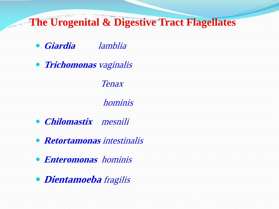
The Urogenital & Digestive Tract Flagellates
Giardia lamblia
Trichomonas vaginalis
Tenax
hominis
Chilomastix mesnili
Retortamonas intestinalis
Enteromonas hominis
Dientamoeba fragilis

Giardiasis
Cause: Giardia lamblia
A flagellate of class Mastigophora
The trophozoite has four pairs of flagella & two nuclei;
giving it a face-like appearance
Grows in the intestinal tract
Cysts are shed in the feces
Transmission and Symptoms
Transmitted via contaminated water & food
Foul-smelling profuse diarrhea
Sometimes chronic
Often misdiagnosed
Diagnosis via microscopic examination, Enterotest capsule

Trichomonas vaginalis (urogenital pathogen)
one of most common STDs
women: itching, discharge, dysuria
men: generally asymptomatic
keep specimens moist for motility
Wet mount: 5-18 um, look for jerky motility
Culture - most sensitive, takes up to 7 days

Trichomoniasis
Cause: Trichomonas vaginalis
Flagellate of the class Mastigophora
No cyst stage
Transmission & Symptoms
Transmitted via sexual contact
Genital itching
Painful urination with a white, mucoid
discharge
Occasional reduction of sperm count or erosion
of the cervix

Chilomastix mesnili (non-pathogen)
World-wide distribution
Found in chimpanzees and monkeys
Trophozoite
pear shaped
10-20 mu by 3-10 mu
elongated cytostome
Cyst - lemon shaped with anterior protrusion

Dientamoeba fragilis (pathogen)
Transmission - no cyst form (oral/fecal route unlikely)
Incidence 9x higher if E. vermicularis infection
Usually asymptomatic but can be carrier
50-80% binucleate, 4-8 karyosomes, no chromatin, 5-12 um
Food vacuoles in cytoplasm

Balantidium coli (pathogen)
only infectious ciliate (covers surface)
largest protozoan
generally asymptomatic or self-limiting diarrhea, vomiting
Trophozoite - oval, vacuoles in cytoplasm
Cyst - 45-75 um; 2 kidney-shaped nuclei
Symptoms: mostly asymptomatic or similar to amebasis
Reservoirs: Human and animals (pigs, himpanzees,…)
Transmission: waterborne, foodborne
Prevention: personal hygiene, water treatment, food safety program

Balantidiasis
Cause: Balantidium coli
Ciliated, in Class Ciliophora
Trophozoites grow in the intestinal tract
Cysts are shed in the feces and may remain
embedded in intestinal walls, causing chronic
infections
Transmission & Symptoms
Contaminated water & food
Ulceration in intestines
Profuse diarrhea


Coccidia
• characterized by thick-walled oocysts excreted in feces
In Humans
• Cryptosporidium
• Isospora
• Cyclospora
• Sarcocystis
• Toxoplasma

Cryptosporidium• 4-5 mm oocysts
• 4 sporozoites
• no sporocysts
Cyclospora• 8-10 mm oocyts
• 2 sporocysts
• 2 sporozoites each
Isospora belli• 30 x 12 mm oocyts
• 2 sporocysts
• 4 sporozoites each

Cryptosporidium• 4-5 mm oocysts
• 4 sporozoites
• no sporocysts
Cyclospora• 8-10 mm oocyts
• 2 sporocysts
• 2 sporozoites each
Isospora belli• 30 x 12 mm oocyts
• 2 sporocysts
• 4 sporozoites each

Cryptosporidium• 4-5 mm oocysts
• 4 sporozoites
• no sporocysts
Cyclospora• 8-10 mm oocyts
• 2 sporocysts
• 2 sporozoites each
Isospora belli• 30 x 12 mm oocyts
• 2 sporocysts
• 4 sporozoites each

Coccidian protozoa
Located worldwide
Primarily infects small intestine
Transmission
Fecal-oral Person-to-person
Animal-to-person
Waterborne
Foodborne
• self-limiting diarrhea in immunocompetent persons
• watery diarrhea associated with AIDS (life threatening)
Cryptosporidium parvum

Cryptosporidiosis
Cause: Cryptosporidium species
In class Sporozoa
A common protozoan parasite in humans
Transmission and Symptoms
Contaminated water (waterborne disease outbreak)
Mild diarrhea in non-immunosuppressed persons
Severe diarrhea in immunosuppressed persons
Is a minute coccidian parasite with worldwide distribution.
20 species of the parasite have been described from a varietyof vertebrates including mammals, birds, reptiles and fish.

Pathogenesis
oocysts infective when passed
+/- auto-infection if cysts rupture in gut
Clinical infection - immunocompetent: rapid but self-
limiting diarrhea and cramps
AIDS patient
life-threatening diarrhea (3-17 L/day)
electrolyte imbalance
possible malabsorption due to injury to villi
Modified Ziehl-Nielsen acid-fast stain
oocyst: red, 4-6 um, yeast: green
Cryptosporidiosis

Cryptosporidium Life Cycle
• Infectious form = oocyst
• Sporozoites ‘invade’ intestinal
epithelial cells
• Merogony
• produce merozoites
• Gametogony
• produce micro- and
macrogametes
• Sporogony
• produce sporozoites
• completed on host cell
• thin (autoinfection) or thick
walled oocysts

Molecular Epidemiology
2 major genotypes identified:
• genotype 1 (C. hominis)• only human sources
• non-infective for mice or calves
• anthroponotic transmission
• genotype 2 (C. parvum)• human and bovine sources
• infective for mice and calves
• zoonotic transmission

Epidemiology
Cosmopolitan ( A serious problem in the warmer ports).
The children are more commonly infected than are adults and that non-breast-fed infants have more cryptosporidiosis than breast-fed infants.
Immune deficiency increase risk of disease
Infection is by fecal-oral contamination
A number of animals can serve as reservoirs of infection
Important for travelers (One of the causative travelers diarrhea & in
boarding house).
Transmited to human by infected water & food & any contaminated
sources
Cryptosporidiosis:

Symptoms & pathgenesis
- In immune competent persons
Self-limited diarrhea that usually lasts about 2 weeks
& less commonly is accompanied by Abdominal discomfort, Anorexia, Fever, Nausea, Weight loss
- Immunodeficient patients
typically have severe diarrhea some times 17 liter water loss/days accompanied by these symptoms.
- In patients with AIDS
may couse life-threatening disease and has been found in sputum in lang biopsy material and in the biliary tract and has been associated with malabsorption.
Cryptosporidiosis:

Diagnosis
Finding oocyst in fecal material
Acid fast modified stains
Oocyst:
- 4-5
- Round
- Without Sporocyst
- With 4 Sporozoite are passed in the feces
Concentration procedures to improve sensitivity:
Sucrose solution or Sheather, s technique
Formalin-ether (Formalin ethyl asetate )
Cryptosporidiosis:

Prevention
Prevention: personal hygiene, surface disinfection, water treatment,
food safety program
Especial in AIDS disease:
Boil drinking water for 1 minue
Filter drinking water with devices that remove particles 1m and
larger
Use bottled drinking water, especially water obtained from
underground sources, i.e., springs or welds which are less likely to be
contaminated by Cryptosporidium oocysts
Diease is self-limited infection in immunocompetent patients
In AIDS patients: Oral paromomycin 1gr 3times/days, 2 weeksappears to suitable
Treatment
Cryptosporidiosis:

Clinical infection same as C. parvum
Life cycle similar to C. parvum except not infective for 24 - 48
hours
Disease - AIDS patients
Diarrhea and weight loss for immunodeficient & suppressed
persistent diarrhea that leads to dehydration and electrolyte
imbalance, which can be fatal
most infections are asymptomatic & self-limiting
Isosporiasis

• wide geographical distribution (higher prevalence in warmer
climates)
• monoxenous, probably not zoonosis
• invades intestinal epithelial cells
• often asymptomatic
• symptoms range from mild gastro-intestinal distress to severe
dysentery
• often self-limiting, but can become chronic (wasting,
anorexia)
• symptoms more severe in AIDS patients
Isosporiasis

Isosporiasis: Pathology / clinical symptoms
Most infections are asymptomatic and self-limited
Symptomatic disease in immune competent individualsincludes diarrhea and malabsorption
AIDS patients have severe persistent diarrhea that leadsto dehydration and electrolyte imbalance, which can befatal
Mucosal epithelial cells in which I. belli develop rupturewhen the organsims are released creating the symptomsdescribed above

Wet preps
Modified acid-fast stain
Permanent smears: best if preserved in
SAF
10% formalin
Mature oocyst: Oval, 20-30 um with 2 sporocysts
Isosporiasis: Diagnosis

Cyclospora
• Infection with cyclospora spp. is widely distributed probably cosmopolitan.
• Countries initially identified as having endemic cyclosporiasis: Haiti,
Guatemala, Peru and Nepal
• Infection most common in HIV/AIDS patients.
• Also important cyclosporosis in travelers.
• Associated with imported food items, specially raspberries and green leafy
vegetables such as basil and mesclun lettuce.

Pathology
• Cyclospora infects enterocytes of the small
bowel where various stages, sexual and
asexual stages have been observed. Villous
blunting, mild crypt hyperplasi.
• The main symptom is watery diarrhea, loss
of appetite, weight loss, abdominal bloating
and cramping, nausea, fatigue and low
grade fever.
• Incubation period averages one week and
illness lasts 6 weeks.
• In the immunocompromised patient, severe
diarrhea can last up to 4 months or longe.
• Extra-intestinal infection appears to be
more common in AIDS patients
• symptoms similar to
Cryptosporidium and Isospora
• watery diarrhea/frequent
stools
• 1-2 week duration typical
• relapses over 1-2 months
Clinical Features

Epidemiology
• Food-borne pathogen
• Not sure if water could also have a role in transmission
• Infected humans are the only known sources of oocysts
• Not other reservoir host identified
• No methods available to grow them in the lab
•Marked seasonality of cyclospora: temp, humidity, may facilitate sporulation
• associated with food-borne outbreaks
• oocysts detected on market vegetables in Peru
• presumed source: contaminated water or human waste as fertilizer
Cyclospora:

Diagnosis and treatment
• Detection of oocysts in stool sample by microscopy
• Recovery of oocysts in intestinal fluid or small bowel biopsy
specimens
•Demonstration of oocyst sporulation
• PCR amplification of Cyclospora DNA
• Can be successfully treated with trimethoprim (TMP)-
sulfamethoxasole: TMP 160 mg/ SMX 800 mg bid × 7-10d
Cyclospora:

Sarcocystis
rare human infection
heteroxenous parasite
predator-prey life cycle
humans support both stages
taxonomic confusion
generally named after host species
Sarcocystis bovihominis
Sarcocystis suihominis
ingest undercooked meat
transient mild to severe diarrhea
excrete sporulated sporocysts
13x10 mm
4 sporozoites

Blastocystis hominis (Possible Pathogen)
one of the most common parasites found in the human
intestinal tract (resides in the colon and cecum).
a nonpathogenic protozoan parasite may be associated with
clinical illnesses such as diarrhea, abdominal pain, abdominal
ballooning, anorexia, vomiting, and urticaria
Controversial: Some labs evaluate when
symptoms with no other enteric pathogens
5-15 um, cytoplasm - dark green, nuclei - dark purple/black

B. hominis reproduces asexually, most likely by binary fission.
B. hominis does not have a cell wall and therefore there is great
variation in its size and shape, ranging from 5 to 40 μm.
Multiple forms of B. hominis have been described in culture including
amoeboid, vacuolar, granular.
The amoeboid form is more likely associated with disease.
Acquisition of B. hominis is thought to occur as a result of frequent
animal–human, human–human and human–animal transmission.
Blastocystis hominis (Possible Pathogen)