Esophagogastric junction distensibility assessed with an endoscopic functional luminal imaging probe...
16
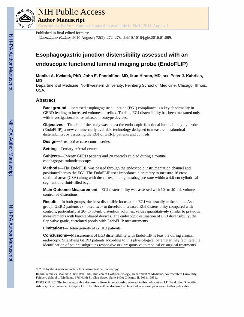
Esophagogastric junction distensibility assessed with an endoscopic functional luminal imaging probe (EndoFLIP) Monika A. Kwiatek, PhD, John E. Pandolfino, MD, Ikuo Hirano, MD, and Peter J. Kahrilas, MD Department of Medicine, Northwestern University, Feinberg School of Medicine, Chicago, Illinois, USA. Abstract Background—Increased esophagogastric junction (EGJ) compliance is a key abnormality in GERD leading to increased volumes of reflux. To date, EGJ distensibility has been measured only with investigational barostatbased prototype devices. Objectives—The aim of the study was to test the endoscopic functional luminal imaging probe (EndoFLIP), a new commercially available technology designed to measure intraluminal distensibility, by assessing the EGJ of GERD patients and controls. Design—Prospective case-control series. Setting—Tertiary referral center. Subjects—Twenty GERD patients and 20 controls studied during a routine esophagogastroduodenoscopy. Methods—The EndoFLIP was passed through the endoscopic instrumentation channel and positioned across the EGJ. The EndoFLIP uses impedance planimetry to measure 16 cross- sectional areas (CSA) along with the corresponding intrabag pressure within a 4.6-cm cylindrical segment of a fluid-filled bag. Main Outcome Measurement—EGJ distensibility was assessed with 10- to 40-mL volume- controlled distentions. Results—In both groups, the least distensible locus at the EGJ was usually at the hiatus. As a group, GERD patients exhibited two- to threefold increased EGJ distensibility compared with controls, particularly at 20- to 30-mL distention volumes, values quantitatively similar to previous measurements with barostat-based devices. The endoscopic estimation of EGJ distensibility, the flap valve grade, correlated poorly with EndoFLIP measurements. Limitations—Heterogeneity of GERD patients. Conclusions—Measurement of EGJ distensibility with EndoFLIP is feasible during clinical endoscopy. Stratifying GERD patients according to this physiological parameter may facilitate the identification of patient subgroups responsive or unresponsive to medical or surgical treatments. © 2010 by the American Society for Gastrointestinal Endoscopy Reprint requests: Monika A. Kwiatek, PhD, Division of Gastroenterology, Department of Medicine, Northwestern University, Feinberg School of Medicine, 676 North St. Clair Street, Suite 1400; Chicago, IL 60611-2951.. DISCLOSURE: The following author disclosed a financial relationship relevant to this publication: J.E. Pandolfino Scientific Advisory Board member, Crospon Ltd. The other authors disclosed no financial relationships relevant to this publication. NIH Public Access Author Manuscript Gastrointest Endosc. Author manuscript; available in PMC 2011 August 1. Published in final edited form as: Gastrointest Endosc. 2010 August ; 72(2): 272–278. doi:10.1016/j.gie.2010.01.069. NIH-PA Author Manuscript NIH-PA Author Manuscript NIH-PA Author Manuscript
-
Upload
independent -
Category
Documents
-
view
0 -
download
0
Transcript of Esophagogastric junction distensibility assessed with an endoscopic functional luminal imaging probe...
Esophagogastric junction distensibility assessed with anendoscopic functional luminal imaging probe (EndoFLIP)
Monika A. Kwiatek, PhD, John E. Pandolfino, MD, Ikuo Hirano, MD, and Peter J. Kahrilas,MDDepartment of Medicine, Northwestern University, Feinberg School of Medicine, Chicago, Illinois,USA.
AbstractBackground—Increased esophagogastric junction (EGJ) compliance is a key abnormality inGERD leading to increased volumes of reflux. To date, EGJ distensibility has been measured onlywith investigational barostatbased prototype devices.
Objectives—The aim of the study was to test the endoscopic functional luminal imaging probe(EndoFLIP), a new commercially available technology designed to measure intraluminaldistensibility, by assessing the EGJ of GERD patients and controls.
Design—Prospective case-control series.
Setting—Tertiary referral center.
Subjects—Twenty GERD patients and 20 controls studied during a routineesophagogastroduodenoscopy.
Methods—The EndoFLIP was passed through the endoscopic instrumentation channel andpositioned across the EGJ. The EndoFLIP uses impedance planimetry to measure 16 cross-sectional areas (CSA) along with the corresponding intrabag pressure within a 4.6-cm cylindricalsegment of a fluid-filled bag.
Main Outcome Measurement—EGJ distensibility was assessed with 10- to 40-mL volume-controlled distentions.
Results—In both groups, the least distensible locus at the EGJ was usually at the hiatus. As agroup, GERD patients exhibited two- to threefold increased EGJ distensibility compared withcontrols, particularly at 20- to 30-mL distention volumes, values quantitatively similar to previousmeasurements with barostat-based devices. The endoscopic estimation of EGJ distensibility, theflap valve grade, correlated poorly with EndoFLIP measurements.
Limitations—Heterogeneity of GERD patients.
Conclusions—Measurement of EGJ distensibility with EndoFLIP is feasible during clinicalendoscopy. Stratifying GERD patients according to this physiological parameter may facilitate theidentification of patient subgroups responsive or unresponsive to medical or surgical treatments.
© 2010 by the American Society for Gastrointestinal EndoscopyReprint requests: Monika A. Kwiatek, PhD, Division of Gastroenterology, Department of Medicine, Northwestern University,Feinberg School of Medicine, 676 North St. Clair Street, Suite 1400; Chicago, IL 60611-2951..DISCLOSURE: The following author disclosed a financial relationship relevant to this publication: J.E. Pandolfino ScientificAdvisory Board member, Crospon Ltd. The other authors disclosed no financial relationships relevant to this publication.
NIH Public AccessAuthor ManuscriptGastrointest Endosc. Author manuscript; available in PMC 2011 August 1.
Published in final edited form as:Gastrointest Endosc. 2010 August ; 72(2): 272–278. doi:10.1016/j.gie.2010.01.069.
NIH
-PA Author Manuscript
NIH
-PA Author Manuscript
NIH
-PA Author Manuscript

Excessive esophagogastric junction (EGJ) compliance is a primary pathophysiologicalabnormality in many cases of GERD.1-4 Increased EGJ compliance allows greater volumesof gastric content to reflux into the esophagus,5 increases the frequency with which transientlower esophageal sphincter (LES) relaxations are elicited by proximal gastric distention,6and allows gastric juice to track within the contracted sphincter.7-9 These physiologicalaberrations result in an increased number of reflux events, increased spatial distribution ofrefluxate within the esophagus, and increased esophageal acid exposure, all of whichincrease the likelihood of esophageal mucosal injury and reflux-related symptoms. Hence,quantifying EGJ compliance may discern clinically relevant subsets of GERD patients. Thecurrent clinical assessment of EGJ function is limited to endoscopic or radiographicimaging, manometric measures, or reflux testing with pH-metry. Although these studies canconfirm excessive reflux, contractile defects in the LES pressure or crural diaphragm, andmisalignment between the 2 manifest as a sliding hiatal hernia, which are all common inGERD,3,10,11 they do not quantify EGJ compliance. Inherent in the definition (change involume when subject to an applied force), measuring EGJ compliance requires it beingchallenged with intraluminal distention.12
To date, the EGJ distensibility has been measured only with purpose-built equipmentprototypes within the domain of research laboratories, which found the narrowest locus to begreater in GERD patients compared with control subjects.3,4 Although a transorally placedesophageal hydrostat or barostat can apply distending pressure at the EGJ and thus restrictdimensional measurements to the area of interest, both techniques are cumbersome andrequire concurrent fluoroscopic imaging.2-4 A more robust method for measuring EGJdistensibility, providing cross-sectional area (CSA) measurements at multiple adjacentsegments without the need for fluoroscopy, adapts the principle of impedanceplanimetry13,14 into a functional luminal imaging probe (FLIP). FLIP recordings allowdynamic imaging of EGJ distention as a cylinder of varying diameter based on instantaneousCSA measurements with concurrent pressure measurements. Quantifying intraluminalpressure along with the CSA permits calculation of EGJ distensibility.15-17 The originalFLIP, designed for transnasal passage, was of relatively large diameter and too poorlytolerated for general clinical use. Consequently, a new smaller probe was designed to fitthrough the instrumentation channel of an endoscope. The distention bag can be localized atthe EGJ for compliance measurements during routine clinical endoscopy, opening thepossibility of measuring EGJ compliance as part of the diagnostic evaluation of GERD.Hence, the aim of the current study was to test the performance of the new endoscopic FLIPsystem (EndoFLIP; Crospon Ltd, Galway, Ireland) and to compare the EGJ distensibility inGERD patients with that of asymptomatic control subjects.
MATERIALS AND METHODSSubjects
Twenty control subjects (4 men; age, 18-42; median age, 27 years) and 20 patients withGERD (10 men; age, 22-79 years; median age, 31 years) were studied. The control subjectswere recruited from a pool of asymptomatic volunteers with no GERD-associatedsymptoms. GERD patients were recruited from a pool of volunteers and patients reportingheartburn at least 2 to 3 days per week while not taking antisecretory medicines. None of thesubjects had a history of GI surgery or significant medical disease. All subjects gave writteninformed consent. The study protocol was approved by the Northwestern UniversityInstitutional Review Board.
Kwiatek et al. Page 2
Gastrointest Endosc. Author manuscript; available in PMC 2011 August 1.
NIH
-PA Author Manuscript
NIH
-PA Author Manuscript
NIH
-PA Author Manuscript

Take-home Message
• The EndoFLIP device yielded values for EGJ distensibility quantitativelysimilar to previous measurements with barostat-based devices. The leastdistensible locus within the EGJ was usually at the hiatus and was 2 to 3 timesmore distensible in GERD patients than controls. Stratifying GERD patients byEGJ distensibility can potentially match GERD patient subgroups to specificmechanistic treatments.
EndoFLIP systemEGJ distensibility was measured by using a commercially developed FLIP, the EndoFLIP,based on the concepts described previously.16 In brief, the EndoFLIP (Fig. 1) usesimpedance planimetry to determine multiple adjacent CSAs within a cylindrical bag placedin a tubular organ during volumetric distention. The additional measure of the correspondingintrabag pressure allows assessment of the CSA pressure response (distensibility) of thedistended area.
The EndoFLIP assembly was 240 cm long with a 3-mm outer diameter. A noncompliant bag(up to a fill-in volume of 40 mL) mounted on the distal 14 cm of the probe was designed toassume a cylindrical shape 7.0 cm long with maximal diameter of 2.5 cm, when fullydistended, between the tapering cone-shaped ends sealed at the assembly. The minimal-to-maximal detectable CSA range was 10 to 450 mm2. Within the bag was a 6.4-cm segmentcomprising 17 ring electrodes spaced 4 mm apart for impedance planimetry measurements.Excitation electrodes at either end emitted a constant low current making the voltagemeasured across each of the 16 adjacent pairs of ring electrodes proportional to theimpedance between them. As the bag was filled with a specially formulated conductivesolution, the impedance across each segment was inversely proportional to the CSA of thebag at that locus. The probe also contained 2 low-compliance perfusion channels connectedto external pressure transducers housed within the recording unit that measured intrabagpressure.
Measurements from the 16 electrode pairs and pressure transducers were sampled at 10 Hzwith the data acquisition system and transmitted to the recording unit, which displayed themin real time as a 6.4-cm cylinder of varying diameters along its length reflecting the 16measured CSAs. The probe and the pressure transducers were precalibrated during themanufacturing process and required no additional calibration before use (CSA resolution 0.8mm2, accuracy ±0.8 mm2; intrabag pressure resolution 0.1 mm Hg, accuracy ±0.8 mm Hg).Before use, the air was removed from the EndoFLIP assembly by using an automated purgesequence. Subsequent establishment of pressure baseline and infusion of the conductivesolution were controlled by using the touch screen on the recording unit positioned at thelevel of the subject's torso.
EndoscopySubjects underwent esophagogastroduodenoscopy in the left lateral decubitus position toevaluate for esophagitis and EGJ anatomy and to place the EndoFLIP probe across the EGJ.A therapeutic gastroscope with an 11.3-mm diameter with a 3.7-mm diameter instrumentchannel was used (Olympus GIF type 1TQ160; Olympus Corp, Tokyo, Japan). Moderatesedation was administered during the procedure (5-15 mg midazolam, 20-200 μg fentanylwith similar doses in each subject group). Esophagitis was graded according to the LosAngeles classification where mucosal breaks were less (A) than 5 mm long, but notextending between the tops of 2 mucosal folds; (B) more than 5 mm long, but not extending
Kwiatek et al. Page 3
Gastrointest Endosc. Author manuscript; available in PMC 2011 August 1.
NIH
-PA Author Manuscript
NIH
-PA Author Manuscript
NIH
-PA Author Manuscript

between the tops of 2 mucosal folds; (C) continuous between the tops of 2 or more mucosalfolds, but involving less than 75% of the circumference; and (D) involving 75% or more ofthe esophageal circumference.18 EGJ morphology was graded according to the “flap valve”concept: (I) a normal ridge of tissue closely approximated to the gastroscope, (II) the ridgeslightly less well defined and opening with respiration, (III) the ridge effaced and the hiatuspatulous, and (IV) the hiatus open at all times and the squamocolumnar junction displacedaxially.19
EndoFLIP protocolAt the end of the endoscopy, the EndoFLIP probe was threaded through the instrumentationchannel until the entire bag was seen within the stomach. The air insufflated duringendoscopy was removed and the intrabag pressure was zeroed within the stomach; therefore,subsequent pressure measurements were relative to the baseline gastric pressure. Theendoscope and probe were withdrawn such that its bag straddled the EGJ and the endoscopetip remained in the distal esophagus. CSA and the corresponding distention pressuremeasurements were made with the bag filled to 10, 20, 30, and 40 mL. EndoFLIPmeasurements were monitored in real-time to ensure proper bag placement by using both thedirect visualization through the endoscope and the display of CSAs on the recording unit. Ifthe bag migrated, it was repositioned, and the measurement was repeated. Measurementsinterrupted by esophageal peristalsis were also repeated.
Data analysisThe 16 CSAs and intrabag pressure were assessed at each distention volume by quantifyingthe 50th percentile of each measure during each 30-second test recording.
EGJ distensibility (CSA vs pressure) was based on the narrowest CSA (invariably thediaphragmatic hiatus) and the corresponding intrabag pressure and expressed as the EGJdistensibility index at each distention volume. To account for the data with 0-mm Hgintrabag pressure, the index was calculated as [narrowest CSA/(intrabag pressure +intragastric pressure offset)], where intragastric pressure was given as 4.0 mm Hg based onprevious reports of typical normal intragastric pressure values across healthy control andGERD patient groups.3
Statistical analysisData were expressed as median (5th-95th percentiles). Statistical comparisons usedWilcoxon matched pairs and Kruskal-Wallis tests. The bivariate relationship betweengastroesophageal flap valve grade (categorical variable) and hiatal CSA or EGJ distensibilityindex (continuous variable) was assessed by using the Kruskal-Wallis test. Significance wasset at P < .05.
RESULTSDemographic data
Demographic data are summarized in Table 1. Eight of the 20 GERD patients hadesophagitis: 5 Los Angeles grade A, 2 grade B, and 1 grade C. Surprisingly,gastroesophageal flap valve grade III was the most common grade reported in both thecontrols (10/20) and the GERD patients (11/20). None of the controls had a grade IV flapvalve indicative of hiatal hernia. Esophageal acid exposure had been assessed in 4 of thepatients with 2 having abnormal values (total percentage time pH <4>, 5%); neither hadesophagitis.
Kwiatek et al. Page 4
Gastrointest Endosc. Author manuscript; available in PMC 2011 August 1.
NIH
-PA Author Manuscript
NIH
-PA Author Manuscript
NIH
-PA Author Manuscript

Patients were included in the study based on the criterion of experiencing heartburn at least2 to 3 days per week while not taking antisecretory medications. Additional symptomspotentially related to reflux included regurgitation (2/20), chest pain (1/20), cough (2/20),throat burning (2/20), and globus sensation (1/20). The majority of the patients (16/20) wereon long-term proton pump inhibitor therapy: esomeprazole (9), omeprazole (4), rabeprazole(2), and lansoprazole (1), which provided adequate symptomatic relief. A minority of thepatients opted to control their symptoms with over-the-counter calcium carbonate productsinstead (2/16) or remain off any medications despite having favorable experience with themin the past (2/16).
Hence, GERD diagnosis was based on objective evidence (esophagitis or positive pH-metry)in 10 of 20 patients and the combination of symptoms and response to antisecretory therapyin 10 of 20 patients.
EGJ distensibilityThe EndoFLIP bag assumed an hourglass shape when distended straddling the EGJ with thecentral constriction at the diaphragmatic hiatus in both control subjects and GERD patients(Fig. 2). The hiatus was typically the least distensible locus with the majority of theconductive solution within the bag displaced proximally or distally to it. The hiatal CSAincreased progressively with distending volumes (P < .0001) to a similar degree in bothgroups (P = .68, Table 2). The hiatal CSA was similar in patients with objectivelydocumented and symptomatically defined GERD (P = .46). However, the hourglass shapewas absent in 2 of 20 control subjects (both with gastroesophageal flap valve grade III) and6 of 20 GERD patients (1 gastroesophageal flap valve grade II, 4 grade III, 1 grade IV). Thebag instead assumed a cylindrical shape of uniform diameter. A “double” hourglass shapewith 2 constriction loci was not seen.
As the hiatal diameter increased with increasing distention volume, so did the pressurewithin the bag in both subject groups (P < .0001, Table 3). However, unlike the hiataldiameter, the distending pressure was consistently lower in GERD patients than in controlsubjects, particularly at 20-, 30-, and 40-mL bag volumes (P < .0005, Table 3). At any givendistensile pressure, the extent of the EGJ opening in GERD patients was greater than incontrol subjects (Fig. 3). The intrabag pressure in patients with objectively documented orsymptomatically defined GERD was comparable (P = .47).
The data in Figure 3 were used to calculate an EGJ distensibility index (defined in the dataanalysis section). The index was significantly greater in the GERD patients (P = .01, Fig. 4).This difference was most prominent at the 20-mL (controls 2 [1-9] mm2/Hg vs GERD 7[1-32] mm2/Hg, P = .02) and 30-mL distention volumes (controls 4 [1-14] mm2/Hg vsGERD 8 [1-46] mm2/Hg, P = .04) at which the index was three- and twofold greater,respectively. As with other measures, the EGJ distensibility index was similar for theobjectively documented or symptomatically defined GERD subgroups (P = .88).
The agreement between endoscopically assessed EGJ morphology and EndoFLIP measureswas tested with pooled data for control and GERD subjects. There was no relationshipbetween the flap valve grade and either the hiatal CSA (P ≥ .62) or the EGJ distensibilityindex (P ≥ .17) at 20- and 30-mL distention volumes (Fig. 5).
DISCUSSIONThis investigation tested the ability of the EndoFLIP, a novel device based on impedanceplanimetry technology, to quantify EGJ distensibility in the course of routine endoscopy.Studies were done on 20 control subjects and 20 GERD patients. The major findings were
Kwiatek et al. Page 5
Gastrointest Endosc. Author manuscript; available in PMC 2011 August 1.
NIH
-PA Author Manuscript
NIH
-PA Author Manuscript
NIH
-PA Author Manuscript

that (1) EndoFLIP provided technically successful measurements in all cases; (2) in bothsubject groups, the least distensible locus within the EGJ (usually the hiatus) was mostsuccessfully isolated with 20- to 30-mL distentions; (3) consistent with previousinvestigations that used prototype barostat-based technology, EGJ distensibility wassignificantly greater in GERD patients than in control subjects; and (4) the nearestendoscopic gauge of EGJ distensibility, the flap valve grade, correlated poorly withEndoFLIP measurements.
The poor correlation between the endoscopic and EndoFLIP estimates of EGJ distensibility(Fig. 5) was somewhat unexpected. The endoscopic assessment is highly qualitative, anddistending pressure is uncontrolled during endoscopy; still, one would have expected acorrelation. This raises the issue of which measure is correct. Addressing that concern, it isuseful to compare the EndoFLIP measurements with previous findings made with hydrostatdevice pressure-controlled distentions in similar groups of GERD and control subjects.3,4The similarities are particularly apparent in Figure 3. In the hydrostat study, at the maximaldistention pressure used (6 mm Hg relative to gastric pressure), the hiatal CSA was 38 ± 5mm2 in controls, and 79 ± 11 mm2 in GERD patients. Extrapolating from Figure 3, thecorresponding EndoFLIP measurements referenced to intragastric pressure would be 40mm2 for control and 80 mm2 for GERD subjects, remarkably similar numbers. Theseopening diameters are less than the diameter of the endoscope used in the current study andemphasize the nonphysiologic circumstances implicit in endoscopic assessment.
Increased EGJ distensibility has major repercussions on the volume of reflux. Whetherreflux events are attributable to transient LES relaxation or otherwise, there is a very robustrelationship in which the volume of flow through the EGJ is proportional to the openingdiameter raised to the fourth power.3,4 Clinical and scientific opinion20-24 suggests this tobe an important distinction that potentially identifies a GERD subgroup with syndromesdriven by large reflux volumes (regurgitation and chest pain) that may benefit fromanatomic correction, by either surgical or evolving endoluminal techniques. It may alsoisolate a GERD subgroup with normal EGJ distensibility and little to gain from suchinterventions. Similarly, EndoFLIP measurements may be potentially useful in the course ofsurgical or endoluminal procedures to calibrate the magnitude of the intervention. That isnot to say that EndoFLIP measurements, or any other putative measure, will represent asingle unifying physiological feature defining GERD. GERD is recognized as aheterogeneous condition, and Figure 4 confirms this once again with respect to the metric ofEGJ distensibility. When we used pooled data, the distensibility index in GERD patients wasdistinctly different from that of controls, but it also varied substantially among GERDpatients.
In conclusion, this experiment appraised the ability of EndoFLIP, a new commerciallyavailable technology, to evaluate EGJ distensibility in the course of clinical endoscopy. Thedevice performed well, both confirming previously described differences in EGJdistensibility between GERD patients and controls and producing results that werequantitatively very similar to measurements made with cumbersome purpose-built barostat-based prototypes requiring concurrent fluoroscopy. As anticipated, GERD patients wereheterogeneous with respect to EGJ distensibility, but stratifying them according to thisphysiological parameter may identify those uniquely responsive to endoluminal or surgicaltherapies. Further studies, especially management trials based on EndoFLIP measures, willbe needed to ascertain the degree to which that is possible.
Kwiatek et al. Page 6
Gastrointest Endosc. Author manuscript; available in PMC 2011 August 1.
NIH
-PA Author Manuscript
NIH
-PA Author Manuscript
NIH
-PA Author Manuscript

AcknowledgmentsSupported and sponsored by Crospon Ltd, Galway, Ireland; grant R01 DC00646 (P.J. Kahrilas and J.E. Pandolfino)from the Public Health Service; and the 2008 AGA June and Donald O. Castell Esophageal Clinical ResearchAward (J.E. Pandolfino).
Abbreviations
CSA cross-sectional area
EGJ esophagogastric junction
FLIP functional luminal imaging probe
LES lower esophageal sphincter
REFERENCES1. Jenkinson AD, Scott SM, Yazaki E, et al. Compliance measurement of lower esophageal sphincter
and esophageal body in achalasia and gastroesophageal reflux disease. Dig Dis Sci 2001;46:1937–42. [PubMed: 11575446]
2. Pandolfino JE, Curry J, Shi G, et al. Restoration of normal distensive characteristics of theesophagogastric junction after fundoplication. Ann Surg 2005;242:43–8. [PubMed: 15973100]
3. Pandolfino JE, Shi G, Trueworthy B, et al. Esophagogastric junction opening during relaxationdistinguishes nonhernia reflux patients, hernia patients, and normal subjects. Gastroenterology2003;125:1018–24. [PubMed: 14517784]
4. Pandolfino JE, Shi G, Curry J, et al. Esophagogastric junction distensibility: a factor contributing tosphincter incompetence. Am J Physiol Gastrointest Liver Physiol 2002;282:G1052–8. [PubMed:12016131]
5. Ghosh SK, Kahrilas PJ, Brasseur JG. Liquid in the gastroesophageal segment promotes reflux, butcompliance does not: a mathematical modeling study. Am J Physiol Gastrointest Liver Physiol2008;295:G920–33. [PubMed: 18718998]
6. Kahrilas PJ, Shi G, Manka M, et al. Increased frequency of transient lower esophageal sphincterrelaxation induced by gastric distention in reflux patients with hiatal hernia. Gastroenterology2000;118:688–95. [PubMed: 10734020]
7. Pandolfino JE, Zhang Q, Ghosh SK, et al. Acidity surrounding the squamocolumnar junction inGERD patients: “acid pocket” versus “acid film.”. Am J Gastroenterol 2007;102:2633–41.[PubMed: 17714553]
8. Fletcher J, Wirz A, Young J, et al. Unbuffered highly acidic gastric juice exists at thegastroesophageal junction after a meal. Gastroenterology 2001;121:775–83. [PubMed: 11606490]
9. Fletcher J, Wirz A, Henry E, et al. Studies of acid exposure immediately above the gastro-oesophageal squamocolumnar junction: evidence of short segment reflux. Gut 2004;53:168–73.[PubMed: 14724145]
10. Kahrilas PJ, Lin S, Manka M, et al. Esophagogastric junction pressure topography afterfundoplication. Surgery 2000;127:200–8. [PubMed: 10686986]
11. Lord RV, DeMeester SR, Peters JH, et al. Hiatal hernia, lower esophageal sphincter incompetence,and effectiveness of Nissen fundoplication in the spectrum of gastroesophageal reflux disease. JGastrointest Surg 2009;13:602–10. Epub 2008 Dec 3. [PubMed: 19050984]
12. Harris LD, Pope CE 2nd. “Squeeze” vs. resistance: an evaluation of the mechanism of sphinctercompetence. J Clin Invest 1964;43:2272–8. [PubMed: 14234823]
13. McMahon BP, Drewes AM, Gregersen H. Functional oesophago-gastric junction imaging. World JGastroenterol 2006;12:2818–24. [PubMed: 16718804]
14. Gregersen, H. Biomechanics of the gastrointestinal tract: new perspectives in motility research anddiagnostics. Springer-Verlag; Heidelberg: 2003.
Kwiatek et al. Page 7
Gastrointest Endosc. Author manuscript; available in PMC 2011 August 1.
NIH
-PA Author Manuscript
NIH
-PA Author Manuscript
NIH
-PA Author Manuscript

15. McMahon BP, Frokjaer JB, Kunwald P, et al. The functional lumen imaging probe (FLIP) forevaluation of the esophagogastric junction. Am J Physiol Gastrointest Liver Physiol2007;292:G377–84. [PubMed: 16950760]
16. McMahon BP, Frokjaer JB, Liao D, et al. A new technique for evaluating sphincter function invisceral organs: application of the functional lumen imaging probe (FLIP) for the evaluation of theoesophago-gastric junction. Physiol Meas 2005;26:823–36. [PubMed: 16088071]
17. Kwiatek MA, Kahrilas PJ, Soper NJ, et al. Esophagogastric junction distensibility afterfundoplication assessed with a novel functional luminal imaging probe. J Gastrointest Surg2009;14:268–76. [PubMed: 19911238]
18. Lundell LR, Dent J, Bennett JR, et al. Endoscopic assessment of oesophagitis: clinical andfunctional correlates and further validation of the Los Angeles classification. Gut 1999;45:172–80.[PubMed: 10403727]
19. Hill LD, Kozarek RA, Kraemer SJ, et al. The gastroesophageal flap valve: in vitro and in vivoobservations. Gastrointest Endosc 1996;44:541–7. [PubMed: 8934159]
20. Kahrilas PJ. Clinical practice. Gastroesophageal reflux disease. N Engl J Med 2008;359:1700–7.[PubMed: 18923172]
21. Lundell L. Anti-reflux surgery in the laparoscopic era. Baillieres Best Pract Res Clin Gastroenterol2000;14:793–810. [PubMed: 11003810]
22. Kahrilas PJ, Shaheen NJ, Vaezi MF. American Gastroenterological Association Institute technicalreview on the management of gastroesophageal reflux disease. Gastroenterology 2008;135:1392–413. 1413.e1-5. Epub 2008 Sep 16. [PubMed: 18801365]
23. Kahrilas PJ, Shaheen NJ, Vaezi MF, et al. American Gastroenterological Association MedicalPosition Statement on the management of gastroesophageal reflux disease. Gastroenterology2008;135:1383–91. 1391.e1-5. [PubMed: 18789939]
24. Sifrim D, Mittal R, Fass R, et al. Review article: acidity and volume of the refluxate in the genesisof gastro-oesophageal reflux disease symptoms. Aliment Pharmacol Ther 2007;25:1003–17.[PubMed: 17439501]
Kwiatek et al. Page 8
Gastrointest Endosc. Author manuscript; available in PMC 2011 August 1.
NIH
-PA Author Manuscript
NIH
-PA Author Manuscript
NIH
-PA Author Manuscript

Figure 1.The Endoscopic Functional Luminal Imaging Probe (EndoFLIP) System. Once the bag isfilled with a conductive solution, 16 intraluminal cross-sectional areas (CSA) are measuredwithin the central part of the bag by the impedance measuring electrodes, whereas thepressure transducers provide the corresponding intrabag pressure. The volume of conductivesolution injected from the syringe to the bag is controlled via the touch screen on therecording unit. The screen displays the calculated CSAs as a cylinder of varying diameter inreal time along with the corresponding intrabag pressure. There is also the option of a split-screen display, as demonstrated here, which can display a snapshot from previous distentionsimultaneously with the current acquisition. (Photograph courtesy of Crospon Ltd., Galway,Ireland.)
Kwiatek et al. Page 9
Gastrointest Endosc. Author manuscript; available in PMC 2011 August 1.
NIH
-PA Author Manuscript
NIH
-PA Author Manuscript
NIH
-PA Author Manuscript

Figure 2.Examples of a set of volumetric EndoFLIP distentions in a control subject (top) and aGERD patient (bottom). Typically the hourglass shape of the EGJ narrowed at the hiatus(white arrow) in both groups. In each panel, EGJ distention is illustrated as a cylinder ofvarying diameter corresponding to the 16 CSAs measured by impedance planimetry withinthe EndoFLIP bag along with the corresponding intrabag pressures. These examples wereselected to illustrate the greater EGJ distensibility in a GERD patient with consistently largerhiatal distention at 20- to 40-mL distention volumes coinciding with lower intrabag pressure.
Kwiatek et al. Page 10
Gastrointest Endosc. Author manuscript; available in PMC 2011 August 1.
NIH
-PA Author Manuscript
NIH
-PA Author Manuscript
NIH
-PA Author Manuscript

Figure 3.EGJ distensibility in control subjects and GERD patients. The EndoFLIP bag distensilepressure (x-axis) and the hiatal CSA (y-axis) were measured with the EndoFLIP bag filled to10 mL (squares), 20 mL (diamonds), 30 mL (circles) and 40 mL (triangles). Both controlsubjects (open symbols) and GERD patients (closed symbols) exhibited measurable EGJdistention, particularly with 20- and 30-mL EndoFLIP bag volumes. However, the intrabagpressures associated with EGJ distention were consistently 6 to 15 mm Hg less in the GERDpatients compared with the control subjects (see also Table 3). Data shown as medians.
Kwiatek et al. Page 11
Gastrointest Endosc. Author manuscript; available in PMC 2011 August 1.
NIH
-PA Author Manuscript
NIH
-PA Author Manuscript
NIH
-PA Author Manuscript

Figure 4.Esophagogastric junction distensibility index in control subjects (white boxes) and GERDpatients (gray boxes). *P = .02 versus controls.
Kwiatek et al. Page 12
Gastrointest Endosc. Author manuscript; available in PMC 2011 August 1.
NIH
-PA Author Manuscript
NIH
-PA Author Manuscript
NIH
-PA Author Manuscript

Figure 5.Endoscopic assessment of EGJ morphology versus functional EGJ assessment withEndoFLIP. The qualitative grading of EGJ patency (gastroesophageal flap valve grade19)was not related to either hiatal CSA measurement (A) or EGJ distensibility index (B) in thepooled data from control subjects and GERD patients at the distention volumes of 20 and 30mL. Bold lines denote medians.
Kwiatek et al. Page 13
Gastrointest Endosc. Author manuscript; available in PMC 2011 August 1.
NIH
-PA Author Manuscript
NIH
-PA Author Manuscript
NIH
-PA Author Manuscript

NIH
-PA Author Manuscript
NIH
-PA Author Manuscript
NIH
-PA Author Manuscript
Kwiatek et al. Page 14
TABLE 1
Subject demographics
Control subjects (n = 20) GERD patients (n = 20)
Male:female 4:16 10:10
Age, y 18-42 22-79
Gastroesophageal flap valve grade I: n = 7; II: n = 3; III: n = 10; IV: n = 0 I: n = 2; II: n = 4; III: n = 11; IV: n = 3
Esophagitis (Los Angeles grading) None 8/20
Abnormal pH study* NA 2/4
*Total percentage time pH <4>, 5%.
Gastrointest Endosc. Author manuscript; available in PMC 2011 August 1.

NIH
-PA Author Manuscript
NIH
-PA Author Manuscript
NIH
-PA Author Manuscript
Kwiatek et al. Page 15
TABLE 2
CSA (mm2) of the diaphragmatic hiatus (narrowest EGJ CSA measured by the EndoFLIP) during volumedistentions reported as median (5th to 95th percentiles)
EndoFLIP bagvolume (mL) Control subjects GERD patients
10 20 (12-110) 20 (12-119)
20 38 (13-94) 36 (13-201)
30 94 (27-225) 116 (33-344)
40 264 (99-496) 180 (86-409)
CSA, Cross-sectional area.
The minimal detectable CSA was approximately 10 mm2.
Gastrointest Endosc. Author manuscript; available in PMC 2011 August 1.

NIH
-PA Author Manuscript
NIH
-PA Author Manuscript
NIH
-PA Author Manuscript
Kwiatek et al. Page 16
TABLE 3
Pressure (mm Hg) within the EndoFLIP bag during distention reported as median (5th to 95th percentiles)
EndoFLIP bagvolume (mL)
Controlsubjects
GERDpatients
10 3 (0-8) 0.5 (0-11)
20 9 (2-20) 3 (0-9)*
30 25 (6-47) 10 (3-20)*
40 39 (17-60) 24 (13-51)*
*P < .01 versus controls.
Gastrointest Endosc. Author manuscript; available in PMC 2011 August 1.