Chagas disease in Italy: breaking an epidemiological silence

BioMed Central
Journal of Negative Results in BioMedicine
ss
Open AcceResearchAortic distensibility measured by pulse-wave velocity is not modified in patients with Chagas' diseaseHumberto Villacorta1,2, Luiz Aparecido Bortolotto*1, Edmundo Arteaga1 and Charles Mady1Address: 1Heart Institute (InCor), University of São Paulo, Medical School, São Paulo, Brazil and 2Hospital Pró-Cardíaco, Rio de Janeiro, Brazil
Email: Humberto Villacorta - [email protected]; Luiz Aparecido Bortolotto* - [email protected]; Edmundo Arteaga - [email protected]; Charles Mady - [email protected]
* Corresponding author
AbstractBackground: Experimental studies demonstrate that infection with trypanosoma cruzi causesvasculitis. The inflammatory lesion process could hypothetically lead to decreased distensibility oflarge and small arteries in advanced Chagas' disease. We tested this hypothesis.
Methods and results: We evaluated carotid-femoral pulse-wave velocity (PWV) in 53 Chagas'disease patients compared with 31 healthy volunteers (control group). The 53 patients wereclassified into 3 groups: 1) 16 with indeterminate form of Chagas' disease; 2) 18 with Chagas'disease, electrocardiographic abnormalities, and normal systolic function; 3) 19 with Chagas'disease, systolic dysfunction, and mild-to-moderate congestive heart failure. No difference wasnoted between the 4 groups regarding carotid-femoral PWV (8.4 ± 1.1 vs 8.2 ± 1.5 vs 8.2 ± 1.4 vs8.7 ± 1.6 m/s, P = 0.6) or pulse pressure (39.5 ± 7.6 vs 39.3 ± 8.1 vs 39.5 ± 7.4 vs 39.7 ± 6.9 mmHg, P = 0.9). A positive, significant, similar correlation occurred between PWV and age in patientswith Chagas' disease (r = 0.42, P = 0.002), in controls (r = 0.48, P = 0.006), and also between PWVand systolic blood pressure in both groups (patients with Chagas' disease, r = 0.38, P = 0.005;healthy subjects, r = 0.36, P = 0.043).
Conclusion: Carotid femoral pulse-wave velocity is not modified in patients with Chagas' disease,suggesting that elastic properties of large arteries are not affected in this disorder.
The elastic properties of the large arteries are importantdeterminants of circulatory physiology [1]. Such proper-ties can be assessed noninvasively by pulse-wave analysis,by ultrasound techniques, or by calculating the velocity ofpulse-wave transit in a given arterial segment. The meas-urement of pulse-wave velocity (PWV) by noninvasivedevices has been considered an index of arterial distensi-bility and stiffness. Indeed, using a modification of theBramwell-Hill equation [2], it can be considered that dis-
tensibility is equal to the inverse of blood viscosity multi-plied by the square of PWV. So, the carotid-femoral PWVis a recognized index of aortic distensibility and stiffnessand has been shown to be an important predictor of car-diovascular events in many disorders, such as end-stagerenal disease, arterial hypertension, diabetes, andischemic heart disease [3-5].
Published: 12 June 2006
Journal of Negative Results in BioMedicine 2006, 5:9 doi:10.1186/1477-5751-5-9
Received: 25 March 2005Accepted: 12 June 2006
This article is available from: http://www.jnrbm.com/content/5/1/9
© 2006 Villacorta et al; licensee BioMed Central Ltd.This is an Open Access article distributed under the terms of the Creative Commons Attribution License (http://creativecommons.org/licenses/by/2.0), which permits unrestricted use, distribution, and reproduction in any medium, provided the original work is properly cited.
Page 1 of 7(page number not for citation purposes)
Journal of Negative Results in BioMedicine 2006, 5:9 http://www.jnrbm.com/content/5/1/9
Chagas' disease, or South American trypanosomiasis, iscaused by the hemoflagellate Trypanosoma cruzi and is animportant cause of heart disease in South and CentralAmerica. Patients with Chagas' disease have an earlyimpairment of baroreflex function [6-8]. A possible mech-anism responsible, at least in part, for this abnormalitycould be an impairment in arterial distensibility. Dataobtained in experimental studies support this issue bydemonstrating that acute infection by trypanosoma cruzicauses inflammatory lesions in large arteries, affectingboth muscular and endothelial layers, besides anincreased production of alpha tumoral necrosis factor andinterleukin [9,10]. Although such alterations could hypo-thetically lead to a decreased distensibility of the largearteries in the chronic stage of Chagas' disease, thishypothesis has never previously been tested. Therefore,we sought to assess carotid-femoral PWV, a well-recog-nized index of aortic distensibility, in patients with differ-ent forms of Chagas' disease and to determine the clinical,echocardiographic, and functional parameters correlatedwith PWV in such patients.
MethodsPopulationFrom July 1999 to August 2000, 53 patients with Chagas'disease (20 men and 33 women, mean age 49 ± 8.6 years)and 31 healthy volunteers (15 men and 16 women, meanage 45.3 ± 8.9 years) were studied. The diagnosis of Cha-gas' disease was based on positive serological reactions toChagas' disease assessed by 2 methods, ELISA andimmunofluorescence. On the basis of medical history,physical examination, roentgenogram of the thorax andelectrocardiogram, all subjects were free of other cardio-vascular or systemic disease. All subjects showed normallaboratory testing including a complete blood account,serum electrolytes, blood glucose, blood urea nitrogen,serum creatinine, total cholesterol, and triglyceride levels.A roentgenogram of the thorax was performed in all indi-viduals. All patients with Chagas' disease underwent bidi-mensional echocardiography (Phillips System-ATL HDI3000, 2–4 MHz transducer, Phillips, Bothell, WA) toassess systolic function. Patients with a left ventricle (LV)fractional shortening ≤25% were considered as havingsystolic dysfunction. In addition, we also evaluated thepeak Vo2 (maximal oxygen consumption) and slope Ve/Vco2 values during a maximal treadmill test in 50 patientswith Chagas' disease. Pulmonary ventilation and gas-exchange data were determined on a breath-by-breathbasis with a computerized system (Model Vmax 229, Sen-sormedics, Yorbalinda, CA). The peak oxygen uptake wasconsidered to occur at the end of the bicycle cardiopulmo-nary exercise test (ramp protocol with a 10- to 15-W incre-ment every minute up to exhaustion), when the subjectno longer maintained the bicycle velocity at 60 rpm.
The exclusion criteria were a prior history of systemichypertension or current blood pressure ≥ 140 × 90 mmHg, age above 65 years, history of diabetes mellitus orserum glucose above 126 mg/dL, total cholesterol above240 mg/dL, serum creatinine above 1.4 mg/dL, chronicatrial fibrillation, concomitant severe chronic disease, andpatients with functional class IV (NYHA) heart failure. Thepatients were classified into 3 groups: a) group 1 – 16patients with an indeterminate form of Chagas' disease,characterized by positive serum Machado Guerreiro reac-tion and no clinical manifestations; b) group 2 – 18patients with Chagas' disease, electrocardiographic abnor-malities, and normal LV systolic function; c) group 3 – 19patients with Chagas' disease, systolic dysfunction, andcongestive heart failure (CHF). The clinical data and PWVmeasurements obtained from the 3 groups of patientswere compared with those obtained in 31 healthy age-sexmatched volunteers with no cardiovascular disease (group4, control group). Among the patients with CHF (group3), 4 were in NYHA functional class I, 12 were in class II,and 3 were in class III. Seven patients in this group wereon digoxin, 7 were on furosemide, 11 were on hydrochlo-rothiazide, 15 were on an angiotensin-converting-enzyme(ACE) inhibitor, 5 were on spironolactone. No patientwas on beta-blocker therapy. For ethical reasons, wedecided not to withdraw the medications. However, tominimize the effects of ACE inhibitors and diuretics onarterial distensibility, patients were asked not to take themorning doses of such drugs on the day of diagnostic test-ing. Written informed consent was obtained from all sub-jects before the study, and the protocol was approved bythe Medical Ethics Committee of the University of SãoPaulo, Medical School, São Paulo, Brazil.
Carotid-femoral PWV measurements and blood pressure determinationThe measurements were performed with the patient in thesupine position. Brachial blood pressure was measuredwith a mercury sphygmomanometer after 15 minutes ofrest. Phases I and V of the Korotkoff sounds were consid-ered respectively as systolic and diastolic blood pressure.Two measurements 5 minutes apart were averaged. Pulsepressure was calculated as the difference between systolicand diastolic blood pressure.
After blood pressure determination, the PWV measure-ment was performed in a controlled environment at22°C. We measured carotid-femoral PWV by using anautomatic device, Complier (Colson, Garges les Gonesses,France), which allows on-line pulse-wave recording andautomatic calculation of PWV with 2 transducers (TY 306,Fukuda, Tokyo, Japan), one positioned at the base of theneck for the common carotid artery and the other over thefemoral artery. The PWV is automatically calculated as thedistance between these points divided by the time the
Page 2 of 7(page number not for citation purposes)
Journal of Negative Results in BioMedicine 2006, 5:9 http://www.jnrbm.com/content/5/1/9
pulse wave takes to go from one point to another. At least10 measurements were taken in each subject, and theaverage was used for the analysis. The validation of thisautomatic method and its reproducibility has been previ-ously described [11].
The correlations between carotid-femoral PWV and thefollowing parameters were also evaluated: age, systolicblood pressure, serum sodium, LV fractional shortening,LV end systolic and diastolic diameters, and peak VO2.
Statistical analysisData are expressed as mean ± SD. The Student t test wasused to compare normally distributed continuous varia-bles. Chi-square (χ2) test was used to compare categoricalbaseline characteristics. Analysis of variance (ANOVA)was used for the comparison between the 3 groups ofpatients and the control group. Linear regression analysis(Pearson's analysis) was used to assess correlations of con-tinuous variables.
The reproducibility of PWV measurements by the deviceused in our study has been described [11], and the accu-racy is better if a consistent number of measurements aredone. In our study, each patient had at least 10 measure-ments, and a mean value of these measurements wasobtained only if the standard deviation was below 0.5 m/s. Based on other studies, the estimated number of sub-jects in each group to give a statistically significant power
in the differences of PWV was at least 15 (all groups of ourstudy had more than 15 subjects). A clinically significantdifference between 2 PWV measurements is probablyabove 1 m/s. P < 0.05 was considered significant. All datawere processed with SPSS System software.
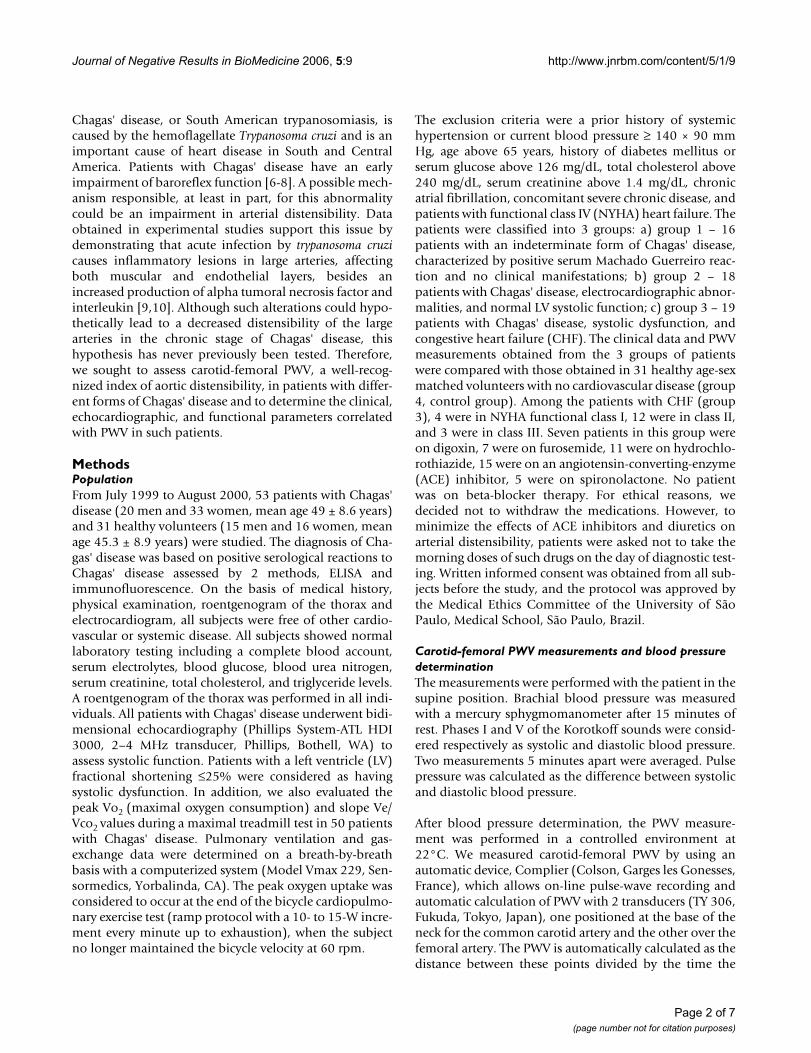
ResultsBaseline characteristicsBaseline demographic, clinical, laboratory, and echocar-diographic parameters of the 4 groups are shown in thetable 1. No difference was observed between the groupsregarding demographic, clinical, and laboratory tests.Likewise, no difference was observed between the 4groups regarding pulse pressure. Thus, the mean values ofgroup 1 were 39.5 ± 7.6 mm Hg, group 2 were 39.3 ± 8.1mm Hg, group 3 were 39.5 ± 7.4 mm Hg, and controlgroup were 39.7 ± 6.9 mm Hg (P = 0.99). All echocardio-graphic measurements were significantly different andobviously impaired in group 3 that comprised patientswith systolic dysfunction. Also, the peak VO2 was signifi-cantly decreased in group 3 patients with systolic dysfunc-tion, in comparison with that in patients without heartfailure.
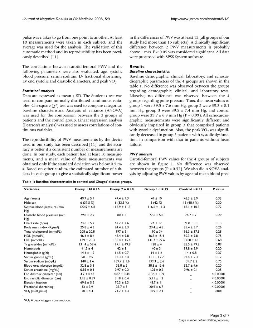
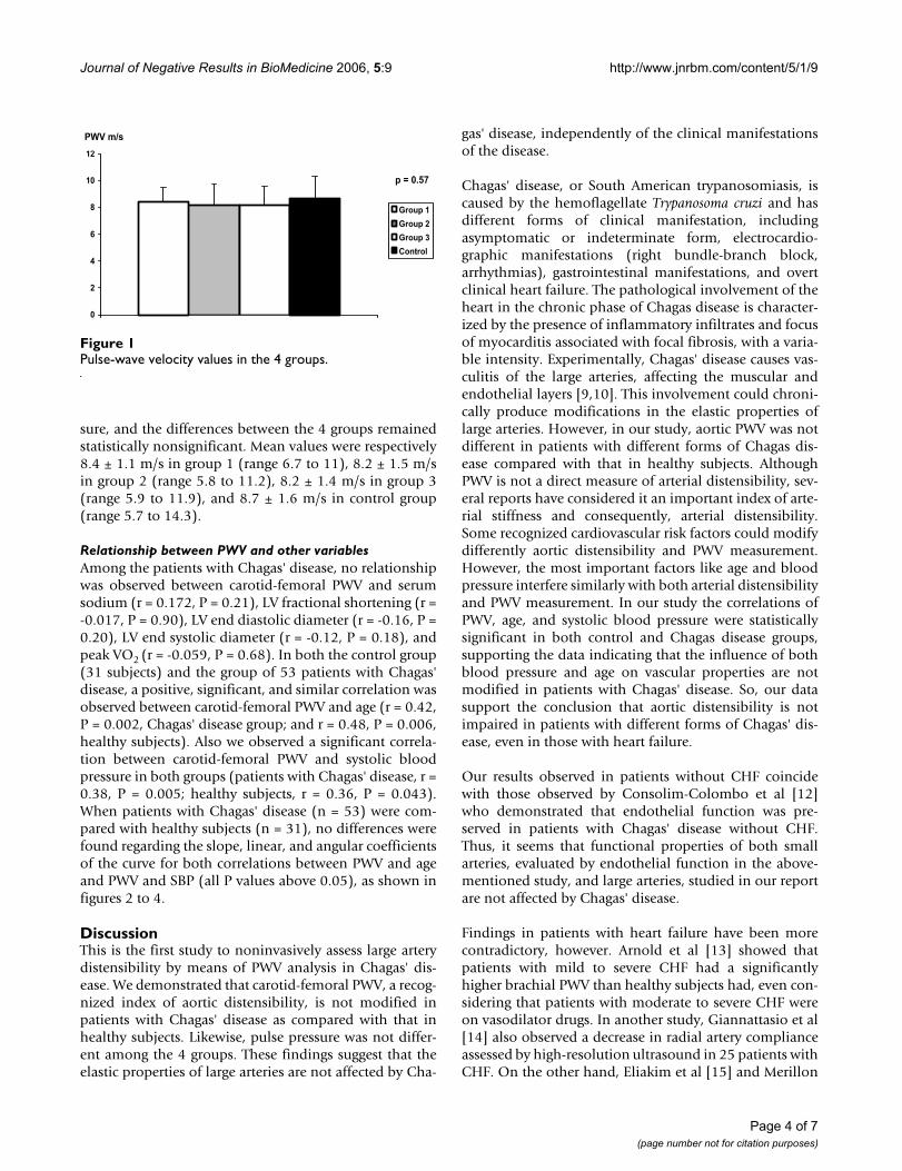
PWV analysisCarotid-femoral PWV values for the 4 groups of subjectsare shown in figure 1. No difference was observedbetween the groups (P = 0.57). We also did ANOVA anal-ysis by adjusting PWV values by age and mean blood pres-
Table 1: Baseline characteristics in control and Chagas' disease groups.
Variables Group 1 N = 16 Group 2 n = 18 Group 3 n = 19 Control n = 31 P value
Age (years) 49.7 ± 5.9 47.4 ± 9.3 49 ± 10 45.3 ± 8.9 0.33Male sex 6 (37.5 %) 6 (33.3 %) 8 (42 %) 15 (48.4 %) 0.30Systolic blood pressure (mm Hg)
120.5 ± 6.8 118.9 ± 11.3 116.6 ± 11.3 118.1 ± 10.3 0.72
Diastolic blood pressure (mm Hg)
79.8 ± 2.9 80 ± 5 77.6 ± 5.8 76.7 ± 7 0.29
Heart rate (bpm) 74.6 ± 5.7 67.7 ± 7.6 74 ± 12 71.8 ± 10 0.13Body mass index (Kg/m2) 25.8 ± 4.3 24.4 ± 3.3 23.4 ± 4.5 25.4 ± 3.7 0.26Total cholesterol (mmol/L) 208 ± 20.8 197 ± 21 190 ± 34 196.3 ± 17.8 0.28HDL (mmol/L) 46.4 ± 8.4 48.4 ± 9.8 46.8 ± 15.4 50.3 ± 9.8 0.75LDL (mmol/L) 139 ± 20.3 130.4 ± 15.4 131.7 ± 27.6 130.8 ± 16 0.68Triglycerides (mmol/L) 131.4 ± 59.6 117.1 ± 49.8 128 ± 4 128.5 ± 49.2 0.89Hematocrit 41.2 ± 4 42 ± 3 40 ± 3 39.8 ± 2.9 0.20Hemoglobin (g/dl) 14.4 ± 1.2 14.5 ± 0.7 14 ± 1.2 14 ± 0.8 0.37Serum glucose (g/dL) 98 ± 9.5 93.3 ± 6.4 101 ± 12.7 93.4 ± 9.3 0.12Serum sodium (mEq/L) 140 ± 1.6 139.7 ± 1.6 139.3 ± 2.6 139.7 ± 2 0.75Blood urea nitrogen (mg/dL) 32.8 ± 5.3 33.8 ± 5 38.8 ± 13.6 32.7 ± 4.6 0.20Serum creatinine (mg/dL) 0.95 ± 0.1 0.97 ± 0.2 1.05 ± 0.2 0.96 ± 0.1 0.25End diastolic diameter (cm) 4.7 ± 0.43 4.87 ± 0.44 6.36 ± 1.09 _ < 0.00001End systolic diameter (cm) 3.18 ± 0.39 3.18 ± 0.4 5.11 ± 1.2 _ < 0.00001Ejection fraction 69.6 ± 5.2 70.3 ± 6.3 48.7 ± 11 _ < 0.00001Fractional shortening 33 ± 3.9 33.7 ± 5 20.9 ± 6.7 _ < 0.00001VO2 (ml/Kg/min) 20 ± 4.3 21.7 ± 7.3 14.9 ± 2.1 _ 0.003
VO2 = peak oxygen consumption.
Page 3 of 7(page number not for citation purposes)
Journal of Negative Results in BioMedicine 2006, 5:9 http://www.jnrbm.com/content/5/1/9
sure, and the differences between the 4 groups remainedstatistically nonsignificant. Mean values were respectively8.4 ± 1.1 m/s in group 1 (range 6.7 to 11), 8.2 ± 1.5 m/sin group 2 (range 5.8 to 11.2), 8.2 ± 1.4 m/s in group 3(range 5.9 to 11.9), and 8.7 ± 1.6 m/s in control group(range 5.7 to 14.3).
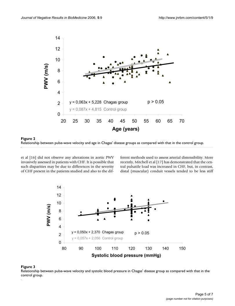
Relationship between PWV and other variablesAmong the patients with Chagas' disease, no relationshipwas observed between carotid-femoral PWV and serumsodium (r = 0.172, P = 0.21), LV fractional shortening (r =-0.017, P = 0.90), LV end diastolic diameter (r = -0.16, P =0.20), LV end systolic diameter (r = -0.12, P = 0.18), andpeak VO2 (r = -0.059, P = 0.68). In both the control group(31 subjects) and the group of 53 patients with Chagas'disease, a positive, significant, and similar correlation wasobserved between carotid-femoral PWV and age (r = 0.42,P = 0.002, Chagas' disease group; and r = 0.48, P = 0.006,healthy subjects). Also we observed a significant correla-tion between carotid-femoral PWV and systolic bloodpressure in both groups (patients with Chagas' disease, r =0.38, P = 0.005; healthy subjects, r = 0.36, P = 0.043).When patients with Chagas' disease (n = 53) were com-pared with healthy subjects (n = 31), no differences werefound regarding the slope, linear, and angular coefficientsof the curve for both correlations between PWV and ageand PWV and SBP (all P values above 0.05), as shown infigures 2 to 4.
DiscussionThis is the first study to noninvasively assess large arterydistensibility by means of PWV analysis in Chagas' dis-ease. We demonstrated that carotid-femoral PWV, a recog-nized index of aortic distensibility, is not modified inpatients with Chagas' disease as compared with that inhealthy subjects. Likewise, pulse pressure was not differ-ent among the 4 groups. These findings suggest that theelastic properties of large arteries are not affected by Cha-
gas' disease, independently of the clinical manifestationsof the disease.
Chagas' disease, or South American trypanosomiasis, iscaused by the hemoflagellate Trypanosoma cruzi and hasdifferent forms of clinical manifestation, includingasymptomatic or indeterminate form, electrocardio-graphic manifestations (right bundle-branch block,arrhythmias), gastrointestinal manifestations, and overtclinical heart failure. The pathological involvement of theheart in the chronic phase of Chagas disease is character-ized by the presence of inflammatory infiltrates and focusof myocarditis associated with focal fibrosis, with a varia-ble intensity. Experimentally, Chagas' disease causes vas-culitis of the large arteries, affecting the muscular andendothelial layers [9,10]. This involvement could chroni-cally produce modifications in the elastic properties oflarge arteries. However, in our study, aortic PWV was notdifferent in patients with different forms of Chagas dis-ease compared with that in healthy subjects. AlthoughPWV is not a direct measure of arterial distensibility, sev-eral reports have considered it an important index of arte-rial stiffness and consequently, arterial distensibility.Some recognized cardiovascular risk factors could modifydifferently aortic distensibility and PWV measurement.However, the most important factors like age and bloodpressure interfere similarly with both arterial distensibilityand PWV measurement. In our study the correlations ofPWV, age, and systolic blood pressure were statisticallysignificant in both control and Chagas disease groups,supporting the data indicating that the influence of bothblood pressure and age on vascular properties are notmodified in patients with Chagas' disease. So, our datasupport the conclusion that aortic distensibility is notimpaired in patients with different forms of Chagas' dis-ease, even in those with heart failure.
Our results observed in patients without CHF coincidewith those observed by Consolim-Colombo et al [12]who demonstrated that endothelial function was pre-served in patients with Chagas' disease without CHF.Thus, it seems that functional properties of both smallarteries, evaluated by endothelial function in the above-mentioned study, and large arteries, studied in our reportare not affected by Chagas' disease.
Findings in patients with heart failure have been morecontradictory, however. Arnold et al [13] showed thatpatients with mild to severe CHF had a significantlyhigher brachial PWV than healthy subjects had, even con-sidering that patients with moderate to severe CHF wereon vasodilator drugs. In another study, Giannattasio et al[14] also observed a decrease in radial artery complianceassessed by high-resolution ultrasound in 25 patients withCHF. On the other hand, Eliakim et al [15] and Merillon
Pulse-wave velocity values in the 4 groupsFigure 1Pulse-wave velocity values in the 4 groups.
0
2
4
6
8
10
12
Group 1
Group 2
Group 3
Control
PWV m/s
p = 0.57
Page 4 of 7(page number not for citation purposes)
Journal of Negative Results in BioMedicine 2006, 5:9 http://www.jnrbm.com/content/5/1/9
et al [16] did not observe any alterations in aortic PWVinvasively assessed in patients with CHF. It is possible thatsuch disparities may be due to differences in the severityof CHF present in the patients studied and also to the dif-
ferent methods used to assess arterial distensibility. Morerecently, Mitchell et al [17] has demonstrated that the cen-tral pulsatile load was increased in CHF, but, in contrast,distal (muscular) conduit vessels tended to be less stiff
Relationship between pulse-wave velocity and systolic blood pressure in Chagas' disease group as compared with that in the control groupFigure 3Relationship between pulse-wave velocity and systolic blood pressure in Chagas' disease group as compared with that in the control group.
y = 0,050x + 2,370 Chagas group
y = 0,057x + 2,056 Control group
0
2
4
6
8
10
12
14
80 90 100 110 120 130 140 150
Systolic blood pressure (mmHg)
PW
V (
m/s
)
p > 0.05
Relationship between pulse-wave velocity and age in Chagas' disease groups as compared with that in the control groupFigure 2Relationship between pulse-wave velocity and age in Chagas' disease groups as compared with that in the control group.
y = 0,063x + 5,228 Chagas group
y = 0,087x + 4,815 Control group
0
2
4
6
8
10
12
14
20 25 30 35 40 45 50 55 60 65 70
Age (years)
PW
V (
m/s
)
p > 0.05
Page 5 of 7(page number not for citation purposes)

Journal of Negative Results in BioMedicine 2006, 5:9 http://www.jnrbm.com/content/5/1/9
(lower carotid radial PWV) in the same patients with CHF.The increased functional stiffness of the central conduitsin CHF observed in Mitchell's study is not apparent in glo-bal measures such as augmentation index or total arterycompliance, probably explained by the contrastingchanges in central and peripheral conduits. In our study,the absence of modifications in carotid pulse-wave veloc-ity in Chagas' disease even in the presence of mild to mod-erate CHF, could be partially explained by the results ofMitchell et al [17].
Three possible mechanisms may be responsible for mod-ifications in arterial distensibility in patients with CHF:increased plasma or tissue concentrations of a number ofvasoconstrictor substances secondary to enhanced sympa-thetic drive; a reduction in the shear endothelium stressand secretion of endothelial relaxing factors as a conse-quence of a reduction in cardiac contractility and output;and arterial wall edema and stiffness due to sodium andwater retention [18]. All these mechanisms are moreintense in patients with severe CHF, patients who wereexcluded in our study so as not to interfere in a possibleeffect of Chagas' disease on the PWV. Thus, as we excludedpatients with severe CHF, and also patients with ischemicheart disease or hypertension, situations that knowinglymodify PWV, our results suggest that the presence of mildto moderate heart failure per se in patients with Chagas'disease does not alter the elastic properties of great arter-ies. However, as we do not assess other vascular functionalproperties like endothelial function in these patients, it is
not possible to totally exclude vascular modifications inpatients with Chagas disease and heart failure.
The lack of differences in carotid-femoral PWV betweenhealthy subjects and the different groups with Chagas dis-ease could be due to type II (beta) statistical error fre-quently observed in a study such as this involving a smallnumber of patients. However, we can observe that thestandard deviation of PWV measurements is very small,and even considering a greater statistical significance(0.10) the values remained not different among thegroups.
In both the control healthy group and patients with Cha-gas disease, a positive and similar relationship betweenPWV and age and between PWV and systolic blood pres-sure was observed, as described previously [10]. We didthis analysis to verify whether the aging and blood pres-sure influence on aortic distensibility could be modifiedby the presence of Chagas disease. Thus, Chagas' diseaseseems not to accelerate the arterial stiffening secondary toaging or elevated blood pressure.
One limitation of our study must be addressed. For ethicalreasons, we did not withdraw diuretic and vasodilatordrugs used by patients with CHF. Therefore, it would bepossible that such drugs may have had a favorable effecton PWV, leading to a "pseudonormalization" of a modi-fied carotid-femoral PWV in such patients.
Relationship between pulse wave velocity and diastolic blood pressure in Chagas' disease group as compared with that in the control groupFigure 4Relationship between pulse wave velocity and diastolic blood pressure in Chagas' disease group as compared with that in the control group.
y = 0,067x + 3,008 Chagas group
y = 0,064x + 3,863 Control group
0
2
4
6
8
10
12
14
50 55 60 65 70 75 80 85 90 95 100
Diastolic blood pressure (mmHg)
PW
V (
m/s
)
p > 0.05
Page 6 of 7(page number not for citation purposes)

Journal of Negative Results in BioMedicine 2006, 5:9 http://www.jnrbm.com/content/5/1/9
Publish with BioMed Central and every scientist can read your work free of charge
"BioMed Central will be the most significant development for disseminating the results of biomedical research in our lifetime."
Sir Paul Nurse, Cancer Research UK
Your research papers will be:
available free of charge to the entire biomedical community
peer reviewed and published immediately upon acceptance
cited in PubMed and archived on PubMed Central
yours — you keep the copyright
Submit your manuscript here:http://www.biomedcentral.com/info/publishing_adv.asp
BioMedcentral
In summary, the present study indicates that carotid-fem-oral PWV is normal in patients with Chagas' disease,despite the presence of electrocardiographic abnormali-ties or mild heart failure, suggesting that large artery dis-tensibility is not primarily affected in this disease.
AcknowledgementsThe authors thank Professor Kathleen A. Dracup, University of California, San Francisco, for reviewing the manuscript.
References1. O'Rourke MF, Mancia G: Arterial stiffness. J Hypertension 1999,
17:1-4.2. Bortolotto LA, Safar ME: Blood pressure profile along arterial
tree and genetics of hypertension. Arq Bras Cardiol 2006,86:166-169.
3. Blacher J, Guerin AP, Pannier B, Marchais SJ, Safar ME, London GM:Impact of aortic stiffness on survival in end-stage renal dis-ease. Circulation 1999, 99:2434-2439.
4. Blacher J, Asmar R, Djane S, London GM, Safar ME: Aortic pulsewave velocity as a marker of cardiovascular risk in hyperten-sive patients. Hypertension 1999, 33:1111-1117.
5. Stefanadis C, Dernellis J, Tsiamis E, Stratos C, Diamantopoulos L,Michaelides A, Toutouzas P: Aortic stiffness as a risk factor forrecurrent acute coronary events in patients with ischaemicheart disease. Eur Heart J 2000, 21:390-396.
6. Marin-Neto JA, Gallo L Jr, Manco JC, Rassi A, Amorim DS: Posturalreflexes in chronic Chagas' heart disease: heart rate andarterial pressure responses. Cardiology 1975, 60:343-357.
7. Palmero HA, Caieiro TF, Iosa DJ: Distinctive abnormal responsesto tilting test in chronic Chagas' disease. Klin Wochenschr 1980,58:1307-1311.
8. Consolim-Colombo FM, Filho JA, Lopes HF, Sobrinho CR, Otto ME,Riccio GM, Mady C, Krieger EM: Decreased cardiopulmonarybaroreflex sensitivity in Chagas' heart disease. Hypertension2000, 36:1035-1039.
9. Rossi MA: Aortic endothelial cell changes in the acute septi-cemic phase of experimental Trypanosoma cruzi infection inrats: scanning and transmission electron microscopic study.Am J Trop Med Hyg 1997, 57:321-327.
10. Sunnermark D, Frostegard J, Orn A, Harris RA: Cellular andcytokine characterization of vascular inflammation in CBA/Jmice chronically infected with Trypanosoma cruzi. Scand JImmunol 1998, 48:480-484.
11. Asmar R, Benetos A, Topouchian J, Laurent P, Pannier B, Brisac AM,Target R, Levy BI: Assessment of arterial distensibility by auto-matic pulse wave velocity measurement: validation and clin-ical application studies. Hypertension 1995, 26:485-490.
12. Consolim-Colombo FM, Rosseto EA, Rocha NN, Lopes HF, BaruzziACA, Rubira MC, Ianni B, Mady C: Endothelial function is pre-served in Chagas' disease. J Hypertens 2001, 6:144. [abstr].
13. Arnold JMO, Marchiori GE, Imrie JR, Burton GL, Plugfelder PW, Kos-tuk WJ: Large artery function in patients with chronic heartfailure: studies of brachial artery diameter and hemodynam-ics. Circulation 1991, 84:2418-2425.
14. Giannattasio C, Failla M, Stella ML, Mangoni AA, Carugo S, Pozzi M,Grassi G, Mansia G: Alterations of radial artery compliance inpatients with congestive heart failure. Am J Cardiol 1995,76:381-385.
15. Eliakim M, Sapoznikov D, Weinman J: Pulse wave velocity inhealthy subjects and in patients with various disease states.Am Heart J 1971, 82:448-457.
16. Merillon JP, Fontenier G, Lerallut JF, Jaffrin MY, Chastre J, Assayag P,Motte G, Gourgon R: Aortic input impedance in heart failure:comparison with normal subjects and its changes duringvasodilator therapy. Eur Heart J 1984, 5:447-455.
17. Mitchell GF, Tardif JC, Arnold JMO, Marchiori G, O'Brien TX, DunlapME, Pfeffer MA: Pulsatile hemodynamics in congestive heartfailure. Hypertension 2001, 38:1433-1439.
18. Ramsey MW, Goodfellow J, Jones CJH, Luddington LA, Lewis MJ,Henderson AH: Endothelial control of arterial distensibility isimpaired in chronic heart failure. Circulation 1995,92:3212-3219.
Page 7 of 7(page number not for citation purposes)
Copyright © 2022 FDOKUMEN