Aortic Annulus Diameter Determination by Multidetector Computed Tomography: Reproducibility,...
11
Aortic Annulus Diameter Determination by Multidetector Computed Tomography Reproducibility, Applicability, and Implications for Transcatheter Aortic Valve Implantation Ronen Gurvitch, MBBS,* John G. Webb, MD,* Ren Yuan, MD,* Mark Johnson, MBBS,* Cameron Hague, MD,* Alexander B. Willson, MBBS,* Stefan Toggweiler, MD,* David A. Wood, MD,* Jian Ye, MD,* Robert Moss, MD,* Christopher R. Thompson, MD,* Stephan Achenbach, MD,† James K. Min, MD,‡ Troy M. LaBounty, MD,‡ Ricardo Cury, MD,§ Jonathon Leipsic, MD* British Columbia, Vancouver, Canada; Erlangen, Germany; Los Angeles, California; and Miami, Florida Objectives This study sought to determine the most reproducible multidetector computed tomog- raphy (MDCT) measurements of the aortic annulus and to determine methods to improve the appli- cability of these measurements for transcatheter aortic valve implantation. Background The reproducibility and applicability of MDCT annular measurements to guide trans- catheter aortic valve implantation remain unclear. Methods Annular measurements were performed in 50 patients planed for transcatheter aortic valve implantation in multiple planes: basal ring (short- and long-axis, mean diameter, area-derived diameter), coronal, sagittal, and 3-chamber projections. A theoretical model was developed taking into account the differences between the most reproducible MDCT measurements and transesopha- geal echocardiography to guide valve size choice. Results The most reproducible measurements were the area-derived diameter and basal ring average diameter (inter-reader intraclass correlation coefficient: 0.87 [95% confidence interval: 0.81 to 0.92] and 0.80 [95% confidence interval: 0.70 to 0.87]; respectively; intrareader 0.90 for all readers). These were generally larger than transesophageal echocardiography diameters (mean difference of 1.5 1.6 mm and 1.1 1.7 mm, respectively). When a strategy of valve-sizing is undertaken using these CT measure- ments using an echocardiographic sizing scale, a different THV size would be selected in 44% and 40% of cases, respectively. When adjusting the sizing cutoffs to account for the differences in observed diam- eters, this was reduced to 10% to 12% (p 0.01 for both, respectively). Conclusions The most reproducible MDCT measurements of the annulus are the area-derived diameter and basal ring average diameter, with derived values generally larger than those obtained with echocardiography. If MDCT is used for valve sizing, a strategy incorporating these differences may be important. MDCT using these easily derived measurements may be ideally suited to sizing transcatheter aortic valves as they account for the eccentricity of the aortic annulus, are reproducible, and are noninvasive. (J Am Coll Cardiol Intv 2011; 4:1235– 45) © 2011 by the American College of Cardiology Foundation From the *St. Paul’s Hospital, University of British Columbia, Vancouver, Canada; †University of Erlangen, Erlangen, Germany; ‡Departments of Medicine, Imaging, and Biomedical Sciences, Cedars-Sinai Medical Center, Los Angeles, California; and the §Baptist Hospital of Miami, Miami, Florida. Dr. Webb is a consultant for Siemens and Edwards Lifesciences. Dr. Wood is a consultant for Edwards Lifesciences. Dr. Ye is a consultant to Edwards Lifesciences. Dr. Achenbach receives research support from Siemens Healthcare and Bayer Schering. Dr. Min is on the Speaker’s Bureau of GE Healthcare; is a consultant for Edwards Lifesciences; and has equity interest in TC3. Dr. Leipsic is on the Speaker’s Bureau and advisory board of Edwards Lifesciences; and he is on the Speaker’s Bureau for GE Healthcare and Equity Stakeholder TC3 Corelab. All other authors have reported that they have no relationships relevant to the contents of this paper to disclose. Manuscript received April 1, 2011; revised manuscript received June 30, 2011, accepted July 21, 2011. JACC: CARDIOVASCULAR INTERVENTIONS VOL. 4, NO. 11, 2011 © 2011 BY THE AMERICAN COLLEGE OF CARDIOLOGY FOUNDATION ISSN 1936-8798/$36.00 PUBLISHED BY ELSEVIER INC. DOI: 10.1016/j.jcin.2011.07.014
-
Upload
independent -
Category
Documents
-
view
0 -
download
0
Transcript of Aortic Annulus Diameter Determination by Multidetector Computed Tomography: Reproducibility,...

J A C C : C A R D I O V A S C U L A R I N T E R V E N T I O N S V O L . 4 , N O . 1 1 , 2 0 1 1
© 2 0 1 1 B Y T H E A M E R I C A N C O L L E G E O F C A R D I O L O G Y F O U N D A T I O N I S S N 1 9 3 6 - 8 7 9 8 / $ 3 6 . 0 0
P U B L I S H E D B Y E L S E V I E R I N C . D O I : 1 0 . 1 0 1 6 / j . j c i n . 2 0 1 1 . 0 7 . 0 1 4
Aortic Annulus Diameter Determinationby Multidetector Computed TomographyReproducibility, Applicability, and Implications forTranscatheter Aortic Valve Implantation
Ronen Gurvitch, MBBS,* John G. Webb, MD,* Ren Yuan, MD,* Mark Johnson, MBBS,*Cameron Hague, MD,* Alexander B. Willson, MBBS,* Stefan Toggweiler, MD,*David A. Wood, MD,* Jian Ye, MD,* Robert Moss, MD,* Christopher R. Thompson, MD,*Stephan Achenbach, MD,† James K. Min, MD,‡ Troy M. LaBounty, MD,‡Ricardo Cury, MD,§ Jonathon Leipsic, MD*
British Columbia, Vancouver, Canada; Erlangen, Germany; Los Angeles, California;and Miami, Florida
Objectives This study sought to determine the most reproducible multidetector computed tomog-raphy (MDCT) measurements of the aortic annulus and to determine methods to improve the appli-cability of these measurements for transcatheter aortic valve implantation.
Background The reproducibility and applicability of MDCT annular measurements to guide trans-catheter aortic valve implantation remain unclear.
Methods Annular measurements were performed in 50 patients planed for transcatheter aorticvalve implantation in multiple planes: basal ring (short- and long-axis, mean diameter, area-deriveddiameter), coronal, sagittal, and 3-chamber projections. A theoretical model was developed takinginto account the differences between the most reproducible MDCT measurements and transesopha-geal echocardiography to guide valve size choice.
Results The most reproducible measurements were the area-derived diameter and basal ring averagediameter (inter-reader intraclass correlation coefficient: 0.87 [95% confidence interval: 0.81 to 0.92] and0.80 [95% confidence interval: 0.70 to 0.87]; respectively; intrareader �0.90 for all readers). These weregenerally larger than transesophageal echocardiography diameters (mean difference of 1.5 � 1.6 mmand 1.1 � 1.7 mm, respectively). When a strategy of valve-sizing is undertaken using these CT measure-ments using an echocardiographic sizing scale, a different THV size would be selected in 44% and 40%of cases, respectively. When adjusting the sizing cutoffs to account for the differences in observed diam-eters, this was reduced to 10% to 12% (p � 0.01 for both, respectively).
Conclusions The most reproducible MDCT measurements of the annulus are the area-derived diameter andbasal ring average diameter, with derived values generally larger than those obtained with echocardiography.If MDCT is used for valve sizing, a strategy incorporating these differences may be important. MDCT usingthese easily derived measurements may be ideally suited to sizing transcatheter aortic valves as they accountfor the eccentricity of the aortic annulus, are reproducible, and are noninvasive. (J Am Coll Cardiol Intv 2011;4:1235–45) © 2011 by the American College of Cardiology Foundation
From the *St. Paul’s Hospital, University of British Columbia, Vancouver, Canada; †University of Erlangen, Erlangen, Germany;‡Departments of Medicine, Imaging, and Biomedical Sciences, Cedars-Sinai Medical Center, Los Angeles, California; and the§Baptist Hospital of Miami, Miami, Florida. Dr. Webb is a consultant for Siemens and Edwards Lifesciences. Dr. Wood is aconsultant for Edwards Lifesciences. Dr. Ye is a consultant to Edwards Lifesciences. Dr. Achenbach receives research support fromSiemens Healthcare and Bayer Schering. Dr. Min is on the Speaker’s Bureau of GE Healthcare; is a consultant for EdwardsLifesciences; and has equity interest in TC3. Dr. Leipsic is on the Speaker’s Bureau and advisory board of Edwards Lifesciences;and he is on the Speaker’s Bureau for GE Healthcare and Equity Stakeholder TC3 Corelab. All other authors have reported thatthey have no relationships relevant to the contents of this paper to disclose.
Manuscript received April 1, 2011; revised manuscript received June 30, 2011, accepted July 21, 2011.
lw
J A C C : C A R D I O V A S C U L A R I N T E R V E N T I O N S , V O L . 4 , N O . 1 1 , 2 0 1 1
N O V E M B E R 2 0 1 1 : 1 2 3 5 – 4 5
Gurvitch et al.
Aortic Annulus Diameter Determination by MDCT
1236
Transcatheter aortic valve implantation (TAVI) is becom-ing an accepted procedure for selected patients with severeaortic stenosis (1). Accurate evaluation of the native aorticvalve annular dimensions is critical to optimize selection ofthe correct bioprosthetic valve size. In contrast to surgicalaortic valve replacement where surgeons perform valvesizing under direct visualization and with the aid of annularsizing probes, noninvasive imaging methods are generallyrequired in the setting of TAVI. Incorrect valve sizing maylead to paravalvular aortic regurgitation, which is a predictorof worse long-term outcome (2), valve embolization, patientprosthesis mismatch, or catastrophic annular rupture (3,4).
Given the complex shape of the aortic annulus, whichincludes a generally noncircular profile coupled with aconical form that is bounded at its nadir by the attachmentof the 3 aortic leaflets (5), accurate evaluation by 2-dimensionalimaging modalities, such as echocardiography, is intrinsi-cally difficult. Recently, 3-dimensional multidetector com-puted tomography (MDCT), which is not limited by planar
imaging, has been shown to pro-vide fine anatomical detail andaccurate assessment of the com-plex anatomical shape of theaortic valve and annulus (6–8).Whereas some have suggested thatMDCT-derived measurementsmay better serve as a gold stan-dard for aortic valve sizing, thereproducibility, reliability, andapplicability of these measure-ments have not been well de-fined. Furthermore, the clinicalutility of MDCT measures re-mains limited. Previous studieshave also suggested that despite
propitious clinical outcomes of patients undergoing TAVIwith echo-based aortic valve sizing, an MDCT-based valvesizing strategy might result in up to 39% of cases requiringa different valve size or exclusion from candidacy for TAVIusing currently available devices (9–11). However, theseprior MDCT studies employed aortic annular measure-ments in limited planes and without systematic adjustmentfor discordance between MDCT and echocardiographicmodalities.
In the present study, we thus aimed to: 1) evaluate theaortic annular dimensions in patients undergoing TAVIusing MDCT in multiple planes; 2) assess which measure-ments have the greatest interobserver and intraobserverreproducibility; and 3) determine the difference in valvesizing recommendations if MDCT measurements are usedaccording to current (transesophageal echocardiography[TEE]–based) sizing cutoffs (“unadjusted criteria”) versus a
Abbreviations andAcronym
CI � confidence interval
ICC � intraclass correlationcoefficient
IQR � interquartile range
MDCT � multidetectorcomputed tomography
TAVI � transcatheter aorticvalve implantation
TEE � transesophagealechocardiography
TTE � transthoracicechocardiography
strategy of incorporating the observed differences between t
MDCT and echocardiography into the sizing criteria (“ad-justed criteria”).
Methods
Patient population. Fifty patients with severe symptomaticaortic stenosis being considered for TAVI underwent pre-procedural evaluation using MDCT imaging. Forty-one ofthese patients underwent TAVI, and all these patients hadmatched transthoracic echocardiography (TTE) and TEEimages. Patients with bicuspid aortic stenosis were excluded.MDCT was clinically indicated for evaluation before theprocedure to assess the peripheral vasculature and determinethe optimal angiographic projection angles for valve implan-tation (12,13). Following implantation, implantationheight was angiographically defined as optimal or subop-timal, with optimal position defined as occurring whenthe native leaflet insertion point was within the middlethird of the stent frame. All other valve positions weredefined as suboptimal.
Patients were evaluated by a team of senior cardiologistsand cardiothoracic surgeons to determine if they posed aprohibitively high surgical risk before being accepted forTAVI. Patients underwent transfemoral or transapical im-plants of Edward Sapien/Sapien XT balloon expandablebioprostheses (Edwards Lifesciences, Irvine, California) of23-mm, 26-mm, or 29-mm diameter with valve size chosenbased on annulus diameters as derived by TEE as previouslydescribed (14–18).CT images acquisition. MDCT examinations were per-formed on a 64-slice Discovery HD 750 High Definitionscanner (GE Healthcare, Milwaukee, Wisconsin), and 80 to120 ml of iodixanol 320 (GE Healthcare, Princeton, NewJersey) were injected at 5 ml/s followed by 30 ml of normalsaline. The timing delay of the scan was determined using asmart prep of the ascending aorta with a preset threshold of150 Hounsfield units. The MDCT examinations wereperformed in the craniocaudal direction with retrospectivegating from the aortic arch through to the diaphragm. Heartrate reduction with beta-blockade was avoided as interpre-tation of the coronary arteries was not required and becauseof clinical concern regarding severe aortic stenosis. MDCTscanner detector collimation width was 0.625 mm, detectorcoverage was 40 mm, reconstructed slice thickness was 1.25mm, and the slice interval was 1.25 mm. Gantry rotationtime was 0.35 s, and the scan pitch ranged between 0.16 and0.20 (adjusted per heart rate). Depending on the patientsize, the maximum tube current ranged between 450 and700 mA with a fixed tube voltage of 100 kVp for patientswith a body mass index �30 kg/m2 and 120 kVp used inarger patients. Electrocardiography-gated dose modulationas used with tube current reduced to 60% of maximum
ube current in systole. This provided adequate image
m
apMmd
in sha
J A C C : C A R D I O V A S C U L A R I N T E R V E N T I O N S , V O L . 4 , N O . 1 1 , 2 0 1 1 Gurvitch et al.
N O V E M B E R 2 0 1 1 : 1 2 3 5 – 4 5 Aortic Annulus Diameter Determination by MDCT
1237
quality for annular assessment in the systolic phases whilereducing the estimated effective radiation dose.Achieving the different imaging planes and measuring theaortic annulus. Following MDCT image acquisition, refor-
ats were performed to allow evaluation of the aortic
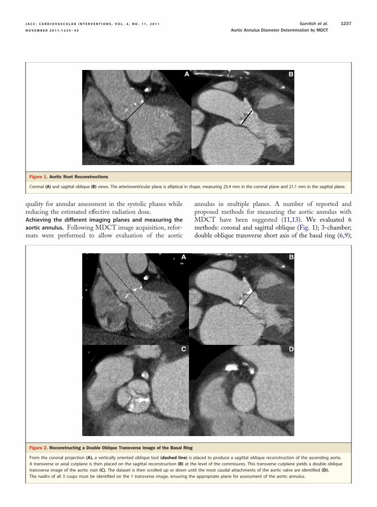
Figure 2. Reconstructing a Double Oblique Transverse Image of the Basal
From the coronal projection (A), a vertically oriented oblique tool (dashed linA transverse or axial cutplane is then placed on the sagittal reconstruction (B)transverse image of the aortic root (C). The dataset is then scrolled up or dow
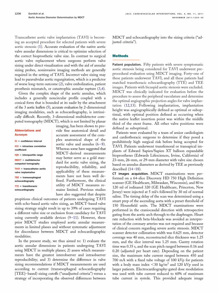
Figure 1. Aortic Root Reconstructions
Coronal (A) and sagittal oblique (B) views. The arterioventricular plane is elliptical
The nadirs of all 3 cusps must be identified on the 1 transverse image, ensuring the
nnulus in multiple planes. A number of reported androposed methods for measuring the aortic annulus withDCT have been suggested (11,13). We evaluated 6ethods: coronal and sagittal oblique (Fig. 1); 3-chamber;
ouble oblique transverse short axis of the basal ring (6,9);
laced to produce a sagittal oblique reconstruction of the ascending aorta.level of the commisures. This transverse cutplane yields a double oblique
il the most caudal attachments of the aortic valve are identified (D).
pe, measuring 25.4 mm in the coronal plane and 21.1 mm in the sagittal plane.
Ring
e) is pat then unt
appropriate plane for assessment of the aortic annulus.

J A C C : C A R D I O V A S C U L A R I N T E R V E N T I O N S , V O L . 4 , N O . 1 1 , 2 0 1 1
N O V E M B E R 2 0 1 1 : 1 2 3 5 – 4 5
Gurvitch et al.
Aortic Annulus Diameter Determination by MDCT
1238
long axis of the basal ring; and an area measurement of thebasal ring of the aorta. In addition, we calculated thearea-derived diameter, as well as the basal ring averagediameter (see the next paragraph). For all these measure-ments, the reader would determine the optimal projectionfor measurement. Importantly, all 3 readers had agreed onthe defined principles behind each measurement and hadread 25 previous cases in collaboration to ensure a standard-ized approach.
As all MDCT studies were performed with retrospectivegating, reading physicians first selected the appropriatephase of the cardiac cycle. By convention, the annulus wasmeasured in systole during maximum valve opening. Fol-lowing this, physicians performed post-processing and anal-ysis beginning in the coronal projection with an axial-cutplane rotated in a counterclockwise direction such that theannular plane ran from right-caudal to left-cranial position.After ensuring that the various imaging planes are locked,attention is turned to the sagittal projection, which isvertically oriented to be orthogonal to the long axis of theaortic annulus (Fig. 2). At this point, the horizontal linerepresenting the axial cutplane is moved up or down oneither the sagittal or coronal projection to create a doubleoblique transverse or axial image of the aortic root. Thishorizontal line is then pulled down through the root of theaorta until the most caudal attachments of the 3 aortic cuspsare identified. Importantly, to accurately define the basalring, the plane must be balanced so that all 3 leaflets comeinto view when scrolling cephalad at the same time (Fig. 2).At this point, the short- and long-axis dimensions of thebasal ring are measured (Fig. 3A), and the area is traced out
Figure 3. Double Oblique Transverse Reconstruction Just Below the CommBasal Ring Measurements
(A) The long- and short-axis dimensions measure 24 mm and 21.6 mm, respectraced, generating a direct measurement of the basal ring area (shaded): 425.
diamete
accurately (Fig. 3B). From the former, a basal ring averagediameter is determined by (basal ring short axis � basal ringlong axis)/2, and from the latter, the area-derived diameteris calculated using:
diameter � 2�area
�
To generate a 3-chamber projection of the aortic annulus,a standardized approach is also used. A transverse image ofthe mitral valve with maximal visualization of the leftventricular cavity that allows for identification of the ven-tricular apex is bisected with an oblique plane creating along-axis reconstruction. The left ventricle is then cut in aplane from the left atrium through the center of the mitralvalve through the left ventricular apex resulting in a4-chamber projection. An oblique plane is then used torotate the left ventricle in a perpendicular fashion to theinterventricular septum to create a short-axis projection.Finally, a cutplane is placed in an oblique fashion at the baseof the short-axis projection out of the left ventricularoutflow tract and aorta to create a 3-chamber projection(Fig. 4).
All measurements were conducted in blinded fashion bythe 3 independent readers. Each reader evaluated theannulus in all planes. Following this, to assess intraobserverreliability, readers reread 20 randomly selected cases at least1 month following the original assessment, with the readersblinded to the original results.
l Insertions of the Aortic Leaflets Demonstrating Various
, with an average diameter of 22.8 mm. (B) The circumference is carefully2. From this, the area-derived diameter is calculated using:
area
�
issura
tively8 mm
r � 2�

m
e8T5
easur
J A C C : C A R D I O V A S C U L A R I N T E R V E N T I O N S , V O L . 4 , N O . 1 1 , 2 0 1 1 Gurvitch et al.
N O V E M B E R 2 0 1 1 : 1 2 3 5 – 4 5 Aortic Annulus Diameter Determination by MDCT
1239
Echocardiography. Scans were performed either before orduring the TAVI procedure and interpreted by an echocar-diographer experienced in TAVI and pre-procedural eval-uation. All patients undergoing TAVI had both TTE andTEE imaging, with all scans clinically indicated as partof the routine clinical evaluation of patients undergoingTAVI. Phillips iE33 or Sonos 5500 ultrasound systems(Philips HealthCare, Minneapolis, Minnesota) were usedfor image evaluation. The aortic annulus was measured atthe base of the leaflet insertion point in a zoomed-up3-chamber view at approximately 120° (TEE), correspond-ing to the nadirs of the noncoronary and right coronaryleaflets, or in a parasternal long-axis view in a zoomed-upview (TTE). All measurements were performed in earlysystole as recommended by the American Society of Echo-cardiography for quantification of stroke volume and aorticstenosis severity (19). All images were stored on disk foroffline analysis. All patients undergoing TAVI had pre-hospital discharge TTE evaluation of valvular function andhemodynamics. The degree and origin of post-proceduralaortic regurgitation was assessed using pre-hospital dis-charge TTE.Impact of using MDCT images on valve size selection. Toevaluate the clinical applicability of MDCT images on valvesize selection, a sizing model was created based on annulardiameters and current implantation guideline cutoffs. Giventhat we found MDCT images to be generally larger thanTEE measurements, the mean difference between the par-ticular MDCT measurements and TEE measurements wereincorporated into an “adjusted” sizing model. A comparisonwas then made between a strategy in which MDCTmeasurements were used with existing TEE-based sizingguidelines for the Sapien valve (23-mm valve for annulardiameters of 18 to 21 mm, a 26-mm valve for 22 to 25 mm,and a 29-mm valve for 26 to 28 mm) and another strategy
Figure 4. The 3-Chamber View
(A) Three-chamber transesophageal view in a zoomed-up image at approximacomputed tomography reconstructed 3-chamber projection (B) is created to m
in which these sizing cutoffs were adjusted to incorporate
the observed mean difference between the specific MDCTviews tested and TEE.Statistical methods. Continuous variables are described as
ean � SD when normally distributed or as medians withinterquartile ranges (IQR) when not. Normality was testedusing the Shapiro-Wilks goodness-of-fit test. Categoricalvariables are described by frequencies and percentages.Comparison of categorical variables was performed using achi-square analysis or the Fisher exact test as appropriate. Amixed-effect model was used to estimate the between-subject variance and within-subject variance. Intraclass cor-relation coefficient (ICC) was defined as the ratio ofbetween-subject variance to the total variance. The 95%confidence interval (CI) was calculated using the deltamethod (20). The Spearman correlation coefficient was usedto assess the relationship between echocardiographic andMDCT measurements. All analyses were performed usingSAS (version 9.1.3, SAS Institute, Cary, North Carolina).
Results
Baseline characteristics. A total of 50 patients underwentvaluation with MDCT (Table 1). The mean age was 81 �
years, 48% were women, and the median Society ofhoracic Surgeons risk score for mortality was 7.5% (IQR:.4% to 9.4%). Mean aortic valve area was 0.67 � 0.21 cm2
0° is used to measure the aortic annulus diameter. The multidetectore a similar plane.
Table 1. Baseline Patient Characteristics (N � 50)
Age, yrs 81 � 8
Female, % 48
STS risk score 7.5 (5.4–9.4)
Mean AVA, cm2 0.67 � 0.21
Mean transaortic gradient, mm Hg 41.2 � 15.1
Baseline atrial fibrillation 7 (17)
Values are mean � SD, n, median (interquartile range), or n (%).
tely 12
AVA � aortic valve area; IQR � interquartile range; STS � Society of Thoracic Surgeons.

mbw
(mohrht2t4ptr
2f
bv
ltv
a3mdads
t
s
J A C C : C A R D I O V A S C U L A R I N T E R V E N T I O N S , V O L . 4 , N O . 1 1 , 2 0 1 1
N O V E M B E R 2 0 1 1 : 1 2 3 5 – 4 5
Gurvitch et al.
Aortic Annulus Diameter Determination by MDCT
1240
and the mean transaortic baseline gradient was 41.2 � 15.1m Hg. Atrial fibrillation was present in 17% of patients at
aseline. The mean heart rate at the time of CT acquisitionas 74 � 8.5 beats/min.
Procedural outcomes. Forty-one patients underwent TAVITable 2). All procedures were successful with no proceduralortality, annular rupture, valve embolization, or coronary
cclusion. All implants were at optimal height but 1 (tooigh), with the latter patient having mild paravalvularegurgitation and no other complications. Thirty patientsad transfemoral procedures, and 11 had access via aransapical route. A 23-mm valve was used in 15 cases,6-mm valve in 23 cases, and 29-mm valve in 3 cases. Afterhe procedure, no paravalvular regurgitation was observed in
patients, trivial/mild paravalvular regurgitation in 34atients, and 3 patients had moderate paravalvular regurgi-ation. No patients had more than trivial transvalvularegurgitation.Interobserver reliability. The interobserver reliability as-sessed by ICC was only acceptable by statistical standards(�0.80) for the area-derived diameter [ICC: 0.87, 95% CI:0.81 to 0.91) and the basal ring average diameter (ICC:0.80, 95% CI: 0.70 to 87) (Table 3). The 3-chamber viewhad lower reliability (ICC: 0.70, 95% CI: 0.58 to 0.80), andthe sagittal oblique measurement showed the lowest inter-observer reliability (ICC: 0.54, 95% CI: 0.39 to 0.69).Intraobserver reliability. The greatest reliability and the only
measurement points that consistently showed ICC �0.90or each reader, were the area-derived diameter and the
Table 2. Procedural Characteristics (N � 41)
Transfemoral procedure 30 (73)
Transapical procedure 11 (27)
Procedural success 41 (100)
Procedural mortality 0 (0)
Valve embolization 0 (0)
23-mm valve 15 (37)
26-mm valve 23 (56)
29-mm valve 3 (7)
Post-procedural paravalvular aortic regurgitation*
None 4 (10)
Trivial/mild 34 (83)
Moderate 3 (7)
Moderate/severe 0 (0)
Severe 0 (0)
Values are n (%) or n. *No patients had more than trivial valvular regurgitation.
Table 3. Inter-Reader Reliability as Measured by ICC, Estimates and 95% C
Coronal Sagittal Oblique Short-Axis Basal Ring Long-Axis Basa
0.78 (0.67–0.85) 0.54 (0.39–0.69) 0.71 (0.59–0.81) 0.72 (0.61–0.
CI � confidence interval(s); ICC � intraclass correlation coefficient.
asal ring average diameter (Table 4). The 3-chamberalues were not as reliable (ICC: 0.75 to 0.98).Correlations between MDCT and echocardiography. Theoverall correlation between MDCT and echocardiographywas higher for TEE than for TTE (Table 5). Whencompared to the TEE-derived annular diameter, the high-est correlation with MDCT was for the 3-chamber view(r � 0.80, 95% CI: 0.64 to 0.89, p � 0.001), followed by thearea-derived diameter (r � 0.79, 95% CI: 0.63 to 0.88, p �0.001) and the basal ring average diameter (r � 0.79, 95%CI: 0.62 to 0.89, p � 0.0001) (Fig. 5). The correlationbetween TEE and TTE was r � 0.73, 95% CI: 0.54 to 0.85.Overall annular dimensions and Bland-Altman plots. Annu-ar dimensions tended to be larger when assessed by TEEhan by TTE. The mean diameter by TEE was 23.0 mmersus 22.1 mm by TTE (p � 0.27).
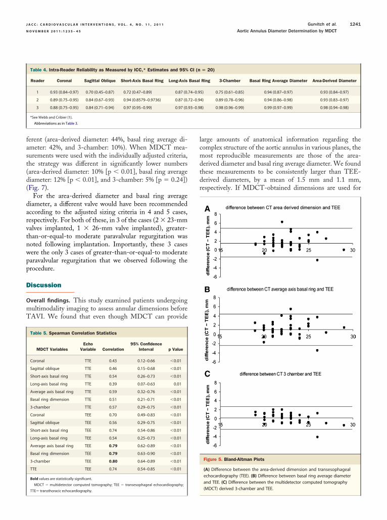
The difference between the TEE measurements andrea-derived diameter, basal ring average diameter, and-chamber dimensions were assessed using Bland-Altmanethods. The mean difference was greatest for the area-
erived diameter (1.5 �1.6 mm), followed by the basal ringverage diameter (1.1 � 1.7 mm) and the 3-chamberimension (0.2 � 1.2 mm). This is demonstrated by thecatters shown in the Bland-Altman plots (Fig. 6).Applicability of MDCT measurements. To evaluate the po-tential clinical applicability of MDCT measurements, westudied the theoretical choice of valve size to be im-planted if the annulus diameter was determined byMDCT. The analysis was performed using the area-derived diameter, basal ring average diameter, and3-chamber view. Given that we have shown these mea-surements to be larger than TEE diameters, a comparisonwas made between using these diameters with currentTEE-based sizing guidelines versus a strategy wherethese observed differences were incorporated into new“adjusted” sizing criteria.
To adjust the sizing criteria, the observed mean differencebetween each of these 3 imaging planes and TEE wereincorporated to current sizing guidelines. For example, thearea-derived diameter is larger than TEE by a mean of 1.5mm. This resulted in the following theoretical “adjusted”sizing criteria if using the MDCT area-derived diameter:23-mm valve for annulus �19.5 to �22.5 mm, 26-mmvalve for �22.5 to �26.5 mm, and 29-mm valve for �26.5o �29.5 mm (Table 6).
When MDCT measurements were used with currentizing criteria, the valve size indicated was frequently dif-
3 Readers (N � 50)
3-Chamber Basal Ring Average Diameter Area-Derived Diameter
0.70 (0.58–0.81) 0.80 (0.70–0.87) 0.87 (0.81–0.92)
I for
l Ring
82)
(
darvtnwpp
D
mT
lcmdtdr
J A C C : C A R D I O V A S C U L A R I N T E R V E N T I O N S , V O L . 4 , N O . 1 1 , 2 0 1 1 Gurvitch et al.
N O V E M B E R 2 0 1 1 : 1 2 3 5 – 4 5 Aortic Annulus Diameter Determination by MDCT
1241
ferent (area-derived diameter: 44%, basal ring average di-ameter: 42%, and 3-chamber: 10%). When MDCT mea-surements were used with the individually adjusted criteria,the strategy was different in significantly lower numbers(area-derived diameter: 10% [p � 0.01], basal ring averagediameter: 12% [p � 0.01], and 3-chamber: 5% [p � 0.24])Fig. 7).
For the area-derived diameter and basal ring averageiameter, a different valve would have been recommendedccording to the adjusted sizing criteria in 4 and 5 cases,espectively. For both of these, in 3 of the cases (2 � 23-mmalves implanted, 1 � 26-mm valve implanted), greater-han-or-equal-to moderate paravalvular regurgitation wasoted following implantation. Importantly, these 3 casesere the only 3 cases of greater-than-or-equal-to moderatearavalvular regurgitation that we observed following therocedure.
iscussion
Overall findings. This study examined patients undergoingultimodality imaging to assess annular dimensions beforeAVI. We found that even though MDCT can provide
Table 4. Intra-Reader Reliability as Measured by ICC,* Estimates and 95%
Reader Coronal Sagittal Oblique Short-Axis Basal Ring Long-Axis
1 0.93 (0.84–0.97) 0.70 (0.45–0.87) 0.72 (0.47–0.89) 0.87 (0.
2 0.89 (0.75–0.95) 0.84 (0.67–0.93) 0.94 (0.8579–0.9736) 0.87 (0.
3 0.88 (0.75–0.95) 0.84 (0.71–0.94) 0.97 (0.95–0.99) 0.97 (0.
*See Webb and Cribier (1).
Abbreviations as in Table 3.
Table 5. Spearman Correlation Statistics
MDCT VariablesEcho
Variable Correlation95% Confidence
Interval p Value
Coronal TTE 0.43 0.12–0.66 �0.01
Sagittal oblique TTE 0.46 0.15–0.68 �0.01
Short-axis basal ring TTE 0.54 0.26–0.73 �0.01
Long-axis basal ring TTE 0.39 0.07–0.63 0.01
Average axis basal ring TTE 0.59 0.32–0.76 �0.01
Basal ring dimension TTE 0.51 0.21–0.71 �0.01
3-chamber TTE 0.57 0.29–0.75 �0.01
Coronal TEE 0.70 0.49–0.83 �0.01
Sagittal oblique TEE 0.56 0.29–0.75 �0.01
Short-axis basal ring TEE 0.74 0.54–0.86 �0.01
Long-axis basal ring TEE 0.54 0.25–0.73 �0.01
Average axis basal ring TEE 0.79 0.62–0.89 �0.01
Basal ring dimension TEE 0.79 0.63–0.90 �0.01
3-chamber TEE 0.80 0.64–0.89 �0.01
TTE TEE 0.74 0.54–0.85 �0.01
Bold values are statistically significant.
MDCT � multidetector computed tomography; TEE � transesophageal echocardiography;
TTE� transthoracic echocardiography.
arge amounts of anatomical information regarding theomplex structure of the aortic annulus in various planes, theost reproducible measurements are those of the area-
erived diameter and basal ring average diameter. We foundhese measurements to be consistently larger than TEE-erived diameters, by a mean of 1.5 mm and 1.1 mm,espectively. If MDCT-obtained dimensions are used for
Figure 5. Bland-Altman Plots
(A) Difference between the area-derived dimension and transesophagealechocardiography (TEE). (B) Difference between basal ring average diameterand TEE. (C) Difference between the multidetector computed tomography
� 20)
Ring 3-Chamber Basal Ring Average Diameter Area-Derived Diameter
5) 0.75 (0.61–0.85) 0.94 (0.87–0.97) 0.93 (0.84–0.97)
4) 0.89 (0.78–0.96) 0.94 (0.86–0.98) 0.93 (0.83–0.97)
8) 0.98 (0.96–0.99) 0.99 (0.97–0.99) 0.98 (0.94–0.98)
CI (n
Basal
74–0.9
72–0.9
93–0.9
(MDCT) derived 3-chamber and TEE.

ascocb
sagit
J A C C : C A R D I O V A S C U L A R I N T E R V E N T I O N S , V O L . 4 , N O . 1 1 , 2 0 1 1
N O V E M B E R 2 0 1 1 : 1 2 3 5 – 4 5
Gurvitch et al.
Aortic Annulus Diameter Determination by MDCT
1242
valve sizing, incorporating these observed differences resultsin less, but persistent and potentially important discrepan-cies when compared with TEE-based cutoffs.Importance of accurate assessment of annular dimensions.The evolution of TAVI has brought the importance ofaccurate noninvasive assessment of the aortic annulus to a
Figure 6. Spearman Correlation Between Various MDCT Dimensions and TE
The 3-chamber dimensions have the highest correlation (r � 0.8), whereas the
Table 6. Valve Size Chosen According to Mode of Annulus Assessmentby TEE Versus MDCT Using Area-Derived Diameter
Imaging Modality andSizing Criteria
Valve Size Chosen
23 mm 26 mm 29 mm
TEE-guided 15 23 3
MDCT unadjusted 5 26 10
MDCT using adjusted criteria 13 23 5
In the top row, valve sizes were chosen according to TEE measurements and the following
guidelines for implantation: 23-mm valve for annulus �18.0 to �21.0 mm; 26-mm valve for �21.0
to �25.0 mm; and 29-mm valve for �25.0 to �28.0 mm. In the middle row, valve sizes were
chosen from the MDCT area-derived diameter according to the same cutoff guidelines for implan-
tation. In the bottom row, valve sizes were chosen from the MDCT area-derived diameter but
adjusted by 1.5 mm to account for the mean difference in this measurement from TEE, hence:
23-mm valve for annulus �19.5 to �22.5 mm; 26-mm valve for �22.5 to �26.5 mm; and 29-mm
valve for �26.5 to �29.5 mm.
bAbbreviations as in Table 5.
new spotlight. Previous work has shown that TEE diame-ters can underestimate the true diameter as measured atsurgery by a mean of 1.2 mm and frequently underestimateit by �2 mm (21). Despite this, operators have becomeaccustomed to echocardiography-guided TAVI with goodresults. Current implantation guidelines use TEE diametersto determine the size of the prosthesis to be implanted.However, aortic regurgitation, valve embolization, annularrupture, and patient prosthetic mismatch continue to occur,underscoring the importance of accurate annular evaluation.Even though MDCT appears to provide data on the trueanatomical shape and dimensions of the aortic annulus, itsapplicability has remained limited due partly to uncertaintyin incorporating the additional information into the clinicalcontext.Most useful MDCT measurements. In this study, we evalu-ted previously described MDCT-derived annular dimen-ions, including the area-derived diameter (11). The latteronsiders the entire directly measured circumferential areaf the complex shape of the annulus, and the mathemati-ally derived diameter attempts to provide a “diameter ofest fit” when provided a circular geometry as seen with a
tal oblique has the lowest (r � 0.56). Abbreviations as in Figure 5.
E
alloon expandable transcatheter valve. We found that in

Mes
atatwS3eCnAotbmMhstrawevFcaATs
incor
J A C C : C A R D I O V A S C U L A R I N T E R V E N T I O N S , V O L . 4 , N O . 1 1 , 2 0 1 1 Gurvitch et al.
N O V E M B E R 2 0 1 1 : 1 2 3 5 – 4 5 Aortic Annulus Diameter Determination by MDCT
1243
fact this area-derived diameter and the basal ring averagediameter were the most reproducible and reliable measure-ments obtained by MDCT. The 3-chamber measurementwas less reproducible, and the sagittal oblique diameter hadthe lowest interobserver reliability with an ICC of 0.54(95% CI: 0.39 to 0.69).
While the 3-chamber measurement was not as reproduc-ible, it had the best correlation with TEE diameters. This isto be expected given that the 3-chamber MDCT projectionin essence is an attempt to replicate the echocardiographicplane of measurement. However, in understanding MDCTdimensions, we need to move beyond attempting to repli-cate the more familiar TEE measurements. The lowercorrelation for the area-derived diameter and the basal ringaverage diameter is simply explained: they are measuringdifferent things and provide more anatomical informationthan does echocardiography.
The unifying feature of the less reproducible measures(sagittal, coronal, or 3-chamber) is that unlike the basal ringmeasurements, they represent single-plane dimensions ofwhat is usually a more complex oval structure. Subtleobliquity or tilting of the root can easily result in significantvariability. Given the elliptical nature of the aortic annulus,even minor changes in orientation can result in potentiallysignificant differences. The 3-chamber reconstruction re-quires additional dataset manipulation to generate and issubject to the same limitations as the coronal and sagittaloblique measures regarding orientation.Application of MDCT measurements. We found that if
DCT area-derived diameter or basal ring average diam-ter were used to guide valve size choice, a different valve
Figure 7. Impact on Valve Size Choice of Unadjusted Versus Adjusted Cuto
The graph demonstrates the effect of using MDCT-derived diameters on valveversus cutoffs in which the mean difference between MDCT and TEE has been
ize might have been selected in 10% to 12% of patients. In t
previous study, Messika-Zeitoun et al. (9) evaluated theheoretical impact of the method of measurement of thennulus diameter on the procedure, finding that when usinghe basal ring average diameter, MDCT measurementsould have modified the TAVI strategy in 38% of patients.imilarly, Tzikas et al. (10) found a change of strategy in6% of cases if the coronal dimension was used, and Schultzt al. (11) found that 39% would not be candidates for aoreValve prosthesis (Medtronic Inc., Minneapolis, Min-esota) if the long-axis diameter of the basal ring was used.n important difference in our study is that having dem-nstrated MDCT measurements to be consistently largerhan TEE measurements, these differences were integratedefore applying diameter criteria that are accepted for TEEeasurements. Using this strategy, we found that whenDCT was used “unadjusted,” the TAVI strategy might
ave been altered in similar frequencies to the previoustudies (42% to 44%), but when the criteria were “adjusted”o incorporate these differences, these were significantlyeduced to 10% to 12% (p � 0.01). These particular casesppear to be especially important. We found in the casesith residual discrepancies between MDCT and TEE that
xceeded these criteria, the frequency of moderate paraval-ular aortic regurgitation following TAVI was very high.or example, moderate regurgitation was noted in 3 of 4ases in which a larger valve size was indicated by MDCTs evaluated by “adjusted” area-derived diameter cutoffs.lthough the cause of aortic regurgitation followingAVI is multifactorial, this nevertheless raises the pos-
ibility that, in specific cases, TEE may underestimate
hoice according to current TEE-based cutoffs (unadjusted criteria [blue])porated (adjusted criteria [brown]). Abbreviations as in Figure 5.
ffs
size c
he true annular dimensions, thus contributing to selec-

J A C C : C A R D I O V A S C U L A R I N T E R V E N T I O N S , V O L . 4 , N O . 1 1 , 2 0 1 1
N O V E M B E R 2 0 1 1 : 1 2 3 5 – 4 5
Gurvitch et al.
Aortic Annulus Diameter Determination by MDCT
1244
tion of undersized prostheses and important paravalvularregurgitation.
Based on these findings, we propose that given theconsistent differences in certain measurements, currentmanufacturer-recommended sizing cutoffs that are based onTEE diameters might not be appropriate to directly trans-late to MDCT cutoffs. For example, 22 mm measured byTEE is not the same as 22 mm measured by the area-derived diameter from MDCT. The aim of our study is tohelp clinicians understand how to best apply MDCTmeasurements and help put them into practical clinicalcontext to aid decision making. We propose that if MDCTmeasurements are used to help guide valve size choice, newstrategies taking into account the differences in specificannular dimensions compared with TEE measurements arerequired. Such strategies should use adjusted criteria utiliz-ing the most reproducible MDCT measurements of thearea-derived diameter or basal ring average diameter.Study limitations. Although we conducted an analysis of thetheoretical choice of valve size according to different annularmeasurements, the choice of valve size is multifactorial.Factors, including annular diameter, degree and extent ofcalcification, symmetry of the leaflets, distance to thecoronaries, diameter of the sinotubular ridge, and curvatureof the sinuses of Valsalva might all affect final valve choice.We could not incorporate these factors into our analysis asthis would be beyond the scope of this study, and weconsidered valve size choice by annular dimensions alone.Also, our study used only the balloon expandable valve andthe results with regards to AR may not be applicable in thesame way to the self-expanding model. However, annulusdetermination is crucial regardless of the valve type chosen.
The MDCT scans and echocardiographic assessmentswere not always performed at exactly the same time of thecardiac cycle for each patient. The MDCT measurementswere, however, taken during systolic phases of the cardiaccycle at the point of maximum valve opening. As well,annulus size variation during the cardiac cycle is minimal(7,22), especially in patients with severely stenotic andrestricted valves.
Not all patients underwent valve implantation: 9 patientsare still awaiting a procedure. This relates to practicalconsiderations of our current waiting list as opposed to anyspecific factors that may affect the results or conclusions ofthis study.
Conclusions
MDCT imaging provides significant anatomical informa-tion regarding the complex elliptical shape of the aorticannulus. The most reproducible MDCT measurements arethe area-derived diameter and basal ring average diameter,with derived values generally larger than those obtained
with echocardiography. For MDCT to be used for valveselection, a strategy incorporating these differences might beimportant. MDCT using these easily derived measurementsmight be ideally suited to sizing transcatheter aortic valvesas they account for the eccentricity of the aortic annulus, arereproducible, and are noninvasive. Further larger studies arerequired to define the specific cutoffs of annular dimensionsas obtained by different imaging modalities for selection ofvalve size.
Reprint requests and correspondence: Dr. Jonathon Leipsic, StPaul’s Hospital, 1081 Burrard Street, Vancouver, British Colum-bia V6Z 1Y6, Canada. E-mail: [email protected].
REFERENCES
1. Webb J, Cribier A. Percutaneous transarterial aortic valve implanta-tion: what do we know? Eur Heart J 2011;32:104–7.
2. Tamburino C, Capodanno D, Ramondo A, et al. Incidence andpredictors of early and late mortality after transcatheter aortic valveimplantation in 663 patients with severe aortic stenosis. Circulation2011;123:299–308.
3. Grube E, Buellesfeld L, Mueller R, et al. Progress and current status ofpercutaneous aortic valve replacement: results of three device genera-tions of the CoreValve Revalving system. Circ Cardiovasc Interv2008;1:167–75.
4. Webb JG, Pasupati S, Humphries K, et al. Percutaneous transarterialaortic valve replacement in selected high-risk patients with aorticstenosis. Circulation 2007;116:755–63.
5. Piazza N, de Jaegere P, Schultz C, Becker AE, Serruys PW, AndersonRH. Anatomy of the aortic valvar complex and its implications fortranscatheter implantation of the aortic valve. Circ Cardiovasc Interv2008;1:74–81.
6. Schultz CJ, Moelker AD, Tzikas A, et al. Cardiac CT: necessary forprecise sizing for transcatheter aortic implantation. EuroIntervention2010;6 Suppl G:G6–13.
7. Tops LF, Wood DA, Delgado V, et al. Noninvasive evaluation of theaortic root with multislice computed tomography implications fortranscatheter aortic valve replacement. J Am Coll Cardiol Img 2008;1:321–30.
8. Wood DA, Tops LF, Mayo JR, et al. Role of multislice computedtomography in transcatheter aortic valve replacement. Am J Cardiol2009;103:1295–301.
9. Messika-Zeitoun D, Serfaty JM, Brochet E, et al. Multimodal assess-ment of the aortic annulus diameter: implications for transcatheteraortic valve implantation. J Am Coll Cardiol 2010;55:186–94.
10. Tzikas A, Schultz CJ, Piazza N, et al. Assessment of the aortic annulusby multislice computed tomography, contrast aortography, and trans-thoracic echocardiography in patients referred for transcatheter aorticvalve implantation. Catheter Cardiovasc Interv 2011;77:686–75.
11. Schultz CJ, Moelker A, Piazza N, et al. Three dimensional evaluationof the aortic annulus using multislice computer tomography: aremanufacturer’s guidelines for sizing for percutaneous aortic valvereplacement helpful? Eur Heart J 2010;31:849–56.
12. Gurvitch R, Wood DA, Leipsic J, et al. Multislice computed tomog-raphy for prediction of optimal angiographic deployment projectionsduring transcatheter aortic valve implantation. J Am Coll Cardiol Intv2010;3:1157–65.
13. Schultz C, Moelker A, Tzikas A, et al. The use of MSCT for theevaluation of the aortic root before transcutaneous aortic valve implan-tation: the Rotterdam approach. Euro Intervention 2010;6:505–11.
14. Jayasuriya C, Moss RR, Munt B. Transcatheter aortic valve implanta-tion in aortic stenosis: the role of echocardiography. J Am SocEchocardiogr 2011;24:15–27.
15. Moss RR, Ivens E, Pasupati S, et al. Role of echocardiography in percutaneous
aortic valve implantation. J Am Coll Cardiol Img 2008;1:15–24.
J A C C : C A R D I O V A S C U L A R I N T E R V E N T I O N S , V O L . 4 , N O . 1 1 , 2 0 1 1 Gurvitch et al.
N O V E M B E R 2 0 1 1 : 1 2 3 5 – 4 5 Aortic Annulus Diameter Determination by MDCT
1245
16. Webb JG, Chandavimol M, Thompson CR, et al. Percutaneous aorticvalve implantation retrograde from the femoral artery. Circulation2006;113:842–50.
17. Webb JG, Altwegg L, Masson JB, Al Bugami S, Al Ali A, Boone RA.A new transcatheter aortic valve and percutaneous valve deliverysystem. J Am Coll Cardiol 2009;53:1855–8.
18. Walther T, Dewey T, Borger MA, et al. Transapical aortic valveimplantation: step by step. Ann Thorac Surg 2009;87:276–83.
19. Baumgartner H, Hung J, Bermejo J, et al. Echocardiographic assess-ment of valve stenosis: EAE/ASE recommendations for clinical prac-tice. J Am Soc Echocardiogr 2009;22:1–23, quiz 101–2.
20. Hankinson SE, Manson JE, Spiegelman D, Willett WC, Longcope C,Speizer FE. Reproducibility of plasma hormone levels in postmeno-
pausal women over a 2–3-year period. Cancer Epidemiol BiomarkersPrev 1995;4:649–54.
21. Babaliaros VC, Liff D, Chen EP, et al. Can balloon aortic valvuloplastyhelp determine appropriate transcatheter aortic valve size? J Am CollCardiol Intv 2008;1:580–6.
22. Shiran A, Adawi S, Ganaeem M, Asmer E. Accuracy and reproduc-ibility of left ventricular outflow tract diameter measurement usingtransthoracic when compared with transesophageal echocardiographyin systole and diastole. Eur J Echocardiogr 2009;10:319–24.
Key Words: aortic annulus � aortic stenosis � computed
tomography � transcatheter aortic valve implantation.