Il rapporto tra le chiose dell'Anonimo Lombardo al Purgatorio e il Commento di Iacomo della Lana
This article appeared in a journal published by Elsevier. The attachedcopy is furnished to the author for internal non-commercial researchand education use, including for instruction at the authors institution
and sharing with colleagues.
Other uses, including reproduction and distribution, or selling orlicensing copies, or posting to personal, institutional or third party
websites are prohibited.
In most cases authors are permitted to post their version of thearticle (e.g. in Word or Tex form) to their personal website orinstitutional repository. Authors requiring further information
regarding Elsevier’s archiving and manuscript policies areencouraged to visit:
http://www.elsevier.com/authorsrights
Author's personal copy
Annals of Anatomy 195 (2013) 401– 408
Contents lists available at ScienceDirect
Annals of Anatomy
jo ur nal ho mepage: www.elsev ier .de /aanat
Research article
Multidetector CT investigation of the mummy of Rosalia Lombardo (1918–1920)
Stephanie Panzera,∗, Heather Gill-Frerkingb, Wilfried Rosendahlc, Albert R. Zinkd,Dario Piombino-Mascalid
a Department of Radiology, Trauma Center Murnau, Biomechanics Laboratory, Paracelsus Medical University Salzburg and Trauma Center Murnau, Prof.-Küntscher-Strasse 8,D-82418 Murnau, Germanyb Mummies of the World Touring Company, NTK Services, P.O. Box 545, Epsom, NH 03234, USAc Reiss-Engelhorn-Museen, C5 Zeughaus, D-68159 Mannheim, Germanyd EURAC-Institute for Mummies and the Iceman, Viale Druso 1, I-39100 Bolzano, Italy
a r t i c l e i n f o
Article history:Received 23 September 2012Received in revised form11 November 2012Accepted 5 March 2013
Keywords:MummyRadiologyComputed tomographyHistoryPaleopathologyEmbalmingPneumonia
s u m m a r y
Whole-body multidetector computed tomography (CT) was performed on the mummified corpse of two-year-old Rosalia Lombardo, an anthropogenic mummy displayed in the Capuchin Catacombs of Palermo,Sicily, Italy. Rosalia Lombardo reportedly died of bronchopneumonia in 1920 and was preserved by theembalmer and taxidermist Alfredo Salafia with a formaldehyde-based fluid. Rosalia Lombardo’s body isstill exhibited in the Capuchin Catacombs inside the original glass-topped coffin in which she was placed.Only her head is visible: the rest of her body is covered by a sheet.
CT images of Rosalia’s body within her coffin were of reduced quality because of distinct metal artifactscaused by the coffin itself. Nevertheless, a detailed radiological analysis was possible for most of the body.Analysis of the data from the CT examination revealed indicators for the historically-reported endovasaland intracavity treatment. Rosalia’s entire body was preserved in a remarkable state. The exceptionalpreservation of her internal organs made it possible to consider a radiological diagnosis of pneumonia.
For this study, CT was determined to be the ultimate method for investigation, since Rosalia’s body hadto be kept untouched in her sealed coffin for conservation purposes. The CT examination offered newinsights into the current preservation status of the body, and the superior contrast of CT allowed detailedassessment of different tissues. Post-processing methods provided reconstructions on any desired plane,as well as three-dimensional reconstruction, for the best possible visualization and interpretation of thebody.
© 2013 Elsevier GmbH. All rights reserved.
1. Introduction
Rosalia Lombardo, who has also been referred to as the “Sicil-ian Sleeping Beauty”, is probably one of the best preserved bodiesin the Capuchin Catacombs of Palermo, Sicily, Italy. The daughterof military officer Mario Lombardo (1890–1980) and Maria Di Cara(1897–1966), Rosalia was born on December 13, 1918, and diedon December 6, 1920, only a week before her second birthday.According to the post-mortem report, she died of bronchopneu-monia. She was “temporarily” brought to the Capuchin Catacombsof Palermo on December 8, 1920, where she remains today (Farella,1982; Piombino-Mascali, 2009; Piombino-Mascali et al., 2010).
∗ Corresponding author.E-mail addresses: [email protected] (S. Panzer),
[email protected] (H. Gill-Frerking), [email protected](W. Rosendahl), [email protected] (A.R. Zink), [email protected](D. Piombino-Mascali).
The Lombardos decided to have their daughter’s corpse pre-served by Professor Alfredo Salafia (1869–1933) prior to herentombment. Salafia, a self-taught taxidermist and embalmer,was responsible for the preparation of many prominent citizensof Palermo from 1902 onwards. The procedure he devised wascharacterized by simplicity, consisting of a single-point injection,preferably into the femoral artery via a gravity injector. Other pro-cedures normally adopted in modern embalming, such as blooddrainage, cavity treatment or additional injections, were believed tobe unnecessary in most cases (Piombino-Mascali, 2009; Piombino-Mascali et al., 2009).
In September 2007, a handwritten memoir by Salafia revealedthe “secret” formula of his preservative. The chemical formulaconsisted of 7 L of one part glycerin, one part formalin saturatedwith both zinc sulfate and chloride, and one part of an alcoholsolution saturated with salicylic acid. Since Salafia developed hispreservative in the early 1900s, it was suggested that his “Perfec-tion Fluid” was one of the earliest formaldehyde-based formulasduring the transition from “old” to “modern” embalming chemicals– a process eventually leading to the replacement of heavy metals.
0940-9602/$ – see front matter © 2013 Elsevier GmbH. All rights reserved.http://dx.doi.org/10.1016/j.aanat.2013.03.009
Author's personal copy
402 S. Panzer et al. / Annals of Anatomy 195 (2013) 401– 408
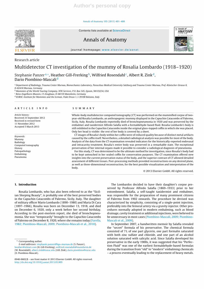
Fig. 1. (A) The body of Rosalia Lombardo, preserved by Alfredo Salafia with hismethod of embalming in 1920. The body is covered by a sheet, with only the headvisible. (B) Close-up of the face of Rosalia Lombardo.
Additionally, Salafia’s memoir also disclosed his occasional useof paraffin wax diluted in ether, which was introduced into thedeceased’s face in order to keep the features plump and lifelike(Piombino-Mascali, 2009).
Rosalia Lombardo’s body is still exhibited in the original glass-topped coffin in which she was placed. Only her head is visible; therest of her body is covered by a sheet. Her face shows a remarkablestate of preservation – she still looks as though she is alive andsleeping (Fig. 1). In the past few years, however, signs of oxidationand decay have become more obvious. Specifically, the child’s hairand accompanying textiles have become lighter, while her face hasdarkened and has shrunk.
Evaluation of anteroposterior (AP) radiographs of the mummy(Panzer et al., 2010) taken in July 2008, revealed that not onlythe head, but the entire body was preserved. Additionally, sev-eral preserved organs were discernible on the radiographs. InDecember 2010, we had the opportunity to investigate Rosalia’sbody by multidetector computed tomography (CT). This technique,considered a “gold standard” in mummy studies, was used to
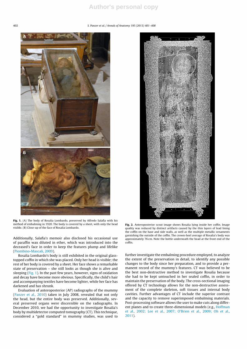
Fig. 2. Anteroposterior scout image shows Rosalia lying inside her coffin. Imagequality was reduced by distinct artifacts caused by the thin layers of lead liningthe coffin on the base and side walls, as well as the multiple metallic ornamentsgarnishing the outside of the coffin. The crown-heel average of Rosalia’s body wasapproximately 76 cm. Note the bottle underneath the head at the front end of thecoffin.
further investigate the embalming procedure employed, to analyzethe extent of the preservation in detail, to identify any possiblechanges to the body since her preparation, and to provide a per-manent record of the mummy’s features. CT was believed to bethe best non-destructive method to investigate Rosalia becauseshe had to be kept untouched in her sealed coffin, in order tomaintain the preservation of the body. The cross-sectional imagingoffered by CT technology allows for the non-destructive assess-ment of the complete skeleton, soft tissues and internal bodycavities. Further advantages of CT include the superior contrastand the capacity to remove superimposed embalming materials.Post-processing software allows the user to make cuts along differ-ent planes and to create three-dimensional models (e.g., Hoffmanet al., 2002; Lee et al., 2007; O’Brien et al., 2009; Oh et al.,2011).
Author's personal copy
S. Panzer et al. / Annals of Anatomy 195 (2013) 401– 408 403
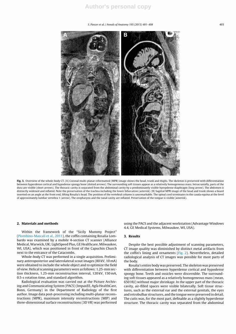
Fig. 3. Overview of the whole-body CT. (A) Coronal multi-planar reformatted (MPR) image shows the head, trunk and thighs. The skeleton is preserved with differentiationbetween hyperdense cortical and hypodense spongy bone (dotted arrows). The surrounding soft tissues appear as a relatively homogeneous mass. Intracranially, parts of thedura are visible (short arrows). The thoracic cavity is separated from the abdominal cavity by a predominantly visible hyerpdense diaphragm (long arrow). The abdomen isdistinctly widened and inflated. Note the preservation of the trachea including the lower bifurcation (asterisk). (B) Sagittal MPR image of the head and trunk shows a boardinserted on an angle at the front end, lifting Rosalia’s head. The position of the vertebral column is unremarkable. The spinal cord terminates in the cauda equina at the levelof approximately lumbar vertebra 1 (arrow). The oropharynx and the nasal cavity are inflated. Preservation of the tongue is visible (asterisk).
2. Materials and methods
Within the framework of the “Sicily Mummy Project”(Piombino-Mascali et al., 2011), the coffin containing Rosalia Lom-bardo was examined by a mobile 4-section CT scanner (AllianceMedical, Warwick, UK; LightSpeed Plus, GE Healthcare, Milwaukee,WI, USA), which was positioned in front of the Capuchin Churchnext to the entrance of the Catacombs.
Whole-body CT was performed in a single acquisition. Prelimi-nary anteroposterior and laterolateral scout images (80 kV, 10 mA)were obtained to include the whole object and to optimize the fieldof view. Helical scanning parameters were as follows: 1.25-mm sec-tion thickness, 1.25-mm reconstruction interval, 120 kV, 150 mA,0.5-s rotation time, and standard algorithm.
Radiological evaluation was carried out at the Picture Archiv-ing and Communicating System (PACS) (ImpaxEE, Agfa HealthCare,Bonn, Germany) in the Department of Radiology of the firstauthor. Image data post-processing including multi-planar recons-tructions (MPR), maximum intensity reconstructions (MIP) andthree-dimensional surface reconstructions (3D VR) was performed
using the PACS and the adjacent workstation (Advantage Windows4.4, GE Medical Systems, Milwaukee, WI, USA).
3. Results
Despite the best possible adjustment of scanning parameters,CT image quality was diminished by distinct metal artifacts fromthe coffin’s lining and ornaments (Fig. 2). Nevertheless, detailedradiological analysis of CT images was possible for most parts ofthe body.
Rosalia’s entire body was preserved. The skeleton was preservedwith differentiation between hyperdense cortical and hypodensespongy bone. Teeth and ossicles were discernible. The surround-ing soft tissues appeared as a relatively homogeneous mass (mean,650 HU) without major shrinkage. In the upper part of the thoraciccavity, air-filled spaces were visible bilaterally. Soft tissue struc-tures, such as the external ear and the external genitals, the eyesand retrobulbar structures, and the tongue were preserved in detail.The cutis was, for the most part, definable as a slightly hyperdensestructure. The thoracic cavity was separated from the abdominal
Author's personal copy
404 S. Panzer et al. / Annals of Anatomy 195 (2013) 401– 408
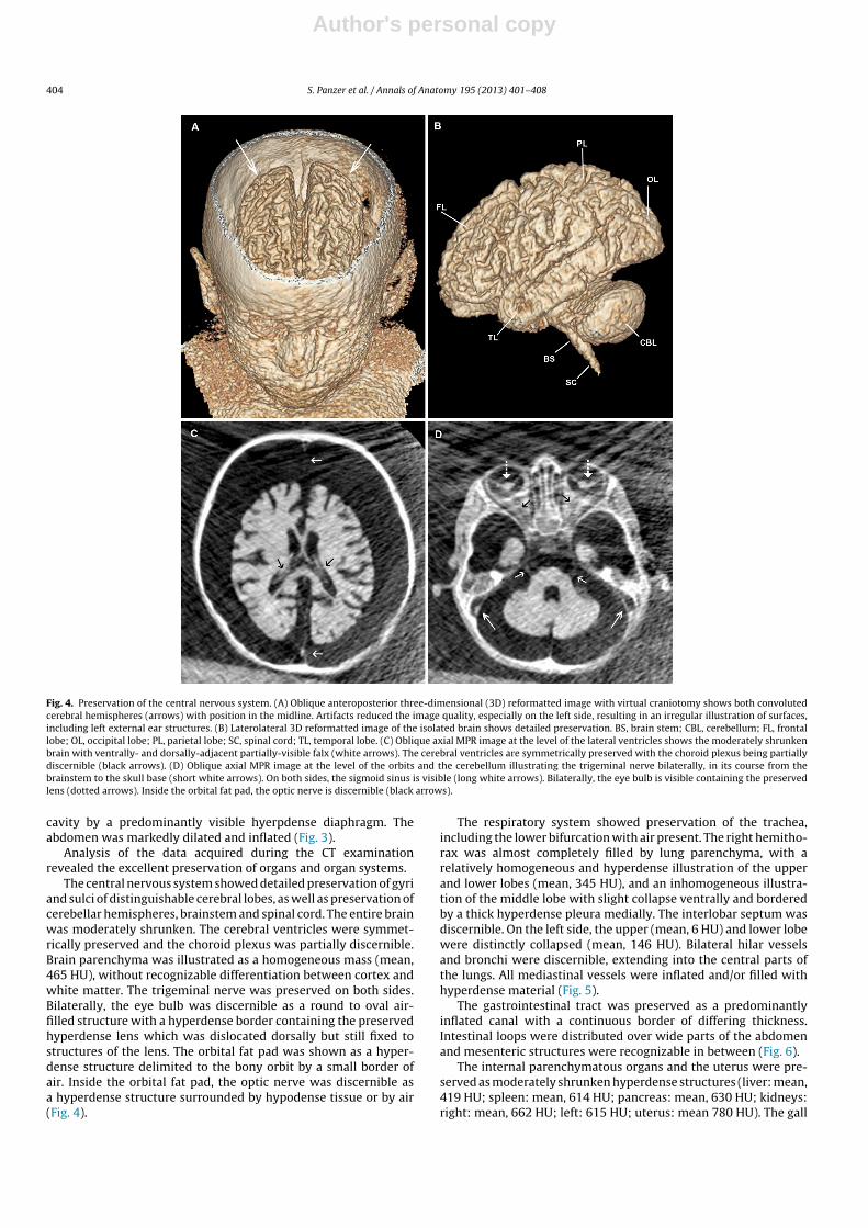
Fig. 4. Preservation of the central nervous system. (A) Oblique anteroposterior three-dimensional (3D) reformatted image with virtual craniotomy shows both convolutedcerebral hemispheres (arrows) with position in the midline. Artifacts reduced the image quality, especially on the left side, resulting in an irregular illustration of surfaces,including left external ear structures. (B) Laterolateral 3D reformatted image of the isolated brain shows detailed preservation. BS, brain stem; CBL, cerebellum; FL, frontallobe; OL, occipital lobe; PL, parietal lobe; SC, spinal cord; TL, temporal lobe. (C) Oblique axial MPR image at the level of the lateral ventricles shows the moderately shrunkenbrain with ventrally- and dorsally-adjacent partially-visible falx (white arrows). The cerebral ventricles are symmetrically preserved with the choroid plexus being partiallydiscernible (black arrows). (D) Oblique axial MPR image at the level of the orbits and the cerebellum illustrating the trigeminal nerve bilaterally, in its course from thebrainstem to the skull base (short white arrows). On both sides, the sigmoid sinus is visible (long white arrows). Bilaterally, the eye bulb is visible containing the preservedlens (dotted arrows). Inside the orbital fat pad, the optic nerve is discernible (black arrows).
cavity by a predominantly visible hyerpdense diaphragm. Theabdomen was markedly dilated and inflated (Fig. 3).
Analysis of the data acquired during the CT examinationrevealed the excellent preservation of organs and organ systems.
The central nervous system showed detailed preservation of gyriand sulci of distinguishable cerebral lobes, as well as preservation ofcerebellar hemispheres, brainstem and spinal cord. The entire brainwas moderately shrunken. The cerebral ventricles were symmet-rically preserved and the choroid plexus was partially discernible.Brain parenchyma was illustrated as a homogeneous mass (mean,465 HU), without recognizable differentiation between cortex andwhite matter. The trigeminal nerve was preserved on both sides.Bilaterally, the eye bulb was discernible as a round to oval air-filled structure with a hyperdense border containing the preservedhyperdense lens which was dislocated dorsally but still fixed tostructures of the lens. The orbital fat pad was shown as a hyper-dense structure delimited to the bony orbit by a small border ofair. Inside the orbital fat pad, the optic nerve was discernible asa hyperdense structure surrounded by hypodense tissue or by air(Fig. 4).
The respiratory system showed preservation of the trachea,including the lower bifurcation with air present. The right hemitho-rax was almost completely filled by lung parenchyma, with arelatively homogeneous and hyperdense illustration of the upperand lower lobes (mean, 345 HU), and an inhomogeneous illustra-tion of the middle lobe with slight collapse ventrally and borderedby a thick hyperdense pleura medially. The interlobar septum wasdiscernible. On the left side, the upper (mean, 6 HU) and lower lobewere distinctly collapsed (mean, 146 HU). Bilateral hilar vesselsand bronchi were discernible, extending into the central parts ofthe lungs. All mediastinal vessels were inflated and/or filled withhyperdense material (Fig. 5).
The gastrointestinal tract was preserved as a predominantlyinflated canal with a continuous border of differing thickness.Intestinal loops were distributed over wide parts of the abdomenand mesenteric structures were recognizable in between (Fig. 6).
The internal parenchymatous organs and the uterus were pre-served as moderately shrunken hyperdense structures (liver: mean,419 HU; spleen: mean, 614 HU; pancreas: mean, 630 HU; kidneys:right: mean, 662 HU; left: 615 HU; uterus: mean 780 HU). The gall
Author's personal copy
S. Panzer et al. / Annals of Anatomy 195 (2013) 401– 408 405
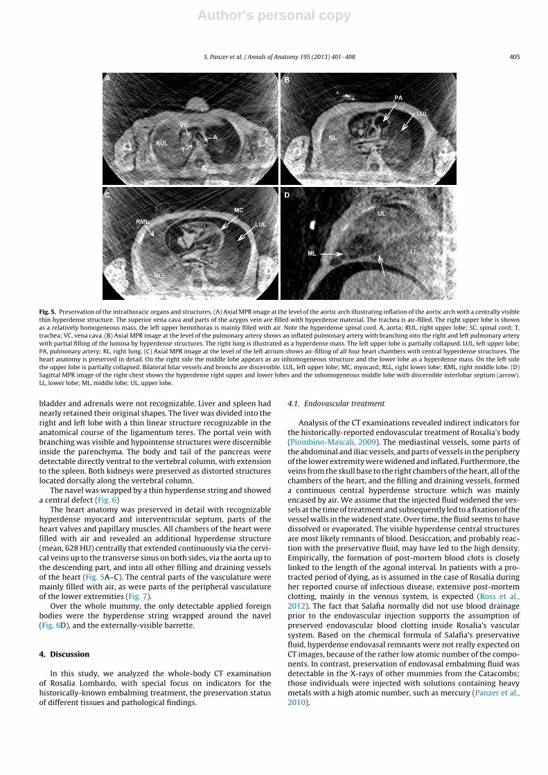
Fig. 5. Preservation of the intrathoracic organs and structures. (A) Axial MPR image at the level of the aortic arch illustrating inflation of the aortic arch with a centrally visiblethin hyperdense structure. The superior vena cava and parts of the azygos vein are filled with hyperdense material. The trachea is air-filled. The right upper lobe is shownas a relatively homogeneous mass, the left upper hemithorax is mainly filled with air. Note the hyperdense spinal cord. A, aorta; RUL, right upper lobe; SC, spinal cord; T,trachea; VC, vena cava. (B) Axial MPR image at the level of the pulmonary artery shows an inflated pulmonary artery with branching into the right and left pulmonary arterywith partial filling of the lumina by hyperdense structures. The right lung is illustrated as a hyperdense mass. The left upper lobe is partially collapsed. LUL, left upper lobe;PA, pulmonary artery; RL, right lung. (C) Axial MPR image at the level of the left atrium shows air-filling of all four heart chambers with central hyperdense structures. Theheart anatomy is preserved in detail. On the right side the middle lobe appears as an inhomogeneous structure and the lower lobe as a hyperdense mass. On the left sidethe upper lobe is partially collapsed. Bilateral hilar vessels and bronchi are discernible. LUL, left upper lobe; MC, myocard; RLL, right lower lobe; RML, right middle lobe. (D)Sagittal MPR image of the right chest shows the hyperdense right upper and lower lobes and the inhomogeneous middle lobe with discernible interlobar septum (arrow).LL, lower lobe; ML, middle lobe; UL, upper lobe.
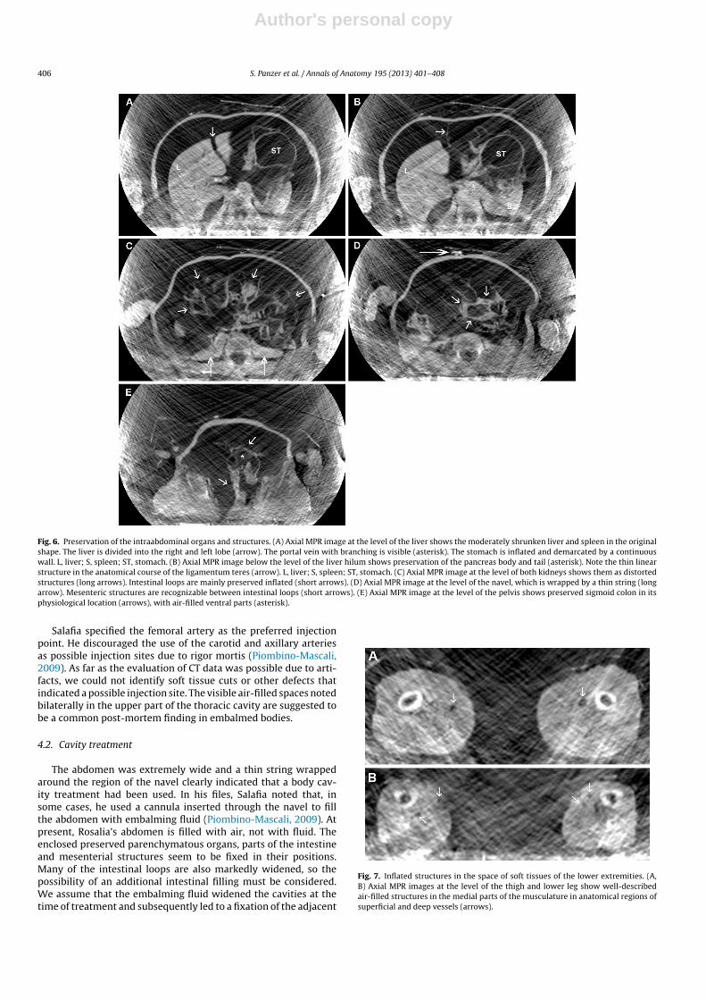
bladder and adrenals were not recognizable. Liver and spleen hadnearly retained their original shapes. The liver was divided into theright and left lobe with a thin linear structure recognizable in theanatomical course of the ligamentum teres. The portal vein withbranching was visible and hypointense structures were discernibleinside the parenchyma. The body and tail of the pancreas weredetectable directly ventral to the vertebral column, with extensionto the spleen. Both kidneys were preserved as distorted structureslocated dorsally along the vertebral column.
The navel was wrapped by a thin hyperdense string and showeda central defect (Fig. 6)
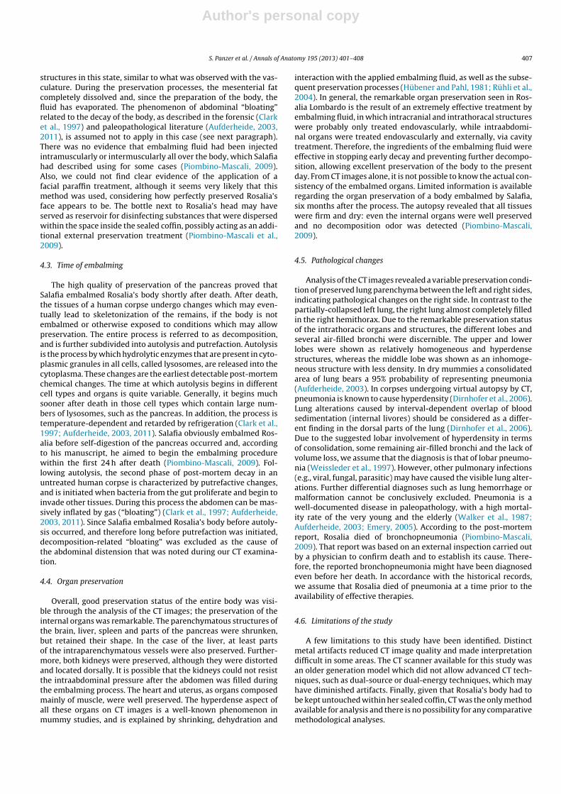
The heart anatomy was preserved in detail with recognizablehyperdense myocard and interventricular septum, parts of theheart valves and papillary muscles. All chambers of the heart werefilled with air and revealed an additional hyperdense structure(mean, 628 HU) centrally that extended continuously via the cervi-cal veins up to the transverse sinus on both sides, via the aorta up tothe descending part, and into all other filling and draining vesselsof the heart (Fig. 5A–C). The central parts of the vasculature weremainly filled with air, as were parts of the peripheral vasculatureof the lower extremities (Fig. 7).
Over the whole mummy, the only detectable applied foreignbodies were the hyperdense string wrapped around the navel(Fig. 6D), and the externally-visible barrette.
4. Discussion
In this study, we analyzed the whole-body CT examinationof Rosalia Lombardo, with special focus on indicators for thehistorically-known embalming treatment, the preservation statusof different tissues and pathological findings.
4.1. Endovascular treatment
Analysis of the CT examinations revealed indirect indicators forthe historically-reported endovascular treatment of Rosalia’s body(Piombino-Mascali, 2009). The mediastinal vessels, some parts ofthe abdominal and iliac vessels, and parts of vessels in the peripheryof the lower extremity were widened and inflated. Furthermore, theveins from the skull base to the right chambers of the heart, all of thechambers of the heart, and the filling and draining vessels, formeda continuous central hyperdense structure which was mainlyencased by air. We assume that the injected fluid widened the ves-sels at the time of treatment and subsequently led to a fixation of thevessel walls in the widened state. Over time, the fluid seems to havedissolved or evaporated. The visible hyperdense central structuresare most likely remnants of blood. Desiccation, and probably reac-tion with the preservative fluid, may have led to the high density.Empirically, the formation of post-mortem blood clots is closelylinked to the length of the agonal interval. In patients with a pro-tracted period of dying, as is assumed in the case of Rosalia duringher reported course of infectious disease, extensive post-mortemclotting, mainly in the venous system, is expected (Ross et al.,2012). The fact that Salafia normally did not use blood drainageprior to the endovascular injection supports the assumption ofpreserved endovascular blood clotting inside Rosalia’s vascularsystem. Based on the chemical formula of Salafia’s preservativefluid, hyperdense endovasal remnants were not really expected onCT images, because of the rather low atomic number of the compo-nents. In contrast, preservation of endovasal embalming fluid wasdetectable in the X-rays of other mummies from the Catacombs;those individuals were injected with solutions containing heavymetals with a high atomic number, such as mercury (Panzer et al.,2010).
Author's personal copy
406 S. Panzer et al. / Annals of Anatomy 195 (2013) 401– 408
Fig. 6. Preservation of the intraabdominal organs and structures. (A) Axial MPR image at the level of the liver shows the moderately shrunken liver and spleen in the originalshape. The liver is divided into the right and left lobe (arrow). The portal vein with branching is visible (asterisk). The stomach is inflated and demarcated by a continuouswall. L, liver; S, spleen; ST, stomach. (B) Axial MPR image below the level of the liver hilum shows preservation of the pancreas body and tail (asterisk). Note the thin linearstructure in the anatomical course of the ligamentum teres (arrow). L, liver; S, spleen; ST, stomach. (C) Axial MPR image at the level of both kidneys shows them as distortedstructures (long arrows). Intestinal loops are mainly preserved inflated (short arrows). (D) Axial MPR image at the level of the navel, which is wrapped by a thin string (longarrow). Mesenteric structures are recognizable between intestinal loops (short arrows). (E) Axial MPR image at the level of the pelvis shows preserved sigmoid colon in itsphysiological location (arrows), with air-filled ventral parts (asterisk).
Salafia specified the femoral artery as the preferred injectionpoint. He discouraged the use of the carotid and axillary arteriesas possible injection sites due to rigor mortis (Piombino-Mascali,2009). As far as the evaluation of CT data was possible due to arti-facts, we could not identify soft tissue cuts or other defects thatindicated a possible injection site. The visible air-filled spaces notedbilaterally in the upper part of the thoracic cavity are suggested tobe a common post-mortem finding in embalmed bodies.
4.2. Cavity treatment
The abdomen was extremely wide and a thin string wrappedaround the region of the navel clearly indicated that a body cav-ity treatment had been used. In his files, Salafia noted that, insome cases, he used a cannula inserted through the navel to fillthe abdomen with embalming fluid (Piombino-Mascali, 2009). Atpresent, Rosalia’s abdomen is filled with air, not with fluid. Theenclosed preserved parenchymatous organs, parts of the intestineand mesenterial structures seem to be fixed in their positions.Many of the intestinal loops are also markedly widened, so thepossibility of an additional intestinal filling must be considered.We assume that the embalming fluid widened the cavities at thetime of treatment and subsequently led to a fixation of the adjacent
Fig. 7. Inflated structures in the space of soft tissues of the lower extremities. (A,B) Axial MPR images at the level of the thigh and lower leg show well-describedair-filled structures in the medial parts of the musculature in anatomical regions ofsuperficial and deep vessels (arrows).
Author's personal copy
S. Panzer et al. / Annals of Anatomy 195 (2013) 401– 408 407
structures in this state, similar to what was observed with the vas-culature. During the preservation processes, the mesenterial fatcompletely dissolved and, since the preparation of the body, thefluid has evaporated. The phenomenon of abdominal “bloating”related to the decay of the body, as described in the forensic (Clarket al., 1997) and paleopathological literature (Aufderheide, 2003,2011), is assumed not to apply in this case (see next paragraph).There was no evidence that embalming fluid had been injectedintramuscularly or intermuscularly all over the body, which Salafiahad described using for some cases (Piombino-Mascali, 2009).Also, we could not find clear evidence of the application of afacial paraffin treatment, although it seems very likely that thismethod was used, considering how perfectly preserved Rosalia’sface appears to be. The bottle next to Rosalia’s head may haveserved as reservoir for disinfecting substances that were dispersedwithin the space inside the sealed coffin, possibly acting as an addi-tional external preservation treatment (Piombino-Mascali et al.,2009).
4.3. Time of embalming
The high quality of preservation of the pancreas proved thatSalafia embalmed Rosalia’s body shortly after death. After death,the tissues of a human corpse undergo changes which may even-tually lead to skeletonization of the remains, if the body is notembalmed or otherwise exposed to conditions which may allowpreservation. The entire process is referred to as decomposition,and is further subdivided into autolysis and putrefaction. Autolysisis the process by which hydrolytic enzymes that are present in cyto-plasmic granules in all cells, called lysosomes, are released into thecytoplasma. These changes are the earliest detectable post-mortemchemical changes. The time at which autolysis begins in differentcell types and organs is quite variable. Generally, it begins muchsooner after death in those cell types which contain large num-bers of lysosomes, such as the pancreas. In addition, the process istemperature-dependent and retarded by refrigeration (Clark et al.,1997; Aufderheide, 2003, 2011). Salafia obviously embalmed Ros-alia before self-digestion of the pancreas occurred and, accordingto his manuscript, he aimed to begin the embalming procedurewithin the first 24 h after death (Piombino-Mascali, 2009). Fol-lowing autolysis, the second phase of post-mortem decay in anuntreated human corpse is characterized by putrefactive changes,and is initiated when bacteria from the gut proliferate and begin toinvade other tissues. During this process the abdomen can be mas-sively inflated by gas (“bloating”) (Clark et al., 1997; Aufderheide,2003, 2011). Since Salafia embalmed Rosalia’s body before autoly-sis occurred, and therefore long before putrefaction was initiated,decomposition-related “bloating” was excluded as the cause ofthe abdominal distension that was noted during our CT examina-tion.
4.4. Organ preservation
Overall, good preservation status of the entire body was visi-ble through the analysis of the CT images; the preservation of theinternal organs was remarkable. The parenchymatous structures ofthe brain, liver, spleen and parts of the pancreas were shrunken,but retained their shape. In the case of the liver, at least partsof the intraparenchymatous vessels were also preserved. Further-more, both kidneys were preserved, although they were distortedand located dorsally. It is possible that the kidneys could not resistthe intraabdominal pressure after the abdomen was filled duringthe embalming process. The heart and uterus, as organs composedmainly of muscle, were well preserved. The hyperdense aspect ofall these organs on CT images is a well-known phenomenon inmummy studies, and is explained by shrinking, dehydration and
interaction with the applied embalming fluid, as well as the subse-quent preservation processes (Hübener and Pahl, 1981; Rühli et al.,2004). In general, the remarkable organ preservation seen in Ros-alia Lombardo is the result of an extremely effective treatment byembalming fluid, in which intracranial and intrathoracal structureswere probably only treated endovascularly, while intraabdomi-nal organs were treated endovascularly and externally, via cavitytreatment. Therefore, the ingredients of the embalming fluid wereeffective in stopping early decay and preventing further decompo-sition, allowing excellent preservation of the body to the presentday. From CT images alone, it is not possible to know the actual con-sistency of the embalmed organs. Limited information is availableregarding the organ preservation of a body embalmed by Salafia,six months after the process. The autopsy revealed that all tissueswere firm and dry: even the internal organs were well preservedand no decomposition odor was detected (Piombino-Mascali,2009).
4.5. Pathological changes
Analysis of the CT images revealed a variable preservation condi-tion of preserved lung parenchyma between the left and right sides,indicating pathological changes on the right side. In contrast to thepartially-collapsed left lung, the right lung almost completely filledin the right hemithorax. Due to the remarkable preservation statusof the intrathoracic organs and structures, the different lobes andseveral air-filled bronchi were discernible. The upper and lowerlobes were shown as relatively homogeneous and hyperdensestructures, whereas the middle lobe was shown as an inhomoge-neous structure with less density. In dry mummies a consolidatedarea of lung bears a 95% probability of representing pneumonia(Aufderheide, 2003). In corpses undergoing virtual autopsy by CT,pneumonia is known to cause hyperdensity (Dirnhofer et al., 2006).Lung alterations caused by interval-dependent overlap of bloodsedimentation (internal livores) should be considered as a differ-ent finding in the dorsal parts of the lung (Dirnhofer et al., 2006).Due to the suggested lobar involvement of hyperdensity in termsof consolidation, some remaining air-filled bronchi and the lack ofvolume loss, we assume that the diagnosis is that of lobar pneumo-nia (Weissleder et al., 1997). However, other pulmonary infections(e.g., viral, fungal, parasitic) may have caused the visible lung alter-ations. Further differential diagnoses such as lung hemorrhage ormalformation cannot be conclusively excluded. Pneumonia is awell-documented disease in paleopathology, with a high mortal-ity rate of the very young and the elderly (Walker et al., 1987;Aufderheide, 2003; Emery, 2005). According to the post-mortemreport, Rosalia died of bronchopneumonia (Piombino-Mascali,2009). That report was based on an external inspection carried outby a physician to confirm death and to establish its cause. There-fore, the reported bronchopneumonia might have been diagnosedeven before her death. In accordance with the historical records,we assume that Rosalia died of pneumonia at a time prior to theavailability of effective therapies.
4.6. Limitations of the study
A few limitations to this study have been identified. Distinctmetal artifacts reduced CT image quality and made interpretationdifficult in some areas. The CT scanner available for this study wasan older generation model which did not allow advanced CT tech-niques, such as dual-source or dual-energy techniques, which mayhave diminished artifacts. Finally, given that Rosalia’s body had tobe kept untouched within her sealed coffin, CT was the only methodavailable for analysis and there is no possibility for any comparativemethodological analyses.
Author's personal copy
408 S. Panzer et al. / Annals of Anatomy 195 (2013) 401– 408
5. Conclusions
The use of whole-body CT examination of the mummy of RosaliaLombardo enabled new insights into the preserved body. Anal-ysis of the CT data provided new information in comparison tothe only medical images previously available: AP radiographs ofa mummy for which only the face and head were visible inside aglass-topped coffin, revealing the preservation of the entire bodyand at least of some organs. Despite the disadvantageous situationcaused by the artifacts due to the coffin’s lead lining and orna-ments, the cross-sectional technique and superior contrast of CTallowed detailed assessment of different tissues. Post-processingmethods provided reconstructions on any desired plane, as well as3D reconstruction, for the most accurate interpretation of findings.Analysis of the data from the CT examination also revealed indi-cators for the historically-reported endovascular and intracavitytreatments. Rosalia’s entire body was in an excellent state of preser-vation, especially the internal organs. The radiological diagnosisof probable pneumonia was possible only due to the exceptionalcondition of the organs. Nevertheless, our investigation representsan assessment only 90 years after Rosalia’s death and subsequentembalming treatment. It is not possible to determine what spe-cific changes occurred at which point in time. Furthermore, it isimpossible to identify what change may occur in the future, orwhen.
Acknowledgments
We are most grateful to the Order of the Capuchin Friars ofPalermo for granting us permission to investigate this mummy.We are also grateful to the Superintendence for the Culturaland Environmental Heritage of Palermo for advocating and sup-porting the project. This study was made possible thanks tothe generosity of Dr. Dariusch Rafiy, 3W Productions, Munich,Germany.
Appendix A. Supplementary data
Supplementary data associated with this article can befound, in the online version, at http://dx.doi.org/10.1016/j.aanat.2013.03.009.
References
Aufderheide, A.C., 2003. The Scientific Study of Mummies. Cambridge UniversityPress, Cambridge, UK, pp. 308, 438.
Aufderheide, A.C., 2011. Soft tissue taphonomy: a paleopathology perspective. Int.J. Paleopathol. 1, 75–80.
Clark, M.A., Worrell, M.B., Pless, J.E., 1997. Postmortem changes in soft tissues. In:Haglund, W.D., Sorg, M.H. (Eds.), Forensic Taphonomy. CRC Press, New York, pp.151–164.
Dirnhofer, R., Jackowski, C., Vock, P., Potter, K., Thali, M.J., 2006. Virtopsy: minimallyinvasive, imaging-guided virtual autopsy. Radiographics 26, 1305–1333.
Emery, A.E., 2005. The mummy state. Clin. Med. 5 (2), 153.Farella, F.D., 1982. Cenni Storici della Chiesa e delle Catacombe dei Cappuccini di
Palermo. Fiamma Serafica, Palermo, Italy, pp. 101–102.Hoffman, H., Torres, W.E., Ernst, R.D., 2002. Paleoradiology: advanced CT in the
evaluation of nine Egyptian mummies. Radiographics 22, 377–385.Hübener, K.H., Pahl, W.M., 1981. Computertomographische Untersuchungen an
altägyptischen Mumien. Fortschr Röntgenstr 135, 213–219.Lee, I.S., Kim, M.J., Yoo, D.S., Lee, Y.S., Park, S.S., Bok, G.D., Han, S.H., Chung, Y.H.,
Chang, B.S., Yi, Y.S., Oh, C.S., Shin, D.H., 2007. Three-dimensional reconstructionof medieval child mummy in Yangju, Korea, using multi-detector computedtomography. Ann. Anat. 189, 558–568.
O’Brien, J.J., Battista, J.J., Romagnoli, C., Chhem, R.K., 2009. CT imaging of humanmummies: a critical review of the literature (1979–2005). Int. J. Osteoarchaeol.19, 90–98.
Oh, C.S., Lee, S.Y., Lee, I.S., Kim, Y.S., Koh, K.S., Shin, D.H., 2011. Differential find-ings in post-factum dissections of medieval Korean mummies exhibiting similarpreservation patterns on computerized tomography images. Ann. Anat. 193,544–549.
Panzer, S., Zink, A.R., Piombino-Mascali, D., 2010. Radiologic evidence ofanthropogenic mummification in the Capuchin Catacombs of Palermo, Sicily.Radiographics 30 (4), 1123–1132.
Piombino-Mascali, D., 2009. Il Maestro del Sonno Eterno. La Zisa, Palermo, Italy, pp.50–62, 72–78.
Piombino-Mascali, D., Aufderheide, A.C., Johnson Williams, M., Zink, A.R., 2009. TheSalafia method rediscovered. Virchows Arch. 454 (3), 355–357.
Piombino-Mascali, D., Aufderheide, A.C., Panzer, S., Zink, A.R., 2010. Mummies fromPalermo. In: Wieczorek, A., Rosendahl, W. (Eds.), Mummies of the World. Prestel,Munich, London, New York, pp. 357–361.
Piombino-Mascali, D., Panzer, S., Marvelli, S., Lösch, S., Aufderheide, A.C., Zink, A.R.,2011. The “Sicily Mummy Project”: first results of the scientific campaigns(2007–2010). In: Sörries, R. (Ed.), Geschichte und Tradition der Mumifizierungin Europa, vol. 18. Kasseler Studien zur Sepulkralkultur, pp. 25–31.
Ross, S.G., Thali, M.J., Bolliger, S., Germerott, T., Ruder, T.D., Flach, P.M., 2012. Sud-den death after chest pain: feasibility of virtual autopsy with postmortem CTangiography and biopsy. Radiology 264 (1), 250–259.
Rühli, F.J., Chhem, R.K., Böni, T., 2004. Diagnostic paleoradiology of mummifiedtissue: interpretation and pitfalls. JACR 55 (4), 218–227.
Walker, R., Parsche, F., Bierbrier, M., McKerrow, J.H., 1987. Tissue identification andhistological study of six lung specimens from Egyptian mummies. Am. J. Phys.Anthropol. 72, 43–48.
Weissleder, R., Rieumont, M.J., Wittenberg, J. (Eds.), 1997. Primer of DiagnosticImaging. , 2nd ed. Mosby, St. Louis, pp. 9–11.
Copyright © 2022 FDOKUMEN