Paravalvular Leak After Transcatheter Aortic Valve Replacement
Upload
independentCategory
view
0download
0
Journal of the American College of Cardiology Vol. 62, No. 5, 2013� 2013 by the American College of Cardiology Foundation ISSN 0735-1097/$36.00Published by Elsevier Inc. http://dx.doi.org/10.1016/j.jacc.2013.04.036
The Impact of Integration of a MultidetectorComputed Tomography Annulus Area SizingAlgorithm on Outcomes of TranscatheterAortic Valve Replacement
A Prospective, Multicenter, Controlled TrialRonald K. Binder, MD,* John G. Webb, MD,* Alexander B. Willson, MBBS,* Marina Urena, MD,yNicolaj C. Hansson, MD,z Bjarne L. Norgaard, MD, PHD,z Philippe Pibarot, MD,yMarco Barbanti, MD,* Eric Larose, MD,y Melanie Freeman, MBBS,* Eric Dumont, MD,yChris Thompson, MD,* Miriam Wheeler, MBCHB,* Robert R. Moss, MD,* Tae-hyun Yang, MD,*
Sergio Pasian, MD,y Cameron J. Hague, MD,* Giang Nguyen, MD,* Rekha Raju, MD,*
Stefan Toggweiler, MD,* James K. Min, MD,x David A. Wood, MD,k Josep Rodés-Cabau, MD,yJonathon Leipsic, MD*
Vancouver and Quebec City, Canada; Aarhus, Denmark; and Los Angeles, California
From the *S
Columbia, C
City, Queb
xCedars-SinHospital, U
Edwards Li
collection, d
Objectives T
t. Paul’s Hospital, Univ
anada; yQuebec Heart
ec, Canada; zAarhus Un
ai Heart Institute, Los A
niversity of British Colu
fesciences funded the im
ata analysis, or manusc
his study prospectively investigated the impact of integration of a multidetector computed tomography (MDCT)annular area sizing algorithm on transcatheter aortic valve replacement (TAVR) outcomes.
Background A
ppreciation of the 3-dimensional, noncircular geometry of the aortic annulus is important for transcatheter heartvalve (THV) sizing.Methods P
atients being evaluated for TAVR in 4 centers underwent pre-procedural MDCT. Recommendations for balloon-expandable THV size selection were based on an MDCT sizing algorithm with an optimal goal of modest annulusarea oversizing (5% to 10%). Consecutive patients who underwent TAVR with the algorithm (MDCT group) werecompared with consecutive patients without the algorithm (control group). The primary endpoint was the incidenceof more than mild paravalvular regurgitation (PAR), and the secondary endpoint was the composite of in-hospitaldeath, aortic annulus rupture, and severe PAR.Results O
f 266 patients, 133 consecutive patients underwent TAVR (SAPIEN XT THV) in the MDCT group and 133consecutive patients were in the control group. More than mild PAR was present in 5.3% (7 of 133) of the MDCTgroup and in 12.8% (17 of 133) in the control group (p ¼ 0.032). The combined secondary endpoint occurred in3.8% (5 of 133) of the MDCT group and in 11.3% (15 of 133) of the control group (p ¼ 0.02), driven by thedifference of severe PAR.Conclusions T
he implementation of an MDCT annulus area sizing algorithm for TAVR reduces PAR. Three-dimensional aorticannular assessment and annular area sizing should be considered for TAVR. (J Am Coll Cardiol 2013;62:431–8)ª 2013 by the American College of Cardiology FoundationThe aortic annulus is a complex, 3-dimensional, nearlyuniformly oval-shaped structure (1). Appreciation of its geo-metry is important for appropriate transcatheter heart valve
ersity of British Columbia, Vancouver, British
and Lung Institute, Laval University, Quebec
iversity Hospital Skejby, Aarhus, Denmark;
ngeles, California; and the kVancouver General
mbia, Vancouver, British Columbia, Canada.
age transfer but had no involvement in data
ript writing. Drs. Binder, Webb, Norgaard,
(THV) size selection (1,2). For transcatheter aortic valve re-placement (TAVR), the dimensions of the aortic annulus havebeen traditionally described by a single diameter measurement
Dumont, Wood, Rodés-Cabau, and Leipsic are consultants to Edwards Lifesciences.
Drs. Binder and Toggweiler received unrestricted research grants from the Swiss
National Foundation. Drs. Hansson and Pibarot received research grants from
Edwards Lifesciences. All other authors have reported that they have no relationships
relevant to the contents of this paper to disclose.
Manuscript received March 7, 2013; revised manuscript received April 13, 2013,
accepted April 23, 2013.

Abbreviationsand Acronyms
MDCT = multidetector
computed tomography
PAR = paravalvular
regurgitation
TAVR = transcatheter aortic
valve replacement
TEE = transesophageal
echocardiography
THV = transcatheter heart
valve
TTE = transthoracic
echocardiography
Binder et al. JACC Vol. 62, No. 5, 2013Computed Tomography Area Sizing for TAVR July 30, 2013:431–8
432
from 2-dimensional transesopha-geal echocardiography (TEE) (3).However, the appropriateness ofthis method for THV sizing hasrecently been questioned (1,4,5).Inappropriate THV size selectionis associated with paravalvularregurgitation (PAR) (2,4), aorticannular rupture (6), coronaryocclusion (7), and device emboli-zation (8). These complicationsstrongly impact morbidity andmortality after TAVR (7). Three-dimensional assessments of theaortic annulus by TEE (9,10) or
multidetector computed tomography (MDCT) have beenshown to predict PAR (2,4) because of better appreciation ofits complex 3-dimensional structure. Our group previouslyfound that MDCT annular area oversizing was the strongestpredictor for PAR (2) in a retrospective, multicenter analysis.However, it is unknown whether this knowledge can betranslated into improved clinical outcomes when practicallyapplied by a readily available, reproducible MDCT annulusarea sizing algorithm (5).We therefore performed a prospec-tive, multicenter, controlled trial to evaluate the impact ofintegration of an MDCT annular area sizing algorithm onTAVR outcomes (5).
Methods
Patients with high surgical risk or inoperable symptomaticsevere aortic stenosis, planed forTAVR in4 experienced centers(St. Paul’sHospital,University ofBritishColumbia,Vancouver,British Columbia, Canada; Quebec Heart and Lung Institute,Laval University, Quebec City, Quebec, Canada; AarhusUniversity Hospital Skejby, Aarhus, Denmark; and VancouverGeneral Hospital, University of British Columbia, Vancouver,British Columbia, Canada) were considered for this trial,which was approved by the local ethical committees. Exclusioncriteria were an estimated glomerular filtration rate of less than35 ml/min (except for patients on chronic hemodialysis),previous aortic valve replacement (“valve-in-valve”), andTAVRwith a THV other than the SAPIEN XT balloon-expandablevalve (Edwards Lifesciences, Irvine, California). Prospectivelyenrolled, consecutive patients who underwent TAVR with theimplementation of the MDCT annular area sizing algorithm(MDCT group) were compared with consecutive patients whounderwent TAVR without the algorithm (control group) priorto trial initiation. For both groups, the availableTHVsizes were20-, 23-, 26-, and 29-mm diameters. Each center contributedequal numbers of patients to theMDCTgroup and the controlgroup. Because the trial was initiated in the beginning of 2012,consecutive patients in the control group were treated in 2011just before trial initiation. Sizing of the THV in the controlgroup was based on an integration of echocardiographymeasurements and angiography (without standardized
measurements) but not on 3-dimensional MDCT areaassessment.Study endpoints. The primary endpoint was the incidenceof more than mild PAR, and the secondary endpoint was thecomposite of in-hospital death, aortic annular rupture (as amarker of excessive oversizing), and severe PAR (as a markerof excessive undersizing). Study endpoint analysis was per-formed by intention to treat.MDCT and THV size selection. All MDCT scans wereread by 1 experienced level 3 cardiac MDCT reader atSt. Paul’s Hospital (MDCT core laboratory) in concert and byconsensus with the local site. Examinations were performedwith either a 64-slice scanner (Discovery HD 750, GEHealthcare, Waukesha, Wisconsin) or a second-generationdual-source CT system (Siemens Somatom Definition Flash,Siemens Healthcare, Erlangen, Germany). The protocol forthe single-source scanner included the injection of 80 to120 ml of radio-contrast medium at 5 ml/s followed by30 ml of normal saline (2). Electrocardiogram-gated dosemodulation was used, but heart rate reduction with beta-blockade was not performed. Depending on patient size,maximum tube current ranged between 450 and 700 mAwith a fixed tube voltage of 100 kVp for patients with a bodymass index (BMI)<30 kg/m2 and 120 kVp for larger patients.For the dual-source scanner, a contrast-enhanced MDCTexamination in the caudocranial direction with retrospectivegating was performed. Commercially available contrastmedia (ioversol 350 mg/ml, Mallinckrodt Inc., Hazelwood,Missouri) was used (20 ml for the test bolus and 70 ml for thespiral scan). Contrast injection was followed by a 50-ml salineflush. Heart rate reduction with beta-blockade was not per-formed. MDCT was performed with a 128 � 0.625 mmcollimation, z-flying spot, gantry rotation time 280 ms, andscan pitch 0.20 to 0.40 (depending on heart rate). Maximumtube current ranged from 450 to 750 mA with fixed tubepotential of 100 (BMI <30 kg/m2) or 120 kV (BMI >30kg/m2). Electrocardiogram-controlled tube current modula-tion was applied with reduction of the current to 20% and fullpulsing applied only from 30% to 70% of the RR interval.
MDCT annular area measurements were performed insystole at 25% or 35% of the RR interval when the annulus isthe largest (5), depending on which of the 2 phase recon-structions displayed better image quality. Recommendationsfor THV size were based on anMDCT sizing algorithm withan optimal goal of modest annulus area oversizing (nominalTHV area/MDCT annular area ¼ 5% to 10%) (Table 1) (5).The algorithm ensures routine oversizing of the MDCTaortic annular area (nominal THV area > MDCT annulararea), calculating a percentage of annular oversizing (nominalTHV area/MDCT annular area). Nominal external THVareas for the 20-, 23-, 26-, and 29-mm SAPIEN XT THVare 3.14 cm2, 4.15 cm2, 5.31 cm2, and 6.61 cm2, respectively.Implantation with nominally filled deployment balloonswas performed by full injection of the indeflator volume.When more than 20% area oversizing was anticipated(or more than 15% oversizing in the presence of adverse aortic

Table 1Multidetector Computed Tomography Annular AreaSizing Algorithm
Annular Area, mm2
Percentage of Annular Area Oversizing, %
20-mmTHV
23-mmTHV
26-mmTHV
29-mmTHV
230 NR
240 NR (30.9)
250 25.7 UE
260 20.8 UE
270 16.4
280 12.2
290 8.3
300 4.7
310 1.3 NR
320 NR (�1.9%) 29.8 UE
330 25.9 UE
340 22.2 UE
350 18.7
360 15.4
370 12.3
380 9.3
390 6.5
400 3.9 NR
410 1.3 NR (29.5)
420 NR (�1.1) 26.4 UE
430 23.5 UE
440 20.7 UE
450 18.0
460 15.4
470 13.0
480 10.6
490 8.4
500 6.2
510 4.1 NR
520 2.1 NR (27.0)
530 0.2 24.6 UE
540 22.3 UE
550 20.1 UE
560 17.9
570 15.9
580 13.9
590 12.0
600 10.1
610 8.3
620 6.5
630 4.8
640 3.2
650 1.6
660 0.1
670 NR
The recommendation is to select a prosthesis with a cross-sectional area modestly larger than thatof the aortic annulus. A target of 5% to 10% area oversizing with upper and lower limits of 1% to20% is suggested. The highlighted grey zones represent annular areas in which valve selection ischallenging. Selecting the larger valve will result in oversizing >20% and the smaller valve willresult in undersizing. Oversizing the annulus >20% may increase the risk of annular injury. Optionsinclude underfilling the transcatheter heart value balloon or selecting a smaller valve size with anattendant risk of paravalvular regurgitation.NR ¼ not recommended; UE ¼ underexpansion.
JACC Vol. 62, No. 5, 2013 Binder et al.July 30, 2013:431–8 Computed Tomography Area Sizing for TAVR
433
root features), intentional underexpansion of the THV wassuggested. This was accomplished by reducing the volume offluid within the valve deployment balloon by 10%. Adverse
root features included more than minimal left ventricularoutflow tract calcification and shallow sinuses of Valsalva.Although 5% to 10% area oversizing was considered optimal,most patients will not meet this target due to the largeincrements between manufactured prosthesis sizes. Wetherefore considered a range from 1% to 15% (20% in theabsence of adverse root features) as acceptable margins in thesizing algorithm, with the integration of balloon underfillingfor those who would require oversizing in excess of 20%by area.
In the MDCT group, TAVR operators were informedabout the MDCT area sizing recommendation before theprocedure. Operators were not obliged to follow the MDCTarea sizing recommendations but could choose the THV sizethey considered most appropriate.Echocardiography. Intraprocedural TEE was routinely per-formed by level 3 echocardiographers highly experienced inTAVR assessments. The aortic annulus was 2-dimensionallyimaged by periprocedural TEE in a 3-chamber view, anda single diameter was documented. Echocardiographers wereblinded toward the MDCT measurements and the MDCTsizing recommendation. Prior to discharge, transthoracicechocardiography (TTE) was performed to assess PAR,valve area, and transvalvular gradients. The grade of PAR wasrated as none, mild, moderate, or severe according to ValveAcademic Research Consortium criteria (11).Statistical analysis. Analyses were performed using SPSSstatistics software (version 20.0, SPSS Inc., Chicago, Illi-nois). Continuous variables are reported as mean � SD andcategorical variables by percentages. Continuous variableswere tested for a normal distribution (QQ plot) andcompared by the Student t test. For comparison of morethan 2 continuous parametric variables, an analysis of vari-ance was used. Categorical variables were compared by theFisher exact test. All tests were 2-tailed, and a p value <0.05was considered statistically significant.
Results
Baseline characteristics. Of 266 patients in the trial, 133consecutive patients underwent TAVR with the MDCTsizing algorithm recommendation (MDCT group) and 133consecutive patients without the algorithm (control group)(University of British Columbia (2 sites): 166 patients; LavalUniversity: 56 patients; Aarhus University Hospital: 44patients) (Fig. 1). Baseline characteristics are shown inTable 2. Patients in the control group had a 1.2% higherSociety of Thoracic Surgeons score (p ¼ 0.007), whichreflects the broadening of TAVR indications to lower-riskpatients over the study period.Valve selection and procedure. All patients received aSAPIEN XT THV (Edwards Lifesciences) with valve sizesof 20-, 23-, 26-, or 29-mm diameter available for bothgroups. Prosthesis sizes and procedural characteristics areshown in Table 3. Within the MDCT group, sizingrecommendations were followed in 80.5% of patients (107 of133). In the 26 patients for which MDCT recommendations

Figure 1 Study Flowchart
MDCT ¼ multidetector computed tomography; SPH ¼ St. Paul’s Hospital; TAVR ¼ transcatheter aortic valve replacement; VHG ¼ Vancouver General Hospital.
Binder et al. JACC Vol. 62, No. 5, 2013Computed Tomography Area Sizing for TAVR July 30, 2013:431–8
434
were not followed, the implanted THV size was chosenbecause of conflicting echocardiography recommendationsin 14 patients, balloon aortic valvuloplasty assessment in2 patients, and operator preference (prosthesis size indiscordance with MDCT as well as echocardiographyrecommendations) in 10 patients. In 9 patients, the MDCTalgorithm would have recommended a larger size and in 17patients a smaller THV size. In this group (26 patients), therewere no cases of severe PAR, no annulus rupture, and noin-hospital mortality. Compared with the control group,the MDCT group was more often associated with theuse of an underfilled deployment balloon (13.5% vs. 0%;p < 0.001) and implantation of the largest available (29-mmdiameter) prosthesis (27.1% vs. 16.5%; p ¼ 0.038). Whetherdeployment balloons were nominally filled or underfilled didnot influence the rates of more than mild PAR (9.3% vs.5.6%; p ¼ 0.595), aortic annulus rupture (0.8% vs. 0%;p ¼ 0.702), THV embolization (0.8% vs. 0%; p ¼ 0.702), orpost-dilation (13.3% vs. 5.6%; p ¼ 0.342) (Fig. 2).Post-implant echocardiography. Pre-discharge TTE doc-umented comparable valve function in both MDCT andcontrol groups. Aortic valve area was 1.6 � 0.3 cm2 versus1.6 � 0.4 cm2 (p ¼ 0.923) and mean gradient 10.0 � 3.8mm Hg versus 9.9 � 4.6 mm Hg (p ¼ 0.96) in the MDCTgroup versus control group, respectively. However, more thanmild PAR was less common in the MDCT group: 5.3% (7 of133) versus 12.8% (17 of 133) (p ¼ 0.032). Similarly, severePAR was less common in the MDCT group: 0% versus 4.5%(6 of 133) (p ¼ 0.013) (Fig. 3). The degree of annulus areaoversizing was significantly associated with a reduction inPAR. Annulus area oversizing was 16.4 � 12.1% in patientswith no PAR, 10.4 � 10.7% in patients with mild PAR, and0.5� 9.4% in patients with more thanmild PAR (p¼ 0.001).
In as-treated analysis, more than mild PAR occurred nearlytwice as often in the control group (6 of 107 patients [5.6%] vs.15 of 159 [9.4%]; p ¼ 0.111). There was no difference inbaseline aortic regurgitation grades between the MDCTgroup and the control group (Table 2). The rates of more thanmild post-procedural PAR did not differ between patientswith or without more than mild pre-procedural aortic regur-gitation (19 of 225 patients [8.4%] vs. 5 of 41 [12.2%]; p ¼0.389). Of the patients with severe pre-procedural aorticregurgitation, none had more than mild post-proceduralPAR.Outcomes. Procedural outcomes are shown in Table 3.There were 5 (3.8%) in-hospital deaths in the MDCT groupand 9 (6.8%) in the control group (p ¼ 0.272). The rates ofannular rupture were 0.8% versus 0.8% (p ¼ 1.0). Thecombined secondary endpoint of in-hospital mortality, aorticannular rupture, and severe PAR occurred in 3.8% (5 of 133)in the MDCT group and 11.3% (15 of 133) in the controlgroup (p ¼ 0.02). In as-treated analysis, the secondaryendpoint occurred twice as often in the control group (5 of107 patients [4.7%] vs. 15 of 159 [9.4%]; p ¼ 0.149), albeitwithout statistical significance. There was no significantdifference in the occurrence of the primary endpointbetween the first and second halves of the control group(17.6% vs. 7.7%; p ¼ 0.119) or the first and second halves ofthe MDCT group (2.9% vs. 7.7%; p ¼ 0.267) (Table 4).
Discussion
In this prospective, multicenter, controlled trial, the inte-gration of an MDCT annular area sizing algorithm reducedthe incidence of more than mild PAR and the compositeendpoint of in-hospital death, aortic annular rupture, and

Table 3Annulus, Echocardiography Assessments, andProcedural Characteristics
MDCT Group(n ¼ 133)
Control Group(n ¼ 133) p Value
Left ventricular ejectionfraction, %
53 � 14 51 � 15 0.175
Mean gradient, mm Hg 42 � 18 38 � 15 0.055
Aortic valve area, cm2 0.7 � 0.2 0.7 � 0.2 0.440
TEE annulus diameter, mm 22.9 � 2.2 22.5 � 3.0 0.224
MDCT short-axis diameter, mm 21.5 � 2.2 n/a n/a
MDCT long-axis diameter, mm 27.3 � 2.9 n/a n/a
MDCT mean diameter, mm 24.4 � 2.3 n/a n/a
MDCT annulus area, cm2 4.8 � 0.8 n/a n/a
Access type
Transfemoral 74 (99) 71 (95) 0.581
Transapical 18 (24) 28 (37) 0.058
Transaortic 8 (10) 1 (1) 0.006
Labeled prosthesis size
20 mm 0.8 (1) 1.5 (2) 0.561
23 mm 26.3 (35) 30.8 (41) 0.415
26 mm 45.9 (61) 51.1 (68) 0.390
29 mm 27.1 (36) 16.5 (22) 0.038
MDCT area oversizing, % 11.6 � 11.5 n/a n/a
Underfilled deployment balloon 13.5 (18) 0 < 0.001
Values are mean � SD or % (n).n/a ¼ not applicable; TEE ¼ transesophageal echocardiography; other abbreviation as in Table 2.
Table 2 Patient Baseline Characteristics
MDCT Group(n ¼ 133)
Control Group(n ¼ 133) p Value
Age, yrs 82 � 8 81 � 8 0.207
Male 57 (76) 63 (84) 0.316
Height, cm 168 � 10 168 � 9 0.991
Weight, kg 77 � 18 75 � 18 0.464
BMI, kg/m2 27 � 6 27 � 6 0.414
Diabetes 32 (43) 35 (47) 0.604
Hypertension 84 (112) 79 (105) 0.268
Dyslipidemia 68 (90) 74 (99) 0.224
Smoking history 43 (58) 38 (50) 0.318
COPD 38 (50) 26 (35) 0.065
NYHA functional class 0.088
Class I 1 (1) 0
Class II 27 (36) 16 (21)
Class III 63 (84) 76 (101)
Class IV 9 (12) 8 (11)
Pre-procedural aorticregurgitation
0.926
None 29.3 (39) 21.8 (29)
Mild 55.6 (74) 62.4 (83)
Moderate 13.5 (18) 14.3 (19)
Severe 1.5 (2) 1.5 (2)
Prior cerebrovascular accident 16 (21) 14 (19) 0.732
Prior open heart surgery 29 (39) 40 (53) 0.094
Porcelain aorta 11 (15) 10 (13) 0.689
Prior permanent pacemaker 12 (16) 13 (17) 0.852
Prior myocardial infarction 21 (28) 30 (40) 0.092
Hemodialysis 3 (4) 2 (3) 0.702
Pulmonary hypertension 40 (53) 29 (38) 0.039
Peripheral vascular disease 20 (26) 19 (25) 0.876
Atrial fibrillation 38 (51) 39 (52) 0.900
STS PROM, % 5.8 � 3.2 7.0 � 3.5 0.007
GFR, ml/min 62 � 24 53 � 20 0.001
Values are mean � SD or % (n).BMI ¼ body mass index; COPD ¼ chronic obstructive pulmonary disease; GFR ¼ glomerular
filtration rate; MDCT ¼ multidetector computed tomography; NYHA ¼ New York Heart Association;PCI ¼ percutaneous coronary intervention; STS PROM ¼ Society of Thoracic Surgeons PredictedRisk of Mortality.
JACC Vol. 62, No. 5, 2013 Binder et al.July 30, 2013:431–8 Computed Tomography Area Sizing for TAVR
435
severe PAR, driven by the difference in severe PAR. Thestrength of our study manifests in a clear, reproducible, andreadily available sizing algorithm, which was prospectivelyimplemented for THV sizing in 4 TAVR centers (3 inCanada and 1 in Europe) and evaluated in a large cohort ofconsecutive patients.
Although TAVR is an approved alternative to surgicalaortic valve replacement in selected patients (12,13), thewidening of its application to lower-risk patients may behampered by complications such as PAR and aortic rootinjury. These TAVR complications can, to some extent, beattributed to inappropriate THV size selection. Operatorsperforming TAVR have to rely on indirect measurements ofthe aortic annulus for annular sizing and THV selection.Since its inception, the traditional measurement of the aorticannulus for TAVR was a single diameter from 2-dimensionalTEE imaging (3), but recently this method has been chal-lenged by 3-dimensional annular assessments with MDCT
(2,4,5,14). In comparison to sizing by direct surgicalinspection of the aortic annulus as a gold standard, somestudies have shown superiority of MDCT over echocardio-graphic measurements (15,16). Our knowledge of thecomplex 3-dimensional geometry of the consistently oval-shaped annulus is expanding (1), and new sizing algorithmsthat address the limitations of 2-dimensional imaging areneeded (5). A single-center retrospective study by Jilaihawiet al. (4) compared the annulus area–derived diameter fromMDCT with the TEE diameter and found that MDCTsizing reduced the incidence of PAR. Another study byHayashida et al. (17) implemented MDCT sizing prospec-tively in 40 patients and found a lower rate of PAR comparedwith that in a retrospective cohort of patients treated withTEE sizing. They studied the cross-sectional maximumdiameter, area-derived diameter, and perimeter-deriveddiameter. Although there was a small, prospective cohort intheir retrospective, single-center trial, this did not translateinto a clear and practical sizing algorithm. In contrast, in ourstudy, a clear and reproducible algorithm was appliedprospectively, which was found to be practical and useful byTAVR operators across different centers for THV selection.MDCT versus TEE. It was not the aim of this trial toperform a head-to-head comparison of TEE sizing versusMDCT sizing. Operators were not bound by recommenda-tions from one or the other imaging modality but were able tofreely choose the THV size based on all available information,including the MDCT area sizing recommendation, TEEmeasurements, TTE evaluation, and aortic root angiograms.For borderline cases, an aortic root angiogram during balloonaortic valvuloplasty may also provide valuable insight for

Figure 2Pre- and Post-TAVR MDCT in a Patient With Adverse Root Features and Intentional Underexpansionof the Transcatheter Heart Valve
Because of adverse aortic root features (A, B) (large calcium nodule in the left ventricular outflow tract), the 26-mm transcatheter heart valve was intentionally underexpanded by
removing 10% of the nominal deployment balloon volume. With this strategy, there was more controlled annular stretch, and displacement of the calcium nodule toward the
ventricular septum was avoided (C), thereby reducing the risk of aortic annulus injury. Abbreviations as in Figure 1.
Binder et al. JACC Vol. 62, No. 5, 2013Computed Tomography Area Sizing for TAVR July 30, 2013:431–8
436
the aortic annular dimensions (18). Because there is no pro-spective, randomized comparison of MDCT versus TEEsizing reported in the literature, it is unknown whether THVsizing by one of these imaging modalities may be superior.We do not think that it is prudent to randomize patientsbetween imaging modalities nor to blind the operator toany pre-procedural data because TAVR remains a complexprocedure with results that are supported by as muchpre-procedural data as possible. Importantly, though, ourdata do confirm the benefit of integration of 3-dimensionalMDCT measurements on TAVR-related outcomes, em-phasizing their importance in THV selection and solidifyingtheir role in TAVR.Rationale for annulus area sizing. We elected to useannular area for sizing because we have consistently found itto be the most reproducible annular measurement (1,2,5)
Figure 3Pre- and Post-TAVR Imaging in an 80-Year-Old Male PatienIntegration of the MDCT Sizing Guideline Showing the Valu
(A) Double oblique transverse reformat of the basal ring/annulus on MDCT with a mean a
echocardiogram suggests an annular diameter of 21.0 mm. (C) A 23-mm balloon-expanda
significant resultant paravalvular regurgitation. Abbreviations as in Figure 1.
and the most predictive of greater than mild PAR (2).Others have suggested that annular sizing should be per-formed with perimeter measures of the annulus owing to itslesser variability across the cardiac cycle (19). Importantly,although perimeter may be less dynamic, there are growingamounts of data to suggest that it does in fact changethrough the cardiac cycle (20) and there remain significantlimitations with these measurements owing to the lack ofappropriate tools on all MDCT image review workstations,resulting in at times erroneous measurements and a lowerdegree of reproducibility (5). Should these tools becomeavailable on all workstations, the area oversizing guidelinesused for this trial could be replaced with a perimeter-basedsizing guideline but would require modification becauseannular perimeter oversizing does not translate to the samepercentage of area oversizing. As an example, 10% annular
t With Severe Symptomatic Aortic Stenosis Enrolled Beforee of 3-Dimensional Imaging of the Aortic Annulus
nnular diameter of 25.3 mm and an area of 5.0 cm2. (B) The long-axis 2-dimensional
ble prosthesis was selected on the basis of the 2-dimensional echocardiogram with
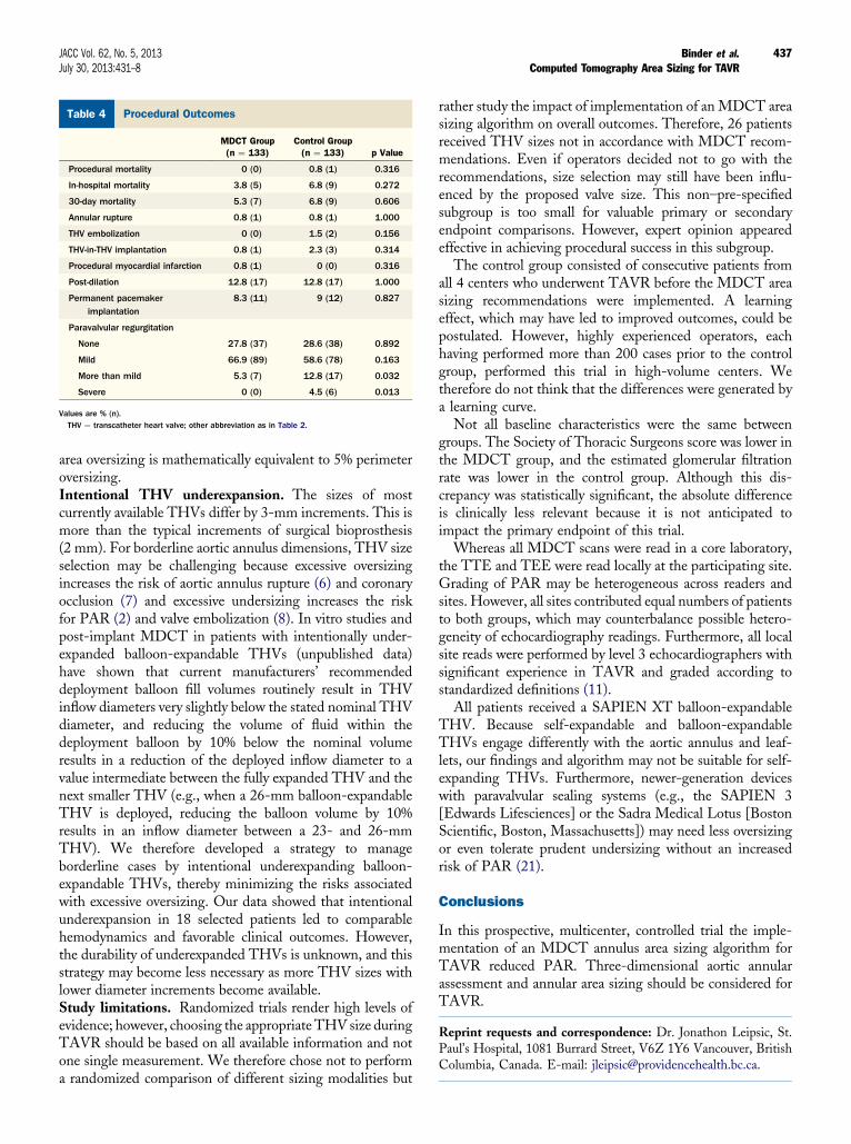
Table 4 Procedural Outcomes
MDCT Group(n ¼ 133)
Control Group(n ¼ 133) p Value
Procedural mortality 0 (0) 0.8 (1) 0.316
In-hospital mortality 3.8 (5) 6.8 (9) 0.272
30-day mortality 5.3 (7) 6.8 (9) 0.606
Annular rupture 0.8 (1) 0.8 (1) 1.000
THV embolization 0 (0) 1.5 (2) 0.156
THV-in-THV implantation 0.8 (1) 2.3 (3) 0.314
Procedural myocardial infarction 0.8 (1) 0 (0) 0.316
Post-dilation 12.8 (17) 12.8 (17) 1.000
Permanent pacemakerimplantation
8.3 (11) 9 (12) 0.827
Paravalvular regurgitation
None 27.8 (37) 28.6 (38) 0.892
Mild 66.9 (89) 58.6 (78) 0.163
More than mild 5.3 (7) 12.8 (17) 0.032
Severe 0 (0) 4.5 (6) 0.013
Values are % (n).THV ¼ transcatheter heart valve; other abbreviation as in Table 2.
JACC Vol. 62, No. 5, 2013 Binder et al.July 30, 2013:431–8 Computed Tomography Area Sizing for TAVR
437
area oversizing is mathematically equivalent to 5% perimeteroversizing.Intentional THV underexpansion. The sizes of mostcurrently available THVs differ by 3-mm increments. This ismore than the typical increments of surgical bioprosthesis(2 mm). For borderline aortic annulus dimensions, THV sizeselection may be challenging because excessive oversizingincreases the risk of aortic annulus rupture (6) and coronaryocclusion (7) and excessive undersizing increases the riskfor PAR (2) and valve embolization (8). In vitro studies andpost-implant MDCT in patients with intentionally under-expanded balloon-expandable THVs (unpublished data)have shown that current manufacturers’ recommendeddeployment balloon fill volumes routinely result in THVinflow diameters very slightly below the stated nominal THVdiameter, and reducing the volume of fluid within thedeployment balloon by 10% below the nominal volumeresults in a reduction of the deployed inflow diameter to avalue intermediate between the fully expanded THV and thenext smaller THV (e.g., when a 26-mm balloon-expandableTHV is deployed, reducing the balloon volume by 10%results in an inflow diameter between a 23- and 26-mmTHV). We therefore developed a strategy to manageborderline cases by intentional underexpanding balloon-expandable THVs, thereby minimizing the risks associatedwith excessive oversizing. Our data showed that intentionalunderexpansion in 18 selected patients led to comparablehemodynamics and favorable clinical outcomes. However,the durability of underexpanded THVs is unknown, and thisstrategy may become less necessary as more THV sizes withlower diameter increments become available.Study limitations. Randomized trials render high levels ofevidence; however, choosing the appropriate THV size duringTAVR should be based on all available information and notone single measurement. We therefore chose not to performa randomized comparison of different sizing modalities but
rather study the impact of implementation of anMDCT areasizing algorithm on overall outcomes. Therefore, 26 patientsreceived THV sizes not in accordance with MDCT recom-mendations. Even if operators decided not to go with therecommendations, size selection may still have been influ-enced by the proposed valve size. This non–pre-specifiedsubgroup is too small for valuable primary or secondaryendpoint comparisons. However, expert opinion appearedeffective in achieving procedural success in this subgroup.
The control group consisted of consecutive patients fromall 4 centers who underwent TAVR before the MDCT areasizing recommendations were implemented. A learningeffect, which may have led to improved outcomes, could bepostulated. However, highly experienced operators, eachhaving performed more than 200 cases prior to the controlgroup, performed this trial in high-volume centers. Wetherefore do not think that the differences were generated bya learning curve.
Not all baseline characteristics were the same betweengroups. The Society of Thoracic Surgeons score was lower inthe MDCT group, and the estimated glomerular filtrationrate was lower in the control group. Although this dis-crepancy was statistically significant, the absolute differenceis clinically less relevant because it is not anticipated toimpact the primary endpoint of this trial.
Whereas all MDCT scans were read in a core laboratory,the TTE and TEE were read locally at the participating site.Grading of PAR may be heterogeneous across readers andsites. However, all sites contributed equal numbers of patientsto both groups, which may counterbalance possible hetero-geneity of echocardiography readings. Furthermore, all localsite reads were performed by level 3 echocardiographers withsignificant experience in TAVR and graded according tostandardized definitions (11).
All patients received a SAPIEN XT balloon-expandableTHV. Because self-expandable and balloon-expandableTHVs engage differently with the aortic annulus and leaf-lets, our findings and algorithm may not be suitable for self-expanding THVs. Furthermore, newer-generation deviceswith paravalvular sealing systems (e.g., the SAPIEN 3[Edwards Lifesciences] or the Sadra Medical Lotus [BostonScientific, Boston, Massachusetts]) may need less oversizingor even tolerate prudent undersizing without an increasedrisk of PAR (21).
Conclusions
In this prospective, multicenter, controlled trial the imple-mentation of an MDCT annulus area sizing algorithm forTAVR reduced PAR. Three-dimensional aortic annularassessment and annular area sizing should be considered forTAVR.
Reprint requests and correspondence: Dr. Jonathon Leipsic, St.Paul’s Hospital, 1081 Burrard Street, V6Z 1Y6 Vancouver, BritishColumbia, Canada. E-mail: [email protected].

Binder et al. JACC Vol. 62, No. 5, 2013Computed Tomography Area Sizing for TAVR July 30, 2013:431–8
438
REFERENCES
1. Gurvitch R, Webb JG, Yuan R, et al. Aortic annulus diameter deter-mination by multidetector computed tomography: reproducibility,applicability, and implications for transcatheter aortic valve implanta-tion. J Am Coll Cardiol Intv 2011;4:1235–45.
2. Willson AB, Webb JG, Labounty TM, et al. 3-dimensional aorticannular assessment by multidetector computed tomography predictsmoderate or severe paravalvular regurgitation after transcatheter aorticvalve replacement: a multicenter retrospective analysis. J Am CollCardiol 2012;59:1287–94.
3. Moss RR, Ivens E, Pasupati S, et al. Role of echocardiography inpercutaneous aortic valve implantation. J Am Coll Cardiol Img 2008;1:15–24.
4. Jilaihawi H, Kashif M, Fontana G, et al. Cross-sectional computedtomographic assessment improves accuracy of aortic annular sizingfor transcatheter aortic valve replacement and reduces the incidenceof paravalvular aortic regurgitation. J Am Coll Cardiol 2012;59:1275–86.
5. WillsonAB,Webb JG, FreemanM, et al. Computed tomography-basedsizing recommendations for transcatheter aortic valve replacementwith balloon-expandable valves: comparison with transesophagealechocardiography and rationale for implementation in a prospective trial.J Cardiovasc Comput Tomogr 2012;6:406–14.
6. Blanke P, Reinohl J, Schlensak C, et al. Prosthesis oversizing in balloon-expandable transcatheter aortic valve implantation is associated withcontained rupture of the aortic root.CircCardiovasc Interv 2012;5:540–8.
7. Gurvitch R, Tay EL, Wijesinghe N, et al. Transcatheter aortic valveimplantation: lessons from the learning curve of the first 270 high-riskpatients. Catheter Cardiovasc Interv 2011;78:977–84.
8. Tay EL, Gurvitch R, Wijeysinghe N, et al. Outcome of patients aftertranscatheter aortic valve embolization. J Am Coll Cardiol Intv 2011;4:228–34.
9. Santos N, de Agustin JA, Almeria C, et al. Prosthesis/annulus dis-congruence assessed by three-dimensional transoesophageal echocar-diography: a predictor of significant paravalvular aortic regurgitationafter transcatheter aortic valve implantation. Eur Heart J CardiovascImaging 2012;13:931–7.
10. Gripari P, Ewe SH, Fusini L, et al. Intraoperative 2D and 3D trans-oesophageal echocardiographic predictors of aortic regurgitation aftertranscatheter aortic valve implantation. Heart 2012;98:1229–36.
11. Kappetein AP, Head SJ, Genereux P, et al. Updated standardizedendpoint definitions for transcatheter aortic valve implantation: the
Valve Academic Research Consortium-2 consensus document. J AmColl Cardiol 2012;60:1438–54.
12. Kodali SK, Williams MR, Smith CR, et al. Two-year outcomes aftertranscatheter or surgical aortic-valve replacement. N Engl J Med 2012;366:1686–95.
13. Makkar RR, Fontana GP, Jilaihawi H, et al. Transcatheter aortic-valvereplacement for inoperable severe aortic stenosis. N Engl J Med 2012;366:1696–704.
14. Altiok E, Koos R, Schroder J, et al. Comparison of two-dimensionaland three-dimensional imaging techniques for measurement of aorticannulus diameters before transcatheter aortic valve implantation. Heart2011;97:1578–84.
15. Kempfert J, Van Linden A, Lehmkuhl L, et al. Aortic annulus sizing:echocardiographic versus computed tomography derived measurementsin comparison with direct surgical sizing. Eur J Cardiothorac Surg2012;42:627–33.
16. Yano M, Nakamura K, Nagahama H, Matsuyama M, Nishimura M,Onitsuka T. Aortic annulus diameter measurement: what is the bestmodality? Ann Thorac Cardiovasc Surg 2012;18:115–20.
17. Hayashida K, Bouvier E, Lefevre T, et al. Impact of CT-guided valvesizing on post-procedural aortic regurgitation in transcatheter aorticvalve implantation. EuroIntervention 2012;8:546–55.
18. Babaliaros VC, Junagadhwalla Z, Lerakis S, et al. Use of balloon aorticvalvuloplasty to size the aortic annulus before implantation of a balloon-expandable transcatheter heart valve. J Am Coll Cardiol Intv 2010;3:114–8.
19. Hamdan A, Guetta V, Konen E, et al. Deformation dynamics andmechanical properties of the aortic annulus by 4-dimensional computedtomography: insights into the functional anatomy of the aortic valvecomplex and implications for transcatheter aortic valve therapy. J AmColl Cardiol 2012;59:119–27.
20. Blanke P, Russe M, Leipsic J, et al. Conformational pulsatile changesof the aortic annulus: impact on prosthesis sizing by computedtomography for transcatheter aortic valve replacement. J Am CollCardiol Intv 2012;5:984–94.
21. Binder RK, Rodés-Cabau J, Wood DA, et al. Transcatheter aorticvalve replacement with the SAPIEN 3: a new balloon-expandabletranscatheter heart valve. J Am Coll Cardiol Intv 2013;6:293–300.
Key Words: annulus area - multidetector computed tomography -
transcatheter aortic valve replacement - transcatheter heart valve sizing.
Copyright © 2022 FDOKUMEN