Paravalvular Leak After Transcatheter Aortic Valve Replacement
12
STATE-OF-THE-ART PAPER Paravalvular Leak After Transcatheter Aortic Valve Replacement The New Achilles’ Heel? A Comprehensive Review of the Literature Philippe Généreux, MD,*†‡ Stuart J. Head, MSC,§ Rebecca Hahn, MD,*† Benoit Daneault, MD,*† Susheel Kodali, MD,*† Mathew R. Williams, MD,*† Nicolas M. van Mieghem, MD, Maria C. Alu, MM,* Patrick W. Serruys, MD, PHD, A. Pieter Kappetein, MD, PHD,§ Martin B. Leon, MD*† New York, New York; Montréal, Québec, Canada; and Rotterdam, the Netherlands Paravalvular leak (PVL) is a frequent complication of transcatheter aortic valve replacement (TAVR) and is seen at a much higher rate after TAVR than after conventional surgical aortic valve replacement. Recent reports indi- cating that PVL may be correlated with increased late mortality have raised concerns. However, the heterogene- ity of methods for assessing and quantifying PVL, and lack of consistency in the timing of such assessments, is a hindrance to understanding its true prevalence, severity, and effect. This literature review is an effort to consol- idate current knowledge in this area to better understand the prevalence, progression, and impact of post-TAVR PVL and to help direct future efforts regarding the assessment, prevention, and treatment of this troublesome complication. (J Am Coll Cardiol 2013;61:1125–36) © 2013 by the American College of Cardiology Foundation Transcatheter aortic valve replacement (TAVR) has become the treatment of choice for inoperable patients with severe aortic stenosis (1) and is comparable to surgical aortic valve replacement (SAVR) for patients at high risk (2). However, paravalvular leak (PVL) is more frequently seen after TAVR than after SAVR, and its potential association with mortal- ity has raised concerns (3–6). Moreover, recent reports have suggested that PVL could negatively impact mid- and long-term prognosis following TAVR (7,8). Although con- cerning, the lack of standardized quantitative and qualitative methods to assess and categorize PVL and the heterogene- ity in the timing of post-procedural assessment of PVL warrant caution in interpretation of these data. Therefore, we sought to perform a systematic review of the current literature to better define the rate, progression over time, predictors, and consequences of PVL after TAVR. Further- more, recommendations for measuring PVL are provided to improve consistency throughout the literature. Rate of PVL Multiple studies have reported the frequency and severity of PVL after TAVR (9). There is, however, significant heter- ogeneity that is caused by differences in: 1) imaging modal- ities (transthoracic echocardiography, transesophageal echo- cardiography, angiography); 2) timing of assessment (immediately after implantation, before discharge, at 30 days); 3) transcatheter heart valve (THV) system; 4) grading scale; and 5) adjudication of events. When PVL was evaluated before hospital discharge and without central core laboratory analysis, its absence was reported in 6% to 59% of patients, whereas moderate or severe PVL was seen in 0% to 24% (1–5,10 –16)(Table 1). Thus far, only the PARTNER (Placement of Aortic Transcatheter Valve) trial has used a central echocardiogra- phy core laboratory to evaluate PVL (1,2). PVL was graded in accordance with the American Society of Echocardiog- raphy recommendations for native valves (17) because there were no recommendations for prosthetic valve assessment at the start of the trial. In addition, because of the inevitable eccentric nature of the jet and the frequent “spray” of the jet contour in the outflow tract, the color Doppler in the available parasternal short-axis view(s) was weighted in a subjective fashion more heavily than From the *Columbia University Medical Center/New York Presbyterian Hospital, New York, New York; †Cardiovascular Research Foundation, New York, New York; ‡Hôpital du Sacré-Coeur de Montréal, Montréal, Québec, Canada; §Department of Cardiothoracic Surgery, Erasmus University Medical Center, Rotterdam, the Neth- erlands; and the Department of Cardiology, Erasmus University Medical Center, Rotterdam, the Netherlands. Dr. Généreux has received speaker honoraria, consulting fees, and research grants from Edwards Lifesciences. Dr. Kodali has received consulting fees from Edwards Lifesciences and St. Jude Medical. Dr. Kappetein is member of a steering committee of the SURTAVI (Surgical Replacement and Transcatheter Aortic Valve Implantation) trial sponsored by Medtronic. Dr. Leon is a nonpaid member of the scientific advisory board of Edwards Lifesciences. All other authors have reported that they have no relationships relevant to the contents of this paper to disclose. Drs. Généreux and Head are joint first authors. Manuscript received June 27, 2012; revised manuscript received August 7, 2012, accepted August 21, 2012. Journal of the American College of Cardiology Vol. 61, No. 11, 2013 © 2013 by the American College of Cardiology Foundation ISSN 0735-1097/$36.00 Published by Elsevier Inc. http://dx.doi.org/10.1016/j.jacc.2012.08.1039
-
Upload
independent -
Category
Documents
-
view
3 -
download
0
Transcript of Paravalvular Leak After Transcatheter Aortic Valve Replacement
Journal of the American College of Cardiology Vol. 61, No. 11, 2013© 2013 by the American College of Cardiology Foundation ISSN 0735-1097/$36.00
STATE-OF-THE-ART PAPER
Paravalvular Leak AfterTranscatheter Aortic Valve ReplacementThe New Achilles’ Heel? A Comprehensive Review of the Literature
Philippe Généreux, MD,*†‡ Stuart J. Head, MSC,§ Rebecca Hahn, MD,*† Benoit Daneault, MD,*†Susheel Kodali, MD,*† Mathew R. Williams, MD,*† Nicolas M. van Mieghem, MD,�Maria C. Alu, MM,* Patrick W. Serruys, MD, PHD,� A. Pieter Kappetein, MD, PHD,§Martin B. Leon, MD*†
New York, New York; Montréal, Québec, Canada; and Rotterdam, the Netherlands
Paravalvular leak (PVL) is a frequent complication of transcatheter aortic valve replacement (TAVR) and is seenat a much higher rate after TAVR than after conventional surgical aortic valve replacement. Recent reports indi-cating that PVL may be correlated with increased late mortality have raised concerns. However, the heterogene-ity of methods for assessing and quantifying PVL, and lack of consistency in the timing of such assessments, isa hindrance to understanding its true prevalence, severity, and effect. This literature review is an effort to consol-idate current knowledge in this area to better understand the prevalence, progression, and impact of post-TAVRPVL and to help direct future efforts regarding the assessment, prevention, and treatment of this troublesomecomplication. (J Am Coll Cardiol 2013;61:1125–36) © 2013 by the American College of Cardiology Foundation
Published by Elsevier Inc. http://dx.doi.org/10.1016/j.jacc.2012.08.1039
Transcatheter aortic valve replacement (TAVR) has becomethe treatment of choice for inoperable patients with severeaortic stenosis (1) and is comparable to surgical aortic valvereplacement (SAVR) for patients at high risk (2). However,paravalvular leak (PVL) is more frequently seen after TAVRthan after SAVR, and its potential association with mortal-ity has raised concerns (3–6). Moreover, recent reports havesuggested that PVL could negatively impact mid- andlong-term prognosis following TAVR (7,8). Although con-cerning, the lack of standardized quantitative and qualitativemethods to assess and categorize PVL and the heterogene-ity in the timing of post-procedural assessment of PVLwarrant caution in interpretation of these data. Therefore,we sought to perform a systematic review of the currentliterature to better define the rate, progression over time,
From the *Columbia University Medical Center/New York Presbyterian Hospital,New York, New York; †Cardiovascular Research Foundation, New York, New York;‡Hôpital du Sacré-Coeur de Montréal, Montréal, Québec, Canada; §Department ofCardiothoracic Surgery, Erasmus University Medical Center, Rotterdam, the Neth-erlands; and the �Department of Cardiology, Erasmus University Medical Center,Rotterdam, the Netherlands. Dr. Généreux has received speaker honoraria, consultingfees, and research grants from Edwards Lifesciences. Dr. Kodali has receivedconsulting fees from Edwards Lifesciences and St. Jude Medical. Dr. Kappetein ismember of a steering committee of the SURTAVI (Surgical Replacement andTranscatheter Aortic Valve Implantation) trial sponsored by Medtronic. Dr. Leon isa nonpaid member of the scientific advisory board of Edwards Lifesciences. All otherauthors have reported that they have no relationships relevant to the contents of thispaper to disclose. Drs. Généreux and Head are joint first authors.
Manuscript received June 27, 2012; revised manuscript received August 7, 2012,accepted August 21, 2012.
predictors, and consequences of PVL after TAVR. Further-more, recommendations for measuring PVL are provided toimprove consistency throughout the literature.
Rate of PVL
Multiple studies have reported the frequency and severity ofPVL after TAVR (9). There is, however, significant heter-ogeneity that is caused by differences in: 1) imaging modal-ities (transthoracic echocardiography, transesophageal echo-cardiography, angiography); 2) timing of assessment(immediately after implantation, before discharge, at 30days); 3) transcatheter heart valve (THV) system; 4) gradingscale; and 5) adjudication of events. When PVL wasevaluated before hospital discharge and without central corelaboratory analysis, its absence was reported in 6% to 59% ofpatients, whereas moderate or severe PVL was seen in 0% to24% (1–5,10–16) (Table 1).
Thus far, only the PARTNER (Placement of AorticTranscatheter Valve) trial has used a central echocardiogra-phy core laboratory to evaluate PVL (1,2). PVL was gradedin accordance with the American Society of Echocardiog-raphy recommendations for native valves (17) because therewere no recommendations for prosthetic valve assessmentat the start of the trial. In addition, because of theinevitable eccentric nature of the jet and the frequent“spray” of the jet contour in the outflow tract, the colorDoppler in the available parasternal short-axis view(s)
was weighted in a subjective fashion more heavily than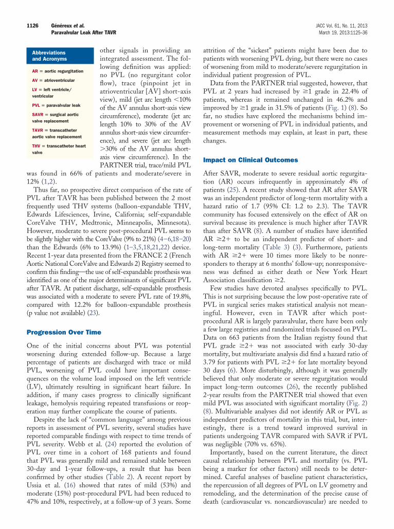
w1
PfECHbtRAciawc(
P
OwpPq(ale
rrPPt3cUm4
apoi
Ppifpmc
I
AtpwhcstAlwsnA
TPipaDP
1126 Généreux et al. JACC Vol. 61, No. 11, 2013Paravalvular Leak After TAVR March 19, 2013:1125–36
other signals in providing anintegrated assessment. The fol-lowing definition was applied:no PVL (no regurgitant colorflow), trace (pinpoint jet inatrioventricular [AV] short-axisview), mild (jet arc length �10%of the AV annulus short-axis viewcircumference), moderate (jet arclength 10% to 30% of the AVannulus short-axis view circumfer-ence), and severe (jet arc length�30% of the AV annulus short-axis view circumference). In thePARTNER trial, trace/mild PVL
as found in 66% of patients and moderate/severe in2% (1,2).Thus far, no prospective direct comparison of the rate of
VL after TAVR has been published between the 2 mostrequently used THV systems (balloon-expandable THV,dwards Lifesciences, Irvine, California; self-expandableoreValve THV, Medtronic, Minneapolis, Minnesota).owever, moderate to severe post-procedural PVL seems to
e slightly higher with the CoreValve (9% to 21%) (4–6,18–20)han the Edwards (6% to 13.9%) (1–3,5,18,21,22) device.ecent 1-year data presented from the FRANCE 2 (Frenchortic National CoreValve and Edwards 2) Registry seemed to
onfirm this finding—the use of self-expandable prosthesis wasdentified as one of the major determinants of significant PVLfter TAVR. At patient discharge, self-expandable prosthesisas associated with a moderate to severe PVL rate of 19.8%,
ompared with 12.2% for balloon-expandable prosthesisp value not available) (23).
rogression Over Time
ne of the initial concerns about PVL was potentialorsening during extended follow-up. Because a largeercentage of patients are discharged with trace or mildVL, worsening of PVL could have important conse-uences on the volume load imposed on the left ventricleLV), ultimately resulting in significant heart failure. Inddition, if many cases progress to clinically significanteakage, hemolysis requiring repeated transfusions or reop-ration may further complicate the course of patients.
Despite the lack of “common language” among previouseports in assessment of PVL severity, several studies haveeported comparable findings with respect to time trends ofVL severity. Webb et al. (24) reported the evolution ofVL over time in a cohort of 168 patients and found
hat PVL was generally mild and remained stable between0-day and 1-year follow-ups, a result that has beenonfirmed by other studies (Table 2). A recent report byssia et al. (16) showed that rates of mild (53%) andoderate (15%) post-procedural PVL had been reduced to
Abbreviationsand Acronyms
AR � aortic regurgitation
AV � atrioventricular
LV � left ventricle/ventricular
PVL � paravalvular leak
SAVR � surgical aorticvalve replacement
TAVR � transcatheteraortic valve replacement
THV � transcatheter heartvalve
7% and 10%, respectively, at a follow-up of 3 years. Some
ttrition of the “sickest” patients might have been due toatients with worsening PVL dying, but there were no casesf worsening from mild to moderate/severe regurgitation inndividual patient progression of PVL.
Data from the PARTNER trial suggested, however, thatVL at 2 years had increased by �1 grade in 22.4% ofatients, whereas it remained unchanged in 46.2% andmproved by �1 grade in 31.5% of patients (Fig. 1) (8). Soar, no studies have explored the mechanisms behind im-rovement or worsening of PVL in individual patients, andeasurement methods may explain, at least in part, these
hanges.
mpact on Clinical Outcomes
fter SAVR, moderate to severe residual aortic regurgita-ion (AR) occurs infrequently in approximately 4% ofatients (25). A recent study showed that AR after SAVRas an independent predictor of long-term mortality with aazard ratio of 1.7 (95% CI: 1.2 to 2.3). The TAVRommunity has focused extensively on the effect of AR onurvival because its prevalence is much higher after TAVRhan after SAVR (8). A number of studies have identifiedR �2� to be an independent predictor of short- and
ong-term mortality (Table 3) (3). Furthermore, patientsith AR �2� were 10 times more likely to be nonre-
ponders to therapy at 6 months’ follow-up; nonresponsive-ess was defined as either death or New York Heartssociation classification �2.Few studies have devoted analyses specifically to PVL.
his is not surprising because the low post-operative rate ofVL in surgical series makes statistical analysis not mean-
ngful. However, even in TAVR after which post-rocedural AR is largely paravalvular, there have been onlyfew large registries and randomized trials focused on PVL.ata on 663 patients from the Italian registry found thatVL grade �2� was not associated with early 30-day
mortality, but multivariate analysis did find a hazard ratio of3.79 for patients with PVL �2� for late mortality beyond30 days (6). More disturbingly, although it was generallybelieved that only moderate or severe regurgitation wouldimpact long-term outcomes (26), the recently published2-year results from the PARTNER trial showed that evenmild PVL was associated with significant mortality (Fig. 2)(8). Multivariable analyses did not identify AR or PVL asindependent predictors of mortality in this trial, but, inter-estingly, there is a trend toward improved survival inpatients undergoing TAVR compared with SAVR if PVLwas negligible (70% vs. 65%).
Importantly, based on the current literature, the directcausal relationship between PVL and mortality (vs. PVLbeing a marker for other factors) still needs to be deter-mined. Careful analyses of baseline patient characteristics,the repercussion of all degrees of PVL on LV geometry andremodeling, and the determination of the precise cause of
death (cardiovascular vs. noncardiovascular) are needed to
1127JACC Vol. 61, No. 11, 2013 Généreux et al.March 19, 2013:1125–36 Paravalvular Leak After TAVR
confirm the strength and the nature of this relationship. Atthis point, any previous observations linking PVL (espe-cially mild) with mortality should be considered hypothesisgenerating.
Predictors of PVL
Significant PVL most commonly results from: 1) incom-plete prosthesis apposition to the native annulus due topatterns or extent of calcification (11,27–30) or annulareccentricity (26); 2) undersizing of the device (10,31,32);and/or 3) malpositioning of the valve (33). These observa-tions seem to be true for both balloon-expandable andself-expandable THVs.
Valve sizing has been shown to be one of the strongestpredictors of PVL. A low cover index reflecting a lowerdegree of oversizing of the prosthesis based on transthoracicechocardiography annulus measurement predicts significantPVL (10). More recently, studies have evaluated the use ofmultidetector computed tomography (MDCT) for THVsizing, and MDCT showed good predictability and reducedrates of significant PVL (34–37). Furthermore, larger andeccentric annuli were identified as predictors of PVL inmultiple studies and most likely reflect inadequate sizing ofthe THV (3,15,26). A smaller aortic valve area was found topredict PVL in one study, but this was likely because thesmaller area indicates a greater degree of calcification (3).The extent of calcification and asymmetrical distribution, aswell as the location of calcium on the aortic wall, valvecommissure, or THV landing zone, as a predictor for PVLhas been confirmed in several studies (11,26,29,37,38).
In studies specifically evaluating the CoreValve (Medtronic), alower depth of implantation and a greater angle between theaorta and LV outflow tract were found to predict PVL(14,15).
Assessment of Paravalvular Regurgitation
Angiographic and hemodynamic assessment. Aortic rootangiography is an established tool for qualitative and semi-quantitative assessment of AR (39). It is readily availableduring the TAVR procedure and can be quickly and safelyexecuted to provide essential information and initiate ad-junctive maneuvers if needed in case of significant (para)valvular AR. Typically, Sellers criteria are applied to gradeAR (40): 1) grade 1 or mild AR corresponds to a smallamount of contrast entering the LV during diastole withoutfilling the entire cavity and clearing with each cardiac cycle;2) grade 2 or moderate AR corresponds to contrast filling ofthe entire LV in diastole but with less density comparedwith contrast opacification of the ascending aorta; 3) grade3 or moderate to severe AR corresponds to contrast filling ofthe entire LV in diastole equal in density to the contrastopacification of the ascending aorta; and 4) grade 4 or severeAR corresponds to contrast filling of the entire LV indiastole on the first beat with greater density compared with
the contrast opacification of the ascending aorta. During thecontrast injection, no material may cross the aortic valveleaflets (e.g., guidewires, catheters) because incomplete valveclosure may artificially be generated, thus resulting in AR.Particularly with self-expanding systems, it is important towait some time (empirically 10 min) after deployment of thebioprosthesis to allow the system to expand to its maximum.The downside of qualitative aortography AR assessment isthat it relies on subjective interpretation of unidimensionalimages; therefore, interobserver and intraobserver variabilitycan be an issue and additional contrast volume required.Moreover, it is difficult to determine the contribution ofPVL and central AR.
Classic findings of acute AR (acute drop in the aorticdiastolic pressure with or without elevated LV end-diastolicpressure [LVEDP]) may be seen after TAVR and may besuggestive of moderate to severe AR. However, thesefindings must be interpreted with caution because theconcomitant use of sedatives, vasopressors, inotropes, andintravenous fluids all impact hemodynamics, and the pres-ence of material through the aortic valve (e.g., wire) mayinterfere temporarily with the THV function. Recently, theAR index, the ratio of the end-diastolic gradient across theaortic valve bioprosthesis to systolic blood pressure ([ADP �LVEDP]/ASP; ADP-aortic diastolic pressure, ASP-aorticsystolic pressure), was described (41). An AR index �25was associated with 1-year mortality. Although this associ-ation is interesting, more data and validation are needed toestablish the role of this new index in the therapeuticdecision process after TAVR.Echocardiographic assessment. Although the native valveregurgitation quantitative grading scheme has been advo-cated for the evaluation of prosthetic valve regurgitation(42), there are limited data to support the use of theseparameters following TAVR. The majority of semiquanti-tative parameters for assessing AR apply to central regurgi-tant jets, which are more uniform, making semiquantitativegrading schemes more reliable.
Unlike central jets, paravalvular regurgitant jets are com-monly eccentric with crescentic, irregular orifices. Becausethese jets occur between the annulus and sewing ring, jetareas and lengths may not represent the same severity ofregurgitation compared with central jets and these param-eters cannot be used to reliably assess regurgitant severity.Although guidelines suggest using the circumferential ex-tent of the regurgitant jet as a semiquantitative measure ofseverity (42), this parameter has not been validated against anyquantitative parameters of regurgitation. Even if we accept thelimited validation of this scheme for surgical prostheses, theanatomy and physiology of THVs are different than that ofsurgical valves. In the balloon-expandable valve, paravalvularregurgitation should be assessed just below the skirt; for centraljets, the regurgitation should be assessed at the coaptationpoint of the leaflets. In addition, there is no scheme thatspecifically addresses the unusual regurgitation that accompa-
nies the THV. The intact calcified cusps and annulus signifi-
Selected Publications Reporting AR After TAVRTable 1 Selected Publications Reporting AR After TAVR
First Author, Year (Ref. #) n Approach Prosthesis Imaging Modality Severity Gradation Adjunctive Techniques AR Post-TAVR
Predictors of AR byMultivariable
Analysis
Detaint, 2009 (10) 74 TF � 46 (62%)TA � 28 (38%)
ES Echocardiogram (TEE)Site reported (blinded
echocardiographist)
0 � absent1 � trace/mild2 � mild/moderate3 � moderate/severe4 � severe
Post-dilation � 5/74Valve-in-valve � 2/74
Early post-TAVR (TEE)0 � 5 (7.0%)1 � 53 (72.0%)2 � 12 (16.0%)3 � 4 (5.0%)4 � 0 (0%)
�2/4 AR● Low cover index● Operator’s
experience
Abdel-Wahab, 2011 (3) 690 TF � 644TA � 26SC � 22TAo � 5
ES � 110 (16%)MCV � 580 (84%)
AngiogramSite reported
0 � absent1 � trace/mild2 � mild/moderate3 � moderate/severe4 � severe
— Early post-TAVR (angiogram)0 � 191 (27.7%)1 � 380 (55.1%)2 � 103 (14.9%)3 � 14 (2.0%)4 � 2 (0.3%)
�2/4 AR● AVA baseline● Annulus baseline● Cardiogenic shock● Renal failure● Male
Sherif, 2010 (14) 50 TF MCV AngiogramEchocardiogramSite reported
1 � trivial/mild2 � moderate3 � moderate/severe4 � severe
— Early post-TAVR (angiogram)0 � 3 (6.0%)1 � 27 (54.0%)2 � 13 (26.0%)3 � 7 (14.0%)4 � 0 (0%)Early post-TAVR (TTE)0 � 9 (18.0%)1 � 24 (48.0%)2 � 13 (26.0%)3 � 4 (8%)4 � 0 (0%)
�2/4 AR● Increase angle of
LVOT andascending aorta
● Depth of device inrelation tononcoronary cups
John, 2010 (78) 100 TF � 97 (97%)SC � 3 (3%)
MCV AngiogramEchocardiogram
01�
2�
3�
4�
Post-dilation � 34/100Snare technique � 4/100Valve-in-valve � 3/100
Early post-TAVR (angiogram)0 � 35 (35.4%)1� � 28 (28.3%)2� � 19 (19.2%)3� � 8 (0.8%)4� � 0 (0%)Early after adjunctivetechnique (angiogram)0 �38 (38.4%)1� � 49 (49.5%)2� � 11 (11.1%)3� � 1 (0.1%)4� � 0 (0%)
AgS and DLZ-CSshowedsignificantcorrelation withgrade of PVLafter initial MCVdeployment
Takagi, 2011 (15) 79 TF � 62 (78.5%)SC � 17(21.5%)
MCV AngiogramEchocardiogramSite reported
0 � absent1 � mild2 � moderate3–4 � severe
Post-dilation � 21/79Snare technique � 1/79Valve-in-valve � 2/79
Final result (angiogram)0 � 21 (26.6%)1 � 42 (53.2%)2 � 13 (16.5%)3 � 3 (3.8%)4 � 0 (0%)
�2/4 AR● Larger annulus
diameter● Low implantation● Peripheral vascular
disease
Continued on the next page
1128Généreux
etal.JACC
Vol.61,No.11,2013Paravalvular
LeakAfter
TAVRM
arch19,2013:1125–36

ei
T
Iint
inue
dab
le1
Con
tinu
ed
irst
Aut
hor,
Yea
r(R
ef.
#)
nA
ppro
ach
Pro
sthe
sis
Imag
ing
Mod
alit
ySev
erit
yG
rada
tion
Adj
unct
ive
Tech
niqu
esA
RP
ost-
TAV
R
Pre
dict
ors
ofA
Rby
Mul
tiva
riab
leA
naly
sis
ambu
rino
,2
01
1(6
)6
63
TFM
CV
Echo
card
iogr
amSite
repo
rted
,eve
nts
revi
ewed
byin
depe
nden
tCEC
—P
ost-d
ilatio
n�
68
/66
3Va
lve-
in-v
alve
�1
39
/66
3Con
vers
ion
toop
ensu
rger
y�
5/6
63
Pos
t-TA
VR�
2P
VL�
13
9(2
1.0
%)
—
otzm
ann,
20
11
(4)
14
5TF
/SC
MCV
Echo
card
iogr
amA
ngio
gram
(If
poor
TTE
qual
ity)
Site
repo
rted
Mild
Mod
erat
eSev
ere
—Ea
rly
post
-TA
VRM
ild�
64
(44
%)
Mod
erat
e�
23
(16
%)
Sev
ere
�2
(1%
)Ea
rly
post
-TA
VR3
0-d
aysu
rviv
ors
only
Mild
�5
5(4
5%
)M
oder
ate
�1
6(1
3%
)Sev
ere
�0
(0%
)
—
oat,
20
11
(5)
87
0TF
�5
99
Oth
er�
27
1ES
�4
10
(47
%)
MCV
�4
59
(53
%)
Ang
iogr
amSite
repo
rted
—Con
vers
ion
toop
ensu
rger
y�
6/8
50
AR
�1
�5
16
(61
%)
AR
�2
�1
15
(13
.6%
)—
�A
gats
ton
scor
e;A
R�
aort
icre
gurg
itatio
n;A
VA�
aort
icva
lve
repl
acem
ent;
CEC
�cl
inic
alev
ents
com
mitt
ee;D
LZ-C
S�
devi
ce-la
ndin
gzo
neca
lcifi
catio
nsc
ore;
ES�
Edw
ards
Sap
ien;
LVO
T�
left
vent
ricu
laro
utflo
wtr
ack;
MCV
�M
edtr
onic
Cor
eVal
ve;P
VL�
para
valv
ular
;SC
�su
bcla
vian
;TA
�tr
ansa
pica
l;TA
o�
tran
saor
tic;T
AVR
�tr
ansc
athe
ter
aort
icva
lve
repl
acem
ent;
TEE
�tr
anse
soph
agea
lech
ocar
diog
raph
y;TF
�tr
ansf
emor
al;T
TE�
tran
stho
raci
cec
hoca
rdio
grap
hy.
1129JACC Vol. 61, No. 11, 2013 Généreux et al.March 19, 2013:1125–36 Paravalvular Leak After TAVR
cantly influence the location and shape of paravalvular jets;typically, these jets appear smaller and more irregular at thelevel of the intact/calcified cusps and larger just apical to theTHV stent.
Quantitative assessment of total AR, or advanced imag-ing techniques for assessing paravalvular regurgitant orifices,may be a more accurate way of assessing severity and thus amore accurate assessment of risk. Quantitative Doppler usescomparative flow measurements across a regurgitant valveand a nonregurgitant valve to calculate regurgitant volumeor fraction (17). The effective regurgitant orifice area is thencalculated by dividing the regurgitant volume by the velocitytime integral of the regurgitant jet continuous wave spectralprofile. Alternatively, the LV stroke volume calculated by2-dimensional (2D) biplane Simpson method of discs (43) canbe used in place of total (regurgitant plus forward) strokevolume; however, systematic underestimation of ventricularvolumes has been reported for this method. Although thisquantitative assessment has been largely validated in theliterature (44–51), has shown reproducibility, and is endorsedby scientific authorities (17,52), it should be acknowledged thatthis assessment is based on 4 parameters, any one of which maybe determined with significant inaccuracy.
Three-dimensional (3D) echocardiography can overcomethe limitations of 2D and standard Doppler measurementsfor quantifying regurgitation (43). Pitfalls of 2D LV imag-ing, including foreshortening, malrotation, and angulation,can be overcome by 3D imaging. However, limitations of3D imaging (lower line density and low volume rates) mayreduce the utility of this method for assessing total strokevolume. Color Doppler 3D volumes can be useful for theidentification and localization of regurgitation jets, as well asplanimetry of the vena contracta area (53,54). This imagingmodality may be particularly useful for post-TAVR assess-ment of PVL (55,56).
With the increased use of multimodality imaging capableof 3D reconstruction of the aortic root (36,57–62), therehas been intense interest in the shape of the annulus andappropriate sizing of the transcatheter heart valve to reducePVL. The oval shape of the annulus has been well documented(36,60,61,63–65), and a single sagittal plane measurement issignificantly smaller than the coronary plane measurement.Algorithms using 3D imaging tools have been suggested toimprove annular sizing and reduce PVL (34,35).
Recently, the Valve Academic Research Consortium(VARC) published the VARC II definitions and suggested theuse of TAVR-specific criteria for the assessment of AR and/orPVL after TAVR (Table 4) (66). Figures 2 and 3 illustratechocardiographic assessment of PVL after TAVR. Figure 4llustrates a case using 3D echocardiography assessment of PVL.
reatment for Significant PVL
mproved positioning of the TAVR could require advancedmaging techniques for angiographic planning; having the
best coplanar view will ensure accurate fluoroscopic local-Co T F T G M
AgS
leak
1130 Généreux et al. JACC Vol. 61, No. 11, 2013Paravalvular Leak After TAVR March 19, 2013:1125–36
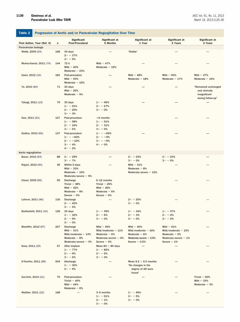
Progression of Aortic and/or Paravalvular Regurgitation Over TimeTable 2 Progression of Aortic and/or Paravalvular Regurgitation Over Time
First Author, Year (Ref. #) nSignificant
Post-ProceduralSignificant at
6 MonthsSignificant at
1 YearSignificant at
2 YearsSignificant at
3 Years
Paravalvular leakage
Webb, 2009 (24) 168 30 days2� � 37%3� � 5%
— “Stable” — —
Muñoz-Garcia, 2011 (79) 144 72 hMild � 40%Moderate � 23%
Mild � 47%Moderate � 19%
— — —
Ussia, 2012 (16) 181 Post-procedureMild � 53%Moderate � 15%
— Mild � 48%Moderate � 18%
Mild � 50%Moderate � 17%
Mild � 47%Moderate � 10%
Ye, 2010 (80) 71 30 daysMild � 26%Moderate � 5%
— — — “Remained unchangedand clinicallyinsignificantduring follow-up”
Takagi, 2011 (15) 79 30 days1� � 51%2� � 20%3� � 3%
1� � 49%2� � 27%3� � 0%
— — —
Ewe, 2011 (81) 107 Post-procedure1� � 58%2� � 16%3� � 5%
�6 months1� � 51%2� � 31%3� � 0%
— —
Godino, 2010 (82) 137 Post-procedure1� � �60%2� � �12%3� � 4%4� � 2%
1� � �65%2� � �9%3� � �5%4� � 0%
— — —
Aortic regurgitation
Bauer, 2010 (83) 88 2� � 29%3� � 7%
— 2� � 24%3� � 0%
2� � 23%3� � 0%
—
Rajani, 2010 (84) 46 Within 5 daysMild � 33%Moderate � 19%Moderate/severe � 5%
— Mild � 31%Moderate � 8%Moderate/severe � 15%
— —
Clavel, 2009 (85) 50 DischargeTrivial � 38%Mild � 42%Moderate � 8%Severe � 0%
6–12 monthsTrivial � 26%Mild � 46%Moderate � 6%Severe � 0%
— —
Lefevre, 2011 (86) 130 Discharge2� � 42%3� � 5%
— 2� � 25%3� � 0%
— —
Buellesfeld, 2011 (20) 126 30 days1� � 32%2� � 9%3� � 0%
1� � 39%2� � 6%3� � 0%
1� � 34%2� � 3%3� � 0%
1� � 37%2� � 0%3� � 0%
—
Bleiziffer, 2012 (87) 227 DischargeMild � 31%Mild/moderate � 13%Moderate � 8%Moderate/severe � 3%
Mild � 45%Mild/moderate � 11%Moderate � 6%Moderate/severe � 0%Severe � 0%
Mild � 40%Mild/moderate � 16%Moderate � 6%Moderate/severe � 0.5%Severe � 0.5%
Mild � 41%Mild/moderate � 15%Moderate � 5%Moderate/severe � 1%Severe � 1%
—
Koos, 2011 (29) 57 After implant1� � 77%2� � 9%3� � 5%
Mean 83 � 80 days1� � 82%2� � 5%3� � 0%
— — —
D’Onofrio, 2011 (88) 504 Discharge1� � 30%2� � 9%
— Mean 9.2 � 6.5 months“No changes in the
degree of AR werefound”
— —
Gurvitch, 2010 (21) 70 Post-procedureTrivial � 40%Mild � 44%Moderate � 6%
— — — Trivial � 60%Mild � 33%Moderate � 3%
Walther, 2011 (22) 168 — 3–6 months1� � 51%2� � 1%
1� � 46%2� � 5%3� � 0%
— —
3� � 0%

1131JACC Vol. 61, No. 11, 2013 Généreux et al.March 19, 2013:1125–36 Paravalvular Leak After TAVR
ization of the valve before implantation. In addition, simul-taneous “real-time” imaging, such as echocardiogram (both2D and 3D), 3D angiographic reconstruction via rotationalaortic root angiogram (67), and the use of novel imagingsystems (68,69), may assist in choosing intraprocedurally theoptimal projection for THV positioning and deployment,leading potentially to less frequent PVL.
Intraprocedurally, several interventional alternatives toreduce regurgitation are available (70). Severe calcificationof the native valve might prevent the implanted valve fromexpanding completely against the annulus, leaving residualorifices through which PVL may occur. Post-implantationballoon dilation of the valve might be effective in reducingPVL and may be considered the initial option for patientswith PVL (71). A slightly oversized balloon is recom-mended to fully expand the valve. Studies have shown thatpost-dilation can be safely performed, with a reduction ofthe regurgitation in a majority of patients (38). Calcificationof the valve significantly influences the success of thisintervention. However, in some patients, post-dilation hasno effect in reducing AR (15); in addition, post-dilation hasbeen shown to be associated with a higher incidence ofcerebrovascular events (38). The effect of post-dilation onsurvival has yet to be determined.
Figure 1 Change in Paravalvular Leak SeverityOver 2-Year Follow-Up
Adapted with permission from Kodali et al. (8).
Outcomes Associated With Aortic and/or Paravalvular RegurgitatioTable 3 Outcomes Associated With Aortic and/or Paravalvular
First Author, Year (Ref. #) n Variable Outco
Abdel-Wahab, 2011 (3) 690 AR �2 In-hospital morta
Gotzmann, 2011 (4) 122 AR �2 6-month mortalityNo clinical improv
Takagi, 2011 (15) 41 AR �2 6-month mortality
Hayashida, 2012 (89) 260 AR �2 Median 217 days
Leber, 2011 (90) 69 AR �2 1-year mortality
Moat, 2011 (5) 870 AR �2 1-year mortality
Sinning, 2012 (91) 152 PVL �2 1-year mortality
Tamburino, 2011 (6) 663 PVL �2 Late mortality
Sinning, 2012 (41) 146 Moderate/severe PVL 1-year survival
Unbehaun, 2012 (26) 358 No vs. trace vs. mild AR 2-year survival
Kodali, 2012 (8) 158 Mild to severe AR 2-year survival
Mild to severe PVL 2-year survival
HR � hazard ratio; IQR � interquartile range; OR � odds ratio; other abbreviations as in Table 1.
Especially with the CoreValve, implantation of the valvethat is too low is associated with PVL. Repositioning to ahigher implantation depth could therefore reduce PVL.However, no retrievable valve is currently available on themarket. Therefore, a snaring maneuver has been de-scribed, in which the valve is pulled up by attaching asnare to one of the frame loops (72,73). Althoughsuccessful cases have been reported (74), the valve mayalso move to the original (too low) position as soon astension is released (70). An extra word of caution iswarranted when the snaring technique is considered inpatients with extensively calcified valves because chunksof calcium may detach as a result of friction. Further-more, there is a risk of damaging the ascending aortaduring the snaring maneuver.
A valve-in-valve procedure may be necessary in somecases in which post-dilation or other techniques do notimprove the degree of PVL. This is specifically indicated forpatients in whom the valve was suboptimally positioned(i.e., too shallow or too deep). In the Italian registry, avalve-in-valve procedure was used in 3.6% of 663 patients
Figure 2 Impact of Paravalvular Leakon 2-Year All-Cause Mortality
Reprinted with permission from Kodali et al. (8). HR � hazard ratio.
rgitation
Univariate Analysis Multivariate Analysis
OR � 2.50 (95% CI 1.37–4.55) OR � 2.43 (95% CI 1.22–4.85)
t— OR � 4.26 (95% CI 1.59–11.45)
OR � 10.1 (95% CI 3.20–31.94)
12.2% vs. 25.0% (p � 0.25) —
54–401) HR � 1.97 (95% CI 1.19–3.28) —
9% vs. 37.5% (95% CI p � 0.07) —
HR � 1.49 (95% CI 1.00–2.21) HR � 1.66 (95% CI 1.10–2.51)
HR � 4.0 (95% CI 2.1–7.5) HR � 4.9 (95% CI 2.5–9.6)
— HR � 3.79 (95% CI 1.57–9.10)
HR � 3.9 (95% CI 2.0–7.5) HR � 2.4 (95% CI 1.0–5.4)
66% vs. 72% vs. 67% (p � 0.77) —
HR � 1.75 (95% CI 1.17–2.61) Not significant
HR � 2.11 (95% CI 1.43–3.10) Not significant
nRegu
me
lity
emen
(IQR:

T �2.5
1132 Généreux et al. JACC Vol. 61, No. 11, 2013Paravalvular Leak After TAVR March 19, 2013:1125–36
(75). Compared with patients who were implanted with asingle valve, those who underwent valve-in-valve had similarsafety and efficacy over a 1-year follow-up. Encouragingresults have been reported from other series as well (76).
As a final option for patients with continued severe PVLin whom interventional therapy does not suffice, conversionto conventional SAVR may be needed (77). SAVR may beundesirable because these patients are generally at high orextreme risk, but the procedure may be unavoidable in somecases.
VARC II Recommendations for Evaluation of Aortic and/or ParavalvTable 4 VARC II Recommendations for Evaluation of Aortic and
Semiquantitative parameters
Diastolic flow reversal in the descending aorta—pulsed wave
Circumferential extent of prosthetic valve paravalvular regurgitation (%)*
Quantitative parameters†
Regurgitant volume (ml/beat)
Regurgitant fraction (%)
Effective regurgitant orifice area (cm2)
*Not well validated and may overestimate severity compared with quantitative Doppler. †For LVOVARC � Valve Academic Research Consortium; other abbreviations as in Table 1.
Figure 3 Quantitative Doppler Echocardiography Can Be Used
(A) Post–transcatheter heart valve (THV) left ventricular outflow tract (LVOT) diame(C) LVOT Doppler with sample volume located just apical to the THV stent alignedStroke volume (SV) across the THV � LVOT area � LVOT velocity time integral (VTume � LVOT SV � RVOT SV � 13 ml. AR � aortic regurgitation; PG � pressure g
Emerging TAVR Technologies
Currently, there is no proven or generally accepted treat-ment for PVL. However, there are emerging THV systemsand technologies that are promising in minimizing PVLafter TAVR (Fig. 5). These devices may reduce PVL bybetter supra-, infra-, or intra-annular sealing (cuff) or byallowing controlled deployment, repositioning, or removalof the THV. Preimplantation calcification debulking (sur-gically or not) also remains one of the most interesting areas
Regurgitation After TAVRaravalvular Regurgitation After TAVR
Mild Moderate Severe
nt or brief early diastolic Intermediate Prominent, holodiastolic
�10 10–29 �30
�30 30–59 �60
�30 30–49 �50
0.10 0.10–0.29 �0.30
cm, significant stenosis criteria is �0.20. Adapted with permission from Kappetein et al. (66).
lculate the Regurgitant Orifice and Volume
st apical to the THV stent). (B) Right ventricular outflow tract (RVOT) diameter.short-axis view of the LVOT pulsed Doppler signal just below the THV stent.
6 ml. (D) RVOT VTI yields an SV across the RVOT of 43 ml. The regurgitant vol-t.
ular/or P
Abse
to Ca
ter (juin theI) � 5radien

1133JACC Vol. 61, No. 11, 2013 Généreux et al.March 19, 2013:1125–36 Paravalvular Leak After TAVR
of development to ensure adequate THV expansion andannulus sealing.
Limitations of the Current Literature
Many limitations of the current literature should be ac-knowledged. Although some studies have used echocardi-
Figure 4 3-Dimensional Echocardiography Can Be Used to Quant
(A) Multiplanar reconstruction of a 3-dimensional color Doppler volume set, aligned inorifices are 4 mm2 and 1 mm2, consistent with a total effective regurgitant orifice are190 ms. The regurgitant volume � EROA � AR VTI � 10 ml (same patient as in Fig.
Figure 5 Emerging TAVR Devices Involving Improved Technolog
(A) SAPIEN 3 (Edwards Lifesciences, Irvine, California). (B) CENTERA (Edwards Lif(D) Portico (St. Jude Medical, St. Paul, Minnesota). (E) Engager (Medtronic, MinneMaple Grove, Minnesota). (G) JenaValve (JenaValve Technology, Munich, Germany
ography, others have used angiography to assess PVLimmediately after THV implantation, making comparisonbetween studies difficult. Most of the studies have used siteself-reported PVL severity and lack independent adjudica-tion of clinical events. Although the PARTNER trial hadthe advantage of a central echocardiography core laboratory
the Regurgitant Orifice and Volume
ort-axis view of the LVOT just below the THV stent. The planimetered regurgitantA) of 5 mm2. (B) Aortic regurgitant continuous wave spectral Doppler with AR VTI ofreviations as in Figure 3.
Potentially Minimizing PVL After TAVR
ces). (C) Direct Flow Medical (Direct Flow Medical, Santa Rosa, California)., Minnesota). (F) Heart Leaflet Technologies (Heart Leaflet Technologies,Sadra Lotus Medical (Boston Scientific SciMed Inc., Maple Grove, Minnesota).
itate
the sha (ERO3). Abb
ies,
escienapolis). (H)

1134 Généreux et al. JACC Vol. 61, No. 11, 2013Paravalvular Leak After TAVR March 19, 2013:1125–36
and adjudication of clinical events, we are still waiting forin-depth analysis of the outcomes associated with PVL.Baseline characteristics of patients with no/trace PVL maybe different than those with mild to severe PVL and mayexplain the difference in mortality and the absence of PVLas a predictor for mortality in several reported multivariableanalyses. Finally, better criteria to establish PVL severity areneeded to ensure appropriate classification and uniformityamong studies.
Conclusions
The association of PVL after TAVR with mortality hasmade it the new “in vogue” Achilles’ heel of TAVR.Although post-procedural moderate to severe PVL canunderstandably be a predictor of a worse outcome, theassociation with mild PVL may be debatable. Given thelimitations of the current literature, the nature and strengthof the relationship between PVL and mortality are still to bedetermined. Future studies should standardize the evalua-tion of PVL and ensure an appropriate classification of itsseverity. Upcoming THV systems should be designed tominimize PVL, and emerging technology, such as nonin-vasive calcification debulking of the aortic valvular complex,brings promises of lower PVL rates after TAVR, potentiallyas low as those after SAVR.
Reprint requests and correspondence: Dr. Martin B. Leon,Columbia University Medical Center, New York-PresbyterianHospital, 177 Fort Washington Avenue, New York, New York10032. E-mail: [email protected].
REFERENCES
1. Leon MB, Smith CR, Mack M, et al. Transcatheter aortic-valveimplantation for aortic stenosis in patients who cannot undergosurgery. N Engl J Med 2010;363:1597–607.
2. Smith CR, Leon MB, Mack MJ, et al. Transcatheter versus surgicalaortic-valve replacement in high-risk patients. N Engl J Med 2011;364:2187–98.
3. Abdel-Wahab M, Zahn R, Horack M, et al. Aortic regurgitation aftertranscatheter aortic valve implantation: incidence and early outcome.Results from the German Transcatheter Aortic Valve InterventionsRegistry. Heart 2011;97:899–906.
4. Gotzmann M, Pljakic A, Bojara W, et al. Transcatheter aortic valveimplantation in patients with severe symptomatic aortic valve steno-sis—predictors of mortality and poor treatment response. Am Heart J2011;162:238–45.
5. Moat NE, Ludman P, de Belder MA, et al. Long-term outcomes aftertranscatheter aortic valve implantation in high-risk patients with severeaortic stenosis: the U.K. TAVI (United Kingdom TranscatheterAortic Valve Implantation) Registry. J Am Coll Cardiol 2011;58:2130–8.
6. Tamburino C, Capodanno D, Ramondo A, et al. Incidence andpredictors of early and late mortality after transcatheter aortic valveimplantation in 663 patients with severe aortic stenosis. Circulation2011;123:299–308.
7. Gilard M, Eltchaninoff H, Iung B, et al. Registry of transcatheteraortic-valve implantation in high-risk patients. N Engl J Med 2012;366:1705–15.
8. Kodali SK, Williams MR, Smith CR, et al. Two-year outcomes aftertranscatheter or surgical aortic-valve replacement. N Engl J Med
2012;366:1686–95.9. Généreux P, Head SJ, Van Mieghem NM, et al. Clinical outcomesafter transcatheter aortic valve replacement using Valve AcademicResearch Consortium definitions: a weighted meta-analysis of 3,519patients from 16 studies. J Am Coll Cardiol 2012;59:2317–26.
10. Detaint D, Lepage L, Himbert D, et al. Determinants of significantparavalvular regurgitation after transcatheter aortic valve: implantationimpact of device and annulus discongruence. J Am Coll Cardiol Intv2009;2:821–7.
11. Ewe SH, Ng AC, Schuijf JD, et al. Location and severity of aorticvalve calcium and implications for aortic regurgitation after transcath-eter aortic valve implantation. Am J Cardiol 2011;108:1470–7.
12. Gurvitch R, Webb JG, Yuan R, et al. Aortic annulus diameterdetermination by multidetector computed tomography: reproducibil-ity, applicability, and implications for transcatheter aortic valve im-plantation. J Am Coll Cardiol Intv 2011;4:1235–45.
13. Piazza N, Schultz C, de Jaegere PP, Serruys PW. Implantation of twoself-expanding aortic bioprosthetic valves during the same procedure—insights into valve-in-valve implantation (“Russian doll concept”).Catheter Cardiovasc Interv 2009;73:530–9.
14. Sherif MA, Abdel-Wahab M, Stocker B, et al. Anatomic andprocedural predictors of paravalvular aortic regurgitation after implan-tation of the Medtronic CoreValve bioprosthesis. J Am Coll Cardiol2010;56:1623–9.
15. Takagi K, Latib A, Al-Lamee R, et al. Predictors of moderate-to-severe paravalvular aortic regurgitation immediately after CoreValveimplantation and the impact of postdilatation. Catheter CardiovascInterv 2011;78:432–43.
16. Ussia GP, Barbanti M, Petronio AS, et al. Transcatheter aortic valveimplantation: 3-year outcomes of self-expanding CoreValve prosthe-sis. Eur Heart J 2012;33:969–76.
17. Zoghbi WA, Enriquez-Sarano M, Foster E, et al. Recommendationsfor evaluation of the severity of native valvular regurgitation withtwo-dimensional and Doppler echocardiography. J Am Soc Echocar-diogr 2003;16:777–802.
18. Eltchaninoff H, Prat A, Gilard M, et al. Transcatheter aortic valveimplantation: early results of the FRANCE (French Aortic NationalCoreValve and Edwards) Registry. Eur Heart J 2011;32:191–7.
19. Zahn R, Gerckens U, Grube E, et al. Transcatheter aortic valveimplantation: first results from a multi-centre real-world registry. EurHeart J 2011;32:198–204.
20. Buellesfeld L, Gerckens U, Schuler G, et al. 2-year follow-up ofpatients undergoing transcatheter aortic valve implantation using aself-expanding valve prosthesis. J Am Coll Cardiol 2011;57:1650–7.
21. Gurvitch R, Wood DA, Tay EL, et al. Transcatheter aortic valveimplantation: durability of clinical and hemodynamic outcomes be-yond 3 years in a large patient cohort. Circulation 2010;122:1319–27.
22. Walther T, Kasimir MT, Doss M, et al. One-year interim follow-upresults of the TRAVERCE trial: the initial feasibility study fortrans-apical aortic-valve implantation. Eur J Cardiothorac Surg 2011;39:532–7.
23. Van Belle E. Perivalvular aortic regurgitation: the main determinant of1-year mortality after a successful TAVI procedure—insights from theFRANCE 2 Registry. Presented at: EuroPCR; May 15–18, 2012;Paris, France.
24. Webb JG, Altwegg L, Boone RH, et al. Transcatheter aortic valveimplantation: impact on clinical and valve-related outcomes. Circula-tion 2009;119:3009–16.
25. Sponga S, Perron J, Dagenais F, et al. Impact of residual regurgi-tation after aortic valve replacement. Eur J Cardiothorac Surg2012;42:486 –92.
26. Unbehaun A, Pasic M, Dreysse S, et al. Transapical aortic valveimplantation: incidence and predictors of paravalvular leakage andtransvalvular regurgitation in a series of 358 patients. J Am CollCardiol 2012;59:211–21.
27. Colli A, D’Amico R, Kempfert J, Borger MA, Mohr FW, Walther T.Transesophageal echocardiographic scoring for transcatheter aorticvalve implantation: impact of aortic cusp calcification on postoperativeaortic regurgitation. J Thorac Cardiovasc Surg 2011;142:1229–35.
28. Haensig M, Lehmkuhl L, Rastan AJ, et al. Aortic valve calciumscoring is a predictor of significant paravalvular aortic insufficiency intransapical-aortic valve implantation. Eur J Cardiothorac Surg 2012;
41:1234–40.
1135JACC Vol. 61, No. 11, 2013 Généreux et al.March 19, 2013:1125–36 Paravalvular Leak After TAVR
29. Koos R, Mahnken AH, Dohmen G, et al. Association of aortic valvecalcification severity with the degree of aortic regurgitation aftertranscatheter aortic valve implantation. Int J Cardiol 2011;150:142–5.
30. Yared K, Garcia-Camarero T, Fernandez-Friera L, et al. Impact ofaortic regurgitation after transcatheter aortic valve implantation: resultsfrom the REVIVAL trial. J Am Coll Cardiol Img 2012;5:469–77.
31. Buzzatti N, Maisano F, Latib A, et al. Computed tomography-basedevaluation of aortic annulus, prosthesis size and impact on earlyresidual aortic regurgitation after transcatheter aortic valve implanta-tion. Eur J Cardiothorac Surg 2013;43:43–51.
32. Schultz CJ, Tzikas A, Moelker A, et al. Correlates on MSCT ofparavalvular aortic regurgitation after transcatheter aortic valve implan-tation using the Medtronic CoreValve prosthesis. Catheter CardiovascInterv 2011;78:446–55.
33. Block PC. Leaks and the “great ship” TAVI. Catheter CardiovascInterv 2010;75:873–4.
34. Jilaihawi H, Kashif M, Fontana G, et al. Cross-sectional computedtomographic assessment improves accuracy of aortic annular sizing fortranscatheter aortic valve replacement and reduces the incidence ofparavalvular aortic regurgitation. J Am Coll Cardiol 2012;59:1275–86.
35. Willson AB, Webb JG, Labounty TM, et al. 3-dimensional aorticannular assessment by multidetector computed tomography predictsmoderate or severe paravalvular regurgitation after transcatheter aorticvalve replacement: a multicenter retrospective analysis. J Am CollCardiol 2012;59:1287–94.
36. Jabbour A, Ismail TF, Moat N, et al. Multimodality imaging intranscatheter aortic valve implantation and post-procedural aorticregurgitation comparison among cardiovascular magnetic resonance,cardiac computed tomography, and echocardiography. J Am CollCardiol 2011;58:2165–73.
37. Schultz C, Rossi A, van Mieghem N, et al. Aortic annulus dimensionsand leaflet calcification from contrast MSCT predict the need forballoon post-dilatation after TAVI with the Medtronic CoreValveprosthesis. EuroIntervention 2011;7:564–72.
38. Nombela-Franco L, Rodes-Cabau J, Delarochelliere R, et al. Predic-tive factors, efficacy, and safety of balloon post-dilation after trans-catheter aortic valve implantation with a balloon-expandable valve.J Am Coll Cardiol Intv 2012;5:499–512.
39. Michel PL, Vahanian A, Besnainou F, Acar J. Value of qualitativeangiographic grading in aortic regurgitation. Eur Heart J 1987;8 SupplC:11–4.
40. Sellers RD, Levy MJ, Amplatz K, Lillehei CW. Left retrogradecardioangiography in acquired cardiac disease: technic, indications andinterpretations in 700 cases. Am J Cardiol 1964;14:437–47.
41. Sinning JM, Hammerstingl C, Vasa-Nicotera M, et al. Aortic regur-gitation index defines severity of peri-prosthetic regurgitation andpredicts outcome in patients after transcatheter aortic valve implanta-tion. J Am Coll Cardiol 2012;59:1134–41.
42. Zoghbi WA, Chambers JB, Dumesnil JG, et al. Recommendations forevaluation of prosthetic valves with echocardiography and Dopplerultrasound: a report from the American Society of Echocardiography’sGuidelines and Standards Committee and the Task Force on Pros-thetic Valves, developed in conjunction with the American College ofCardiology Cardiovascular Imaging Committee, Cardiac ImagingCommittee of the American Heart Association, the European Asso-ciation of Echocardiography, a registered branch of the EuropeanSociety of Cardiology, the Japanese Society of Echocardiography andthe Canadian Society of Echocardiography, endorsed by the AmericanCollege of Cardiology Foundation, American Heart Association,European Association of Echocardiography, a registered branch of theEuropean Society of Cardiology, the Japanese Society of Echocardi-ography, and Canadian Society of Echocardiography. J Am SocEchocardiogr 2009;22:975–1014.
43. Lang RM, Badano LP, Tsang W, et al. EAE/ASE recommendationsfor image acquisition and display using three-dimensional echocardio-graphy. J Am Soc Echocardiogr 2012;25:3–46.
44. Huntsman LL, Stewart DK, Barnes SR, Franklin SB, Colocousis JS,Hessel EA. Noninvasive Doppler determination of cardiac output inman. Clinical validation. Circulation 1983;67:593–602.
45. Lewis JF, Kuo LC, Nelson JG, Limacher MC, Quinones MA. PulsedDoppler echocardiographic determination of stroke volume and car-diac output: clinical validation of two new methods using the apical
window. Circulation 1984;70:425–31.46. Labovitz AJ, Buckingham TA, Habermehl K, Nelson J, Kennedy HL,Williams GA. The effects of sampling site on the two-dimensionalecho-Doppler determination of cardiac output. Am Heart J 1985;109:327–32.
47. Dittmann H, Voelker W, Karsch KR, Seipel L. Influence of samplingsite and flow area on cardiac output measurements by Dopplerechocardiography. J Am Coll Cardiol 1987;10:818–23.
48. Darmon PL, Hillel Z, Mogtader A, Mindich B, Thys D. Cardiacoutput by transesophageal echocardiography using continuous-waveDoppler across the aortic valve. Anesthesiology 1994;80:796–805.
49. Stoddard MF, Prince CR, Ammash N, Goad JL, Vogel RL. PulsedDoppler transesophageal echocardiographic determination of cardiacoutput in human beings: comparison with thermodilution technique.Am Heart J 1993;126:956–62.
50. Maslow A, Comunale ME, Haering JM, Watkins J. Pulsed waveDoppler measurement of cardiac output from the right ventricularoutflow tract. Anesth Analg 1996;83:466–71.
51. Gorcsan J 3rd, Diana P, Ball BA, Hattler BG. Intraoperative deter-mination of cardiac output by transesophageal continuous wave Dopp-ler. Am Heart J 1992;123:171–6.
52. Quinones MA, Otto CM, Stoddard M, Waggoner A, Zoghbi WA.Recommendations for quantification of Doppler echocardiography: areport from the Doppler Quantification Task Force of the Nomen-clature and Standards Committee of the American Society of Echo-cardiography. J Am Soc Echocardiogr 2002;15:167–84.
53. Chin CH, Chen CH, Lo HS. The correlation between three-dimensional vena contracta area and aortic regurgitation index inpatients with aortic regurgitation. Echocardiography 2010;27:161–6.
54. Fang L, Hsiung MC, Miller AP, et al. Assessment of aortic regurgi-tation by live three-dimensional transthoracic echocardiographic mea-surements of vena contracta area: usefulness and validation. Echocar-diography 2005;22:775–81.
55. Goncalves A, Almeria C, Marcos-Alberca P, et al. Three-dimensionalechocardiography in paravalvular aortic regurgitation assessment aftertranscatheter aortic valve implantation. J Am Soc Echocardiogr 2012;25:47–55.
56. Zamorano JL, Badano LP, Bruce C, et al. EAE/ASE recommen-dations for the use of echocardiography in new transcatheterinterventions for valvular heart disease. J Am Soc Echocardiogr2011;24:937– 65.
57. Koos R, Altiok E, Mahnken AH, et al. Evaluation of aortic root fordefinition of prosthesis size by magnetic resonance imaging andcardiac computed tomography: implications for transcatheter aorticvalve implantation. Int J Cardiol 2012;158:353–8.
58. Leipsic J, Gurvitch R, Labounty TM, et al. Multidetector computedtomography in transcatheter aortic valve implantation. J Am CollCardiol Img 2011;4:416–29.
59. Santos N, de Agustin JA, Almerı́a C, et al. Prosthesis/annulusdiscongruence assessed by three-dimensional transoesophageal echo-cardiography: a predictor of significant paravalvular aortic regurgita-tion after transcatheter aortic valve implantation. Eur Heart J Cardio-vasc Imaging 2012;13:931–7.
60. Schultz CJ, Moelker AD, Tzikas A, et al. Cardiac CT: necessary forprecise sizing for transcatheter aortic implantation. EuroIntervention.2010;6 Suppl G:G6–13.
61. Tops LF, Wood DA, Delgado V, et al. Noninvasive evaluation of theaortic root with multislice computed tomography implications fortranscatheter aortic valve replacement. J Am Coll Cardiol Img 2008;1:321–30.
62. Tzikas A, Schultz CJ, Piazza N, et al. Assessment of the aortic annulusby multislice computed tomography, contrast aortography, and trans-thoracic echocardiography in patients referred for transcatheter aorticvalve implantation. Catheter Cardiovasc Interv 2011;77:868–75.
63. Hamdan A, Guetta V, Konen E, et al. Deformation dynamics andmechanical properties of the aortic annulus by 4-dimensional com-puted tomography. J Am Coll Cardiol 2012;59:119–27.
64. Ng ACT, Delgado V, van der Kley F, et al. Comparison of aortic rootdimensions and geometries before and after transcatheter aortic valveimplantation by 2-and 3-dimensional transesophageal echocardiogra-phy and multislice computed tomography. Circ Cardiovasc Imaging2010;3:94–102.
65. Schultz CJ, Weustink A, Piazza N, et al. Geometry and degree of
apposition of the CoreValve revalving system with multislice com-
6
6
6
6
7
7
7
7
7
7
7
7
7
7
8
8
8
8
8
8
8
8
8
8
9
9
1136 Généreux et al. JACC Vol. 61, No. 11, 2013Paravalvular Leak After TAVR March 19, 2013:1125–36
puted tomography after implantation in patients with aortic stenosis.J Am Coll Cardiol 2009;54:911–8.
6. Kappetein AP, Head SJ, Généreux P, et al. Updated standardizedendpoint definitions for transcatheter aortic valve replacement: theValve Academic Research Consortium-2 consensus document. EurJ Cardiothorac Surg 2013;145:6–23.
7. Binder RK, Leipsic J, Wood D, et al. Prediction of optimaldeployment projection for transcatheter aortic valve replacement:angiographic 3-dimensional reconstruction of the aortic root versusmultidetector computed tomography. Circ Cardiovasc Interv 2012;5:247–52.
8. Tzikas A, Schultz C, Van Mieghem NM, de Jaegere PP, Serruys PW.Optimal projection estimation for transcatheter aortic valve implanta-tion based on contrast-aortography: validation of a prototype software.Catheter Cardiovasc Interv 2010;76:602–7.
9. Dvir D, Lavi I, Eltchaninoff H, et al. Multicenter evaluation ofEdwards Sapien positioning during transcatheter aortic valve implan-tation with correlates for device movement during final deployment.J Am Coll Cardiol Intv 2012;5:563–70.
0. Eggebrecht H, Doss M, Schmermund A, Nowak B, Krissel J,Voigtlander T. Interventional options for severe aortic regurgitationafter transcatheter aortic valve implantation: balloons, snares, valve-in-valve. Clin Res Cardiol 2012;101:503–7.
1. Holmes DR Jr, Mack MJ, Kaul S, et al. 2012 ACCF/AATS/SCAI/STS expert consensus document on transcatheter aortic valve replace-ment. J Am Coll Cardiol 2012;59:1200–54.
2. Majunke N, Doss M, Steinberg DH, et al. How should I treat amisplaced self-expanding aortic bioprosthetic valve? EuroIntervention2010;6:537–42.
3. Vavuranakis M, Vrachatis D, Stefanadis C. CoreValve aortic biopros-thesis: repositioning techniques. J Am Coll Cardiol Intv 2010;3:565–6.
4. Ussia GP, Barbanti M, Sarkar K, et al. Transcatheter aortic biopros-thesis dislocation: technical aspects and midterm follow-up. EuroIn-tervention 2012;7:1285–92.
5. Ussia GP, Barbanti M, Ramondo A, et al. The valve-in-valvetechnique for treatment of aortic bioprosthesis malposition: an analysisof incidence and 1-year clinical outcomes from the Italian CoreValveRegistry. J Am Coll Cardiol 2011;57:1062–8.
6. Gerckens U, Latsios G, Mueller R, et al. Procedural and mid-termresults in patients with aortic stenosis treated with implantation of 2(in-series) CoreValve prostheses in 1 procedure. J Am Coll CardiolIntv 2010;3:244–50.
7. Raffa GM, Malvindi PG, Settepani F, et al. Aortic valve replacementfor paraprosthetic leak after transcatheter implantation. J Card Surg2012;27:47–51.
8. John D, Buellesfeld L, Yuecel S, et al. Correlation of device landingzone calcification and acute procedural success in patients undergoing
transcatheter aortic valve implantations with the self-expanding Cor-eValve prosthesis. J Am Coll Cardiol Intv 2010;3:233–43.9. Muñoz-Garcı́a AJ, Alonso-Briales JH, Jiménez-Navarro MF, et al.Mechanisms, treatment and course of paravalvular aortic regurgitationafter percutaneous implantation of the CoreValve aortic prosthesis. IntJ Cardiol 2011;149:389–92.
0. Ye J, Cheung A, Lichtenstein SV, et al. Transapical transcatheteraortic valve implantation: follow-up to 3 years. J Thorac CardiovascSurg 2010;139:1107–13.
1. Ewe SH, Delgado V, Ng AC, et al. Outcomes after transcatheteraortic valve implantation: transfemoral versus transapical approach.Ann Thorac Surg 2011;92:1244–51.
2. Godino C, Maisano F, Montorfano M, et al. Outcomes after trans-catheter aortic valve implantation with both Edwards-Sapien andCoreValve devices in a single center: the Milan experience. J Am CollCardiol Intv 2010;3:1110–21.
3. Bauer F, Lemercier M, Zajarias A, Tron C, Eltchaninoff H, Cribier A.Immediate and long-term echocardiographic findings after transcath-eter aortic valve implantation for the treatment of aortic stenosis: theCribier-Edwards/Edwards-Sapien valve experience. J Am Soc Echo-cardiogr 2010;23:370–6.
4. Rajani R, Kakad M, Khawaja MZ, et al. Paravalvular regurgitation oneyear after transcatheter aortic valve implantation. Catheter CardiovascInterv 2010;75:868–72.
5. Clavel MA, Webb JG, Pibarot P, et al. Comparison of thehemodynamic performance of percutaneous and surgical biopros-theses for the treatment of severe aortic stenosis. J Am Coll Cardiol2009;53:1883–91.
6. Lefevre T, Kappetein AP, Wolner E, et al. One year follow-up of themulti-centre European Partner Transcatheter Heart Valve Study. EurHeart J 2011;32:148–57.
7. Bleiziffer S, Mazzitelli D, Opitz A, et al. Beyond the short-term:clinical outcome and valve performance 2 years after transcatheteraortic valve implantation in 227 patients. J Thorac Cardiovasc Surg2012;143:310–7.
8. D’Onofrio A, Rubino P, Fusari M, et al. Clinical and hemodynamicoutcomes of “all-comers” undergoing transapical aortic valve implan-tation: results from the Italian Registry of Trans-Apical Aortic ValveImplantation (I-TA). J Thorac Cardiovasc Surg 2011;142:768–75.
9. Hayashida K, Morice MC, Chevalier B, et al. Sex-related differencesin clinical presentation and outcome of transcatheter aortic valveimplantation for severe aortic stenosis. J Am Coll Cardiol 2012;59:566–71.
0. Leber AW, Kasel M, Ischinger T, et al. Aortic valve calcium score asa predictor for outcome after TAVI using the CoreValve revalvingsystem. Int J Cardiol 2011 Dec 22 [E-pub ahead of print].
1. Sinning JM, Scheer AC, Adenauer V, et al. Systemic inflammatoryresponse syndrome predicts increased mortality in patients aftertranscatheter aortic valve implantation. Eur Heart J 2012;33:1459–68.
Key Words: aortic stenosis y paravalvular leak y TAVI y TAVR.