DIAGNOSTIC CHALLENGES IN AORTIC DISEASE
248
DIAGNOSTIC CHALLENGES IN AORTIC DISEASE Wouter W. Jansen Klomp
-
Upload
khangminh22 -
Category
Documents
-
view
3 -
download
0
Transcript of DIAGNOSTIC CHALLENGES IN AORTIC DISEASE

diag
no
stic ch
alleng
es in aortic d
isease wo
uter w. jan
sen klom
p
diagnostic challenges
in aortic disease
Wouter W. Jansen Klomp
UITNODIGINGVoor het bijwonen van de
openbare verdediging van het proefschrift
DIAGNOSTIC CHALLENGES IN AORTIC DISEASE
doorWouter W. Jansen Klomp
Op vrijdag 2 december om 14.30u stipt
in de prof dr. G. Berkhoffzaal, gebouw “de Waaier”,
Hallenweg 25 te Enschede
U bent van harte uitgenodigd voor de receptie.
Deze zal aansluitend aan de promotie plaatsvinden
U bent ook van harte welkom op het feest
vanaf 21.00u in café “de Koperen Kees”
Spinhuisplein 14, te Zwolle
ParanimfenWouter Bosschaart
Thomas Herngreen0683598257
Wouter W. Jansen KlompAssiesstraat 10 8011 XT Zwolle


DIAGNOSTIC CHALLENGES IN AORTIC DISEASE
Wouter W. Jansen Klomp

DIAGNOSTIC CHALLENGES IN AORTIC DISEASE ISBN 978-94-6233-488-5Cover: Evelien JagtmanLay-out and printing: Gildeprint, Enschede
Part of the research described in this thesis was supported by a grant by ZonMw, the Netherlands Organization for Health and Development (number 945-27-009). Financial support by “Zwols Wetenschapsfonds Isala Klinieken” for the publication of this thesis was gratefully acknowledged”. Additional financial support for the printing of this thesis was generously provided by Getinge Group and Stroke2Prevent b.v
Copyright © 2016 by W.W. Jansen Klomp. All rights reserved. No part of this book may be reproduced, stored in a retrieval system, or transmitted in any form or by any means, without prior permission of the author. The copyright of the articles that have been accepted for publication of published have been transferred to the respective journals.

DIAGNOSTIC CHALLENGES IN AORTIC DISEASE
PROEFSCHRIFT
ter verkrijging van de graad van doctor aan de Universiteit Twente
op gezag van de rector magnificusprof. dr. T.T.M. Palstra,
volgens besluit van het College voor Promoties in het openbaar te verdedigenop vrijdag 2 December 2016
om 14.45 uur precies
door
Wouter Willem Jansen Klompgeboren op 20 mei 1983
te Zwolle

PROMOTIECOMMISSIE
Promotor: prof. dr. J.G. Grandjean
Copromotoren: dr. A.W.J. van ‘t Hof dr. L.M. Peelen
Overige leden: prof. dr. L.P.H.J. Aarts prof. dr. C. von Birgelen prof. dr. M.J. IJzerman prof. dr. K.G.M. Moons prof. dr. W.J. Morshuis dr. A.P. Nierich prof. dr. G.P.M.R. Dewulf
De promotor en copromotoren hebben de inhoud van dit proefschrift goedgekeurd.

TABLE OF CONTENTS
Chapter 1 General introduction and outline 7
PART ONE: AORTIC ATHEROSCLEROSIS 15
Chapter 2 Imaging techniques for the diagnosis of thoracic aortic athero-sclerosis
17
Chapter 3 Added value of modified transoesophageal echocardiography in the diagnosis of atherosclerosis of the distal ascending aorta in cardiac surgery patients
31
Chapter 4 Prognostic value of modified transoesophageal echocardiogra-phy in patients undergoing cardiac surgery
55
Chapter 5 Impact of modified transesophageal echocardiography on stroke and mortality after cardiac surgery – a large cohort study
75
Chapter 6 Impact of preoperative screening with modified transesophageal echocardiography on cerebral DW-MRI lesions after coronary artery bypass grafting – a randomized controlled pilot study
93
Chapter 7 Survival and quality of life after surgical aortic valve replacement In octogenarians
117
PART TWO: AORTIC DISSECTION 137
Chapter 8 Acute aortic dissections; clinical suspicion and long-term prog-nosis
139
Chapter 9 Modified transesophageal echocardiography of the dissected aorta a novel diagnostic approach
157
Chapter 10 General discussion 173Supplement Protocol for the standardized diagnosis and management of
aortic atherosclerosis in cardiac surgery193
Summary 213Samenvatting 221List of publications 231Dankwoord 235Curriculum Vitae 243


1General introduction and outline


9
General introduction and outline
GENERAL INTRODUCTION
Cardiothoracic surgery is considered a major surgical procedure with a postopera-tive period that is characterized by a considerable impairment of the quality of life1 and cognitive function.2,3 From a patients’ perspective, this short-term burden of cardiothoracic surgery is acceptable only in the prospect of an improvement in the long-term prognosis and quality of life. Post-operative neurological complications directly interfere with these aims of cardiac surgery. Improving the safety of surgery and reducing post-operative complications should thus be a constant objective. Post-operative stroke is a multifactorial process, but the mobilization of emboli during manipulation of aortic atherosclerosis is believed to play a central role.4–10 Release of emboli has also been associated with post-operative delirium, cogni-tive dysfunction,11 renal dysfunction,12 and mortality.13,14 The central hypothesis underlying this thesis is that accurate detection of aortic atherosclerosis can lead to effective changes in the surgical management and a subsequent reduction in post-operative complications.
The distal ascending aorta (DAA) is an area of particular interest, since this is the part of the aorta most often manipulated during surgery, e.g. during aortic cannulation or the placement of a cross-clamp.15 Although accurate visualization of this part of the aorta is possible with epiaortic ultrasound,16 this imaging modality is infrequently used.17 Transesophageal echocardiography (TEE) is applied more routinely for perioperative monitoring. Although TEE can accurately visualize atherosclerosis of the proximal ascending- and descending aorta,34,35 the sensitivity for atherosclerosis of the DAA, aortic arch and branching vessels is severely impaired by the air-filled trachea.36
A pragmatic and effective solution for this limitation of conventional TEE is the positioning of a balloon in the trachea and left main bronchus, which after inflation with saline provides a view to the upper thoracic aorta.18–20 This method (“modified TEE” or “A-View”) was first described in a feasibility study in 2007.18 The diagnostic accuracy of modified TEE for the diagnosis of atherosclerosis of the DAA was studied in 465 patients and compared to epiaortic ultrasound.20 This study showed that modified TEE is an accurate test (area under curve: 0.89) with a high sensitivity but lower specificity (95% and 79% respectively). Modified TEE is thus more suited for

Chapter 1
10
the exclusion than for the inclusion of atherosclerosis. It was also concluded that modified TEE is a safe test, provided that the introduction of the balloon is per-formed carefully. Furthermore, modified TEE is more cost-effective than a diagnostic protocol with manual palpation.21 These studies formed the heart of a previous thesis by van Zaane.
The evaluation of a new diagnostic test should not be limited to the diagnostic ac-curacy however, but should follow a phased approach to study the test-treatment(-outcome) pathway.22 First, modified TEE should provide more accurate or more timely information compared to existing tests, i.e. primarily conventional TEE. Second the results of modified TEE should have prognostic value. Third, the results of modified TEE should result in changes in the surgical management, which itself should be effective to improve patient outcomes. The aim of part I of this thesis was to study this pathway for modified TEE.
OUTLINE OF THE THESIS
PART ONE: AORTIC ATHEROSCLEROSIS
Chapter 2 gives an overview of the current imaging modalities for the diagnosis of atherosclerosis of the distal ascending aorta in cardiac surgery patients. Based on a review of the literature we discuss the diagnostic accuracy and practical strengths and limitations of each test. In Chapter 3 we studied the incremental diagnostic value of modified TEE for the diagnosis of atherosclerosis of the distal ascending aorta, compared to patient characteristics and conventional TEE imaging of the proximal ascending- and descending aorta. In Chapter 4 we studied if the degree of atherosclerosis of the DAA, visualized with modified TEE, had prognostic con-sequences. For this aim, we assessed the incidence of post-operative stroke and long-term mortality in the patients who participated in the above-mentioned diag-nostic accuracy study, in which the attending surgeon was unaware of the results of modified TEE. Additionally, we studied if modified TEE carried prognostic informa-tion beyond conventional TEE imaging. In Chapter 5 we present the results of a non-randomized intervention study, which compared the outcomes after cardiac surgery in patients with- and without perioperative modified TEE screening. The crude and adjusted incidence of stroke and mortality in both groups was compared.

11
General introduction and outline
To overcome the limitations of an observational study would require a randomized diagnostic intervention study, in Chapter 6 we present the results of a pilot phase of such a study. Patients were randomly allocated to a perioperative diagnostic pro-tocol with conventional TEE (control) or the addition of modified TEE (intervention). The primary outcome was the incidence of new diffusion-weighted lesions on a post-operative MRI. The aims of this pilot study were to assess the feasibility of our study design, and to estimate the incidence of the primary end-point.
The introduction of percutaneous options for aortic valve replacement introduced questions regarding the optimal approach in patients with symptomatic aortic ste-nosis. Severe aortic atherosclerosis is a well established reason for a percutaneous approach. From registry data it is known that advanced age is also a frequent reason for a transcatheter, rather than a surgical approach; this indication is however not established in guidelines. In Chapter 7 we studied the outcomes after aortic valve replacement for aortic stenosis in patients aged >80 (octogenarians). Aortic valve replacement is known to improve the survival of patients with aortic stenosis, but in elderly patients an improvement in the quality of life may be of greater relevance. We compared the incidence of post-operative complications, changes in the quality-of-life and the long-term survival in octogenarians, and compared these outcomes with patients aged <80 years.
PART TWO: AORTIC DISSECTION
This second part of the thesis describes two disease areas in which accurate visualiza-tion of the aorta is also important and modified TEE may also be applied. Acute aortic dissection is characterized by a diverse clinical picture which, combined with a low incidence, presents a diagnostic challenge. Insights in the diagnostic pathway of this disease combined with accurate diagnostic tests may improve the recognition of future patients. In Chapter 8 we present a cohort of patients with acute aortic dissection as the final diagnosis. We retrospectively assessed if the suspicion of an acute aortic dissection was included in the initial differential diagnosis. We studied which character-istics were associated with the absence of a dissection in the first differential diagnosis. Furthermore, we studied if this absence was associated with differences in patient management and outcomes. In Chapter 9 we present how modified TEE can improve the diagnosis and surgical management of patients with an acute aortic dissection.

Chapter 1
12
REFERENCES 1. Markou ALP, de Jager MJ, Noyez L. The impact of coronary artery disease on the qual-
ity of life of patients undergoing aortic valve replacement. Interact Cardiovasc Thorac Surg. 2011; 13: 128–32.
2. Bucerius J, Gummert JF, Borger MA, Walther T, Doll N, Falk V, et al. Predictors of delirium after cardiac surgery delirium: Effect of beating-heart (off-pump) surgery. J Thorac Cardiovasc Surg 2004; 127 :57–64.
3. Reynolds MR, Magnuson EA, Wang K, Thourani VH, Williams M, Zajarias A, et al. Health-Related Quality of Life After Transcatheter or Surgical Aortic Valve Replace-ment in High-Risk Patients With Severe Aortic Stenosis: Results From the PARTNER (Placement of AoRTic TraNscathetER Valve) Trial (Cohort A). J Am Coll Cardiol 2012; 60: 558–548.
4. Dittrich R, Ringelstein EB. Occurrence and clinical impact of microembolic signals dur-ing or after cardiosurgical procedures. Stroke 2008; 39: 503–11.
5. Borger M. Stroke during coronary bypass surgery: principal role of cerebral macroem-boli. Eur J Cardio-Thoracic Surg 2001; 19: 627–32.
6. Djaiani GN. Aortic arch atheroma: stroke reduction in cardiac surgical patients. Semin Cardiothorac Vasc Anesth 2006; 10: 143–57.
7. Challa VR, Moody DM, Troost BT. Brain embolic phenomena associated with cardiopul-monary bypass. J Neurol Sci 1993; 117: 224–31.
8. Gottesman RF, McKahnn GM, Hogue CW. Neurological Complications of Cardiac Sur-gery. Semin Neurol 2008; 28: 703–15.
9. Moody DM, Brown WR, Challa VR, Stump DA., Reboussin DM, Legault C. Brain micro-emboli associated with cardiopulmonary bypass: A histologic and magnetic resonance imaging study. Ann Thorac Surg 1995; 59: 1304–7.
10. Bergman P. Aortic atheroma is related to number of particulates captured by intra-aortic filtration in CABG. Eur J Cardiothorac Surg 2002; 22: 539–44.
11. Evered LA, Silbert BS, Scott DA. Postoperative cognitive dysfunction and aortic ath-eroma. Ann Thorac Surg 2010; 89: 1091–7.
12. Dávila-Román VG, Kouchoukos NT, Schechtman KB, Barzilai B. Atherosclerosis of the ascending aorta is a predictor of renal dysfunction after cardiac operations. J Thorac Cardiovasc Surg 1999; 117: 111–6.
13. Dávila-Román VG, Murphy SF, Nickerson NJ, Kouchoukos NT, Schechtman KB, Barzilai B. Atherosclerosis of the ascending aorta is an independent predictor of long-term neurologic events and mortality. J Am Coll Cardiol 1999; 33: 1308–16.
14. Thambidorai SK, Jaffer SJ, Shah TK, Stewart WJ, Klein AL, Lauer MS. Association of atheroma as assessed by intraoperative transoesophageal echocardiography with long-term mortality in patients undergoing cardiac surgery. Eur Heart J 2007; 28: 1454–61.
15. van der Linden J, Bergman P, Hadjinikolaou L. The topography of aortic atherosclerosis enhances its precision as a predictor of stroke. Ann Thorac Surg 2007; 83: 2087–92.
16. Glas KE, Swaminathan M, Reeves ST, Shanewise JS, Rubenson D, Smith PK, et al. Guidelines for the performance of a comprehensive intraoperative epiaortic ultraso-nographic examination: recommendations of the American Society of Echocardiogra-phy and the Society of Cardiovascular Anesthesiologists; endorsed by the Society of Thoracic S. J Am Soc Echocardiogr 2007; 20: 1227–35.
17 Reeves ST, Glas KE, Eltzschig H, Mathew JP, Rubenson DS, Hartman GS, et al. Guide-lines for performing a comprehensive epicardial echocardiography examination:

13
General introduction and outline
recommendations of the American Society of Echocardiography and the Society of Cardiovascular Anesthesiologists. Anesth Analg 2007; 105: 22–8.
18. van Zaane B, Nierich AP, Buhre WF, Brandon Bravo Bruinsma GJ, Moons KGM. Resolv-ing the blind spot of transoesophageal echocardiography: a new diagnostic device for visualizing the ascending aorta in cardiac surgery. Br J Anaesth 2007; 98: 434–41.
19. Nierich AP, van Zaane B, Buhre WF, Coddens J, Spanjersberg AJ, Moons KGM. Visualiza-tion of the distal ascending aorta with A-Mode transesophageal echocardiography. J Cardiothorac Vasc Anesth. 2008; 22: 766–73.
20. van Zaane B, Nierich AP, Brandon Bravo Bruinsma GJ, Rosseel PMJ, Ramjankhan FZ, de Waal EEC, et al. Diagnostic accuracy of modified transoesophageal echocardiography for pre-incision assessment of aortic atherosclerosis in cardiac surgery patients. Br J Anaesth 2010; 105: 131–8.
21. Koffijberg H, van Zaane B, Moons KG. From accuracy to patient outcome and cost-effectiveness evaluations of diagnostic tests and biomarkers: an exemplary modelling study. BMC Med Res Methodol 2013; 13: 12.
22. Ferrante di Ruffano L, Hyde CJ, McCaffery KJ, Bossuyt PMM, Deeks JJ. Assessing the value of diagnostic tests: a framework for designing and evaluating trials. BMJ 2012; 344: e686–e686.


PART ONEAORTIC ATHEROSCLEROSIS


2Imaging techniques for the diagnosis of thoracic
aortic atherosclerosis
W.W. Jansen Klomp, G.J. Brandon Bravo Bruinsma, A.W.J. van ‘t Hof, J.G. Grandjean, A.P. Nierich
Published in: International Journal of Vascular Medicine, 2016

Chapter 2
18
ABSTRACT
The most severe complications after cardiac surgery are neurological com-plications including stroke, which is often caused by emboli merging from atherosclerosis in the ascending aorta to the brain. Information about the thoracic aorta is crucial in reducing the embolization risk for both surgical open and closed chest procedures such as transaortic heart valve implan-tation. Several techniques are available to screen the ascending aorta, for example, transesophageal echocardiography, epiaortic ultrasound, A-view method, manual palpation, computed tomography, and magnetic resonance imaging. This paper provides a description of the advantages and disadvan-tages of these imaging techniques.

19
Imaging techniques for the diagnosis of thoracic aortic atherosclerosis
INTRODUCTION
Neurological complications including stroke are amongst the dreaded complications of cardiothoracic surgery. The incidence of stroke reported in previous studies varied from 1.6% to 17%.1-7 This wide range may be explained by two factors. First, it has been shown that different definitions of stroke and different strategies to es-tablish the diagnosis greatly impact the incidence; a structured diagnostic protocol for stroke was shown to result in a doubled incidence.7 Second, the occurrence of stroke is a multifactorial process, and the individual stroke risk depends on multiple pre- and perioperative factors, which include age, female gender, previous cere-brovascular disease, diabetes mellitus, prior heart surgery, prior vascular surgery, history of pulmonary disease, impaired left ventricular function, type of surgery, cardiopulmonary bypass (CPB) time, and aortic atherosclerosis.2, 8-12
ROLE OF AORTIC ATHEROSCLEROSIS IN NEUROLOGICAL COMPLICATIONS
From a pathophysiological perspective, particulate emboli originating from aortic atheroma have been shown to play a pivotal role in the occurrence of postoperative neurological complications.13-18 Histopathology of emboli captured with an intra-aortic filter showed that 85% of emboli consisted of fibrous atheroma or cap.19 Also, an autopsy study including 262 patients who died after cardiac surgery showed that cerebral circulatory disturbances were present in 49% of the brains, which primarily consisted of (micro) infarction, followed by cerebral and subarachnoid hemorrhage; however, the incidence of generalized hypoxemia was low (1.9–2.7% if heart trans-plant was excluded).20 The cause of death was considered to be of primary cerebral etiology in 12.9% of patients.20
Stroke is the most evident clinical characteristic after cerebral embolization, but the occurrence of postoperative cognitive dysfunction (POCD), delirium, and dementia may be associated with cerebral emboli. Indeed, presence of aortic atherosclerosis has been associated with an increased risk of postoperative stroke,2, 9-12 POCD,21 renal dysfunction,22 and mortality.23-24.

Chapter 2
20
IMAGING TECHNIQUES OF THE DISTAL ASCENDING AORTA
TRANSOESOPHAGEAL ECHOCARDIOGRAPHY Several guidelines have been developed to recommend the position of perioperative use of transesophageal echocardiography (TEE) in cardiac surgery. Whereas in earlier guidelines TEE was recommended primarily in more complicated procedures (e.g., valve and dissec-tion),25-27 more recent guidelines recommend its use in all cardiac and thoracic aortic surgeries.28-31 Detailed descriptions of the technique and views that should be obtained have previously been described.29, 31-33 Assessment of the thoracic aorta for atherosclerosis or aortic wall pathology constitutes part of this standard examina-tion.
TEE can be performed after induction of anesthesia and before sternotomy, which offers more time from diagnosis of atherosclerosis to the actual change in the surgi-cal management than epiaortic ultrasound (EAU). Transesophageal echocardiog-raphy allows adequate visualization of the proximal ascending aorta and thoracic descending aorta.34,35 However, visualization of the distal ascending aorta (DAA) and its branches is hampered by the so-called blind spot caused by the air-filled trachea which interposes the oesophagus and aorta.36,37 A meta-analysis of diag-nostic accuracy studies showed that the sensitivity of TEE in the diagnosis of severe atherosclerosis of the DAA was a mere 21%.36 This caveat of TEE was also recognized in the aforementioned guidelines.28,29,36
EPIAORTIC ULTRASOUND In epiaortic ultrasound imaging, accurate visualization of the ascending aorta, both proximal and distal, is possible with the direct application of an echo probe onto the aorta. However, visualization of the arch and the origins of the cerebral vessels is limited by anatomic borders such as the pericardium. This method is considered as the gold standard for visualization of the DAA.35 Its use is recommended in patients with an increased stroke risk, such as patients with prior cerebral or vascular disease, and in patients with evidence of atherosclerosis based on other diagnostic tests.38 Also, EAU can be used as an alternative test in patients with a contraindication for TEE and has not been associated with complications itself, although some concerns have been raised regarding interference with the surgical field. Since EAU requires sternotomy, it can only be applied in a later stage of the

21
Imaging techniques for the diagnosis of thoracic aortic atherosclerosis
operation compared to TEE.38 Also optimal EAU visualization of the DAA, aortic arch, and its branches is performed with the pericardium still closed since opening of the pericardium will reduce visualization of the arch and its side branches.
Screening for aortic atherosclerosis with EAU has been shown to result in changes of the surgical management in 4.1–31%in patients undergoing cardiac surgery.39-43 Also, several retrospective cohort studies suggested a reduction in stroke43-44 and POCD,45 associated with changes in the surgical management after EAU screening. Djaiani et al. included 113 patients in a randomized comparison of screening for atherosclerosis by TEE with manual palpation or the addition of EAU and found that although the surgical management changed more frequently in the EAU group (12% versus 29%, 𝑝 = 0.025), the incidence of cerebral emboli and cognitive dysfunction was similar in both groups. The most frequent change in the surgical plan was an adjustment of the cannulation site (𝑁 = 14), followed by distal arch cannulation (𝑁 = 4), fibrillatory arrest (𝑁 = 3), and change to off-pump coronary artery bypass grafting (CABG; 𝑁 = 2). However, aortic arch imaging was limited by only TEE monitoring of the distal arch and left subclavian artery, probably resulting in underestimation of atherosclerosis. Multiple guidelines recommended the use of EAU in (high-risk) cardiothoracic surgery.33, 37, 38 Its use is diminishing however since TEE is often the preferred test as this allows for continuous monitoring and does not interfere with the surgical procedure.37
MODIFIED TRANSOESOPHAGEAL ECHOCARDIOGRAPHY (OR A-VIEW METHOD) technique has been developed to eliminate this so called blind spot to be used as an additional diagnostic tool prior to cardiac surgery. A modification of TEE has been shown to accurately diagnose aortic atherosclerosis of the DAA, through the place-ment of a balloon positioned in the trachea, which provides an echocardiographic window to the aorta after inflation with saline.46-48 The method allows also visualiza-tion of the aortic arch and the origins of the cerebral arteries. Therefore, a complete overview of the thoracic aorta and branch vessels can be achieved before surgical incision or sternotomy.
After conventional TEE imaging, during which the thoracic aorta is visualized as good as possible, the A-View balloon is introduced into the trachea and left main

Chapter 2
22
bronchus and inflated with saline after pre-oxygenation of the patient. During a period of apnea, the remaining part of the thoracic aorta, that is the DAA, aortic arch, and its branches can be visualized.46-48 Compared to EAU, modified TEE had a good overall diagnostic accuracy (area under the receiver operating curve [AUC] of 0.89) for atherosclerosis of the DAA grade 3 or greater, with a positive predictive value (PPV) of 67% and negative predictive value (NPV) of 97%.48 Also, the diagnosis improved beyond patient characteristics and conventional TEE imaging49 (athero-matous disease of the aorta was defined by grading the disease using the Katz clas-sification: Grade 1, normal appearing intima of the aorta, Grade 2, extensive intimal thickening, Grade 3, sessile atheroma protruding <5mm into the aorta, Grade 4, atheroma protruding >5 mm, and Grade 5, mobile atheroma).50
Compared to EAU, modified TEE has the advantage to be performed before the start of the operation. Important decision time for the surgeon is gained in making the right decision in treatment strategy by discussing the patient risk factors and TEE diagnostic information including atherosclerosis before incision. In 12% of the procedures, surgical adaptions were applied, mostly based on change of cannula-tion site (38%). Also EAU was frequently added to supplement the modified TEE examination giving a more direct guided visualization of the plaque with a more detailed view due to the high frequency probe used in EAU. Implementing the so-called Isala safety check reduced mortality from 2010 till 2013 each year with 15% in the presumed low risk procedures such as coronary artery bypass grafting (CABG), aortic valve replacement (AVR), and combined AVR-CABG.51
MANUAL PALPATION Although manual palpation for aortic plaques is routinely per-formed, it is well known that presence of atherosclerosis is underestimated using this method with sensitivity of 21%,52-54 which also results in fewer changes in the surgical management compared to EAU (see Section3.2).42 The lesions most likely to be missed are non-calcified plaques, which are on the contrary most likely to cause distal embolization. Furthermore, it is conceivable that the manipulation itself causes plaque disturbance. Therefore, we do not consider manual palpation to be of value in the diagnosis of aortic atherosclerosis.

23
Imaging techniques for the diagnosis of thoracic aortic atherosclerosis
COMPUTED TOMOGRAPHY Although this paper focuses on the diagnosis of ath-erosclerosis during surgery, preoperative screening for aortic atherosclerosis can also be achieved using computed tomography (CT) or magnetic resonance imaging (MRI).55 The diagnostic accuracy of computed tomography compared to TEE for the presence of aortic atherosclerosis was studied in 47 stroke patients; CT angiography had low sensitivity (52.6%) compared to TEE with positive and negative predictive values of 84.6% and 75.8%, respectively.56 Another study similarly showed that presence of aortic atherosclerosis was underestimated with CT imaging compared to EAU, with poor reliability between the two methods (kappa: 0.45).57 This would imply that CT-angiography is not a good test to exclude aortic atherosclerosis, which could be related to the limited ability to detect (non-calcified) soft plaques. This hypothesis was not addressed in these studies however. Of note, a smaller study (𝑁 = 32) showed good correlation between aortic arch atheroma thickness diagnosed with CT and TEE imaging (Pearson’s 𝑅: 0.82).58 Although the diagnostic accuracy of CT imaging appears to be inferior to EAU and TEE imaging, its results do have prognostic consequences. A “total plaque burden score” calculated from multi-detector-row CT angiography (MDCTA) prior to cardiothoracic surgery was associ-ated with increased all-cause mortality; atherosclerosis located in the ascending but not in the descending aorta was an independent predictor.59 Another study, which included 141 patients planned for minimally invasive mitral valve surgery without sternotomy, showed that in 30 patients MDCTA screening resulted in a change in the final approach, primarily because of visualization of aortoiliac atherosclerosis. In 29 patients a (partial) sternotomy was performed, while in one patient surgery was cancelled.60 Also, a retrospective cohort study using a historical comparison group suggested that implementation of preoperative non-contrast CT screening in pa-tients with an increased stroke risk resulted in a reduction of stroke and mortality.61
The timelier diagnosis of aortic atherosclerosis with CT compared to (modified) TEE and EAU is an important advantage, as this provides more time to plan changes in the surgical management. Disadvantages however are beside a logistic burden, a nephrotoxic risk, and radiation exposure. Moreover, since CT imaging cannot be performed during surgery, the intraoperative guidance of (subtle) changes in the surgical management and continuous monitoring during surgery are impossible. Therefore, although CT imaging may have an important role in specific procedures

Chapter 2
24
(e.g., transcatheter aortic valve replacement or minimally invasive mitral valve replacement) in which the aortic anatomy is also of importance, (modified) TEE or EAU is preferable for the detection of aortic atherosclerosis.
MAGNETIC RESONANCE IMAGING Using MR imaging various aspects of aortic atheroma can be characterized, including fibrous cap, lipid core, and thrombus.62 Several studies compared the diagnosis of aortic atheroma with MRI to TEE.63-64 In 99 patients with cryptogenic stroke, the imaging quality (defined as the percentage of the wall circumference assessable with a high level of confidence) of MRI was shown to be superior compared to TEE in the ascending aorta and aortic arch, which was attributed to air artefacts.64 A good imaging quality of the ascending aorta was observed in 7% and 73% of examinations with TEE and MR imaging, respectively (𝑝 < 0.001), although TEE quality was superior for the descending aorta. Accordingly, magnetic resonance imaging showed more complex plaques compared to TEE in
the ascending aorta (13 versus 7, 𝑝 = 0.179), aortic arch (37 versus 11, 𝑝 = 0.003), and descending aorta (101 versus 70, 𝑝 < 0.001). Despite the advantages of MRI, its use in general practice is limited because of several limitations, including current imaging times, availability, costs, and the lack of intraoperative imaging.
CONCLUSIONS
A complete examination of the thoracic aorta is important to guide surgical decision making in treatment algorithms. Information about the thoracic aorta is crucial in reducing the embolization risk for both surgical open and closed chest procedures such as transaortic heart valve implantation. All imaging modalities do contribute to diagnostic imaging; however, only echo provides real time imaging during the different phases of treatment. If conventional TEE imaging quality is insufficient, additional screening with modified TEE or EAU is advised. The choice for either test depends on availability and operator experience. Modified TEE has the advantage to be performed before surgical incision, when changes in surgical management or a crossover to a nonsurgical management can still be considered.

25
Imaging techniques for the diagnosis of thoracic aortic atherosclerosis
REFERENCES 1. Almassi GH, Sommers T, Moritz TE, Shroyer AL, London MJ, Henderson WG, Sethi GK,
Grover FL, Hammermeister KE. Stroke in cardiac surgical patients: determinants and outcome. Ann Thorac Surg. 1999;68:391–7; discussion 397–8.
2. Van der Linden J, Hadjinikolaou L, Bergman P, Lindblom D. Postoperative stroke in cardiac surgery is related to the location and extent of atherosclerotic disease in the ascending aorta. J Am Coll Cardiol. 2001;38:131–5.
3. Bucerius J, Gummert JF, Borger MA, Walther T, Doll N, Onnasch JF, Metz S, Falk V, Mohr FW. Stroke after cardiac surgery: a risk factor analysis of 16,184 consecutive adult patients. Ann Thorac Surg. 2003;75:472–8.
4. Barber PA, Hach S, Tippett LJ, Ross L, Merry AF, Milsom P. Cerebral ischemic lesions on diffusion-weighted imaging are associated with neurocognitive decline after cardiac surgery. Stroke. 2008;39:1427–33.
5. Grocott HP, Tran T. Aortic atheroma and adverse cerebral outcome: risk, diagnosis, and management options. Semin Cardiothorac Vasc Anesth. 2010;14:86–94.
6. Hogue CW, Murphy SF, Schechtman KB, Dávila-román VG. Risk Factors for Early or Delayed Stroke. Circulation. 2012;642–647.
7. Messé SR, Acker M a, Kasner SE, Fanning M, Giovannetti T, Ratcliffe SJ, Bilello M, Szeto WY, Bavaria JE, Hargrove WC, Mohler ER, Floyd TF. Stroke after Aortic Valve Surgery: Results from a Prospective Cohort. Circulation. 2014;
8. Newman MF, Wolman R, Kanchuger M, Marschall K, Mora-Mangano C, Roach G, Smith LR, Aggarwal A, Nussmeier N, Herskowitz A, Mangano DT. Multicenter preop-erative stroke risk index for patients undergoing coronary artery bypass graft surgery. Multicenter Study of Perioperative Ischemia (McSPI) Research Group. Circulation. 1996;94:II74–80.
9. Van der Linden J, Bergman P, Hadjinikolaou L. The topography of aortic atherosclerosis enhances its precision as a predictor of stroke. Ann Thorac Surg. 2007;83:2087–92.
10. Hartman GS, Yao FS, Bruefach M, Barbut D, Peterson JC, Purcell MH, Charlson ME, Gold JP, Thomas SJ, Szatrowski TP. Severity of aortic atheromatous disease diagnosed by transesophageal echocardiography predicts stroke and other outcomes associated with coronary artery surgery: a prospective study. Anesth Analg. 1996;83:701–8.
11. Djaiani G, Fedorko L, Borger M, Mikulis D, Carroll J, Cheng D, Karkouti K, Beattie S, Karski J. Mild to moderate atheromatous disease of the thoracic aorta and new ischemic brain lesions after conventional coronary artery bypass graft surgery. Stroke. 2004;35:e356–8.
12. Elias-Smale SE, Odink AE, Wieberdink RG, Hofman A, Hunink MGM, Krestin GP, Koudstaal PJ, Breteler MMB, van der Lugt A, Witteman JCM. Carotid, aortic arch and coronary calcification are related to history of stroke: the Rotterdam Study. Atheroscle-rosis. 2010;212:656–60.
13. Dittrich R, Ringelstein EB. Occurrence and clinical impact of microembolic signals dur-ing or after cardiosurgical procedures. Stroke. 2008;39:503–11.
14. Borger M. Stroke during coronary bypass surgery: principal role of cerebral macroem-boli. Eur J Cardio-Thoracic Surg. 2001;19:627–632.
15. Djaiani GN. Aortic arch atheroma: stroke reduction in cardiac surgical patients. Semin Cardiothorac Vasc Anesth. 2006;10:143–57.
16. Challa VR, Moody DM, Troost BT. Brain embolic phenomena associated with cardiopul-monary bypass. 1993;117:224–231.

Chapter 2
26
17. Gottesman RF, McKahnn GM, Hogue CW. Neurological Complications of Cardiac Sur-gery. Semin Neurol. 2008;28:703 – 715.
18. Moody DM, Brown WR, Challa VR, Stump D a., Reboussin DM, Legault C. Brain micro-emboli associated with cardiopulmonary bypass: A histologic and magnetic resonance imaging study. Ann Thorac Surg. 1995;59:1304–1307.
19. Bergman P. Aortic atheroma is related to number of particulates captured by intra-aortic filtration in CABG. Eur J Cardio-Thoracic Surg. 2002;22:539–544.
20. Emmrich P, Hahn J, Ogunlade V, Geiger K, Schober R, Mohr FW. [Neuropathological find-ings after cardiac surgery-retrospective study over 6 years]. Z Kardiol. 2003;92:925–37.
21. Evered L a, Silbert BS, Scott D a. Postoperative cognitive dysfunction and aortic ath-eroma. Ann Thorac Surg. 2010;89:1091–7.
22. Dávila-Román VG, Kouchoukos NT, Schechtman KB, Barzilai B. Atherosclerosis of the ascending aorta is a predictor of renal dysfunction after cardiac operations. J Thorac Cardiovasc Surg. 1999;117:111–6.
23. Dávila-Román VG, Murphy SF, Nickerson NJ, Kouchoukos NT, Schechtman KB, Barzilai B. Atherosclerosis of the ascending aorta is an independent predictor of long-term neurologic events and mortality. J Am Coll Cardiol. 1999;33:1308–16.
24. Thambidorai SK, Jaffer SJ, Shah TK, Stewart WJ, Klein AL, Lauer MS. Association of ath-eroma as assessed by intraoperative transoesophageal echocardiography with long-term mortality in patients undergoing cardiac surgery. Eur Heart J. 2007;28:1454–61.
25. Cheitlin MD, Armstrong WF, Aurigemma GP, Beller G a, Bierman FZ, Davis JL, Douglas PS, Faxon DP, Gillam LD, Kimball TR, Kussmaul WG, Pearlman AS, Philbrick JT, Rakowski H, Thys DM, Antman EM, Smith SC, Alpert JS, Gregoratos G, Anderson JL, Hiratzka LF, Hunt SA, Fuster V, Jacobs AK, Gibbons RJ, Russell RO. ACC/AHA/ASE 2003 guideline update for the clinical application of echocardiography: summary article: a report of the American College of Cardiology/American Heart Association Task Force on Practice Guidelines (ACC/AHA/ASE Committee to Update the 1997 Guid. Circulation. 2003;108:1146–62.
26. Abiad M, Guarracino F, Zimpfer M, Perioperative E. The influence of transoesophageal echocardiography on intra-operative decision making A European multicentre study. 1998;767–773.
27. Schmidlin D, Bettex D, Bernard E, Germann R, Tornic M, Jenni R, Schmid ER. Tran-soesophageal echocardiography in cardiac and vascular surgery : implications and observer variability. 2001;86:497–505.
28. Practice guidelines for perioperative transesophageal echocardiography. An updated report by the American Society of Anesthesiologists and the Society of Cardiovascular Anesthesiologists Task Force on Transesophageal Echocardiography. Anesthesiology. 2010;112:1084–96.
29. Flachskampf F a, Badano L, Daniel WG, Feneck RO, Fox KF, Fraser AG, Pasquet A, Pepi M, Perez de Isla L, Zamorano JL, Roelandt JRTC, Piérard L. Recommendations for tran-soesophageal echocardiography: update 2010. Eur J Echocardiogr. 2010;11:557–76.
30. Flachskampf FA, Wouters PF, Edvardsen T, Evangelista A, Habib G, Hoffman P, Hoffmann R, Lancellotti P, Pepi M. Recommendations for transoesophageal echocardiography: EACVI update 2014. Eur Heart J Cardiovasc Imaging. 2014;15:353–65.
31. Hahn RT, Abraham T, Adams MS, Bruce CJ, Glas KE, Lang RM, Reeves ST, Shanewise JS, Siu SC, Stewart W, Picard MH. Guidelines for performing a comprehensive transesoph-ageal echocardiographic examination: recommendations from the American Society of Echocardiography and the Society of Cardiovascular Anesthesiologists. 2014.

27
Imaging techniques for the diagnosis of thoracic aortic atherosclerosis
32. Reeves ST, Finley AC, Skubas NJ, Swaminathan M, Whitley WS, Glas KE, Hahn RT, Shanewise JS, Adams MS, Shernan SK. Basic perioperative transesophageal echocar-diography examination: a consensus statement of the American Society of Echocar-diography and the Society of Cardiovascular Anesthesiologists. J Am Soc Echocardiogr. 2013;26:443–56.
33. Troianos C a, Hartman GS, Glas KE, Skubas NJ, Eberhardt RT, Walker JD, Reeves ST. Guidelines for performing ultrasound guided vascular cannulation: recommendations of the American Society of Echocardiography and the Society of Cardiovascular Anes-thesiologists. J Am Soc Echocardiogr. 2011;24:1291–318.
34. Wilson MJ, Boyd SYN, Lisagor PG, Rubal BJ, Cohen DJ, Services CS, Army B, Houston FS. Intraoperatively by Epiaortic and Transesophageal Echocardiography. 2000;
35. Glas KE, Swaminathan M, Reeves ST, Shanewise JS, Rubenson D, Smith PK, Mathew JP, Shernan SK. Guidelines for the performance of a comprehensive intraoperative epiaortic ultrasonographic examination: recommendations of the American Society of Echocardiography and the Society of Cardiovascular Anesthesiologists; endorsed by the Society of Thoracic S. J Am Soc Echocardiogr. 2007;20:1227–35.
36. Van Zaane B, Zuithoff NPA, Reitsma JB, Bax L, Nierich AP, Moons KGM. Meta-analysis of the diagnostic accuracy of transesophageal echocardiography for assessment of atherosclerosis in the ascending aorta in patients undergoing cardiac surgery. Acta Anaesthesiol Scand. 2008;52:1179–87.
37. Reeves ST, Glas KE, Eltzschig H, Mathew JP, Rubenson DS, Hartman GS, Shernan SK. Guidelines for performing a comprehensive epicardial echocardiography examination: recommendations of the American Society of Echocardiography and the Society of Cardiovascular Anesthesiologists. Anesth Analg. 2007;105:22–8.
38. Glas KE, Swaminathan M, Reeves ST, Shanewise JS, Rubenson D, Smith PK, Mathew JP, Shernan SK. Guidelines for the performance of a comprehensive intraoperative epiaortic ultrasonographic examination: recommendations of the American Society of Echocardiography and the Society of Cardiovascular Anesthesiologists; endorsed by the Society of Thoracic S. Anesth Analg. 2008;106:1376–84.
39. Davila-Roman VG, Barzilai B, Wareing TH, Murphy SF, Kouchoukos NT. Intraoperative ultrasonographic evaluation of the ascending aorta in 100 consecutive patients under-going cardiac surgery. Circulation. 1991;84:III47–53.
40. Hangler HB, Nagele G, Danzmayr M, Mueller L, Ruttmann E, Laufer G, Bonatti J. Modi-fication of surgical technique for ascending aortic atherosclerosis: impact on stroke reduction in coronary artery bypass grafting. J Thorac Cardiovasc Surg. 2003;126:391–400.
41. Bolotin G, Domany Y, de Perini L, Frolkis I, Lev-Ran O, Nesher N, Uretzky G. Use of intraoperative epiaortic ultrasonography to delineate aortic atheroma. Chest. 2005;127:60–5.
42. Djaiani G, Ali M, Borger M a, Woo A, Carroll J, Feindel C, Fedorko L, Karski J, Rakowski H. Epiaortic scanning modifies planned intraoperative surgical management but not cerebral embolic load during coronary artery bypass surgery. Anesth Analg. 2008;106:1611–8.
43. Rosenberger P, Shernan SK, Löffler M, Shekar PS, Fox J a, Tuli JK, Nowak M, Eltzschig HK. The influence of epiaortic ultrasonography on intraoperative surgical management in 6051 cardiac surgical patients. Ann Thorac Surg. 2008;85:548–53.
44. Zingone B, Rauber E, Gatti G, Pappalardo A, Benussi B, Dreas L, Lattuada L. The impact of epiaortic ultrasonographic scanning on the risk of perioperative stroke. Eur J Cardio-thorac Surg. 2006;29:720–8.

Chapter 2
28
45. Royse AG, Royse CF, Ajani AE, Symes E, Maruff P, Karagiannis S, Gerraty RP, Grigg LE, Davis SM. Reduced neuropsychological dysfunction using epiaortic echocardiography and the exclusive Y graft. Ann Thorac Surg. 2000;69:1431–8.
46. Van Zaane B, Nierich AP, Buhre WF, Brandon Bravo Bruinsma GJ, Moons KGM. Resolv-ing the blind spot of transoesophageal echocardiography: a new diagnostic device for visualizing the ascending aorta in cardiac surgery. Br J Anaesth. 2007;98:434–41.
47. Nierich AP, van Zaane B, Buhre WF, Coddens J, Spanjersberg AJ, Moons KGM. Visualiza-tion of the distal ascending aorta with A-Mode transesophageal echocardiography. J Cardiothorac Vasc Anesth. 2008;22:766–73.
48. Van Zaane B, Nierich AP, Brandon Bravo Bruinsma GJ, Rosseel PMJ, Ramjankhan FZ, de Waal EEC, Buhre WF, Moons KGM. Diagnostic accuracy of modified transoesophageal echocardiography for pre-incision assessment of aortic atherosclerosis in cardiac surgery patients. Br J Anaesth. 2010;105:131–8.
49. Jansen Klomp WW, Peelen LM, Spanjersberg SJ, Brandon Bravo Bruinsma GJ, Lange F de, Van’t Hof AWJ, Moons KGM. Added value of modified transoesophageal echocar-diography in the diagnosis of atherosclerosis of the distal ascending aorta in cardiac surgery patients. Eur Heart J Cardiovasc Imaging. 2014;15:623–30.
50. Katz ES, Tunick PA, Rusinek H, Ribakove G, Spencer FC, and Kronzon I, “Protruding aortic atheromas predict stroke in elderly patients undergoing cardiopulmonary by-pass: experience with intraoperative transesophageal echocardiography,” Journal of the American College of Cardiology, vol. 20-21, no. 1, pp. 70–77, 1992.
51. Nierich AP, “Effects of the introduction of a cardiac surgery safety checklist on 30 day mortality and operative team culture: a cohort study,” Journal of Cardiothoracic and Vascular Anesthesia, vol. 29, supplement 2, p. S46, 2015.
52. Sylivris S, Levi C, Matalanis G, Rosalion a, Buxton BF, Mitchell a, Fitt G, Harberts DB, Sal-ing MM, Tonkin a M. Pattern and significance of cerebral microemboli during coronary artery bypass grafting. Ann Thorac Surg. 1998;66:1674–8.
53. Royse AG, Royse CF. Epiaortic ultrasound assessment of the aorta in cardiac surgery. Best Pract Res Clin Anaesthesiol. 2009;23:335–341.
54. Sylivris S, Calafiore P, Matalanis G, Rosalion a, Yuen HP, Buxton BF, Tonkin a M. The intraoperative assessment of ascending aortic atheroma: epiaortic imaging is superior to both transesophageal echocardiography and direct palpation. J Cardiothorac Vasc Anesth. 1997;11:704–7.
55. Gottsegen JM, Coplan NL. The Atherosclerotic Aortic Arch : Considerations in Diagnos-tic Imaging. 2008;162–167.
56. Benyounes N, Lang S, Savatovsky J, Cohen A, Lacroix D, Devys J-M, Gout O, Obadia M. Diagnostic performance of computed tomography angiography compared with trans-esophageal echocardiography for the detection and the analysis of aortic atheroma. Int J Stroke. 2013;8:E22.
57. Bergman P, van der Linden J, Forsberg K, Ohman M. Preoperative computed tomogra-phy or intraoperative epiaortic ultrasound for the diagnosis of atherosclerosis of the ascending aorta? Heart Surg Forum. 2004;7:E245–9; discussion E249.
58. Hussain SI, Gilkeson RC, Suarez JI, Tarr R, Schluchter M, Landis DMD, Zaidat OO. Comparing multislice electrocardiogram-gated spiral computerized tomography and transesophageal echocardiography in evaluating aortic atheroma in patients with acute ischemic stroke. J Stroke Cerebrovasc Dis. 2008;17:134–40.
59. Kurra V, Lieber ML, Sola S, Kalahasti V, Hammer D, Gimple S, Flamm SD, Bolen MA, Hal-liburton SS, Mihaljevic T, Desai MY, Schoenhagen P. Extent of thoracic aortic atheroma

29
Imaging techniques for the diagnosis of thoracic aortic atherosclerosis
burden and long-term mortality after cardiothoracic surgery: a computed tomography study. JACC Cardiovasc Imaging. 2010;3:1020–9.
60. Moodley S, Schoenhagen P, Gillinov AM, Mihaljevic T, Flamm SD, Griffin BP, Desai MY. Preoperative multidetector computed tomograpy angiography for planning of minimally invasive robotic mitral valve surgery: impact on decision making. J Thorac Cardiovasc Surg. 2013;146:262–8.e1.
61. Lee R, Matsutani N, Polimenakos AC, Levers LC, Lee M, Johnson RG. Preoperative non-contrast chest computed tomography identifies potential aortic emboli. Ann Thorac Surg. 2007;84:38–41; discussion 42.
62. Thenappan T, Ali Raza J, Movahed A. Aortic atheromas: current concepts and contro-versies-a review of the literature. Echocardiography. 2008;25:198–207.
63. Fayad Z a., Nahar T, Fallon JT, Goldman M, Aguinaldo JG, Badimon JJ, Shinnar M, Che-sebro JH, Fuster V. In Vivo Magnetic Resonance Evaluation of Atherosclerotic Plaques in the Human Thoracic Aorta : A Comparison With Transesophageal Echocardiography. Circulation. 2000;101:2503–2509.
64. Harloff A, Brendecke SM, Simon J, Assefa D, Wallis W, Helbing T, Weber J, Frydrychowicz A, Vach W, Weiller C, Markl M. 3D MRI provides improved visualization and detection of aortic arch plaques compared to transesophageal echocardiography. J Magn Reson Imaging. 2012;36:604–11.

3Added value of modified transoesophageal
echocardiography in the diagnosis of
atherosclerosis of the distal ascending aorta in
cardiac surgery patients
W.W. Jansen Klomp, L.M. Peelen, A.J. Spanjersberg, G.J. Brandon Bravo Bruinsma, A.W.J. van ‘t Hof, F. de Lange, K.G.M. Moons
Published in: European Heart Journal – Cardiovascular Imaging, 2014

Chapter 3
32
ABSTRACT
AIMS Accurate visualization of the distal ascending aorta (DAA) can guide the surgical management and hence prevent dislodgment of atherogenic emboli during cardiac surgery. Conventional transoesophageal echocardiography (TEE) has a poor sensitivity; modified TEE was previously shown to accurately visualize atherosclerosis of the DAA. We studied the added value of modified TEE beyond the patient history and TEE screening.
METHODS AND RESULTS Included were 421 patients from a previous diag-nostic study, which compared the diagnosis of severe atherosclerosis with modified TEE and epiaortic ultrasound (EUS; reference test). We fitted three models, which predicted presence of atherosclerosis Grade ≥3 of the DAA. Model 1 included preoperative patient characteristics; in Model 2 conven-tional TEE was added; Model 3 additionally included modified TEE results. For each model, the area under the receiver-operating curve (AUC), the ‘net reclassification improvement’ (NRI) and the ‘integrated discrimination improvement’ (IDI) were determined. Missing data were imputed.
The AUCs of Models 1, 2, and 3 were 0.73 (95% CI: 0.68–0.78), 0.80 (95% CI: 0.76–0.85), and 0.93 (95% CI: 0.9 - 0.96), respectively. Comparing Model 3 with Model 2, the AUC was significantly higher (P <0.001), the NRI was 0.60 (95% CI: 0.54–0.66; P <0.001), and the IDI was 0.30 (95% CI: 0.28–0.32; P <0.001), indicating that visualization of the DAA with modified TEE signifi-cantly improved reclassification.
CONCLUSION Visualization of atherosclerosis of the DAA with modified TEE provided information beyond patient history and conventional TEE screen-ing, which resulted in an improved diagnosis of atherosclerosis.

33
The diagnosis of atherosclerosis of the distal ascending aorta in cardiac surgery patients
INTRODUCTION
In patients undergoing cardiac surgery, severe atherosclerosis of the ascending aorta is an independent predictor of post-operative cognitive dysfunction, stroke, and mortality.1–5 Release of atherogenic emboli in the cerebral circulation after aortic manipulation is assumed to play a pivotal role in the development of these complications.6 Atherogenic emboli can be elicited by external manipulation (e.g. placement of an aortic cross-clamp) or by direct endovascular trauma (e.g. aortic cannulation, or delivery of a transcatheter aortic valve). The distal ascending aorta (DAA) is an area of particular interest, since most of these manipulations are applied on this part of the aorta.7 Transoesophageal echocardiography (TEE) can accurately visualize atherosclerosis of the proximal ascending- and descending-aorta. However, a well-known caveat of TEE is the poor accuracy to visualize the DAA, caused by the interposition of the air-filled trachea between the oesophagus and DAA.8 Modified TEE (‘A-View method’) aims to address this limitation of transoesophageal echo-cardiography. With this novel diagnostic test, a balloon is introduced in the trachea and left main bronchus during conventional TEE. After inflation of the balloon with saline, a sonographic window to the aorta is created.9–11 A previous diagnostic ac-curacy study showed that the overall diagnostic accuracy of modified TEE was good (AUC 0.89; 95% CI: 0.86–0.92), with a high negative predictive value (97%; 95% CI: 95–99%) and a somewhat lower positive predictive value (67%; 95% CI: 60–73%).11 We followed a widely recommended phased approach for the evaluation of modi-fied TEE,12–14 which was initiated with a feasibility study,9 and followed by a single-test diagnostic accuracy study.11 The logical next step before performing a large and costly randomized trial to quantify the impact of modified TEE on clinical endpoints was to study the added diagnostic value beyond information already known. We hypothesized that modified TEE screening would improve the diagnosis of aortic atherosclerosis beyond clinical characteristics and conventional TEE imaging.
METHODS
STUDY DESIGN AND POPULATION The study followed a cross-sectional diagnostic design and has been described in detail previously.11 In short, between May 2006 and December 2008, 465 patients were included in a multi-center cross-sectional

Chapter 3
34
diagnostic study aimed at evaluating the single-test diagnostic accuracy of modified TEE. The outcome of interest was the presence of severe atherosclerosis of the DAA visualized with modified TEE (index test) compared with epiaortic ultrasound (EUS; reference test). In the current study, we included patients who had been operated at one of the participating centers (Isala Clinics, Zwolle, the Netherlands), since the clinical characteristics essential to quantify the added value of modified TEE were available for that center only. Included were patients who underwent any cardiothoracic procedure that required sternotomy and an age ≥65 years. The study conformed to the principles outlined in the Helsinki declaration; all patients gave written informed consent and the local medical ethics board approved the study.
CONVENTIONAL TRANSOESOPHAGEAL ECHOCARDIOGRAPHY Before sternotomy, but after induction of general anesthesia and endotracheal intubation, a general echocardiographic assessment of the cardiac function and of the thoracic aorta was performed with conventional TEE (S7–2 omniplane, Philips, Eindhoven, the Netherlands). The proximal ascending- and descending-aorta were screened for atherosclerosis. Furthermore, an attempt was made to visualize the DAA, aortic arch, and the innominate artery.
MODIFIED TEE (INDEX TEST) After TEE assessment, the transoesophageal probe stayed in situ. A separate TEE A-View catheter (Stroke2prevent, Hilversum, The Netherlands) was introduced via the endotracheal (ET) tube until the 24 cm marker lined up with a 24 cm marker on the ET-tube. After an echocardiographic check of an adequate position of the catheter in the left main bronchus, the balloon was inflated with 20–50 mL of sterile saline. Ventilation was not possible during balloon-inflation; pre-oxygenation of the patient provided a time span of 2–3 min in which the imaging could be safely performed.9,10 According to a standard protocol, three views of the aorta were acquired. First, the DAA TEE A-View short-axis view was obtained by retracting the TEE probe to a depth of 25–30 cm of the incisors, starting from a conventional transoesophageal proximal ascending aorta short-axis view. Upon rotating the multi-plane to 70 – 120 degrees the TEE A-View DAA long-axis view appeared. Upon further retracting the probe in a zero-degree multi-plane angle, the aortic arch and its branch vessels were visualized by the TEE A-View aortic arch view.10

35
The diagnosis of atherosclerosis of the distal ascending aorta in cardiac surgery patients
EPIAORTIC ULTRASOUND (REFERENCE TEST) EUS (L15-7io epiaortic probe, Philips, Eindhoven, the Netherlands) served as the reference test. Short- and long-axis im-ages of the proximal and DAA were obtained.15 EUS was performed after sternotomy and opening of the pericardium, the surgeon performing and interpreting the EUS was unaware of the results of modified TEE.
DATA COLLECTION During surgery, the attending anesthesiologist registered the highest degree of atherosclerosis visualized with TEE, modified TEE, and EUS on dedicated forms. For all three modalities, the degree of atherosclerosis was re-corded in four segments of the thoracic aorta, i.e. the proximal and DAA, the aortic arch and descending aorta. Severity of atherosclerosis was determined according to the Katz’s classification (Figure 1), severe atherosclerosis was defined as grade 3 or greater.16
The DAA was defined as the aorta distal from the crossing with the right pulmo-nary artery until the innominate artery. Patient characteristics and perioperative data were extracted from a prospective ongoing registry in which pre-, peri- and post-operative data from all patients undergoing cardiothoracic surgery in the Isala Clinics were collected.
STATISTICAL ANALYSIS Baseline characteristics were described by mean (standard deviation) for normally distributed and median (interquartile range) for non-nor-mally distributed variables; statistical differences were assessed using a Student’s t-test or Mann–Whitney U test, respectively. Nominal variables were presented as frequencies and percentages of total; statistical difference was tested using a c2 or Fisher’s exact test as appropriate. A contingency table was constructed to compare the diagnosis of severe atherosclerosis of the DAA with the index and reference test, respectively. From this table, we calculated sensitivity and specificity, the positive and negative predictive value (PPV and NPV, respectively) and positive and negative likelihood ratios of modified TEE.

Chapter 3
36
Figure 1. Distal, long-axis view of the DAA as imaged with modified TEE, showing, (A) Normal aorta, (B) extensive intimal thickening, (C) protruding atheroma,5 mm, (D) protruding atheroma >5 mm, (E) mobile plaque. (1, anterior wall; 2, posterior wall; 3, inflated A-View balloon (arrow indicates atheroma).
37
The diagnosis of atherosclerosis of the distal ascending aorta in cardiac surgery patients
To quantify the added diagnostic value of modified TEE, we fitted three multivariable logistic regression models with presence of severe atherosclerosis (diagnosed with EUS) as the outcome or dependent variable. In the first model, we included known predictors of aortic atherosclerosis derived from literature: logistic EuroSCORE, age, sex, diabetes mellitus, hypertension, preoperative creatinine, peripheral atheroscle-rosis, neurological dysfunction, aortic stenosis, and previous vascular intervention [percutaneous coronary intervention (PCI) and/or coronary artery bypass grafting surgery (CABG)].17–21 In the second model, the grade of atherosclerosis (Grade I–V) of the proximal ascending- and descending aorta visualized with conventional TEE was added to Model 1. Finally, in Model 3 we added the grade of atherosclerosis of the DAA visualized with modified TEE.
DIAGNOSTIC PERFORMANCE For each model, we assessed its discrimination (the ability to distinguish between patients with and without severe aortic athero-sclerosis) and calibration (the ability to correctly predict the probability of having severe atherosclerosis) with a receiver-operating characteristic (ROC) curve, the ac-companying area under the receiver-operating curve (AUC), and a calibration plot, respectively. Furthermore, a reclassification table comparing Model 2 with Model 3 was constructed, and the inherent ‘Net Reclassification Improvement’ (NRI) was calculated.22–24 classified into a lower risk stratum by the larger model, respectively. The predicted probabilities of the separate models were stratified into three strata, arbitrarily defined as low (<0.25), intermediate (0.25–0.75), or high (>0.75). The NRI was calculated according to formula S1 (see Supplementary data).22–24
A correct reclassification was a patient with atherosclerosis classified into a higher risk stratum, or a patient without atherosclerosis As the NRI depends on subjectively defined probability thresholds, we also estimated the integrated discrimination improvement (IDI).22 This measure of reclassification calculates the difference in the mean predicted probabilities derived by two models, for patients with and without atherosclerosis (see Supplementary data, formula S2).22
The IDI can be interpreted as a change in the sensitivity given that the specificity re-mains constant.24 Finally, the cumulative proportions of the predicted probabilities of the respective models were graphically displayed in a predictiveness curve.24,25

Chapter 3
38
On the X-axis of this curve, the cumulative percentage of patients was depicted, while on the Y-axis the corresponding predicted probabilities were shown. Unlike other indices of diagnostic accuracy, the predictiveness curve does not require dichotomization, but offers insight in the full range of predicted risks.
Missing data were imputed, as it is well known that data are seldom missing com-pletely at random, but rather selectively, i.e. subjects with missing values are differ-ent than those with completely observed data. Accordingly, discarding patients with missing data may lead to biased results and a loss in precision.26,27 Imputation was performed in R using the MICE library; five imputed datasets were created.28 The above analyses were performed for each separate dataset, and then pooled accord-ing to Rubin’s rule.26 Throughout the analyses, tests were performed two-sided and a P-value of <0.05 was regarded statistically significant. Analyses were performed in R 2.13.1 and SPSS 19.0.0.
Results
Included were 421 patients operated at the Isala Clinics, Zwolle, The Netherlands. In 316 (77.0%) of these patients, both the index and reference test had been per-formed. There were no significant differences in baseline characteristics between patients with or without missing outcome data, except for non-elective surgery, which was more common in patients with a missing index- or reference-test (2.9% vs. 0.3%, P <0.05; see Supplementary data, S1). To account for this (selective) miss-ingness, all analyses presented below were based on imputed data. However, our findings did not change substantially when performed with the complete data (see Supplementary data, table S2-S3).
Table 1 presents the baseline characteristics, stratified by presence of severe ath-erosclerosis of the DAA as diagnosed by EUS. Median age of all patients was 73 (IQR: 69 - 77) years, 63.2% of patients were male, and the median EuroSCORE was 7.5% (IQR: 4.3% - 14.7%). Patients with atherosclerosis of the DAA more often had atherosclerosis Grade ≥3 of the proximal ascending (82.2% vs. 63.3%, P <0.001) and descending (99.0% vs. 84.5%, P <0.001) aorta, compared with those without severe atherosclerosis of the DAA. The NPV of absence of atherosclerosis in both the proxi-

39
The diagnosis of atherosclerosis of the distal ascending aorta in cardiac surgery patients
mal ascending- and descending-aorta was 98.8% (95% CI: 76.1% - 99.9%), while the PPV of presence of severe atherosclerosis in both segments was 49.6% (95% CI: 43.6% - 55.7%). The contingency table of EUS and modified TEE is depicted in Table 2. Sensitivity and specificity of modified TEE to detect atherosclerosis Grade ≥3 of the DAA were 94.6% (95% CI: 89.8% - 97.4%) and 77.6% (95% CI: 72.0% - 82.2%), respectively, with a PPV of 73.7% (95% CI: 67.2% - 79.3%) and NPV of 95.6% (95% CI: 91.6% - 97.9%).
Table 1. Baseline Characteristics
Atherosclerosis DAA (with epiaortic ultrasound)
Grade £2 Grade ³3
Total N = 253 Total N = 168 p
Male gender 164 (64.8) 102 (60.8) 0.47
Age, median (IQR) 73 (69 – 77) 73 (70 – 78) 0.24
History of
Hypertension 143 (56.3) 94 (56.3) 0.93
Diabetes mellitus 56 (22.0) 47 (28.1) 0.19
COPD 36 (14.3) 33 (19.5) 0.20
Extracardiac atherosclerosis 25 (9.8) 45 (26.9) <0.001
Neurological dysfunction 33 (13.0) 29 (17.3) 0.28
Myocardial infarction 24 (9.3) 18 (11.0) 0.70
PCI 24 (9.6) 33 (19.5) 0.005
Cardiac surgery 10 (3.9) 18 (10.8) 0.009
LVEF* >50% 144 (56.9) 94 (56.0) 0.44
30 – 50% 92 (36.2) 56 (33.6)
<30% 18 (7.0) 17 (10.4)
Aortic Stenosis 76 (30.0) 88 (52.4) <0.001
EuroSCORE, median (IQR) 6.6 (4.0 – 12.0) 9.7 (5.0 – 18.0) <0.001
Atherosclerosis, grade ³3
Proximal AA 160 (63.3) 138 (82.2) <0.001
Descending aorta 214 (84.5) 166 (99.0) <0.001
Values are number (%) of patients in corresponding group; continuous variables are presented as the median (interquartile range). *Counts do not add up to total due to rounding. DAA, distal ascending aorta; EUS, epiaortic ultrasound; IQR, interquartile range; PCI, Percutaneous Coronary Intervention
Chapter 3
40
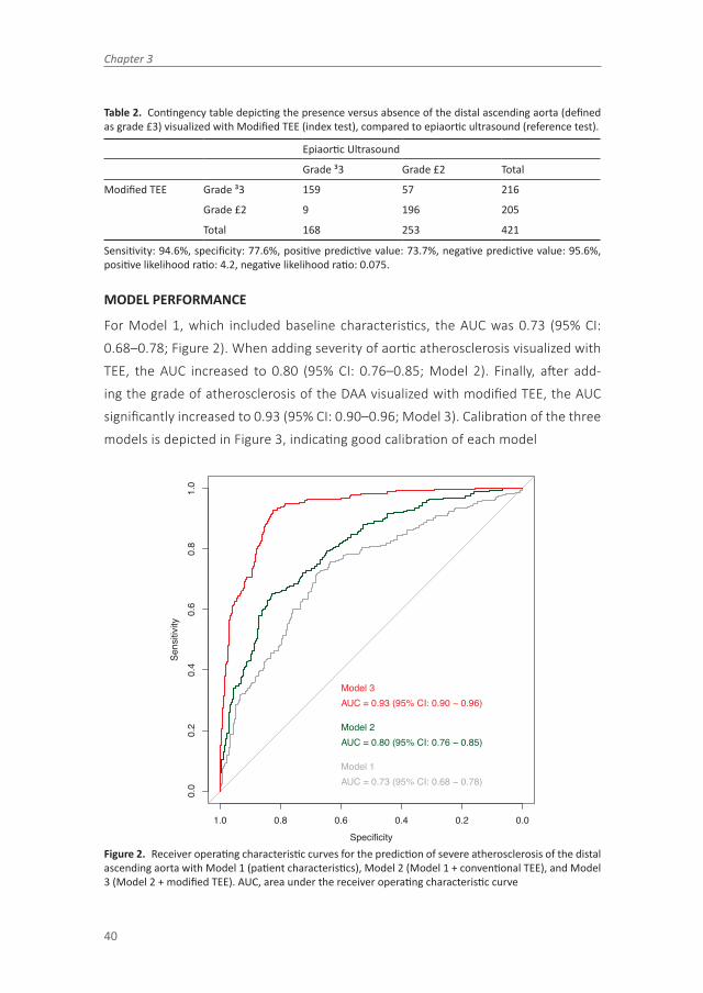
Table 2. Contingency table depicting the presence versus absence of the distal ascending aorta (defined as grade £3) visualized with Modified TEE (index test), compared to epiaortic ultrasound (reference test).
Epiaortic Ultrasound
Grade ³3 Grade £2 Total
Modified TEE Grade ³3 159 57 216
Grade £2 9 196 205
Total 168 253 421
Sensitivity: 94.6%, specificity: 77.6%, positive predictive value: 73.7%, negative predictive value: 95.6%, positive likelihood ratio: 4.2, negative likelihood ratio: 0.075.
MODEL PERFORMANCE
For Model 1, which included baseline characteristics, the AUC was 0.73 (95% CI: 0.68–0.78; Figure 2). When adding severity of aortic atherosclerosis visualized with TEE, the AUC increased to 0.80 (95% CI: 0.76–0.85; Model 2). Finally, after add-ing the grade of atherosclerosis of the DAA visualized with modified TEE, the AUC significantly increased to 0.93 (95% CI: 0.90–0.96; Model 3). Calibration of the three models is depicted in Figure 3, indicating good calibration of each model
Specificity
Sens
itivi
ty
0.0
0.2
0.4
0.6
0.8
1.0
1.0 0.8 0.6 0.4 0.2 0.0
Model 1AUC = 0.73 (95% CI: 0.68 − 0.78)
Model 2AUC = 0.80 (95% CI: 0.76 − 0.85)
Model 3AUC = 0.93 (95% CI: 0.90 − 0.96)
Figure 2. Receiver operating characteristic curves for the prediction of severe atherosclerosis of the distal ascending aorta with Model 1 (patient characteristics), Model 2 (Model 1 + conventional TEE), and Model 3 (Model 2 + modified TEE). AUC, area under the receiver operating characteristic curve

41
The diagnosis of atherosclerosis of the distal ascending aorta in cardiac surgery patients
Figu
re 3
. Ca
libra
tion
plot
s of M
odel
1 (p
atien
t cha
ract
eristi
cs),
Mod
el 2
(Mod
el 1
+ c
onve
ntion
al T
EE),
and
Mod
el 3
(Mod
el 2
+ m
odifi
ed T
EE)

Chapter 3
42
RECLASSIFICATION
The net reclassification index comparing Model 3 with Model 1 was 0.84 (95% CI: 0.77–0.91; reclassification table not shown), with an IDI of 0.41 (P <0.001). The reclassification table comparing Models 3 and 2 is presented in Table 3. As can be interpreted, 73 (43.5%) of the 168 cases were correctly reclassified into a higher risk category, while eight (4.8%) were incorrectly reclassified into a lower risk cat-egory, resulting in a net gain in reclassification of 38.7%. Similarly, among the 253 non-cases, the net gain in reclassification was 21.3%. The overall NRI was thus 0.60 (95% CI: 0.54–0.66), indicating that adding modified TEE greatly and significantly improved the classification of patients. The mean predicted probabilities were 0.73 and 0.56 for Model 3 and 2 among cases, and 0.17 and 0.29 among non-cases, respectively.
Table 3. Reclassification table comparing the risk of atherosclerosis of the distal ascending aorta pre-dicted by model 2 (patient history and TEE) to model 3 (model 2 + modified TEE).
Atherosclerosis (N=168) Model 3 – Modified TEE
Model 2 – TEE Low Intermediate High Total
Low 1 17 1 20* (11.7)
Intermediate 5 44 55 104 (62.2)
High 1 2 40 44* (26.0)
Total 8* (4.7) 64* (38.0) 96 (57.2) 168 (100)
No atherosclerosis (N=253) Model 3 – Modified TEE
Model 2 – TEE Low Intermediate Low Total
Low 107 24 0 131 (61.4)
Intermediate 78 28 8 106 (34.8)
High 1 7 0 8 (3.8)
Total 186 (68.4) 59* (27.8) 8 (3.8) 253 (100)
Table depicts the number of patients classified to a low (<0.25), intermediate (0.25 - 0.75) or high (³0.75) probability of atherosclerosis in the DAA. The upper panel represents patients with presence of severe atherosclerosis (grade ³3) diagnosed with epiaortic ultrasound, the lower panel with absence of athero-sclerosis. The white cells contain the number of patients in the same risk group; patients in the green cells were correctly reclassified by model 3, while patients in the red cells were incorrectly reclassified. The net reclassification improvement of model 3 vs. model 2 = 0.60 (95% CI: 0.54 – 0.66). Figures are N (%). *Counts do not add up to total due to rounding of imputed data
Following formula S2, this resulted in an IDI of 0.30 (P <0.001). Finally, the predic-tiveness curves of the three models are depicted in Figure 4. As an example, the previously mentioned risk categories have been indicated in this figure. Applying the

43
The diagnosis of atherosclerosis of the distal ascending aorta in cardiac surgery patients
second model, 36% of patients were classified in the low-, 52% in the intermediate-, and 12% in the high-risk group. In contrast, applying Model 3 these figures were 46%, 30%, and 24%, respectively.
Figure 4. Curves depict the cumulative proportion of predicted probabilities for Model 1 (patient charac-teristics), Model 2 (Model 1 + conventional TEE), and Model 3 (Model 2 + modified TEE)
DISCUSSION
The main finding of this study was that modified TEE improved the diagnosis of ath-erosclerosis of the DAA considerably and significantly, beyond information available from patient history and visualization of the thoracic aorta with conventional TEE. The improved diagnosis of atherosclerosis of the DAA should guide the operative management and prevent perioperative cerebral embolic complications. Accurate visualization of the (distal) ascending aorta during cardiothoracic aorta is of great importance, as presence of aortic atherosclerosis can guide subtle (e.g. change of positioning aortic cannula or cross-clamp) and more extensive (e.g. off-pump sur-

Chapter 3
44
gery, femoral cannulation) changes in the surgical management. Indeed, multiple guidelines recommend perioperative screening for aortic atherosclerosis during car-diothoracic surgery,15,29 as well as during transcatheter aortic valve replacements.30 EUS is considered the gold standard of visualization of the DAA in cardiac surgery, in daily practice it is not routinely used however.5 Furthermore, it cannot be applied in a growing volume of closed-chest or minimally invasive procedures, as direct access to the ascending aorta is required. Transoesophageal echocardiography is widely used during cardiothoracic surgery, but its sensitivity to diagnose severe athero-sclerosis of the DAA is only 21% (95% CI: 13– 32%).8 Also, the agreement between TEE and EUS is very poor (kappa 0.12, 95% CI: 0.0–0.25) due to an underestimation of the degree of aortic atherosclerosis with TEE in 66% of patients.31 As mentioned, the main reason for this poor accuracy is the so-called ‘blind-spot’ caused by the interposition of the trachea between oesophagus and the DAA. Previously, modified TEE was shown to accurately detect, and especially exclude, atherosclerosis of the DAA in a single-test diagnostic accuracy study.11 Alike most diagnostic work-ups, however the diagnosis of aortic atherosclerosis with modified TEE is a multivari-able process.32 – 34 The perioperative management can only improve if additional information is obtained beyond parameters already known.35 – 37 Indeed, patients’ characteristics (AUC 0.73; 95% CI: 0.68–0.78) and additional TEE imaging (AUC 0.80; 95% CI: 0.76–0.85) already predicted the presence of atherosclerosis in the DAA reasonably well. However, addition of modified TEE further improved the diagnostic accuracy (AUC 0.93; 0.90–0.96), and improved patient classification as shown by an IDI and NRI of 0.60 and 0.30, respectively (both P <0.001). A more accurate diagnosis of aortic atherosclerosis is a prerequisite, but obviously not a guarantee that the implementation of modified TEE will also improve clinical out-comes. The timing of the test is another important parameter. In contrast to EUS, with modified TEE the thoracic aorta can be visualized before start of surgery after intubation. In our experience, this time span is sufficient also for the performance of major adaptations such as transfemoral cannulation or conversion to off-pump surgery. Computed tomography and magnetic resonance imaging can offer a more timely diagnosis of aortic atherosclerosis. An increased CT-detected atherosclerotic burden has been associated with a worse prognosis after cardiac surgery,38 and its implementation as a preoperative screening modality may result in changes in the surgical management and a reduction of post-operative ischemic strokes.39 It is

45
The diagnosis of atherosclerosis of the distal ascending aorta in cardiac surgery patients
well known, however, that non-calcified and mobile plaques carry a higher risk of dislodgement and (cerebral) embolization during cardiac surgery.3,5 In contrast to real-time imaging with echocardiography-based techniques, this cannot be offered with these offline modalities. It has indeed been shown that the sensitivity of CT-imaging for aortic atherosclerosis was inferior to EUS1 and TEE.40 In the latter study, 47% of TEE-detected severe atherosclerotic plaques were missed with CT imaging.40 Another interesting new technique for the intraoperative visualization of aortic ath-erosclerosis is phased-array intra-cardiac echocardiography (ICE). In this technique, an ultrasound unit is placed in the right atrium, through which the aortic valve and proximal ascending aorta can be visualized.41 Direct intra-aortic echocardiography of the descending aorta using the same device has also been described.42 To our knowledge, the diagnostic accuracy of both methods for aortic atherosclerosis has not yet been studied however.
Some limitations apply to our study. First, since EUS served as the reference test, we could not study the added value of modified TEE compared with the added value of EUS. In daily practice, however, screening for aortic atherosclerosis is initiated with modified TEE, as this is performed before sternotomy. Given its high negative predictive value, surgery can continue as planned when severe atherosclerosis is absent. Depending on location and severity of atherosclerosis, a subsequent EUS ex-amination may be considered to confirm a positive result of modified TEE. Secondly, generalizability may be impaired as modified TEE was mainly performed by two senior anesthesiologists, and all patients were included at a single institution with extensive experience in the field of cardiothoracic surgery. Future research will have to show whether these results can be replicated in other settings and with other, less experienced, observers. Finally, there was a substantial amount of missing data, especially for the results of the index and reference test. The main reason for not undergoing these tests was an unavailability of trained personnel to operate modi-fied TEE or EUS. Thus, it seems reasonable to assume that the missingness of these variables did not depend on patient characteristics, and did therefore not affect validity of the results. Indeed, there were only minor imbalances in the distribution of preoperative characteristics in patients with and without a missing index and/or reference test (see Supplementary data online, SA). Also, we aimed to minimize any possible bias through multiple imputation of the missing data. To conclude,

Chapter 3
46
detection of atherosclerosis in the DAA with modified TEE was not only accurate, but also offered information beyond that obtained from patient characteristics and transoesophageal echocardiography, which resulted in an improved classification of patients.

47
The diagnosis of atherosclerosis of the distal ascending aorta in cardiac surgery patients
References 1. Bergman P, van der Linden J, Forsberg K, Ohman M. Preoperative computed tomogra-
phy or intraoperative epiaortic ultrasound for the diagnosis of atherosclerosis of the ascending aorta? Heart Surg Forum 2004;7:E245–9.
2. Evered LA, Silbert BS, Scott DA. Postoperative cognitive dysfunction and aortic ath-eroma. Ann Thorac Surg 2010;89:1091–7.
3. Katz ES, Tunick PA, Rusinek H, Ribakove G, Spencer FC, Kronzon I. Protruding aortic atheromas predict stroke in elderly patients undergoing cardiopulmonary bypass: experience with intraoperative transoesophageal echocardiography. J Am Coll Cardiol 1992;20:70–7.
4. Davila-Roman VG, Murphy SF, Nickerson NJ, Kouchoukos NT, Schechtman KB, Barzilai B. Atherosclerosis of the ascending aorta is an independent predictor of long-term neurologic events and mortality. J Am Coll Cardiol 1999;33:1308–16.
5. Van der Linden J, Hadjinikolaou L, Bergman P, Lindblom D. Postoperative stroke in cardiac surgery is related to the location and extent of atherosclerotic disease in the ascending aorta. J Am Coll Cardiol 2001;38:131–5.
6. Borger M. Stroke during coronary bypass surgery: principal role of cerebral macroem-boli. Eur J Cardthorac Surg 2001;19:627–32.
7. Van der Linden J, Bergman P, Hadjinikolaou L. The topography of aortic atherosclerosis enhances its precision as a predictor of stroke. Ann Thorac Surg 2007;83: 2087–92.
8. Van Zaane B, Zuithoff NPA, Reitsma JB, Bax L, Nierich AP, Moons KGM. Meta-analysis of the diagnostic accuracy of transoesophageal echocardiography for assessment of atherosclerosis in the ascending aorta in patients undergoing cardiac surgery. Acta Anaesthsiol Scand 2008;52:1179–87.
9. Van Zaane B, Nierich AP, BuhreWF, Brandon Bravo Bruinsma GJ, Moons KGM. Resolv-ing the blind spot of transoesophageal echocardiography: a new diagnostic device for visualizing the ascending aorta in cardiac surgery. Br J Anaesth 2007;98:434–41.
10. Nierich AP, Van Zaane B, BuhreWF, Coddens J, Spanjersberg AJ, Moons KGM. Visualiza-tion of the distal ascending aorta with A-Mode transoesophageal echocardiography. J Cardiothor Vasc Anaesth 2008;22:766–73.
11. Van Zaane B, Nierich AP, Brandon Bravo Bruinsma GJ, Rosseel PMJ, Ramjankhan FZ. Diagnostic accuracy of modified transoesophageal echocardiography for preinci-sion assessment of aortic atherosclerosis in cardiac surgery patients. Br J Anaesth 2010;105:131–8.
12. Fryback DG, Thornbury JR. The efficacy of diagnostic imaging. Med Decis Making 1991;11:88–94.
13. Guyatt GH, Tugwell PX, Feeny DH, Haynes RB, Drummond M. A framework for clinical evaluation of diagnostic technologies. Can Med Assoc J 1986;134:587–94.
14. Lijmer JG, LeeflangMMG, BossuytPMM. Proposal for a phased evaluation of medical tests. Med Decis Making 2009;29:E13–21.
15. Glas KE, Swaminathan M, Reeves ST, Shanewise JS, Rubenson D, Smith PK. Guidelines for the performance of a comprehensive intraoperative epiaortic ultrasonographic examination. Am Soc Echocardiogr 2007;20:1227–35.
16. KatzNM,Chase GA. Risks of cardiac operations for elderly patients: reduction of the age factor. Ann Thorac Surg 1997;63:1309–14.
17. Jensky NE, Criqui MH, Wright MC,Wassel CL, Brody SA, Allison MA. Blood pressure and vascular calcification. Hypertension 2010;55:990–7.

Chapter 3
48
18. Iribarren C, Sidney S, Sternfeld B, Browner WS. Calcification of the aortic arch: risk factors and association with coronary heart disease, stroke, and peripheral vascular disease. J Am Med Assoc 2000;283:2810–5.
19. Saimanen E. Risk factors for ascending aortic atheromatosis--a retrospective analysis of 2,263 patients undergoing coronary artery bypass surgery. Ann Chir Gynaecol 1999; 88:280–4.
20. Tessitore E, Rundek T, Jin Z,HommaS, Sacco RL, Di Tullio MR. Association between carotid intima-media thickness and aortic arch plaques. J Am Soc Echocardiogr 2010; 23:772–7.
21. Biancari F, Heikkinen J, Mosorin M, Rasinaho E, Lahtinen J, Niemela¨ E et al. Predictors of diseased ascending aorta in patients undergoing off-pump coronary artery bypass surgery. Heart Surg Forum 2006;9:E857–60.
22. Pencina MJ, D’Agostino RB, Vasan RS. Evaluating the added predictive ability of a new marker: from area under the ROC curve to reclassification and beyond. Stat Med 2008;27:157–72.
23. Moons KGM, Kengne AP,Woodward M, Royston P, Vergouwe Y, Altman DG et al. Risk prediction models: I. Development, internal validation, and assessing the incremental value of a new (bio)marker. Heart 2012;98:683–90.
24. Moons KGM, De Groot JAH, Linnet K, Reitsma JB, Bossuyt PMM. Quantifying the added value of a diagnostic test or marker. Clin Chem 2012;58:1408–17.
25. Pepe MS, Feng Z, Huang Y, Longton G, Prentice R, Thompson IM et al. Integrating the predictiveness of a marker with its performance as a classifier. Am J Epidemiol 2008; 167:362–8.
26. Rubin DB. Multiple Imputation for Nonresponse in Surveys. New York: J. Wiley & Sons; 1987.
27. Groenwold RHH, Donders ART, Roes KCB, Harrell FE, Moons KGM. Dealing with miss-ing outcome data in randomized trials and observational studies. Am J Epidemiol 2012;175:210–7.
28. Van Buuren S, Groothuis-oudshoorn K. Journal of Statistical Software MICE: multivari-ate imputation by chained. J Stat Softw 2011;45:1–67.
29. Cheitlin MD, Armstrong WF, Aurigemma GP, Beller GA, Bierman FZ, Davis JL et al. ACC/AHA/ASE 2003 guideline update for the clinical application of echocardiography. Circulation 2003;108:1146–62.
30. Zamorano JL, Badano LP, Bruce C, Chan KL, Gonc¸alves A, Hahn RT et al. EAE/ASE recommendations for the use of echocardiography in new transcatheter interventions for valvular heart disease. J Am Soc Echocardiogr 2011;24:937–65.
31. Davila-Roman VG, Phillips KJ, Daily BB, Da´vila RM, Kouchoukos NT, Barzilai B. Intraop-erative transoesophageal echocardiography and epiaortic ultrasound for assessment of atherosclerosis of the thoracic aorta. J Am Coll Cardiol 1996;28:942–7.
32. Ledley RS, Lusted LB. Reasoning foundations of medical diagnosis. Science 1959;130: 9–21.
33. Moons KGM, Van Es GA, Deckers JW, Habbema JDF, Grobbee DE. Limitations of sen-sitivity, specificity, likelihood ratio, and Bayes’ theorem in assessing diagnostic prob-abilities. Epidemiology 1997;8:12–7.
34. Miettinen OS, Henschke CI, Yankelevitz DF. Evaluation of diagnostic imaging tests. J Clin Epimiol 1998;51:1293–8.
35. Moons KGM. Criteria for scientific evaluation of novel markers: a perspective. Clin Chem 2010;56:537–41.

49
The diagnosis of atherosclerosis of the distal ascending aorta in cardiac surgery patients
36. Bossuyt PMM, Reitsma JBR, Linnet K, Moons KGM. Beyond diagnostic accuracy: the clinical utility of diagnostic tests. Clin Chem 2012;58:1636–43.
37. Ferrante di Ruffano L, Hyde CJ, McCaffery KJ, Bossuyt PMM, Deeks JJ. Assessing the value of diagnostic tests: a framework for designing and evaluating trials. Br Med J 2012;344:e686.
38. Kurra V, Lieber ML, Sola S, Kalahasti V, Hammer D, Gimple S et al. Extent of thoracic aortic atheroma burden and long-term mortality after cardiothoracic surgery: a com-puted tomography study. J Am Coll Cardiol 2010;3:1020–9.
39. Lee R, Matsutani N, Polimenakos AC, Levers LC, Lee M, Johnson RG. Preoperative non-contrast chest computed tomography identifies potential aortic emboli. Ann Thorac Surg 2007;84:38–41.
40. Barazangi N, Wintermark M, Lease K, Rao R, SmithW, Josephson SA. Comparison of computed tomography angiography and transesophageal echocardiography for evalu-ating aortic arch disease. Stroke 2011;20:436–42.
41. Kim SS, Hijazi ZM, Lang RM, Knight BP. The use of intracardiac echocardiography and other intracardiac imaging tools to guide noncoronary cardiac interventions. J Am Coll Cardiol 2009;53:2117–28.
42. Bartel T, Müller L, Müller S. Intra-aortic phased-array imaging: new guiding tool for transcatheter aortic valve implantation. Eur Heart J 2009;30:2368

Chapter 3
50
Formula S1. Net Reclassification Improvement
(1) NRI = [P(up | D=1) - P(down | D=1)] - [P(up | D=0) - P(down | D=0)],
where P is the proportion of patients, “up” and “down” denote a reclassification into a higher or lower risk-class respectively, and D is the disease status (0 = atheroscle-rosis grade £2, 1 = atherosclerosis grade ³3).
Formula S2. Integrated discrimination Improvement
(2) IDI = (Pnew, atherosclerosis - Pold, atherosclerosis) - (Pnew, no atherosclerosis - Pold, no atherosclerosis),
where P is the mean probability per category
Figure S1. Receiver Operating Characteristic Curves for the three models in patients with complete data. Model 1 = Patient characteristics, Model 2 = Patient characteristics + TOE, model 3 = Patient characteris-tics + TOE + A-View method. TOE = Transoesophageal echocardiography, AUC = Area under the receiver operating curve

51
The diagnosis of atherosclerosis of the distal ascending aorta in cardiac surgery patients
Table S1. Distribution of preoperative characteristics in patients with and without a missing index and / or reference test.
TOE A-View and epiaortic ultrasound images
Not obtained(total N=105 )
Obtained(total N=316 )
N (%) N (%) p
Male Sex 72 (68.6) 194 (61.4) 0.23
Age; median (IQR) 73 (71 – 77) 73 (69 – 77) 0.85
Hypertension 57 (54.3) 180 (57.0) 0.71
Diabetes Mellitus 20 (19.0) 83 (26.3) 0.17
COPD 22 (21.0) 47 (14.9) 0.19
Extracardiac atherosclerosis 21 (20.0) 49 (15.5) 0.36
Neurological Dysfunction 16 (15.2) 46 (14.6) 0.99
Active Endocarditis 0 (0.0) 2 (0.6) 1.00
Pulmonary Hypertension 4 (3.8) 22 (7.0) 0.35
History of MI 20 (19.0) 64 (20.3) 0.90
PCI 17 (16.2) 40 (12.7) 0.45
Cardiac Surgery 9 (8.6) 19 (6.0) 0.49
Instable Angina 3 (2.9) 29 (9.2) 0.06
Additive EuroSCORE 7.0 (5.0 – 9.0) 7.0 (5.0 – 9.0) 0.91
Logistic EuroSCORE 7.0 (4.4 – 16.8) 7.5 (4.3 – 14.3) 0.82
Non-elective surgery 3 (2.9) 1 (0.3) 0.05*
Isolated CABG 39 (37.1) 98 (31.0) 0.30
Isolated valve (any) 6 (5.7) 38 (12.0) 0.10
Combined CABG + valve 25 (23.8) 57 (18.0) 0.25
Additive and Logistic EuroSCORE are median (IQR). EUS = Epiaortic ultrasound, COPD = Chronic obstruc-tive pulmonary disease, MI = Myocardial infarction, PCI = Percutaneous coronary intervention, CABG = Coronary artery bypass grafting. * Fishers’ exact test.
Table S2. Contingency table depicting the presence versus absence of relevant atherosclerosis of the distal ascending aorta (defined as grade ³3) in patients with complete data (N= 316)
Epiaortic Ultrasound
Presence Absence Total
AVIEW Presence 122 38 160
Absence 4 152 156
Total 126 190 316
Sensitivity: 97.4%, specificity: 76.2%, Positive predictive value: 76.2%, Negative predictive value: 97.4%, Positive likelihood ratio: 4.1, Negative likelihood ratio: 0.034.

Chapter 3
52
Table S3. Baseline characteristics of patients with complete data (N = 316), stratified by presence of rel-evant atherosclerosis
Atherosclerosis DAA (with epiaortic ultrasound)
Grade £2 Grade ³3
Total N = 190 Total N = 126 p
Male gender 118 (62.1) 76 (60.3) 0.75
Age. median (IQR) 72 (68 – 77) 73 (69 – 78) 0.11*
Hypertension 109 (57.4) 71 (56.3) 0.86
Diabetes mellitus 46 (24.2) 37 (29.4) 0.31
COPD 25 (13.2) 22 (17.5) 0.29
Extracardiac atherosclerosis 17 (8.9) 32 (25.4) <0.001
Neurological dysfunction 24 (12.6) 22 (17.5) 0.23
LVEF Good 106 (55.8) 69 (54.7) 0.94
Moderate 68 (35.8) 45 (35.7)
Poor 16 (8.4) 12 (9.5)
Aortic Stenosis 59 (31.1) 70 (55.6) <0.001
Previous MI 20 (10.5) 15 (11.9) 0.70
Previous PCI 17 (8.9) 23 (18.3) 0.015
Any cardiac surgery 8 (4.2) 11 (8.7) 0.10
COPD = chronic obstructive pulmonary disease, LVEF = left ventricular ejection fraction, MI = myocardial infarction, PCI = percutaneous coronary intervention, * Mann-Whitney-U


4Prognostic value of modified transesophageal
echocardiography in patients undergoing cardiac
surgery
W.W. Jansen Klomp, A.P. Nierich, G.J. Brandon Bravo Bruinsma, A.W.J. van ‘t Hof, K.G.M. Moons, L.M. Peelen
Resubmitted

Chapter 4
56
ABSTRACT
BACKGROUND Modified transesophageal echocardiography (TEE) enables visualization of the distal ascending aorta (DAA) through the placement of a saline-filled balloon in the left main bronchus. Previous studies showed a good diagnostic accuracy of modified TEE. The present study assessed the prognostic value of Katz grade ³3 atherosclerosis of the DAA detected by modified TEE, which was also compared to the results and prognostic performance of conventional TEE imaging of the proximal ascending- and descending aorta .
METHODS A follow-up study of 421 patients who participated in a large pre-viously published diagnostic accuracy study, which compared modified TEE to epiaortic ultrasound. We studied the association between atherosclerosis of the distal ascending aorta (DAA) and short- and long-term outcomes, and compared the prognostic performance of modified TEE with conventional TEE imaging of the proximal ascending- and descending aorta and the original EuroSCORE. For this aim, we predicted the one-year mortality with logistic regression modelling, and compared a model with- and without modified TEE imaging with respect to (the difference in) the area under the curve (AUC), the net reclassification improvement (NRI) at preselected thresholds (£4%, 4-8%, ³8%) and the integrated discrimination index (IDI).
RESULTS Median age was 73 years, 63.2% was male; patients with severe atherosclerosis had a higher original EuroSCORE (7.0% vs. 5.9%, p = 0.009). Severe atherosclerosis of the DAA based on modified TEE imaging was as-sociated with an increased long-term all-caused mortality (1.82, 95% CI: 1.10 – 3.24), but not with in-hospital stroke. The prognostic accuracy of modified TEE (AUC: 0.64, 95% CI: 0.53 – 0.75) for one-year mortality was superior compared to conventional TEE (AUC: 0.53, 95% CI: 0.43 – 0.64, p <0.001). Similarly, the NRI and IDI (0.14 and 0.0132, p both <0.001) indicated a small but significant improvement of the prognostic performance of modified TEE

57
Prognostic value of modified transesophageal echocardiography
beyond conventional TEE imaging. Adding the EuroSCORE to this model fur-ther improved the predictive value (AUC 0.78, 95% CI 0.69 – 0.83, P<0.001).
CONCLUSION In cardiac surgery patients, severe atherosclerosis of the DAA visualized with modified TEE is a predictor of long-term mortality, which pro-vides further evidence that the degree of atherosclerosis can be accurately detected with modified TEE. The prognostic performance of modified TEE was superior to conventional TEE.

Chapter 4
58
INTRODUCTION
In cardiac surgery patients, atherosclerosis of the ascending aorta is associated with an increased risk of postoperative stroke,1–5 cognitive dysfunction,6 renal dysfunc-tion7, and short- and long-term mortality.8,9 Aortic atherosclerosis is most likely the direct cause of these complications rather than a mere indicator of a worse general health status, as these associations were found to be independent of other patient characteristics.1–11 Manipulation of the distal ascending aorta (e.g. aortic cannula-tion, placement and release of a cross- or side biting-clamp, and the “sand-blasting” effect of cardiopulmonary bypass) is believed to play a pivotal role in the occurrence of cerebral emboli leading to cerebral complications.12
Transesophageal echocardiography (TEE) is widely used as a monitoring tool in cardiothoracic procedures to screen for aortic atherosclerosis, however the air-filled trachea precludes accurate visualization of the distal part of the ascending aorta (DAA).13 Epiaortic ultrasound (EAU) can accurately visualize atherosclerosis of the (distal) ascending aorta, but not the complete aortic arch and its branches.5 Since the EAU probe is applied directly onto the aorta, surgical exposure of the aorta is a prerequisite. Cardiothoracic interventions are increasingly performed during minimally invasive or percutaneous procedures in a closed-chest setting, such as transcatheter aortic valve replacement (TAVR), which requires alternative diagnostic tools to visualize the thoracic aorta.
The thoracic aorta can also be visualized with modified transesophageal echocar-diography. In this TEE-based technique, an acoustic window to the thoracic aorta is obtained through a fluid-filled balloon positioned in the distal trachea and left main bronchus, which enables visualization of the DAA.14–16 Previous diagnostic accuracy studies showed that, compared to epiaortic scanning as reference standard, modi-fied TEE provides an accurate diagnosis of the degree of atherosclerosis of the DAA (AUC: 0.89, NPV 97%, PPV 67%).16 Also, we showed that modified TEE improved the diagnosis of atherosclerosis of the DAA beyond conventional TEE.17
The goal of perioperative screening with modified TEE is to improve patient out-comes with the targeted prevention of plaque disruption during aortic manipula-
59
Prognostic value of modified transesophageal echocardiography
tions. Therefore, the first aim of this study was to assess if aortic atherosclerosis of the DAA detected by modified TEE is a predictor of post-operative stroke and mortality. In the absence of such an association, it would be unlikely that modified TEE could improve patient outcomes. The second aim was to study if the results of modified TEE improved this prognostication beyond conventional TEE results. Finally, the prognostic value of modified TEE was compared with a non-invasive prediction model, i.e. the original EuroSCORE.
METHODS
STUDY DESIGN AND POPULATION Between May 2006 and December 2008, a total of 465 patients were included in a multi-center study evaluating the diagnostic accu-racy of a modified TEE method in patients undergoing cardiothoracic surgery.16 The aim of the initial study was to compare the diagnosis of atherosclerosis as visualized with modified TEE to the “gold standard” epiaortic ultrasound. Before sternotomy, examination of the thoracic aorta with modified TEE was performed (see below), results of this test were blinded for the attending surgeon. Second, an EAU assess-ment of the ascending aorta was performed.18 Eligible patients were aged over 65 years and underwent any cardiac surgical procedure, excluded were patients with a contraindication for one of the applied diagnostic tests.
In the current prospective cohort study we included 421 participants of this study operated at a single center (ISALA Clinics, Zwolle, the Netherlands), as the outcome data used in our analyses was available for this center only. The study was con-ducted in accordance with the principles outlined in the Helsinki declaration; all patients gave written informed consent and the study was approved by the local medical ethics board.
(MODIFIED) TEE After induction of general anesthesia, conventional TEE was first applied to assess the degree of atherosclerosis of the proximal ascending- and de-scending aorta. Also, visualization of the distal ascending aorta was attempted. Then, the catheter mounted A-View balloon (Stroke2prevent, Zwolle, The Netherlands) was introduced in the distal trachea and left main bronchus via the endotracheal tube.14–16 After inflation of the balloon with saline, a sonographic window to the dis-

Chapter 4
60
tal ascending aorta was obtained. Since patients cannot be ventilated after balloon inflation, patients need to be pre-oxygenated. We previously showed that modified TEE can be performed safely during 2-5 minutes of apnoea,14 yet the procedure is contra-indicated in patients with a severely compromised pulmonary function, and in patients with tracheal or bronchial pathology that precludes a safe introduction and expansion of the balloon.15 Possible complications of modified TEE include hypoxemia and hypercapnia after prolonged ventilator arrest, damage to tracheal and bronchial wall, (pulmonary) bleeding and leakage of sterile saline after balloon rupture.15,16 A previous publication showed that of the 465 patients included in this study, ten (2.9%) had blood on the “A-View” catheter without relevant findings dur-ing bronchoscopy; one additional patient required prolonged ventilation for several days because of a pulmonary bleeding probably related to the balloon expansion.
From the conventional mid-esophageal ascending aortic view the probe is retracted until a depth of 25 to 30cm from the incisors, which should reveal the short axis distal ascending aorta view with the multiplane angle adjusted to 30°. The long-axis view can obtained through the adjustment of the multiplane angle to 70° – 120°. For the current study we only used the degree of atherosclerosis in the distal ascending aorta, yet modified TEE also allows visualization of the aortic arch by retracting the probe another 2 to 5cm, with the multiplane angle in 0° and 70° and short- and long-axis view can be obtained respectively. Further retraction of the probe will show the aortic branch view, in which the innominate- and carotid arteries can be visualized. The degree of atherosclerosis was recorded following the Katz’ classification, which classifies the severity of atherosclerosis in five degrees (Table 1).19 Severe atherosclerosis was defined as a grade three or greater atheroma (i.e. protruding atheroma or mobile plaque).
Table 1. Katz’ classification of aortic atherosclerosis
Degree of atherosclerosis
I Normal aorta
II Extensive Intimal Thickening
III Protruding atheroma <5mm
IV Protruding atheroma >5mm
V Mobile plaque

61
Prognostic value of modified transesophageal echocardiography
OUTCOME The primary outcome measure was one-year all-cause mortality, which was prospectively registered. Also, we present the long-term all-cause mortality as extracted from the Dutch Municipal Personal Records Database and completed through review of the electronic charts of each patients. Follow-up of the all-cause mortality was completed until July 3th 2016. Secondary outcomes were the occur-rence of operative (i.e. in-hospital or 30-day) mortality, and in-hospital stroke, which were both prospectively registered. Stroke was defined as a clinical suspicion of cerebral ischemia lasting longer than 24 hours confirmed by an attending neurolo-gist, typically although not mandatory, after cerebral computed tomography (CT) imaging.
STATISTICAL ANALYSIS The baseline characteristics and TEE findings are presented. We compared the grade of atherosclerosis in the DAA (modified TEE) and the proximal- and distal ascending aorta (conventional TEE) respectively. From these tables we calculated the positive predictive values (PPV) for the presence of grade ³3 atherosclerosis of the DAA, in the presence of grade ³3 atherosclerosis in the PAA and/or descending aorta. Dichotomous variables were described by counts and per-centages, and differences were tested for statistical significance with a Chi-square test or Fisher’s exact test as appropriate. Continuous data were described as mean (standard deviation [SD]) for normally distributed, and median (interquartile range [IQR]) for non-normally distributed variables; differences were tested for statistical significance with a Student’s t-test or Mann-Whitney-U test respectively, or with one-way ANOVA for EuroSCORE.
The in-hospital stroke rate and 30-days mortality rate were studied per grade of ath-erosclerosis. We did not analyze the in-hospital outcomes in a multivariable model because of the limited number of events. Long-term all-cause survival was depicted in a Kaplan-Meier curve; the survival was depicted per grade of atherosclerosis, as well as for the absence versus presence of atherosclerosis as defined above. The latter was tested for significance using a log-rank test.
Then, we studied whether the information of modified TEE improved the prognostic performance for one-year mortality beyond the imaging results of TEE. For this aim we modelled the one-year mortality using logistic regression modelling. We per-

Chapter 4
62
formed bootstrapping (N=1000) to correct for optimism in the confidence intervals. These models included:– Model 1: Degree of atherosclerosis of the PAA and descending aorta (conven-
tional TEE)– Model 2: Model 1 + degree of atherosclerosis DAA (modified TEE)– Model 3: Model 2 + Logistic EuroSCOREThe predicted probabilities of the three models were compared for difference in overall performance using the (differences in) area under the receiver-operating characteristic (ROC) curve. Furthermore, we studied the net reclassification im-provement (NRI) comparing model II to model I. The NRI compares the ability of two models to differentiate patients with- and without the outcome of interest.20,21 This method requires the definition of risk groups, here we arbitrarily defined a pre-dicted probability for one-year mortality <0.04 as low-, a probability between 0.04 and 0.08 as intermediate-, and a probability >0.08 as high risk. In our case, a correct reclassification is either a patient who is alive and was moved to a lower risk-group in the second model, or a deceased patient who was moved to a higher risk group in the second model. As a sensitivity analysis the same analyses were performed with different cut-off values, i.e. 0.03-0.06 and 0.03-0.10. We also studied the integrated discrimination improvement, since this metric does not require cut-of values.
Visualization of the distal ascending aorta with modified TEE and EAU was per-formed in 326 (77.4%) and 322 (76.5%) of 421 patients respectively. The body-mass index was missing in two patients (0.5%) and follow-up of the operative mortality in one patient (0.2%). Discarding patients with missing data can lead to biased results and a loss of precision.22–24 We therefore therefore imputed missing cases rather than exclude subjects with missing data. Using the appropriate software in SPSS, twenty imputed datasets were created. Using R statistics, the analyses were then performed for each separate dataset and pooled according to Rubin’s rule. Numbers in tables may not add up to total because of rounding of the imputed values. Statisti-cal analyses were performed in R version 3.1.0 and SPSS version 22.0.0;22,25 all tests were performed 2-sided with a p-value < 0.05 indicating statistical significance.

63
Prognostic value of modified transesophageal echocardiography
RESULTS
BASELINE CHARACTERISTICS The preoperative patient characteristics are depicted in table 2. Median age of all patients was 73 (IQR: 69 – 77) years, 63.2% was male, 56.3% of patients had hypertension and 56.5% had a good left ventricular ejection fraction. Modified TEE showed severe atherosclerosis in 51.1% (N=215) of patients, which was associated with a higher original EuroSCORE (7.0% vs. 5.9%, p = 0.009), extracardiac atherosclerosis (23.3 vs. 9.6%, p = 0.003) and previous percutaneous coronary intervention (PCI; 18.0 vs. 8.8%, p = 0.03).
Table 2. Baseline characteristics
TotalN = 421
DAA Grade £2N = 206
DAA Grade 3-5N = 215
p
Male gender 266 (63.2) 129 (62.9) 138 (63.5) 0.92
Age 73 (69 – 77) 73 (69 – 77) 73 (69 – 77) 0.27
History of
Hypertension 237 (56.3) 115 (56.2) 122 (56.3) 0.99
Diabetes mellitus 103 (24.5) 47 (23.1) 56 (25.7) 0.57
COPD 69 (16.4) 29 (9.6) 40 (18.3) 0.37
Extracardiac atherosclerosis 70 (16.6) 20 (9.6) 50 (23.3) 0.003
Stroke 62 (14.7) 28 (13.8) 34 (15.6) 0.66
MI 84 (20.0) 34 (16.8) 50 (22.9) 0.16
PCI 57 (13.5) 18 (8.8) 39 (18.0) 0.03
Cardiac surgery 28 (6.7) 11 (5.5) 17 (7.7) 0.41
LVEF >50% 238 (56.5) 124 (60.6) 114 (52.2) 0.02
30 – 50% 148 (35.2) 65 (31.9) 83 (38.2)
<30% 35 (8.3) 15 (7.5) 20 (9.1)
EuroSCORE I, % 6.2 (3.7 – 11.1) 5.9 (3.5 – 9.4) 7.0 (3.9 – 13.8) 0.009
Isolated CABG 271 (64.4) 125 (61.3) 146 (67.3) 0.25
Baseline characteristics for patients with (grade 3-5) and without (grade £2) severe atherosclerosis of the distal ascending aorta diagnosed with modified transesophageal echocardiography. Age is depicted as mean (95% confidence interval), the EuroSCORE as median (interquartile range). DAA = distal ascending aorta, COPD = chronic obstructive pulmonary disease, MI = myocardial infarction, PCI = percutaneous coronary intervention, LVEF = left ventricular ejection fraction, CABG = coronary artery bypass grafting.
Atherosclerosis diagnosed with conventional and modified TEE Conventional TEE showed severe atherosclerosis of the PAA in 68.1% (N = 286) of patients and of the descending aorta in 90.9% (N = 382) of patients. Modified TEE showed grade I

Chapter 4
64
to V atherosclerosis of the DAA in 142 (33.1%), 66 (15.4%), 170 (41.5%), 33 (7.6%) and 11 (2.4%) patients respectively. Tables 3A and 3B compare the degree of ath-erosclerosis in the PAA and descending aorta (conventional TEE) with the degree of atherosclerosis in the the DAA (modified TEE). The presence of grade ³3 athero-sclerosis in the PAA and descending aorta was associated with a PPV of 58.7% and 54.6% for the presence of grade ³3 atherosclerosis in the DAA respectively; the PPV in the presence of grade ³3 atherosclerosis of both segments was 61.2%. Grade £2 atherosclerosis of the PAA and descending aorta had a NPV of 64.9% and 83.5% for grade £2 atherosclerosis of the DAA; the NPV in the absence of atherosclerosis in both segments was 93.1%.
Table 3A. Degree of atherosclerosis in the PAA and DAA
Modified TEE – Distal Ascending Aorta
TEE – Proximal Ascending aorta
I II III IV V
I 39 7 17 1 0
II 25 15 25 3 1
III 68 37 110 15 6
IV 6 6 19 12 2
V 1 0 2 1 1
Table 3B. Degree of atherosclerosis in the descending aorta and DAA
Modified TEE – Distal Ascending Aorta
TEE – Descending aorta
I II III IV V
I 15 3 3 0 0
II 9 4 4 0 0
III 68 24 74 9 2
IV 31 21 45 8 3
V 16 13 48 14 5
Table compares the degree of atherosclerosis in the DAA diagnosed with modified TEE with the degree of atherosclerosis in the PAA (Panel A) and descending aorta (Panel B). The degree of atherosclerosis in the DAA and PAA was similar in 177 (42.1%) patients, the degree of atherosclerosis in the DAA was higher 77 patients (18.4%) and lower in 165 (39.5%) patients. Comparing the degree of atherosclerosis in the DAA and descending aorta, the degree was similar in 106 (25.4%) patients, higher in the DAA in 24 patients (5.6%) and lower in the DAA in 289 (69.0%).
IN-HOSPITAL STROKE AND MORTALITY Eleven patients (2.6%) experienced a stroke during hospitalization, of whom six had grade ³ 3 atherosclerosis of the DAA. Opera-tive mortality was 3.1% (N = 13), including one patient with a stroke. As shown in

65
Prognostic value of modified transesophageal echocardiography
figure 1, the EuroSCORE-predicted operative risk increased with each increase in the degree of atherosclerosis (p = 0.01). The association between atherosclerosis of the DAA detected with modified TEE and in-hospital stroke and with operative mortality showed a similar distribution, but both associations were non-significant (Chi-square for trend: p = 0.10 and p = 0.15 respectively).
Figure 1. The EuroSCORE I predicted mortality and observed in-hospital stroke and operative mortality for each degree of atherosclerosis of the distal ascending aorta (DAA). N = number, IQR = interquartile range. P value for trend; EuroSCORE: p = 0.01, mortality: p = 0.15, stroke: p = 0.10.
LONG-TERM MORTALITY Median follow-up time of the vital status was 4.2 years (IQR 3.6 – 4.8) with a total follow-up of 1645 person-years; in this period 74 people died (17.6%). The mortality rate per 1000 person-years was 58.1 in patients with, and 32.0 in patients without severe atherosclerosis of the DAA respectively (RR 1.82, 95% CI: 1.10 – 3.24). Kaplan-Meier plots in figure 2 shows the unadjusted association between atherosclerosis of the DAA and the long-term all-cause mortal-ity. Patients with grade ³3 atherosclerosis of the DAA had an increased all-cause mortality compared to patients with grade £2 atherosclerosis (p = 0.026).
PROGNOSTIC PERFORMANCE First, we compared the overall prognostic perfor-mance of the three models for all-cause one-year mortality in an ROC-curve (figure 3). Model I included the degree of atherosclerosis in the PAA and descending aorta diagnosed with conventional TEE, which did not predict one-year all-cause mortal-

Chapter 4
66
Figu
re 2
. Pa
nel A
: Kap
lan-
Mei
er e
stim
ated
surv
ival
stra
tified
by
abse
nce
(gra
de £
2) o
r pre
senc
e (g
rade
³3) o
f ath
eros
cler
osis
of th
e di
stal
asc
endi
ng a
orta
(DAA
) visu
al-
ized
with
mod
ified
tran
seso
phag
eal e
choc
ardi
ogra
phy.
Pane
l B: K
apla
n-M
eier
esti
mat
ed s
urvi
val s
trati
fied
by d
egre
e of
ath
eros
cler
osis
of th
e di
stal
asc
endi
ng a
orta
visu
alize
d w
ith m
odifi
ed tr
anse
soph
agea
l ech
ocar
diog
ra-
phy.

67
Prognostic value of modified transesophageal echocardiography
ity (AUC: 0.53, 95% CI: 0.43 – 0.64). In model 2, the degree of atherosclerosis of the DAA visualized with modified TEE was added to model 1, which significantly improved the model performance (AUC: 0.64, 95% CI: 0.53 – 0.75; p for difference with model I <0.001). In model 3 the original EuroSCORE was added to model 2, which provided a superior prognosis of the one-year mortality compared to the two previous models (AUC 0.76, 95% CI: 0.69 – 0.84; p for difference <0.001).
Specificity
Sens
itivi
ty
0.0
0.2
0.4
0.6
0.8
1.0
1.0 0.8 0.6 0.4 0.2 0.0
Model 1:AUC = 0.53 (95% CI: 0.43 − 0.64)
Model 2:AUC = 0.64 (95% CI: 0.53 − 0.75)
Model 3:AUC = 0.78 (95% CI: 0.69 − 0.83)
Figure 3. Receiver Operating Characteristics Curves depicting the predicted probability for one-year mor-tality. AUC = Area Under (operating receiver) Curve.Model 1 = TEE: degree of atherosclerosis of the proximal ascending aorta and descending aortaModel 2 = Modified TEE: Model 1 + degree of atherosclerosis of the distal ascending aortaModel 3 = logistic EuroSCORE
The NRI comparing models II and I was 0.14 (p < 0.001; figure 4), indicating a small increase in the reclassification with the second model. The sensitivity analyses showed a higher NRI using different cut-off values (supplement A) Finally, the IDI comparing models 1 and 2 was 0.01323 (p <0.001), also indicating a small but significant improvement of reclassification with the second model (supplement B).

Chapter 4
68
Deceased Model 2 (Modified TEE)
Low Intermediate High
Model 1 (TEE) Low 4 1 1
Intermediate 5 13 6
High 0 0 3
Alive Model 2 (Modified TEE)
Low Intermediate High
Model 1 (TEE) Low 129 13 2
Intermediate 77 73 60
High 0 16 17
Figure 4. Net reclassification improvement comparing model 2 (modified TEE) to model 1 (conventional TEE). A low, intermediate and high risk was defined as a predicted mortality of <4%, 4-8% and >8% respec-tively. The upper panel includes patients who died <1 year after surgery, the lower panel shows patients who were alive one year after surgery. A correct reclassification (green figures) is a non-deceased patient reclassified into a lower risk stratum or a deceased patient reclassified into a higher risk stratum. The NRI comparing models 2 and 1 was 0.14 (p <0.001).
DISCUSSION
Our first aim was to study the prognostic information contained within the severity of atherosclerosis of the DAA as visualized with modified TEE. Indeed, in this cohort of cardiac surgery patients, grade ³3 atherosclerosis of the DAA was associated with an increased risk for long-term all-cause mortality (RR 1.82, 95% CI: 1.10 – 3.24). Although this association itself was already known, previous studies predominantly used epiaortic ultrasound to visualize aortic atherosclerosis, and this study is the first to show that the results of modified TEE have similar prognostic consequences for patients. This is of importance, since cardiothoracic interventions are increas-ingly performed during minimally invasive (e.g. robotic surgery) or percutaneous procedures (e.g. TAVR), in which the thoracic aorta is no longer exposed for exami-nation with epiaortic ultrasound.26
Other tests including conventional TEE, may partly already provide the same infor-mation as modified TEE. This would make the use of a more invasive test question-able. In a previous study we already showed that the use of modified TEE improved the diagnosis of atherosclerosis of the DAA beyond conventional TEE results.17 In the current study we showed that the presence of atherosclerosis in the PAA and

69
Prognostic value of modified transesophageal echocardiography
descending aorta are poor predictors of atherosclerosis in the DAA; even if severe atherosclerosis was detected in both segments, the PPV for atherosclerosis in the DAA was only 61.2%. It is thus reasonable to perform modified TEE to confirm the presence of atherosclerosis of the DAA and to identify the exact location of athero-sclerosis to guide the subtle changes in the surgical management. If conventional TEE shows no atherosclerosis in both segments it is not necessary to perform ad-ditional modified TEE imaging given an NPV of 93.1%.
The second aim was to study if modified TEE results improved the prognostic per-formance for long-term all-cause mortality beyond conventional TEE. The degree of atherosclerosis in the PAA and descending aorta visualized with conventional TEE results did not have predictive value for the one-year mortality (AUC 0.53, 95% CI: 0.43 – 0.64). Addition of the degree of atherosclerosis of the DAA, visualized with modified TEE, indeed improved the prognostic performance (AUC 0.64, 95% CI: 0.53 – 0.75, p <0.001). The NRI and IDI similarly showed a significant but small improve-ment of the prognostic performance with modified TEE imaging. Of importance, the results of modified TEE were blinded in this diagnostic accuracy study. Therefore, these results show an association which is not influenced by changes made in the management, based on the imaging findings itself. The original EuroSCORE had a superior prognostic performance compared to the second model (AUC 0.76, 95% CI: 0.69 – 0.84, p <0.001). The ultimate goal of modified TEE is however not to provide a new invasive tool to predict mortality in the coming years, but rather to prevent intra-operative complications in the coming minutes to hours. This requires the visualization of the exact location and severity of an aortic plaque, and not a probability of atherosclerosis based on a non-invasive risk score.
In contrast to previous studies, the degree of atherosclerosis of the DAA was not associated with a higher risk for in-hospital stroke.1–4,8 We assessed the same as-sociation for the degree of atherosclerosis of the DAA diagnosed with EAU, which showed similar results (data not shown). Given this absence of an association for the “gold standard”, an association is also not likely for the “reference test”. There are several potential explanations for this absence of an association. First, the lim-ited number of patients with stroke may have resulted in a type II error, Second, severe atherosclerosis was defined as grade ³3, since this was cut-off was used in
Chapter 4
70
previous studies.16,17 Yet, the incidence of post-operative stroke in patients with grade 3 atherosclerosis was comparable to patients with grade 1-2 atherosclerosis. Therefore, it may be clinically more relevant to identify patients with grade ³4, as these patients are at increased risk for post-operative stroke.19 Third, although the results of modified TEE were blinded to the surgeon, the degree of atherosclerosis was known after EAU imaging. Given a high correlation between both tests, embolic complications may have been prevented through adaptations of the procedure in patients with severe atherosclerosis diagnosed with modified TEE, in whom EAU also showed aortic atherosclerosis.
LIMITATIONS Modified TEE imaging was missing in a substantial proportion of pa-tients, which was primarily caused by changes in the operation schedule resulting in the absence of an anesthesiologist who was trained to perform modified TEE. Therefore, the missingness was not associated to patient characteristics, but to an external factor. Indeed, the baseline characteristics and complete cases analyses showed similar results. Although the imputation of test results may feel counter-intuitive, this provides more accurate results than discarding these patients.22–24
CONCLUSIONS
In cardiac surgery patients, severe atherosclerosis of the DAA visualized with modi-fied TEE is a predictor of long-term mortality, which provides further evidence that the degree of atherosclerosis can be accurately detected with modified TEE. Also, the prognostic performance of modified TEE was superior to conventional TEE imag-ing of the PAA and descending aorta. The results of modified TEE should guide the operative management to prevent intraoperative embolic complications. Modified TEE should not be used as a purely prognostic test, since the EuroSCORE is a better predictor of one-year mortality.

71
Prognostic value of modified transesophageal echocardiography
REFERENCES 1. van der Linden J, Hadjinikolaou L, Bergman P, Lindblom D. Postoperative stroke in
cardiac surgery is related to the location and extent of atherosclerotic disease in the ascending aorta. J Am Coll Cardiol 2001;38:131–5.
2. van der Linden J, Bergman P, Hadjinikolaou L. The topography of aortic atherosclerosis enhances its precision as a predictor of stroke. Ann Thorac Surg 2007;83:2087–92.
3. Hartman GS, Yao FS, Bruefach M, Barbut D, Peterson JC, Purcell MH, Charlson ME, Gold JP, Thomas SJ, Szatrowski TP. Severity of aortic atheromatous disease diagnosed by transesophageal echocardiography predicts stroke and other outcomes associated with coronary artery surgery: a prospective study. Anesth Analg 1996;83:701–8.
4. Djaiani G, Fedorko L, Borger M, Mikulis D, Carroll J, Cheng D, Karkouti K, Beattie S, Karski J. Mild to moderate atheromatous disease of the thoracic aorta and new ischemic brain lesions after conventional coronary artery bypass graft surgery. Stroke 2004;35:e356–8.
5. Elias-Smale SE, Odink AE, Wieberdink RG, Hofman A, Hunink MGM, Krestin GP, Koud-staal PJ, Breteler MMB, van der Lugt A, Witteman JCM. Carotid, aortic arch and coro-nary calcification are related to history of stroke: the Rotterdam Study. Atherosclerosis 2010;212:656–60.
6. Evered L a, Silbert BS, Scott DA. Postoperative cognitive dysfunction and aortic ath-eroma. Ann Thorac Surg 2010;89:1091–7.
7. Dávila-Román VG, Kouchoukos NT, Schechtman KB, Barzilai B. Atherosclerosis of the ascending aorta is a predictor of renal dysfunction after cardiac operations. J Thorac Cardiovasc Surg 1999;117:111–6.
8. Dávila-Román VG, Murphy SF, Nickerson NJ, Kouchoukos NT, Schechtman KB, Barzilai B. Atherosclerosis of the ascending aorta is an independent predictor of long-term neurologic events and mortality. J Am Coll Cardiol 1999;33:1308–16.
9. Thambidorai SK, Jaffer SJ, Shah TK, Stewart WJ, Klein AL, Lauer MS. Association of ath-eroma as assessed by intraoperative transoesophageal echocardiography with long-term mortality in patients undergoing cardiac surgery. Eur Heart J 2007;28:1454–61.
10. Bergman P, van der Linden J, Forsberg K, Ohman M. Preoperative computed tomogra-phy or intraoperative epiaortic ultrasound for the diagnosis of atherosclerosis of the ascending aorta? Heart Surg Forum. 2004;7:E245–9; discussion E249.
11. Mackensen GB, Ti LK, Phillips-Bute BG, Mathew JP, Newman MF, Grocott HP. Cerebral embolization during cardiac surgery: impact of aortic atheroma burden. Br J Anaesth. 2003;91:656–61.
12. Barbut D, Hinton RB, Szatrowski TP, Hartman GS, Bruefach M, Williams-Russo P, Charl-son ME, Gold JP. Cerebral emboli detected during bypass surgery are associated with clamp removal. Stroke 1994;25:2398–2402.
13. Van Zaane B, Zuithoff NPA, Reitsma JB, Bax L, Nierich AP, Moons KGM. Meta-analysis of the diagnostic accuracy of transesophageal echocardiography for assessment of atherosclerosis in the ascending aorta in patients undergoing cardiac surgery. Acta Anaesthesiol Scand 2008 52:1179–87.
14. van Zaane B, Nierich AP, Buhre WF, Brandon Bravo Bruinsma GJ, Moons KGM. Resolv-ing the blind spot of transoesophageal echocardiography: a new diagnostic device for visualizing the ascending aorta in cardiac surgery. Br J Anaesth 2007;98:434–41.
15. Nierich AP, van Zaane B, Buhre WF, Coddens J, Spanjersberg AJ, Moons KGM. Visualiza-tion of the distal ascending aorta with A-Mode transesophageal echocardiography. J Cardiothorac Vasc Anesth. 2008;22:766–73.

Chapter 4
72
16. van Zaane B, Nierich AP, Brandon Bravo Bruinsma GJ, Rosseel PMJ, Ramjankhan FZ, de Waal EEC, Buhre WF, Moons KGM. Diagnostic accuracy of modified transoesophageal echocardiography for pre-incision assessment of aortic atherosclerosis in cardiac surgery patients. Br J Anaesth 2010;105:131–8.
17. Jansen Klomp WW, Peelen LM, Spanjersberg SJ, Brandon Bravo Bruinsma GJ, Lange F de, Van’t Hof AWJ, Moons KGM. Added value of modified transoesophageal echocar-diography in the diagnosis of atherosclerosis of the distal ascending aorta in cardiac surgery patients. Eur Heart J Cardiovasc Imaging 2014;15:623–30.
18. Glas KE, Swaminathan M, Reeves ST, Shanewise JS, Rubenson D, Smith PK, Mathew JP, Shernan SK. Guidelines for the performance of a comprehensive intraoperative epiaortic ultrasonographic examination: recommendations of the American Society of Echocardiography and the Society of Cardiovascular Anesthesiologists; endorsed by the Society of Thoracic S. J Am Soc Echocardiogr 2007;20:1227–35.
19. Katz ES, Tunick P a, Rusinek H, Ribakove G, Spencer FC, Kronzon I. Protruding aortic atheromas predict stroke in elderly patients undergoing cardiopulmonary bypass: experience with intraoperative transesophageal echocardiography. J Am Coll Cardiol 1992;20:70–7.
20. Pencina MJ, D’Agostino RB, Vasan RS. Evaluating the added predictive ability of a new marker: from area under the ROC curve to reclassification and beyond. Stat Med 2008;27:157–72.
21. Moons KGM, Kengne AP, Woodward M, Royston P, Vergouwe Y, Altman DG, Grobbee DE. Risk prediction models: I. Development, internal validation, and assessing the incremental value of a new (bio)marker. Heart 2012;98:683–90.
22. Rubin DB. Multiple Imputation for Nonresponse in Surveys. 1987. 23. Little RJ. Regression with Missing X’s: A Review. J Am Stat Assoc. 1992;87:1227–1237. 24. Donders ART, van der Heijden GJMG, Stijnen T, Moons KGM. Review: a gentle introduc-
tion to imputation of missing values. J Clin Epidemiol 2006;59:1087–91. 25. van Buuren S, Groothuis-oudshoorn K. Journal of Statistical Software MICE : Multivari-
ate Imputation by Chained. J Stat Softw. 2011;45. 26. Iribarne A, Easterwood R, Chan EYH, Yang J, Soni L, Russo MJ, Smith CR, Argenziano
M. The golden age of minimally invasive cardiothoracic surgery: current and future perspectives. Future Cardiol 2011;7:333–46.

73
Prognostic value of modified transesophageal echocardiography
Supplement A1. Net reclassification improvement comparing model 2 (modified TEE) to model 1 (con-ventional TEE). A low, intermediate and high risk was defined as a predicted mortality of <3%, 3-6% and >6% respectively. The NRI comparing models 2 and 1 was 0.25 (p <0.001).
Deceased Model 2 (Modified TEE)
Low Intermediate High
Model 1 (TEE) Low 1 3 1
Intermediate 0 7 7
High 0 3 10
Alive Model 2 (Modified TEE)
Low Intermediate High
Model 1 (TEE) Low 84 35 2
Intermediate 31 61 44
High 0 46 85
Supplement A2. Net reclassification improvement comparing model 2 (modified TEE) to model 1 (con-ventional TEE). A low, intermediate and high risk was defined as a predicted mortality of <3%, 3-10% and >10% respectively. These values represent an often used risk stratification for the EuroSCORE, but are not well applicable to our study, given that the predicted probabilities of model 1 were all <0.10. The NRI comparing models 2 and 1 was 0.16 (p <0.001).
Deceased Model 2 (Modified TEE)
Low Intermediate High
Model 1 (TEE) Low 1 4 0
Intermediate 0 23 4
High 0 0 0
Alive Model 2 (Modified TEE)
Low Intermediate High
Model 1 (TEE) Low 84 37 0
Intermediate 31 216 20
High 0 0 0
Supplement B. Integrated Discrimination Improvement
One-year follow-up
Alive Ceased
Model 1 0.05071 0.06148
Model 2 0.05001 0.07465
Model 3 0.04936 0.08646
Table shows the mean predicted probabilities for mortality in patients alive and ceased one year after surgery respectively. A perfect model would have a predicted probability of 0.00 and 1.00 in both groups respectively. The integrated discrimination improvement (IDI) compares this prediction across different models, a model is more accurate if patients who are alive have a lower predicted probability, and patients who ceased have a higher predicted probability. Thus, the IDI can be calculated as follows:Model 2 vs. 1: 0.05071 – 0.05001 + 0.07465 – 0.06148 = 0.01323Model 3 vs. 2: 0.05001 – 0.04936 + 0.08636 – 0.07465 = 0.01236

5Impact of modified transesophageal
echocardiography on mortality and stroke after
cardiac surgery – a large cohort study
W.W. Jansen Klomp, K.G.M. Moons, A.P. Nierich, G. J. Brandon Bravo Bruinsma, A.W.J. van ‘t Hof, J.G. Grandjean, L.M. Peelen
Submitted

Chapter 5
76
ABSTRACT
OBJECTIVE Atherosclerosis of the upper thoracic aorta can be accurately vi-sualized with modified transesophageal echocardiography (A-View method). We investigated the impact of perioperative screening with modified TEE on postoperative stroke and mortality.
METHODS A single-center observational study including consecutive patients who underwent cardiac surgery between 2006 until 2014. We compared 30-day mortality and in-hospital stroke incidence in patients who were operated with perioperative modified TEE screening (intervention group) to those without the use of modified TEE screening (control group). The associa-tion between modified TEE use (yes/no) and both outcomes was adjusted for confounding with multivariable logistic regression and propensity score analysis.
RESULTS The study included 8,605 patients, modified TEE screening was used in 1,391 (16.2%) patients. Patients in the intervention group were on average older (71 vs. 68 years, p <0.001), were more often females (31.0% vs. 28.0%, p <0.001), and had a higher predicted mortality (EuroSCORE I: 5.9% vs. 4.0%, p <0.001). The observed 30-day mortality was 2.2% and 2.5% in both groups respectively, with multivariable and propensity-score adjusted relative risks (RRs) of 0.70 (95% CI: 0.50 – 1.00, p = 0.05) and 0.67 (95% CI: 0.45 – 0.98, p = 0.04). In-hospital stroke was 2.9% and 2.1% in both groups respectively, with adjusted RRs of 1.03 (95% CI: 0.73 – 1.45) and 1.01 (95% CI: 0.71 – 1.43).
CONCLUSIONS In patients undergoing cardiac surgery, use of perioperative screening for aortic atherosclerosis with modified TEE was associated with lower postoperative mortality, but not stroke, as compared to patients oper-ated without such screening.

77
Impact of modified transesophageal echocardiography on mortality and stroke after cardiac surgery
INTRODUCTION
In patients who undergo cardiac surgery, presence of aortic atherosclerosis is asso-ciated with an increased risk of post-operative stroke and mortality.1–4 Atherogenic emboli released during aortic manipulation are thought to play a pivotal role in the pathophysiology of these complications, as these particles can cause cerebral isch-emia. Other symptoms associated with these embolic complications are transient ischemic attack (TIA), convulsions, delirium and post-operative cognitive decline.5,6 Screening for the presence of aortic atherosclerosis allows for a change in the surgi-cal technique if aortic atherosclerosis is diagnosed, aimed to prevent the formation of such emboli. These changes include a different aortic cannulation site, choosing a different position for the aortic cross-clamp, the use of a dispersion cannula, cardio-pulmonary bypass (CPB) through femoral or subclavian cannulation, or avoidance of CPB in a beating-heart “no touch aorta” procedure.7
The distal ascending aorta (DAA) is most prone for the development of athero-sclerosis.3 Guidelines stress the importance of perioperative imaging of this part of the aorta with transesophageal echocardiography (TEE) or epiaortic ultrasound (EAU).8–10 Although epiaortic ultrasound is a highly accurate test, its use in practice is limited. On the contrary, TEE is widely used and has the advantage that it can be applied before sternotomy, yet visualization of the upper thoracic aorta is limited by the interposition of the air-filled trachea. Due to this so-called “blind-spot” the sensitivity of TEE for the diagnosis of atherosclerosis of the DAA is only 21%.11
Echocardiographic visualization of these structures is possible using a modification to TEE, in which a balloon is introduced in the trachea. After inflation of the balloon with saline, the aortic arch and its branches can be accurately visualized through the trachea.12–14 Previous studies have shown that modified TEE is safe to apply,12 that the sensitivity for detection of atherosclerosis of the DAA was 95% (95% confidence interval [CI]: 92% – 99%),14 and its diagnostic accuracy was significantly better as compared to conventional TEE imaging.15
Better diagnostic accuracy for modified TEE as compared to conventional does not necessarily imply that it leads to improved care and thus outcomes of patients.16,17

Chapter 5
78
We therefore studied the impact of using modified TEE screening in patients un-dergoing cardiothoracic surgery, as compared to not using such screening, on the actual occurrence of postoperative mortality and stroke.
METHODS
STUDY DESIGN AND PATIENTS This observational large cohort study was conducted in the ISALA Clinics, Zwolle, The Netherlands. All patients undergoing surgery that required a sternotomy between January 1st 2008 and December 31th 2014 were eligible. Excluded were patients aged below 18 (N = 1), patients who did not give informed consent (N = 163), those included in a competing study (N = 189), and patients who underwent non-cardiac surgery (N = 133), thoracic stenting (N = 7) or re-operations within 30 days after the index surgery (figure 1).14 Patients provided preoperative written informed consent for use of their data for clinical research, and the study conformed to the principals outlined in the declaration of Helsinki. The institution’s ethical committee waived formal evaluation of the study protocol.
Figure 1. Flowchart of patient inclusion.

79
Impact of modified transesophageal echocardiography on mortality and stroke after cardiac surgery
INTERVENTIONS
INDEX GROUP Patients underwent TEE examination before sternotomy to screen for atherosclerosis of the proximal ascending aorta (PAA) and descending aorta. Severity of atherosclerosis was graded following the Katz’ classification (figure 2).18 In general, if TEE-imaging showed grade three or higher atherosclerosis, modified TEE was added to visualize the DAA and aortic arch (figure 3).18 This technique was described elaborately in previous publications.12–14 In short, following a conventional TEE examination the “A-View catheter” (Stroke2prevent, Zwolle, The Netherlands) was introduced via an endotracheal (ET) tube until the 24 cm marker lined up with a 24 cm marker on the ET-tube. The correct position of the catheter in the left main bronchus was ascertained, and patients were pre-oxygenated. Then the balloon was inflated with 20 – 50 ml of sterile saline, after which imaging could be safely performed within two to three minutes. Three views of the aorta were acquired fol-lowing a predefined protocol. First, starting from a conventional proximal ascending aortic transesophageal view, the DAA was visualized in a short-axis view by retract-ing the TEE probe to a depth of 25 - 30 cm of the incisors. Then, a long axis view was obtained by rotating the multi-plane to 70 – 120 degrees. Finally, the aortic arch and its branch vessels were visualized by further retracting the probe in a 0 degrees multiplane angle.
Figure 2. Katz’ classification of aortic atherosclerosis
Grade atherosclerosis Aspect
Mild 1 Normal appearing aorta
2 Extensive intimal thickening
Severe 3 Atheroma protruding <5mm
4 Atheroma protruding >5mm
5 Mobile atheroma
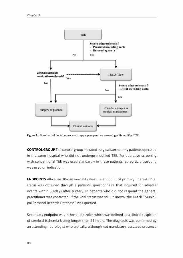
Chapter 5
80
Figure 3. Flowchart of decision process to apply preoperative screening with modified TEE
CONTROL GROUP The control group included surgical sternotomy patients operated in the same hospital who did not undergo modified TEE. Perioperative screening with conventional TEE was used standardly in these patients, epiaortic ultrasound was used on indication.
ENDPOINTS All-cause 30-day mortality was the endpoint of primary interest. Vital status was obtained through a patients’ questionnaire that inquired for adverse events within 30-days after surgery. In patients who did not respond the general practitioner was contacted. If the vital status was still unknown, the Dutch “Munici-pal Personal Records Database” was queried.
Secondary endpoint was in-hospital stroke, which was defined as a clinical suspicion of cerebral ischemia lasting longer than 24 hours. The diagnosis was confirmed by an attending neurologist who typically, although not mandatory, assessed presence
81
Impact of modified transesophageal echocardiography on mortality and stroke after cardiac surgery
of cerebral ischemia with cerebral computed tomography (CT) imaging. Stroke was recorded in the ongoing registry; to ascertain that all strokes were identified we also performed a chart review of all patients in whom a cerebral CT was performed during the index-hospitalization.
Finally, as an intermediate endpoint, modifications in the operative management of patients were assessed in both groups. Data was available for two surgical modifica-tions: use of off-pump coronary artery bypass grafting (OPCAB) and cannulation with a different type of cannula (“Select 3D cannula”, Medtronic Inc. Minneapolis, MN).19 This cannula aims at preventing dislodgement of aortic atheroma by converting the outflow in three streams with a lower velocity. 20
POTENTIAL CONFOUNDERS Due to our observational cohort design, the allocation to the use of modified TEE was clearly not random. Particularly, patients with a higher risk of having aortic atherosclerosis, based on the TEE results as well as on patient characteristics, were likely more indicated to undergo modified TEE screen-ing during surgery Since the same characteristics that indicate the use of modified TEE are also associated with a higher risk of postoperative stroke and mortality, the crude estimated effect association of the use of modified TEE (as compared to the control group) and the outcomes was likely distorted by these factors. We therefore considered the following confounders to be adjusted for in the analysis: logistic EuroSCORE, sex, age, hypertension, diabetes mellitus, unstable angina pectoris, previous myocardial infarction, previous percutaneous transluminal coronary an-gioplasty, previous cardiac surgery, preoperative creatinin, left ventricular ejection fraction (LVEF; <30%, 30-50%, >50%), type of surgery (isolated coronary artery by-pass grafting [CABG], isolated valve, CABG + valve, other) and emergency indication. Age and EuroSCORE were modelled using a log transformation; the preoperative creatinin was modelled as ((1/creatinin)*100).
STATISTICAL ANALYSIS
UNADJUSTED ANALYSIS First, the crude association of the use of modified TEE (intervention) as compared to not using this technique (control) was assessed for both outcomes using the risk ratio (RR) and 95% CI.

Chapter 5
82
MULTIVARIABLE REGRESSION Second, the association between the use of modified TEE as compared to the control group was corrected for the above-mentioned con-founders, again for both end-points. Rather than multivariable logistic regression we used a binomial distribution with a log-link to obtain RR’s with a 95% CI, which are more easily interpretable than odds ratios and are synchronous with the crude association measure.21,22
PROPENSITY SCORE ANLAYSIS Additionally, we corrected for confounding with pro-pensity score (PS) analysis.11 Using logistic regression analysis a PS was created, with modified TEE as the dependent variable and the confounders as covariates. After checking that the range of the PS was similar for the exposed and the unexposed, and that the score was well balanced over quintiles of the PS, the individual pro-pensity scores were added to a model with only modified TEE status as a covariate.
Analyses were performed in SPSS 22.0 and R version 3.1.0. A p-value <0.05 was regarded statistically significant throughout this article.
RESULTS
Of the 8,605 study patients, modified TEE was applied in 1,391 patients (16.2%). Table 1 summarizes the baseline characteristics in comparison between both groups. The modified TEE (index) group included more females, and patients were on average older. These patients had more comorbid diseases, including hypertension, diabetes mellitus, extracardiac atherosclerosis, previous TIA or stroke, and COPD. Surgery in the index group more often included replacement of the aortic valve, ascending aorta or (partial) aortic arch (table 2). Duration of the extracorporeal circulation was similar in both groups, as was the postoperative ventilation time.

83
Impact of modified transesophageal echocardiography on mortality and stroke after cardiac surgery
Table 1. Baseline characteristics comparing patients with and without perioperative screening with modi-fied TEE.
Modified TEE(N = 1,391)
No modified TEE(N = 7,214)
P
Age 71 (65 – 78) 68 (61 – 75) <0.001
Female sex 431 (31.0) 2,023 (28.0) 0.026
BMI 27.0 (24.9 – 30.0) 27.0 (25.0 – 30.0) 0.38
History of
Hypertension 867 (62.3) 3,490 (48.4) <0.001
DM 365 (26.2) 1,629 (22.6) 0.003
Peripheral atherosclerosis 264 (19.0) 651 (9.0) <0.001
TIA or stroke 192 (13.8) 545 (7.6) <0.001
COPD 255 (18.4) 1015 (14.1) <0.001
Myocardial Infarction 411 (29.5) 2,028 (28.1) 0.27
PTCA 242 (17.4) 1,064 (14.7) 0.012
Cardiothoracic surgery 109 (7.8) 405 (5.6) 0.001
Instable angina 80 (5.8) 350 (4.9) 0.16
LVEF >50% 870 (62.5) 4,725 (61.2) 0.069
30 – 50% 406 (29.2) 1,976 (31.3)
<30% 115 (8.2) 508 (7.0)
Preoperative creatinin 87 (75 – 103) 85 (73 – 100) <0.001
Logistic EuroSCORE 5.9 (2.9 – 12.5) 4.0 (2.1 – 8.2) <0.001
BMI = Body mass index, DM = Diabetes mellitus, COPD = Chronic obstructive pulmonary disease, TIA = Transient ischaemic attack, PTCA = Percutaneous transluminal coronary angioplasty, LVEF = left ventricular ejection fraction.

Chapter 5
84
Table 2. Operative characteristics comparing patients with and without perioperative screening with modified TEE.
Modified TEE(N = 1,410)
No modified TEE(N = 7,214)
P
Emergency Indication 25 (1.8) 205 (2.8) <0.001
Surgery
CABG 942 (67.7) 5,033 (69.8) 0.13
Off-pump CABG 63 (9.9)* 488 (15.7)* <0.001
Aortic valve surgery 481 (34.6) 1,860 (25.8) <0.001
Mitral valve surgery 202 (14.5) 1,090 (15.1) 0.57
Aortic root replacement 36 (2.6) 164 (2.3) 0.48
Ascending aorta replacement 81 (5.8) 246 (3.4) <0.001
(Partial) arch replacement 27 (1.9) 73 (1.0) 0.003
Extracorporeal Circulation (min) 109 (75 – 164) 105 (78 – 153) 0.23
Blood loss during surgery (ml) 200 (150 – 300) 200 (200 – 350) <0.001
Postoperative Ventilation 6 (4 – 9) 5 (4 – 8) 0.54
CK (max) 343 (206 – 692) 332 (210 – 598) 0.61
CKMb (max) 42 (28 – 68) 35 (25 - 63) 0.003
*Percentage of patients with isolated coronary revascularization. CABG = coronary artery bypass grafting, CK = Creatinin Kinase, CKMb = Creatinin Kinase MB isoenzyme.
CHANGE IN SURGICAL MANAGEMENT In the subgroup of patients who underwent coronary revascularization, the use of off-pump techniques was lower in the index group as compared to the control group (9.9% vs. 15.7%; p <0.001). Aortic cannula-tion with the “Select 3D cannula” was used more frequently in the modified TEE group than in the control group (2.5% vs. 0.4%; p < 0.001).
MORTALITY The EuroSCORE-predicted mortality was 5.9% in the intervention group, and 4.0% in the control group (p <0.001). The observed 30-day mortality was 2.2% and 2.5% in both groups respectively (RR: 0.89; 95% CI: 0.61 – 1.30, p = 0.55; table 3 and figure 4). Perioperative screening for aortic atherosclerosis was associated with a lower mortality both after multivariable adjustment, (RR 0.70, 95% CI: 0.48 – 1.00, p = 0.050) and 0.67 (95% CI: 0.45 – 0.98, p = 0.040).

85
Impact of modified transesophageal echocardiography on mortality and stroke after cardiac surgery
Figure 4 Crude, multivariable-adjusted and propensity-score adjusted relative risk of 30-day mortality and in-hospital stroke in patients with preoperative modified TEE screening compared to those without modified TEE.
Table 3. Primary outcome measures comparing patients with and without perioperative screening with modified TEE.
Modified TEE(N = 1,410)
No modified TEE(N = 7,214)
P
Mortality, 30-day 31 (2.2) 177 (2.5) 0.55
Stroke, in-hospital 41 (2.9) 155 (2.1) 0.067

Chapter 5
86
STROKE The predicted incidence of stroke was 4.1% (1.2% – 4.8%) in patients with modified TEE and 2.8% (0.9% – 3.4%) in the control group. The observed in-hospital stroke rate was 2.9% and 2.1% respectively (RR 1.37; 95% CI: 0.98 – 1.93, p = 0.067). The multivariable- and propensity-score adjusted RRs for in-hospital stroke were 1.03 (0.73 – 1.45) and 1.01 (0.71 – 1.43), indicating that stroke risk is similar in patients with and without modified TEE screening.
DISCUSSION
This large non-randomized intervention study showed that perioperative screening for aortic atherosclerosis with modified TEE was associated with a lower mortal-ity after correction for confounding, as compared to patients without screening. The use of modified TEE was also associated with limited changes in the surgical management, but not with a lower incidence of stroke.
The evaluation of any new test should move beyond diagnostic accuracy, since an accurate test does not result in improved patient outcomes per se. First, a test should lead to an improved (or more timely) diagnosis, second it should lead to effective changes in patient management.16,17,23–26 Considering modified TEE, we already showed that the diagnosis of aortic atherosclerosis was accurate, and that the diagnosis was improved beyond conventional TEE imaging.14,15 Therefore, it was a logical next step to study the clinical effects of this test.7 An ideal design would be a randomized diagnostic intervention study. However, randomized studies are expensive and time-consuming. Moreover, if the current observational data would not show an improved outcome in patients with modified TEE imaging, this would also be unlikely in a randomized comparison.
Modified TEE was used on indication, primarily if atherosclerosis of the DAA or descending aorta was observed with conventional TEE. The baseline characteris-tics clearly reflect this “confounding by indication”, as patients with modified TEE screening were on average older and had more co-morbid conditions than patients without modified TEE. Although the EuroSCORE predicted mortality was higher in the former group (5.9% vs. 4.1%, p <0.001), the observed 30-day mortality was non-significantly lower (2.2% vs. 2.5%, p = 0.55) and its use was associated with a

87
Impact of modified transesophageal echocardiography on mortality and stroke after cardiac surgery
lower propensity-score adjusted 30-day mortality (RR: 0.67 95% CI: 0.45 – 0.98) as compared to the control group. Emboli related death is a common cause for post-operative mortality and may have attributed to this difference.27 Another explana-tion for this difference may be that attending anesthesiologists differed not only to the use of modified TEE, but also to other aspects of patient management. Since the effect of any diagnostic modality can only be operationalized through the actions of the observer it would however be incorrect to adjust for the observers.
Both the expected and the observed incidence of stroke were higher in the modified-TEE group; after correction for confounding the stroke incidence was similar in both groups. This contrasted our expectation that modified TEE would be associated with a reduced incidence of stroke. Several explanations can be hypothesized for this find-ing. First, although we consider the large cohort a strength of this study, potentially we were still underpowered to show statistical significance of a difference in stroke incidence. Second, it is possible that there is still residual confounding because of unmeasured covariates. To conclude that use of modified TEE indeed reduces the risk of stroke, with similar incidences and a power of 0.80, would require an RCT with inclusion of 3825 patients per group.
Changes in patient outcome can only be achieved through improved patient man-agement. We showed that the use of a so-called “3D dispersion cannula” was higher in the modified TEE group. This catheter is aimed to reduce the sandblasting effects of CPB on the posterior aortic wall by diverting the flow into multiple jets.19 We also studied the use of off-pump revascularization, which is associated with a reduc-tion in stroke especially in patients with aortic atherosclerosis.28 Yet, in contrast to our expectations, an off-pump approach was used less often in the modified TEE group. Possibly, screening for aortic atherosclerosis was considered less needed in procedures without aortic manipulations.
It would have been of interest to have a more complete picture of other steps between test application and outcome such as subtle changes in the positioning of the aortic cross-clamp or CPB cannula. These were not collected in the registry and cannot be assessed retrospectively. Also, at the time of this study we did not yet

Chapter 5
88
describe the indications for modified TEE in a protocol and the changes in patient management in the presence of aortic atherosclerosis were not standardized.
Despite these limitations, the results of this hypothesis-generating study warrant further research to establish the association between modified TEE and patient outcomes. A randomized diagnostic design should be considered to overcome the current limitations associated with “confounding by indication”. In such a study, the changes in the surgical management in the presence of aortic atherosclerosis should be further protocolized and registered in detail.

89
Impact of modified transesophageal echocardiography on mortality and stroke after cardiac surgery
REFERENCES 1. Dávila-Román VG, Murphy SF, Nickerson NJ, Kouchoukos NT, Schechtman KB, Barzilai
B. Atherosclerosis of the ascending aorta is an independent predictor of long-term neurologic events and mortality. J Am Coll Cardiol 1999; 33: 1308–16.
2. Djaiani G, Fedorko L, Borger M, Mikulis D, Carroll J, Cheng D, Karkouti K, Beattie S, Kar-ski J. Mild to moderate atheromatous disease of the thoracic aorta and new ischemic brain lesions after conventional coronary artery bypass graft surgery. Stroke 2004; 35: e356–8.
3. van der Linden J, Hadjinikolaou L, Bergman P, Lindblom D. Postoperative stroke in cardiac surgery is related to the location and extent of atherosclerotic disease in the ascending aorta. J Am Coll Cardiol 2001; 38: 131–5.
4. van der Linden J, Bergman P, Hadjinikolaou L. The topography of aortic atherosclerosis enhances its precision as a predictor of stroke. Ann Thorac Surg 2007; 83: 2087–92.
5. van Dijk D, Keizer AMA, Diephuis JC, Durand C, Vos LJ, Hijman R. Neurocognitive dys-function after coronary artery bypass surgery: A systematic review. J Thorac Cardiovasc Surg 2000; 120: 632–639.
6. Bucerius J, Gummert JF, Borger MA, Walther T, Doll N, Falk V, Schmitt DV, Mohr FW. Predictors of delirium after cardiac surgery delirium: Effect of beating-heart (off-pump) surgery. J Thorac Cardiovasc Surg 2004; 127: 57–64.
7. Engelman RM, Engelman DT. Strategies and Devices to Minimize Stroke in Adult Car-diac Surgery. Semin Thorac Cardiovasc Surg 2015; 27: 24–29.
8. Cheitlin MD, Armstrong WF, Aurigemma GP, Beller G a, Bierman FZ, Davis JL, Douglas PS, Faxon DP, Gillam LD, Kimball TR, Kussmaul WG, Pearlman AS, Philbrick JT, Rakowski H, Thys DM, Antman EM, Smith SC, Alpert JS, Gregoratos G, Anderson JL, Hiratzka LF, Hunt SA, Fuster V, Jacobs AK, Gibbons RJ, Russell RO. ACC/AHA/ASE 2003 guideline update for the clinical application of echocardiography: summary article: a report of the American College of Cardiology/American Heart Association Task Force on Practice Guidelines. Circulation 2003; 108: 1146–62.
9. Practice Guidelines for Perioperative Transesophageal Echocardiography. Anesthesiol-ogy 2010; 112: 1084–96.
10. Flachskampf FA, Badano L, Daniel WG, Feneck RO, Fox KF, Fraser AG, Pasquet A, Pepi M, Perez de Isla L, Zamorano JL, Roelandt JRTC, Piérard L. Recommendations for tran-soesophageal echocardiography: update 2010. Eur J Echocardiogr 2010; 11: 557–76.
11. Van Zaane B, Zuithoff NPA, Reitsma JB, Bax L, Nierich AP, Moons KGM. Meta-analysis of the diagnostic accuracy of transesophageal echocardiography for assessment of atherosclerosis in the ascending aorta in patients undergoing cardiac surgery. Acta Anaesthesiol Scand 2008; 52: 1179–87.
12. van Zaane B, Nierich AP, Buhre WF, Brandon Bravo Bruinsma GJ, Moons KGM. Resolv-ing the blind spot of transoesophageal echocardiography: a new diagnostic device for visualizing the ascending aorta in cardiac surgery. Br J Anaesth 2007; 98: 434–41.
13. Nierich AP, van Zaane B, Buhre WF, Coddens J, Spanjersberg AJ, Moons KGM. Visualiza-tion of the distal ascending aorta with A-Mode transesophageal echocardiography. J Cardiothorac Vasc Anesth 2008; 22: 766–73.
14. van Zaane B, Nierich AP, Brandon Bravo Bruinsma GJ, Rosseel PMJ, Ramjankhan FZ, de Waal EEC, Buhre WF, Moons KGM. Diagnostic accuracy of modified transoesophageal echocardiography for pre-incision assessment of aortic atherosclerosis in cardiac surgery patients. Br J Anaesth 2010; 105: 131–8.
Chapter 5
90
15. Jansen Klomp WW, Peelen LM, Spanjersberg SJ, Brandon Bravo Bruinsma GJ, Lange F de, van’t Hof AWJ, Moons KGM. Added value of modified transoesophageal echocar-diography in the diagnosis of atherosclerosis of the distal ascending aorta in cardiac surgery patients. Eur Heart J Cardiovasc Imaging 2014; 15: 623–30.
16. Moons KGM. Criteria for scientific evaluation of novel markers: a perspective. Clin Chem 2010; 56: 537–41.
17. Bossuyt PMM, Reitsma JBR, Linnet K, Moons KGM. Beyond Diagnostic Accuracy: The Clinical Utility of Diagnostic Tests. Clin Chem 2012; 58: 1636 - 43.
18. Katz ES, Tunick PA, Rusinek H, Ribakove G, Spencer FC, Kronzon I. Protruding aortic atheromas predict stroke in elderly patients undergoing cardiopulmonary bypass: experience with intraoperative transesophageal echocardiography. J Am Coll Cardiol 1992; 20: 70–7.
19. Lata AL, Hammon JW, Deal DD, Stump DA, Kincaid EH, Kon ND. Cannula design reduces particulate and gaseous emboli during cardiopulmonary bypass for coronary revascu-larization. Perfusion 2011; 26: 239–44.
20. Minakawa M, Fukuda I, Yamazaki J, Fukui K, Yanaoka H, Inamura T. Effect of cannula shape on aortic wall and flow turbulence: hydrodynamic study during extracorporeal circulation in mock thoracic aorta. Artif Organs 2007; 31: 880–6.
21. McNutt L-A. Estimating the Relative Risk in Cohort Studies and Clinical Trials of Com-mon Outcomes. Am J Epidemiol 2003; 157: 940–943.
22. Groenwold RHH, Moons KGM, Peelen LM, Knol MJ, Hoes AW. Reporting of treatment effects from randomized trials: a plea for multivariable risk ratios. Contemp Clin Trials 2011; 32: 399–402.
23. Biesheuvel CJ, Grobbee DE, Moons KGM. Distraction from randomization in diagnostic research. Ann Epidemiol 2006; 16: 540–4.
24. Ferrante di Ruffano L, Hyde CJ, McCaffery KJ, Bossuyt PMM, Deeks JJ. Assessing the value of diagnostic tests: a framework for designing and evaluating trials. BMJ 2012; 344: e686–e686.
25. Ferrante di Ruffano L, Davenport C, Eisinga A, Hyde C, Deeks JJ. A capture-recapture analysis demonstrated that randomized controlled trials evaluating the impact of diagnostic tests on patient outcomes are rare. J Clin Epidemiol 2012; 65: 282–7.
26. Reilly BM, Evans AT. Translating Clinical Research into Clinical Practice: Impact of Using Prediction Rules To Make Decisions. Ann Intern Med 2006; 144: 201–209.
27. Emmrich P, Hahn J, Ogunlade V, Geiger K, Schober R, Mohr FW. [Neuropathological findings after cardiac surgery-retrospective study over 6 years]. Z Kardiol 2003; 92: 925–37.
28. Afilalo J, Rasti M, Ohayon SM, Shimony A, Eisenberg MJ. Off-pump vs. on-pump coronary artery bypass surgery: an updated meta-analysis and meta-regression of randomized trials. Eur Heart J 2012; 33: 1257–1267.


6The impact of preoperative screening with
modified transesophageal echocardiography on
cerebral DW-MRI lesions after coronary artery
bypass grafting – a randomized controlled pilot
study.
W.W. Jansen Klomp, L.M. Peelen, A.W.J. van ‘t Hof, J.G. Grandjean, A.P. Nierich
Submitted

Chapter 6
94
ABSTRACT
BACKGROUND Modified transesophageal echocardiography (TEE) is an ac-curate test for the diagnosis of atherosclerosis of the distal ascending aorta (DAA) and provides information beyond conventional TEE. A randomized diagnostic trial to study the impact of modified TEE on patient management and outcomes is a logical next step. The current pilot study aimed to assess the feasibility of such a study.
METHODS A single-center pragmatic, randomized, controlled, double-blinded, parallel group trial. Included were patients >65 years who underwent isolated coronary artery bypass grafting (CABG), excluded were patients with atrial fi-brillation or a contraindication for modified TEE or magnetic resonance imaging (MRI). Patients were randomized to a perioperative diagnostic protocol with- and without modified TEE imaging. The primary end-point was the incidence of diffusion-weighted (DW) lesions on a post-operative cerebral MRI. The current aims were to assess the feasibility, study attrition to the post-operative MRI, and improve the estimation of the incidence of post-operative lesions.
RESULTS Between March 2011 and March 2012, 32 patients were included. Mean age was 71 years, 94% was male and the median EuroSCORE was 2.6%. A preoperative MRI was not feasible in the majority of patients, and was removed from the protocol after inclusion of four patients. Modified TEE showed severe atherosclerosis of the DAA in 24% of patients, the surgical management was changed in 12% of patients based on imaging results. Thirty-one patients (97%) underwent a postoperative MRI, with an incidence of DW lesions of 29% (95% CI: 16 – 47%).
CONCLUSION The prevalence of aortic atherosclerosis was low, possibly due to the inclusion of isolated CABG only. The post-operative MRI was com-pleted in 97% of patients, which is higher than reported in previous studies.

95
The impact of preoperative screening with modified transesophageal echocardiography
The incidence of ischemic lesions was lower than anticipated however. We recommended changes in the design of a potential full-cohort trial.

Chapter 6
96
INTRODUCTION
Presence of aortic atherosclerosis in patients undergoing cardiothoracic surgery is associated with an increased risk of postoperative stroke,1–5 cognitive dysfunction,6 renal dysfunction7, and both short- and long-term mortality.8,9 From studies using transcranial Doppler (TCD), it is known that patients with aortic atherosclerosis have a higher cerebral embolic load during cardiac surgery.4,10 It is assumed that the abovementioned neurological complications are directly caused by atherogenic emboli originating from the thoracic aorta.11 Atherosclerosis of the distal ascending aorta (DAA) has been shown to bear the largest risk of late stroke (odds ratio [OR] 2.9, 95% CI: 1.5 – 5.9).2
Epiaortic ultrasound (EAU) is considered the “gold standard” for screening the DAA for atherosclerosis. However, since it is applied directly on the aorta, EAU can only be used after sternotomy. Also, the visual field is limited to the part of the aorta exposed during surgery; it is therefore not feasible to visualize the aortic branches. Modified transesophageal echocardiography (TEE) is widely used for perioperative monitoring during cardiothoracic surgery. A major limitation of TEE is the poor sensitivity of the diagnosis of atherosclerosis in the DAA (21%, 95% CI: 13% – 32%), which is caused by interposition of the air-filled trachea between the oesophagus and the ascending aorta and aortic arch.12
In modified TEE, a catheter-mounted balloon is placed in the left main bronchus during conventional TEE and filled with saline, thus enabling the echocardiographic visualization of the DAA.13–15 It was previously shown that the sensitivity and speci-ficity of modified TEE were 95% (95% CI: 92% – 99%) and 79% (75% – 84%) respec-tively, with an area under the receiver operating curve of 0.89 (0.86 – 0.92).15 We also showed that modified TEE increased the diagnostic yield compared to conven-tional TEE,16 and that its results have prognostic implications for long-term all-cause mortality (this thesis). A logical next step is to move beyond test accuracy, and study the impact of perioperative modified TEE screening on cerebral embolization. A non-randomized study showed that modified TEE screening was associated with lower all-cause mortality, but not stroke, compared to a diagnostic strategy without modified TEE (this thesis). To overcome the limitations of this observational study,

97
The impact of preoperative screening with modified transesophageal echocardiography
mainly confounding by indication, requires a randomized comparison between perioperative screening for aortic atherosclerosis with- and without modified TEE imaging. The hypothesis underlying this randomized controlled trial (RCT) is that improved visualization of atherosclerosis of the DAA with modified TEE will improve the surgical management, and will thus reduce the incidence of atherosclerotic cerebral emboli.
METHODS
Study design This pilot study was designed as a single-center pragmatic, random-ized, controlled, double-blinded, parallel group trial. The results of this study can guide the design of a potential full-cohort RCT, aimed to study the impact of preop-erative screening for aortic atherosclerosis with modified TEE on the incidence of new cerebral diffusion-weighted (DW) magnetic resonance imaging (MRI) lesions. The aims of the current study were to assess the infrastructural feasibility of our protocol, quantify the attrition to a pre- and post operative MRI, and estimate the incidence of DW lesions.
The study was conducted in the ISALA clinics (Zwolle, Netherlands) and complied with the Declaration of Helsinki. The ethical committee approved the research protocol, all patients gave written informed consent prior to study participation. We followed the CONSORT statement for the report of randomized trials,17,18 the study was registered in http://ClinicalTrials.gov (identifier: NCT01310608).
Participants Included were patients undergoing isolated coronary artery bypass grafting (CABG) surgery with an increased risk for postoperative stroke, as indicated by a stroke risk index (SRI) over 75.19 Excluded were patients with a contraindication for MRI or (modified) TEE imaging, and patients with atrial fibrillation on admission.
INTERVENTION The intervention under investigation in this study was the periopera-tive screening protocol for aortic atherosclerosis. Patients were allocated either to a standard diagnostic regimen (control), or to screening with modified TEE additional to the standard regimen (intervention). The severity of aortic atherosclerosis was graded following Katz’ classification. The absence of atherosclerosis was defined

Chapter 6
98
as a normal aorta or extensive intimal thickening (i.e. grade I-II respectively), while presence of atherosclerosis was defined as a protruding plaque smaller or larger than 5mm or a mobile plaque (i.e. grade III – V respectively).20 The degree of aortic atherosclerosis, diagnosed with TEE, modified TEE and EAU, was interpreted and registered during the surgical procedure.
CONTROL In the control group, the diagnostic work-up for aortic atherosclerosis was left at the discretion of the attending anesthesiologist and surgeon. Transesopha-geal echocardiography was used in all patients to screen for atherosclerosis of the proximal ascending- and descending aorta, and was performed after induction of general anesthesia, but before sternotomy. Other possible diagnostic tests included, but were not limited to, manual palpation and EAU. Also, the protocol allowed for a crossover to modified TEE.
In the intervention group, screening for aortic atherosclerosis was similar to that in the control group (i.e. TEE and optional EAU and manual palpation), with the addition of modified TEE to visualize the DAA and the aortic branches.13–15 Modified TEE was performed immediately after the conventional TEE examination. Patients were pre-oxygenated to obtain a span of 2-3 minutes in which images could be obtained, since ventilation is not possible after inflation of the tracheal balloon. A special catheter-mounted balloon was placed in the left main bronchus and inflated with approximately 25 – 50 milliliters of saline. Using a regular TEE probe, it was then possible to visualize the upper thoracic aorta through the fluid-filled balloon. Starting from a conventional proximal ascending aortic view the probe was retracted to obtain a short- and long axis view of the proximal ascending aorta. After further retraction of the probe the aortic arch, and finally the innominate- and carotid arter-ies can be visualized.13–15
OUTCOMES
PRIMARY OUTCOME The primary outcome measure was the incidence of cerebral ischaemic lesion(s) on a post-operative DW-MRI (1.5-T Intera Achieva XR A-serie/SE, Philips Medical Systems, Eindhoven, Netherlands). Two experienced radiolo-gists blinded to patient data and group assignment evaluated the number, size and location of the new ischemic lesions (figure 1). Diffusion weighted images were
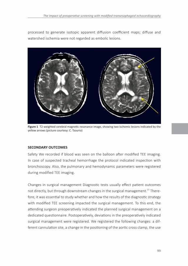
99
The impact of preoperative screening with modified transesophageal echocardiography
processed to generate isotopic apparent diffusion coefficient maps; diffuse and watershed ischemia were not regarded as embolic lesions.
Figure 1 T2-weighted cerebral magnetic resonance image, showing two ischemic lesions indicated by the yellow arrows (picture courtesy: C. Tzourio)
SECONDARY OUTCOMES
Safety We recorded if blood was seen on the balloon after modified TEE imaging. In case of suspected tracheal hemorrhage the protocol indicated inspection with bronchoscopy. Also, the pulmonary and hemodynamic parameters were registered during modified TEE imaging.
Changes in surgical management Diagnostic tests usually effect patient outcomes not directly, but through downstream changes in the surgical management.21 There-fore, it was essential to study whether and how the results of the diagnostic strategy with modified TEE screening impacted the surgical management. To this end, the attending surgeon preoperatively indicated the planned surgical management on a dedicated questionnaire. Postoperatively, deviations in the preoperatively indicated surgical management were registered. We registered the following changes: a dif-ferent cannulation site, a change in the positioning of the aortic cross clamp, the use

Chapter 6
100
of a different aortic cannula, proceeding to off-pump surgery, or adaptation of the site of the proximal anastomosis.
CLINICAL ENDPOINTS Postoperatively, the occurrence of several clinical symptoms associated with cerebral embolization were recorded (i.e. transient ischemic attack (TIA), stroke, delirium, and epileptic insults). Presence of delirium was scored using the “Visual Analogue Scale”, which was completed on admission and twice daily after surgery until discharge from the intensive care unit (ICU). Also, we registered any possible harms of modified TEE, i.e. intraoperative hypoxemia, esophageal hemorrhage, and prolonged postoperative ventilation time.
QUALITY OF LIFE Finally, the quality of life was assessed using the SF-36 Health Survey. This questionnaire was filled-out at home, both pre-operatively and 30 days and 1-year after surgery. The SF-36 is a generic questionnaire, which enables the comparison of the quality of life pre-and postoperatively, and with a healthy age-matched population.22 From the 36 questions, eight subscores (i.e. physical function, role-physical, bodily pain, general health, vitality, social functioning, role-emotional and mental health) and two principal component scores (i.e. physical health and mental health) were calculated. The subscores were compared to the average scores of age-matched healthy Dutch inhabitants.23
PROTOCOL CHANGES To compensate for a slower than expected inclusion rate, two protocol changes were introduced after the inclusion of four patients (May 30th 2011). First, we replaced the need of an SRI over 75 by a minimum age of 65 years, as the former was met in fewer patients than expected. Second, the preoperative MRI was removed from the protocol, as it was frequently infeasible to perform an MRI within the short time-span in which patients were hospitalized before surgery.
SAMPLE SIZE Few studies previously reported the incidence of ischemic lesions after isolated CABG,4,24,25 and no information was available on the expected reduction of these lesions with the use of modified TEE. Therefore, prior to a large clinical trial, this pilot study aimed to assess the feasibility of the study design, the proportion of patients unable to undergo a postoperative MRI (e.g. because of postoperative delirium or stroke), and the incidence of postoperative cerebral ischemic lesions in

101
The impact of preoperative screening with modified transesophageal echocardiography
our study domain. We expected that the inclusion of 32 patients would yield suf-ficient information for these aims. Of note, the current pilot phase was not powered to detect differences between the intervention and control group.
RANDOMIZATION Computer-generated assignment to the intervention or control group was performed in an online environment. This website was controlled by a third party, and ensured concealment of allocation. A research nurse performed randomization on the day of admission prior to surgery.
BLINDING The study was double-blinded, i.e. both patients and observers of the end-points were unaware of group allocation. Of note, the anesthesiologist and surgeon were explicitly aware of group assignment, since they were responsible for the diagnosis and management of aortic atherosclerosis.
STATISTICAL ANALYSIS Dichotomous variables are presented as frequencies and percentages of total, accompanied by a 95% CI for the primary end-point. Continu-ous variables are described as mean and standard deviation (SD) when normally distributed (as visualized by a Q-Q plot and tested with a Kolmogorov-Smirnov test), or as the median and interquartile range (IQR) when not following a normal distribu-tion. We did not test for statistical differences between the intervention and control group. Data were analyzed according to the intention-to-treat principle.
When analyzing quality of life, the separate categorical items of the SF-36 were recoded into 0 – 100 scores, and combined into eight subscores and two summary scores. Using the mean and SD of healthy age-matched Dutch inhabitants, Z-scores and subsequent standardized scores (50 + Z * SDreference) were calculated. The latter can be interpreted more easily than the crude scores, with a score of 50 (SD 10) equal to the population mean. Analyses were performed in RStudio version 0.95.263 and SPSS 19.0.0
RESULTS
Recruitment and numbers analyzed Between March 1st 2011 and March 27th 2012, 125 patients were screened for study eligibility (figure 6.2). Ten patients did not
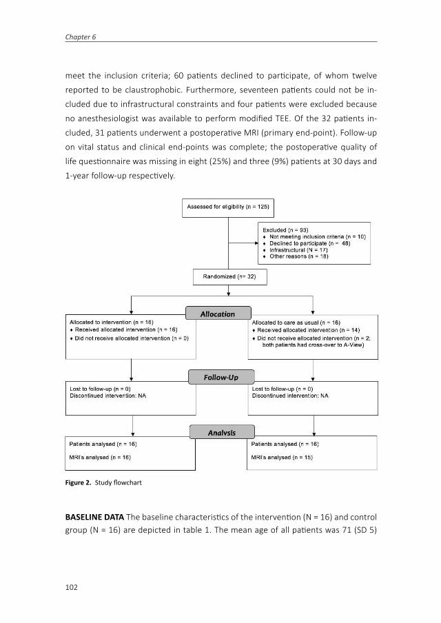
Chapter 6
102
meet the inclusion criteria; 60 patients declined to participate, of whom twelve reported to be claustrophobic. Furthermore, seventeen patients could not be in-cluded due to infrastructural constraints and four patients were excluded because no anesthesiologist was available to perform modified TEE. Of the 32 patients in-cluded, 31 patients underwent a postoperative MRI (primary end-point). Follow-up on vital status and clinical end-points was complete; the postoperative quality of life questionnaire was missing in eight (25%) and three (9%) patients at 30 days and 1-year follow-up respectively.
Figure 2. Study flowchart
BASELINE DATA The baseline characteristics of the intervention (N = 16) and control group (N = 16) are depicted in table 1. The mean age of all patients was 71 (SD 5)

103
The impact of preoperative screening with modified transesophageal echocardiography
years, 94% (N = 30) of patients were male, and 16% (N = 5) had a history of TIA or stroke. The median EuroSCORE was 2.6% (IQR: 2.1% – 4.4%).
Table 1. Baseline characteristics for patients allocated to preoperative screening for aortic atherosclerosis with- (intervention) and without (control) perioperative modified TEE screening for aortic atherosclerosis.
Total(N = 32)
Intervention(N = 16)
Control(N = 16)
Age (years) 71(5) 72 (5) 70 (5)
Male gender (%) 30 (94) 14 (88) 16 (100)
History
Hypertension 16 (50) 7 (44) 9 (56)
COPD 4 (13) 1 (6) 3 (19)
Diabetes Mellitus 6 (21) 2/14 (14) 4/14 (29)
TIA or stroke 5 (16) 3 (19) 2 (13)
Extracardiac atherosclerosis 4 (13) 1 (6) 3 (19)
Myocardial infarction 12 (38) 6 (38) 6 (38)
Logistic EuroSCORE 2.6 (2.1 – 4.4) 3.1 (2.2 – 4.2) 2.6 (2.0 – 5.2)
LVEF ³ 50% 22/31 (71) 11/15 (73) 11 (69)
30 – 50% 9/31 (29) 4/15 (27) 5 (31)
NYHA class ³3 14/27 (28) 7/11 (64) 7 (54)
CCS class ³3 13/24 (54) 7/11 (64) 6/13 (46)
Numbers are N (%) or median (interquartile range). COPD = chronic obstructive pulmonary disease, TIA = transient ischemic attack, LVEF = left ventricular ejection fraction, NYHA = New York Heart Association, CCS = Canadian Cardiovascular Society
DIAGNOSTIC TESTS AND SAFETY Conventional TEE was applied in all patients. Modified TEE was used in all patients allocated to the intervention; additionally, two patients in the control group received modified TEE imaging (i.e. cross-over) because the attending anesthesiologist considered not performing modified TEE in these patients unethical.
Manual palpation was performed in fourteen patients, of whom two had a sus-pected aortic plaque. In none of the patients the surgical management was changed based on the findings of manual palpation. Epiaortic ultrasound was performed in 6 patients, of whom 4 were included in the intervention group.

Chapter 6
104
Modified TEE was performed in all patients in the intervention group. In the control group two a cross-over to modified TEE was made in two patients. The incidence of severe atherosclerosis of the DAA was 24% (4/17). Performance of modified TEE or the other diagnostic tests was not associated with complications; specifically, no esophageal hemorrhages were observed.
SURGICAL MANAGEMENT Preoperatively, the attending surgeon had indicated in all patients that surgery would be performed according to a standard procedure, i.e. on-pump CABG with aortic cannulation using a single-lumen straight-tip cannula (Select Series®, REF 76122; Medtronic Inc., Minneapolis, MN). Aortic cannulation was planned on the distal, mid or proximal ascending aorta in 77% (N = 23), 13% (N = 4) and 10% (N = 3) of patients respectively. In 26 patients the pre- and postoperative form had been completed, in three of these patients (12%) the surgical procedure was altered based on the perioperative screening for aortic atherosclerosis. These changes included a different cannulation site in two patients, and an adaptation of the proximal anastomosis site in one of these patients and in an additional patient.
CEREBRAL MRI Before the protocol changes, four patients underwent a baseline cerebral DW-MRI, none of which showed ischemic lesions. The postoperative MRI was obtained within 6 days after surgery in 30 (94%) patients. In one patient the MRI could not be performed until the 35th day because of a prolonged stay at the ICU with intermittent respiratory insufficiency requiring reintubation. One additional patient (male, 68 years, intervention group) refused to undergo an MRI postoperatively, be-cause of dyspnea and claustrophobia. Initially, the two observers identified ischemic lesions in seven and nine of 31 postoperative MR images respectively (kappa = 0.83, 95% CI: 0.61 – 1.00). After an expert meeting, the observers agreed that nine of 31 (29%, 95% CI: 16 – 47%) postoperative MRIs showed ischemic lesions.
The incidence of cerebral ischemic lesions was 30% (5/16) in the intervention group, and 27% (4/15) in the control group (table 2). In seven patients, a single lesion was observed, none of these patients showed signs of a neurological complication. One patient included in the control group had two ischemic lesions on the cerebral MRI performed on the sixth day after surgery. This patient suffered from a transient expressive aphasia and delirium on the second day after surgery. Finally, one patient

105
The impact of preoperative screening with modified transesophageal echocardiography
included in the intervention group had thirteen postoperative DW-detected lesions. This patient, who had also developed postoperative atrial fibrillation, was diagnosed with a delirium on the third day after surgery. Of note, the patient in whom the MRI was performed on the 36th day after surgery had one DW-lesion, but did not have neurological sequelae during follow-up.
Table 2. Postoperative MRI results
Total(N = 32)
Intervention(N = 16)
Control(N = 16)
MRI £ 6 days after surgery
31 (97) 16 (100) 15 (94)
Days to MRI 4 (3 – 36) 4 (3 – 36) 3 (3 – 6)
New cerebral DW-lesion(s)
9 (29) 5 (30) 4 (27)
Number lesions 1 (1 - 16) 1 (1 – 16) 1 (1 – 2)
Numbers are N (%) or median (range; min - max). MRI = magnetic resonance imaging, DW = diffusion-weighted.
ISCHEMIA VS. NO ISCHEMIA In table 3 the preoperative characteristics, the degree of aortic atherosclerosis and in-hospital complications of patients with and without cerebral ischemia on the postoperative cerebral MRI are depicted.
Table 3. Comparison of patients with and without ischemic lesions on postoperative MRI
Ischemic lesions(N = 9)
No ischemic lesions(N = 21)
Age (years) 68 (66 – 72) 72 (69 – 76)Male gender (%) 9 (100) 20 (91)HistoryTIA or stroke 1 (11) 4 (18)Extracardiac atherosclerosis 2 (22) 2 (9)Logistic EuroSCORE 3.2 (2.4 – 5.8) 2.5 (2.1 – 4.0)LVEF < 50% 5 (63) 4 (18)Atherosclerosis grade ³3 (TEE)Proximal ascending aorta 1/7 (14) 9/20 (45)Distal ascending aorta* 0/5 (0) 4/9 (31)Descending aorta 3/7 (43) 19/20 (95)In-hospital complicationsDelirium 2 (22) 0 (-)TIA or stroke 1 (11) 0 (-)*Visualized with modified TEE. Numbers are N (%) or median (interquartile range). TIA = transient ischemic attack, LVEF = left ventricular ejection fraction, TEE = transesophageal echocardiography.

Chapter 6
106
CLINICAL ENDPOINTS AND QUALITY OF LIFE The incidence of in-hospital complica-tions is depicted in table 4. All patients were alive one year after surgery; two pa-tients suffered from a post-operative delirium and one patient from a minor stroke. Figure 3 shows the physical and mental component scores at baseline, 30-days and 1-year follow-up, stratified by group assignment. The physical component scores are below the population average both before surgery (intervention: 46 ± 10, control: 45 ± 9) and at 30 days follow-up (intervention: 47 ± 6), control: 49 ± 6). One year after surgery, the component scores were above population average both in the intervention (53 ± 8) and in the control (55 ± 7) group.
Figure 3. Postoperative physical component score (PCS) and mental component score (MCS) at baseline, 30-days and 1-year follow-up respectively. A principal component (PC) score of 50 equals the mean score in age-matched Dutch inhabitants.
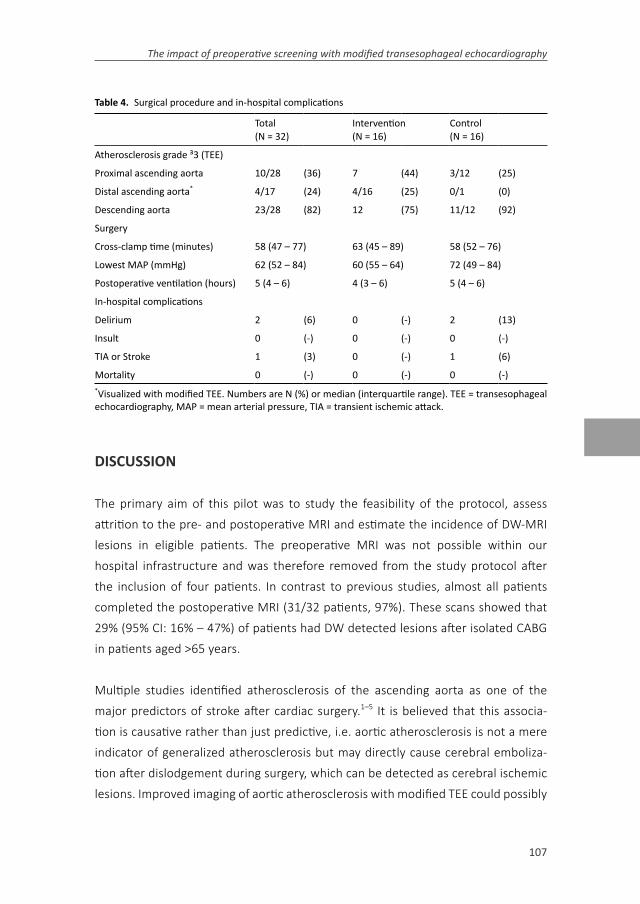
107
The impact of preoperative screening with modified transesophageal echocardiography
Table 4. Surgical procedure and in-hospital complications
Total(N = 32)
Intervention(N = 16)
Control(N = 16)
Atherosclerosis grade ³3 (TEE)
Proximal ascending aorta 10/28 (36) 7 (44) 3/12 (25)
Distal ascending aorta* 4/17 (24) 4/16 (25) 0/1 (0)
Descending aorta 23/28 (82) 12 (75) 11/12 (92)
Surgery
Cross-clamp time (minutes) 58 (47 – 77) 63 (45 – 89) 58 (52 – 76)
Lowest MAP (mmHg) 62 (52 – 84) 60 (55 – 64) 72 (49 – 84)
Postoperative ventilation (hours) 5 (4 – 6) 4 (3 – 6) 5 (4 – 6)
In-hospital complications
Delirium 2 (6) 0 (-) 2 (13)
Insult 0 (-) 0 (-) 0 (-)
TIA or Stroke 1 (3) 0 (-) 1 (6)
Mortality 0 (-) 0 (-) 0 (-)*Visualized with modified TEE. Numbers are N (%) or median (interquartile range). TEE = transesophageal echocardiography, MAP = mean arterial pressure, TIA = transient ischemic attack.
DISCUSSION
The primary aim of this pilot was to study the feasibility of the protocol, assess attrition to the pre- and postoperative MRI and estimate the incidence of DW-MRI lesions in eligible patients. The preoperative MRI was not possible within our hospital infrastructure and was therefore removed from the study protocol after the inclusion of four patients. In contrast to previous studies, almost all patients completed the postoperative MRI (31/32 patients, 97%). These scans showed that 29% (95% CI: 16% – 47%) of patients had DW detected lesions after isolated CABG in patients aged >65 years.
Multiple studies identified atherosclerosis of the ascending aorta as one of the major predictors of stroke after cardiac surgery.1–5 It is believed that this associa-tion is causative rather than just predictive, i.e. aortic atherosclerosis is not a mere indicator of generalized atherosclerosis but may directly cause cerebral emboliza-tion after dislodgement during surgery, which can be detected as cerebral ischemic lesions. Improved imaging of aortic atherosclerosis with modified TEE could possibly

Chapter 6
108
lead to effective changes in the surgical management and thereby in reduction of DW-detected lesions and ultimately (neurological) complications.
Before engaging in a full-sized randomized diagnostic trial, we aimed to study the fea-sibility of a protocol which compared the incidence of post-operative DW-detected lesions in patients allocated to modified TEE or a conventional diagnostic approach. This pilot study was not powered to detect differences in outcomes between the intervention and control group. However, several findings are of importance for the performance of a potential full-sized trial in specific, and for the research of aortic atherosclerosis in general.
First, it was often impossible to perform a MRI scan in the limited time period between hospital admission and surgery. Therefore, the pre-operative MRI was removed from the study protocol after inclusion of four patients. In the absence of a pre-operative MRI scan it is not possible to accurately differentiate new and existing ischemic lesions on the post-operative MRI. However, in a sufficiently large trial the incidence of existing DW-detected lesions is expected to be similar in both groups after successful randomization. Hence, a difference in the incidence of post-operative ischemic lesions can still be interpreted as a difference in the incidence of new lesions.
A second aim of this study was to quantify the percentage of patients in whom a post-operative MRI could be performed. This is of importance for the sample size calculation, but the percentage of patients who cannot undergo an MRI after cardiac surgery varied widely in previous studies ranging from 45% to 93%;4,26,27 although most studies did not present the percentage of missing scans.24,25,28,29 The main reasons reported to not undergo a postoperative MRI were claustrophobia (44%) and confusion (18%).4 In our study, the post-operative MRI was missing in one patient (3%) only, because of dyspnea and claustrophobia. The reasons why the number of missing scans was so much lower in our study have to be speculated, but it is conceivable that the inclusion of patients undergoing isolated elective CABG in our study was associated with a faster post-operative recovery with fewer complica-tions that may interfere with the performance of a post-operative MRI.

109
The impact of preoperative screening with modified transesophageal echocardiography
Third, we aimed to estimate the incidence of post-operative ischemic lesions. The observed incidence of 29% was lower than anticipated. Possibly, this was associated with a low risk of cerebral embolization in patients with isolated CABG, given that the original EuroSCORE was only 2.6%, and the prevalence of atherosclerosis of the proximal- and distal ascending aorta was low. In previous studies the incidence of DW-detected lesions after cardiac surgery ranged from 0 – 51%,4,24–33 and is influ-enced by factors including the baseline risk, surgical procedure, presence of atrial fibrillation, and the timing and technique of the MRI. As an example, one studied showed that the incidence of DW-detected lesions was 26% after isolated CABG and 40%% after surgical AVR.27
Interestingly, in our study only one patient with post-operative ischemic lesions had severe atherosclerosis of the proximal ascending aorta; on the contrary, nine of twenty patients without ischemic lesions had severe atherosclerosis of the same segment. Definite conclusions cannot be made given the small sample-size, but this finding seems to oppose the main hypothesis underlying our study that ischemic lesions are associated with (the manipulation of) aortic atherosclerosis. A previous study similarly reported that postoperative ischemic lesions were unrelated to the degree of aortic atherosclerosis.29 This may reflect the multifactorial etiology of neurological complications, which includes other factors such as inflammation,34 hypotension induced cerebral ischemia,35 and postoperative hypercoagulation36.
Generalizability Our results are of importance for future studies, but cannot be used to assess the efficacy of modified TEE on patient management and outcomes. The percentage of patients who were willing to participate in this study was limited (26%), but exclusion was seldom related to a contra-indication for, or unavailability of modified TEE (N = 4; 4%).
Implications for future studies The results from this pilot study have implications for the infrastructure and sample size of a potential full-cohort RCT. First, although it would be preferable to have information on the prevalence of preoperative DW- lesions, the current study showed that a pre-operative MRI was not feasible in this study population, unless patients are admitted to the hospital earlier for study purposes. Second, the inclusion criteria should be reconsidered to include patients
Chapter 6
110
with a higher risk of aortic atherosclerosis and therefore cerebral embolization. In the current protocol we included patients with isolated CABG only, which aimed to reduce the occurrence of ischemic lesions after manipulation of structures other than the aorta, e.g. during valve replacement. However, in a population with a low burden of aortic atherosclerosis, it is unlikely that modified TEE will reduce the incidence of ischemic lesions. Inclusion of more complex surgery should thus be considered, which may also increase the number of eligible patients.
Finally, the lower than expected incidence of postoperative ischemic lesions has implications for the sample size calculation. Previously, it was anticipated that 60% of patients in the control group would have new ischemic lesions postoperatively. Assuming a risk reduction of 33%, alpha = 0.05 and beta = 0.80, it was estimated that 107 patients would have to be included in each group. If assuming an incidence of new ischemic lesions of 29% (this study) with a similar RR, alpha and beta, the sample size would have to be increased to 305 patients per group.
LIMITATIONS The small sample size of this pilot study does not allow for a compari-son between the intervention- and control group. Furthermore, although there was a well-defined diagnostic protocol and a registration of the changes in the surgical management based on the imaging findings, these changes were left at the discre-tion of the attending surgeon and were thus not uniform. To quantify the impact of a diagnosis-treatment combination, it is essential to also capture the therapeutic ac-tions in a protocol.21 To our knowledge, such a protocol has not yet been published with respect to the therapeutic actions that should follow visualization of aortic atherosclerosis. We recommend such a protocol to be designed and implemented in a full-sized study. To further elucidate the pathway from aortic atherosclerosis towards the occurrence of ischemic lesions, we intend to perform TCD screening in a future study to quantify the embolic load in the middle cerebral artery during surgery. As in any study, data collection was not complete, with values missing both in the patient characteristics and in the outcome data. It is generally recommended to impute missing data, as this can reduce the possibility of biased results due to selective missingness. We decided not to impute data in this small size study, as imputation techniques rely on regression modelling, which is difficult in a study of this size.

111
The impact of preoperative screening with modified transesophageal echocardiography
CONCLUSION
This pilot study assessed the feasibility of a randomized diagnostic trial compar-ing a diagnostic strategy with- and without perioperative modified TEE imaging. The prevalence of aortic atherosclerosis was low, possibly due to the inclusion of isolated CABG only. A preoperative MRI was not feasible; a post-operative MRI was completed in 97% of patients, which is higher than reported in previous studies. The incidence of ischemic lesions was 29% (95% CI: 16% – 47%), which was lower than anticipated. We recommended changes in the design of a potential full-cohort trial.

Chapter 6
112
REFERENCES 1. van der Linden J, Hadjinikolaou L, Bergman P, Lindblom D. Postoperative stroke in
cardiac surgery is related to the location and extent of atherosclerotic disease in the ascending aorta. J Am Coll Cardiol 2001;38:131–5.
2. van der Linden J, Bergman P, Hadjinikolaou L. The topography of aortic atherosclerosis enhances its precision as a predictor of stroke. Ann Thorac Surg 2007;83:2087–92.
3. Hartman GS, Yao FS, Bruefach M, Barbut D, Peterson JC, Purcell MH, et al. Severity of aortic atheromatous disease diagnosed by transesophageal echocardiography predicts stroke and other outcomes associated with coronary artery surgery: a prospective study. Anesth Analg 1996;83:701–8.
4. Djaiani G, Fedorko L, Borger M, Mikulis D, Carroll J, Cheng D, et al. Mild to moder-ate atheromatous disease of the thoracic aorta and new ischemic brain lesions after conventional coronary artery bypass graft surgery. Stroke 2004;35:e356–8.
5. Elias-Smale SE, Odink AE, Wieberdink RG, Hofman A, Hunink MGM, Krestin GP, et al. Carotid, aortic arch and coronary calcification are related to history of stroke: the Rot-terdam Study. Atherosclerosis 2010;212:656–60.
6. Evered LA, Silbert BS, Scott DA. Postoperative cognitive dysfunction and aortic ath-eroma. Ann Thorac Surg 2010;89:1091–7.
7. Dávila-Román VG, Kouchoukos NT, Schechtman KB, Barzilai B. Atherosclerosis of the ascending aorta is a predictor of renal dysfunction after cardiac operations. J Thorac Cardiovasc Surg 1999;117:111–6.
8. Dávila-Román VG, Murphy SF, Nickerson NJ, Kouchoukos NT, Schechtman KB, Barzilai B. Atherosclerosis of the ascending aorta is an independent predictor of long-term neurologic events and mortality. J Am Coll Cardiol 1999;33:1308–16.
9. Thambidorai SK, Jaffer SJ, Shah TK, Stewart WJ, Klein AL, Lauer MS. Association of ath-eroma as assessed by intraoperative transoesophageal echocardiography with long-term mortality in patients undergoing cardiac surgery. Eur Heart J 2007;28:1454–61.
10. Mackensen GB, Ti LK, Phillips-Bute BG, Mathew JP, Newman MF, Grocott HP. Cerebral embolization during cardiac surgery: impact of aortic atheroma burden. Br J Anaesth 2003;91:656–61.
11. Borger M. Stroke during coronary bypass surgery: principal role of cerebral macroem-boli. Eur J Cardio-Thoracic Surg 2001;19:627–32.
12. Van Zaane B, Zuithoff NPA, Reitsma JB, Bax L, Nierich AP, Moons KGM. Meta-analysis of the diagnostic accuracy of transesophageal echocardiography for assessment of atherosclerosis in the ascending aorta in patients undergoing cardiac surgery. Acta Anaesthesiol Scand 2008;52:1179–87.
13. van Zaane B, Nierich AP, Buhre WF, Brandon Bravo Bruinsma GJ, Moons KGM. Resolv-ing the blind spot of transoesophageal echocardiography: a new diagnostic device for visualizing the ascending aorta in cardiac surgery. Br J Anaesth 2007.
14. Nierich AP, van Zaane B, Buhre WF, Coddens J, Spanjersberg AJ, Moons KGM. Visualiza-tion of the distal ascending aorta with A-Mode transesophageal echocardiography. J Cardiothorac Vasc Anesth 2008;22:766–73.
15. van Zaane B, Nierich AP, Brandon Bravo Bruinsma GJ, Rosseel PMJ, Ramjankhan FZ, de Waal EEC, et al. Diagnostic accuracy of modified transoesophageal echocardiography for pre-incision assessment of aortic atherosclerosis in cardiac surgery patients. Br J Anaesth 2010;105:131–8.
16. Jansen Klomp WW, Peelen LM, Spanjersberg SJ, Brandon Bravo Bruinsma GJ, Lange F de, Van’t Hof AWJ, et al. Added value of modified transoesophageal echocardiography

113
The impact of preoperative screening with modified transesophageal echocardiography
in the diagnosis of atherosclerosis of the distal ascending aorta in cardiac surgery patients. Eur Heart J Cardiovasc Imaging 2014;15:623–30.
17. Moher D, Hopewell S, Schulz KF, Montori V, Gøtzsche PC, Devereaux PJ, et al. CONSORT 2010 Explanation and Elaboration: Updated guidelines for reporting parallel group randomised trials. J Clin Epidemiol 2010;63:e1–37.
18. Schulz KF, Altman DG, Moher D. CONSORT 2010 Statement: Updated guidelines for reporting parallel group randomised trials. J Clin Epidemiol 2010;63:834–40.
19. Newman MF, Wolman R, Kanchuger M, Marschall K, Mora-Mangano C, Roach G, et al. Multicenter preoperative stroke risk index for patients undergoing coronary artery bypass graft surgery. Multicenter Study of Perioperative Ischemia (McSPI) Research Group. Circulation 1996;94:II74–80.
20. Katz NM, Chase GA. Risks of cardiac operations for elderly patients: reduction of the age factor. Ann Thorac Surg 1997;63:1309–14.
21. Guyatt GH, Tugwell PX, Feeny DH, Haynes RB, Drummond M. A framework for clinical evaluation of diagnostic technologies. CMAJ 1986;134:587–94.
22. Wareing TH, Davila-Roman VG, Barzilai B, Murphy SF, Kouchoukos NT. Management of the severely atherosclerotic ascending aorta during cardiac operations. A strategy for detection and treatment. J Thorac Cardiovasc Surg 1992;103:453–62.
23. Aaronson NK, Muller M, Cohen PDA, Essink-Bot M-L, Fekkes M, Sanderman R, et al. Translation, Validation, and Norming of the Dutch Language Version of the SF-36 Health Survey in Community and Chronic Disease Populations. J Clin Epidemiol 1998;51:1055–68.
24. Restrepo L, Wityk RJ, Grega MA., Borowicz L, Barker PB, Jacobs MA., et al. Diffusion- and Perfusion-Weighted Magnetic Resonance Imaging of the Brain Before and After Coronary Artery Bypass Grafting Surgery. Stroke 2002;33:2909–15.
25. Knipp SC, Matatko N, Wilhelm H, Schlamann M, Massoudy P, Forsting M, et al. Evalu-ation of brain injury after coronary artery bypass grafting. A prospective study using neuropsychological assessment and diffusion-weighted magnetic resonance imaging. Eur J Cardiothorac Surg 2004;25:791–800.
26. Stolz E, Gerriets T, Kluge A, Klövekorn W-P, Kaps M, Bachmann G. Diffusion-weighted magnetic resonance imaging and neurobiochemical markers after aortic valve replace-ment: implications for future neuroprotective trials? Stroke 2004;35:888–92.
27. Cook DJ, Huston J, Trenerry MR, Brown RD, Zehr KJ, Sundt TM. Postcardiac surgical cognitive impairment in the aged using diffusion-weighted magnetic resonance imag-ing. Ann Thorac Surg 2007;83:1389–95.
28. Knipp SC, Matatko N, Schlamann M, Wilhelm H, Thielmann M, Forsting M, et al. Small ischemic brain lesions after cardiac valve replacement detected by diffusion-weighted magnetic resonance imaging: relation to neurocognitive function. Eur J Cardiothorac Surg 2005;28:88–96.
29. Floyd TF, Shah PN, Price CC, Harris F, Ratcliffe SJ, Acker MA, et al. Clinically silent cerebral ischemic events after cardiac surgery: their incidence, regional vascular oc-currence, and procedural dependence. Ann Thorac Surg 2006;81:2160–6.
30. Barber PA, Hach S, Tippett LJ, Ross L, Merry AF, Milsom P. Cerebral ischemic lesions on diffusion-weighted imaging are associated with neurocognitive decline after cardiac surgery. Stroke 2008 39:1427–33.
31. Agarwal R, Kalita J, Pandey S, Agarwal SK, Misra UK. Evaluation of cognitive function and P300 in patients undergoing cardiac surgery. Electromyogr Clin Neurophysiol 2010;50:259–64.

Chapter 6
114
32. Knipp SC, Matatko N, Wilhelm H, Schlamann M, Thielmann M, Losch C, et al. Cognitive outcomes three years after coronary artery bypass surgery: relation to diffusion-weighted magnetic resonance imaging. Ann Thorac Surg 2008;85:872–9.
33. Patel N, Minhas JS, Chung EML. The Presence of New MRI Lesions and Cognitive De-cline After Cardiac Surgery: A Systematic Review. J Card Surg 2015;30:808–12.
34. Murphy GS, Sherwani SS, Szokol JW, Avram MJ, Greenberg SB, Patel KM, et al. Small-dose dexamethasone improves quality of recovery scores after elective cardiac surgery: a randomized, double-blind, placebo-controlled study. J Cardiothorac Vasc Anesth 2011;25:950–60.
35. Hogue CW, Palin CA, Arrowsmith JE. Cardiopulmonary bypass management and neurologic outcomes: an evidence-based appraisal of current practices. Anesth Analg 2006;103:21–37.
36. Lo B, Fijnheer R, Nierich AP, Kalkman CJ, van Dijk D. Activation of hemostasis is as-sociated with early cognitive decline after off-pump coronary artery bypass surgery. J Thromb Haemost 2005;3:2114–7.


7Survival and quality of life after surgical aortic
valve replacement in octogenarians
W.W. Jansen Klomp, A.P. Nierich, L.M. Peelen, G.J. Brandon Bravo Bruinsma, J.H.E. Dambrink, K.G.M. Moons, A.W.J. van ‘t Hof
Published in: Journal of Cardiothoracic Surgery, 2016
Chapter 7
118
ABSTRACT
BACKGROUND In patients with symptomatic severe aortic stenosis, advanced age is often a reason for a transcatheter rather than surgical aortic valve replacement. In this pre-transcathter cohort we had the unique oportunity to study outcomes after surgical aortic valve replacement for severe aortic stenosis in patients who might currently be triaged to a percutaneous ap-proach.
METHODS In a prospective single-center cohort study we compared the inci-dence of peri-operative complications, mortality, and health-related quality of life in octogenarians versus patients aged <80 years. The quality of life was measured using the SF-36 questionnaire and expressed as a physical and mental component score (PCS and MCS respectively); a score of 50 equals the average score in the age-matched general population. The association between age and the component scores at one-year follow-up was studied with the use of linear regression, corrected for a set of confounding variables.
RESULTS We included 762 patients, of whom 21.4% was aged >80 and 49.0% underwent concomitant revascularization. In octogenarians, the incidence of post-operative delirium was 11.0%, which was higher than in patients aged below 80 (6.2%, p = 0.034); the operative mortality (1.9% vs. 2.9%; p = 0.59) and long-term survival were not different however (log-rank p = 0.75). In octogenarians, the quality of life was impaired 30-days after surgery (PCS 45.01, p <0.001; MCS 48.21, p = 0.04), which improved towards or above normal values at one-year follow-up (PCS: 49.92, p = 0.67, MCS: 52.55, p < 0.001). After correction for confounding, age was not significantly associated with the one-year PCS (b 0.08 per year, p = 0.34) or MCS (b 0.08 per year, p = 0.32).
CONCLUSIONS This pre-transcatheter study showed that surgical aortic valve replacement in octogenarians could be performed with very low mortality,

119
Survival and quality of life after surgical aortic valve replacement in octogenarians
and with a relevant and significant increase of the quality of life towards normal values. Also, age was not associated with a lower PCS or MCS one-year after surgery.

Chapter 7
120
INTRODUCTION
Surgical aortic valve replacement (AVR) has long been the only effective treatment for symptomatic severe aortic stenosis (AS).1–3 The introduction of catheter-based techniques questioned existing treatment strategies for patients with AS. One obvi-ous, but difficult challenge is to assess in which patients transcatheter aortic valve replacement (TAVR) is preferable, and in whom a surgical intervention remains the treatment of choice. Based on the results of four randomized trials4–7 and multiple registries, current guidelines recommend TAVR in patients who are either not suit-able for surgery or have a very high surgical risk.2,8 These recommendations leave room for interpretation and in daily clinical practice advanced age alone is often the reason to prefer a percutaneous approach.9 This may indicate that physicians expect a poorer outcome after surgical AVR in elderly patients.
In this study we investigate data from a large cohort of surgical AVR patients that underwent surgery before TAVR became available in our clinic. We will hence not directly compare TAVR with surgical AVR, but we have the unique opportunity to assess outcomes of surgical AVR in patients who may nowadays be triaged to a percutaneous approach.
The aim of our study was to investigate the influence of age on postoperative outcomes, in particular comparing octogenarians to younger patients. Additional to post-operative complications and long-term mortality we also studied the health-related quality of life (HRQoL) after one year, an outcome increasingly recognized as an outcome of importance2, and its change from baseline HRQoL for patients of different age groups.
METHODS
In this prospective, observational cohort study we included consecutive patients with symptomatic AS who underwent elective surgery for aortic stenosis, either iso-lated AVR or AVR with concomitant coronary artery bypass grafting (CABG). Patients were operated in a single institution (Isala clinics, Zwolle, The Netherlands) between November 1st 2007 and August 1st 2011. During this period, the institution did not

121
Survival and quality of life after surgical aortic valve replacement in octogenarians
perform percutaneous procedures yet; therefore, patients who would currently be considered for a percutaneous approach were part in this surgical cohort.
Patients gave written informed consent for the systematic collection of data in an ongoing registry of cardiothoracic surgery in our institution. We excluded patients who refused informed consent, who had a non-stenotic valve, active endocarditis or a previous AVR during the study period from our analysis. The institution’s ethical committee approved the protocol (reference: 12.0881n) and waived the need for formal evaluation according to the Dutch Law on Scientific Medical Research with Humans. Data were anonymized before data analysis, and the study conforms to the principles outlined in the Helsinki declaration.
DATA COLLECTION AND END-POINTS Data were collected in an ongoing registry, which included baseline characteristics, perioperative data, in-hospital outcomes, and follow-up for clinical events and quality of life until one year after surgery. The aortic peak gradient was retrospectively registered through a review of electronic charts. End-points were reported according to the “Valve Academic Research Con-sortium-2 (VARC-2) criteria.10 In-hospital stroke was diagnosed by an attending neurologist, and usually supported by cerebral computed tomography imaging. Presence and severity of postoperative kidney injury were defined according to the Acute Kidney Injury Network (AKIN) classification.11 Delirium was assessed using the delirium observation screening (DOS) scale, which was performed daily until hos-pital discharge. Operative mortality was defined as 30-day mortality or in-hospital mortality if postoperative hospital stay exceeded 30 days. Long-term all-cause mor-tality was extracted from the Dutch Municipal Personal Records Database on April 6th, 2012.
The HRQoL was measured with the Medical Outcomes Study Short-Form 36 (SF-36), a widely used and well-validated self-administered instrument.12,13 Patients completed the test preoperatively, and at 30-days and one year after intervention. According to the guidelines for this instrument the 36 questions were subsequently grouped into eight health scales (physical functioning, role-physical, bodily pain, general health, vitality, social functioning, role-emotional, and mental health) and

Chapter 7
122
two summary measures (the physical component score [PCS] and mental compo-nent score [MCS]).
STATISTICAL ANALYSIS Throughout the analyses we compared octogenarians (defined as an age >80 years) with patients aged less than 80 years. The baseline characteristics and in-hospital outcomes are presented as frequencies and per-centages of total for dichotomous variables, as the mean and standard deviation (SD) for normally distributed data or the median and interquartile range (IQR) for non-normally distributed data respectively. Differences in categorical variables were assessed as appropriate with a chi-square test or a Fisher’s exact test. Differences in continuous variables were tested using a Students’ t-test or Mann-Whitney-U test. Survival plots were calculated with Kaplan-Meier (KM) statistics, as a reference we also depicted the predicted survival of age- and sex matched Dutch inhabitants re-ported by Statistics Netherlands (www.cbs.nl). Differences in KM-estimated survival were tested with a log-rank test.
The PCS and MCS were normalized such that a score of 50 (standard deviation [SD] 10) equalled the mean score of an age-matched Dutch reference population.13 We calculated the difference between the summary scores at baseline versus the scores at 30-days and one year follow-up, and tested these differences with a paired t-test. Also, we tested for a difference in the change in summary scores in octogenarians compared to younger patients using an independent samples t-test, which was cor-rected for the baseline scores.
To study the clinical relevance of a difference (rather than only statistical significance) we also calculated Cohen’s effect size by dividing the difference between two mea-surements through the SD at baseline. Furthermore, we calculated the percentage of patients in whom a minimal clinically important difference (MCID) was observed, defined as a difference ³2.5 points above or below the baseline score.14
Finally, we studied the influence of age on the difference in the component scores at one-year follow-up compared to baseline. The respective scores of the PCS and MCS were included as the dependent variable in a univariable generalized linear re-gression model (crude association), and were then corrected for several predefined

123
Survival and quality of life after surgical aortic valve replacement in octogenarians
covariables (see supplement A). Sex was included in the multivariable model to study gender specific changes. Furthermore, the baseline component scores were also included, since patients with higher baseline scores generally improve less on follow-up measurements than patients with lower baseline scores.15
Missing data were imputed since data are usually not missing completely at random, but rather selectively.16,17 Discarding patients with missing data may lead to biased results and furthermore a loss in precision.16,17 Imputation was performed in SPSS using five imputed datasets, and then pooled using Rubin’s rule (see supplement A for imputation model). Throughout the analysis a level of significance of 0.05 was used. Analyses were performed in SPSS Statistics version 21.0.0 and R version 2.13.1.
RESULTS
Characteristics of the patients During the inclusion period, 5,069 cardiothoracic op-erations were performed, of which 862 were isolated AVR or AVR with concomitant CABG. Twenty-three patients were excluded because they refused consent for the registration; another 77 patients met one or more of the exclusion criteria (figure 1). The final study population thus comprised 762 patients. Follow-up of the vital status was completed in 757 (99.3%) of patients, with a median follow-up of 2.4 years (IQR 1.0 – 3.3). Follow-up of the SF-36 was completed by 82.2% of patients at baseline, by 83.9% at 30-days follow-up and 84.7% of patients at one-year follow-up. The baseline characteristics of patients with and without missing one-year follow-up of the SF-36 questionnaire are shown in supplement B.

Chapter 7
124
Figure 1. Long-term survival after aortic valve replacement. Patients were included between November 1st 2007 and August 1st 2011*One hundred patients were excluded in total; five patients met two exclusion criteria, i.e. active endocarditis and non-elective surgery. AVR = aortic valve replacement, CABG = coronary artery bypass grafting, FU = follow-up.
One-hundred and sixty-three patients (21.4%) were aged 80 years or over at the date of surgery; median age of this group was 82 years, with a female predominance of 52.1%. Octogenarians had a lower systolic left ventricular function than younger patients (p = 0.044), other baseline characteristics were not significantly different (table 1). A bioprosthesis was implanted more often in octogenarians than in non-octogenarians (97.6% vs. 90.6%, p <0.001); the total cross-clamp time was shorter (82 vs. 91 minutes, p = 0.004), and combined surgery with CABG occurred in almost half of the procedures in both age groups less frequently (46.0% vs. 49.9%, p = 0.426). The EuroSCORE I predicted risk of operative mortality was higher in octoge-narians than in patients aged below 80 years (10.7% vs. 4.9%, p < 0.001).

125
Survival and quality of life after surgical aortic valve replacement in octogenarians
Table 1. Baseline characteristics and operative characteristics
Age < 80 Age ³ 80
N = 597 N = 163 P
Male sex 363 (60.8) 85 (52.1) 0.057
Age, years 71 (66 – 75) 82 (81 – 83) <0.001
History of:
DM 145 (24.4) 37 (22.7) 0.727
Hypertension 325 (54.4) 98 (60.1) 0.228
COPD 116 (19.4) 30 (18.4) 0.855
Peripheral VD 70 (11.7) 17 (10.4) 0.748
Stroke 66 (11.1) 20 (12.3) 0.768
MI 74 (12.4) 21 (12.9) 0.979
PCI 65 (10.9) 18 (11.0) 1.000
Cardiac surgery 42 (7.0) 8 (4.9) 0.428
EuroSCORE 6 (5 – 7) 8 (8 – 10) <0.001
Log EuroSCORE 4.9 (3.1 – 7.6) 10.7 (8.4 – 15.7) <0.001
LVEF >50% 468 (78.7) 113 (69.3) 0.044
30 – 50% 103 (17.3) 40 (24.5)
<30% 24 (4.0) 10 (6.1)
NYHA Class 1 145 (24.3) 45 (27.6) 0.149
Class 2 342 (57.4) 80 (49.1)
Class 3 109 (18.3) 38 (23.3)
Peak gradient AV (mmHg) 73 (60 – 90) 71 (58 – 88) 0.55
Operative characteristics
Bioprosthesis 541 (90.6) 159 (97.6) <0.001
CABG 298 (49.9) 75 (46.0) 0.426
X-clamp time (min) 91 (75 – 111) 82 (68 – 107) 0.004
Baseline chararcteristics and operative characteristics in octogenarians and patients aged less than 80 years. Values are number (%), continuous variables are presented as median (25th – 75th percentile). DM = diabetes mellitus, COPD = chronic obstructive pulmonary disease, VD = vascular disease, MI = myocardial infarction, PCI = percutanaeous coronary intervention, LVEF = left ventricular ejection fraction, NYHA = New York Heart Association, AV = aortic valve, peak gradient = maximum gradient over the aortic valve, CABG = coronary artery bypass grafting.
IN-HOSPITAL OUTCOMES AND MORTALITY During hospitalization, octogenar-ians more frequently developed a delirium (11.0% vs. 6.2%, p = 0.034), and total hospital-stay was longer (median (IQR) of 7 (5 - 9) and 7 (5 - 12) respectively, p = 0.031; table 2). The incidence of stroke (3.1% vs. 3.5%, p = 0.97), myocardial infarc-tion (2.6% vs. 3.0%, p = 1.00) and acute kidney injury (9.2% vs. 7.6%, p = 0.22)

Chapter 7
126
did not differ significantly between the two groups. Two octogenarians died before hospital discharge, one additional patient died within 30-days of surgery, thus the operative mortality was 1.9%, compared to 2.9% in patients aged <80 (p = 0.59). Both one-year mortality (6.5% vs. 6.3%, p = 1.00) and long-term mortality were similar for octogenarians and patients aged < 80 years (log-rank p = 0.745; figure 2).
Figure 2. Long-term survival after aortic valve replacement. Kaplan-Meier estimated survival in octoge-narians (red line and 95% confidence interval [CI]) compared to patients aged <80 years (green line and 95% CI); as a reference the dotted black lines depict the predicted survival for Dutch inhabitants with a similar distribution of age and sex as the two study groups.

127
Survival and quality of life after surgical aortic valve replacement in octogenarians
Table 2. Patient outcomes
Age < 80 Age ³ 80
N = 597 N = 163 P
In-hospital Outcomes
Delirium 37 (6.2) 18 (11.0) 0.034
Stroke 21 (3.5) 5 (3.1) 0.97
MI 17 (3.0) 4 (2.6) 1.00*
AKIN No injury 537 (92.4) 148 (90.8) 0.22
Stage 1 36 (6.2) 12 (7.4)
Stage 2 5 (0.9) 0 (0)
Stage 3 3 (0.5) 3 (1.8)
Hospital stay (days) 7 (5 – 9) 7 (5 – 12) 0.031
Mortality
In-hospital 13 (2.2) 2 (1.2) 0.75*
Operative 17 (2.9) 3 (1.9) 0.59*
1-Year 33 (6.3) 9 (6.5) 1.00
Comparison of operative chararcteristics and patient outcomes. Operative mortality is either 30-day or in-hospital mortality. * Fisher’s Exact test. MI = myocardial infarction, AKIN = acute kidney injury network.
HEALTH-RELATED QUALITY OF LIFE The PCS in octogenarians was 44.68 at baseline, which was lower than the average score of 50 in the age-matched reference popula-tion (p <0.001; table 3). The PCS remained impaired 30-days after surgery (45.01, p <0.001) but improved towards a normal level one year after surgery (49.92, p = 0.67). The one-year PCS in octogenarians was also comparable to patients aged below 80 (49.92 vs. 51.34, p = 0.12), but the increase from baseline was smaller in octogenarians (4.84 vs. 6.95, p = 0.04), as was Cohen’s D (0.54 vs. 0.78, p = 0.04).

Chapter 7
128
Tabl
e 3.
Hea
lth-r
elat
ed q
ualit
y of
life
in p
atien
ts u
nder
goin
g su
rgic
al A
VR.
Base
line
30 d
ays
1 ye
ar
Scor
eSc
ore
Chan
ge fr
om
base
linea
P fo
r cha
nge
Cohe
n’s
effec
t size
b%
reac
hing
MCI
DSc
ore
Chan
ge fr
om
base
linea
P fo
r cha
nge
Cohe
n’s
effec
t size
b%
reac
hing
MCI
D
PCS
Age
< 80
44.2
744
.11
-0.1
60.
73-0
.02
39.0
51.3
46.
95<0
.001
0.78
66.8
Age
> 80
44.8
645
.01
0.09
†0.
930.
0140
.149
.92
4.84
†<0
.001
0.54
60.7
MCS
Age
< 80
49.1
248
.33
-0.8
10.
073
-0.5
236
.250
.83
1.67
0.00
60.
2244
.2
Age
> 80
51.1
748
.21
-2.9
6†0.
002
-0.6
432
.552
.55
1.31
†0.
126
0.14
39.6
a The
“cha
nge
from
bas
elin
e” in
oct
ogen
aria
ns w
as co
mpa
red
to p
atien
ts a
ged
<80
year
s usin
g an
inde
pend
ent s
ampl
es t
test
and
corr
ecte
d fo
r the
bas
elin
e co
mpo
nent
sc
ore,
whe
re †
indi
cate
s p >
0.0
5. b An
effe
ct si
ze o
f <0.
20 c
an b
e co
nsid
ered
as c
linic
ally
irre
leva
nt, 0
.20-
0.49
as s
mal
l, 0.
50-0
.79
as m
oder
ate
and
> 0.
80 a
s lar
ge. P
CS =
ph
ysic
al c
ompo
nent
scor
e, M
CS =
men
tal c
ompo
nent
scor
e, M
CID
= m
inim
al c
linic
ally
impo
rtan
t diff
eren
ce.

129
Survival and quality of life after surgical aortic valve replacement in octogenarians
The MCS in octogenarians was 51.17 at baseline, which was higher than the average score in the reference population (p = 0.04). Thirty-days after surgery the MCS had reduced towards a below-average score (48.21, p = 0.04), but at one-year follow-up the MCS was again above expected (52.55, p < 0.001). The MCS one year after surgery was also higher in octogenarians compared to patients aged below 80 years (MCS 52.55 vs. 50.83, p < 0.001); however, the increase from baseline was similar (1.31 vs. 1.67, p = 0.38) and Cohen’s effect size was lower (0.14 vs. 0.22, p = 0.01).
ASSOCIATION BETWEEN AGE AND HRQOL Increasing age was associated with a higher PCS at 30-days follow-up, both in a univariable model (b 0.20 per year, 95% CI: 0.14 – 0.27, p <0.001) and when corrected for baseline characteristics (b 0.30, 95% CI: 0.08 – 0.15, p <0.001; table 4). One year after surgery age was however not associated with the PCS (multivariable model: b 0.08, 95% CI: -0.11 – 0.16, p = 0.34). Also, age was not associated with the MCS at 30-days follow-up (b -0.07, 95% CI: -0.22 – 0.83, p = 0.37), or 1-year after surgery (b 0.08, 95% CI: -0.08 – 0.25, p = 0.32).
Table 4. Association between age and the PCS and MCS scores at 30 days and 1 year follow-up
PCS – 30 days PCS – 1 year
b (95% CI) P b (95% CI) P
Age 0.20 (0.14 - 0.27) <0.001 -0.00 (-0.11 – 0.10) 0.90
Age (+ baseline PCS) 0.16 (0.10 – 0.22) <0.001 -0.07 (-0.17 – 0.04) 0.19
Age (full model) 0.30 (0.08 – 0.15) <0.001 0.08 (-0.11 – 0.16) 0.34
MCS – 30 days MCS – 1 year
b (95% CI) P b (95% CI) P
Age -0.05 (-0.14 – 0.03) 0.22 0.07 (-0.02 – 0.15) 0.11
Age (+ baseline) -0.08 (-0.16 – 0.00) 0.05 0.05 (-0.03 – 0.14) 0.20
Age (full model) -0.07 (-0.22 – 0.83) 0.37 0.08 (-0.08 – 0.25) 0.32
Table shows the univariable and multivariably adjusted asociation between age and the component scores; the full model also included: sex, chronic obstructive pulmonary disease, diabetes mellitus, extra-cardiac atherosclerosis, history of stroke, history of myocardial infarction, previous percutaneous coronary intervention, previous cardic surgery, NYHA class, CCS class, left ventricular function, concomitant coro-nary artery bypass grafting, aortic peak gradient, logistic EuroSCORE and baseline physical component score for the PCS model, or baseline mental component score for the MCS model. PCS = physical compo-nent score, MCS = mental component score.
Chapter 7
130
DISCUSSION
This study of pre-TAVR patients showed that surgical AVR in octogenarians could be performed with very low postoperative mortality, and a relevant and significant increase of the HRQoL towards normal values. Also, age was not associated with a lower PCS or MCS one-year after surgery.
OUTCOMES AND SURVIVAL Post-operative delirium was more often observed in octogenarians compared to younger patients (11.0% vs. 6.2%, p = 0.03), which may reflect an increased vulnerability of the aging brain for ischemic cerebral damage. Several perioperative processes are associated with this type of injury, including the embolization of particulate emboli, hypotension and inflammation.18 Indeed, age was previously found to be one of the most important risk-factors for post-operative delirium.19 However, the incidence of stroke, which is regarded to be in the same spectrum of cerebral (embolic) injury as delirium, was not increased in octogenar-ians.
Mortality rates were low, with an observed operative mortality of 1.9% in octoge-narians and 2.9% (p = 0.59) in younger patients. These rates are considerably lower than reported in previous studies, which ranged from 4.0 – 16%.20–23 One year after surgery 93.5% of octogenarians were alive, and the long-term survival up to 4 years after surgery was similar compared to younger patients. These outcomes contrast with the grim prognosis of medically treated severe AS,1 and based on our results one may question if age alone is a valid motivation to either refrain from surgery or to prefer a percutaneous treatment. Interestingly, from one year after surgery onwards, the long-term survival of octogenarians was better than that of Dutch inhabitants of the same age and sex.
Although it is well accepted that surgery for aortic stenosis improves life expectancy, the improved performance compared to the general population is likely to also in-dicate the selection of a healthy population in our study, which may have positively influenced the results of our study. Yet, over 20% of patients was aged ³80 years, which is in the higher range of other registries, and indicates a pro-active attitude towards surgical management in these patients.20,22 Also, we included patients with

131
Survival and quality of life after surgical aortic valve replacement in octogenarians
concomitant revascularization as recommended, since both survival and HRQoL are known to be reduced in these patients.24
HEALTH-RELATED QUALITY OF LIFE One month after surgery the PCS and MCS seemed to decrease rather than to improve, which may be related to the burden-some effects associated with sternotomy. This was also shown in the PARTNER trial, which randomly allocated patients to a surgical or percutaneous AVR. In that study, one month after intervention the HRQoL had increased in the latter, but not in the former group.25 However, at 6 and 12 months follow-up a similar increase of the PCS was observed after both approaches. Similarly in our study, the PCS and MCS one year after surgery had increased relevantly and significantly towards a level expected for the reference population.
ASSOCIATION BETWEEN AGE AND HRQOL Age was not associated with the PCS score at thirty days and one-year follow-up. Furthermore, age was positively as-sociated with the MCS at 30-days follow-up. Although the reasons for this positive association have to be speculated, these results indicate that older patients can expect a relative quality of life at least similar to younger patients.
As expected, a higher baseline component score was associated with a lower com-ponent score at one-year follow-up. This is probably caused by regression to the mean, as was described previously in the serial analysis of HRQoL measurements.15 That is, patients who already have a high baseline score are less likely to have an even higher score at follow-up, while patients with a low baseline score are less likely to have an even lower score. To correct for this potential bias, we included the baseline component scales in the multivariable linear regression analyses.
LIMITATIONS The results of this study should be appreciated with consideration of its limitations. First, as in any observational study, the efficacy of the interven-tion itself could not be studied, as this would require a control group of similar patients without surgical AVR. However, as a general reference, we compared both the survival and HRQoL to a matched general population. Long-term survival in oc-togenarians was better than expected for age- and sex matched Dutch inhabitants, which is likely to indicate that the octogenarians selected as suitable candidates for

Chapter 7
132
surgery were in relative good physical health. Yet, as mentioned, the proportion of octogenarians in our study is comparable to other registries. Also, mortality was well below predicted based on the original EuroSCORE.
One-year follow-up of the SF-36 questionnaire was missing in approximately 15% of patients. Missing follow-up status was associated a higher preoperative EuroS-CORE, but this difference was non-significant within patients alive at one-year (i.e. missingness not due to mortality; supplement B). In such a situation where data are not “missing completely at random”, it has been recommended to multiply impute missing data.17 In a sensitivity analysis that included patients with complete cases only, our results did not change substantially (supplement C).
Finally, as recommended by the VARC-2 criteria, postoperative kidney injury was defined according to the AKIN classification in which kidney injury is categorized both according to creatinine levels and urine output (whichever falls in the worse category).10 The latter was not recorded however, which may thus have resulted in misclassification of patients.
CONCLUSION
This study of pre-TAVR patients showed that surgical AVR in octogenarians could be performed with very low mortality and a relevant and significant increase of the quality of life towards normal values. Age was not associated with a lower PCS or MCS one-year after surgery after adjustment for comorbidities. Hence, based on these results, age by itself does not serve as a contraindication for surgical AVR.

133
Survival and quality of life after surgical aortic valve replacement in octogenarians
REFERENCES 1. Rosenhek R, Zilberszac R, Schemper M, Czerny M, Mundigler G, Graf S, Bergler-Klein J,
Grimm M, Gabriel H, Maurer G. Natural history of very severe aortic stenosis. Circula-tion. 2010;121:151–6.
2. Vahanian A, Alfieri O, Andreotti F, Antunes MJ, Barón-Esquivias G, Baumgartner H, Borger MA, Carrel TP, De Bonis M, Evangelista A, Falk V, Iung B, Lancellotti P, Pierard L, Price S, Schäfers H-J, Schuler G, Stepinska J, Swedberg K, Takkenberg J, Von Oppell UO, Windecker S, Zamorano JL, Zembala M. Guidelines on the management of valvular heart disease (version 2012). Eur Heart J. 2012;33:2451–96.
3. Bonow R, Carabello BA, Chatterjee KM, Leon ACJ de, Faxon DP, Freed MD, Gaasch WH, Lytle BW, Nishimura RA, O’Gara PT, O’Rourke RA, Otto CM, Shah PM, Shanewise JS. 2008 Focused Update Incorporated Into the ACC/AHA 2006 Guidelines for the Man-agement of Patients With Valvular Heart Disease: A Report of the American College of Cardiology/American Heart Association Task Force on Practice Guidelines (Writing Committee to. Circulation. 2008;118:523–661.
4. Leon MB, Smith CR, Mack M, Miller DC, Moses JW, Svensson LG, Tuzcu EM, Webb JG, Fontana GP, Makkar RR, Brown DL, Block PC, Guyton RA, Pichard AD, Bavaria JE, Herrmann HC, Douglas PS, Petersen JL, Akin JJ, Anderson WN, Wang D, Pocock S. Transcatheter aortic-valve implantation for aortic stenosis in patients who cannot undergo surgery. N Engl J Med. 2010;363:1597–1607.
5. Smith CR, Leon MB, Mack MJ, Miller DC, Moses JW, Svensson LG, Tuzcu EM, Webb JG, Fontana GP, Makkar RR, Williams M, Dewey T, Kapadia S, Babaliaros V, Thourani VH, Corso P, Pichard AD, Bavaria JE, Herrmann HC, Akin JJ, Anderson WN, Wang D, Pocock SJ. Transcatheter versus surgical aortic-valve replacement in high-risk patients. N Engl J Med. 2011;23:2187–2198.
6. Nielsen HHM, Klaaborg KE, Nissen H, Terp K, Mortensen PE, Kjeldsen BJ, Jakobsen C-J, Andersen HR, Egeblad H, Krusell LR, Thuesen L, Hjortdal VE. A prospective, randomised trial of transapical transcatheter aortic valve implantation vs. surgical aortic valve replacement in operable elderly patients with aortic stenosis: the STACCATO trial. EuroIntervention. 2012;8:383–389.
7. Adams DH, Popma JJ, Reardon MJ, Yakubov SJ, Coselli JS, Deeb GM, Gleason TG, Buchbinder M, Hermiller J, Kleiman NS, Chetcuti S, Heiser J, Merhi W, Zorn G, Tadros P, Robinson N, Petrossian G, Hughes GC, Harrison JK, Conte J, Maini B, Mumtaz M, Chenoweth S, Oh JK. Transcatheter Aortic-Valve Replacement with a Self-Expanding Prosthesis. N Engl J Med. 2014;370:1790–8.
8. Holmes DR, Mack MJ, Kaul S, Agnihotri A, Alexander KP, Bailey SR, Calhoon JH, Cara-bello BA, Desai MY, Edwards FH, Francis GS, Gardner TJ, Kappetein AP, Linderbaum JA, Mukherjee C, Mukherjee D, Otto CM, Ruiz CE, Sacco RL, Smith D, Thomas JD. 2012 ACCF/AATS/SCAI/STS Expert Consensus Document on Transcatheter Aortic Valve Re-placement. Ann Thorac Surg. 2012;93:1395–1340.
9. Deutsch M-A, Bleiziffer S, Elhmidi Y, Piazza N, Voss B, Lange R, Krane M. Beyond adding years to life: health-related quality-of-life and functional outcomes in patients with severe aortic valve stenosis at high surgical risk undergoing transcatheter aortic valve replacement. Curr Cardiol Rev. 2013;9:281–94.
10. Kappetein AP, Head SJ, Généreux P, Piazza N, van Mieghem NM, Blackstone EH, Brott TG, Cohen DJ, Cutlip DE, van Es G-A, Hahn RT, Kirtane AJ, Krucoff MW, Kodali S, Mack MJ, Mehran R, Rodés-Cabau J, Vranckx P, Webb JG, Windecker S, Serruys PW, Leon MB. Updated standardized endpoint definitions for transcatheter aortic valve implantation: The Valve Academic Research Consortium-2 consensus document. J Thorac Cardiovasc Surg. 2012;1–18.

Chapter 7
134
11. Mehta RL, Kellum JA, Shah S V, Molitoris B a, Ronco C, Warnock DG, Levin A. Acute Kidney Injury Network: report of an initiative to improve outcomes in acute kidney injury. Crit care. 2007;11:R31.
12. Ware JE, Sherbourne CD. The MOS 36-item short-form health survey (SF-36). I. Con-ceptual framework and item selection. Med Care. 1992;30:473–83.
13. Aaronson NK, Muller M, Cohen PDA, Essink-Bot M-L, Fekkes M, Sanderman R, Sprang-ers MAG, te Velde A, Verrips E. Translation, Validation, and Norming of the Dutch Language Version of the SF-36 Health Survey in Community and Chronic Disease Populations. J Clin Epidemiol. 1998;51:1055–1068.
14. Ware JE. Kosinski M, Bjorner JB, Turner-Bowkes DM, Gandek B, Maruish ME. Determin-ing important differences in scores. In: User’s Manual for the SF-36v2 Health Survey. Lincoln, RI: QualityMetric Incorporated, 2007.
15. Vickers AJ, Altman DG. Analysing controlled trials with baseline and follow up mea-surements. Br Med J. 2001;323:1123–1124.
16. Donders ART, van der Heijden GJMG, Stijnen T, Moons KGM. Review: a gentle introduc-tion to imputation of missing values. J Clin Epidemiol. 2006;59:1087–91.
17. Groenwold RHH, Donders ART, Roes KCB, Harrell FE, Moons KGM. Dealing with miss-ing outcome data in randomized trials and observational studies. Am J Epidemiol. 2012;175:210–7.
18. Evered LA, Silbert BS, Scott DA. Postoperative cognitive dysfunction and aortic ath-eroma. Ann Thorac Surg. 2010;89:1091–7.
19. Bucerius J, Gummert JF, Borger M a, Walther T, Doll N, Falk V, Schmitt D V, Mohr FW. Predictors of delirium after cardiac surgery delirium: Effect of beating-heart (off-pump) surgery. J Thorac Cardiovasc Surg. 2004;127:57–64.
20. Saxena A, Poh C-L, Dinh DT, Reid CM, Smith JA, Shardey GC, Newcomb AE. Early and late outcomes after isolated aortic valve replacement in octogenarians: an Austral-asian Society of Cardiac and Thoracic Surgeons Cardiac Surgery Database Study. Eur J Cardiothorac Surg. 2012;41:63–8.
21. Leung Wai Sang S, Chaturvedi RK, Iqbal S, Lachapelle K, de Varennes B. Functional quality of life following open valve surgery in high-risk octogenarians. J Card Surg. 2012;27:408–14.
22. Di Eusanio M, Fortuna D, Cristell D, Pugliese P, Nicolini F, Pacini D, Gabbieri D, La-marra M. Contemporary outcomes of conventional aortic valve replacement in 638 octogenarians: insights from an Italian Regional Cardiac Surgery Registry (RERIC). Eur J Cardiothorac Surg. 2012;41:1247–53.
23. Florath I, Albert A, Boening A, Ennker IC, Ennker J. Aortic valve replacement in octoge-narians: identification of high-risk patients. Eur J Cardiothorac Surg. 2010;37:1304–10.
24. Dewey TM, Brown DL, Herbert MA, Culica D, Smith CR, Leon MB, Svensson LG, Tuzcu M, Webb JG, Cribier A, Mack MJ. Effect of concomitant coronary artery disease on procedural and late outcomes of transcatheter aortic valve implantation. Ann Thorac Surg. 2010;89:758–67; discussion 767.
25. Reynolds MR, Magnuson EA, Wang K, Thourani VH, Williams M, Zajarias A, Rihal CS, Brown DL, Smith CR, Leon MB, Cohen DJ. Health-Related Quality of Life After Trans-catheter or Surgical Aortic Valve Replacement in High-Risk Patients With Severe Aortic Stenosis: Results From the PARTNER (Placement of AoRTic TraNscathetER Valve) Trial (Cohort A). J Am Coll Cardiol. 2012;60:558–548.


PART TWOAORTIC DISSECTION


8Clinical recognition of acute aortic dissections;
insights from a large single-center cohort study
W.W. Jansen Klomp, G.J. Brandon Bravo Bruinsma, L.M. Peelen, A.P. Nierich, J.G. Grandjean, A.W.J. van ‘t Hof
Published in: Netherlands Heart Journal, 2016

Chapter 8
140
ABSTRACT
AIMS Acute aortic dissection (AD) requires immediate treatment, but is a diagnostic challenge. We studied how often AD was missed initially, which patients were more likely to be missed and how this influenced patient man-agement and outcomes.
METHODS A retrospective cohort study including 200 consecutive patients with AD as the final diagnosis, admitted to a tertiary hospital between 1998 and 2008. The first differential diagnosis was identified, patients with and without AD included were compared. Characteristics associated with a lower suspicion were identified using multivariable logistic regression, and cox-regression was used for survival analyses. Missing data were imputed.
RESULTS Mean age was 63 years, 39% was female and 76% had Stanford type A dissection. In 69% of patients an AD was included in the first differ-ential diagnosis; this was less likely in in women (adjusted relative risk [aRR]: 0.66, 95% CI: 0.44–0.99), in the absence of back pain (aRR: 0.51, 95% CI: 0.30–0.84), and in patients with extracardiac atherosclerosis (aRR: 0.64, 95% CI: 0.43–0.96). Absence of an AD in the differential diagnosis was associated with the use of more imaging tests (1.8 vs 2.3, p = 0.01) and increased time from admission to surgery (1.8 vs. 10.1 hours, p < 0.01), but not with a differ-ence in the adjusted long-term all-cause mortality (hazard ratio: 0.76, 95% CI: 0.46–1.27).
CONCLUSIONS Acute aortic dissection was initially not suspected in almost one third of patients, which was more likely in women, in the absence of back pain and in patients with extracardiac atherosclerosis. Although the number of imaging tests was higher and time to surgery longer, patient outcomes were similar in both groups.

141
Clinical recognition of acute aortic dissections; insights from a large single-center cohort study
INTRODUCTION
BACKGROUND Acute aortic dissection (AD) is a vascular emergency that requires immediate treatment to prevent morbidity and mortality. Its diagnosis is challenging however, given heterogeneous symptoms and a low incidence estimated at 2.6 – 6 cases per 100.000 person-years.1-3 Although 8 – 10 % of all patients admitted to a general emergency care department present with acute chest pain, only one in 980 of these patients will have an acute aortic dissection.4,5
IMPORTANCE The mortality of untreated AD has been estimated to be 1 % per hour;1 a prompt diagnosis is thus paramount. Despite the diagnostic tests for aortic dissections being highly sensitive, dissections are frequently missed due to insufficient recognition of its symptoms. It has been estimated that only half of all AD patients are admitted to a hospital,3 and 23% of patients with Stanford type A dissections are not identified until surgery for another indication or necropsy.6 One of the steps to improve the recognition of future AD patients is to identify the characteristics associated with a higher likelihood of a missed diagnosis.
GOALS OF THIS INVESTIGATION We aimed to study the clinical awareness for AD during the initial work-up of patients with AD as the final diagnosis. Since it is by definition difficult to identify patients with a missed diagnosis, we used the first documented differential diagnosis to identify patients in whom AD was not suspect-ed initially. Absence of AD in the differential diagnosis indicates that the symptoms were not attributed to acute AD as the underlying condition. We studied in what proportion of patients AD was not recognized initially. Secondly, we identified the characteristics which were associated with a lower clinical recognition, and, thirdly, we evaluated the impact on patient management and outcome.
METHODS
STUDY DESIGN Included in this retrospective cohort study were all consecutive patients with AD as the final diagnosis, admitted to a large tertiary hospital (ISALA, Zwolle, Netherlands) between January 1998 and June 2008. Excluded were patients with a traumatic, iatrogenic or chronic dissection, as were patients aged less than

Chapter 8
142
18 years and patients referred for recovery after the diagnosis and treatment of AD in another hospital. Our institution’s ethical committee waived formal evaluation of the study, which conformed to the principles outlined in the Helsinki declaration.
Patient identification and data collection. Surgically treated patients were identified from a prospective surgical registry, which included patient history, perioperative data, and follow-up of in-hospital complications.7 Patients treated medically were identified retrospectively from a central electronic database containing all hospital discharge diagnoses. We used the search term “dissection”, which was kept broad in order to capture all AD patients. The final diagnosis was established after review of the electronic discharge letter, or if this did not suffice from the paper medi-cal records. After identification of all AD patients, data were abstracted form the electronic and paper charts, and captured in a predefined electronic collection instrument. Missing data were recorded as such.8
CLINICAL SUSPICION We documented the first differential diagnosis upon admis-sion, if available. The correct suspicion of AD was defined as the inclusion of this diagnosis in the first differential diagnosis. We did not differentiate between patients in whom AD was considered the most likely diagnosis, and those in whom AD was ranked second or lower. Clinical suspicion for AD was considered absent if AD was not included in the first differential diagnosis.
We compared the baseline characteristics, symptoms and signs and the applied diagnostic between both groups. Then, we identified characteristics associated with the likelihood of a correct clinical suspicion after correction for other characteristics. In the subset of patients with Stanford type A dissection we compared the time from hospital admission to surgery in both groups.
PATIENT OUTCOMES All-cause mortality during follow-up was registered from medical charts and by contacting the general practitioner, and was completed until November 2013. We studied the association between the clinical suspicion and long-term mortality corrected for a set of clinical variables: i.e. sex, age, hyperten-sion, extracardiac atherosclerosis, chronic obstructive pulmonary disease, a pulse

143
Clinical recognition of acute aortic dissections; insights from a large single-center cohort study
deficit, and the hemoglobin on admission. Since this association has not been stud-ied previously, variables were selected based on clinical reasoning.
STATISTICAL ANALYSIS Throughout the study we compared patients with and with-out AD in the first differential diagnosis, unless stated otherwise. Nominal variables were presented as frequencies and percentage of total, continuous variables as the median and 25th and 75th percentile. Statistical difference of nominal variables was tested with a Chi-square test or Fisher’s exact test as appropriate; differences in the distribution of continuous variables were tested with a Mann-Whitney-U test. Associates of the clinical suspicion were assessed using a multivariable log-binomial regression model, which can be interpreted as a relative risk. Survival plots were calculated with Kaplan-Meier statistics; as a reference we depicted the expected survival of age- and sex matched Dutch inhabitants. Differences in the survival were assessed using a uni- and multivariable Cox proportional hazards model, which included the previously mentioned variables. Missing data were imputed, since discarding subjects with missing data can result in a lower statistical power and may introduce bias due to the non-random missingness of variables.9-12 Twenty imputed datasets were created using the appropriate software in SPSS version 22.0, the results were then pooled using Rubin’s rule.13 Figures were created in R version 2.13.1. A p-value <0.05 was regarded statistically significant throughout the study.
RESULTS
STUDY POPULATION AND MISSING DATA We identified 213 patients diagnosed with AD of whom thirteen were excluded because of iatrogenic or traumatic dissec-tion (N = 9), surgical treatment in a referring hospital (N = 3) or an age <18 years (N = 1). The cohort thus consisted of 200 patients, including 152 (76.0%) patients with Stanford type A AD and 10 (5.0%) patients with an intramural hematoma. Acute aor-tic dissection was included in the first differential diagnosis in 138 patients (69.0%), while in 62 patients (31.0%) AD was not suspected initially. Baseline characteristics were missing in £3.0% of patients and any component of the physical examination was missing in £11.5% of patients.

Chapter 8
144
PATIENT CHARACTERISTICS, SYMPTOMS AND SIGNS The baseline characteristics of both groups are depicted in table 1. The median age of all patients was 64 years, which was similar in both groups. An AD was more often not suspected initially in women (50.1% vs. 34.0%, p = 0.046); other baseline variables were not statistically different between both groups.
Table 1. Baseline characteristics
In first DD(N = 138)
Not in first DD(N = 62)
P
Age 64 (54 – 73) 65 (56 – 72) 0.74
Female sex 47 (34.0) 31 (50.1) 0.046
BMI 26 (24 – 29) 26 (24 – 29) 0.90
Current smoking 56 (40.9) 23 (36.9) 0.67
History of
Hypertension 78 (56.3) 41 (66.5) 0.22
COPD 26 (18.8) 13 (21.6) 0.66
DM 9 (6.5) 5 (7.2) 0.84
Extracardiac atherosclerosis 25 (18.4) 19 (30.0) 0.09
Connective tissue disease 17 (12.3) 2 (3.0) 0.10
Myocardial Infarction 14 (10.2) 7 (11.0) 0.88
Neurological Dysfunction 17 (12.6) 12 (19.2) 0.24
Previous Aortic Dissection 7 (5.2%) 1 (1.4%) 0.99
Cardiac Surgery 13 (9.6) 4 (6.8) 0.56
Bicuspid Aortic Valve 10 (12.3) 2 (3.4) 0.36
Table depicts the baseline characteristics of patients with- and without the inclusion of acute aortic dis-section (AD) in the first differential diagnosis (DD). BMI = Body mass index, COPD = chronic obstructive pulmonary disease, DM = diabetes mellitus.
Chest pain was reported similarly in both groups (64.7% vs. 61.3%, p = 0.68, table 2), as were the absence of pain (10.2% vs 18.2%, p = 0.15) and a sudden onset (79.4% vs. 67.9%, p = 0.16). Patients differed significantly however with respect to the presence of back pain, which was higher in the group suspected of having an AD (56.1% vs. 31.0%, p = 0.004). Hemodynamic parameters including the heart rate, blood pressure and hemoglobin level were similar.
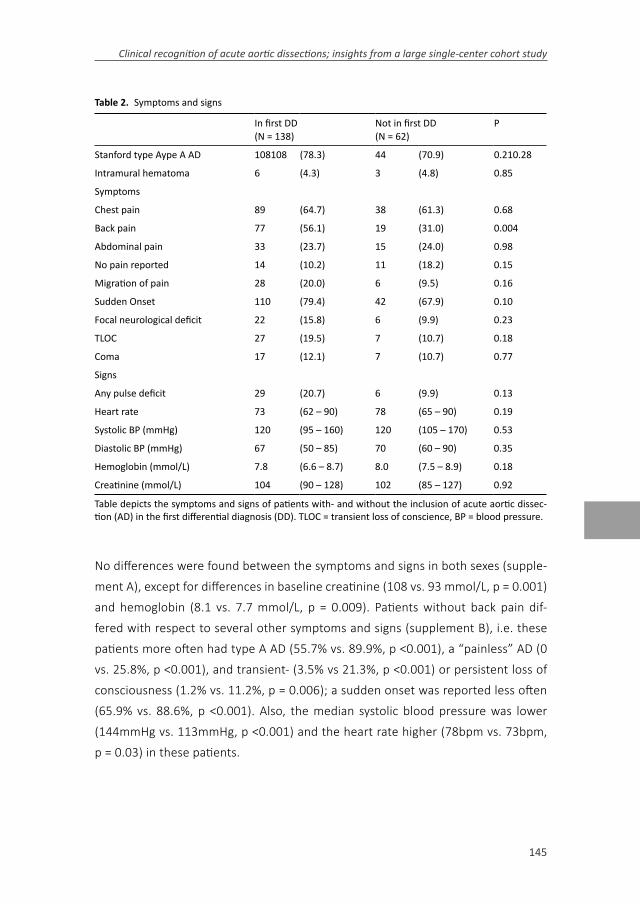
145
Clinical recognition of acute aortic dissections; insights from a large single-center cohort study
Table 2. Symptoms and signs
In first DD(N = 138)
Not in first DD(N = 62)
P
Stanford type Aype A AD 108108 (78.3) 44 (70.9) 0.210.28
Intramural hematoma 6 (4.3) 3 (4.8) 0.85
Symptoms
Chest pain 89 (64.7) 38 (61.3) 0.68
Back pain 77 (56.1) 19 (31.0) 0.004
Abdominal pain 33 (23.7) 15 (24.0) 0.98
No pain reported 14 (10.2) 11 (18.2) 0.15
Migration of pain 28 (20.0) 6 (9.5) 0.16
Sudden Onset 110 (79.4) 42 (67.9) 0.10
Focal neurological deficit 22 (15.8) 6 (9.9) 0.23
TLOC 27 (19.5) 7 (10.7) 0.18
Coma 17 (12.1) 7 (10.7) 0.77
Signs
Any pulse deficit 29 (20.7) 6 (9.9) 0.13
Heart rate 73 (62 – 90) 78 (65 – 90) 0.19
Systolic BP (mmHg) 120 (95 – 160) 120 (105 – 170) 0.53
Diastolic BP (mmHg) 67 (50 – 85) 70 (60 – 90) 0.35
Hemoglobin (mmol/L) 7.8 (6.6 – 8.7) 8.0 (7.5 – 8.9) 0.18
Creatinine (mmol/L) 104 (90 – 128) 102 (85 – 127) 0.92
Table depicts the symptoms and signs of patients with- and without the inclusion of acute aortic dissec-tion (AD) in the first differential diagnosis (DD). TLOC = transient loss of conscience, BP = blood pressure.
No differences were found between the symptoms and signs in both sexes (supple-ment A), except for differences in baseline creatinine (108 vs. 93 mmol/L, p = 0.001) and hemoglobin (8.1 vs. 7.7 mmol/L, p = 0.009). Patients without back pain dif-fered with respect to several other symptoms and signs (supplement B), i.e. these patients more often had type A AD (55.7% vs. 89.9%, p <0.001), a “painless” AD (0 vs. 25.8%, p <0.001), and transient- (3.5% vs 21.3%, p <0.001) or persistent loss of consciousness (1.2% vs. 11.2%, p = 0.006); a sudden onset was reported less often (65.9% vs. 88.6%, p <0.001). Also, the median systolic blood pressure was lower (144mmHg vs. 113mmHg, p <0.001) and the heart rate higher (78bpm vs. 73bpm, p = 0.03) in these patients.
Chapter 8
146
Diagnostic tests A chest x-ray was more often made in patients initially not suspect-ed of an AD (69.5% vs. 84.6%, p = 0.04); the presence of a widened mediastinum (44.2% vs. 46.1%, p = 0.89) or pleural effusion (9.3% vs. 7.8%, p = 0.76) was similar in both groups. The electrocardiogram showed atrial fibrillation in 5.7% and 5.3% (p = 0.92) in both groups respectively; the incidence of ST-depression (18.0% vs. 30.2%, p = 0.08) and ST-elevation (9.9% vs. 15.1%, p = 0.33) was not significantly different in both groups. D-dimer was measured in 16 patients, 12 of whom had an AD included in the first differential diagnosis. All d-dimer assays were positive, i.e. above the conventional threshold of 0.50 mg/ml and also above the age-adjusted cut-off (age /100 mg/ml).
Table 3 compares the primary- and total number of imaging modalities in both groups. In patients not suspected of an AD initially, the primary imaging test was more often transthoracic echocardiography (TTE; 34.7% vs. 50.9%) or coronary angiography (9.1% vs. 0.8%) and less often computed tomography (CT; 44.5% vs. 29.1%) or transesophageal echocardiography (TEE; 5.5% vs. 15.1%; p for all catego-ries = 0.002). The average number of imaging tests applied was higher in patients not suspected of AD initially (1.8 vs. 2.3, p = 0.01). Comparing all imaging tests applied, TTE (52.1% vs. 70.9%, p = 0.02) and coronary angiography (2.5% vs. 18.2%, p < 0.001) were more often used in the “not-suspected” group, while CT (77.3% vs. 67.3%, p = 0.16), TEE (45.4% vs. 42.6%, p = 0.73) and MRI (5.0% vs. 7.3%, p = 0.76) were used similarly in both groups.
CLINICAL SUSPICION In 179 patients we could retrieve a differential diagnosis; 124 (69.3%) of these patients had an AD included, and in 91 (50.8%) patients it was considered the most likely diagnosis. Among the 124 patients suspected of an AD, this diagnosis was positioned first, second and third or higher in 73.4%, 19.4%, and 7.2% of patients respectively. Corrected for other baseline characteristics, AD was less likely to be included in the first differential diagnosis in patients without back pain (RR 0.51, 95% CI: 0.30 – 0.84; table 4), in female patients (RR 0.66, 95% CI: 0.44 – 0.99) and in patients with extracardiac atherosclerosis (RR 0.64, 95% CI: 0.43 – 0.96).

147
Clinical recognition of acute aortic dissections; insights from a large single-center cohort study
Table 3. Imaging test used
In first DD(N = 138)
Not in first DD(N = 62)
P
First imaging test
TTE 43 (36.1) 28 (50.9) 0.002*
CT 53 (44.5) 16 (29.1)
TEE 18 (15.1) 3 (5.5)
MRI 3 (2.5) 0 (0)
CAG 1 (0.8) 5 (9.1)
All imaging tests
TTE 62 (52.1) 39 (70.9) 0.02
CT 92 (77.3) 37 (67.3) 0.16
TEE 54 (45.4) 23 (42.6) 0.73
MRI 6 (5.0) 4 (7.3) 0.56
Table 3 Imaging test used CAG
3 (2.5) 10 (18.2) <0.001
Table compares the first and total number of tests used in patients with and without AD in the first dif-ferential diagnosis. *P-value for all categories. TTE = transthoracic echocardiography, CT = computed to-mography, TEE = transesophageal echocardiography, MRI = magnetic resonance imaging, CAG = coronary angiography.
PATIENT MANAGEMENT AND OUTCOMES Type A AD was treated surgically in 136 of 152 (89.5%) patients. The median time to surgery was 1.8 (0.8 – 4.1) hours in the “suspected” group versus 10.1 (2.2 – 26.9) hours in the “not-suspected” group (p <0.001). Seven patients with type A AD died before surgery could be initiated. Nine patients died during surgery for type A AD, with a similar distribution among patients with and without an initial suspicion for AD (5.6% vs. 9.4%, p = 0.56). In nine patients a conservative treatment was chosen because of patient preference, old age or comorbidity; one of these patients survived until hospital discharge. The overall in-hospital mortality was similar in both groups (24.2% vs, 27.3; p = 0.66).
Median follow-up of all-cause mortality was 6.3 (interquartile range: 3.3 – 9.4) years, during which 92 (46.0%) patients died. Thirty-day mortality was similar in both groups (23.4% and 25.5% respectively, p = 0.77), as was the long-term survival (figure 1). The hazard ratio comparing patients with- and without an AD in the first

Chapter 8
148
differential diagnosis was 0.97 (95% CI: 0.61 – 1.55), and 0.76 (95% CI: 0.46 – 1.27) after multivariable correction.
Figure 1. Long-term survival of patients in whom an AD was included in the first differential diagnosis (group I, blue line) and patients in whom an AD was not included in the first differential diagnosis (group 2, red line); as a reference the age- and sex matched survival of Dutch inhabitants is depicted (black dashed line). A p-value is not given because lines are crossing.
DISCUSSION
This study showed that AD was not recognized during the primary clinical assess-ment in one-third of patients who were eventually diagnosed with AD. Although this was associated with the use of additional diagnostic test and a prolonged time from admission to surgery, the short- and long-term outcomes were similar. Reduc-tion of the time-laps between recognition and treatment of aortic dissections is of the utmost importance to lower perioperative mortality. We suggest that physi-cians who attend patients with suspected AD need to be familiar with the pitfalls in identifying symptoms and signs. Primarily, these are a low incidence combined with a broad range of symptoms which can mimic other conditions such as stroke, myocardial infarction and other abdominal pathology. Our study extends to the

149
Clinical recognition of acute aortic dissections; insights from a large single-center cohort study
insights that have been gained in the past decade from larger registries such as the IRAD to provide adequate information regarding the symptoms and signs of AD.14,15 It is also important to know how many AD patients are missed and what makes these patients different from patients who are correctly identified.
In our study, 31% of patients were not recognized initially, which was more likely in the absence of back pain, in female patients, and in patients with extracardiac atherosclerosis. It is important to be aware of these associations since they may improve the awareness for an AD in these patients. However, it is likely that these factors are a proxy for a different clinical picture rather than the cause for a missed diagnosis. The absence of back pain as a presenting symptom was indeed associ-ated with other differences in the clinical presentation, as these patients more often had type A AD, presented with “painless” AD, or had a transient or persistent loss of consciousness, and less often reported a sudden onset; also, they had a worse hemodynamic profile. It is thus likely that these underlying differences in the clinical picture are the actual reason that patients were not adequately identified as having an AD. A study by Imamura et al similarly showed that patients who presented with “painless AD” more often had an “atypical” presentation with higher incidences of hemodynamic shock and neurological symptoms.16 Therefore, to improve the identification of AD patients, physicians should be more aware of the possibility of an AD in patients with shock of unknown cause and in patients with (transient) loss of consciousness, specifically if combined with thoracic pain.
Our results did not indicate that female sex was similarly a proxy for a different clini-cal profile, as the distribution of symptoms and signs was similar among males and females except for the sex-dependent hemoglobin and creatinine levels. Previous studies however showed that women less often have an abrupt onset of symptoms or a pulse deficit, and more often present with coma and congestive heart failure.17 These factors are itself associated with a delayed recognition of AD.18 Indeed, it has been shown that the time from admission to diagnosis is longer in women compared to men.18 It is thus likely that an “a-typical” presentation in women is recognized less (timely), a phenomenon that is also well described in other cardiovascular diseases.19
Chapter 8
150
Based on our results and on previously reported findings it is clear that the diagnostic process of patients with suspected AD needs to be improved. Several aspects of the diagnostic process can be considered. First, risk scores should be used to calculate the a-priori risk of an AD in suspected patients.20,21 Patients with a predicted high risk should receive expedited imaging with CT, TEE or MRI. Second, measurement of D-dimer should be considered in patients with a low or intermediate a-priori risk.22,23 The use of d-dimer was still limited during our study, but is currently an important tool to further stratify the diagnostic process. In our study none of the patients had a false negative test; previous studies showed that d-dimer is specifi-cally useful to exclude AD, since the likelihood of AD after a negative test (0.50 mg/ml) is only 0.3%.24
Finally, the inadvertent administration of anticoagulant or thrombolytic therapy to AD patients who present with symptoms suggestive of myocardial- or cerebral infarc-tion may lead to acute hemorrhage and death.25-27 The former should be suspected in patients who present with ST-elevation in the inferior leads, since progression of an intimal tear is usually to the right coronary cusp. Specifically, alternating ST-elevation and concomitant neurological symptoms should raise suspicion of an AD. For the timely recognition of dissections mimicking stroke, routine visualization of the aortic arch and its branching vessels during cerebral CT may be considered. Such a protocol could, at the costs of little extra scanning time and radiation exposure, identify patients with an underlying aortic dissection.
LIMITATIONS Some limitations apply to our study; mainly the retrospective study design may have precluded several biases. First, despite a large effort to register all AD patients, we may have missed patients with a conservative management, and those who were misclassified as another condition or who died before the diagnosis was established.3 Also, registration of (baseline) characteristics may have been incomplete due to the retrospective data collection. Rather than discarding missing data, we imputed missing covariates, as this has been shown to reduce bias and increase the precision of results.10,11 Furthermore, the study was not designed to directly capture the effect of test results on the clinical suspicion for an AD. Finally, although we consider the registration of the presence versus absence of AD in the first differential diagnosis a strength of this study, it would have been informative to
151
Clinical recognition of acute aortic dissections; insights from a large single-center cohort study
also know which alternative diagnoses were considered and how this affected the diagnostic strategy.
In summary, acute aortic dissection was initially not suspected in almost one third of patients. This was more likely in women, patients with extracardiac atheroscle-rosis or absence of back pain. Patients without AD in the first differential diagnosis underwent more imaging tests and had a longer time from admission to surgery, but a similar short- and long-term mortality. An improved recognition of patients with acute aortic dissection could lead a reduction in the number of diagnostic tests, a shorter time to therapy, and possibly improved patient outcomes.

Chapter 8
152
REFERENCES 1. Mészáros I, Mórocz J, Szlávi J, et al. Epidemiology and clinicopathology of aortic dissec-
tion. Chest. 2000;117:1271-8. 2. Clouse WD, Hallett JW Jr, Schaff HV, et al. Acute aortic dissection: population-based
incidence compared with degenerative aortic aneurysm rupture. Mayo Clin Proc. 2004;79:176-80.
3. Howard DPJ, Banerjee A, Fairhead JF, Perkins J, Silver LE, Rothwell PM. Population-based study of incidence and outcome of acute aortic dissection and premorbid risk factor control: 10-year results from the Oxford Vascular Study. Circulation. 2013;127:2031-7.
4. Kontos MC, Diercks DB, Kirk JD. Emergency department and office-based evaluation of patients with chest pain. Mayo Clin Proc. 2010;85:284-99.
5. Alter S, Eskin B, Allegra J. Diagnosis of aortic dissection in emergency department patients is rare. West J Emerg Med. 2015;16:629-31.
6. Roberts WC, Vowels TJ, Ko JM, Guileyardo JM. Acute aortic dissection with tear in ascending aorta not diagnosed until necropsy or operation (for another condition) and comparison to similar cases receiving proper operative therapy. Am J Cardiol. 2012;110:728-35.
7. Svircevic V, Nierich AP, Moons KGM, Brandon Bravo Bruinsma GJ, Kalkman CJ, van Dijk D. Fast-track anesthesia and cardiac surgery: a retrospective cohort study of 7989 patients. Anesth Analg. 2009;108:727-33.
8. Kaji AH, Schriger D, Green S. Looking through the retrospectoscope: Reducing bias in emergency medicine chart review studies. Ann Emerg Med. 2014;64:292-8.
9. Donders ART, van der Heijden GJMG, Stijnen T, Moons KGM. Review: a gentle introduc-tion to imputation of missing values. J Clin Epidemiol. 2006;59:1087-91.
10. Groenwold RHH, Donders ART, Roes KCB, Harrell FE, Moons KGM. Dealing with miss-ing outcome data in randomized trials and observational studies. Am J Epidemiol. 2012;175:210-7.
11. Newgard CD, Haukoos JS. Advanced statistics: Missing data in clinical research-Part 2: Multiple imputation. Acad Emerg Med. 2007;14:669-78.
12. Li P, EA S, DB A. Multiple imputation: A flexible tool for handling missing data. JAMA. 2015;314:1966-7.
13. Rubin D. An overview of multiple imputation. In: Proceedings of the Survey Research Methods Section, American Statistical Association 1988; 79-84.
14. Hagan PG, Nienaber CA, Isselbacher EM, et al. The international registry of acute aortic dissection (IRAD). JAMA 2000;283:897-903.
15. Pape LA, Awais M, Woznicki EM, et al. Presentation, diagnosis, and outcomes of acute aortic dissection: 17-year trends from the International Registry of Acute Aortic Dis-section. J Am Coll Cardiol. 2015;66:350-8.
16. Imamura H, Sekiguchi Y, Iwashita T et al. Painless Acute Aortic Dissection - Diagnostic, prognostic and Clinical implications. Circ J. 2011;75:59-66.
17. Nienaber CA. Gender-related differences in acute aortic dissection. Circulation. 2004;109:3014-21.
18. Harris KM, Strauss CE, Eagle K, et al. Correlates of delayed recognition and treatment of acute type A aortic dissection: the International Registry of Acute Aortic Dissection (IRAD). Circulation. 2011;124:1911-8.
153
Clinical recognition of acute aortic dissections; insights from a large single-center cohort study
19. EUGenMed; Cardiovascular Clinical Study Group, Regitz-Zagrosek V, Oertelt-Prigione S, Prescott E, et al. Gender in cardiovascular diseases: impact on clinical manifesta-tions, management, and outcomes. Eur Heart J. 2016;37:24-34.
20. Erbel R, Aboyans V, Boileau C, et al. 2014 ESC Guidelines on the diagnosis and treat-ment of aortic diseases: Document covering acute and chronic aortic diseases of the thoracic and abdominal aorta of the adult. The Task Force for the Diagnosis and Treat-ment of Aortic Diseases of the European. Eur Heart J. 2014;35:2873-2926.
21. Hiratzka LF, Bakris GL, Beckman JA, et al. 2010 ACCF/AHA/AATS/ACR/ ASA/SCA/SCAI/SIR/STS/SVM guidelines for the diagnosis and management of patients with Thoracic Aortic Disease: a report of the American College of Cardiology Foundation/American Heart Association Task Force on Practice Guidelines. Circulation. 2010;121:e266-e369.
22. Suzuki T, Distante A, Zizza A, et al. Diagnosis of acute aortic dissection by D-dimer: the International Registry of Acute Aortic Dissection Substudy on Biomarkers (IRAD-Bio) experience. Circulation. 2009;119:2702-7.
23. Nazerian P, Morello F, Vanni S, et al. Combined use of aortic dissection detection risk score and D-dimer in the diagnostic workup of suspected acute aortic dissection. Int J Cardiol. 2015;175:78-82.
24. Asha SE, Miers JW. A systematic review and meta-analysis of D-dimer as a rule-out test for suspected acute aortic dissection. Ann Emerg Med. 2015;66:1-11.
25. Ho CH, Chen YL, Lin YY, et al. Acute aortic dissection complicated by acute ischemic stroke: diagnostic challenges. Am J Emerg Med. 2012;30:2070-3.
26. Tanoue S, Yanagawa Y. Ischemic stroke with left hemiparesis or shock should be evalu-ated by computed tomography for aortic dissection. Am J Emerg Med. 2012;30:836.e3-e4.
27. Huang CH, Huang HC, Lin KH, Chen WK, Tsai CH. Identification of painless aortic dis-section before thrombolytic treatment for acute ischemic stroke. Am J Emerg Med. 2013;31:762.e5-e6.

Chapter 8
154
Supplement A
Male(N = 122)
Female(N = 78)
P
Stanford Type A 94 (77.0) 58 (74.4) 0.66
Symptoms
Any pain reported
107 (87.9) 68 (86.6) 0.80
Chest 80 (65.4) 48 (61.0) 0.55
Back 57 (47.0) 39 (50.5) 0.65
Abdominal 24 (19.4) 23 (30.4) 0.10
Migration 16 (13.1) 17 (21.9) 0.14
Sudden Onset 96 (78.5) 56 (71.9) 0.31
Focal neurological deficit
18 (14.5) 5 (6.8) 0.11
TLOC 19 (15.6) 14 (18.2) 0.66
Coma 15 (12.6) 8 (9.7) 0.56
Signs
Any pulse deficit
19 (15.6) 14 (18.2) 0.72
Heart rate 76 (62 – 94) 72 (65 – 88) 0.37
Systolic BP (mmHg)
120 (104 – 155) 120 (80 – 170) 0.66
Diastolic BP (mmHg)
70 (57 – 85) 65 (50 – 90) 0.31
Hemoglobin (mmol/L)
8.1 (7.1 – 8.9) 7.7 (6.5 – 8.4) 0.009
Creatinine (mmol/L)
108 (95 – 134) 93 (80 – 111) 0.001
Table comparing the symptoms and signs in both sexes. TLOC = transient loss of conscience, BP = blood pressure.

155
Clinical recognition of acute aortic dissections; insights from a large single-center cohort study
Supplement B
Back pain(N = 88)
No back pain(N = 89)
P
Stanford Type A 49 (55.7) 80 (89.9) <0.001
Symptoms
Chest 50 (56.8) 61 (68.5) 0.11
Abdominal 24 (27.3) 14 (15.7) 0.06
Migration 18 (20.5) 3 (3.4) 0.001
Painless dissection
0 (0) 23 (25.8) <0.001
Sudden Onset 78 (88.6) 58 (65.9) <0.001
Focal neurological deficit
9 (10.5) 10 (11.2) 0.87
TLOC 3 (3.5) 19 (21.3) <0.001
Coma 1 (1.2) 10 (11.2) 0.006
Signs
Any pulse deficit
11 (12.9) 11 (12.8) 0.98
Heart rate 73 (60 – 85) 78 (65 – 100) 0.03
Systolic BP (mmHg)
144 (118 – 176) 113 (90 – 140) <0.001
Diastolic BP (mmHg)
80 (61 – 97) 63 (50 – 80) <0.001
Hemoglobin (mmol/L)
8.0 (7.3 – 8.9) 7.8 (7.3 – 8.7) 0.39
Creatinine (mmol/L)
73 (60 – 85) 78 (65 – 100) 0.44
Table comparing the symptoms and signs in patients with and without back pain on admission. TLOC = transient loss of conscience, BP = blood pressure.

9Modified transesophageal echocardiography of the
dissected thoracic aorta;
a novel diagnostic approach.
W.W. Jansen Klomp, L.M. Peelen, G.J. Brandon Bravo Bruinsma, A.W.J. van ‘t Hof, J.G. Grandjean, A.P. Nierich
Published in: Cardiovascular Ultrasound, 2016

Chapter 9
158
ABSTRACT
BACKGROUND Transesophageal echocardiography (TEE) is a key diagnostic modality in patients with acute aortic dissection, yet its sensitivity is limited by a “blind-spot” caused by air in the trachea. After placement of a fluid-filled balloon in the trachea visualization of the thoracic aorta becomes possible. This method, modified TEE, has been shown to be an accurate test for the diagnosis of upper aortic atherosclerosis. In this study we discuss how we use modified TEE for the diagnosis and management of patients with (suspected) acute aortic dissection.
NOVEL DIAGNOSTIC APPROACH OF THE DISSECTED AORTA Modified TEE provides the possibility to obtain a complete echocardiographic overview of the thoracic aorta and its branching vessels with anatomical and functional information. It is a bedside test, and can thus be applied in hemodynamic instable patients who cannot undergo computed tomography. Visualization of the aortic arch allows differentiation between Stanford type A and B dis-sections and visualization of the proximal cerebral vessels enables a timely identification of impaired cerebral perfusion.
During surgery modified TEE can be applied to identify the true lumen for cannulation, and to assure that the true lumen is perfused. Also, the in-nominate- and carotid arteries can be assessed for structural integrity and adequate perfusion during multiple phases of the surgical repair.
CONCLUSIONS Modified TEE can reveal the “blind-spot” of conventional TEE. In patients with (suspected) aortic dissection it is thus possible to obtain a complete echocardiographic overview of the thoracic aorta and its branches. This is of specific merit in hemodynamically unstable patients who cannot undergo CT. Modified TEE can guide also guide the surgical management and monitor perfusion of the cerebral arteries.

159
Modified transesophageal echocardiography of the dissected thoracic aorta
BACKGROUND
Acute aortic dissection (AD) is a life-threatening condition, which requires a prompt diagnosis to prevent morbidity and mortality. This necessitates sensitive and conclusive diagnostic tests. Transthoracic echocardiography is often the first test used since it is directly available, may reveal a proximal dissection, and can detect pericardial and pleural effusion.1,2 However, the focused screening for AD requires tests with a superior diagnostic accuracy, i.e. computed tomography (CT), magnetic resonance imaging (MRI) or transesophageal echocardiography (TEE).3,4
Guidelines recommend the use of CT or MRI as the primary test in patients who are hemodynamically stable.5 Both modalities can accurately visualize an intimal tear, and complications can be detected, including aneurysmal widening of the aorta, pericardial and pleural effusion and involvement of coronary or distal arteries.3-6 A major limitation of both tests is the need to move patients out of the acute care environment. Moreover, CT is lacking the possibility of functional imaging, while the use of MRI is limited by its longer imaging-time and lower availability.
In hemodynamically instable patients, TEE is recommended as the primary test because it can be performed in the emergency care department or operating room.3-5 Transesophageal echocardiography has the advantage of providing a quick overview of the heart and aorta with both anatomical and functional information. The diagnostic accuracy of TEE is considered inferior however compared to CT and MRI, mainly because of the impaired visualization of the distal ascending aorta and aortic arch caused by the interposition of the air-filled trachea.3,4,7,8 Indeed, limited dissections can be missed due to this so-called “blind-spot”.9
The diagnostic accuracy of conventional TEE can be improved using a modification which enables the visualization of the distal ascending aorta, aortic arch and its branches through a fluid-filled balloon placed in the trachea and left main bronchus (Figure 1).10 The original aim of “modified TEE” was to improve the diagnosis of aortic atherosclerosis during cardiothoracic surgery and thereby reduce the inci-dence of emboli-related complications. Modified TEE is indeed a sensitive test for

Chapter 9
160
the diagnosis of atherosclerosis of the distal ascending aorta,10-12 with a superior diagnostic accuracy compared to conventional TEE.13
Figure 1. Schematic overview of modified transesophageal echocardiography. 1. Esophagus with TEE probe; 2. Trachea and left main bronchus with inflated endotracheal “A-View” balloon creating an echo-cardiographic window to the aortic arch; 3. Distal ascending aorta, aortic arch and branching vessels; 4. Pulmonary artery.
We also routinely use modified TEE in patients with (suspected) AD. Most impor-tantly, modified TEE provides the possibility to obtain a complete echocardiographic overview of the thoracic aorta and its branches in patients who cannot undergo CT. Second, modified TEE can guide essential steps in the surgical repair of the dissected aorta. In this study we discuss how we use modified TEE to aid the diagnosis and management of AD patients.

161
Modified transesophageal echocardiography of the dissected thoracic aorta
Technique of modified TEE. A conventional TEE examination of the heart and aorta is performed following the prevailing guidelines.14 In the setting of suspected AD, it is recommended to perform TEE under general anesthesia as the introduction of the probe may cause an inadvertent increase of the blood pressure in conscious patients,4 which is associated with a higher likelihood of rupture of the adventitial layer.15 The endotracheal-tube should be placed directly distal from the vocal chords to ensure enough space for the positioning of the tracheal balloon. Pre-oxygenation permits for an apneic period of 2 – 3 minutes in which imaging with modified TEE can be performed safely. During cardiac bypass or deep hypothermic arrest, modified TEE can be used continuously. After preparation of the specially designed “A-View” catheter,12 the ventilator is disconnected, and the A-View catheter is introduced and positioned in the trachea and left main bronchus. For a correct positioning, the 24 cm markers on the endotracheal tube and catheter should line-up; a correct positioning of the catheter can also be checked using TEE. Inflation of the balloon with 20-50 ml of saline should give an echocardiographic window to the “blind-spot” (figures 2 and 3).
Figure 2. Distal ascending aorta long-axis view with modified TEE showing a normal aorta. T = Trachea with inflated endotracheal balloon. RPA = Right Pulmonary Artery, DAA = Distal Ascending Aorta, P =Pos-terior wall, A = Anterior wall.

Chapter 9
162
Figure 3. Visualization of the distal ascending aorta (DAA) during elective aortic root replacement in a 43-year old woman with Marfan syndrome; the bispectral index indicated a compromised cerebral perfusion.Panel A Conventional TEE with inadequate visualization of the cannulation site due to the “blind-spot”.Panel B Same view (DAA, long-axis) after inflation of the endotracheal balloon, showing a limited iatrogenic dissection (red line) with extracorporeal perfusion of the false lumen. 1. Trachea with inflated endotracheal balloon; 2. True lumen; 3. False lumen; 4. Perfusion jet.
Modified TEE is compatible with additional modalities including color-Doppler and 3D imaging. Contraindications are similar as for conventional TEE, with the addition of tracheomalacia (e.g. in connective tissue diseases) and tracheal stenosis which may predispose to complications related to the balloon inflation.
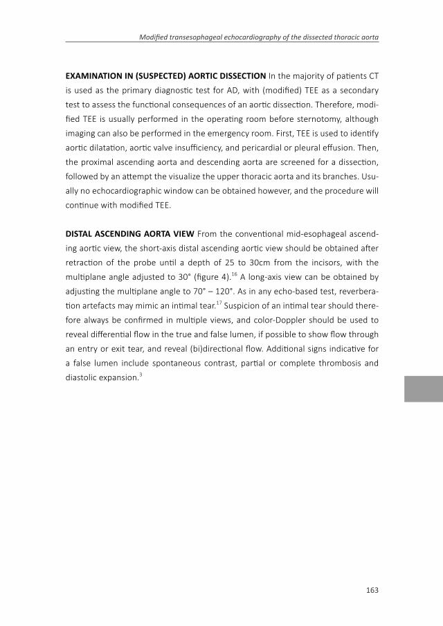
163
Modified transesophageal echocardiography of the dissected thoracic aorta
EXAMINATION IN (SUSPECTED) AORTIC DISSECTION In the majority of patients CT is used as the primary diagnostic test for AD, with (modified) TEE as a secondary test to assess the functional consequences of an aortic dissection. Therefore, modi-fied TEE is usually performed in the operating room before sternotomy, although imaging can also be performed in the emergency room. First, TEE is used to identify aortic dilatation, aortic valve insufficiency, and pericardial or pleural effusion. Then, the proximal ascending aorta and descending aorta are screened for a dissection, followed by an attempt the visualize the upper thoracic aorta and its branches. Usu-ally no echocardiographic window can be obtained however, and the procedure will continue with modified TEE.
DISTAL ASCENDING AORTA VIEW From the conventional mid-esophageal ascend-ing aortic view, the short-axis distal ascending aortic view should be obtained after retraction of the probe until a depth of 25 to 30cm from the incisors, with the multiplane angle adjusted to 30° (figure 4).16 A long-axis view can be obtained by adjusting the multiplane angle to 70° – 120°. As in any echo-based test, reverbera-tion artefacts may mimic an intimal tear.17 Suspicion of an intimal tear should there-fore always be confirmed in multiple views, and color-Doppler should be used to reveal differential flow in the true and false lumen, if possible to show flow through an entry or exit tear, and reveal (bi)directional flow. Additional signs indicative for a false lumen include spontaneous contrast, partial or complete thrombosis and diastolic expansion.3
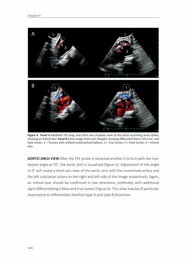
Chapter 9
164
Figure 4. Panel A Modified TEE long- and short axis (X-plane) view of the distal ascending aorta (DAA), showing an intimal tear. Panel B Same image with color-Doppler showing differential flow in the true- and false lumen. 1 = Trachea with inflated endotracheal balloon; 2 = True lumen; 3 = False lumen; 4 = Intimal tear.
AORTIC ARCH VIEW After the TEE probe is retracted another 2 to 5cm with the mul-tiplane angle at 70°, the aortic arch is visualized (figure 5). Adjustment of the angle to 0° will reveal a short-axis view of the aortic arch with the innominate artery and the left subclavian artery on the right and left side of the image respectively. Again, an intimal tear should be confirmed in two directions, preferably with additional signs differentiating a false and true lumen (Figure 6). This view may be of particular importance to differentiate Stanford type A and type B dissection.

165
Modified transesophageal echocardiography of the dissected thoracic aorta
Figure 5. Modified TEE aortic arch short axis view in a patient with a normal aortic arch and flow pattern. 1 = Trachea with inflated endotracheal balloon; 2 = Innominate artery; 3 = Aortic arch; 4 = Left carotid artery.
Figure 6. Modified TEE aortic arch short axis view Panel A Plain 2D image showing a dissection starting in the proximal aortic arch, which continued distally into the descending aorta. The intimal tear did not progress into the innominate artery. Panel B Similar image with color-Doppler showing flow through the true aortic lumen, and a normal perfusion of the innominate artery. 1 = Trachea with inflated endotracheal balloon; 2 = Innominate artery; 3 = True lumen; 4 = False lumen; red lines delineate the intimal tear.
Aortic Branch Vessels View Finally, the proximal part of the innominate artery (fig-ures 7 and 8), and the left carotid- and subclavian artery are visualized by further retracting the TEE probe. Navigation between the vessels is achieved by rotation of the TEE probe both to the left and right.

Chapter 9
166
Figure 7. Modified TEE showing the innominate artery with three different modalities. Panel A 2D-image showing an intimal tear and differential flow. Panel B 3D-image in a different patient without dissection. Panel C M-Mode image of the same patient without dissection. 1 = Trachea with inflated endotracheal balloon; 2 = Innominate artery; 3 = Intimal tear.
Figure 8. Modified TEE long axis view of the innominate artery in a patient with Stanford type A aortic dissection with progression into the innominate artery. 1. Trachea with inflated endotracheal balloon 2. Innominate artery; red line delineating the intimal tear.
PERIOPERATIVE MONITORING During surgery for AD, modified TEE is used to guide the surgical management and to support cerebral monitoring. Specific attention is given to the perfusion of the branching vessels (i.e. the brachiocephalic- and carotid artery), which can be impaired because of a continuing dissection, obstruction by an intimal tear, or compression by an extravascular hematoma. The early detection of impaired cerebral perfusion allows for timely changes in the management, e.g. direct initiation of deep-hypothermic arrest, maintenance of a higher blood pres-sure and application of additional cerebral monitoring tools, e.g. bispectral index and near-infrared spectroscopy.
During surgery we use modified TEE to guide aortic cannulation and to confirm that the true lumen is perfused during CPB. If right axillary cannulation is considered,
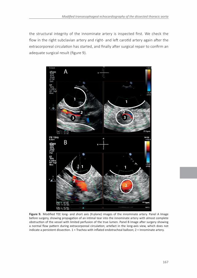
167
Modified transesophageal echocardiography of the dissected thoracic aorta
the structural integrity of the innominate artery is inspected first. We check the flow in the right subclavian artery and right- and left carotid artery again after the extracorporeal circulation has started, and finally after surgical repair to confirm an adequate surgical result (figure 9).
Figure 9. Modified TEE long- and short axis (X-plane) images of the innominate artery. Panel A Image before surgery, showing propagation of an intimal tear into the innominate artery with almost complete obstruction of the vessel with limited perfusion of the true lumen. Panel B Image after surgery showing a normal flow pattern during extracorporeal circulation; artefact in the long-axis view, which does not indicate a persistent dissection. 1 = Trachea with inflated endotracheal balloon; 2 = Innominate artery.

Chapter 9
168
LIMITATIONS OF MODIFIED TEE Modified TEE has some limitations. First, experience with conventional TEE is a prerequisite and additional training should be considered since a learning curve exists. Second, the spatial resolution can be lower than in conventional TEE because of some scattering by the balloon. Finally, we already addressed the procedures to prevent false positive results. Despite these measures we are aware of one patient (female, 43 years) who underwent sternotomy because of a false positive finding with modified TEE. This patient was referred for surgery based on the suspicion of an AD on CT. During preoperative visualization of the thoracic aorta with modified TEE no intimal tear was confirmed, but a structure outside the aorta appeared as an extramural hematoma of the aortic arch. However, during intraoperative inspection this structure was revealed to be thymus tissue.
CONCLUSIONS Modified TEE can reveal the “blind-spot” of conventional TEE. In patients with suspected acute aortic dissection it is thus possible to obtain a complete echocardiographic overview of the thoracic aorta and its branches with both anatomical and functional information. This may be specifically useful in he-modynamically unstable patients who cannot undergo CT. During surgery for aortic dissection, modified TEE can guide the surgical management and monitor perfusion of the cerebral arteries.

169
Modified transesophageal echocardiography of the dissected thoracic aorta
REFERENCES 1. Evangelista A, Avegliano G, Aguilar R, Cuellar H, Igual A, González-Alujas T, et al. Impact
of contrast-enhanced echocardiography on the diagnostic algorithm of acute aortic dissection. Eur Heart J. 2010; 31: 472–9.
2. Sobczyk D, Nycz K. Feasibility and accuracy of bedside transthoracic echocardiography in diagnosis of acute proximal aortic dissection. Cardiovasc Ultrasound. 2015;13:15.
3. Hiratzka LF, Bakris GL, Beckman JA, Bersin RM, Carr VF, Casey DE, et al. 2010 ACCF/AHA/AATS/ACR/ASA/SCA/SCAI/SIR/STS/SVM guidelines for the diagnosis and manage-ment of patients with Thoracic Aortic Disease: a report of the American College of Cardiology Foundation/American Heart Association Task Force on Practice Guidelines. Circulation. 2010; 121: e266–369.
4. Baliga RR, Nienaber CA, Bossone E, Oh JK, Isselbacher EM, Sechtem U, et al. The role of imaging in aortic dissection and related syndromes. JACC-Cardiovasc Imag. 2014; 7: 406–24.
5. Erbel R, Aboyans V, Boileau C, Bossone E, Bartolomeo RD, Eggebrecht H, et al. 2014 ESC Guidelines on the diagnosis and treatment of aortic diseases: Document covering acute and chronic aortic diseases of the thoracic and abdominal aorta of the adult. Eur Heart J. 2014; 35: 2873–926.
6. Moore AG, Eagle KA, Bruckman D, Moon BS, Malouf JF, Fattori R, et al. Choice of com-puted tomography, transesophageal echocardiography, magnetic resonance imaging, and aortography in acute aortic dissection: International Registry of Acute Aortic Dis-section (IRAD). Am J Cardiol. 2002; 89: 1235–8.
7. Van Zaane B, Zuithoff NPA, Reitsma JB, Bax L, Nierich AP, Moons KGM. Meta-analysis of the diagnostic accuracy of transesophageal echocardiography for assessment of atherosclerosis in the ascending aorta in patients undergoing cardiac surgery. Acta Anaesthesiol Scand. 2008; 52: 1179–87.
8. Glas KE, Swaminathan M, Reeves ST, Shanewise JS, Rubenson D, Smith PK, et al. Guidelines for the performance of a comprehensive intraoperative epiaortic ultraso-nographic examination: recommendations of the American Society of Echocardiogra-phy and the Society of Cardiovascular Anesthesiologists; endorsed by the Society of Thoracic Surgeons. J Am Soc Echocardiogr. 2007; 20: 1227–35.
9. Demertzis S, Casso G, Torre T, Siclari F. Direct epiaortic ultrasound scanning for the rapid confirmation of intraoperative aortic dissection. Interact Cardiovasc Thorac Surg. 2008 Aug; 7: 725–6.
10. van Zaane B, Nierich AP, Buhre WF, Brandon Bravo Bruinsma GJ, Moons KGM. Resolv-ing the blind spot of transesophageal echocardiography: a new diagnostic device for visualizing the ascending aorta in cardiac surgery. Br J Anaesth. 2007; 98: 434–41.
11. Nierich AP, van Zaane B, Buhre WF, Coddens J, Spanjersberg AJ, Moons KGM. Visualiza-tion of the distal ascending aorta with A-Mode transesophageal echocardiography. J Cardiothorac Vasc Anesth. 2008; 22: 766–73.
12. van Zaane B, Nierich AP, Brandon Bravo Bruinsma GJ, Rosseel PMJ, Ramjankhan FZ, de Waal EEC, et al. Diagnostic accuracy of modified transesophageal echocardiography for pre-incision assessment of aortic atherosclerosis in cardiac surgery patients. Br J Anaesth. 2010; 105: 131–8.
13. Jansen Klomp WW, Peelen LM, Spanjersberg SJ, Brandon Bravo Bruinsma GJ, Lange F de, van ’t Hof AWJ, et al. Added value of modified transesophageal echocardiography in the diagnosis of atherosclerosis of the distal ascending aorta in cardiac surgery patients. Eur Heart J Cardiovasc Imaging. 2014; 15: 623–30.

Chapter 9
170
14. Flachskampf FA, Wouters PF, Edvardsen T, Evangelista A, Habib G, Hoffman P, et al. Recommendations for transesophageal echocardiography: EACVI update 2014. Eur Heart J Cardiovasc Imaging. 2014; 15: 353–65.
15. Silvey SV, Stoughton TL, Pearl W, Collazo WA, Belbel RJ. Rupture of the outer partition of aortic dissection during transesophageal echocardiography. Am J Cardiol. 1991; 68: 286–7.
16. Shiga T, Wajima Z, Apfel CC, Inoue T, Ohe Y. Diagnostic accuracy of transesophageal echocardiography, helical computed tomography, and magnetic resonance imaging for suspected thoracic aortic dissection: systematic review and meta-analysis. Arch Intern Med. 2006; 166: 1350–6.
17. Losi MA, Betocchi S, Briguori C, Manganelli F, Ciampi Q, Pace L, et al. Determinants of aortic artifacts during transesophageal echocardiography of the ascending aorta. Am Heart J. 1999; 137: 967–72


10General discussion


175
Modified transesophageal echocardiography of the dissected thoracic aorta
PART I
This thesis investigated the role for modified TEE in timely detection of aortic atherosclerosis in patients undergoing cardiothoracic surgery (part I) and explored other potential areas where modified TEE could be of use (part II). In this chapter we will first position the findings regarding its use in cardiothoracic surgery in a framework for diagnostic test research and investigate which aspects still require further research. Subsequently we will discuss to which extent a similar extensive approach is required for the disease areas discussed in part II of the thesis.
Cardiac surgery is a burdensome treatment for patients in the short term, which is acceptable in the prospect of an improved life-expectancy and quality of life in the long term. Neurological complications directly interfere with these aims of cardiac surgery. Reducing the incidence of post-operative neurological complications is thus paramount, both for the patients exposed to these hazards, but also to reduce the financial burden of cardiac surgery.
Post-operative neurological complications include stroke, delirium, cognitive de-cline, renal dysfunction, coma and death.1–9 The etiology of these complications is multifactorial, but cerebral embolization after manipulation and dislodgement of aortic atherosclerosis is believed to play a pivotal role.10–15 The hypothesis underly-ing the majority of the studies included in this thesis is that the incidence of post-operative neurological complications can be reduced through accurate detection of aortic atherosclerosis followed by effective changes in the surgical management.
Several diagnostic tests are available for the diagnosis of aortic atherosclerosis (Chapter 2), none of which is perfect. Epiaortic ultrasound is considered the gold standard for the visualization of the DAA,16 but is infrequently used.17 Transesopha-geal echocardiography (TEE) is most often used for the perioperative screening for aortic atherosclerosis, but the visualization of the upper thoracic aorta is usually hampered by the air-filled trachea.18 Since the distal ascending aorta is the part of the aorta most likely to be manipulated during cardiac surgery, it is important to obtain accurate information on the degree of atherosclerosis in this aortic seg-ment.2 Modified TEE enables visualization of this part of the aorta after inflation of a
Chapter 10
176
fluid-filled balloon which is positioned in the trachea. The simple pragmatism of this method is appealing, but the risk of test-related complications as well as increased costs should be outweighed by a more accurate diagnosis and ultimately improved patient outcomes.
A previous thesis by van Zaane assessed the technical feasibility, safety, diagnostic accuracy and cost-effectiveness (based on modeling studies) of modified TEE.18–21 The main conclusion was that modified TEE was an accurate test for the diagnosis of atherosclerosis of the distal ascending aorta (DAA) with a high negative predictive value, at a low risk of test-related complications. The main limitation was a limited inter-and intra-observer variability, which was attributed to a lack of uniformity in the reporting of the test results in the absence of clear definitions. It was thus recommended to improve the agreement within and across future observers of modified TEE through improved training and the establishment of definitions of the severity of atherosclerosis. Also, it was advised to study the impact of modified TEE on patient management and emboli-related cerebral complications, preferably in a randomized diagnostic trial. Finally, the results of such a study could be used to study the cost-effectiveness of modified TEE in practice.
TEST-TREATMENT PATHWAY
The following section will assess the value of modified TEE alongside the pathway from test application towards patient outcomes. Ferrante di Ruffano and colleagues advocated to move beyond diagnostic accuracy, and designed a framework for the systematic evaluation of the test-treatment(-outcome) pathway of diagnostic tests.22 We followed this structural
evaluation for the evaluation of modified TEE using the available studies (table 1), which will naturally show the shortcomings in the current investigations and recom-mendations for future research.

177
Modified transesophageal echocardiography of the dissected thoracic aorta
Table 1 Assessment of the test-treatment pathway of modified TEE following the framework presented by Ferrante di Ruffano et al.
Pathway component Modified TEE in this respect Reference
Timing of test Modified TEE is conducted before sternotomy; current reference standard EUS after sternotomy. Before sternotomy allows for conversion to other approaches
19,23
Feasibility Successful view of DAA in 41/41 patients (first study) and 357/370 patients (second study)
20,23
Safety Major complications: 2/1802 (0.1%). Blood on balloon: 17/429 (4.0%) without clinical consequences. Minor bleeding in current use in practice unknown.
19,23Chapter 5,6
Diagnostic accuracy Diagnosis of grade ³3 atherosclerosis DAA, compared to epiaortic ultrasound:Sensitivity: 95%, specificity: 79%, PPV: 79%, NPV: 97%. Overall: AUC 0.89
20
Added value Modified TEE improved diagnostic accuracy when added to patient characteristics and conventional TEE (AUCs: 0.80 -> 0.93; NRI: 0.60 and IDI: 0.30)
Chapter 3
Prognostic implications Atherosclerosis DAA associated with increased mortality (HR 1.82), predictive value beyond conventional TEE. Not be used for prognostic prediction only, then EUROSCORE is preferred.
Chapter 4
Impact on management Observational study: use of “Select 3D cannula” higher (2.5 vs. 0.4%), but OPCAB lower in modified TEE group (9.9% vs. 15.7%). Possibly due to confounding.Pilot study: 12% (3/26). Different cannulation site: 2; site adaptation: 1.
Chapter 5,6
Impact on patient outcome Observational study: Lower 1-year mortality (adjusted RR 0.67, 95% CI: 0.45 – 0.98), but similar incidence of in-hospital stroke (adjusted RR: 1.01, 95% CI: 0.71 – 1.43). Major limitation: modified TEE was used on indication in high-risk patients (EuroSCORE 5.9% vs. 4.0%), results may be confounded. Randomized comparison desirable.Pilot diagnostic RCT: Not powered to assess impact, cerebral lesions occurred in 29% of all patients.
Chapter 5,6
TIMING OF TEST A tests can improve patient outcomes through a more accurate- or a timelier diagnosis, compared to conventional tests. Unlike epiaortic ultrasound, which is considered the gold-standard for the diagnosis of atherosclerosis of the DAA, modified TEE can be applied before sternotomy. An earlier diagnosis enables changes in the surgical management such as conversion to off-pump revasculariza-
Chapter 10
178
tion, distal aortic cannulation or even discontinuation of surgery. An example of the latter is the conversion of a surgical- to a percutaneous aortic valve replacement in a patient with a severely diseased aorta. For minor changes in the surgical manage-ment, e.g. a change in the cannulation site, this difference in the timing of the test is probably not clinically relevant.
TEST FEASIBILITY The test feasibility is well established for modified TEE. In the first feasibility study the DAA was visible in all 41 patients.19 In a larger diagnostic accuracy study, the DAA was visualized in 357 of 370 (96.5%) of modified TEE stud-ies.20 These figures indicate that it is usually possible to obtain an adequate window to the DAA.
TEST SAFETY These two studies also meticulously assessed the safety of modi-fied TEE. Overall, the test was considered safe. However, in two patients a major complication was observed; in one patient a deep desaturation to 53% occurred after dislocation of the ET-tube. A second patient needed prolonged ventilation after a pulmonary bleeding occurred at the end of surgery. In the latter case, a bronchoscopy had not been performed after blood was observed on the balloon. Both patients recovered fully.
In the retrospective study presented in Chapter 5 of this thesis, none of the 1,391 patients in whom modified TEE was applied developed a major complication re-quiring a change in the patient management or outcomes. Also, the post-operative ventilation time was similar in patients with- and without modified TEE. The pres-ence of blood on the catheter was not recorded however, and clinically insignificant complications can thus not be excluded in this cohort.
Given the low incidence of major complications, modified TEE can be considered safe. Yet, the introduction of the endotracheal balloon is invasive and requires vigi-lance with respect to patient selection. Modified TEE is contra-indicated in patients with an increased risk of bleeding or rupture of the trachea and main bronchus.23 Furthermore, if blood is seen on the catheter, a bronchoscopy should follow to inspect the mucosa of the trachea and left main bronchus.

179
Modified transesophageal echocardiography of the dissected thoracic aorta
DIAGNOSTIC ACCURACY The accuracy of a new test is usually assessed in com-parison to a gold-standard, in this case epiaortic ultrasound. Modified TEE has a good diagnostic accuracy for the diagnosis of atherosclerosis of the DAA (area under curve: 0.89, 95% CI: 0.86 – 0.92).20 In a population of cardiac surgery patients aged >65, the test had a high sensitivity and negative predictive value (95% and 97% respectively), but a lower specificity and positive predictive value (79% and 67%) for grade ³3 atherosclerosis. A negative test result thus safely excludes the pres-ence of atherosclerosis, but in case of a positive result additional confirmation with epiaortic ultrasound can be considered.
IMPROVED DIAGNOSTIC ACCURACY The aim of a diagnostic process is to increase the probability of one disease and make competing diagnoses less probable. This process is seldom based on a single test result, but rather on a combination of infor-mation from patient history, symptoms and signs, often combined with other diag-nostic tests. A new test should improve the probability of a certain disease beyond the information that is already available in order to improve patient management (or change the timing of the diagnosis).
In Chapter 3 we studied if modified TEE imaging improved the diagnosis of athero-sclerosis of the DAA beyond clinical characteristics and TEE results, using data from the previously mentioned diagnostic accuracy study. Using three statistical models we showed that the presence of atherosclerosis in the DAA was predicted reasonably well with clinical variables (AUC: 0.73, 95% CI: 0.68–0.78) and addition of the degree of atherosclerosis in the proximal ascending- and descending aorta (AUC: 0.80, 95% CI: 0.76–0.85). However, modified TEE improved the diagnosis of atherosclerosis of the DAA beyond these parameters (AUC 0.93, 95% CI: 0.90 – 0.96). This was also indicated by the net reclassification improvement (0.60, 95% CI: 0.54 – 0.66) and integrated discrimination improvement (0.30, 95% CI: 0.28 – 0.32).
In addition to better prediction of the presence of atherosclerosis, modified TEE also provides information on the exact location of atherosclerosis, which is necessary to decide on changes in treatment strategy if necessary

Chapter 10
180
BEYOND DIAGNOSTIC ACCURACY – EFFECTS ON PATIENTS OUTCOMES
Diagnostic research often focuses on test accuracy only, without investigating its consequences on clinical decisions and patient outcomes.22,24–26 The primary aim of the this thesis was to study such downstream effects of modified TEE imaging.
PROGNOSTIC IMPLICATIONS As shown in Chapter 4 the degree of atherosclerosis of the DAA, visualized with modified TEE, is associated with long-term all-cause mortality. This study included the patients who had participated in the diagnostic accuracy study, in which the results of modified TEE were blinded to the surgeon who could thus not chance the surgical management based on the imaging results. These results provide further evidence that the results of modified TEE indeed dif-ferentiate patients with different disease status. In the absence of such an associa-tion, it would also be unlikely that the results of modified TEE can improve patient outcomes. We also showed that the results of modified TEE improved the prediction of long-term all-cause mortality beyond TEE. However, although associated with the patient’s prognosis, the findings on modified TEE are not intended to be used as a prognostic instrument by themselves; as a reference we compared its performance with the original EuroSCORE, a non-invasive clinical model designed for risk predic-tion, which provided a better prediction of the one-year mortality after surgery.
INTERVENTION STUDIES:
OBSERVATIONAL A retrospective comparison of 1,391 patients with- and 7,214 patients without perioperative screening with modified TEE showed that its use was associated with lower 30-day mortality after correction for confounding (RR 0.67 [95% CI: 0.45 – 0.98]), but not with a lower incidence of in-hospital stroke (RR 1.01 [95% CI: 0.71 – 1.43]; Chapter 5). This is obviously an important finding, but is not the definite proof of a causative association. We discussed the limitations of this study, which was mainly the clear presence of “confounding by indication”. We used multivariable logistic regression and propensity score analysis to estimate the true (i.e. unconfounded) associations with mortality and stroke. It is however possible that this correction for confounding was incomplete. Second, the pathway from test application towards an improved outcome was insufficiently registered in this study. The true degree of aortic atherosclerosis was not known for all patients and the changes in the surgical management based on modified TEE imaging were also not

181
Modified transesophageal echocardiography of the dissected thoracic aorta
registered prospectively. Finally, it was surprising to find a reduction in mortality, but not stroke, as the latter would be expected based on the hypothesis that modified TEE reduces the incidence of cerebral embolization. A randomized allocation to modified TEE or a conventional diagnostic protocol would be a solution for most of these limitations.
RANDOMIZED PILOT STUDY In intervention research (e.g. for drug), randomized evidence of the efficacy to improve patient outcomes is paramount. For diagnos-tic tests this is different. Although diagnostic trials provide the highest quality of evidence for the efficacy of a test-treatment-outcome pathway they are are infre-quently performed27, and often not necessary.28 Primarily, a trial may not be neces-sary if the intervention that follows the test result is clearly effective, and the test itself provides a more accurate or more timely diagnosis than existing tests.27 Al-though for modified TEE an improved diagnosis of atherosclerosis is well established (Chapter 3), the efficacy of the interventions that follow are less well established. Off-pump coronary artery bypass surgery (OPCAB) is the only intervention which has been extensively studied. Although a meta-analysis including 59 trials reported that OPCAB reduced post-operative stroke (RR 0.70, 95% CI: 0.49 – 0.99),29 many of the including studies showed conflicting results. Other interventions, e.g. a different location of cannulation to avoid mobilization of an aortic plaque, are logical from a clinical perspective, but do not have definite proof of their efficacy.
Second, if another test has been shown effectively effect patient outcomes, it may suffice to show that a new test is at least as accurate, to safely assume that it will impact patient outcomes similarly.27 Although a non-randomized study showed an association between the use of EAU and a lower post-operative incidence of stroke (OR 0.27; 0.14 – 0.38),30 another study which randomly allocated 113 patients to a diagnostic protocol with- and without epiaortic ultrasound did not find a reduction in the incidence of perioperative cerebral emboli, although EAU did result in more changes in the surgical management (29% vs. 12% of patients, p = 0.025).31
Randomized diagnostic trials typically require inclusion of large amounts of patients.32 Therefore we first conducted a pilot study to assess logistical aspects of such a study and obtain an estimate of the incidence of postoperative embolic
Chapter 10
182
lesions.32 The pilot study presented in Chapter 6 was not powered to detect differ-ences between the intervention and control group, and should thus not be used to determine the efficacy of modified TEE. However, its results do have implications for the design of a potential full-sized study. First, it was not feasible to perform a pre-operative cerebral MRI, which was thus removed from the study protocol. Although a pre-operative MRI is not essential to establish the diagnosis of post-operative diffusion-weighted lesions, it was no longer possible to study if these lesions were new or pre-existing. However, the aim of randomization is to make the baseline risk similar in both groups, therefore, given that the future RCT is adequately large, a difference in post-operative lesions can still be attributed to the test-treatment pathway of modified TEE. Second, the incidence of the primary end-point (post-operative DW-lesions) was lower than expected (29% vs. 60% as found in the literature). This may have been caused by the inclusion of isolated CABG patients only, who had an average original EuroSCORE of 2.6% and a prevalence of grade ³3 atherosclerosis of the DAA of 24%. Therefore, the inclusion criteria of a future study should be reconsidered to include more patients with aortic atherosclerosis. Also, the sample-size calculation should be reconsidered based on the incidence of cerebral lesions in our study.
RECOMMENDATIONS FOR FUTURE RESEARCH
Given the above, it can be concluded that modified TEE is a safe and accurate test, which provides additional diagnostic and prognostic information beyond conventional TEE imaging. Also, the diagnostic accuracy is comparable to epiaortic ultrasound (gold standard), with the advantage of a timelier diagnosis. The impact on patient management and the subsequent effectiveness to improve patient out-comes remains insufficiently known however. The observational study showed that modified TEE is already frequently used as a perioperative test for aortic atheroscle-rosis, and indicated that its use may be associated with a lower one-year mortality, but not with a lower incidence of in-hospital stroke.
However, confounding by indication makes definite conclusions regarding the effica-cy of modified TEE impossible. A randomized diagnostic trial is therefore desirable, but would require a large cohort and considerable (financial) resources. It is not

183
Modified transesophageal echocardiography of the dissected thoracic aorta
likely that such a study will soon be performed. However, given that modified TEE is regularly used, well-designed observational studies should fill the current gaps.
First, the registration of test results and subsequent changes in the (surgical) man-agement should be improved. For this aim a registration form was developed which includes an overview of the degree of atherosclerosis in each aortic segment, and the changes in the surgical management that were advised. The registration of these findings should result in an improved understanding of the test-treatment pathway.
Second, the treatment-outcome pathway should also be further elucidated. Again, accurate registration is needed. Moreover, improved uniformity in the diagnosis and management of aortic atherosclerosis may result in a more effective treatment. For this aim we designed a protocol for the diagnosis and management of aortic atherosclerosis (Supplement A). The efficacy of the implementation of this protocol can be studied in a before-after study.
Modified TEE is currently used in potential candidates for transcatheter aortic valve replacement (TAVR). This treatment has been associated with an increased risk of stroke compared to conventional AVR,42 although more recent studies do not show such a difference.43 An accurate visualization of aortic atherosclerosis is crucial to select the route for valve delivery with the lowest risk of complications. In the case of severe calcifications of the ascending aorta, aortic arch or descend-ing aorta it may thus be preferable to choose an apical- or direct aortic approach. Computed tomography is standardly performed in these patients to assess the size and anatomy of the aortic valve and sinus. Although this test may also show aortic atherosclerosis, this test is not sufficiently accurate for this indication as was shown in Chapter 2. Also, these patients receive regular TEE imaging to assess the anatomy and function of the aortic- and mitral valve. Patients are therefore already ventilated and addition of modified TEE is a minor addition for a potential improvement of the diagnosis of aortic atherosclerosis in these patients. Accurate registration of test results and subsequent changes in the (surgical) management should enable a future non-randomized assessment of the impact of modified TEE imaging on patient management and outcomes. Although the indications for TAVR are broaden-ing, the results of Chapter 7 showed that surgical AVR should not be dismissed as a

Chapter 10
184
treatment from the past as its results are excellent both with regards to the survival and quality of life.
PART II
In the previous part the test-treatment pathway was discussed with regard to the di-agnosis of aortic atherosclerosis during cardiac surgery. This second part will explore the potential indication of modified TEE imaging in the diagnosis and management of the dissected aorta. After a short discussion of the possible role of modified TEE, the implications for future research are discussed.
Previous studies have indicated that a timely diagnosis of AD is paramount to prevent complications, given an estimated mortality rate of 1% per hour.35 Stan-ford type A aortic dissection specifically carries a risk for sudden death, e.g. after tamponade, sheer rupture or progression into the cerebral vessels. Indeed, it has been shown that many AD patients die before hospital admission.36 The diagnosis of AD is often challenging however given a low incidence combined with a diverse clinical picture which may mimic several other conditions.37 In our cohort of patients included between 1998 and 2008 with AD as the final diagnosis, we showed that AD was not included in the initial differential diagnosis in 31% of patients (Chapter 8). This implies that the attending physician was insufficiently able to associate the symptoms and signs of the patient with the final diagnosis. If the symptoms of an AD are not correctly recognized in the early phase, it is likely that the following diag-nostic strategy is not focused to a quick confirmation of an AD. Indeed, we showed that the absence of an AD in the initial differential diagnosis was associated with the performance of additional diagnostic tests, and with a longer time from admission to surgery.
These results indicate that there is room for improvement in the initial phase of the diagnostic process. An important aspect is to increase the awareness among attending physicians for the diverse clinical symptoms that can accompany this diagnosis.38–40 We showed that absence of back pain and female sex were associated with the absence of an AD in the initial differential diagnosis. This finding is perhaps somewhat difficult to apply in daily practice (many patients fall into the category
185
Modified transesophageal echocardiography of the dissected thoracic aorta
female, no back pain) but the knowledge that symptoms can also be absent in AD may result in an increased recognition. Similarly, one of the major aims of the international registry of acute aortic dissections (IRAD, over 2000 patients) was to improve the outcome of future AD patients through a better understanding of its risk factors and symptoms and signs.39,40
Second, diagnostic tests should establish a quick and accurate diagnosis. Computed tomography (CT) fulfills both criteria, and is indeed considered the primary test of choice.37 Advantages are an overview of the complete aorta and branching vessels, with information regarding possible complications including pericardial and pleural infusion. The main limitation of CT imaging is the need to move patients from an acute care environment, which precludes its use in patients with an unstable he-modynamic profile. In these patients the diagnosis may thus rely on a diagnostic strategy with TEE only.37
In a TEE-only strategy however, crucial information regarding the progression of a dissection into the aortic arch and cerebral vessels is missing. Given the possibility of modified TEE imaging to accurately visualize these parts of the aorta, this modal-ity is also used in the diagnosis and management of AD patients. In Chapter 9 we described how modified TEE can aid the diagnosis and management of AD patients. A major advantage is the possibility to visualize the arch vessel, which is helpful to identify a possible impaired cerebral perfusion, and also guide therapies to restore the perfusion. Also, modified TEE may be used to differentiate type A and type B dissections, which determines if patients require a surgical or medical treatment respectively. Finally, we showed how a limited iatrogenic AD that was not seen with conventional TEE, could be diagnosed modified TEE.
This indication for modified TEE seems promising, since many steps in the diagnosis and (operative) management of AD require accurate visualization of the upper thoracic aorta. Also, there are well-established changes in the surgical management aimed to improve post-operative outcomes, mainly through an improved cerebral perfusion. But how should we move from this point? Is the diagnostic accuracy for aortic atherosclerosis also valid for the diagnosis of the dissected aorta, or should this be subject to a new diagnostic accuracy study? What is the test-treatment

Chapter 10
186
pathway for this indication? And should additional outcome studies be performed before modified TEE is widely used?
From a legal point of view there is no need for additional studies. The FDA and CE approval are not limited to a single indication, but rather to the test application in general. This is in contrast to intervention research (especially drug research) which requires evidence for the efficacy for each separate indication. The technical aspects of modified TEE imaging for the identification of AD are identical to the screening for atherosclerosis. Conventional TEE imaging for suspected AD already requires intubation and ventilation of patients to prevent a sudden increase in the blood pressure. Placement and inflation of the balloon are additionally performed, but it is unlikely that this carries more risks than during modified TEE imaging for other indications, although the blood pressure and oxygenation should be closely monitored.
From a clinical point of view additional studies should be considered however. The sensitivity and negative predictive value for aortic atherosclerosis were both high (95% and 97% respectively). If similar for AD, this would imply that 5% of patients would be missed, which is slightly higher than after TEE, CT and MRI (sensitivity: 98%, 100%, 98% respectively).41 More worrisome are the specificity and positive predictive values that were shown for aortic atherosclerosis (79% and 67% respec-tively). A false positive diagnosis of aortic atherosclerosis does not have important consequences, as it probably only leads to a more cautious surgical management. A false positive diagnosis of an AD does have important consequences however, as this may lead to unnecessary sternotomy. Indeed, one patient who underwent sternotomy for suspected presence of an extramural hematoma as the cause of thoracic pain had a normal aorta, the mass was later confirmed to be thymus tissue. Of note, CT imaging also gave the false impression of an aortic dissection, which was the reason for referral for surgery.
A possible design of such a study could be to include hemodynamically stable AD patients who first undergo computed tomography imaging for the establishment of the diagnosis. Modified TEE imaging can be performed before surgery (i.e. in Stanford type A). Usually the index test is performed before the reference test to

187
Modified transesophageal echocardiography of the dissected thoracic aorta
prevent “incorporation bias”, but blinding the results of CT imaging for the observer of modified TEE could prevent such a potential bias. Such a study could be followed by similar studies as discussed in Part I.
As a final remark, this discussion is a critical appraisal of the available studies of modified TEE. The performance of these studies required a large effort, which did however not (yet) result in conclusive answers. Yet, it is good to realize that the same is true for the other diagnostic tests that were reviewed (CT, MRI, EAU), none of which have randomized evidence of their efficacy to improve clinical out-comes.16,33,34 The use of these tests is recommended in guidelines because of the believe that these improve patient management and outcomes.

Chapter 10
188
REFERENCES 1. van der Linden J, Hadjinikolaou L, Bergman P, Lindblom D. Postoperative stroke in
cardiac surgery is related to the location and extent of atherosclerotic disease in the ascending aorta. J Am Coll Cardiol. 2001;38:131-135.
2. van der Linden J, Bergman P, Hadjinikolaou L. The topography of aortic atherosclerosis enhances its precision as a predictor of stroke. Ann Thorac Surg. 2007;83:2087-2092.
3. Hartman GS, Yao FS, Bruefach M, et al. Severity of aortic atheromatous disease diagnosed by transesophageal echocardiography predicts stroke and other out-comes associated with coronary artery surgery: a prospective study. Anesth Analg. 1996;83:701-708.
4. Djaiani G, Fedorko L, Borger M, et al. Mild to moderate atheromatous disease of the thoracic aorta and new ischemic brain lesions after conventional coronary artery bypass graft surgery. Stroke. 2004;35:e356-e358.
5. Elias-Smale SE, Odink AE, Wieberdink RG, et al. Carotid, aortic arch and coronary calcification are related to history of stroke: the Rotterdam Study. Atherosclerosis. 2010;212:656-660.
6. Evered L a, Silbert BS, Scott D a. Postoperative cognitive dysfunction and aortic ath-eroma. Ann Thorac Surg. 2010;89:1091-1097.
7. Dávila-Román VG, Kouchoukos NT, Schechtman KB, Barzilai B. Atherosclerosis of the ascending aorta is a predictor of renal dysfunction after cardiac operations. J Thorac Cardiovasc Surg. 1999;117:111-116.
8. Dávila-Román VG, Murphy SF, Nickerson NJ, Kouchoukos NT, Schechtman KB, Barzilai B. Atherosclerosis of the ascending aorta is an independent predictor of long-term neurologic events and mortality. J Am Coll Cardiol. 1999;33:1308-1316.
9. Thambidorai SK, Jaffer SJ, Shah TK, Stewart WJ, Klein AL, Lauer MS. Association of atheroma as assessed by intraoperative transoesophageal echocardiography with long-term mortality in patients undergoing cardiac surgery. Eur Heart J. 2007;28:1454-1461.
10. Dittrich R, Ringelstein EB. Occurrence and clinical impact of microembolic signals dur-ing or after cardiosurgical procedures. Stroke. 2008;39:503-511.
11. Borger M. Stroke during coronary bypass surgery: principal role of cerebral macroem-boli. Eur J Cardio-Thoracic Surg. 2001;19:627-632.
12. Djaiani GN. Aortic arch atheroma: stroke reduction in cardiac surgical patients. Semin Cardiothorac Vasc Anesth. 2006;10:143-157.
13. Challa VR, Moody DM, Troost BT. Brain embolic phenomena associated with cardiopul-monary bypass. 1993;117:224-231.
14. Gottesman RF, McKahnn GM, Hogue CW. Neurological Complications of Cardiac Sur-gery. Semin Neurol. 2008;28:703-715.
15. Moody DM, Brown WR, Challa VR, Stump D a., Reboussin DM, Legault C. Brain micro-emboli associated with cardiopulmonary bypass: A histologic and magnetic resonance imaging study. Ann Thorac Surg. 1995;59:1304-1307.
16. Glas KE, Swaminathan M, Reeves ST, et al. Guidelines for the performance of a comprehensive intraoperative epiaortic ultrasonographic examination: recommenda-tions of the American Society of Echocardiography and the Society of Cardiovascular Anesthesiologists; endorsed by the Society of Thoracic S. J Am Soc Echocardiogr. 2007;20:1227-1235.
17. Reeves ST, Glas KE, Eltzschig H, et al. Guidelines for performing a comprehensive epi-cardial echocardiography examination: recommendations of the American Society of

189
Modified transesophageal echocardiography of the dissected thoracic aorta
Echocardiography and the Society of Cardiovascular Anesthesiologists. Anesth Analg. 2007;105:22-28.
18. Van Zaane B, Zuithoff NPA, Reitsma JB, Bax L, Nierich AP, Moons KGM. Meta-analysis of the diagnostic accuracy of transesophageal echocardiography for assessment of atherosclerosis in the ascending aorta in patients undergoing cardiac surgery. Acta Anaesthesiol Scand. 2008;52:1179-1187.
19. van Zaane B, Nierich AP, Buhre WF, Brandon Bravo Bruinsma GJ, Moons KGM. Resolv-ing the blind spot of transoesophageal echocardiography: a new diagnostic device for visualizing the ascending aorta in cardiac surgery. Br J Anaesth. 2007;98:434-441.
20. van Zaane B, Nierich AP, Brandon Bravo Bruinsma GJ, et al. Diagnostic accuracy of modified transoesophageal echocardiography for pre-incision assessment of aortic atherosclerosis in cardiac surgery patients. Br J Anaesth. 2010;105:131-138.
21. Koffijberg H, van Zaane B, Moons KG. From accuracy to patient outcome and cost-effectiveness evaluations of diagnostic tests and biomarkers: an exemplary modelling study. BMC Med Res Methodol. 2013;13:12.
22. Ferrante di Ruffano L, Hyde CJ, McCaffery KJ, Bossuyt PMM, Deeks JJ. Assessing the value of diagnostic tests: a framework for designing and evaluating trials. BMJ. 2012;344(feb21 1):e686-e686.
23. Nierich AP, van Zaane B, Buhre WF, Coddens J, Spanjersberg AJ, Moons KGM. Visualiza-tion of the distal ascending aorta with A-Mode transesophageal echocardiography. J Cardiothorac Vasc Anesth. 2008;22:766-773.
24. Bossuyt PM, Irwig L, Craig J, Glasziou P. Comparative accuracy: assessing new tests against existing diagnostic pathways. BMJ. 2006;332:1089-1092.
25. Lijmer JG, Leeflang MMG, Bossuyt PMM. Proposal for a phased evaluation of medical tests. Med Decis Making. 2009:E13-E21.
26. Bossuyt PM, Reitsma JB, Bruns DE, et al. Academia and Clinic Towards Complete and Accurate Reporting of Studies of Diagnostic Accuracy : The STARD Initiative. 2012.
27. Biesheuvel CJ, Grobbee DE, Moons KGM. Distraction from randomization in diagnostic research. Ann Epidemiol. 2006;16:540-544.
28. Ferrante di Ruffano L, Davenport C, Eisinga A, Hyde C, Deeks JJ. A capture-recapture analysis demonstrated that randomized controlled trials evaluating the impact of diagnostic tests on patient outcomes are rare. J Clin Epidemiol. 2012;65:282-287.
29. Afilalo J, Rasti M, Ohayon SM, Shimony A, Eisenberg MJ. Off-pump vs. on-pump coronary artery bypass surgery: an updated meta-analysis and meta-regression of randomized trials. Eur Heart J. 2012;33:1257-1267.
30. Rosenberger P, Shernan SK, Löffler M, et al. The influence of epiaortic ultrasonography on intraoperative surgical management in 6051 cardiac surgical patients. Ann Thorac Surg. 2008;85:548-553.
31. Djaiani G, Ali M, Borger M a, et al. Epiaortic scanning modifies planned intraoperative surgical management but not cerebral embolic load during coronary artery bypass surgery. Anesth Analg. 2008;106:1611-1618.
32. Van den Bruel A, Cleemput I, Aertgeerts B, Ramaekers D, Buntinx F. The evaluation of diagnostic tests: evidence on technical and diagnostic accuracy, impact on patient outcome and cost-effectiveness is needed. J Clin Epidemiol. 2007;60:1116-1122.
33. Davila-Roman VG, Phillips KJ, Daily BB, Davila RM, Kouchoukos NT, Barzilai B. Intraop-erative transesophageal echocardiography and epiaortic ultrasound for assessment of atherosclerosis of the thoracic aorta. J Am Coll Cardiol. 1996;28:942-947.
34. Practice guidelines for perioperative transesophageal echocardiography. An updated report by the American Society of Anesthesiologists and the Society of Cardiovascular
Chapter 10
190
Anesthesiologists Task Force on Transesophageal Echocardiography. Anesthesiology. 2010;112:1084-1096.
35. Mészáros I. Epidemiology and Clinicopathology of Aortic Dissection. CHEST J. 2000;117(5):1271.
36. Howard DPJ, Banerjee A, Fairhead JF, Perkins J, Silver LE, Rothwell PM. Population-based study of incidence and outcome of acute aortic dissection and premorbid risk factor control: 10-year results from the Oxford Vascular Study. Circulation. 2013;127:2031-2037.
37. Erbel R, Aboyans V, Boileau C, et al. 2014 ESC Guidelines on the diagnosis and treat-ment of aortic diseases: Document covering acute and chronic aortic diseases of the thoracic and abdominal aorta of the adult * The Task Force for the Diagnosis and Treatment of Aortic Diseases of the European. Eur Heart J. 2014;35:2873-2926.
38. Harris KM, Strauss CE, Eagle K a, et al. Correlates of delayed recognition and treatment of acute type A aortic dissection: the International Registry of Acute Aortic Dissection (IRAD). Circulation. 2011;124:1911-1918.
39. Pape LA, Awais M, Woznicki EM, et al. Presentation, Diagnosis, and Outcomes of Acute Aortic Dissection: 17-Year Trends From the International Registry of Acute Aortic Dis-section. J Am Coll Cardiol. 2015;66:350-358.
40. Hagan PG, Nienaber CA, Isselbacher EM, Bruckman D, Karavite DJ, Russman PL, Evan-gelista A, Fattori R, Suzuki T, Oh JK, Moore AG, Malouf JF, Pape LA, Gaca C, Sechtem U, Lenferink S, Deutsch HJ, Diedrichs H, Marcos y Robles J, Llovet A, Gilon D, Das SK, Arm EK. The international registry of acute aortic dissection (IRAD). JAMA. 2000;283:897-903.
41. Shiga T, Wajima Z, Apfel CC, Inoue T, Ohe Y. Diagnostic accuracy of transesophageal echocardiography, helical computed tomography, and magnetic resonance imaging for suspected thoracic aortic dissection: systematic review and meta-analysis. Arch Intern Med. 2006;166(13):1350-1356.
42. Smith CR, Leon MB, Mack MJ, et al. Transcatheter versus surgical aortic-valve replace-ment in high-risk patients. N Engl J Med. 2011;23:2187-2198.
43. Adams DH, Popma JJ, Reardon MJ, et al. Transcatheter Aortic-Valve Replacement with a Self-Expanding Prosthesis. N Engl J Med. 2014;370:1790-1798.


Supplement
A protocol for the standardized diagnosis and
operative management of aortic atherosclerosis in
cardiac surgery patients.
W.W. Jansen Klomp, G.J. Brandon Bravo Bruinsma, A.W.J. van ‘t Hof, J.G. Grandjean, A.P. Nierich


195
Supplement
We designed a protocol, which combined the diagnosis of atherosclerosis of tho-racic aorta and the subsequent considerations with respect to the intraoperative management (figure 1). This protocol consists of two parts; A. Diagnosis of aortic atherosclerosis and B. Operative management and provides a systematic approach to reduce the risk of cerebral complications.
Part A - DIAGNOSIS OF AORTIC ATHEROSCLEROSIS
As recommended, transesophageal echocardiography (TEE) should be performed in all patients at the start of surgery, i.e. before sternotomy, to perform a complete cardiac interrogation, and to subsequently screen for atherosclerosis of the proxi-mal ascending aorta and the descending aorta. Absence of severe atherosclerosis of the descending aorta has a negative predictive value of 94% for the absence of severe atherosclerosis in the ascending aorta.1 Therefore, if no severe atherosclero-sis is visualized in these parts, the chance of atherosclerosis of the distal ascending aorta (DAA) is limited, and surgery can be continued as planned without further imaging of the DAA. If atherosclerosis grade 3 or greater is visualized in either part, additional imaging of the DAA, the aortic arch and (if possible) its branches should be performed, which may be performed before sternotomy with modified TEE. This additional diagnostic tool has been shown to accurately diagnose aortic atherosclerosis of the DAA.2–4 Epiaortic ultrasound (EAU) imaging can be performed after sternotomy (additional to modified TEE). With EAU, accurate visualization of both the proximal and distal ascending aorta is possible. Although still not routinely applied in all settings, EAU might be considered to verify grade ≥3 atherosclerosis before altering the surgical strategy.
Part B - OPERATIVE MANAGEMENT
A complete investigation of the aorta before surgery provides important information about the atherosclerotic burden of the aorta and guides the surgical team in mak-ing appropriate decisions for the individual patient.
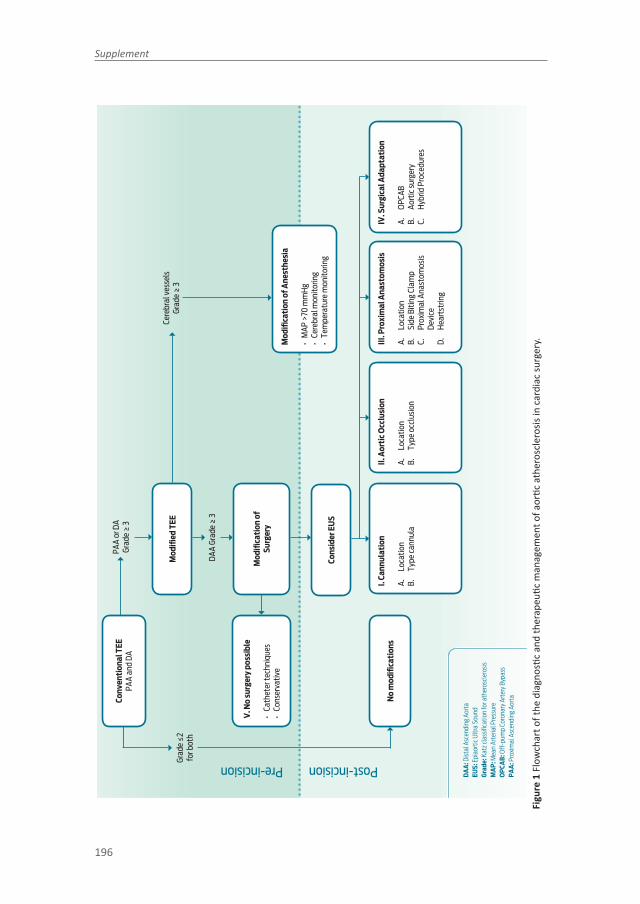
Supplement
196
No
mod
ifi ca
tion
s
V. N
o su
rger
y po
ssib
le
• Ca
thet
er te
chni
ques
• Co
nser
vativ
e
Mod
ifi ed
TEE
Mod
ifi ca
tion
of A
nest
hesi
a
• M
AP >
70 m
mH
g•
Cere
bral
mon
itorin
g•
Tem
pera
ture
mon
itorin
g
I. Ca
nnul
atio
n
A.
Loca
tion
B.
Type
cann
ula
II. A
orti
c O
cclu
sion
A.
Loca
tion
B.
Type
occ
lusi
on
III. P
roxi
mal
Ana
stom
osis
A.
Loca
tion
B.
Side
Biti
ng C
lam
pC.
Pr
oxim
al A
nast
omos
is
Devi
ceD.
H
eart
strin
g
IV. S
urgi
cal A
dapt
atio
n
A.
OPC
ABB.
Ao
rtic
surg
ery
C.
Hyb
rid P
roce
dure
s
Mod
ifi ca
tion
of
Surg
ery
Cons
ider
EU
S
PAA
or D
AGr
ade
≥ 3
Cere
bral
ves
sels
Grad
e ≥
3
DAA
Grad
e ≥
3
Grad
e ≤2
for b
oth
Conv
enti
onal
TEE
PA
A an
d DA
Post-incision Pre-incision
DA
A: D
istal
Asc
endi
ng A
orta
EUS:
Epi
aort
ic U
ltra
Soun
dG
rade
: Kat
z cla
ssifi
catio
n fo
r ath
eros
cler
osis
MA
P: M
ean
Arte
rial P
ress
ure
OP
CAB
: Off
-pum
p Co
rona
ry A
rter
y By
pass
PA
A: P
roxi
mal
Asc
endi
ng A
orta
Figu
re 1
Flo
wch
art o
f the
dia
gnos
tic a
nd th
erap
eutic
man
agem
ent o
f aor
tic a
ther
oscl
eros
is in
car
diac
surg
ery.
197
Supplement
SURGICAL MODIFICATIONS
Presence of severe atherosclerosis of the thoracic aorta should raise several ques-tions regarding the optimal perioperative management. We summarized the possible modifications of the surgical and anesthesiological management if atherosclerosis of the aorta is present. These changes were depicted in the lower part of figure 1, and can be categorized in the following groups:I. CannulationII. Aortic OcclusionIII. Proximal AnastomosisIV. Surgical Adaptation V. No surgery possible
The figure aimed to provide a simple and comprehensive overview of the possible changes in the surgical management. In the respective sections below we provide further information for each subject with an overview of the prevailing literature.
I. CANNULATION
The DAA is typically preferred for cannulation since this is most easily accessible. A different location may be preferable in the presence of atherosclerosis or an-eurysmal widening of the ascending aorta or aortic arch.5 We will briefly describe the advantages and disadvantages of each alternative, which were also elaborately described in recent guidelines.5 The eventual strategy should be weighted in each separate case, based on the severity and extent of aortic atherosclerosis of the ascending aorta, aortic arch and main cerebral vessels, type of cerebral protection, anatomical considerations and surgeon’s capabilities.
A. LOCATION
In many cases of localized atherosclerosis, simply moving the cannula away from atherosclerotic areas may suffice. Indeed, a different cannulation site is the most frequent change in the surgical technique if imaging shows atherosclerosis of the ascending aorta.6 Multiple non-randomized studies have suggested that the inci-dence of stroke can be reduced by avoiding manipulation of aortic atheroma during cannulation. A randomized study which compared a strategy with- and without perioperative screening with epiaortic ultrasound did however not find a difference

Supplement
198
in perioperative embolization or post-operative neurological complications, despite a significant change in the positioning of cannulation.6
Femoral In patients with atherosclerosis of the ascending aorta, femoral cannulation has been a much used alternative to central cannulation.7 The aim is to prevent stroke through the prevention of plaque mobilization in the ascending aorta. Several retrospective studies have questioned its use however, because of concerns that this technique may actually increase the incidence stroke because of flow reversal and the possible mobilization of the usually even more atherosclerotic descending aorta.8 Although a more recent study did not confirm this supposed association between femoral cannulation and increased stroke or mortality, central cannulation may be preferable in the sclerotic aorta.7 Femoral cannulation has an important place for individual patients, for example in surgery for type A aortic dissection.
Axillary or subclavian Reports on the above mentioned increased incidence of stroke after femoral cannulation led to an increasing use of axillary or innominate artery cannulation.7,9,10 There are still some controversies regarding the potential lower risk with axillary or subclavian cannulation compared to femoral or direct aortic cannulation.7–9,11 According to recent guidelines on the surgical management of aortic valve and ascending aortic disease, axillary cannulation should be considered in patients with a calcified or porcelain aorta.5
B. TYPE OF CANNULA
The type of cannula is important since it directs the high flow during extracorpo-real circulation into the aorta.12,13 Typically, aortic cannulation is performed with a straight cannula. After an angulated introduction in the anterior wall, the jet is directed at the posterior wall of the ascending aorta, which has been suggested to cause a “sand-blasting effect” which may release atherogenic emboli.14 Also changes in aortic rotational flow have been documented during cardiopulmonary bypass (CPB) and could explain the erosive effect of the high-velocity jet from aortic cannula.15,16
Therefore, other types of cannulas were designed, aimed to reduce the stress on the aortic wall. Bent-tip catheters should prevent a flow directed at the aortic wall,

199
Supplement
however they create a potential sand-blasting effect into de aortic arch and cerebral vessels.17 The incidence of stroke was lower in patients with aortic cannulation us-ing a bent-tip catheter (0.9 vs. 1.8%); the authors did however not corrected for differences in baseline risk between both groups.17 Another type of aortic catheter (Select 3D® arterial cannula; Medtronic Inc., Minneapolis, MN) produces four jets with a reduced outflow velocity.12,18,19 The tip is also curved, which together should reduce the shear stress on the aortic wall. No studies yet studied the impact of this catheter on the prevention of postoperative complications.
II. AORTIC OCCLUSION
The default procedure for aortic occlusion is the placement of an aortic crossclamp. Other possibilities are endoballoon occlusion and hypothermic circulatory arrest.
A. LOCATION
During CPB, aortic cross clamping is a normal procedure to allow cardioplegic solu-tion to enter the coronary arteries to arrest the heart. The placement and release of a clamp are however associated with increased transcranial Doppler detected emboli, and may cause overt stroke. This was nicely demonstrated in a post-mortem study, which associated the positioning of a cross clamp with stroke.20 They con-cluded that before placement of a clamp on the aorta, echo evaluation is mandatory in order to reduce adverse events.
B. TYPE OF OCCLUSION
Endoballoon occlusion Placement and release of a traumatic aortic cross-clamp has been associated with an increased incidence of cerebral emboli. Alternatively, it has been hypothesized that inflation of a balloon in the aorta is less traumatic and may thus reduce cerebral embolization. In a randomized comparison of CPB with endoclamping or transthoracic clamping in patients who underwent minimal invasive mitral valve replacement, the incidence of transcranial Doppler (TCD)-measured solid emboli in the middle cerebral arteries was lower in the endovas-cular group when applying and releasing the clamp.21 However, patients with aortic atherosclerosis were excluded from this study, while it would be interesting to know if the use of an endoballoon also reduces cerebral embolization when applied in a diseased aorta. It is unlikely that in a diseased aorta, a sliding aortic balloon is

Supplement
200
a stroke risk-reducing procedure in robotic or minimally invasive cardiac surgery. Reports have described its use in severely atheromatous aortas where the risks of dislodgment after external crossclamping or intermittent fibrillatory arrest were deemed too high.22 Severe disease of the ascending aorta is however considered a relative contra-indication for this endoaortic balloon. Placement of an endoaortic crossclamp requires cannulation of the femoral artery or ascending aorta, both of which may cause dislodgement of atheroma itself.
Hypothermic Circulatory Arrest and Cerebral Perfusion In the severely diseased aorta, replacement of the ascending aorta might be considered as a last resort treatment necessitating profound hypothermic circulatory arrest (PHCA) in order to perform the procedure. In a review by Lee and colleagues the use of antegrade- and retrograde cerebral perfusion (ACP and RCP respectively) and profound hypother-mic circulatory arrest were discussed.23 Although many non-randomized studies have reported outcomes associated with ACP, RCP and PHCA, these are difficult to compare due to heterogeneity of study populations, and of techniques for cerebral protection and other aspects of the surgical procedure. The stroke rate after as-cending aortic and aortic arch surgery using PHCA may be as low as 3.1%, with an increased risk if PHCA time exceeded 40 minutes (13.1%). One study randomized patients to PHCA or ACP, and showed a higher jugular venous oxygen saturation in the ACP group, but no differences in neurological outcomes.24 Three studies compared ACP and RCP,24–26 none of which showed a difference in the mortality or stroke rate, although ACP was associated with improved cerebral perfusion,25 and a lower incidence of (transient) neurologic deficits.26 Since embolic complications rather than metabolic insufficiency causes stroke, it has been hypothesized that the beneficial hemodynamic properties of ACP are counterbalanced by a higher risk of cerebral emboli associated with the introduction of catheters in the arch vessels. As-sessing these arteries before manipulation is a possible solution in order to reduce embolic load to the brain.23
III. PROXIMAL ANASTOMOSIS
Proximal anastomosis and “no-touch surgery” Total arterial or T-graft revasculariza-tion may obviate the need for a proximal anastomosis, coronary revascularization can thus be performed during no-touch OPCAB. Although theoretically this would

201
Supplement
reduce aortic manipulation and thus may prevent dislodgement of aortic atheroma, limited randomized studies have been performed to study this hypothesis. In a small prospective OPCAB study, it was determined whether a clampless facilitat-ing device (CFD) to perform proximal aortocoronary anastomoses would result in a lower incidence of cerebral embolic events compared with a partial clamping strategy during OPCAB. After epiaortic ultrasound confirmed the mild aortic disease (grades I and II, i.e. plaques up till 3 mm), 57 patients were randomly assigned to have proximal anastomoses using a partial-occluding clamp (CL, n = 28) or a CFD [Heartstring, n =29]. Solid and gaseous emboli in the middle cerebral arteries were detected using TCD. The mean number of proximal anastomoses was similar between groups 1.93±0.72 (CL) and 1.72 ± 0.70 (HS) (P = 0.28). The mean number of gaseous plus solid emboli was greater in the CL group than the HS group (90.0±64.0 vs. 50.8±36.6, P=0.01). Emboli were fewest in patients undergoing HS anastomoses using the suction device. The number of intraoperative cerebral emboli was propor-tional to the number of proximal anastomoses in the CFD groups, but independent of the number of proximal anastomoses in the CL groups. Among patients with a low burden of aortic atherosclerosis, partial clamping of the ascending aorta dur-ing OPCAB was associated with more cerebral embolic events compared with an anastomosis with a CFD. Extrapolation of these results to daily practice is however limited since patients with plaques > 3 mm were excluded.27 In a large cohort study of 4314 patients, both OPCAB and aortic no-touch technique reduce stroke after CABG. They evaluated the impact of partial aortic clamping versus a no-touch tech-nique using either the HEARTSTRING system or total arterial revascularization on the incidence of stroke. OPCAB was superior with regard to risk-adjusted outcomes, but only if no touch of the aorta was applied with a superiority of the CFD group. However, no information was provided about aortic disease as one of the major risk factor of perioperative stroke.28 In a retrospective study including 160 patients aged >75 years who underwent OPCAB a no-touch strategy was compared to the use of a side-biting clamp; the latter group had more neurological events or mortal-ity (12.3 vs. 2.9%, p = 0.035), which remained significant after correction for other variables.29 A second study similarly identified partial aortic clamping as a risk factor for stroke after OPCAB.

Supplement
202
IV. SURGICAL ADAPTATION
A. OFF-PUMP SURGERY
Coronary artery bypass surgery on a beating heart precludes multiple of the previ-ously mentioned risk–factors for the development of postoperative neurological complications, i.e. aortic cannulation, aortic cross clamping, and antegrade cardio-plegia, as well as the possible cerebral inflammatory response to CPB.30,31 In earlier randomized trials OPCAB was associated with reduced short-term post-operative cognitive dysfunction and a lower rate of bleeding, transfusion and respiratory complications, an increased risk of early revascularisation, and no effect on stroke, myocardial infarction, renal-failure or death.32–34 The largest randomized study until date, the Randomized On/Off Bypass (ROOBY) trial, showed however no benefit of OPCAB with an increased incidence of the primary composite endpoint (9.9% vs. 7.4%, p = 0.04) and cardiac mortality (2.7 vs. 1.3%, p = 0.03); also, graft patency was reduced in the OPCAB group (82.6 vs. 87.8%, p < 0.01).35 A more recent meta-analysis showed that OPCAB was associated with a 30% (95% CI: 1 – 51%) relative risk reduction for stroke, in the absence of a difference in 30-day mortality or myo-cardial infarction.36 This is an important finding, as the incidence of stroke is too low to expect single trials to show a difference in the stroke rate.36 Given an absolute difference in post-operative stroke of 0.7% (2.1 minus 1.4%), the number needed to treat with OPCAB to prevent one stroke is approximately 143.
A flaw of these trials however was that the individual preoperative stroke risk was not used to select patients at a higher risk of developing stroke, e.g. with an in-creased risk based on patient characteristics,37 or with preoperative visualization of the aorta to identify patients with aortic atherosclerosis. The reported trials did also not stratify their results for presence and absence of aortic atherosclerosis in a post-hoc analysis, which would explain the limited effect on stroke reduction. Most studies, however, have not differentiated between clampless and off-pump tech-niques. Avoiding partial aortic clamping during OPCAB provided superior neurologic outcome and identified partial aortic clamping as the only independent predictor of stroke.29,38 In a comparison with percutaneous interventions, OPCAB with no touch treatment showed the same low incidence of stroke of 0,8%, compared to on-pump CABG of 2,2%.39 Also, in a recent meta-analysis comparing the risk of stroke in

203
Supplement
CABG versus OPCAB, off-pump surgery does not seem to reduce the risk of stroke. A pooled analysis of more than 4000 patients from 11 randomized clinical trails did not reveal any benefit of avoiding CPB on stroke risk. A possible explanation is that most OPCAB patients still undergo aortic manipulation because the side biting clamp is used for proximal anastomoses. This seems to be confirmed in the same meta-analysis that compares also bypass surgery with and without manipulation of the ascending aorta. It reports a spectacular reduction in stroke risk from 1.3 percent to 0.3 percent.40
B. AORTIC SURGERY
Replacement of the ascending aorta is recommended in the aneurysmal aorta (>45mm), but guidelines do not give advice with regards to the optimal treatment of the severely sclerotic aorta. It can however be considered to prevent perioperative stroke, but also to prevent late strokes caused by mobile plaques. In a study by Zin-gone et al., a major change to the surgical procedure was deemed necessary after EAU evaluation in 152 of 1927 (7.9%) of patients who underwent cardiothoracic surgery, of whom 36 received a replacement of the ascending aorta.41 Stroke was observed in one patient, two patients died before hospital discharge. Another study similarly reported the outcomes of 36 patients with AA-replacement because of in-traoperative identification of AA atherosclerosis; one patient developed stroke and died during hospitalization.42 In a third study however, both the stroke rate (3/17) and mortality (4/17) were substantial.43 Furthermore, aortic arch endarterectomy has been shown to be associated with an increased risk of post-operative stroke (OR 3.6, p = 0.001) in an already high incidence of stroke among patients with aortic atheroma (15.3%).44
C. HYBRID PROCEDURES
Alternatively, a hybrid procedure combining surgery and percutaneous intervention may be useful in selected patients, e.g. with surgical revascularization using the left internal thoracic artery and subsequent percutaneous complete revascularization. However, passing the diseased aorta during cardiac catheterization carries a (lim-ited) risk itself for cerebral complications.45

Supplement
204
V. NO SURGERY POSSIBLE
The first aim of any treatment is “Primum non nocere”. In very unusual cases, the newly acquired information on the severity of aortic atherosclerosis may result in a reconsideration of the surgical procedure itself. After consideration of the previ-ously addressed aspects of surgery, the risk of perioperative complications may be deemed to high to proceed. Alternative treatments should be considered, e.g. a percutaneous coronary intervention for coronary artery disease, or percutaneous valve replacement for aortic stenosis.
MODIFICATION OF ANESTHESIA
A. PERIOPERATIVE BLOOD PRESSURE
Fundamental monitors for cardiac surgery include invasive hemodynamic monitor-ing of blood pressure and central venous pressure. The cause of perioperative brain injury in the cardiac surgery population is, at least in part, secondary to cerebral em-bolism, but hypoperfusion and subsequent ischemia – reperfusion injury also play a role.46 Intraoperative hemodynamic variability can result in insufficient cerebral per-fusion.47 And patients with intracranial arterial stenosis are particularly susceptible to variations in cerebral perfusion and are at increased risk for perioperative strokes. In patients with symptomatic cerebrovascular disease and pre-existing ischaemic symptoms, a stroke rate of up to 13% has been reported.48 Several studies have identified prolonged hypotension as a risk factor for adverse cerebral outcomes.49,50 Increased blood pressure in order to maintain adequate cerebral perfusion pres-sure should be implemented in case of cerebrovascular disease, unless cerebral monitoring depicts otherwise.47 A threshold mean blood pressure of approximately 70 mmHg is necessary since studies in awake healthy subjects indicate that this is the lower limit of autoregulation, which is higher than the mostly recommended 50 mmHg from current textbooks.51 In a randomized trial, Gold et al. showed that a higher mean arterial pressure (MAP; 80–100 mmHg) during bypass improves outcomes in long- and short-term follow-up when compared to low MAP (50–60 mmHg).52

205
Supplement
B. CEREBRAL MONITORING
Different types of cerebral monitoring devices such as bispectral index (BIS) and ce-rebral oximetry using near-infrared spectroscopy (NIRS) have been developed over the last decades in order to reduce cerebral morbidity related to the perioperative period. The BIS remains a mainstay of brain monitoring in cardiac surgery, although it is not directly related to improving neurologic outcome. Independent of its direct application to awareness, the information provided with BIS, when integrated with other sources of information (such as blood pressure), can be used to help balance the anaesthetic requirements with vasoactive therapies. It can be valuable in titrat-ing the balance of very complex intraoperative hemodynamic and anaesthetic goals.
Cerebral oximetry using near-infrared spectroscopy has been available for more than a decade. In much the same way that a pulse oximeter can determine periph-eral arterial saturation, cerebral oximetry can similarly determine the saturation of the cerebral tissue; in essence, a compilation of both venous and arterial blood within the brain. One of the first studies using a NIRS treatment algorithm showed not an improvement in overall outcome but also a trend towards stroke reduction in patients who had the interventional algorithm.53,54
Ultrasound guided aortic instrumentation (epiaortic scanning) may decrease atheroembolic load and risk of stroke. Unrecognized sources of microgaseous em-boli, including air entrainment from surgical purse string sutures and perfusionist interventions, can be identified and reduced by transcranial Doppler monitoring. Cerebral hypoperfusion from unrecognized cerebral venous obstruction, inadequate mean arterial pressure, or from hypocapnic cerebral alkalosis can be identified by multi-modality neuromonitoring using regional cerebral oxygen saturation and TCD. In a large, nonrandomized series of 1698 cardiac surgical patients reported by Goldman and colleagues, a significant reduction in perioperative stroke rate, from 2.0% to 1.0%, was observed in patients in whom rSO2 cerebral oximetry was used to optimize and maintain intraoperative cerebral oxygenation versus an untreated comparator group of 2077 similar patients operated on in the immediately preced-ing 18-month interval.55 Overall patient outcomes can be improved, and hospital length of stay shortened, by applied neuromonitoring techniques, especially in the severe aortic disease patients.56

Supplement
206
C. TEMPERATURE MONITORING
The benefits of hypothermia are related to its effect on lowering cerebral metabolic rate, but, equally important, it has significant effects on the inhibition of excita-tory neurotransmitter release as well as other protective cellular mechanisms.57 Although mild-to-moderate hypothermia during CPB has not been shown to have a sustained benefit on post-operative neurologic outcome, there are data that outline the detrimental effect of hyperthermia in the perioperative period. For example, in the cardiac surgery population, maximum postoperative temperature has been associated with greater postoperative cognitive dysfunction at 6 weeks. Modern strategies for perioperative temperature management are more directed towards preventing cerebral hyperthermia and its physiologic consequences.58,59
LIMITATIONS
The evidence for the efficacy of the individual changes in the surgical management is mostly limited to observational studies of limited size, except for OPCAB, which has been intensively compared to revascularization during CPB in multiple random-ized trials. The absence of evidence should however not be a reason to ignore the well-known risks. We hope that this systematic approach to the diagnosis and man-agement of aortic atherosclerosis can result in a reduction of postoperative cerebral complications. Future studies should focus on the effect of the implementation of this protocol on chances in the surgical management and on patient outcomes.
CONCLUSIONS
Neurological complications associated with cardiac surgery have been a primary concern. Many risk factors for stroke after cardiac surgery are known, of which atherosclerosis of the ascending aorta is of major interest. The stroke risk during cardiac surgery depends on the location and extent of disease, and on the amount of aortic manipulation. Routine use of intraoperative monitoring with (modified) transesophageal and/or epiaortic ultrasound should be applied in all cardiac sur-gery procedures in order to identify the patients at risk. The acquired diagnostic information should be used to tailor surgical strategies. We propose a systematic approach to identify aortic atherosclerosis, and consider changes in the surgical

207
Supplement
perioperative management. These include aortic cannulation, aortic occlusion, proximal anastomosis, and surgical adaptation. Also modifications of anesthesia should be considered. Finally, in unusual cases a reappraisal of surgery itself may be appropriate, and percutaneous treatments should be considered. Although none of the described changes in the surgical management have unequivocal evidence of its efficacy, we hope that a holistic approach to this challenge will result in a reduction of stroke and other embolic related.
Figure 2. Example of a vascular report in the preoperative screening for possible aortic valve replacement in a patient with severe aortic stenosis. (Modified) transesophageal echocardiography showed extensive calcifications of the complete thoracic aorta, it was therefore concluded to perform a transapical aortic valve replacement to minimize manipulation of the aorta.

Supplement
208
REFERENCES 1. Royse C, Royse A, Blake D, Grigg L. Screening the thoracic aorta for atheroma: a
comparison of manual palpation, transesophageal and epiaortic ultrasonography. Ann Thorac Cardiovasc Surg. 1998;4(6):347-350. http://www.ncbi.nlm.nih.gov/pubmed/9914464. Accessed September 13, 2013.
2. van Zaane B, Nierich AP, Buhre WF, Brandon Bravo Bruinsma GJ, Moons KGM. Resolv-ing the blind spot of transoesophageal echocardiography: a new diagnostic device for visualizing the ascending aorta in cardiac surgery. Br J Anaesth. 2007;98(4):434-441. doi:10.1093/bja/aem009.
3. Nierich AP, van Zaane B, Buhre WF, Coddens J, Spanjersberg AJ, Moons KGM. Visualiza-tion of the distal ascending aorta with A-Mode transesophageal echocardiography. J Cardiothorac Vasc Anesth. 2008;22(5):766-773. doi:10.1053/j.jvca.2008.07.017.
4. van Zaane B, Nierich AP, Brandon Bravo Bruinsma GJ, et al. Diagnostic accuracy of modified transoesophageal echocardiography for pre-incision assessment of aortic atherosclerosis in cardiac surgery patients. Br J Anaesth. 2010;105(2):131-138. doi:10.1093/bja/aeq122.
5. Svensson LG, Adams DH, Bonow RO, et al. Aortic valve and ascending aorta guidelines for management and quality measures. Ann Thorac Surg. 2013;95(6 Suppl):S1-66. doi:10.1016/j.athoracsur.2013.01.083.
6. Djaiani G, Ali M, Borger M a, et al. Epiaortic scanning modifies planned intraoperative surgical management but not cerebral embolic load during coronary artery bypass sur-gery. Anesth Analg. 2008;106(6):1611-1618. doi:10.1213/ane.0b013e318172b044.
7. Di Eusanio M, Pantaleo A, Petridis FD, et al. Impact of different cannulation strate-gies on in-hospital outcomes of aortic arch surgery: a propensity-score analysis. Ann Thorac Surg. 2013;96(5):1656-1663. doi:10.1016/j.athoracsur.2013.06.081.
8. Modi P, Chitwood WR. Retrograde femoral arterial perfusion and stroke risk during minimally invasive mitral valve surgery: is there cause for concern? Ann Cardiothorac Surg. 2013;2(6):E1. doi:10.3978/j.issn.2225-319X.2013.11.13.
9. Svensson LG, Blackstone EH, Rajeswaran J, et al. Does the arterial cannulation site for circulatory arrest influence stroke risk? Ann Thorac Surg. 2004;78(4):1274-84-84. doi:10.1016/j.athoracsur.2004.04.063.
10. Etz CD, Plestis K a, Kari F a, et al. Axillary cannulation significantly improves survival and neurologic outcome after atherosclerotic aneurysm repair of the aortic root and ascending aorta. Ann Thorac Surg. 2008;86(2):441-6-7. doi:10.1016/j.athorac-sur.2008.02.083.
11. Sabik JF, Lytle BW, McCarthy PM, Cosgrove DM. Axillary artery: an alternative site of arterial cannulation for patients with extensive aortic and peripheral vascular disease. J Thorac Cardiovasc Surg. 1995;109(5):885-90-1. http://www.ncbi.nlm.nih.gov/pubmed/7739248. Accessed June 17, 2014.
12. Minakawa M, Fukuda I, Yamazaki J, Fukui K, Yanaoka H, Inamura T. Effect of cannula shape on aortic wall and flow turbulence: hydrodynamic study during extracorporeal circulation in mock thoracic aorta. Artif Organs. 2007;31(12):880-886. doi:10.1111/j.1525-1594.2007.00481.x.
13. Fukuda I, Fujimori S, Daitoku K, Yanaoka H, Inamura T. Flow velocity and turbulence in the transverse aorta of a proximally directed aortic cannula: hydrodynamic study in a transparent model. Ann Thorac Surg. 2009;87(6):1866-1871. doi:10.1016/j.athorac-sur.2009.03.054.
14. Swaminathan M, Grocott HP, Mackensen GB, Podgoreanu M V, Glower DD, Mathew JP. The ‘sandblasting’ effect of aortic cannula on arch atheroma during

209
Supplement
cardiopulmonary bypass. Anesth Analg. 2007;104(6):1350-1351. doi:10.1213/01.ane.0000264090.24756.08.
15. Koh TW, Parker KH, Kon M, Pepper JR. Changes in aortic rotational flow during car-diopulmonary bypass studied by transesophageal echocardiography and magnetic resonance velocity imaging: a potential mechanism for atheroembolism during car-diopulmonary bypass. Heart Vessels. 2001;16(1):1-8.
16. White JK, Jagannath A, Titus J, Yoneyama R, Madsen J, Agnihotri AK. Funnel-tipped aortic cannula for reduction of atheroemboli. Ann Thorac Surg. 2009;88(2):551-557. doi:10.1016/j.athoracsur.2009.04.108.
17. Albert AA, Beller CJ, Arnrich B, et al. Is there any impact of the shape of aortic end-hole cannula on stroke occurrence? clinical evaluation of straight and bent-tip aortic cannu-lae. Perfusion. 2002;17(6):451-456. http://www.ncbi.nlm.nih.gov/pubmed/12470037. Accessed June 5, 2014.
18. Grooters RK, Ver Steeg DA, Stewart MJ, Thieman KC, Schneider RF. Echocardiographic comparison of the standard end-hole cannula, the soft-flow cannula, and the disper-sion cannula during perfusion into the aortic arch. Ann Thorac Surg. 2003;75(6):1919-1923. http://www.ncbi.nlm.nih.gov/pubmed/12822636. Accessed May 13, 2013.
19. Lata AL, Hammon JW, Deal DD, Stump D a, Kincaid EH, Kon ND. Cannula design reduces particulate and gaseous emboli during cardiopulmonary bypass for coronary revascu-larization. Perfusion. 2011;26(3):239-244. doi:10.1177/0267659110394905.
20. Boivie P, Hansson M, Engström KG. Intraluminal aortic manipulation by means of intra-aortic filter, cannulation, and external clamp maneuvers evaluated versus dislodged embolic material. J Thorac Cardiovasc Surg. 2006;131(2):283-289. doi:10.1016/j.jtcvs.2005.10.015.
21. Maselli D, Pizio R, Borelli G, Musumeci F. Endovascular balloon versus transtho-racic aortic clamping for minimally invasive mitral valve surgery: impact on cerebral microemboli. Interact Cardiovasc Thorac Surg. 2006;5(2):183-186. doi:10.1510/icvts.2005.123372.
22. Liddicoat JR, Doty JR, Scott R. Management of the Atherosclerotic Ascending Aorta With Endoartic Occlusion. 1998;4618(98):1133-1135.
23. Lee T, Safi HJ, Estrera AL. Cerebral Perfusion in Aortic Arch Surgery. 2011;38(6). 24. Harrington DK, Walker a S, Kaukuntla H, et al. Selective antegrade cerebral perfusion
attenuates brain metabolic deficit in aortic arch surgery: a prospective randomized trial. Circulation. 2004;110(11 Suppl 1):II231-6. doi:10.1161/01.CIR.0000138945.78346.9c.
25. Tanoue Y, Tominaga R, Ochiai Y, Fukae K. Comparative Study of Retrograde and Selec-tive. 1999:3-6.
26. Okita Y, Minatoya K, Tagusari O, Ando M, Nagatsuka K, Kitamura S. Prospective comparative study of brain protection in total aortic arch replacement: deep hypo-thermic circulatory arrest with retrograde cerebral perfusion or selective antegrade cerebral perfusion. Ann Thorac Surg. 2001;72(1):72-79. http://www.ncbi.nlm.nih.gov/pubmed/11465234.
27. El Zayat H, Puskas JD, Hwang S, et al. Avoiding the clamp during off-pump coronary artery bypass reduces cerebral embolic events: results of a prospective randomized trial. Interact Cardiovasc Thorac Surg. 2012;14(1):12-16. doi:10.1093/icvts/ivr004.
28. Emmert MY, Seifert B, Wilhelm M, Grünenfelder J, Falk V, Salzberg SP. Aortic no-touch technique makes the difference in off-pump coronary artery bypass grafting. J Thorac Cardiovasc Surg. 2011;142(6):1499-1506. doi:10.1016/j.jtcvs.2011.04.031.

Supplement
210
29. Lev-Ran O, Loberman D, Matsa M, et al. Reduced strokes in the elderly: the benefits of untouched aorta off-pump coronary surgery. Ann Thorac Surg. 2004;77(1):102-107. doi:10.1016/S0003-4975(03)01334-1.
30. Jansen EW, Borst C, Lahpor JR, et al. Coronary artery bypass grafting without car-diopulmonary bypass using the octopus method: results in the first one hundred patients. J Thorac Cardiovasc Surg. 1998;116(1):60-67. http://www.ncbi.nlm.nih.gov/pubmed/9671898. Accessed February 14, 2015.
31. Nussmeier NA. A review of risk factors for adverse neurologic outcome after car-diac surgery. J Extra Corpor Technol. 2002;34(1):4-10. http://www.ncbi.nlm.nih.gov/pubmed/11911628. Accessed February 14, 2015.
32. Van Dijk D. Cognitive Outcome After Off-Pump and On-Pump Coronary Artery Bypass Graft Surgery: A Randomized Trial. JAMA J Am Med Assoc. 2002;287(11):1405-1412. doi:10.1001/jama.287.11.1405.
33. van Dijk D, Spoor M, Hijman R, et al. Cognitive and cardiac outcomes 5 years after off-pump vs on-pump coronary artery bypass graft surgery. JAMA. 2007;297(7):701-708. doi:10.1001/jama.297.7.701.
34. Lamy A, Devereaux PJ, Prabhakaran D, et al. Off-pump or on-pump coronary-artery bypass grafting at 30 days. N Engl J Med. 2012;366(16):1489-1497. doi:10.1056/NEJMoa1200388.
35. Shroyer AL, Grover FL, Hattler B, et al. On-pump versus off-pump coronary-artery by-pass surgery. N Engl J Med. 2009;361(19):1827-1837. doi:10.1056/NEJMoa0902905.
36. Afilalo J, Rasti M, Ohayon SM, Shimony A, Eisenberg MJ. Off-pump vs. on-pump coronary artery bypass surgery: an updated meta-analysis and meta-regression of randomized trials. Eur Heart J. 2012;33:1257-1267. doi:10.1093/eurheartj/ehr307.
37. Katz NM, Chase GA. Risks of cardiac operations for elderly patients: reduction of the age factor. Ann Thorac Surg. 1997;63(5):1309-1314. http://www.ncbi.nlm.nih.gov/pubmed/9146320. Accessed January 31, 2012.
38. Lev-Ran O, Braunstein R, Sharony R, et al. No-touch aorta off-pump coronary surgery: the effect on stroke. J Thorac Cardiovasc Surg. 2005;129(2):307-313. doi:10.1016/j.jtcvs.2004.06.013.
39. Halbersma WB, Arrigoni SC, Mecozzi G, et al. Four-year outcome of OPCAB no-touch with total arterial Y-graft: making the best treatment a daily practice. Ann Thorac Surg. 2009;88(3):796-801. doi:10.1016/j.athoracsur.2009.04.104.
40. Edelman JJ, Yan TD, Bannon PG, Wilson MK, Vallely MP. Coronary artery bypass grafting with and without manipulation of the ascending aorta--a meta-analysis. Heart Lung Circ. 2011;20(5):318-324. doi:10.1016/j.hlc.2011.02.003.
41. Zingone B, Rauber E, Gatti G, et al. Diagnosis and management of severe atherosclerosis of the ascending aorta and aortic arch during cardiac surgery: focus on aortic replace-ment. Eur J Cardiothorac Surg. 2007;31(6):990-997. doi:10.1016/j.ejcts.2007.02.017.
42. Gullu AU, Okten EM, Akay MH, et al. Replacement of the ascending aorta for severe atherosclerosis during coronary artery bypass surgery. J Card Surg. 2012;27(5):538-542. doi:10.1111/j.1540-8191.2012.01510.x.
43. King RC, Kanithanon RC, Shockey KS, Spotnitz WD, Tribble CG, Kron IL. Replacing the atherosclerotic ascending aorta is a high-risk procedure. Ann Thorac Surg. 1998;66(2):396-401. http://www.ncbi.nlm.nih.gov/pubmed/9725375.
44. Stern a, Tunick P a, Culliford a T, et al. Protruding aortic arch atheromas: risk of stroke during heart surgery with and without aortic arch endarterectomy. Am Heart J. 1999;138(4 Pt 1):746-752. http://www.ncbi.nlm.nih.gov/pubmed/10502222.

211
Supplement
45. Hamon M, Gomes S, Clergeau M-R, Fradin S, Morello R, Hamon M. Risk of acute brain injury related to cerebral microembolism during cardiac catheterization performed by right upper limb arterial access. Stroke. 2007;38(7):2176-2179. doi:10.1161/STROKEAHA.107.482414.
46. Hogue CW, Palin C a, Arrowsmith JE. Cardiopulmonary bypass management and neurologic outcomes: an evidence-based appraisal of current practices. Anesth Analg. 2006;103(1):21-37. doi:10.1213/01.ANE.0000220035.82989.79.
47. Laiwalla AN, Ooi YC, Van De Wiele B, et al. Rigorous anaesthesia management protocol for patients with intracranial arterial stenosis: a prospective controlled-cohort study. BMJ Open. 2016;6(1):e009727. doi:10.1136/bmjopen-2015-009727.
48. Nakamura M, Okamoto F, Nakanishi K, et al. Does intensive management of cerebral hemodynamics and atheromatous aorta reduce stroke after coronary artery surgery? Ann Thorac Surg. 2008;85(2):513-519. doi:10.1016/j.athoracsur.2007.08.056.
49. Roach G, Kanchuger M. Adverse cerebral outcomes after coronary bypass surgery. Engl J. 1996;335:1857-1863. http://www.nejm.org/doi/full/10.1056/NEJM199612193352501. Accessed April 10, 2012.
50. Selim M. Perioperative stroke. N Engl J Med. 2007;356(7):706-713. doi:10.1056/NEJMra062668.
51. Bijker JB, Gelb AW. Review article: the role of hypotension in perioperative stroke. Can J Anaesth. 2013;60(2):159-167. doi:10.1007/s12630-012-9857-7.
52. Gold JP, Charlson ME, Williams-Russo P, et al. Improvement of outcomes after coro-nary artery bypass. A randomized trial comparing intraoperative high versus low mean arterial pressure. J Thorac Cardiovasc Surg. 1995;110(5):1302-11-4. http://www.ncbi.nlm.nih.gov/pubmed/7475182. Accessed February 15, 2015.
53. Murkin JM. Is it better to shine a light, or rather to curse the darkness? Cerebral near-infrared spectroscopy and cardiac surgery. Eur J Cardiothorac Surg. 2013;43(6):1081-1083. doi:10.1093/ejcts/ezt186.
54. Denault A, Deschamps A, Murkin JM. A proposed algorithm for the intraoperative use of cerebral near-infrared spectroscopy. Semin Cardiothorac Vasc Anesth. 2007;11(4):274-281. doi:10.1177/1089253207311685.
55. Goldman S, Sutter F, Ferdinand F, Trace C. Optimizing intraoperative cerebral oxygen delivery using noninvasive cerebral oximetry decreases the incidence of stroke for cardiac surgical patients. Heart Surg Forum. 2004;7(5):E376-81. doi:10.1532/HSF98.20041062.
56. Murkin JM. Applied neuromonitoring and improving CNS outcomes. Semin Cardiothorac Vasc Anesth. 2005;9(2):139-142. http://www.ncbi.nlm.nih.gov/pubmed/15920638. Accessed February 14, 2015.
57. Fedorow C, Grocott HP. Cerebral monitoring to optimize outcomes after cardiac surgery. Curr Opin Anaesthesiol. 2010;23(1):89-94. doi:10.1097/ACO.0b013e3283346d10.
58. Shann KG, Likosky DS, Murkin JM, et al. An evidence-based review of the practice of cardiopulmonary bypass in adults: a focus on neurologic injury, glycemic control, hemo-dilution, and the inflammatory response. J Thorac Cardiovasc Surg. 2006;132(2):283-290. doi:10.1016/j.jtcvs.2006.03.027.
59. Bechtel A, Huffmyer J. Anesthetic Management for Cardiopulmonary Bypass: Update for 2014. Semin Cardiothorac Vasc Anesth. 2014;18(2):101-116. doi:10.1177/1089253214529607.


Summary


215
Summary
PART I – AORTIC ATHEROSCLEROSIS
During cardiac surgery, manipulation of the thoracic aorta can cause the release of emboli into the systemic circulation. Aortic atherosclerosis has indeed been associ-ated with post-operative stroke and other embolic complications. Atherosclerosis of the distal ascending aorta has the largest risk for post-operative stroke, because surgical procedures including aortic cannulation and placement of a cross-clamp are usually performed on this part of the aorta. Accurate identification of aortic plaques can guide the surgical management to avoid dislodgement of plaques.
Multiple diagnostic tests are available to visualize atherosclerosis of the distal as-cending aorta; these modalities were reviewed in Chapter 2. Computed tomography (CT) has a low sensitivity and negative predictive value, and is thus not very useful for the focused screening for aortic atherosclerosis. Magnetic resonance imaging (MRI) is an accurate test with anatomical and functional information, but is infrequently used. More importantly, both tests lack the possibility of real-time intraoperative visualization. Epiaortic ultrasound (EAU) is considered the gold standard for the intraoperative visualization of atherosclerosis of the distal ascending aorta. Its use has been found to lead to changes in the surgical management, but an improvement of patient outcomes has not yet been established. Manual palpation is frequently applied, but is known to underestimate the atherosclerotic burden and may itself cause the release of emboli. Transesophageal echocardiography (TEE) is used most often for the focused screening for aortic atherosclerosis, as it has the advantages of visualizing the aorta before sternotomy and to provide real-time imaging during surgery. A major limitation however is the poor sensitivity for atherosclerosis of the distal part of the ascending aorta due to the air-filled trachea. A possible solu-tion for this limitation is offered with modified TEE, in which a fluid-filled balloon is positioned in the trachea and left main bronchus thereby allowing for visualization of the distal ascending aorta and its branches is possible. Compared to EAU, in previ-ous studies modified TEE was found to have a good overall diagnostic accuracy (area under curve: 0.89), with a high sensitivity and negative predictive value (95% and 97% respectively) but limited specificity and positive predictive value (79% and 67% respectively). However, good diagnostic accuracy by itself is not going to improve

216
Summary
patient outcomes. This thesis consists of studies investigating next steps, by putting modified TEE to use in practice, to improve patient outcomes.
The aim of modified TEE screening is to improve patient outcomes through a more accurate diagnosis of atherosclerosis of the distal ascending aorta. Therefore, modified TEE should provide information that is not already captured by other tests. In Chapter 3 the added value of modified TEE was studied, compared to patient characteristics and conventional TEE imaging. Modified TEE indeed improved the diagnosis of atherosclerosis of the distal ascending aorta beyond conventional TEE, as indicated by the “net reclassification improvement” and “integrated discrimina-tion improvement”.
In Chapter 4 we studied the prognostic implications of atherosclerosis of the distal ascending aorta (DAA) diagnosed with modified TEE. For this aim we studied the follow-up of patients included in a diagnostic accuracy study, in which the results of modified TEE were blinded during surgery. Atherosclerosis of the DAA was as-sociated with increased one-year and long-term all-cause mortality (hazard ratio [HR]1.82, 95% CI: 1.10 – 3.24). The predictive performance was superior to the results of conventional TEE, but inferior to the prognosis of one-year mortality with the original EuroSCORE. Also, there was no association with short-term mortality or stroke, possibly related to the limited number of patients with these end-points. These results indicate that the findings of modified TEE are of relevance for the prognosis of patients, and that this information is not already available using con-ventional TEE.
The ultimate goal of modified TEE is however not to provide a new invasive tool to predict mortality in the coming years, but to prevent intra-operative complications during surgery. The aim of Chapter 5 was to study if the use of modified TEE was indeed associated with improved patient outcomes. This non-randomized observa-tional study compared 1,391 patients who underwent perioperative modified TEE-with 7,124 patients without modified TEE imaging. In this study, modified TEE was used on indication, i.e. in patients with a higher probability of atherosclerosis based on clinical characteristics and conventional TEE imaging. The EuroSCORE predicted operative mortality was indeed higher in the intervention- compared to the control

217
Summary
group (5.9% vs. 4.0%, p<0.001); the observed 30-day mortality was similar in both groups (2.2% vs. 2.5%, p = 0.55). The use of modified TEE was associated with a lower mortality after multivariable- (relative risk [RR] 0.70, 95% CI: 0.50 – 1.00) and propensity score adjustment (RR 0.67, 95% CI: 0.45 – 0.98). In contrast to our expec-tations, modified TEE was not associated with a lower incidence of post-operative stroke (adjusted RR: 1.01, 95% CI: 0.71 – 1.43). Limitations of this study, besides the observational design, were the absence of a protocol to guide the diagnosis and management of aortic atherosclerosis, and an insufficient registration of the steps between test application and patient outcomes.
To overcome these limitations would require randomization to a diagnostic protocol with- or without modified TEE. In Chapter 6 the results of the pilot-phase of such a diagnostic trial were presented. This study included 32 patients who were randomly allocated to perioperative screening with conventional TEE (and optional use of epiaortic ultrasound; control), or the addition of modified TEE (intervention). The primary end-point was the incidence of new diffusion-weighted lesions on a post-operative MRI. The aims of this pilot study were to asses the feasibility of the study design and to estimate the incidence of the primary end-point. A pre-operative MRI turned out not to be feasible and was therefore removed from the study protocol after inclusion of four patients. Modified TEE showed severe atherosclerosis of the DAA in 24% of patients, the surgical management was changed in 12% of all patients based on imaging results. A post-operative MRI was completed in 97% of patients, which was considerably more than expected from previous studies; one patient refused because of claustrophobia and dyspnea. The incidence of post-operative lesions was 29%, which was considerably lower than expected. We recommended changes in the design of a future full-cohort trial, i.e. to remove the preoperative MRI from the study protocol, to include patients with a higher prevalence of aortic atherosclerosis, and to perform a new sample-size calculation based on the current incidence of post-operative ischemic lesions.
In Chapter 7 we studied the outcomes after conventional AVR in patients aged >80 years (octogenarians). In the past decade transcatheter aortic valve replacement (TAVR) has become available as an alternative treatment for symptomatic aortic stenosis. The indication for this newer, less-invasive treatment is a very high surgical

218
Summary
risk, or presence of contra-indications for conventional surgery. The latter may also constitute a severely calcified aorta, in which case a transapical or direct aortic ap-proach may be preferable to prevent (cerebral) embolization. We therefore perform modified TEE imaging in all these patients before deciding on the best treatment.
Registries have reported the regular off-label use of TAVR in elderly patients also. We compared the outcomes after conventional surgery in octogenarians. In a pre-TAVR cohort of 762 patients, we compared the outcomes of octogenarians 163 (21.4%) and patients aged <80 years. Octogenarians more often had a postop-eratieve delirium compared to patients aged <80 years (11.0% vs 6.2%, p = 0.03), but the operative mortality was similar (1.9% vs. 2.9%, p = 0.59). The mortality at one-year follow-up was low, and comparable to younger patients (6.5% vs. 6.3%, P = 0.75); more importantly, the quality of life was at or above the expected level for age-matched Dutch inhabitants. Our results indicate that, in octogenarians deemed suitable, surgical AVR was associated with a good long-term prognosis and quality of life.
PART II – AORTIC DISSECTION
Acute aortic dissection (AD) is a rare but highly lethal condition. A timely diagnosis is thus of the utmost importance. This first requires an adequate interpretation of the symptoms and signs and second requires accurate diagnostic tests.
In Chapter 8 we studied a cohort of 200 consecutive patients with an acute aortic dissection as the final diagnosis. As shown in previous studies, the symptoms on admission were diverse. The study focused on the initial differential diagnosis, which did not include an aortic dissection in 31% of the patients. Women and pa-tients without back pain were more likely to be missed initially. Additional analyses indicated that “painless” AD was more likely to be missed, possibly because of a different clinical presentation with hemodynamic instability. Patients without an AD in the initial differential diagnosis had a longer time from admission to surgery, but similar short- and long-term outcomes.

219
Summary
In hemodynamically stable patients with suspected acute aortic dissection, CT (or MRI) should be used as a primary test. Recent guidelines no longer recommend TEE imaging in these patients, because of limited visualization of the upper tho-racic aorta. In hemodynamically unstable patients, TEE is recommended however, since this allows continuous hemodynamic monitoring. In Chapter 9, we describe how modified TEE can improve the diagnosis and management of patients with a dissected aorta. In the acute setting, modified TEE can be used to establish the diagnosis of a dissection, assess progression into the cerebral vessels and differenti-ate Stanford type A and type B dissections. Second, we describe the potential of intraoperative monitoring, which includes the guidance of cannulation of the true lumen and monitoring of the flow in the proximal cerebral vessels. Finally, modified TEE can be used for the diagnosis of intraoperative (iatrogenic) aortic dissection which, as we present, can be missed using conventional TEE.


Samenvatting


223
Samenvatting
DEEL I – ATHEROSCLEROSE VAN DE AORTA
Tijdens hartchirurgie moet de chirurg meestal een aantal handelingen uitvoeren op de aorta, onder andere bij het aansluiten van de hart-long machine en bij het maken van een omleiding naar een kransslagvat. Tijdens deze manipulatie van de aorta kan eventuele aanwezige aderverkalking (“atherosclerose”) van de aortawand losraken en met de bloedstroom meegenomen worden naar de hersenen. Deze zogenoemde cerebrale embolisatie kan zich na de operatie manifesteren als een herseninfarct, maar ook als een delier, nierinsufficiëntie of overlijden. De distale ascenderende aorta (DAA) is het gedeelte van de aorta waar de meeste handelingen plaatsvinden (o.a. plaatsen van een canule en het klemmen van de aorta) en atherosclerose van dit gedeelte van de aorta is dan ook het sterkst geassocieerd met het optreden van postoperatieve herseninfarcten. Accurate beeldvorming van atherosclerose van de aorta moet leiden tot veranderingen in het chirurgische beleid, met als gevolg een afname van de door embolisatie veroorzaakte postoperatieve complicaties.
Diverse diagnostische tests zijn beschikbaar voor de beeldvorming van van de dis-tale ascenderende aorta, deze zijn beschreven in Hoofdstuk 2. Computertomografie
(CT) heeft een lage sensitiviteit en negatieve voorspellende waarde, en draagt dus weinig bij aan de gerichte screening naar atherosclerose van de aorta. “Magnetic
resonance imaging” (MRI) is een accurate test en geeft zowel anatomische als functionele informatie, maar wordt in praktijk weinig gebruikt voor deze indicatie. Een beperking van beide testen is het ontbreken van de mogelijkheid om de aorta tijdens de operatie in beeld te brengen. Epiaortale echografie wordt beschouwd als de goudstandaard voor de beeldvorming van atherosclerose van de distale aorta tijdens hartchirurgie. Uit eerdere studies is gebleken dat screening met deze test kan leiden tot veranderingen in het chirurgische beleid, maar er bestaat geen eenduidig bewijs dat er hierdoor ook een verbetering van klinische uitkomsten wordt bereikt. Manuele palpatie (“voelen met de hand”) wordt frequent gebruikt tijdens hartchirurgie, maar het is bekend dat de ernst van atherosclerose met deze methode wordt onderschat; bovendien is het zelf een manipulatie van de aorta met potentiele mobilisatie van plaques. Slokdarm- of transoesophageale echografie
(TEE) wordt het vaakst gebruikt voor de gerichte screening naar atherosclerose van de aorta tijdens hartchirurgie, en heeft als voordeel dat het aan het begin van een

224
Samenvatting
operatie kan worden toegepast nog voor het borstbeen wordt geopend. Ook kan deze test gedurende de hartoperatie worden toegepast, waarmee chirurgische han-delingen direct kunnen worden begeleid. Een belangrijk nadeel van TEE is een lage sensitiviteit voor het in beeld brengen van atherosclerose in de DAA, hetgeen wordt veroorzaakt door lucht in de luchtpijp. Gemodificeerde TEE is beschikbaar als oplos-sing voor deze beperking. Bij deze techniek wordt, nadat een patiënt is beademend, een ballonnetje in de luchtpijp ingebracht en opgeblazen met steriel vocht. Hierna kan met reguliere TEE een beeld worden verkregen van de distale ascenderende aorta, de aortaboog en de boogvaten. Vergeleken met epiaortale echografie heeft gemodificeerde TEE een goede diagnostische accuratesse (oppervlakte onder de curve: 0.89) met een hoge sensitiviteit en negatief voorspellende waarde (respec-tief 95% en 97%) maar een lagere specificiteit en positief voorspellende waarde (respectievelijk 79% en 67%). Het belangrijkste nadeel van gemodificeerde TEE is de noodzaak van een periode van ademstilstand, hetgeen met de juiste voorbereiding echter veilig mogelijk is.
Het doel van beeldvorming met gemodificeerde TEE is om uitkomsten van hartope-raties te verbeteren door een betere diagnose van atherosclerose in de distale as-cenderende aorta. Het is daarom van belang dat de verkregen informatie niet reeds bekend is door de uitkomsten van andere testen. In Hoofdstuk 3 onderzochten we deze toegevoegde waarde van gemodificeerde TEE ten opzichte van patiëntkarakte-ristieken en beeldvorming van de aorta met conventionele TEE. Gemodificeerde TEE gaf inderdaad een betere beeldvorming van atherosclerose van de DAA, hetgeen werd aangetoond middels twee statistische methoden, de “net reclassification improvement” and “integrated discrimination improvement”.
In Hoofdstuk 4 werden de prognostische consequenties van de aanwezigheid van atherosclerose in de DAA onderzocht. Voor dit doel werden de patiënten uit de diagnostische accuratesse studie gevolgd. De testresultaten waren geblindeerd, de chirurg heeft het beleid dus niet aangepast naar aanleiding van de ernst van athe-rosclerose gediagnosticeerd met gemodificeerde TEE. Atherosclerose van de DAA was geassocieerd met een hogere mortaliteit gedurende de follow-up (hazard ratio: 1.82, 95% CI: 1.10 – 3.24). De resultaten van gemodificeerde TEE gaven een betere voorspelling van de kans op mortaliteit dan de uitkomsten van conventionele TEE,

225
Samenvatting
maar een mindere voorspelling van deze kans dan de EuroSCORE. De uitkomsten van gemodificeerde TEE waren niet voorspellend voor het optreden van herseninfarcten na de operatie, hetgeen mogelijk wordt verklaard doordat het aantal patiënten met een infarct te gering was om een verschil statistisch aan te tonen.
Beeldvorming van de aorta met gemodificeerde TEE heeft echter niet als primair doel om de mortaliteit op lange termijn te voorspellen, maar om complicaties tij-dens hartchirurgie te voorkomen. Het doel van Hoofdstuk 5 was om te onderzoeken of het gebruik van gemodificeerde TEE ook heeft geleid tot een afname van posto-peratieve complicaties. Voor deze onderzoeksvraag werden registratiedata gebruikt uit onze dagelijkse praktijk, waarin we onderzochten of het toepassen van periope-ratieve screening met gemodificeerde TEE (interventie, N = 1,391) geassocieerd was met betere postoperatieve uitkomsten dan zonder gemodificeerde TEE (controle, N = 7,214). Het betreft niet-gerandomiseerd observationeel onderzoek, waarbij zoge-heten “confounding by indication” op kan treden. Dit betekent dat de kenmerken van de patiënten mede bepalend zijn voor welke onderzoek toegepast wordt, maar ook voor de kans op complicaties. De interventie werd toegepast in patiënten met een hoger risico op atherosclerose, hetgeen geassocieerd is met een hoger risico op sterfte (EuroSCORE 5.9% vs. 4.0%, p<0.001). Het is daarom noodzakelijk om de uitkomsten te corrigeren voor verschillen in het basale risico. De ongecorrigeerde mortaliteit 30 dagen na de operatie was gelijk in beide groepen (2.2% vs. 2.5%, p = 0.55); na correctie was echter sprake van een lagere mortaliteit in de interven-tiegroep (relatief risico [RR] 0.67, 95% CI: 0.45 – 0.98). In tegenstelling tot onze verwachting was het optreden van herseninfarcten na de operatie in beide groepen gelijk (gecorrigeerd RR: 1.01, 95% CI: 0.71 – 1.43). Beperkingen van deze studie waren, naast het observationele karakter, het ontbreken van een protocol om de diagnostiek en behandeling van aortale atherosclerose te bepalen, en onvoldoende registratie van de therapeutische stappen die op gemodificeerde TEE volgden.
Een gerandomiseerde diagnostische studie zou deze beperkingen kunnen oplos-sen. Hoofdstuk 6 betreft de beschrijving van een pilotstudie waarin 32 patiënten werden gerandomiseerd naar een diagnostisch protocol met het gebruik van ge-modificeerde TEE (interventie) of een diagnostisch protocol zonder gemodificeerde TEE (controle). In beide groepen werd conventionele TEE, en desgewenst epiaortale

226
Samenvatting
echografie toegepast. Het primaire eindpunt was de aanwezigheid van ischemische diffusie gewogen laesies op een postoperatieve MRI. Deze afwijkingen duiden op de aanwezigheid van embolisatie naar het brein, maar dit hoeft niet gepaard te gaan met klinische symptomen. Deze pilotstudie had tot doel om de praktische uitvoer-baarheid van het protocol te testen en om een betere schatting te verkrijgen van de incidentie van het primaire eindpunt. Ten eerste bleek het lastig om de preoperatieve MRI in te passen in de strakke preoperatieve planning, na de inclusie van vier patiën-ten werd derhalve besloten om de preoperatieve MRI uit het protocol te schrappen. In 24% van de patiënten toonde gemodificeerde TEE graad >3 atherosclerose van de DAA, in 12% van alle patiënten werd het beleid aangepast op basis van de beeldvor-ming. De postoperatieve MRI werd in 97% (31/32) van de patiënten verkregen, dit percentage lag aanzienlijk hoger dan verwacht uit eerdere studies. De incidentie van ischemische laesies was lager dan verwacht (29% vs. 60%). De bevindingen van deze studie kunnen gebruikt worden bij het ontwerp en de uitvoering van een mogelijke toekomstige gerandomiseerde studie. Met name moet overwogen worden om af te zien van een preoperatieve MRI; ook kunnen de inclusiecriteria aangepast worden om de incidentie van atherosclerose en ischemische laesies te vergroten, en moet een nieuwe berekening van de studiegrootte volgen.
In hoofdstuk 7 onderzochten we de uitkomsten na chirurgische aortaklepvervan-ging bij patiënten ouder dan 80 jaar. Als achtergrond van dit artikel is van belang dat in het afgelopen decennium een nieuwe, minder invasieve behandeling mogelijk is geworden waarbij het borstbeen gesloten blijft. Deze nieuwe behandeling wordt momenteel geadviseerd in patiënten met een contra-indicatie voor conventionele chirurgie of een zeer hoog operatief risico. De aanwezigheid van ernstige atheros-clerose is een andere reden om voor deze nieuwere behandeling te kiezen. Gemodi-ficeerde TEE wordt om deze reden gebruikt in het preoperatieve traject om aortale atherosclerose in kaart te brengen en zodoende de beste therapeutische strategie te kiezen.
Leeftijd wordt in richtlijnen niet genoemd als reden voor een percutane behande-ling, uit registraties is echter bekend dat oudere leeftijd in praktijk ook vaak als reden wordt aangevoerd voor de keuze voor deze nieuwe behandeling. Deze studie werd uitgevoerd voordat de percutane behandeling zijn intrede deed. Van de 762

227
Samenvatting
geïncludeerde patiënten was 163 (21.3%) ouder dan 80 jaar. Deze patiënten hadden vaker een delier na de operatie dan de jongere groep (11.0% vs. 6.2%), maar de mortaliteit gedurende de ziekenhuisopname was gelijk (1.9% vs. 2.9%) evenals de mortaliteit 1 jaar na de operatie (6.5% vs. 6.3%). Ten minste net zozeer van belang was de bevinding dat ook de kwaliteit van leven een jaar na de operatie gestegen was tot boven het gemiddelde niveau van leeftijdsgenoten. Overigens was 30 dagen na de operatie de kwaliteit van leven (met name fysiek) nog wel lager dan verwacht. Deze resultaten toonden dat, met de thans toegepaste selectie van patiënten, aor-taklepchirurgie in 80-plussers geassocieerd is met een goede prognose zowel qua overleving als qua kwaliteit van leven.
DEEL II – ACUTE DISSECTIE VAN DE AORTA
Acute aortadissectie (AD) is een zeldzame maar hoogst dodelijke aandoening. Bij een aortadissectie ontstaat er een scheuring tussen de lagen van de bekleding van de aorta. Een snelle diagnose is daarom van groot belang. Voorwaarden hiervoor zijn uiteraard ten eerste de klinische verdenking op de aanwezigheid van een aorta dissectie, en ten tweede de beschikbaarheid van nauwkeurige diagnostiek.
Hoofdstuk 8 beschrijft een observationele studie waarin onze ervaring met de di-agnose en behandeling van 200 AD-patiënten wordt beschreven. Conform eerdere studies was sprake van uiteenlopende symptomen bij presentatie. Het focus van dit artikel lag op de initiële differentiaaldiagnose, welke in 31% van de patiënten niet de verdenking op een dissectie bevatte. Vrouwelijk geslacht en de afwezigheid van rugpijn bij opname waren geassocieerd met de afwezigheid van AD in de eerste differentiaaldiagnose. In deze groep werden meer diagnostische tests uitgevoerd en bleek de tijd tussen opname en chirurgie langer dan in patiënten bij wie een AD wel in de eerste differentiaaldiagnose was opgenomen. De klinische uitkomsten in beide groepen waren echter gelijk, zowel op de korte- als lange termijn.
Bij verdenking op een AD is een snelle en accurate diagnose van belang. CT en MRI voldoen aan beide voorwaarden, en zijn dan ook de primaire keus in hemodyna-misch stabiele patiënten. Een nadeel van beide modaliteiten is echter dat goede hemodynamische controle tijdens het onderzoek niet mogelijk is. Slokdarmecho is

228
Samenvatting
daarom een geschiktere keuze voor patiënten met hemodynamische instabiliteit. Zoals beschreven is de diagnostische accuratesse van TEE met betrekking tot de beeldvorming van de DAA, aortaboog en afgaande vaten echter beperkt.
Hoofdstuk 9 beschrijft de toepassing van gemodificeerde TEE voor de diagnose en het beleid van AD-patiënten. Gemodificeerde TEE kan gebruikt worden in de initi-ele diagnostiek, met name om vast te stellen of de hersenvaten ook een dissectie bevatten en om het onderscheid te kunnen maken tussen Stanford type A- en type B dissecties. Ten tweede wordt gemodificeerde TEE gebruikt tijdens de operatieve correctie van dissecties, waarbij middels deze methode kan worden gecontroleerd of de hart-longmachine inderdaad het ware lumen van bloed voorziet. Ook kan de doorstroming van de boogvaten worden gecontroleerd, indien deze onvoldoende is kan vroegtijdige actie worden ondernomen gericht op het behoud van de cerebrale functie. Ten slotte kan gemodificeerde TEE gebruikt worden voor de diagnose van (beperkte) door de arts veroorzaakte aorta dissecties, welke gemist kunnen worden met conventionele TEE.


List of publications


233
List of publications
1. Jansen Klomp WW, Brandon Bravo Bruinsma GJ, Peelen LM, Nierich AP, Grand-jean JG, van ‘t Hof AWJ. Survival and quality of life after surgical aortic valve replacement in octogenarians. Netherlands Heart Journal 2016 (accepted)
2. Jansen Klomp WW, Peelen LM, Brandon Bravo Bruinsma GJ, van ‘t Hof AWH, Grandjean JG, Nierich AP. Modified transesophageal echocardiography of the dissected thoracic aorta; a novel diagnostic approach. Cardiovascular Ultrasound 2016; 14: 28.
3. Jansen Klomp WW, Nierich AP, Peelen LM, Brandon Bravo Bruinsma GJ, van ’t Hof AWJ, Dambrink JH, van ’t Hof AWJ. Survival and quality of life after surgical aortic valve replacement in octogenarians. Journal of Cardiothoracic Surgery. 2016; 11: 38.
4. Jansen Klomp WW, Brandon Bravo Bruinsma GJ, van ’t Hof AWJ, Grandjean JG, Nierich AP. Imaging Techniques for Diagnosis of Thoracic Aortic Atherosclerosis. International Journal of Vascular Medicine. 2016: 4726094.
5. Jansen Klomp WW, Peelen LM, Spanjersberg SJ, Brandon Bravo Bruinsma GJ, Lange F de, van ’t Hof AWJ, et al. Added value of modified transesophageal echo-cardiography in the diagnosis of atherosclerosis of the distal ascending aorta in cardiac surgery patients. European Heart Journal - Cardiovascular Imaging. 2014; 15: 623–30.


Dankwoord


237
Dankwoord
Ongelofelijk trots ben ik op het feit dat mijn boekje klaar is. Zes jaren vol leermo-menten, inspirerende collega’s, vermakelijke congressen, en de vreugde van “ont-dekkingen” en geaccepteerde artikelen. Ook jaren van hard werken aan een project waarvan ik soms dacht dat het nooit tot een einde zou komen. Voor mij voelt het behalen van de eindstreep dan ook als een brevet van doorzettingsvermogen, met als beloning het tastbare resultaat dat u nu in handen heeft.
In december 2010 startte ik het traject aan het Julius centrum in Utrecht. De witte jas werd verruild voor een bureau met beeldscherm en ik herontdekte de college-zaal. In twee jaar tijd leerde ik de ins- en outs van de klinische epidemiologie kennen en maakte ik een goede start met mijn onderzoek. Een hele inspirerende omgeving en tijd, vooral door alle slimme collega’s van allerlei pluimage. Ook na het werk was het goed vertoeven met vele etentjes, feestjes, en (après)ski.
Gaandeweg het promotietraject kwam ik tot ontdekking dat er meer uitdagingen waren dan enkel de “diagnostic challenges” die in dit proefschrift ontrafeld worden. Zonder financiën geen gerandomiseerde studie en ook geen onderzoeker. Gelukkig kon ik starten met mijn opleiding en bleef de steun van het promotieteam aanwezig. Hiervoor ben ik jullie erg dankbaar. Uiteindelijk heb ik van Utrecht afscheid moeten nemen, en mag ik het proefschrift in Twente verdedigen. Mijn naam staat op de kaft, maar dit resultaat was enkel mogelijk met de steun van alle directbetrokkenen, en de op afstand soms net zo belangrijke hulptroepen.
Graag maak ik dan ook van de gelegenheid gebruik om jullie hiervoor te bedan-ken. Als eerste breng ik mijn dank uit naar alle patiënten die deelnamen aan de A-View studies, aan de thoraxanesthesiologen en hartchirurgen in de ISALA, en de medewerkers van onderzoeksbureau “Athena Care”. Jullie vormen de basis van dit onderzoek, dat zonder jullie niet plaats had kunnen vinden. Ik heb veel waardering voor de manier waarop wij binnen het hartcentrum in Zwolle zorg leveren. Dit proefschrift is een getuige van de zelfkritische instelling die er heerst, gericht op verbetering van zorg en het voorkomen van complicaties. Een open cultuur waar geleerd wordt van de zaken die nog niet gaan zoals gewenst. Ik denk dat dit de kern is van de uitstekende resultaten die worden behaald.

238
Dankwoord
Geachte prof. Grandjean, beste Jan, ik had niet verwacht om jou op deze plek als mijn promotor te mogen bedanken. De eerste keer dat we elkaar spraken was ongeveer anderhalf jaar geleden. Sindsdien is het project in een stroomversnelling geraakt die heeft geleid tot een vruchtbaar jaar en het afronden van dit proefschrift. Dit is jouw verdienste, waarvoor mijn dank. Jij temperde mijn perfectionisme en door jouw pragmatische visie en hands-on mentaliteit (ja die chirurgen hè) zorgde je ervoor dat ik dit jaar het boekje heb kunnen afronden. Ik heb van je geleerd om buiten de kaders te denken en ook eens een andere aanpak te proberen.
Geachte dr. van ’t Hof, beste Arnoud, in 2008 schreef ik je als coassistent een brief met de vraag of ik onderzoek kon komen doen bij de cardiologie in Zwolle. Je inspireerde mij direct door je enthousiasme over de cardiologie en wetenschap en je nieuws-gierigheid in het algemeen. Dit motiveerde me om het onderzoek voort te zetten. Ik durf dan ook te stellen dat dit dankwoord er zonder jou nooit was geweest. Je bent heel goed in het motiveren van mensen, en bent altijd gericht op vooruitgang. Jouw vastberadenheid en vertrouwen neem ik mee in mijn verdere carrière. Ik keek altijd uit naar onze overleggen, meestal bespraken we in 5 minuten de lijn van het proefschrift, om daarna snel over te stappen op onze gezamenlijke passie, toerskiën. Ik hoop dan ook dat we deze overwinning binnenkort samen op een top kunnen vieren.
Geachte dr. Peelen, beste Linda, wat ben ik ontzettend blij om jou als copromotor getroffen te hebben. Ik had me geen betere mentor kunnen wensen. Ik heb het tra-ject dan ook wat gerekt om hier zo lang mogelijk gebruik van te maken. Je was altijd vol van vertrouwen en positiviteit, ook als mij dat soms volstrekt onterecht leek. Je hebt me veel geleerd over de epidemiologie, en had hierbij altijd opvallend veel geduld. Mede dankzij jou ben ik in staat geweest om alle statistiek zelf uit te voeren, en heb ik zelfs geleerd om te programmeren. Heel erg spijtig dat onze wegen voor nu scheiden, maar wie weet waar we elkaar weer zullen treffen.
Geachte dr. Nierich, beste Arno, jij bent de grondlegger en aanjager van dit onder-zoek. De pragmatische oplossing voor een klinisch probleem was wat me aantrok in A-View. Je bent een enorm veelzijdig persoon met een schijnbaar onuitputtelijke energie. Je hebt een sterke innerlijke drang om dingen te verbeteren, met vele in-novaties als resultaat. Ik heb geleerd van de manier waarop je zaken voor elkaar

239
Dankwoord
krijgt, en ben blij dat ik een beetje heb kunnen afkijken hoe je als arts ook innovator en ondernemer kunt zijn. Veel belangrijker dan dat was het altijd erg gezellig, niet in de laatste plaats tijdens (of na..) de congres bezoeken.
Geachte prof. Moons, beste Carl, jammer dat ik je uiteindelijk niet als mijn promotor mag bedanken voor alles wat ik van je geleerd heb. Maar dat neemt niets weg aan waar ik je dankbaar voor ben. Zowel binnen de begeleiding van het onderzoek als in de collegezaal. Ik ben jaloers op het overzicht dat jij altijd had over de stukken en het proefschrift. Ook was ik altijd onder de indruk van de mooiste volzinnen die je zo uit de mouw schudde. Dank voor alles.
Geachte promotiecommissie, prof. L.P.H.J. Aarts, prof. C. von Birgelen, prof. M.J. IJzerman, prof. W.J. Morshuis en prof. G.P.M.R. Dewulf, mijn dank voor jullie tijd en aandacht voor de beoordeling en verdediging van het manuscript.
Geachte dr. Brandon Bravo Bruinsma, beste George, eigenlijk een beetje vreemd natuurlijk, een cardioloog i.o. die schrijft over de complicaties van onze thoraxchi-rurgen, en dan ook nog met de boodschap dat het anders en wellicht beter kan. Dit kan natuurlijk enkel bij de gratie van de uitgebreide klinische en wetenschappelijke ervaring van jou en anderen. Hoewel jij geen onderdeel uitmaakte van het promo-tieteam voelde dit wel zo. Je bent bij alle studies betrokken geweest. Vrijwel zonder uitzondering was je de eerste om revisies terug te sturen, en meestal waren ze ook nog eens uitgebreid en scherp. Dank!
Ook Jan-Henk Dambrink, Fellery de Lange en Sander Spanjersberg wil ik bedanken voor hun bijdrage als coauteur.
Ik wil alle cardiologen bedanken voor de uitstekende opleiding die in de Zwolse periferie gevestigd is. Mijn specifieke dank voor mijn opleider, dr. Ramdat Misier, voor het gestelde vertrouwen en de begeleiding van de opleiding. Vera Derks, jij bent de stille kracht achter vele promoties en biedt altijd een luisterend oor voor de hobbels die we zo af en toe tegen komen.

240
Dankwoord
De afgelopen twee jaar heb ik in de keuken mogen kijken van de interne genees-kunde, longgeneeskunde en intensive care. Dank aan alle specialisten en arts-assistenten waarmee ik samen heb mogen werken. Mijn aandacht ging soms meer uit naar het onderzoek wat ook moest gebeuren, maar aan het einde van de rit heb ik toch geleerd dat er meer in een lichaam zit dan alleen het hart.
Sinds juli van dit jaar ben ik weer terug op het thuishonk, de cardiologie. Wat een mooie groep mensen! Als jullie vers geïnaugureerde Commissaris voor gezelligheden pleit ik voor een significante en vooral klinisch relevante toename van de hoeveel-heid quality-time buiten het ziekenhuis. Speciale dank voor alle mooie momenten met mijn onderzoeksmaatjes: Mo, Rik, José, Amber, Annemiek, Dominique, Else-mieke, Mark en Thomas. Ik heb veel respect voor hoe jullie opleiding en onderzoek combineren. Heren en dame van de Fietsala, de woensdagavond staat heel 2017 geblokkeerd op jacht naar KOMmetjes. Mauri, dank voor je morele ondersteuning. Anneloes en Willemijn, ik vond het erg leuk om betrokken te mogen zijn bij jul-lie scriptie, mooi dat jullie nu allebei een plek hebben! Ook de samenwerking met de verpleegkundigen mag niet ongenoemd blijven. Zeker tijdens mijn recente stage op de hartbewaking heb ik veel lol met jullie gehad.
Al mijn lieve vrienden! Ik heb jullie de afgelopen tijd veel te weinig gezien. Nu mijn computer weer de kast in kan is het tijd voor wat inhaalwerk.
JC Karakter, 15 jaar geleden is het alweer dat dat deze illustere groep mensen tot formatie kwam. Ik spreek jullie niet dagelijks, maar als we samen zijn voelt het alsof we terug zijn in Groningen. Aangezien ik nog nooit een afstudeerborrel heb gegeven maak ik me wel wat zorgen over de oude koeien in jullie speech.
Heren van “Zwaar 2004”, kun je spreken van een succesvolle ploeg als je nog nooit winnend over de streep bent gekomen? Zeker! Het roeijaar was zonder twijfel het mooiste uit Groningen, en het leverde ook nog eens fantastische vrienden op. Kern-ploeg, het is tijd om de balans na 5 jaar op te maken, wat wordt de bestemming?
Co-genootjes uit Deventer, wat een mooie tijd was het daar! Speciale dank voor de “Brown-Alley-Boys” Jan en Sjoerd. Douwe, Emmerik en Faas, het is tijd voor kip!
Joram, hoe bijzonder om een vriend te hebben die ik al als peuter kende. Van speelgoed treintjes tot Franse vakantieliefdes, later als clubgenoten en nu als fietsfanaten. Zoveel samen gedeeld!

241
Dankwoord
Lieve “schoonfamilie”, Albert, Bieke, Roelof en Reinier, nu ruim drie jaar geleden kwamen jullie in mijn leven. Wat een heerlijk warm bad was dat. Jullie zijn altijd betrokken en geïnteresseerd in hoe het me verging. Door jullie Vlaamse invloeden heb ik geleerd om prestaties te vieren. Ik kijk uit naar het grote feest in Italië.
Lieve Michiel en Julie, wat fijn om zo’n goede band met jullie te hebben. Als grote broer daagde je me vroeger altijd al uit om je bij te houden, en ook later zorgde jouw voorbeeld dat ik het beste uit mezelf probeerde te halen. Sinds een paar weken is daar jullie prachtigste cadeau, Max! Ik ben nu al zijn grootste fan.
Lieve pa en ma, dank voor alles wat jullie me gegeven hebben! Jullie hebben me ge-leerd om altijd te vertrouwen op mijn eigen kunnen, en om soms door te zetten om mooie dingen te bereiken. Meer nog ben ik gezegend met jullie onvoorwaardelijke liefde, ik hoop hier nog heel lang van te mogen genieten.
Een van de mooiste momenten van dit hele traject was het vragen van mijn pa-ranimfen. Hoewel inhoudelijk misschien niet een hele uitdagende functie, zal de aanwezigheid van twee zulke goede vrienden ongetwijfeld helpen tijdens de ver-dediging.
Thomas, we leerden elkaar kennen tijdens de coschappen en na wat aandringen van mijn kant als huisgenoten in de o zo mooie (en koude) boerderie in Diepenveen. Ik heb altijd veel respect gehad voor je keus om de witte jas in te ruilen voor macro’s, business cases, en nu je eigen bedrijf.
Wouter, als boeg en slag zaten we in de boot ver van elkaar verwijderd, buiten de boot zeker niet. Hoogtepunten waren het lustrum op Oktoberfest in München en natuurlijk jullie recente huwelijk, ik vond het een eer om daar je getuige te mogen zijn. Dat er vele mooie avonden mogen volgen, misschien met iets minder whiskey.
Katrien, liefste, ik ben trots om zo’n mooie, oprechte en lieve dame naast me te hebben. Eindelijk iemand die mijn slechte grappen waardeert. Je vult me aan, daagt me uit en voorziet me altijd van eerlijke feedback. Op naar nieuwe avonturen in de sneeuw of op het water, met de fiets of gewoon lekker op de bank.


Curriculum vitae


245
Curriculum vitae
Wouter Jansen Klomp was born on May 20th 1983 in Zwolle. After graduation from high school in 2001 (Thorbecke College, Zwolle) he attended medical school at the Rijksuniversiteit Groningen. In 2006 Wouter started his clinical internships at the Deventer Hospital. Surprisingly, his interest in the field of cardiology was raised by a German army doctor during an internship tropical medicine in Ghana. Back in the Netherlands, he graduated in 2009 with a thesis on the diagnosis and management of acute aortic dissections, supervised by dr. van ‘t Hof.
He started his career as a medical doctor at the Department of Cardiology in Zwolle. Before commencing his training as a cardiologist he wished to continue with medical research. In 2010 he therefore started his work as a PhD student at the Department of Clinical Epidemiology at the University of Utrecht, which led into the current thesis. During his PhD he further participated in a post-graduate Master of Clinical Epidemiology under the supervision of dr. Peelen, from which he graduated Cum Laude. Wouter is currently in the fourth year of his residency and aims to registers as a Cardiologist in 2019. His fields of interest are interventional cardiology and cardiovascular imaging. He is an aspiring guitar player and an enthusiast for outdoor activities, like cycling and backcountry skiing. He lives together with his great love Katrien.

diag
no
stic ch
alleng
es in aortic d
isease wo
uter w. jan
sen klom
p
diagnostic challenges
in aortic disease
Wouter W. Jansen Klomp
UITNODIGINGVoor het bijwonen van de
openbare verdediging van het proefschrift
DIAGNOSTIC CHALLENGES IN AORTIC DISEASE
doorWouter W. Jansen Klomp
Op vrijdag 2 december om 14.30u stipt
in de prof dr. G. Berkhoffzaal, gebouw “de Waaier”,
Hallenweg 25 te Enschede
U bent van harte uitgenodigd voor de receptie.
Deze zal aansluitend aan de promotie plaatsvinden
U bent ook van harte welkom op het feest
vanaf 21.00u in café “de Koperen Kees”
Spinhuisplein 14, te Zwolle
ParanimfenWouter Bosschaart
Thomas Herngreen0683598257
Wouter W. Jansen KlompAssiesstraat 10 8011 XT Zwolle